THE RESPIRATORY SYSTEM
Reading edition
The organs of respiration (Apparatus respiratorius) comprise the nasal cavity,[120] the pharynx, the larynx, the trachea, the bronchi, and the lungs. The lungs are the central organs in which the exchange of gases between the blood and the air takes place; the other parts of the system are passages by which the air passes to and from the lungs. The nasal cavity opens externally at the nostrils, and communicates behind with the pharynx through the posterior nares (Choanæ); it contains the peripheral part of the olfactory apparatus, which mediates the sense of smell. The pharynx is a common passage for the air and food—a remnant of the primitive embryonic arrangement; it has been described as a part of the digestive tube. The larynx is a complex valvular apparatus which regulates the volume of air passing through the tract; it is also the chief organ of voice. The trachea, and the bronchi formed by its bifurcation, are permanently open conducting tubes. The thorax, the pleural sacs which it contains, and the muscles which increase or diminish the size of the cavity are also parts of the system. The bones, joints, and muscles of the thorax have already been described.
For topographic reasons two ductless glands, the thyroid and the thymus, are usually described in this section, although they are in no sense a part of the respiratory system.
RESPIRATORY SYSTEM OF THE HORSE
THE NASAL CAVITY
The nasal cavity (Cavum nasi), the first segment of the respiratory tract, is a long, somewhat cylindrical passage, inclosed by all the facial bones except the mandible and hyoid. It is separated from the mouth ventrally by the palate. It opens externally at the nostrils, and communicates posteriorly with the pharynx through the posterior nares.
The nostrils or anterior nares (Nares) are somewhat oval in outline, and are placed obliquely, so that they are closer together below than above. They are bounded by two alæ or wings (Alæ nasi), which meet above and below, forming the commissures. The outer ala is concave; the inner one is convex above, concave below. The upper commissure is narrow, the lower one wide and rounded. If the finger is passed into the nostril at the upper commissure, it enters the so-called false nostril, which leads backward to a blind cutaneous pouch. This cul-de-sac, the diverticulum nasi, extends backward to the angle of junction of the nasal bone and the nasal process of the premaxilla. The posterior part of the diverticulum is blind, but the false nostril communicates below and internally with the vestibule of the nasal cavity. The external opening of the naso-lacrimal duct is seen when the nostril is dilated; it is situated on the floor of the vestibule, perforating the skin close to its junction with the mucous membrane, about two inches (ca. 5 cm.) from the lower commissure. (It is not rare to find one or two accessory orifices further back.)
Structure.—The skin around the nostrils presents long tactile hairs as well as the ordinary ones. It is continued around the alæ and lines the vestibule. In the 437posterior part of the diverticulum the skin is thin and usually black, and is covered with exceedingly fine hairs; it is provided with numerous sebaceous glands. The inner wing is supported by the alar cartilage (Cartilago alaris), which is shaped somewhat like a comma, the convex margin being direct medially. The cartilages are attached by fibrous tissue to the extremity of the septal cartilage. Each consists of an upper, quadrilateral curved plate, the lamina, and a narrow cornu which curves downward and outward, supporting the inner wing and the lower commissure, but not entering into the formation of the outer wing. The lamina causes the projection of the upper part of the internal wing, from which the thick alar fold (Plica alaris) passes backward along the external wall of the nasal cavity to cover the cartilaginous prolongation of the inferior turbinal bone. This fold separates the true from the false nostril. The extremity of the cornu usually causes a slight projection of the skin a short distance behind and below the lower commissure.
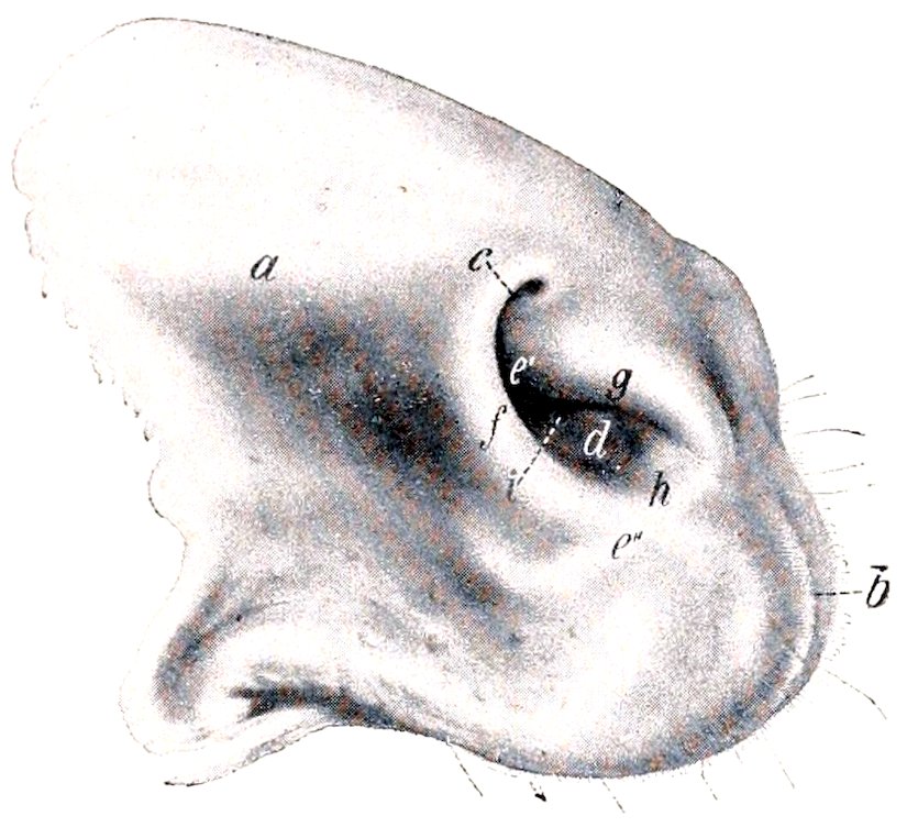
Fig. 344.—Nostrils of Horse. a, Diverticulum nasi (shown as if distended); b, philtrum; c, false nostril; d, true nostril; e′, prominence caused by lamina of alar cartilage; e″, prominence over cornu of same; f, external wing of nostril; g, internal wing; h, lower commissure; i, orifice of naso-lacrimal duct. (Ellenberger-Baum, Anat. d. Haustiere.)
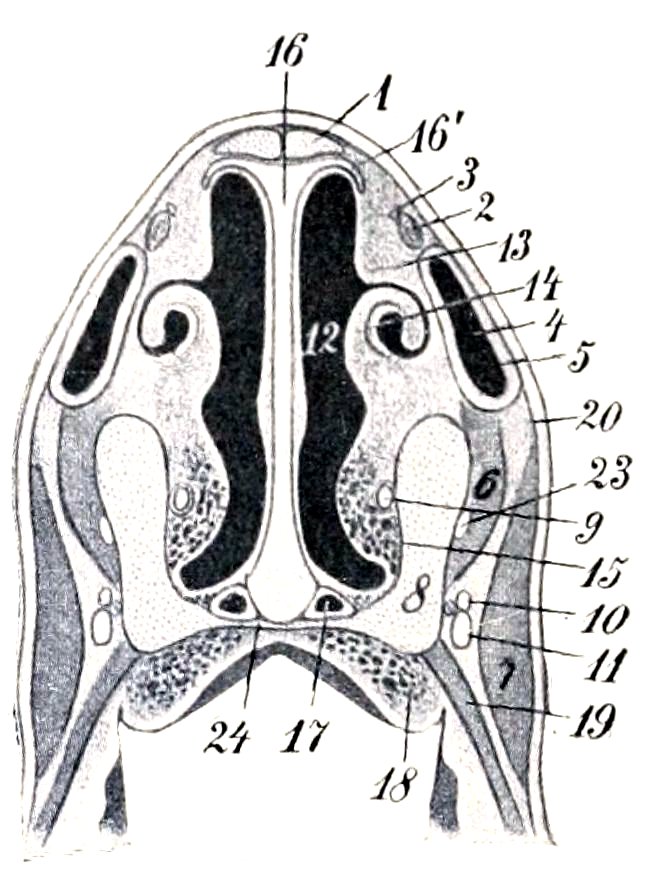
Fig. 345.—Cross-section of Narial Region of Horse. 1, Nasal bone; 2, tendon of levator labii superioris proprius; 3, external nasal nerve; 4, nasal diverticulum; 5, outer wall of 4; 6, dilatator naris inferior; 7, levator nasolabialis + dilatator naris lateralis; 8, maxilla and premaxilla; 9, naso-lacrimal duct; 10, 11, chief branches of superior labial nerve, with superior labial artery at their inner side; 12, nasal cavity; 13, superior turbinal fold; 14, inferior turbinal fold, inclosing cartilaginous prolongation of inferior turbinal bone; 15, venous plexus; 16, septum nasi; 16′, parietal cartilage; 17, vomero-nasal organ (of Jacobson); 18, venous plexus of palate; 19, buccinator; 20, skin; 23, anterior nasal nerve; 24, palatine process of premaxilla. (After Ellenberger-Baum, Top. Anat. d. Pferdes.)
The muscles of the nostrils have been described.
Blood-supply.—Palato-labial, superior labial, and lateral nasal arteries.
Nerve-supply.—Trigeminal and facial nerves.
The nasal cavity is divided into two symmetrical halves by the median septum nasi. The osseous part of the septum (Septum osseum) is formed behind by the perpendicular plate of the ethmoid and below by the vomer. A few ridges on the former correspond to the ethmoidal meatuses. The major part of it, however, is formed by the septal cartilage (Cartilago septi nasi). The surfaces of the cartilage are marked by faint grooves for the vessels and nerves which course over it. The dorsal border is attached along the frontal and nasal sutures, and extends beyond the apices of the nasal bones about two inches (ca. 5 cm.). From this border a thin, narrow plate, the parietal cartilage (Cartilago parietalis) curves outward for a short distance on either side. Near the nostrils these plates are somewhat wider, partially making good the defect in the bony roof of the cavity 438in this situation. The ventral border is thick and rounded; it lies chiefly in the groove of the vomer, but anteriorly it occupies the space between the premaxillary bones. The posterior border is continuous with the perpendicular plate of the ethmoid bone.[121] The alar cartilages are attached to the anterior extremity by fibrous tissue in such a manner as to allow very free movement—in fact, an actual joint may be found. The ventral part of the cartilage is about half an inch (ca. 1 cm.), the middle part about one-tenth of an inch (ca. 2.5 mm.), and the dorsal part about a quarter to a third of an inch (ca. 6 to 8 mm.) in thickness.
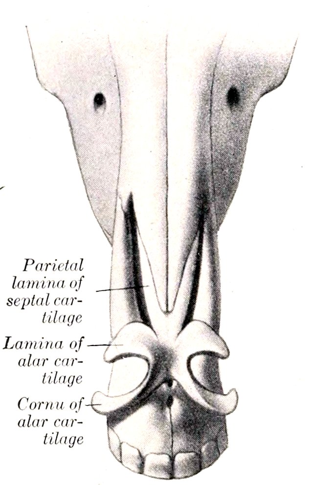
Fig. 346.—Nasal Cartilages of Horse, Dorsal View. (After Ellenberger-Baum, Anat. für Künstler.)
The two turbinal bones (Conchæ nasales) project from the lateral wall, and divide the outer part of the cavity into three meatuses—superior, middle, and inferior.
The superior meatus (Meatus nasi dorsalis) is a narrow passage, bounded dorsally by the roof of the cavity, and ventrally by the superior turbinal bone; its posterior end is closed by the junction of the inner plate of the frontal bone with the cribriform plate and lateral mass of the ethmoid. It transmits air to the upper part of the olfactory region.
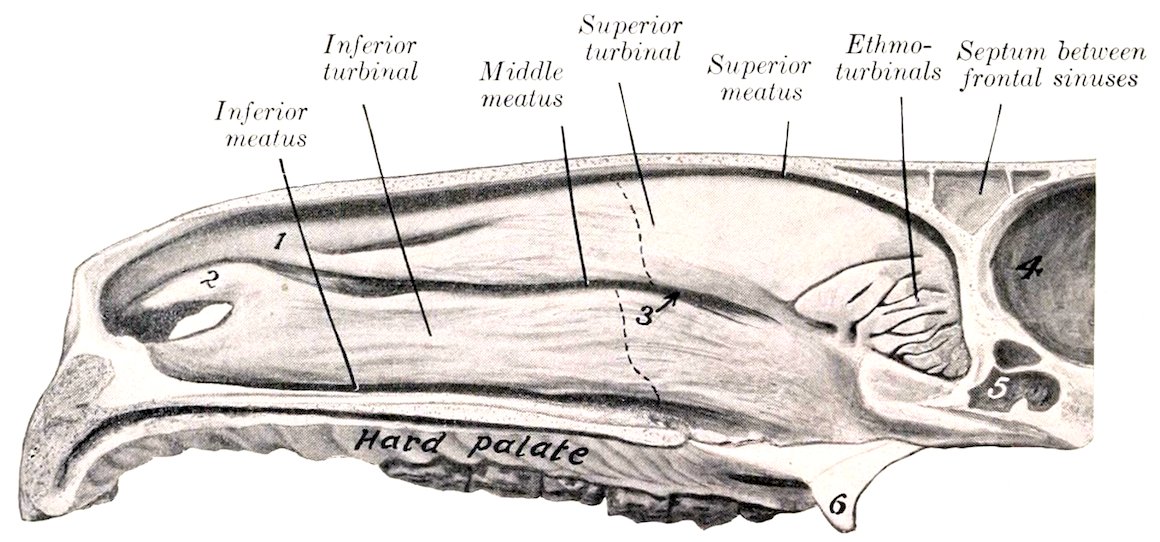
Fig. 347.—Nasal Cavity of Horse, Sagittal Section with Septum Removed. 1, Superior turbinal fold; 2, alar fold, containing cartilaginous prolongation of inferior turbinal; 3, arrow pointing to naso-maxillary fissure; 4, cranial cavity; 5, sphenoidal sinus; 6, hamulus of pterygoid bone. The olfactory mucous membrane is shaded. Dotted lines indicate anterior limit of uncoiled parts of turbinal bones, which inclose parts of frontal and maxillary sinuses.
The middle meatus (Meatus nasi medius) is between the two turbinal bones. It is somewhat larger than the superior meatus, and does not extend back so far; it ends near the great ethmoturbinal and the ethmoidal meatuses. In its posterior part is found the extremely narrow, slit-like interval by which the 439maxillary sinus communicates with the nasal cavity—the naso-maxillary fissure (Aditus naso-maxillaris). The fissure is not visible from the nasal side, being concealed by the overhanging superior turbinal. A fine flexible probe, passed outward and somewhat backward between the turbinal bones, enters the maxillary sinus; if introduced a little further in the same direction, it usually passes through the orifice of communication between the maxillary and the frontal sinus and enters the latter. A small part of the fissure usually brings the anterior division of the maxillary sinus into communication with the nasal cavity. The spaces inclosed by the coiled portions of the turbinals also open into the middle meatus. This passage may be characterized as the sinus-meatus, but it also conducts air to the olfactory region.
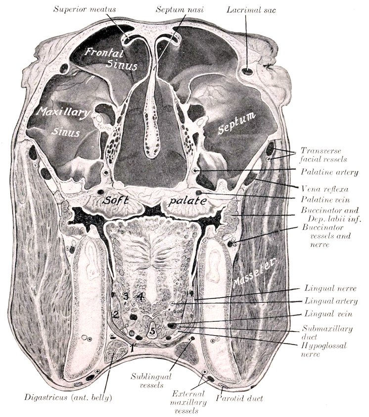
Fig. 348.—Cross-section of Head of Horse. The Section Passes Through the Internal Canthi. 1, Mylo-hyoideus; 2, stylo-glossus; 3, hyo-glossus; 4, genio-glossus; 5, lingual process of hyoid bone. An arrow points to the naso-maxillary fissure.
The inferior meatus (Meatus nasi ventralis) is situated between the inferior 440turbinal and the floor of the cavity. It is much larger than the other two, and is the direct passage between the nostrils and the pharynx.
The common meatus (Meatus nasi communis) is situated between the septum and the turbinals, and is continuous externally with the other meatuses. It is very narrow dorsally, but widens ventrally.
The lateral masses of the ethmoid bone project forward into the posterior part (fundus) of the nasal cavity (Fig. 347). Between the ethmoturbinals of which each mass is composed there are three principal and numerous small passages, the ethmoidal meatuses (Meatus ethmoidales).
The posterior nares (Choanæ) are two elliptical orifices by which the nasal cavity and pharynx communicate. They are in the same plane as the floor of the nasal cavity, and are separated from each other by the vomer. They are, taken together, about two inches (ca. 5 cm.) wide and three to four inches (ca. 8 to 10 cm.) long.
The nasal mucous membrane (Membrana mucosa nasi) is thick, highly vascular, and is, in general, firmly attached to the underlying periosteum and perichondrium. It is continuous in front with the skin which lines the nostrils, and behind with the mucous membrane of the pharynx. It is also continuous at the naso-maxillary fissure with the very thin and only slightly vascular mucous membrane which lines the sinuses. In the anterior part of the cavity it forms prominent thick folds on the lateral wall, which extend from the turbinal bones to the nostril. There are usually two superior turbinal folds which unite anteriorly. The upper one incloses a thin plate of cartilage which is continuous with the superior turbinal bone. The inferior turbinal fold is curved, and incloses an ᔕ-shaped cartilaginous plate which prolongs the inferior turbinal bone; this fold is continuous with the alar fold of the nostril, and forms with it the upper margin of the entrance from the true nostril to the nasal cavity. Below this there is a rounded ridge produced by the nasal process of the premaxilla. The mucous membrane of the greater part of the cavity (Regio respiratoria) is red in color, and is covered with a stratified ciliated epithelium. It contains numerous acinous nasal glands (Glandulæ nasales). The submucosa contains rich venous plexuses which form in certain situations a sort of cavernous tissue (Plexus cavernosus), composed of several strata of freely anastomosing veins, between which there are unstriped muscle-fibers. This arrangement is most marked in the turbinal folds, on the lower part of the inferior turbinal bone, and the lower part of the septum. On the posterior part of the lateral masses of the ethmoid and the adjacent part of the superior turbinal and the septum (Regio olfactoria) the mucous membrane is brownish-yellow in color and thicker; it contains the olfactory nerve-endings in a special non-ciliated epithelium. In it are numerous olfactory glands (Glandulæ olfactoriæ); these are long, tubular, and often branched.
The vomero-nasal organ of Jacobson (Organon vomeronasale) is situated on the floor of the nasal cavity, on either side of the ventral border of the septum (Fig. 345.) It consists of a tubular cartilage lined with mucous membrane, to which fibers of the olfactory nerve may be traced. Its anterior part communicates with the nasal cavity by a slit-like orifice.
The paranasal sinuses are described in the Osteology.
THE LARYNX
The larynx is a short tube which connects the pharynx and trachea. It is a complex valvular apparatus, which regulates the volume of air in respiration, prevents aspiration of foreign bodies, and is the chief organ of voice.
It is situated partly between the internal pterygoid muscles, partly in the neck 441between the parotid glands.[122] Its long axis is practically horizontal when the head and neck are extended (Fig. 254).
It is related dorsally to the pharynx and the origin of the œsophagus. Ventrally it is covered by the skin, fascia, and sterno-hyoid and omo-hyoid muscles. Laterally it is related to the parotid and submaxillary glands and to the internal pterygoid, stylo-maxillaris, digastricus, stylo-hyoid, and pharyngeal constrictor muscles.
It is attached to the body and thyroid cornua of the hyoid bone, and thus indirectly to the base of the cranium.
Its cavity communicates anteriorly with the pharynx and (potentially) with the mouth; posteriorly, with the trachea.
The wall of the larynx consists of a framework of cartilages, connected by joints and ligaments or membranes, and moved by extrinsic and intrinsic muscles. The cavity is lined with mucous membrane.
Cartilages of the Larynx
There are three single cartilages and one pair; the single cartilages are the cricoid, thyroid, and epiglottis; the arytenoid cartilages are paired.
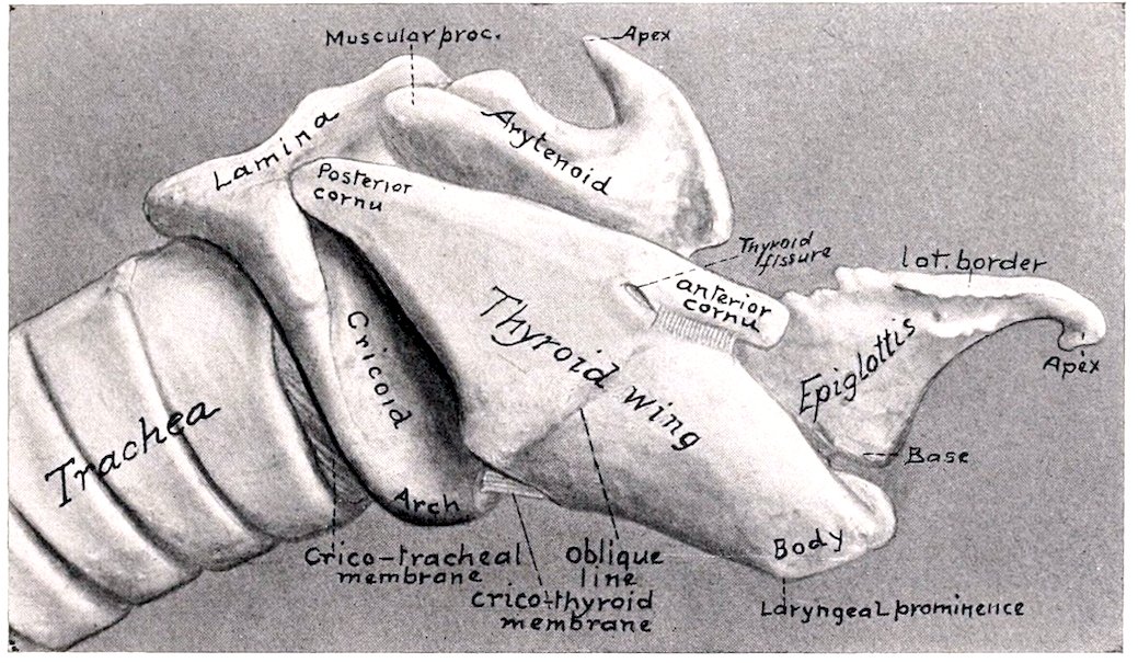
Fig. 349.—Laryngeal Cartilages of Horse, Right View.
The cricoid cartilage (Cartilago cricoidea) is shaped like a signet ring. The dorsal part is a broad, thick, quadrilateral plate termed the lamina; the outer (dorsal) surface of this is marked by a median ridge separating two shallow cavities, which give attachment to the posterior crico-arytenoid muscles. On either side of these depressions are two articular facets. The anterior facets (Facies articulares arytænoideæ) are placed at the anterior border, are oval and convex, and articulate with the arytenoid cartilages. The posterior facets (Facies articulares thyreoideæ) are situated on the front of a ridge, a short distance from the posterior border; they are concave, and articulate with the posterior cornua of the thyroid cartilage. The ventral and lateral parts of the ring are formed by a curved band, called the arch (Arcus), which is narrowest below. The lateral surfaces of the arch are grooved for the crico-thyroid muscle. The anterior border of the 442lamina is thick and slightly concave; the posterior border is thin and irregularly notched. The anterior border of the arch is concave ventrally and gives attachment to the crico-thyroid membrane; laterally it is thicker and gives attachment to the crico-arytenoideus lateralis muscle. The posterior border is attached to the first ring of the trachea by an elastic membrane. The inner surface is smooth and is covered with mucous membrane.
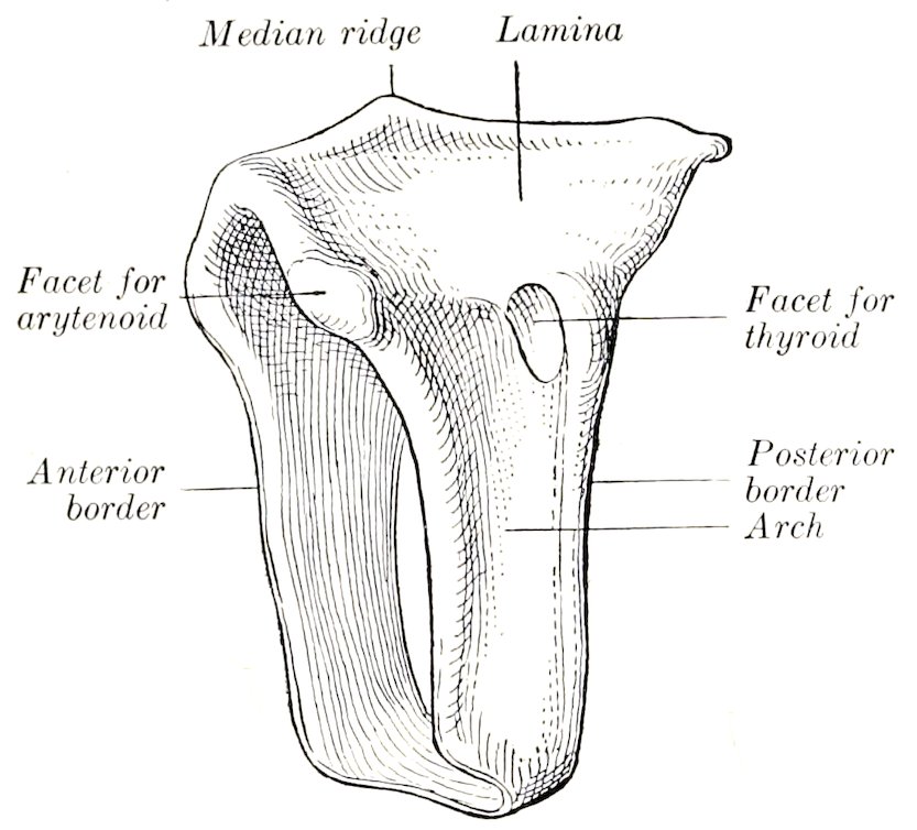
Fig. 350.—Cricoid Cartilage of Horse, Left Lateral View.
The thyroid cartilage (Cartilago thyreoidea) consists of a median thickened portion, termed the body, and two lateral laminæ or wings. The body forms ventrally a slight prominence (Prominentia laryngea), which can be felt, but is not visible in the living subject; it supports the base of the epiglottis, which is attached to it by an elastic ligament. The wings or laminæ spring from the body on either side and form a large part of the lateral wall of the larynx. Each is a rhomboid plate, presenting a slightly convex outer surface, which is divided into two areas by an oblique line (Linea obliqua), on which the thyro-hyoid and thyro-pharyngeus muscles meet. The superior (dorsal) border is nearly straight; it gives attachment to the pharyngeal fascia and the palato-pharyngeus muscle, and bears a cornu at each end. The anterior cornu (Cornu orale) is attached to the thyroid cornu of the hyoid bone; below it is a notch, which is converted into a foramen (Foramen thyreoideum) by a fibrous band, and transmits the superior laryngeal nerve to the interior of the larynx. The posterior cornu (Cornu caudale) articulates with the cricoid cartilage. The inferior (ventral) border is fused with the body in front; behind, it diverges from its fellow to inclose a triangular space (Incisura thyreoidea caudalis), which is occupied by the crico-thyroid membrane. The anterior border is slightly convex, and is attached to the hyoid bone by the thyro-hyoid membrane. The posterior border overlaps the arch of the cricoid cartilage, and gives attachment to the crico-thyroid muscle. The inner surface is concave, and is covered by mucous membrane to a small extent.
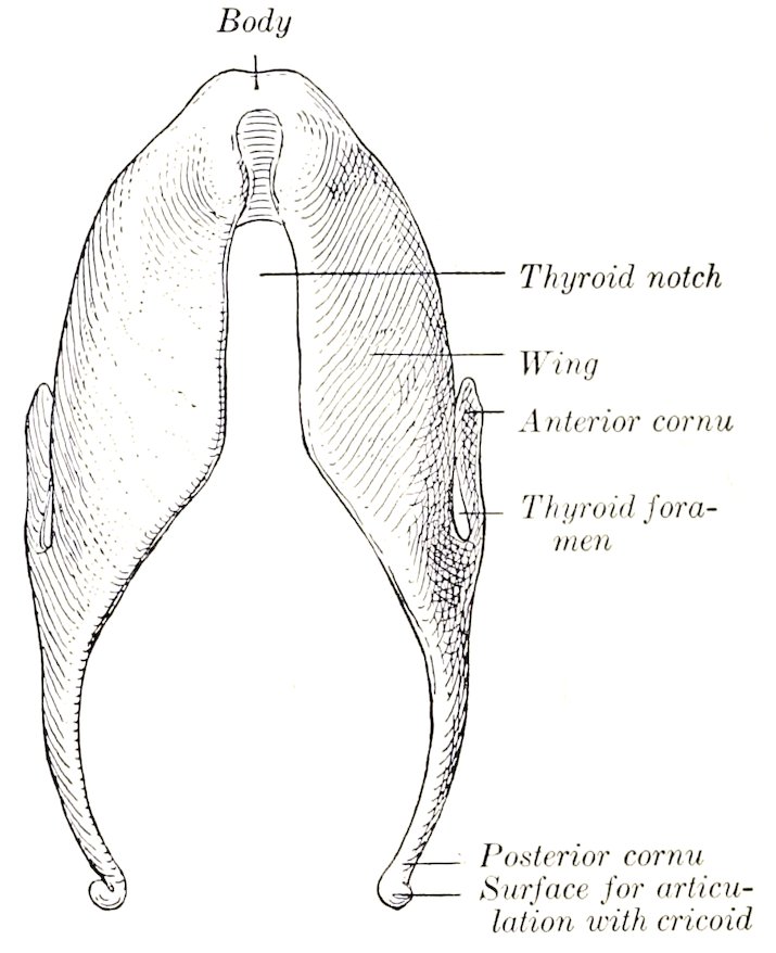
Fig. 351.—Thyroid Cartilage of Horse, Ventral View.
The epiglottis (Cartilago epiglottica) is situated above the body of the thyroid 443cartilage and curves toward the root of the tongue. It is shaped somewhat like a pointed ovate leaf, and presents two surfaces, two borders, a base, and an apex. The oral (or anterior) surface is concave in its length, convex transversely; the pharyngeal (or posterior) surface has the reverse configuration. The borders are thin, denticulated, and somewhat everted. The base is thick, and is attached to the dorsal surface of the body of the thyroid cartilage by elastic tissue. From each side of it a cartilaginous bar projects backward; these processes correspond to the cuneiform cartilages of man. The apex is pointed and curved ventrally. Nearly all of the epiglottis is covered with mucous membrane. It lies sometimes in front of, sometimes behind, the soft palate.
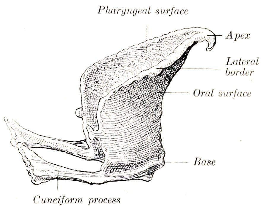
Fig. 352.—Epiglottis of Horse, Right Lateral View.
The arytenoid cartilages (Cartilagines arytenoideæ) are situated on either side, in front of the cricoid, and partly between the alæ of the thyroid cartilage. They are somewhat pyramidal in form, and may be described as having two surfaces, two borders, a base, and an apex. The internal surface is concavo-convex, smooth, and covered by mucous membrane. The external surface is divided by a ridge into two areas: the lower flat area faces outward, and is covered by the thyro-arytenoid and lateral crico-arytenoid muscles; the upper concave area faces chiefly forward, and is covered by the transverse arytenoid muscle. The anterior border is convex, and at the base of the cartilage forms a projection termed the vocal process (Processus vocalis); it is so named because it gives attachment to the vocal or thyro-arytenoid ligament, which forms the basis of the true vocal cord. The posterior border is straight, and extends from the vocal process to the facet on the base. The dorsal border forms a deep notch with the apex. The base is concave and faces chiefly backward; it presents an oval, concave facet for articulation with the anterior border of the lamina of the cricoid cartilage. The external angle of the base forms a strong eminence termed the muscular process (Processus muscularis), which gives attachment to the crico-arytenoid muscles. The internal angle is attached to its fellow by the transverse arytenoid ligament. The apex (Cartilago corniculata) curves upward and backward, forming with its fellow the pitcher-shaped lip, from which the cartilages derive their name.
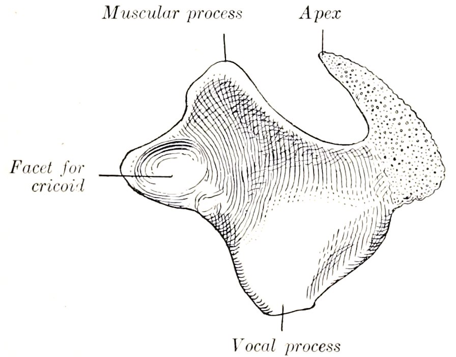
Fig. 353.—Left Arytenoid Cartilage of Horse, Inner View.
The cricoid and thyroid cartilages, and the greater part of the arytenoid cartilages, consist of hyaline cartilage. The apices and vocal processes of the arytenoid cartilages and the epiglottis (including the cuneiform processes) consist 444of elastic cartilage; they show no tendency toward ossification at any age. The thyroid and cricoid cartilages regularly undergo considerable ossification; the process begins in the body of the thyroid, and often involves the greater part of the cartilage.
Joints, Ligaments, and Membranes of the Larynx
The crico-thyroid joints (Articulationes crico-thyreoideæ) are diarthroses formed by the apposition of the convex facets on the ends of the posterior cornua of the thyroid cartilage with corresponding facets on the sides of the cricoid cartilage. The capsule is thin, but is strengthened by accessory bands dorsally, externally, and internally. The movements are chiefly rotation around a transverse axis passing through the centers of the two joints.
The crico-arytenoid joints (Articulationes crico-arytænoidea) are also diarthrodial. They are formed by the apposition of the convex facets on the anterior border of the cricoid cartilages with the concave facets on the bases of the arytenoid cartilages. Each has a very thin, loose capsule, strengthened by accessory bundles dorsally and internally. The movements are gliding and rotation. In the former the arytenoid cartilage glides inward or outward; in the latter it rotates around a longitudinal axis, so that the vocal process swings outward or inward.
The thyro-hyoid joints (Articulationes hyo-thyreoideæ) are formed by the anterior cornua of the thyroid cartilage and the extremities of the thyroid cornua of the hyoid bone. They permit rotation around a transverse axis passing through the two joints.
The crico-thyroid membrane (Ligamentum crico-thyreoideum) occupies the thyroid notch and extends backward to the arch of the cricoid cartilage. It is triangular in form, and is attached by its base to the anterior border of the arch of the cricoid cartilage, while its borders are attached to the margins of the thyroid notch. It is strong, tightly stretched, and composed largely of elastic tissue. It is reinforced ventrally by longitudinal fibers, dorsally by fibers which stretch across the thyroid notch.
The thyro-hyoid membrane (Membrana hyo-thyreoidea) connects the anterior border of the thyroid cartilage with the body and thyroid cornua of the hyoid bone.
The hyo-epiglottic ligament (Ligamentum hyo-epiglotticum) attaches the lower part of the oral surface of the epiglottis to the body of the hyoid bone. It forms an elastic sheath for the hyo-epiglottic muscle. Ventrally it blends with the thyro-hyoid membrane, and dorsally it is not well defined.
The thyro-epiglottic ligament (Ligamentum thyreo-epiglotticum) is strong and thick; it is composed chiefly of elastic tissue. It connects the base of the epiglottis with the body and the adjacent inner surface of the laminæ of the thyroid cartilage. Other fibers attach the cuneiform processes somewhat loosely to the thyroid laminæ.
The transverse arytenoid ligament (Ligamentum arytænoideum transversum) is a slender band which connects the inner angles of the bases of the arytenoid cartilages.
The vocal (or inferior thyro-arytenoid) ligament (Ligamentum vocale) forms the basis of the true vocal cord (Fig. 254). It is elastic and is attached ventrally to the body of the thyroid cartilage and the crico-thyroid membrane close to its fellow of the opposite side, and ends dorsally on the processus vocalis of the arytenoid cartilage.
The ventricular (or superior thyro-arytenoid) ligament (Ligamentum ventriculare) is included in the false vocal cord. It consists of loosely arranged bundles which arise chiefly from the cuneiform process and end on the outer surface of the processus vocalis and adjacent part of the arytenoid cartilage.
445The crico-tracheal membrane (Ligamentum crico-tracheale) connects the cricoid cartilage with the first ring of the trachea.
Muscles of the Larynx
A. Extrinsic Muscles
The sterno-thyro-hyoideus is described on p. 226.
The thyro-hyoideus (M. hyo-thyreoideus) is a flat, quadrilateral muscle, which lies on the outer surface of the thyroid lamina and the lateral part of the thyro-hyoid membrane. It arises from the thyroid cornu of the hyoid bone, and ends on the oblique line on the lamina of the thyroid cartilage. Its action is to draw the larynx toward the root of the tongue.
The hyo-epiglotticus (M. hyo-epiglotticus) is a feeble muscle which occupies a median position above the central part of the thyro-hyoid membrane, inclosed by an elastic sheath, the hyo-epiglottic ligament. It arises from the body of the hyoid bone by two branches which unite and are inserted into the front of the base of the epiglottis. Its action is to draw the epiglottis toward the root of the tongue.
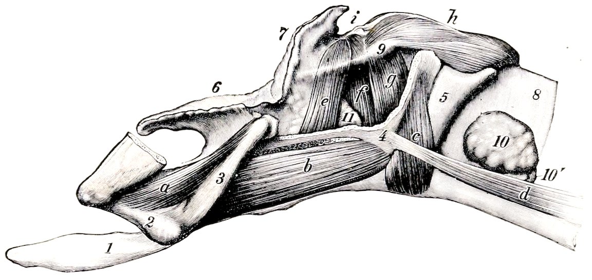
Fig. 354.—Laryngeal Muscles of Horse, Seen from Left Side After Removal of Major Part of Lamina of Thyroid Cartilage. a, Kerato-hyoideus; b, thyro-hyoideus; c, crico-thyroideus; d, sterno-thyroideus; e, ventricularis; f, vocalis; g, crico-arytenoideus lateralis; h, crico-arytenoideus post. s, dorsalis; i, arytenoideus transversus; 1, lingual process; 2, small cornu; 3, thyroid cornu, of hyoid bone; 4, thyroid cartilage; 5, cricoid cartilage; 6, epiglottis; 7, apex of arytenoid cartilage; 8, trachea; 9, muscular process of arytenoid; 10, lateral lobe of thyroid gland; 10′, isthmus of thyroid gland; 11, laryngeal saccule. (Ellenberger-Baum, Anat. d. Haustiere.)
B. Intrinsic Muscles
The crico-thyroideus (M. crico-thyreoideus) is a short muscle which fills the groove on the lateral surface of the cricoid cartilage; its fibers are directed dorsally and somewhat forward. It arises on the lower half of the lateral surface and posterior edge of the cricoid cartilage, and is inserted into the posterior border and adjacent part of the surfaces of the lamina of the thyroid cartilage. Its action is to draw the thyroid cartilage and the ventral part of the cricoid cartilage together. In this action the cricoid cartilage is probably rotated, carrying the bases of the arytenoid cartilages with it and thus tensing the vocal cords.
The crico-arytenoideus dorsalis s. posterior (M. crico-arytænoideus dorsalis) is a strong, somewhat fan-shaped muscle, which, with its fellow, covers the dorsal surface of the lamina of the cricoid cartilage. It is partially divisible into two layers. It has a broad origin on half of the lamina of the cricoid cartilage, and its fibers converge to be inserted into the processus muscularis of the arytenoid cartilage. 446Its action is to dilate the rima glottidis by rotating the arytenoid cartilage so as to carry the vocal process and cord outward.
The crico-arytenoideus lateralis (M. crico-arytænoideus lateralis) lies on the inner face of the thyroid lamina. It arises from the anterior border of the lateral part of the arch of the cricoid cartilage. The fibers pass in a dorsal direction and converge on the processus muscularis of the arytenoid cartilage. It closes the rima glottidis by rotating the arytenoid cartilage inward.
The arytenoideus transversus (M. arytænoideus transversus) is an unpaired muscle which stretches across the concave dorsal surface of the arytenoid cartilages. Its fibers are attached on either side to the processus muscularis and the ridge which extends forward from it. The right and left parts of the muscle meet at a fibrous raphé which is connected with the transverse arytenoid ligament. Filters of the thyro-arytenoideus muscle overlap its anterior part. It narrows the rima by drawing the arytenoid cartilages together.
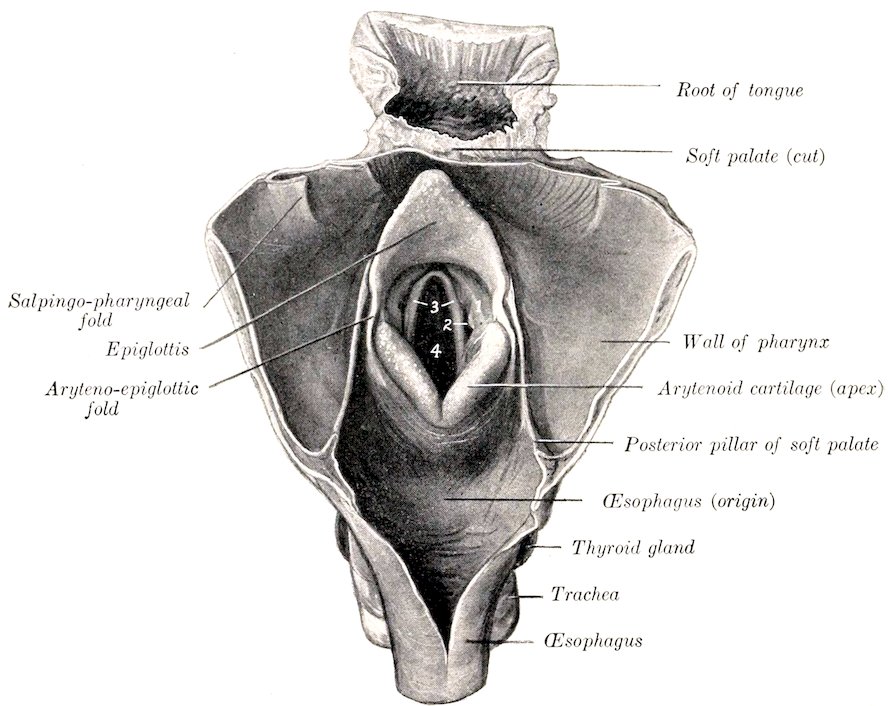
Fig. 355.—Aditus Laryngis of Horse, Exposed by Opening Pharynx and Beginning of Œsophagus Along Median Dorsal Line. 1, False vocal cord; 2, lateral ventricle; 3, true vocal cords; 4, rima glottidis.
The thyro-arytenoideus (Mm. ventricularis et vocalis) is situated in the lateral wall of the larynx, covered by the lamina of the thyroid cartilage. It consists of two parts, between which the mucous membrane of the lateral ventricle pouches to form the saccule. The anterior part (M. ventricularis) arises from the anterior part of the crico-thyroid membrane and the ventral border of the thyroid lamina. Its fibers pass upward and backward to end partly on the processus muscularis, partly on the arytenoideus transversus, meeting its fellow. The posterior part (M. vocalis) has a more extensive origin on the crico-thyroid membrane. Its direction corresponds 447with that of the true vocal cord. It is inserted into the outer surface of the arytenoid cartilage below the processus muscularis. The muscle closes the rima and slackens the vocal cords. With the arytenoideus transversus and crico-arytenoideus lateralis it forms a sphincter which closes the entrance to the larynx.
Cavity of the Larynx
The cavity of the larynx (Cavum laryngis) is smaller than one would expect from its external appearance. On looking into it through the pharyngeal end two folds are seen projecting from its lateral walls. These are termed the vocal cords, and they divide the cavity into two portions.
The aditus laryngis, or pharyngeal aperture, is a large, oblique, oval opening, which faces forward and upward into the ventral part of the pharynx. It is bounded in front by the epiglottis, behind by the arytenoid cartilages, and laterally by the aryteno-epiglottic folds of mucous membrane (Plicæ aryepiglotticæ), which stretch between the edges of the epiglottis and the arytenoid cartilages.
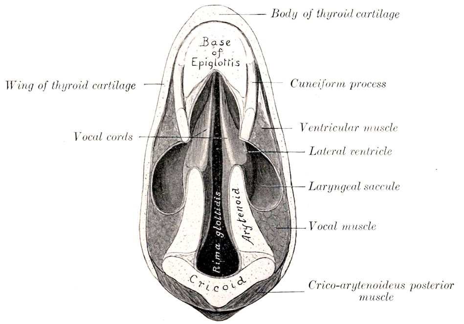
Fig. 356.—Section of Larynx of Horse. The plane of the section is parallel with the vocal cords.
The vestibule of the larynx (Vestibulum laryngis) is that part of the cavity which extends from the aditus to the vocal cords. On its lateral walls are the false vocal cords (Plicæ ventriculares), each of which consists of a fold of mucous membrane covering the anterior part of the thyro-arytenoid ligament and the cuneiform process. Between the false and true vocal cord there is a pocket-like depression termed the lateral ventricle of the larynx (Ventriculus lateralis laryngis). This is the entrance to the laryngeal saccule (Appendix ventriculi laryngis), a cul-de-sac of the mucous membrane which is about one inch (ca. 2 to 3 cm.) long and extends upward and backward on the inner surface of the thyroid lamina. There is a small middle ventricle (Ventriculus laryngis medianus) at the base of the epiglottis.
The middle, narrow part of the cavity is termed the glottis or rima glottidis.[123] 448It is bounded on either side by the true vocal cord and the inner surface of the arytenoid cartilage. The true vocal cords (Labia vocales) are situated behind the false cords and the lateral ventricles. They extend from the angle between the body and laminæ of the thyroid cartilage to the vocal processes of the arytenoid cartilages. They are prismatic in cross-section, and their free edges look forward and somewhat upward. The mucous membrane of the cord (Plica vocalis) is very thin and smooth, and is intimately attached to the underlying ligament. In ordinary breathing the rima is somewhat lanceolate in form; when dilated, it is diamond-shaped, the widest part being between the vocal processes. The narrow part of the glottis between the vocal cords is termed the glottis vocalis (Pars intermembranacea), while the wider part between the arytenoid cartilages is the glottis respiratoria (Pars intercartilaginea).
The posterior compartment of the laryngeal cavity is directly continuous with the trachea. It is inclosed by the cricoid cartilage and the crico-thyroid membrane. It is oval in form, the transverse diameter being an inch and a half to two inches (ca. 4 to 5 cm.), and the dorso-ventral diameter two to two and a half inches (ca. 5 to 6 cm.).
The mucous membrane which lines the larynx (Tunica mucosa laryngis) is reflected around the margin of the aditus to become continuous with that of the pharynx, and behind it is continuous with that which lines the trachea. It is closely attached to the pharyngeal surface of the epiglottis, but elsewhere in the aditus and vestibule it is loosely attached by submucous tissue which contains many elastic fibers.[124] It is thin and very closely adherent over the vocal cords and the inner surfaces of the arytenoid cartilages. The epithelium is of the stratified squamous type from the aditus to the glottis, beyond which it is columnar ciliated in character. There are numerous mucous glands (Glandulæ laryngeæ), except over the glottis and the pharyngeal surface of the epiglottis, in which situations they are scanty.
Blood-supply.—Thyro-laryngeal artery.
Nerve-supply.—Superior laryngeal and recurrent laryngeal nerves (from the vagus).
THE TRACHEA
The trachea extends from the larynx to the roots of the lungs, where it divides into the right and left bronchi. It is kept permanently open by a series of about fifty to fifty-five incomplete cartilaginous rings embedded in its wall. It occupies a median position, except near its termination, where it is pushed a little to the right by the arch of the aorta. It is approximately cylindrical, but its cervical portion is for the most part depressed dorso-ventrally by contact with the longus colli muscle, so that the dorsal surface is flattened. The average caliber is about two to two and one-half inches (ca. 5 to 6 cm.), but in the greater part of the neck the transverse diameter is greater and the dorso-ventral smaller. It is inclosed by a fascia propria.
In its cervical part the trachea is related dorsally to the œsophagus for a short distance, but chiefly to the longus colli muscle. Laterally it is related to the thyroid gland, the carotid artery, the jugular vein, the vagus, sympathetic, and recurrent laryngeal nerves, and the tracheal lymph ducts and cervical lymph glands. The œsophagus lies on its left face from the third cervical vertebra backward. The sterno-cephalicus muscles cross it very obliquely, passing from the ventral surface forward over its sides, and diverging to reach the angles of the jaw. The omo-hyoidei also cross it very obliquely, passing over the lateral surfaces of the tube, and converging ventrally to the body of the hyoid bone. The sterno-thyrohyoideus 449lies on the ventral surface.[125] The scaleni lie on either side near the entrance to the thorax.
The thoracic part of the trachea passes backward between the pleural sacs and divides into the two bronchi over the left atrium of the heart. It is related dorsally to the longus colli for a short distance, and beyond this to the œsophagus. Its left face is crossed by the aortic arch, the left brachial artery, and the thoracic duct. Its right face is crossed by the vena azygos, the dorso-cervical and vertebral vessels, and the right vagus nerve. Ventrally it is related to the anterior vena cava, the brachiocephalic and common carotid trunks, and the left recurrent nerve.
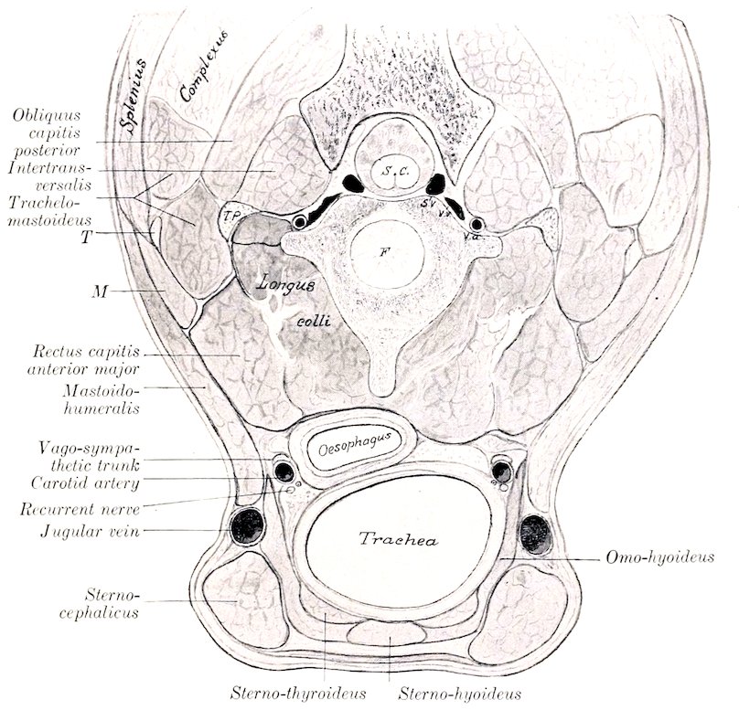
Fig. 357.—Cross-section of Ventral Part of Neck of Horse. The section is cut at right angles to the long axis of the neck, passing through the junction of the second and third cervical vertebræ. S.c., Spinal cord; S.v., spinal vein; V.v., V.a., vertebral vein and artery; T.p., transverse process (tip); F, intervertebral fibro-cartilage; T, atlantal tendon common to mastoido-humeralis, splenius, and lower part of trachelo-mastoideus; M, digitation of mastoido-humeralis inserted by T.
The bifurcation of the trachea (Bifurcatio tracheæ) is situated opposite to the fifth intercostal space, and about five or six inches (ca. 12 to 15 cm.) below the seventh and eighth thoracic vertebræ.
The trachea is composed of—(1) A fibro-elastic membrane in which are embedded (2) the cartilaginous rings; (3) a muscular layer; (4) the mucous membrane. The elastic membrane is intimately attached to the perichondrium 450of the rings. In the intervals between the latter it constitutes the annular ligaments (Ligamenta annularia). The rings of the trachea (Cartilagines tracheales) are composed of hyaline cartilage. They are incomplete dorsally, and when their free ends are drawn apart, resemble somewhat the letter C. In the cervical part the thin wide free ends overlap, while in the thoracic part they do not meet; here the deficiency is made up by thin plates of variable size and form, embedded in a membrana transversa. Ventrally the rings are about one-half inch (ca. 1 to 1.2 cm.) wide and much thicker than dorsally. The first ring is attached to the cricoid cartilage by the crico-tracheal membrane, and is usually fused dorsally with the second ring. Various irregularities, such as partial bifurcation or partial or complete fusion with an adjacent ring, are common. The arrangement in the terminal part is very irregular. The muscular layer (Musculus trachealis) consists of unstriped fibers which stretch across the dorsal part of the tube. It is separated from the ends of the rings and the membrana transversa by a quantity of areolar tissue. When it contracts the caliber of the tube is diminished. The mucous membrane is pale, normally, and presents numerous fine longitudinal folds, in which are bundles of elastic fibers. The epithelium is stratified columnar ciliated. Numerous tubular mucous glands (Glandulæ tracheales) are present.
Blood-supply.—Common carotid arteries.
Nerve-supply.—Vagus and sympathetic nerves.
THE BRONCHI
The two bronchi, right and left (Bronchus dexter, sinister), are formed by the bifurcation of the trachea. Each passes backward and outward to the hilus of the corresponding lung. The right bronchus is a little wider and less oblique in direction than the left. They are related ventrally to the divisions of the pulmonary artery, and dorsally to the branches of the bronchial artery and the bronchial lymph glands. Their structure is similar to that of the trachea.
THE THORACIC CAVITY
The thoracic cavity (Cavum thoracis) is the second in point of size of the body cavities. In form it is somewhat like a truncated cone, much compressed laterally in its anterior part, and with the base cut off very obliquely.
The dorsal wall or roof is formed by the thoracic vertebræ and the ligaments and muscles connected with them.
The lateral walls are formed by the ribs and the intercostal muscles.
The ventral wall or floor is formed by the sternum, the cartilages of the sternal ribs, and the muscles in connection therewith. It is about one-half as long as the dorsal wall.
The posterior wall, formed by the diaphragm, is very oblique and is strongly convex.
The anterior aperture or inlet (Apertura thoracis cranialis) is relatively small, and of narrow, oval form. It is bounded dorsally by the first thoracic vertebra and laterally by the first pair of ribs. It is occupied by the longus colli muscles, the trachea, œsophagus, vessels, nerves, and the prepectoral lymph glands.
The cavity is lined by the endothoracic fascia and by the pleuræ.
A longitudinal septum, termed the mediastinum (Septum mediastinale), extends from the dorsal wall to the ventral and posterior walls, and subdivides the cavity into two lateral chambers which contain the lungs. Each of these chambers is lined by a serous membrane called the pleura, and is called a pleural cavity (Cavum pleuræ). The mediastinum is, for the most part, not median in position; this is largely due to the fact that the largest organ contained in it, the heart, is 451placed more on the left side; consequently the right pleural cavity and lung are larger than the left. Practically all the organs in the thorax are in the mediastinal space with the exception of the lungs, the posterior vena cava, and the right phrenic nerve. The part in which the heart and the pericardium are situated, together with that above it, is usually called the middle mediastinal space; the parts before and behind this are termed the anterior and posterior mediastinal spaces respectively.
THE PLEURÆ
The pleuræ are two serous sacs which line the pleural cavities and are reflected at the roots of the lungs to invest those organs. We, therefore, distinguish parietal and visceral parts of the pleuræ.
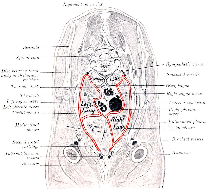
Fig. 358.—Cross-section of the Thorax of New-born Foal. Tr., Trachea; S, left brachial artery; B, brachiocephalic artery; l.g., lymph gland. Pleura indicated by red line.
The parietal pleura (Pleura parietalis) lines the cavity in which each lung is situated (Cavum pleuræ). On the lateral thoracic wall it is adherent to the ribs and intercostal muscles and is termed the costal pleura (Pleura costalis). Behind it is closely attached to the diaphragm, forming the diaphragmatic pleura (Pleura diaphragmatica). The part which is in apposition with the opposite sac or covers the mediastinal organs is termed the mediastinal pleura (Pleura mediastinalis); 452where this layer is adherent to the pericardium, it is distinguished as pericardiac pleura (Pleura pericardiaca).[126]
The pleura is reflected at the root of the lung, which it covers, constituting the visceral or pulmonary pleura (Pleura pulmonalis). Behind the root of the lung a considerable triangular area is not covered by the pleura, the two lungs being attached to each other by connective tissue in this situation.
The ligament of the lung (Lig. pulmonale) is a fold formed by the reflection of the pleura from the mediastinum and the diaphragm to the lung, behind the triangular area of adhesion just mentioned. It is seen when the base of the lung is drawn outward. It contains elastic tissue, especially in its posterior part.
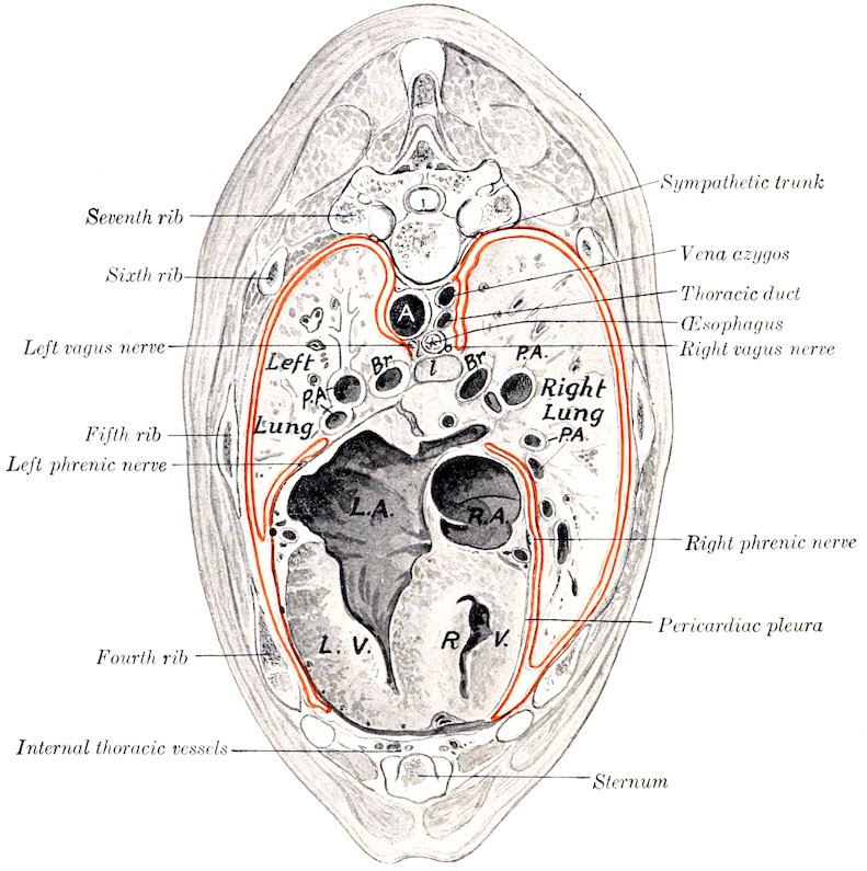
Fig. 359.—Cross-section of Thorax of New-born Foal. A, Aorta; Br., bronchi; P.A., pulmonary arteries; l, lymph gland; R.A., right atrium; L.A.; left atrium; R.V., right ventricle; L.V., left ventricle of heart.
The right pleura forms a special sagittal fold (Plica venæ cavæ) about a handbreadth to the right of the median plane, which incloses the posterior vena cava in its upper edge and gives off a small accessory fold for the right phrenic nerve. The fold arises from the thoracic floor and from the diaphragm below the foramen venæ cavæ, and intervenes between the mediastinal lobe and the body of the right lung. It is delicate and lace-like.
453The posterior mediastinum is very delicate below the œsophagus, and usually appears fenestrated; when these apertures are present, the two pleural cavities communicate with each other.[127]
The pleural sacs contain a serous fluid, the liquor pleuræ; in health there is only a sufficient amount to moisten the surface, but it accumulates rapidly after death.
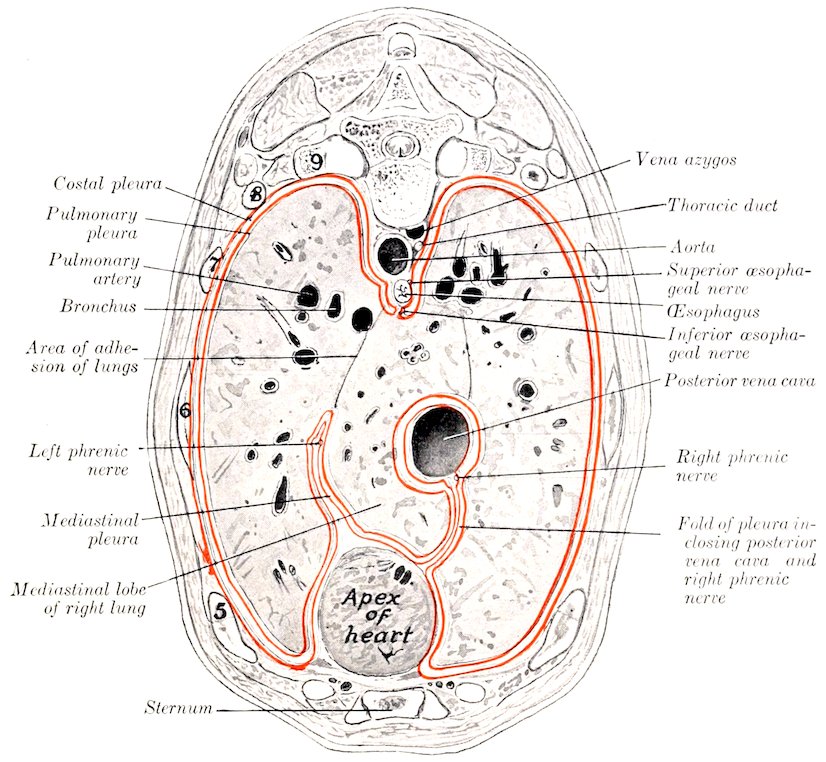
Fig. 360.—Cross-section of Thorax of New-born Foal. Ribs are numbered.
THE LUNGS
The lungs (Pulmones) occupy much the greater part of the thoracic cavity. They are accurately adapted to the walls of the cavity and the other organs contained therein. The two lungs are not alike in form or size, the right one being considerably larger than the left; the difference is chiefly in width, in conformity with the projection of the heart to the left.
The lung is soft, spongy, and highly elastic. It crepitates when pressed between the finger and thumb, and floats in water. When the thoracic cavity of the unpreserved subject is opened, the lung collapses immediately to about one-third of its original size, and loses its proper form; this is due to its highly elastic character and the fact that the tension of the lung 454tissue caused by the air pressure in its cavities has been relieved by the external air pressure.[128]
The color varies according to the amount of blood contained in the lung. During life the lung has a pink color, but in subjects which have been bled for dissection it is light gray or faintly tinged with red. In unbled subjects it is dark red; the depth of color varies, and is often locally accentuated by gravitation of blood to the most dependent parts (hypostasis).
The fœtal lung, since it contains no air and has a relatively small blood-supply, differs from that of an animal which has breathed in the following respects: (1) It is much smaller; (2) it is firmer and does not crepitate; (3) it sinks in water; (4) it is pale gray in color.
In form the lungs are like casts of the pleural cavities in which they are situated. When well hardened in situ, their surfaces present impressions and elevations corresponding exactly to the structures with which they are in contact. Each lung presents two surfaces, two borders, a base, and an apex.
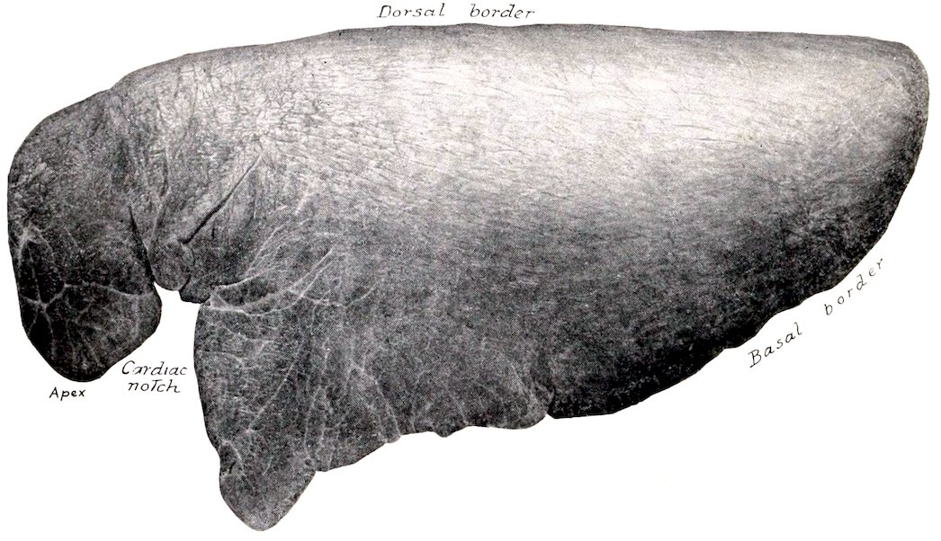
Fig. 361.—Left Lung of Horse, Costal Surface. Specimen hardened in situ.
The costal (or external) surface (Facies costalis) is convex, and lies against the lateral thoracic wall, to which it is accurately adapted.
The mediastinal (or internal) surface (Facies mediastinalis) is less extensive than the costal surface. It is molded on the mediastinum and its contents. It presents a large cavity adapted to the pericardium and heart; this is termed the cardiac impression (Impressio cardiaca), and is larger and deeper on the left lung than on the right. Above and behind this is the hilus, at which the bronchus, vessels, and nerves enter the lungs. The bronchial lymph glands are also found here. Behind this the two lungs are adherent to each other over a triangular area. Above this is a groove for the œsophagus (Sulcus œsophageus), which is deepest on the left lung. A groove for the aorta (Sulcus aortæ) curves upward and backward over the hilus, and passes backward near the dorsal border of the lung; the curved part of the groove for the aortic arch is absent on the right lung, on which there is a groove for the vena azygos. Anteriorly there 455are grooves for the trachea, the anterior aorta, the anterior vena cava, and other vessels.
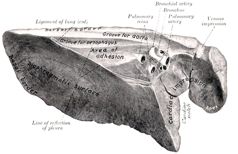
Fig. 362.—Left Lung of Horse, Mediastinal and Diaphragmatic Surfaces. Organ hardened in situ. (Venous impression for common dorso-cervico-vertebral stem.)
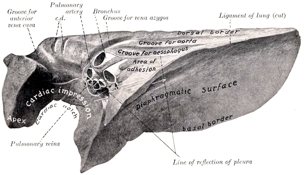
Fig. 363.—Right Lung of Horse, Mediastinal and Diaphragmatic Surfaces. Organ hardened in situ. c, d, Grooves for superior cervical and dorsal veins. Arrows indicate canal for posterior vena cava between mediastinal lobe and main part of lung.
The dorsal (or superior) border (Margo obtusus) is long, thick, and rounded; it lies in the groove alongside of the bodies of the thoracic vertebræ.
The ventral (or inferior) border (Margo acutus) is thin and short. It presents, 456opposite to the heart, the cardiac notch (Incisura cardiaca). On the left lung this notch is opposite to the third, fourth, and fifth ribs, so that a considerable area of the pericardium here lies in direct contact with the chest-wall. On the right lung the notch is less extensive, and is an intercostal space further forward.
The base of the lung (Basis pulmonis) is oval in outline; its surface (Facies diaphragmatica) is deeply concave in adaptation to the thoracic surface of the diaphragm. Laterally and posteriorly it is limited by a thin basal border which fits into the narrow recess (Sinus phrenico-costalis) between the diaphragm and the chest-wall. The position of this border, of course, varies during respiration. In the deepest inspiration it may reach the bottom of this recess; in ordinary inspiration it lies about four or five inches (ca. 10 to 12 cm.) from the costal arch, to which it is nearly parallel; in ordinary expiration it lies about twice as far from the costal arch.
The apex of the lung (Apex pulmonis) is prismatic, narrow, and flattened transversely. It is partially marked off from the rest of the lung by the cardiac notch. It curves downward, and is related internally to the anterior mediastinum and behind to the anterior part of the pericardium.
In the horse the lungs are not divided into distinct lobes by deep fissures, as in most mammals. In the case of the left lung there is no lobation, but the right lung presents a mediastinal lobe, separated from the body of the lung by a fissure which forms in its dorsal part a canal for the posterior vena cava and the right phrenic nerve, inclosed in a special fold of the right pleura.[129]
The root of the lung (Radix pulmonis) is composed of the structures which enter or leave the lung at the hilus on the mediastinal surface. These are: (1) The bronchus; (2) the pulmonary artery; (3) the pulmonary veins; (4) the bronchial artery; (5) the pulmonary nerves; (6) the lymph vessels, which open into the bronchial lymph glands. The bronchus is situated dorsally, with the bronchial artery on its upper surface and the pulmonary artery immediately below it. The pulmonary veins lie chiefly below and behind the artery.
The lobulation of the lungs is not very evident on account of the small amount of interlobular tissue. The lobules appear on the surface or on sections as irregular polygonal areas of different sizes.[130]
Bronchial Branches.—Each bronchus at its entrance into the lung gives off a branch to the apex of the lung. It is then continued backward as the main stem bronchus, parallel with the dorsal border of the lung, giving off branches (Rami bronchiales) in monopodic fashion; these branch similarly and reach all parts of the lung. The right bronchus gives off a special branch to the mediastinal lobe.
The arrangement of the larger bronchi is not the same in the two lungs. The right bronchus, after detaching the apical branch, gives off a large ventral branch which shortly bifurcates. One of the divisions passes ventrally into the part of the lung behind the cardiac notch (homologue of cardiac lobe), while the other passes downward and backward into the lower part of the base. The stem bronchus then gives off the branch to the mediastinal lobe, and divides, about six inches from the hilus of the lung, into two nearly equal branches; the dorsal branch is a direct continuation of the stem, the ventral one passes into the middle part of the base. The left bronchus, after a short course, divides into two nearly equal branches. The dorsal branch may be regarded as the continuation of the stem; it passes backward parallel with the dorsal border of the lung, giving off a ventral branch much further back than in the right lung. The ventral division of the stem bronchus divides, after a very short course, into two branches; the anterior one passes ventrally into the homologue of the cardiac lobe, while the posterior one passes downward and backward into the base of the lung.
The structure of the larger bronchial tubes is, in general, similar to that of the trachea. Their walls contain irregular plates of cartilage instead of rings. There is 457a continuous layer of unstriped muscle, composed of circularly arranged bundles. The mucous membrane presents numerous longitudinal folds; it contains many elastic fibers, mucous glands, and lymph nodules, and is lined by ciliated columnar epithelium. As the tubes diminish in size the coats become thinner and the cartilages smaller; in tubes about 1 mm. in diameter the cartilages and mucous glands are absent.
By repeated branching the interlobular bronchi are formed, and from these arise the lobular bronchioles. The latter enter a lobule and branch within it, forming the respiratory bronchioles; these give off the alveolar ducts, the walls of which are pouched out to form hemispherical diverticula, the alveoli or air-cells.
A lobule of the lung, the unit of lung structure, is made up of a lobular bronchiole with its branches, and their air-cells, blood- and lymph vessels, and nerves. Between the lobules is the interlobular tissue, which forms the supporting framework of the lung.
Vessels of the Lungs.—The branches of the pulmonary artery carry venous blood to the lungs. They accompany the bronchi, and form rich capillary plexuses on the walls of the alveoli. Here the blood is arterialized, and is returned to the heart by the pulmonary veins. The bronchial arteries are relatively small vessels which carry arterial blood for the nutrition of the lungs. The branches of these arteries accompany the bronchial ramifications as far as the alveolar ducts, but do not extend to the alveoli. The œsophageal artery also supplies pulmonary branches (in the horse) which reach the lung by way of the ligament of the latter. These branches vary much in size, and ramify chiefly in the subpleural tissue of the basal part of the lung.[131] The lymph vessels are numerous, and are arranged in two sets. The superficial set forms close networks in and under the pleura, while the deep set accompanies the bronchi and pulmonary vessels. All converge to the root of the lung and enter the bronchial lymph glands.
Nerve-supply.—The pulmonary nerves come from the vagus and sympathetic nerves. They enter at the hilus and supply branches to the bronchial arteries and the air-tubes.
THE THYROID GLAND
The thyroid gland (Glandula thyreoidea) is a very vascular ductless gland, situated on the trachea close to the larynx. It is red brown in color, and consists of two lateral lobes and a connecting isthmus (Fig. 354).
The lateral lobes are situated on either side of the first and second or second and third rings of the trachea, to which they are loosely attached. Each is about the size of a walnut, and has a convex superficial face which is related to the parotid gland and the omo-hyoid muscle, and a slightly concave, deep face, applied to the trachea.
The isthmus in the adult horse is usually very rudimentary. It may occur as a very narrow glandular band which connects the posterior extremities of the lateral lobes, extending across the ventral surface of the trachea, but it is frequently only a small strand of connective tissue, and sometimes is entirely absent. It may be represented by a small tail-like process of one lobe.
The lateral lobes vary in size and position, and are often dissimilar on the two sides. The anterior extremity is the larger; it is rounded and usually lies about a finger’s breadth behind the larynx, but may be in contact with it. The posterior extremity is often produced to form a narrow tail-like process, which is continuous with the isthmus when one is present. In the ass and mule the isthmus is usually well developed. There is sometimes a narrow band of thyroid 458tissue which extends forward from the isthmus for a variable distance; this is termed the pyramidal lobe. The position of the lateral lobe is indicated approximately by the junction of the external maxillary and jugular veins.
Structure.—The gland is enveloped by a thin, fibrous capsule, from which trabeculæ pass into the gland, dividing it into lobules. The lobules consist of non-communicating alveoli or follicles of varying form and size. The alveoli are lined by cubical epithelium, and contain a viscid colloid substance.
Blood-supply.—The arteries are relatively very large; there are usually two thyroid arteries, which arise from the carotid and enter each extremity of the gland.
Nerve-supply.—The nerves are derived from the sympathetic system.
Nodules of thyroid tissue of variable size, sometimes as large as a pea, may be found near the anterior extremity of the lateral lobes, on the course of the anterior thyroid artery or on the trachea, even at a considerable distance from the thyroid region. They are termed accessory thyroids (Glandulæ thyreoidæ accessoriæ).
The parathyroids are small glandular bodies found in the thyroid region. They are paler and not so dense as the thyroid tissue, but often cannot be differentiated by their naked-eye appearance. In the horse there is usually only one on each side in the connective tissue over the anterior extremity of the lateral lobe of the thyroid. They resemble in structure embryonic thyroid tissue.
THE THYMUS GLAND
The thymus is a ductless gland which has a close resemblance to the lymphoid tissues. It is well developed only in late fœtal life and for a few months after birth. After this it undergoes rapid atrophy, fatty infiltration, and amyloid degeneration, so that in the adult it is represented by a thin remnant in the lower part of the anterior mediastinum or has entirely disappeared. In the new-born foal it is of a grayish-pink color, and consists of two lateral lobes (Fig. 358). The greater part of the gland is situated in the anterior mediastinum, but the two lobes are continued into the neck by a chain of lobules which lie along the course of the carotid artery, extending sometimes as far as the thyroid gland.[132]
Structure.—The thymus is inclosed in a thin, connective-tissue capsule, and consists of secondary lobules (Lobuli thymi), of varying shape and size, held together by areolar tissue and fat, and all attached to a cord of adenoid tissue termed the tractus centralis. These lobules are subdivided into polyhedral primary lobules, which are composed of lymphoid tissue, and present a dark vascular cortex, and a lighter medulla that contains Hassal’s corpuscles.
Blood-supply.—Internal thoracic and carotid arteries.
Nerve-supply.—Sympathetic and vagus nerve.
RESPIRATORY SYSTEM OF THE OX
THE NASAL CAVITY
The nostrils, situated on either side of the muzzle, are relatively small, and are much less dilatable than those of the horse. The alæ are thick and firm. The superior commissure is narrow. There is no false nostril or diverticulum nasi. The skin is smooth, bare, and moist, and there is no clear line of demarcation between 459it and the nasal mucosa. The opening of the naso-lacrimal duct is not visible, as it is on the outer wall of the nostril, and is concealed by the cartilaginous prolongation of the inferior turbinal. There are two parietal or lateral cartilages on either side, united by fibrous tissue. The dorsal cartilages are thin laminæ which curve outward and downward from the dorsal margin of the septal cartilage; they are prolongations of the nasal bones, and carry the alar cartilages on their anterior extremities. The ventral pair are lateral continuations of the basal lamellæ of the inferior turbinals. They lie along the nasal processes of the premaxillæ; anteriorly, they become thicker, turn a little upward, and each blends with the cartilaginous prolongation of the upper coil of the inferior turbinal. The alar cartilages are of peculiar form. The lamina is oblong, and curves outward and downward from the anterior extremity of the dorsal parietal cartilage, with which it is connected. The cornu springs from the outer part of the lamina and curves outward and upward into the outer wing of the nostril; it carries on its extremity a small transverse bar, thus having some resemblance to the fluke of an anchor.
The nasal cavity is short, wide anteriorly, narrow behind. It is not completely divided by the septum, which in its posterior third is separated from the floor of the cavity by an interval that increases from before backward. The middle meatus is very narrow, and divides posteriorly into two branches; the upper division leads to the ethmoidal meatuses, and communicates with the frontal sinus and the cavity of the superior turbinal. Anterior to the division are communications with the upper cavity of the ventral turbinal and the maxillary sinus. The main facts in regard to the sinuses were stated in the osteology.
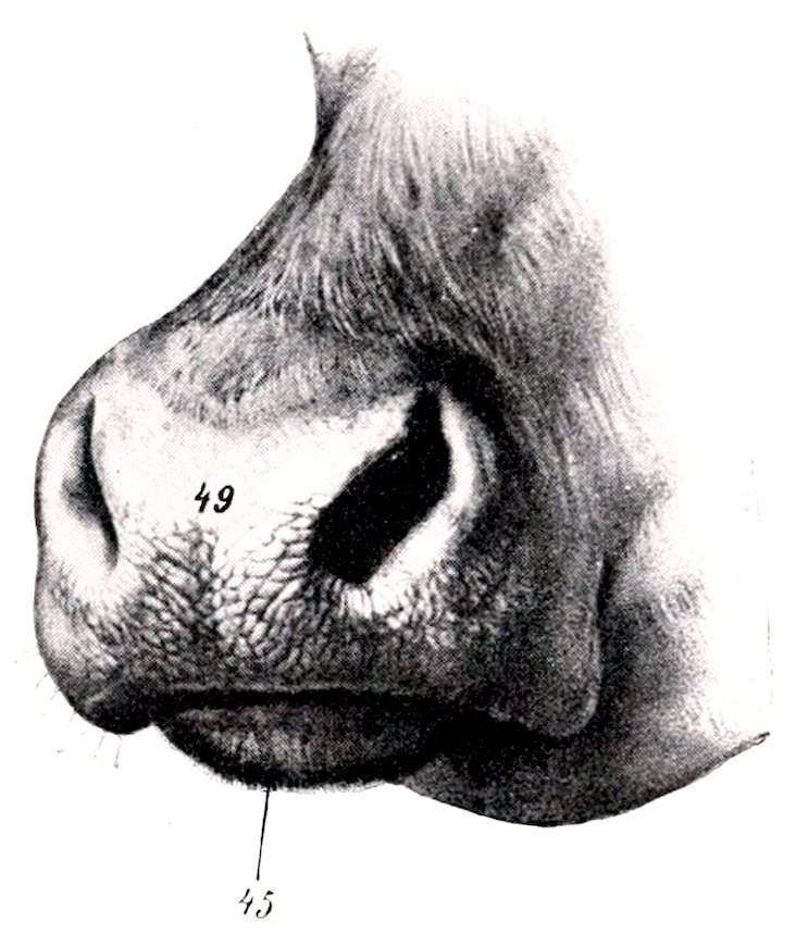
Fig. 364.—Muzzle of Ox. 49, Naso-labial region; 45, lower lip. (After Ellenberger-Baum, Anat. für Künstler.)
THE LARYNX
The larynx is more compact than in the horse, and presents numerous differential features. The cricoid cartilage is compressed laterally. The lamina is not distinctly marked off from the arch; it slopes downward and backward and has a large median ridge. The thyroid cartilage is complete ventrally, i. e., the laminæ are united throughout to form a long plate which is notched in front and behind; the laryngeal prominence is small and is situated posteriorly. The height and thickness of the cartilage increase from before backward. The posterior cornua are about an inch long, and form syndesmoses with the cricoid cartilage; the short anterior cornua unite similarly with the hyoid bone. The epiglottis is oval in outline and its apex is rounded; its base rests on the thyro-hyoid membrane, to which it is rather loosely attached. The cuneiform cartilages are absent. The arytenoid cartilages present only slight differences. The muscular process is well developed, and the vocal process is narrow and long. The vocal cords project very little from the wall, so that the rima glottidis is wide; the vocal ligament is attached ventrally to the crico-thyroid membrane; it has the form of a half tube, open behind. The lateral ventricles are extremely shallow, and the saccules are absent. The thyro-arytenoideus muscle is not divided; it is fan-shaped, thick, and narrow dorsally; ventrally, it is attached to the base of the 460epiglottis, the angle of the union of the thyroid laminæ, and the crico-thyroid membrane. The hyo-epiglotticus muscle is large and bifid.
THE TRACHEA
The caliber of the trachea is relatively small, its width being about an inch and a half (ca. 4 cm.) and its height one and a half to two inches (ca. 4 to 5 cm.). The rings are smaller, and number about fifty. In the neck their free ends are separated by a varying interval, so that the tube is flattened and membranous dorsally. In the thorax the ends are in apposition, so that they form a ridge dorsally. The trachea is adherent to the right lung from the second rib backward. The bifurcation is opposite the fourth intercostal space.
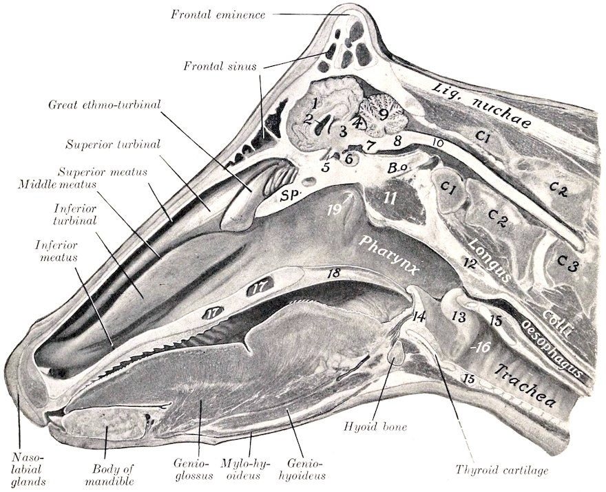
Fig. 365.—Sagittal Section of Head of Cow. 1, Cerebral hemisphere; 2, lateral ventricle; 3, thalamus; 4, corpora quadrigemina; 5, optic chiasma; 6, pituitary body; 7, pons; 8, medulla oblongata; 9, cerebellum; 10, spinal cord; 11, ventral straight muscles; 12, pharyngeal lymph gland; 13, arytenoid cartilage; 14, epiglottis; 15, cricoid cartilage; 16, vocal cord; 17, palatine sinus; 18, soft palate; 19, Eustachian opening; C1, C2, C3, first, second, and third cervical vertebræ; B.o., basioccipital; Sp., presphenoid.
THE BRONCHI
There are three chief bronchi. The bronchus for the apical and the anterior cardiac lobe of the right lung is detached from the trachea at a considerable distance anterior to the bifurcation.
461
THE THORACIC CAVITY AND PLEURA
The thoracic cavity is relatively small; it is especially short dorsally, and is diminished laterally by the mode of attachment of the diaphragm to the ribs (vide Myology). The endothoracic fascia is better developed than in the horse, and is distinctly elastic. The pleura is also thick, and there are no perforations of the mediastinum. The pleural sacs are more unequal in extent than in the horse, and the ventral part of the mediastinum is further to the left; in front of the pericardium it lies for the most part on the left wall of the chest.
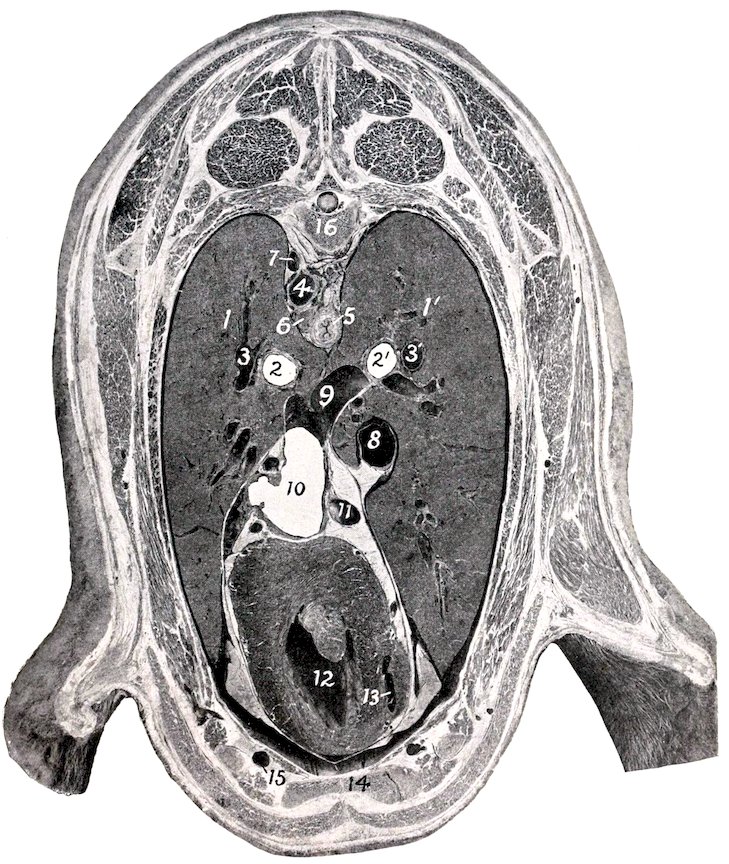
Fig. 366.—Cross-section of Thorax of Sheep, Passing Through Middle of Fifth Pair of Ribs, Posterior View. 1, 1′, Lungs; 2, 2′, bronchi; 3, 3′, pulmonary arteries; 4, thoracic aorta; 5, œsophagus; 6, lymph gland; 7, vena hemiazygos; 8, posterior vena cava; 9, large pulmonary vein; 10, left atrium, 11, vena hemiazygos; 12, left ventricle; 13, right ventricle; 14, sternum; 15, internal thoracic vessels; 16, sixth thoracic vertebra.
462
THE LUNGS
The difference in size between the two lungs is greater than in the horse, the right lung weighing about half as much again as the left one. The average weight of the lungs is about 7½ pounds (ca. 3 to 4 kg.); they form about ¹⁄₁₇₀ of the body-weight.
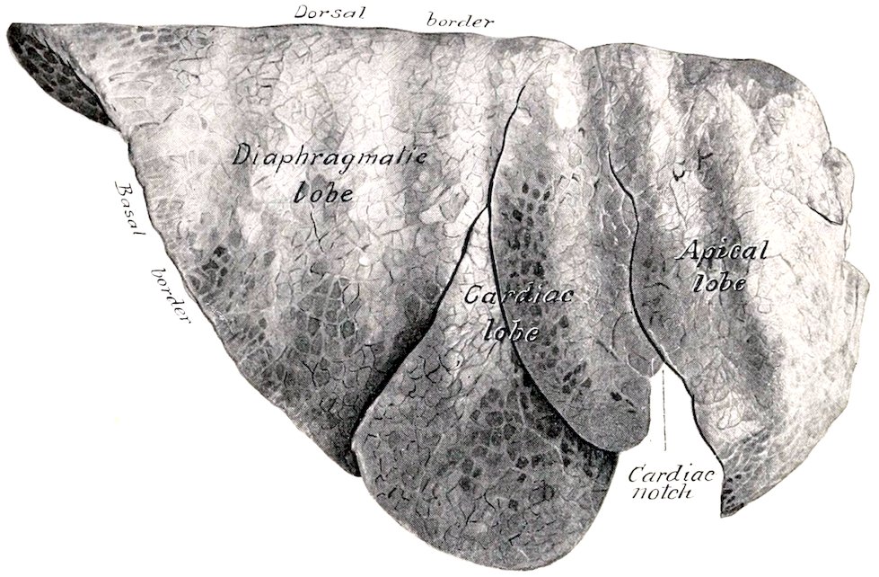
Fig. 367.—Right Lung of Ox, Costal Surface. Organ hardened in situ.
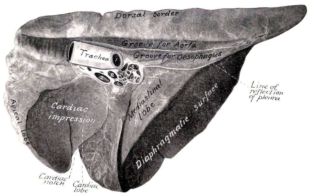
Fig. 368.—Right Lung of Ox, Mediastinal Aspect. Hardened in situ. 1, Left bronchus; 2, pulmonary veins; 3, pulmonary artery.
They are divided into lobes by deep fissures (Incisuræ interlobares). The left lung is divided into three lobes, named from before backward apical, 463cardiac, and diaphragmatic. The right lung has four or five lobes; the apical lobe is much larger than that of the left lung, and occupies the space in front of the pericardium, pushing the mediastinum against the left wall;[133] the mediastinal lobe resembles that of the horse, while the cardiac lobe is often divided by a fissure into two parts. The apical lobe of the right lung receives a special bronchus from the trachea opposite the third rib. The right lung is adherent to the trachea from the second rib backward.
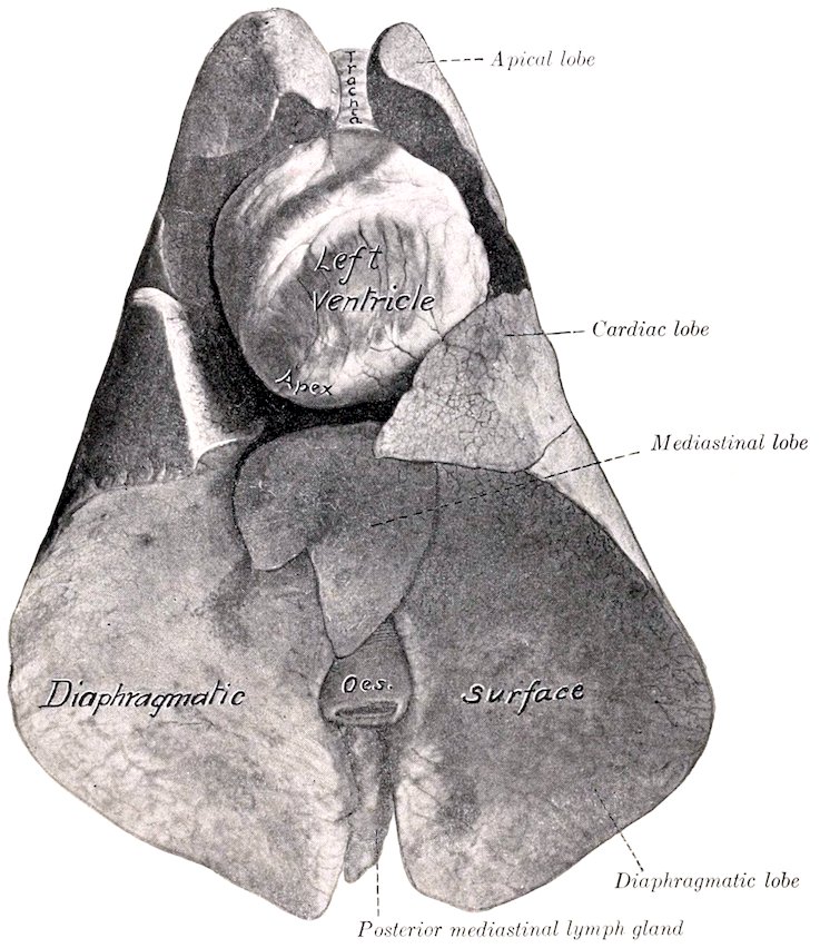
Fig. 369.—Lungs and Heart of Sheep, Ventral View. Specimen hardened in situ. Space between heart and lungs was occupied by pericardium and fat.
The interlobar fissures begin at the ventral margin of the lung and pass toward the root. Those of the left lung lie opposite to the fourth and sixth ribs. The diaphragmatic lobe is the largest, and has the form of a three-sided pyramid with its base resting on the diaphragm. The cardiac lobe is prismatic and forms the posterior margin of the cardiac notch; its long axis corresponds to the fifth rib. The apical lobe of the left lung is small and pointed; its ventral margin lies on the pulmonary artery and the anterior aorta; below this the space in front of the heart is occupied by the apical lobe of the right lung. The cardiac lobe of the right lung is much larger than that of the left lung, and covers the right face of the pericardium, so that the latter has no contact with the lateral wall of the thorax.
The lobulation is very distinct on account of the extremely large amount of interlobular tissue.
464In the sheep the costal attachment of the diaphragm resembles that of the horse, and the basal part of the lung corresponds thereto. The lobation of the lungs resembles that of the ox, but the lobulation is very indistinct. The pleural sacs extend under the first lumbar vertebra.
THE THYROID GLAND
The lobes of the thyroid gland are more extensive and paler in color than in the horse; they extend further forward, overlapping the crico-pharyngeus muscle to a small extent, and come in contact dorsally with the œsophagus. They are flattened, and have an irregular, lobulated surface. In the young subject the isthmus is well developed, being about half an inch (ca. 1 to 1.5 cm.) in width.
Accessory thyroids may be found, and parathyroids also occur near the posterior extremity or inner surface of the lateral lobes of the thyroid.
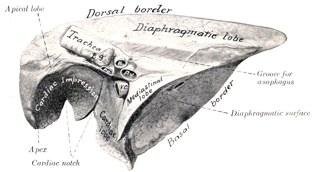
Fig. 370.—Right Lung of Sheep, Mediastinal Aspect. Hardened in situ. B, Bronchus; P.a., pulmonary artery; P.v., pulmonary vein; V.c., posterior vena cava; L.g., bronchial lymph gland.
THE THYMUS GLAND
The thymus is pale and distinctly lobulated. It is much larger in the calf than in the foal, weighing at five or six weeks about 15 to 25 ounces (ca. 425 to 600 grams). The cervical part is specially large, thick, and compact; it extends to the thyroid gland. It undergoes atrophy slowly and remnants of the thoracic part often remain even in advanced age.
RESPIRATORY SYSTEM OF THE PIG
THE NASAL CAVITY
The nostrils are small, and are situated on the flat anterior surface of the rostrum or snout (Rostrum suis). The latter is a short cylindrical projection, with which the upper lip is fused, and is circumscribed by a prominent circular margin. The skin on the snout is thin and highly sensitive; it presents small pores, and scattered over it are fine short hairs. In the snout between the nostrils is the os rostri, which is to be regarded as a special development of the extremity of 465the septum nasi in adaptation to the habit of rooting. A plate of cartilage, representing the lamina of the alar cartilage of the horse, curves outward and downward from the upper part of the os rostri and a pointed bar of cartilage curves upward from the lower part of the bone in the outer wing of the nostril. The notch between the nasal bone and the premaxilla is closed in by parietal cartilages which resemble those of the ox.
The nasal cavity is long and narrow. It is divided behind by a horizontal plate into an upper olfactory part, which leads to the ethmoidal meatuses, and a lower respiratory part, which is a direct continuation of the inferior meatus. The posterior part of the septum is membranous.
The turbinal bones resemble in general those of the ox. The superior meatus is exceedingly small. The middle meatus is a deep fissure between the two turbinals: it divides posteriorly into two branches; one of these extends upward and backward between the lateral mass of the ethmoid bone and the superior turbinal; the other widens and joins the inferior meatus. The opening between the maxillary sinus and the middle meatus lies in a plane through the last cheek teeth. The middle meatus is continuous with the space inclosed by the dorsal part of the inferior turbinal and with the cavity of the superior turbinal. The dorsal division of the middle meatus presents several openings into the frontal sinus. The inferior meatus is relatively roomy; it communicates with the space inclosed by the ventral coil of the inferior turbinal. The opening of the naso-lacrimal duct is found in the posterior part of the inferior meatus. The naso-palatine duct and the vomero-nasal organ (of Jacobson) resemble those of the ox.
THE LARYNX
The larynx is remarkable for its great length and mobility. The cartilages are more loosely attached to each other than in the other animals. The cricoid cartilage is thick and compressed laterally; its lamina is long and narrow; its arch is directed obliquely downward and backward. The thyroid cartilage is very long; its laminæ are united ventrally and form a median ridge. The anterior cornua being absent, there are no joints formed with the hyoid bone. The posterior cornua are broad, bent inward, and articulate with the cricoid cartilage. The epiglottis is relatively very large, and is more closely attached to the hyoid bone than to the rest of the larynx. The middle part of its base is turned forward, and rests on the thyro-hyoid membrane; it is closely connected with the body of the hyoid bone by the hyo-epiglottic ligament and the strong hyo-epiglottic muscle.[134] The apex of the arytenoid cartilage is very large, and is divided into two parts at its extremity; the inner part is fused with that of the opposite cartilage. There is a small interarytenoid cartilage in the transverse ligament. The rima glottidis is very narrow. The vocal cords are directed obliquely downward and backward; and each is pierced by a long, slit-like opening, which leads into the large laryngeal saccule. The thyro-arytenoid or vocal ligament is similarly divided into a larger anterior and a smaller posterior part. There is a middle ventricle near the base of the epiglottis. The thyro-arytenoid muscle is very strong and is undivided. The arytenoideus is very small. The crico-thyroid consists of two strata; the superficial layer corresponds to the muscle of the other animals; the deep part consists of transverse fibers.
466
TRACHEA AND BRONCHI
The trachea is circular in cross-section, and contains thirty or more rings which almost meet dorsally. A special bronchus is detached for the apical lobe of the right lung, as in the ox.
THE LUNGS (Figs. 324, 325)
The right lung has four or five lobes, according to whether the apical is subdivided or not. The left lung resembles that of the ox in form and lobation. The lobulation is distinct, but the interlobular septa are thinner than in the ox.
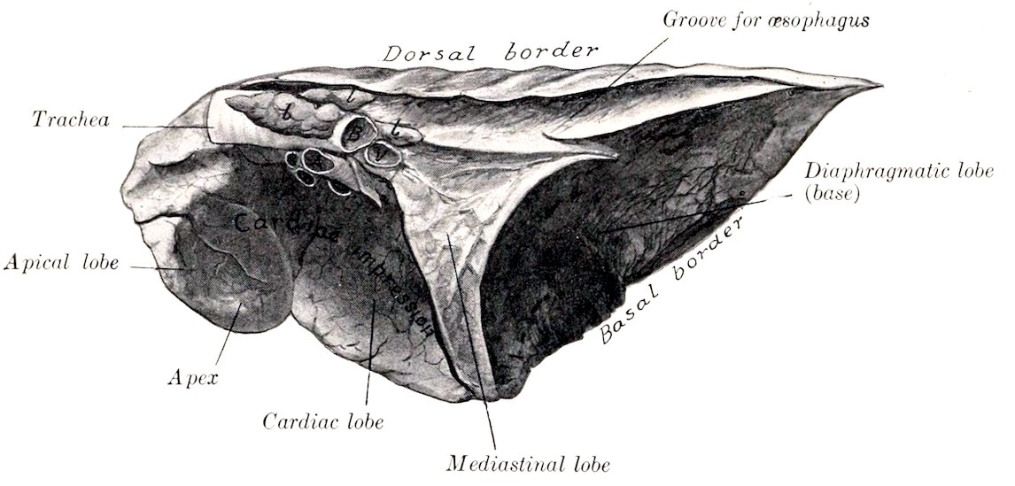
Fig. 371.—Right Lung of Pig, Mediastinal Aspect. Hardened in situ. B, Left bronchus; V., pulmonary veins; A., pulmonary artery; l, lymph glands.
THE THYROID GLAND
The two lobes of the thyroid are long and flattened; they are situated close together, on the ventral surface of the trachea, extending as far forward as the cricoid cartilage. The gland is dark red in color.
THE THYMUS
The thymus is very large, extending to the larynx or even to the submaxillary space in young subjects.
RESPIRATORY SYSTEM OF THE DOG
THE NASAL CAVITY
The nostrils are situated on the muzzle, with which the upper lip blends. They are shaped somewhat like a comma, with the broad part next to the septum and the narrow part directed backward and outward. The skin around the nostrils is bare, usually black, and in health moist and cool. The muzzle is marked by a median furrow (philtrum) or a deep fissure in some breeds. The cartilaginous framework is formed essentially by the septal cartilage and the parietal cartilages which proceed from it. The septal cartilage projects beyond the premaxilla, and is much thickened at its extremity; it gives off from its upper and lower margins 467the two parietal cartilages, which curve laterally and toward each other. A grooved plate extends outward from the septal cartilage in the floor of the nostril, and another lamina supports the alar fold of the inferior turbinal bone.
The length of the nasal cavity varies greatly in different breeds, corresponding, of course, to the length of the face. The cavity is roomy, but is very largely occupied by the turbinals and the lateral masses of the ethmoid bone. The middle meatus is short and narrow, and divides posteriorly into two branches; the upper branch leads to the ethmoidal meatuses; the lower branch joins the inferior meatus. The inferior meatus is very small in its middle part, owing to the great development here of the inferior turbinal. The posterior part of the nasal cavity is divided by a horizontal plate, the lamina transversalis, into an upper olfactory part and a lower respiratory part. The sinuses have been described (vide Osteology).
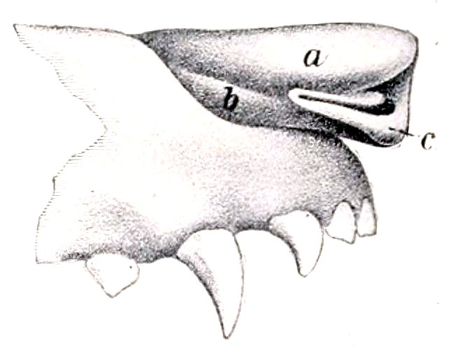
Fig. 372.—Nasal Cartilages of Dog. a, Upper parietal cartilage; b, lower parietal cartilage; c, alar cartilage. (After Ellenberger, in Leisering’s Atlas.)
THE LARYNX
The larynx is relatively short. The lamina of the cricoid cartilage is wide; the arch is grooved laterally. The laminæ of the thyroid cartilage are high, but short; they unite ventrally to form the body, on which there is anteriorly a marked prominence, and posteriorly a deep notch. The oblique line on the outer surface of the lamina is prominent. There is a rounded notch (Fissura thyreoidea) below the short anterior cornua for the passage of the superior laryngeal nerve. The posterior cornu is strong, and has a rounded surface for articulation with the cricoid cartilage. The arytenoid cartilages are relatively small, and have between them a small interarytenoid cartilage. The epiglottis is quadrilateral; its lower part or stalk is narrow, fitting into the angle of the thyroid cartilage. The cuneiform cartilages are large and somewhat crescent-shaped; they are not blended with the epiglottis.
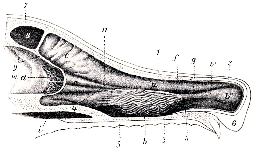
Fig. 373.—Sagittal Section of Nasal Region of Dog. 1, Skin; 2, parietal cartilage; 3, floor of nasal cavity; 4, lamina transversalis; 5, mucous membrane of hard palate; 6, upper lip; 7, frontal bone; 8, frontal sinus; 9, cranial plate of frontal bone; 10, cranial cavity; 11, dotted line indicating anterior limit of olfactory region; a, superior turbinal; b, inferior turbinal; b′, b″, inferior turbinal fold; c, e, ethmoturbinals; d, cribriform plate of ethmoid; f, superior meatus; g, middle meatus; h, inferior meatus; i, naso-pharyngeal meatus. (After Ellenberger, in Leisering’s Atlas.)
The false vocal cords extend from the cuneiform cartilages to the thyroid. 468The true vocal cords are large and prominent. The entrance to the large lateral ventricle and saccule is a long slit parallel with the anterior margin of the true vocal cord.
The crico-thyroid muscle is thick. The hyo-epiglotticus is well developed, and is double at its hyoid attachment. The anterior part of the thyro-arytenoideus arises on the cuneiform cartilage. Hence Lesbre has suggested the name cuneo-arytenoideus for it.
THE TRACHEA AND BRONCHI
The trachea is very slightly flattened dorsally. It contains forty to forty-five U-shaped rings; the ends of the rings do not meet dorsally, so that here the wall of the trachea is membranous and is composed of a layer of transverse smooth muscle-fibers outside of the rings, the fibrous membrane, and the mucous membrane.
The stem bronchi diverge at an obtuse angle, and each divides into two branches before entering the lung. The anterior branch goes to the apical and cardiac lobes, the posterior one to the diaphragmatic lobe, supplying also the mediastinal lobe of the right lung. The anterior branch of the left bronchus crosses under the pulmonary artery.
THE LUNGS
The lungs (Figs. 342, 343) differ in shape from those of the horse and ox in conformity with the shape of the thorax, which is relatively very wide in the dog; the lateral thoracic walls are strongly curved, and the costal surface of the lungs is correspondingly convex. There is no cardiac notch on the left lung, although a small part of the pericardium is not covered by the lung at the lowest part of the fifth and sixth intercostal spaces. On the right side the cardiac notch is opposite the fourth and fifth intercostal spaces. Each lung is divided by deep fissures into three lobes—apical, cardiac, and diaphragmatic; the right lung has in addition a mediastinal lobe. The apical lobe of the right lung is considerably larger than that of the left. The latter is often more or less fused with the cardiac lobe. Variable accessory lobes may be found. On account of the small amount of interlobular tissue the lobulation is not distinct.
THE THYROID GLAND
The lateral lobes of the thyroid gland are long and narrow, and have a flattened, ellipsoidal form; they are placed on the lateral surfaces of the trachea near the larynx. The extremities are small, the posterior one often being pointed. The isthmus is inconstant and variable; in large dogs it usually has the form of a glandular band which may be nearly half an inch (ca. 1 cm.) wide; in small dogs it is usually absent.
Accessory thyroids are frequently present; three or four may be found on either side, as well as a median one near the hyoid bone.
The parathyroids, about the size of millet or hemp seed, are four in number usually. Two are placed on the deep face of the thyroid lobes; the others are placed laterally near the anterior extremity of the thyroid.
THE THYMUS
The thymus is relatively small, and is situated almost entirely in the thorax. The left lobe is much larger than the right, and extends back as far as the sixth rib at the time of its greatest size. According to Baum the gland increases in size during the first two weeks after birth, and atrophies rapidly in the next two or three months. Traces of it are present at two or three years of age, and may be found even in old subjects.
469