THE MUSCULAR SYSTEM (continued 4)
Reading edition
The external intercostals do not occupy the spaces between the costal cartilages.
The diaphragm has a small tendinous center. The œsophageal opening is between the pillars. The fleshy rim is attached at the costo-chondral junctions from the eighth backward, and along the thirteenth rib nearly its entire length.
Muscles of the Back and Loins
The serratus anticus arises from the median raphé of the neck and the first six or seven thoracic spines, and is inserted into the second to the ninth ribs. It is well developed. The serratus posticus—much weaker—arises on the lumbo-dorsal fascia, and is inserted into the last three or four ribs. Thus one or two ribs intervene between the two.
The transversalis costarum (Iliocostalis) is well developed, and extends from the ilium to the sixth, fifth, or fourth cervical vertebra.
The longissimus resembles that of the other animals, but the spinalis et semispinalis separates clearly at the sixth or seventh thoracic vertebra. It is inserted into the articular and spinous processes of the last six cervical vertebræ. It has no depression in the lumbar region for the gluteus medius.
The intertransversales are fleshy, as in the ox.
The interspinales are distinct, especially in the lumbar region.
Muscles of the Tail
These present the same general arrangement as in the horse. The sacro-coccygei, however, arise on the lumbar vertebræ also, and the coccygeus on the ischiatic spine. There is found a sacro-coccygeus accessorius, which arises on the internal border of the ilium, the edge of the sacrum, and the transverse processes of the first coccygeal vertebræ, and is inserted between the superior and lateral sacro-coccygei. It is homologous with the intertransversales.
326
Muscles of the Abdomen
The abdominal tunic is practically absent.
The obliquus abdominis externus has an extensive fleshy portion. It arises from the last eight or nine ribs and the lumbo-dorsal fascia.
The obliquus abdominis internus arises from the external angle of the ilium and the lumbo-dorsal fascia. The fibers have an almost vertical direction, and there is a fleshy attachment to the last rib.
The rectus abdominis is attached by a long tendon on the first five or six costal cartilages, and by fleshy fibers on the xiphoid cartilage. It has three to six indistinct tendinous inscriptions.
The transversus abdominis presents no special features except that the posterior part of its aponeurosis splits into two layers which include the rectus between them.
Muscles of the Pelvic Limb
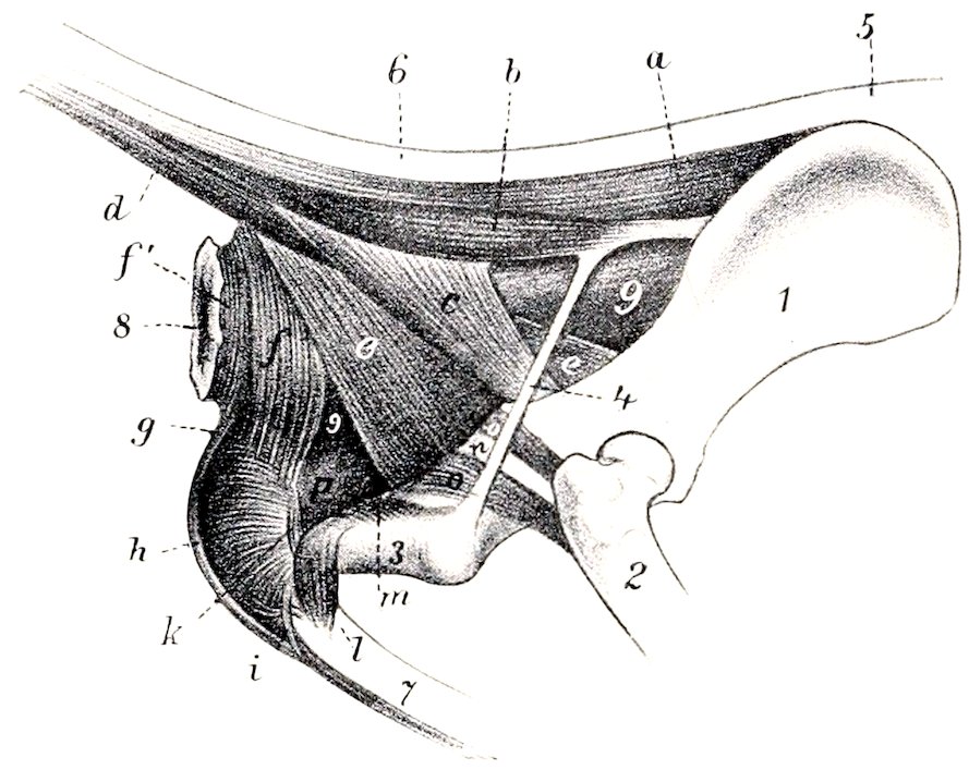
Fig. 235.—Muscles of Tail, Anus, and Genital Organs of Dog. 1, Ilium; 2, femur; 3, tuber ischii; 4, sacro-sciatic lig.; 5, sacral region; 6, tail; 7, penis; 8, anus; 9, rectum; a, sacro-coccygeus superior; b, sacro-coccygeus accessorius; c, coccygeus; d, sacro-coccygeus inferior; e, retractor ani; f, f′, sphincter ani externus; g, retractor penis; h, bulbo-cavernosus; i, transversus perinei (?); k, m, ischio-urethrales; l, ischio-cavernosus; n, tendon of obturator internus; o, gemellus; p, urethral muscle. (After Ellenberger, in Leisering’s Atlas.)
The psoas minor arises on the last thoracic and first four or five lumbar vertebræ, and is inserted into the ilio-pectineal line.
The psoas major is short, arising from the last four lumbar vertebræ.
The outer head of the iliacus is small, while the inner head is large and fuses with the psoas major.
The quadratus lumborum is well developed, and extends laterally beyond the outer edge of the ilio-psoas. It arises from the last four ribs and the lumbar transverse processes, and ends on the pelvic surface of the wing of the ilium.
The tensor fasciæ latæ consists of two parts. The anterior part is long and rounded; the posterior is shorter and fan-like.
The gluteus superficialis is small. It arises on the sacrum, first coccygeal vertebræ, and sacro-sciatic ligament. It is inserted below and behind the trochanter major of the femur, on the outer branch of the linea aspera.
The gluteus medius has no lumbar portion. It is inserted into the trochanter major by a strong tendon.
The gluteus profundus is broad and fan-shaped. It arises on the superior ischiatic spine and on the ilium as far forward as the gluteal line, and is inserted into the trochanter major below the medius.
The pyriformis is not blended with the gluteus medius. It arises from the border of the sacrum and from the sacro-sciatic ligament, and ends on the trochanter major.
The biceps femoris has two heads of origin which soon fuse. The larger head arises from the sacro-sciatic ligament and tuber ischii, the smaller one from the tuber 327ischii. The aponeurosis of insertion ends on the patella, the patellar ligament, and the tibial crest. There is also a tendinous band, which comes from the deep face of the muscle and terminates at the tarsus, as in the horse.
The abductor cruris posterior may be regarded as an accessory head of the biceps femoris. It is a thin, muscular band which arises on the sacro-sciatic ligament, passes downward between the biceps and semimembranosus, and ends by blending with the former.
The semitendinosus arises from the tuber ischii only. It ends as in the horse.
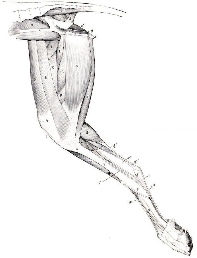
Fig. 236.—Muscles of Pelvic Limb of Dog, Internal View. a, Ilio-psoas; b, tendon of psoas minor; c, sacro-coccygeus ventralis lateralis; d, coccygeus; e, pyriformis; f, obturator internus; g, origin of retractor ani; h, h′, sartorius; i, rectus femoris; k, vastus internus; l, pectineus; m, adductor; n, gracilis; o, semitendinosus; p, semimembranosus; q, gastrocnemius, inner head; q′, tendon of gastrocnemius; r, superficial digital flexor; r′, tendon of r; s, tarsal tendon of biceps femoris; t, popliteus; u, deep digital flexor, outer head (flexor hallucis longus); u′, long digital flexor; u″, common tendon of u and u′; v, tendon of tibialis posterior; w, tibialis anterior; x, tendon of anterior or long digital extensor; 1, pelvic surface of ilium; 2, section of symphysis pelvis; 3, tuber ischii; 4, internal condyle of femur; 5, ligamentum patellæ; 6, 7, internal surface of tibia; 8, tarsus; 8′, fibular tarsal bone; 9, metatarsus. (Ellenberger-Baum, Anat. d. Hundes.)
The semimembranosus is large and arises from the ischium only. It divides into two portions: the anterior portion ends on the tendon of the pectineus, on the femur above the internal condyle, and on the inner (Vesalian) sesamoid bone; the posterior portion ends on the internal condyle of the tibia, the tendon passing under the internal lateral ligament of the stifle joint.
The rectus femoris has only one tendon of origin. The single patellar ligament acts as the tendon of insertion of the quadriceps.
328The capsularis is usually present, but is small and pale.
The sartorius consists of two portions. It arises from the external angle and border of the ilium. The anterior portion ends on the patella, the posterior on the internal surface of the tibia, its tendon blending with that of the gracilis.
The graciles are not so much fused at their origin as in the other animals.
The pectineus is long and slender. It arises from the ilio-pectineal eminence and ends on the internal branch of the linea aspera above the distal end of the femur.
There are two distinct adductors. The small anterior one ends on the proximal third of the femur, the posterior one on the distal part of the femur and on the inner face of the stifle joint.
The quadratus femoris is short and strong. The other external rotators of the thigh present no special features.
The peroneus tertius is represented by a tendinous band which arises on the internal surface of the tibia below the crest. It passes downward on the inner surface of the tibialis anterior, blends with the annular ligament above the tarsus, and is attached to the joint capsule and the proximal end of the third metatarsal bone.
Arloing and Lesbre say: “The third peroneus is a proper extensor of the fifth digit; it is a very feeble, fleshy band, situated behind the peroneus brevis, which it partially covers. It is attached to the upper part of the fibula, and is continued by a long, delicate tendon which passes in the same malleolar groove with the muscle mentioned; it then crosses behind the tendon of the peroneus longus and extends to the phalanges of the outer digit, where it joins one of the branches of the common extensor.”
The tibialis anterior is large and superficial. It arises on the external condyle and crest of the tibia, and is inserted into the first metatarsal bone, or into the first tarsal and second metatarsal.
There are four extensors of the digits.
1. The anterior or long digital extensor (M. extensor digitalis longus) is fusiform, and lies largely under the preceding muscle. It arises from the extensor fossa of the femur. The tendon is bound down by two annular ligaments, and divides below the tarsus into four branches, which end on the distal phalanges of the digits (second to fifth).
2. The lateral digital extensor (M. extensor digitalis lateralis) is feeble and semipennate. It arises on the fibula below the head. The tendon passes under the lateral ligament of the tarsus and joins the branch of the tendon of the long extensor for the fifth digit.
3. The extensor hallucis longus is a very thin muscle which arises from the fibula under the long extensor. Its delicate tendon accompanies that of the tibialis anterior to the first metatarsal bone, or becomes lost in the fascia.
4. The extensor digitalis brevis has three divisions. It arises on the fibular tarsal bone and the adjacent ligaments. The three tendons are inserted into the second, third, and fourth digits, blending with the interossei. (Sometimes there is found a tendon to the rudimentary first digit, which may represent the extensor hallucis brevis. There may be a fourth belly for the tendon to the second digit.)
The peroneus longus arises on the external condyle of the tibia, the head of the fibula, and the lateral ligament. The long tendon passes down the leg parallel to the fibula, crosses the plantar (posterior) surface of the tarsus transversely, and ends on the first metatarsal bone. A short branch is detached to the external metatarsal bone.
The peroneus brevis is semipennate and arises from the distal half or more of the outer face of the tibia and the fibula. Its tendon accompanies that of 329the lateral extensor over the external malleolus of the tibia, and ends on the proximal end of the fifth metatarsal bone.
The soleus is absent. (It is present and large in the cat.)
The gastrocnemius arises on the rough lines above the condyles of the femur. The heads of origin each contain a bone about the size of a pea—the sesamoid of Vesal—which articulates with the corresponding condyle of the femur. The tendon comports itself as in the horse.
The superficial digital flexor has a large round belly. It arises in common with the outer head of the gastrocnemius from the external rough line and Vesalian sesamoid bone, and from the aponeurosis of the vastus externus. The tendon winds around that of the gastrocnemius, passes over the tuber calcis (where it is arranged as in the horse), and divides below the tarsus into two branches. Each of these divides into two branches which end as in the fore limb. The outer and inner branches detach slips to the suspensory ligaments of the large digital pad. Muscle-fibers often occur in the tendon in the metatarsal region.
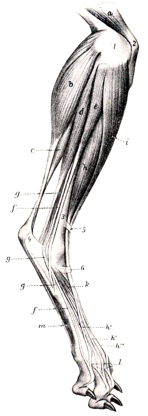
Fig. 237.—Muscles of Leg and Foot of Dog, External View. a, Quadriceps femoris; b, gastrocnemius, external head; c, superficial digital flexor; d, deep digital flexor; e, peroneus longus; f, tendon of lateral extensor; g, peroneus brevis; h, long or anterior digital extensor; h′, h″, h‴, tendons of preceding; i, tibialis anterior; k, extensor brevis; l, slips from interossei (m); 1, external condyle of femur; 2, patella; 3, tibia; 4, tuber calcis; 5, 6, annular ligaments. (After Ellenberger, in Leisering’s Atlas.)
The deep digital flexor has two heads. The large outer head, the flexor hallucis longus, arises from the tibia and fibula, filling the interosseous space. The small inner head, flexor digitalis pedis longus, also arises from the tibia and fibula, its tendon joining that of the large head below the tarsus. The common tendon detaches a branch to the large digital pad and terminates as in the fore limb.
The tibialis posterior is a small but distinct muscle, which arises on the proximal part of the fibula. The thin tendon accompanies that of the flexor longus and ends on the internal lateral ligament of the tarsus.
The tendon of the popliteus contains a small sesamoid bone.
The adductors of the second and fifth digits, the lumbricales, and the interossei are arranged as in the thoracic limb.
The quadratus plantæ arises on the outer surface of the distal end of the fibular tarsal bone and on the lateral tarsal ligament, passes downward and inward, and terminates on a thin tendon which fuses with that of the deep flexor.
The abductor digiti quinti is a very small muscle which consists of two parts. One of these is a tendinous slip which extends from the posterior surface of the proximal part of the fibular tarsal bone to the head of the fifth metatarsal bone; the other part arises from the inner surface of the fibular tarsal bone (or from the tendinous part) and ends on the first phalanx of the fifth digit.
In case the skeleton of the first digit is well developed, there are found three muscles homologous with those of the same digit in the fore limb. These are the abductor hallucis, adductor hallucis, and flexor hallucis brevis.
330