NEUROLOGY (continued 2)
Reading edition
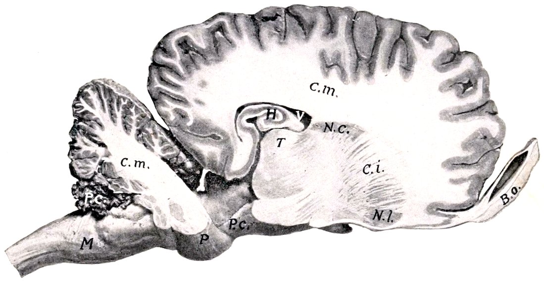
Fig. 513.—Sagittal Section of Brain of Horse. Section is cut about 1.5 cm. to the right of the median plane. M, Medulla oblongata; P, pons; P.c. (above M), chorioid plexus of fourth ventricle; C.m., central white matter (corpus medullare) of cerebellum and of cerebrum; P.c. (in front of P), cerebral peduncle; H, hippocampus; V, lateral ventricle; T, thalamus; N.c., caudate nucleus; C.i., internal capsule; N.l., lenticular nucleus; B.o., olfactory bulb.
The corpus striatum[202] is the great basal ganglion of the hemisphere. It is situated in front of the thalamus and the cerebral peduncle, and its anterior rounded end appears on the base of the hemisphere at the trigonum olfactorium. It is composed of two masses of gray matter, the caudate and lenticular nuclei, separated incompletely by tracts of white matter which are known collectively as the internal capsule. The caudate nucleus (Nucleus caudatus) is the dorso-medial and larger of the two gray masses; it has been seen in the examination of the floor of the lateral ventricle. The lenticular nucleus (Nucleus lentiformis) lies ventro-laterally, over the trigonum olfactorium and the fossa transversa. It is related externally to a layer of white matter termed the external capsule, which separates it from a stratum of gray substance known as the claustrum. The two nuclei are fused in front, and 676further back they are connected by strands of gray matter which intersect the internal capsule.
The amygdaloid nucleus (Nucleus amygdalæ) (Fig. 506) is an ovoid mass of gray matter situated external to the ventral horn of the lateral ventricle and below the posterior part of the lenticular nucleus. Some fibers of the stria terminalis are connected with it.
The internal capsule (Capsula interna) is a broad band of white matter situated between the thalamus and caudate nucleus internally and the lenticular nucleus externally. A sagittal section through the brain shows that it is in great part directly continuous with the basis or ventral part of the cerebral peduncle. It contains most of the so-called projection fibers of the hemisphere, which connect the cerebral cortex with nuclei of other and more posterior parts of the brain. When the fibers of the internal capsule are traced forward it is evident that they spread out in all directions to reach the cerebral cortex. This arrangement, in which the fibers of the corpus callosum participate, is termed the corona radiata.
The internal capsule also contains fibers which connect the corpus striatum with the thalamus. These are termed the thalamo-striate and strio-thalamic fibers respectively, according to the direction in which they conduct impulses.
The fibers of the stria terminalis connect the amygdaloid nucleus with the septum pellucidum and trigonum olfactorium. It is therefore probably part of the complex connections between the primary and secondary olfactory centers.
The Cranial Nerves
The cranial or cerebral nerves (Nn. cerebrales) comprise twelve pairs which are designated from before backward numerically and by name. Their number, names, and functional characters are given in the subjoined table:
I. Olfactory Sensory (Small)
II. Optic Sensory (Sight)
III. Oculomotor Motor
IV. Trochlear Motor
V. Trigeminal Mixed
VI. Abducent Motor
VII. Facial Mixed
VIII. Auditory Sensory (Hearing and Equilibration)
IX. Glosso-pharyngeal Mixed
X. Vagus or Pneumogastric Mixed
XI. Spinal accessory Motor
XII. Hypoglossal Motor
THE FIRST OR OLFACTORY NERVE
The olfactory nerve (N. olfactorius) is peculiar in that its fibers are not aggregated to form a trunk, but are connected in small bundles with the olfactory bulb. They are non-medullated, and are the central processes of the olfactory cells which are situated in the olfactory region of the mucous membrane of the nasal cavity. This region is distinguished by its brown color and comprises most of the lateral mass of the ethmoid, a small adjacent area of the superior turbinal, and the corresponding surface of the septum nasi. The nerve-bundles are inclosed in sheaths derived from the membranes of the brain and pass through the foramina of the cribriform plate to join the convex surface of the olfactory bulb. Some fibers come from the vomero-nasal organ of Jacobson.
THE SECOND OR OPTIC NERVE
The optic nerve (N. opticus) is composed of fibers which are the central processes of the ganglion cells of the retina. The fibers converge within the eyeball to 677the optic papilla, where they are collected into a round trunk, the optic nerve. The nerve thus formed pierces the chorioid and sclera, emerges from the posterior part of the eyeball, and passes backward and inward to the optic foramen. After traversing the latter it decussates with its fellow of the opposite side to form the optic chiasma or commissure. In the orbit the nerve is slightly flexuous and is embedded in the fat behind the eyeball and surrounded by the retractor oculi muscle. Its intraosseous part is an inch or more (ca. 3 cm.) long. The sheath of the nerve is formed by prolongations of the membranes of the brain, and includes continuations of the subdural and subarachnoid spaces.
The greater part of the fibers of the optic nerve cross in the chiasma to the tract of the opposite side. In the tract the fibers proceed to (1) the internal geniculate body, (2) the posterior part of the thalamus, and (3) the anterior quadrigeminal body (indirectly). The fibers which go to the internal geniculate body appear to belong to Gudden’s commissure and to be nonvisual in function. The visual fibers, which come from the outer part of the retina of the same side and the inner part of the retina of the opposite side, terminate about cells in the anterior quadrigeminal body and the part of the thalamus which corresponds to the pulvinar and external geniculate body of man. From the cells of the former fibers pass to the nuclei of the motor nerves of the eyeball, and complete the reflex arc. Fibers proceed from the cells of the thalamus to the visual area of the cortex in the occipital part of the hemisphere.
THE THIRD OR OCULOMOTOR NERVE
The oculomotor nerve (N. oculomotorius) arises by several radicles from the basal surface of the cerebral peduncle, a little lateral to the interpeduncular furrow. It turns sharply outward and forward, crosses over the cavernous sinus, and continues above the maxillary nerve and in company with the ophthalmic nerve to the foramen lacerum orbitale. It emerges through the foramen with the latter nerve and the abducens and divides into two branches. The superior branch is short and divides into twigs which supply the rectus superior, retractor, and levator palpebræ superioris. The inferior branch (Figs. 438, 439) is larger and much longer. It supplies the motor fibers to the ciliary ganglion (which lies directly on this branch in the horse) and short branches to the rectus internus and rectus inferior, and continues forward on the latter to end in the inferior oblique muscle.
The deep origin of the fibers of the oculomotor nerve is in the oculomotor nucleus situated in the gray matter of the floor of the cerebral aqueduct in the region of the anterior corpora quadrigemina.
THE FOURTH OR TROCHLEAR NERVE
The trochlear or pathetic (N. trochlearis) is the smallest of the cranial nerves. It arises from the anterior cerebellar peduncle just behind the corpora quadrigemina, curves outward and forward, pierces the tentorium cerebelli, and passes forward along the outer border of the maxillary nerve (Figs. 504, 505). It emerges from the cranium through a small foramen immediately above the foramen lacerum orbitale or through the latter and passes forward along the inner wall of the orbit to end in the posterior part of the superior oblique muscle of the eyeball (Fig. 439).
The fibers of the fourth nerve spring from a nucleus in the gray matter of the floor of the cerebral aqueduct behind the oculomotor nucleus. The fibers run backward in the tegmentum, then turn upward and inward and undergo total decussation with those of the opposite nerve in the anterior part of the anterior medullary velum. In addition to this peculiarity it is the only nerve which is connected with the dorsal aspect of the brain.
THE FIFTH, TRIGEMINAL, OR TRIFACIAL NERVE
The trigeminal nerve (N. trigeminus) is the largest of the cranial series. It is connected with the lateral part of the pons by a large sensory root and a smaller motor root (Fig. 499).
678The sensory root (Portio major) extends forward through a notch on the lower part of the petrosal crest and widens out to join the semilunar ganglion.
The semilunar (or Gasserian) ganglion (Ganglion semilunare) is a crescent-shaped mass of nerve-fibers and cells which overlies the antero-external part of the foramen lacerum basis cranii, and is partly embedded in the dense fibrous tissue which occupies the foramen except where vessels and nerves pass through. Its long axis, which is about an inch (2.5 cm.) in length, is directed forward and inward and its convex anterior face gives rise to the ophthalmic, the maxillary, and the sensory part of the mandibular division of the nerve. The surface of the ganglion is irregularly striated. It is connected by filaments with the adjacent carotid plexus of the sympathetic, and sends delicate twigs to the dura mater.
The fibers of the sensory root arise from the ganglion as axones of the ganglion cells, and the fibers of the nerves which extend peripherally from the ganglion are dendrites of the cells. The sensory root-fibers enter the tegmentum of the pons and divide into anterior and posterior branches, which terminate about the cells of the sensory nucleus of termination of the trigeminus. This nucleus extends from the pons to the sixth cervical segment of the spinal cord (Dexler). The posterior branches of the fibers are collected into a compact bundle, the spinal tract or root of the trigeminus, which lies lateral to the substantia gelatinosa in the medulla. The central connections of the sensory part of the trigeminus are very extensive. The most important paths are: (1) Axones of cells of the sensory nucleus and the substantia gelatinosa pass chiefly as arcuate fibers across the raphé to the thalamus, whence impulses are transmitted by thalamo-cortical fibers to the cerebral cortex. In ungulates a distinct tract extends from the anterior part of the sensory nucleus to the thalamus of the same side (Wallenberg). It is probable that collaterals of the arcuate fibers go to the motor nuclei of the fifth, seventh, ninth, and tenth cranial nerves. (2) Axones of cells of the sensory nucleus enter the posterior cerebellar peduncle of the same side and reach the cerebellar cortex. (3) Collaterals are distributed to the nuclei of origin of the hypoglossal and of the motor part of the trigeminal and facial nerves.
The motor root (Portio minor) extends forward beneath the sensory root and the semilunar ganglion and is incorporated with the mandibular division of the nerve. Its fibers arise chiefly from the so-called masticatory nucleus, which is situated in the pons near the inner face of the sensory nucleus; a few of these fibers come from the nucleus of the opposite side and cross in the raphé. Other fibers, which constitute the mesencephalic root, arise from cells in the outer part of the central gray matter of the mid-brain.
It is evident from the foregoing statements that the trigeminus has essentially the same arrangement as a typical spinal nerve. It divides into three branches.
I. The ophthalmic nerve (N. ophthalmicus) (Figs. 438, 439, 440) is purely sensory and is the smallest of the three branches of the trigeminus. It arises from the inner part of the front of the semilunar ganglion, passes forward along the outer side of the cavernous sinus, and is blended with the maxillary nerve for some distance. It enters the foramen lacerum orbitale with the third and sixth nerves and divides into three branches.
1. The lacrimal nerve (N. lacrimalis) runs forward on the rectus superior and the levator palpebræ superioris and ramifies chiefly in the lacrimal gland and the upper eyelid. A branch (Ramus zygomatico-temporalis) exchanges twigs with the zygomatic branch of the maxillary nerve, perforates the periorbita, and emerges from the orbital fossa behind the supraorbital process; it forms a plexus with branches of the auriculo-palpebral and frontal nerves, and ramifies in the skin of the temporal region.
2. The frontal nerve (N. frontalis), also termed the supraorbital, runs forward almost parallel with the superior oblique muscle, at first within, then outside of, the periorbita. It passes through the supraorbital foramen with the artery of like name and ramifies in the skin of the forehead and upper eyelid, forming a plexus with the lacrimal and auriculo-palpebral nerves. It divides into three branches.
3. The naso-ciliary nerve (N. naso-ciliaris), also termed the palpebro-nasal, runs forward along the inner side of the optic nerve between parts of the retractor muscle and divides into two branches. Of these the ethmoidal nerve (N. ethmoidalis) 679is the continuation of the parent trunk. It accompanies the ethmoidal artery through the foramen of like name into the cranial cavity and crosses the lower part of the ethmoidal fossa. Leaving the cranium through an opening in the cribriform plate close to the crista galli, it enters the nasal cavity and ramifies in the mucous membrane of the septum nasi and the superior turbinal. The infratrochlear nerve (N. infratrochlearis) runs forward to the inner canthus and ramifies in the skin in this region; it detaches twigs to the conjunctiva and caruncula lacrimalis, and a long branch which supplies the third eyelid and the lacrimal ducts and sac. The naso-ciliary nerve furnishes the sensory or long root (Radix longa) of the ciliary ganglion.
The ciliary ganglion is placed on the inferior branch of the oculomotor nerve. It is usually not larger than a millet-seed, and is best found by following the nerve to the inferior oblique muscle back to its origin. The ganglion receives (a) sensory fibers from the naso-ciliary nerve; (b) motor fibers from the oculomotor nerve; and (c) sympathetic fibers from the sphenopalatine plexus. It detaches filaments which unite with twigs from the ophthalmic and maxillary nerves and from the sphenopalatine ganglion to form the ciliary plexus. From the latter emanate five to eight delicate short ciliary nerves (Nn. ciliares breves), which pursue a somewhat flexuous course along the optic nerve, pierce the sclera near the entrance of that nerve, and run forward between the sclera and chorioidea to the circumference of the iris. Here the branches of adjacent nerves anastomose to form a circular plexus (Plexus gangliosus ciliaris), from which filaments go to the ciliary body, iris, and cornea.
The circular fibers of the iris and the ciliary muscle are innervated by fibers derived from the oculomotor nerve, the radial fibers of the iris by the sympathetic.
II. The maxillary nerve (N. maxillaris), also termed the superior maxillary, is purely sensory and is much larger than the ophthalmic. It extends forward from the semilunar ganglion in the middle cranial fossa in the large groove on the root of the temporal wing of the sphenoid. It is related internally to the cavernous sinus and superiorly to the ophthalmic nerve, with which it is blended for some distance. It emerges through the foramen rotundum, passes forward in the pterygo-palatine fossa above the internal maxillary artery and embedded in fat, and is continued in the infraorbital canal as the infraorbital nerve (Fig. 439). Its branches are as follows:
1. The zygomatic nerve (N. zygomaticus s. subcutaneus malæ), also termed the orbital branch, arises before the maxillary nerve reaches the pterygo-palatine fossa (Figs. 438, 439, 514). It pierces the periorbita and divides into two or three delicate branches which pass along the surface of the external straight muscle to the external canthus and ramify chiefly in the lower lid and the adjacent skin. Anastomoses are formed with branches of the lacrimal nerve.
2. The sphenopalatine nerve (N. sphenopalatinus) is given off in the pterygo-palatine fossa from the lower border of the maxillary nerve (Fig. 439). It is broad and flat and forms a plexus in which several small sphenopalatine ganglia are interposed. It divides into posterior nasal, and greater and lesser palatine nerves. (1) The posterior nasal nerve (N. nasalis aboralis) passes through the sphenopalatine foramen, in which it bears one or more minute ganglia, enters the nasal cavity, and divides into internal and external branches (Figs. 440, 464). The internal branch (N. septi narium) runs forward in the submucous tissue of the lower part of the septum nasi, gives twigs to the mucous membrane here and to the vomero-nasal organ (of Jacobson), passes through the palatine cleft and ramifies in the anterior part of the hard palate. The external branch (Ramus lateralis) ramifies in the mucous membrane of the inferior turbinal and the middle and inferior meatus nasi. (2) The greater or anterior palatine nerve (N. palatinus major s. anterior) (Figs. 680438, 439) is the largest of the three branches. It runs forward in the palatine canal and groove and ramifies in the hard palate and gums. It also supplies twigs to the soft palate, and gives off branches which pass through the accessory palatine foramina to supply the mucous membrane of the inferior meatus.
The branches of the two nerves anastomose in the hard palate and form a plexus about the branches of the palatine arteries.
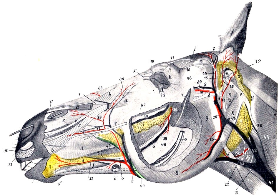
Fig. 514.—Dissection of Head of Horse. The masseter and superficial muscles and the parotid gland are in great part removed. a, a, Levator labii superioris proprius; b, b, levator nasolabialis; c, dilatator naris inferior; d, buccinator; e, common mass of buccinator and depressor labii inferioris; f, depressor labii inferioris; g, masseter; h, orbicularis oculi; i, temporalis; k, occipito-hyoideus; k′, stylo-maxillaris; l, sterno-cephalicus; l′, tendon of same; m, omo-hyoideus; n, crico-pharyngeus; o, mastoid tendon of mastoido-humeralis; p, splenius; q, inferior buccal and labial glands; r, superior buccal glands; s, remnant of parotid gland; t, submaxillary gland; u, anterior cervical (subparotid) lymph glands; v, probe passed into diverticulum nasi; w, cornu of alar cartilage; x, internal palpebral ligament; y, wing of atlas; z, scutiform cartilage of ear; 1, external nasal nerve; 2, anterior nasal nerve; 3, superior labial nerve; 4, anterior part of superior buccal nerve; 5, buccinator nerve; 6, 6′, inferior alveolar nerve; 6″, mental nerve-continuation of 6; 7, masseteric nerve; 8, facial nerve (cut); 9, superficial temporal nerve; 10, anastomosis between 9 and 8; 11, internal auricular nerve; 12, posterior auricular nerve; 13, digastric nerve; 14, cervical branch of facial nerve (cut); 15, auriculo-palpebral nerve; 16, lacrimal nerve; 17, frontal nerve; 18, infratrochlear nerve; 19, n. zygomaticus s. subcutaneus malæ; 20, spinal accessory nerve; 21, ventral branch of spinal accessory nerve (to sterno-cephalicus); 22, ventral end branch of first cervical nerve; 23, thyro-laryngeal artery; 24, internal maxillary artery; 25, masseteric artery; 26, great (posterior) auricular artery; 27, external branch of 26; 28, deep auricular artery; 29, superficial temporal artery; 30, transverse facial artery; 31, facial artery; 32, inferior labial artery; 33, superior labial artery; 34, lateral nasal artery; 35, dorsal nasal artery; 36, angular artery of eye; 37, labial twigs of palato-labial artery—left uncolored by oversight; 38, branch of buccinator artery; 39, buccinator vein; 40, vena reflexa; 41, 42, jugular vein; 43, external maxillary vein; 44, inferior cerebral vein; 45, stump of great auricular vein; 46, ramus of mandible; 47, facial crest; 48, zygomatic arch; 49, parotid duct. (After Ellenberger-Baum, Top. Anat. d. Pferdes.)
(3) The lesser or posterior palatine nerve (N. palatinus minor s. posterior), also termed the staphyline, is much the smallest of the three branches (Figs. 438, 439). It passes downward and forward with the palatine vein in the groove at the inner side of the tuber maxillare and ramifies in the soft palate.
The sphenopalatine ganglia and plexus (Fig. 515) lie on the perpendicular part of the palate bone and the pterygoid process under cover of the maxillary nerve. The afferent fibers of the 681plexus and ganglia come chiefly from the branches of the sphenopalatine nerve and the nerve of the pterygoid canal. Interspersed in these are several minute ganglia and one or more larger ones. The nerve of the pterygoid canal or Vidian nerve (N. canalis pterygoidei) is formed by the union of sympathetic fibers with the superficial petrosal branch of the facial nerve. It passes forward at first between the Eustachian tube and the sphenoid bone, enters the canal between the pterygoid bone and process, and joins the posterior part of the plexus. It is probable that it furnishes the motor fibers to the levator palati and palatinus muscles. Efferent filaments go to the periorbita and the ophthalmic vessels, and others accompany the branches of the maxillary nerve, around which they have a plexiform arrangement.
3. The infraorbital nerve (N. infraorbitalis) is the continuation of the maxillary trunk. It traverses the infraorbital canal, emerges through the infraorbital foramen, and divides into nasal and superior labial branches. Along its course it gives off superior alveolar or dental branches (Rami alveolares maxillæ), which supply the teeth, alveolar periosteum, and gums.
The delicate posterior alveolar branches are given off in the pterygo-palatine fossa, pass through small foramina in the tuber maxillare, and supply the posterior molar teeth and the maxillary sinus. The middle alveolar branches are given off in the infraorbital canal, and constitute the chief nerve-supply to the cheek teeth and the maxillary sinus. The anterior or incisor branch runs forward in the anterior alveolar or incisor canal and supplies branches to the canine and incisor teeth. The foregoing unite with each other to form the superior dental plexus, from which the dental and gingival branches are given off.
The external nasal branches (Rami nasales externi), two or three in number, accompany the levator labii superioris proprius and ramify in the dorsum nasi and the nasal diverticulum.
The large anterior nasal branch (Ramus nasalis anterior) passes over the nasal process of the premaxilla under cover of the dilatator naris inferior, gives branches to the nasal mucous membrane, and terminates in the skin of the upper lip.
The superior labial branch (Ramus labialis dorsalis) is the largest of the terminals of the infraorbital nerve. It passes downward and forward under the levator nasolabialis and, after supplying the skin of the anterior part of the cheek, forms a rich terminal ramification in the skin and mucous membrane of the upper lip. It anastomoses with the superior buccal branch of the facial nerve.
III. The mandibular nerve (N. mandibularis), also termed the inferior maxillary branch, is formed by the union of two roots; of these the large sensory root comes from the semilunar ganglion, and the small motor root is the pars minor of the trigeminus. It emerges from the cranium through the oval notch of the foramen lacerum, between the temporal wing of the sphenoid bone and the muscular process of the petrous temporal, and gives off the following branches:
1. The masseteric nerve (N. massetericus) (Figs. 437, 515) passes outward through the sigmoid notch of the mandible and enters the deep face of the masseter muscle, in which it ramifies.
2. The deep temporal nerves (Nn. temporales profundi) (Fig. 437), two or three in number, arise by a common trunk with the masseteric. They supply the temporal muscle.
3. The buccinator nerve (N. buccinatorius) passes obliquely forward through the anterior part of the external pterygoid muscle, then between the internal pterygoid and the tuber maxillare (Fig. 514). It continues forward in the submucous tissue of the cheek along the lower border of the buccinator and divides into branches which ramify in the mucous membrane and glands of the lips in the vicinity of the commissure. It supplies small branches to the external pterygoid and temporal muscles and detaches numerous collateral twigs to the mucous membrane of the cheek and to the buccal glands. It also communicates with the inferior buccal branch of the facial nerve.
4. The pterygoid nerve (N. pterygoideus) arises in common with the preceding, passes forward on the guttural pouch, and divides into branches for the pterygoid muscles (Fig. 515).
The otic ganglion (G. oticum) is situated near the origin of the pterygoid and 682buccinator nerves, and is related internally to the tensor palati and the Eustachian tube. It receives motor fibers from the pterygoid nerve and sensory fibers by the small superficial petrosal nerve from the tympanic plexus, through which communications are made with the facial and glosso-pharyngeal nerves. Sympathetic fibers are derived from the plexus on the internal maxillary artery. Efferent filaments go to the tensor palati, tensor tympani, and pterygoid muscles, and to the Eustachian tube.
The ganglion is small and somewhat difficult to demonstrate. In many cases it is replaced by a number of minute ganglia interspersed in a fine plexus.
5. The superficial temporal nerve (N. temporalis superficialis) (Figs. 437, 514, 515) runs outward across the pterygoideus externus, passes between the parotid gland and the neck of the ramus of the mandible, turns around the latter, and divides into two branches. The upper branch (Ramus transversus faciei) accompanies the transverse facial vessels and ramifies in the skin of the cheek. The larger inferior branch unites with the inferior buccal division of the facial nerve.
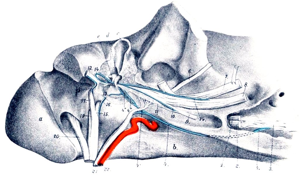
Fig. 515.—Deep Dissection of Base of Cranium, Viewed from the Right and Below. The tympanic cavity is opened and the paramastoid process is sawn off. a, Occipital condyle; b, body of sphenoid; c, external auditory meatus (part removed); d, malleus; e, incus; 1, stump of ophthalmic nerve; 2, maxillary nerve (cut off); 3, sphenopalatine plexus; 4, Vidian nerve; 4′, deep petrosal nerve; 4″, branch to tympanic plexus; 4‴, superficial petrosal nerve; 5, superficial temporal nerve (cut off); 6, masseteric nerve (cut off); 7, mandibular nerve (raised); 8, lingual nerve (cut off); 9, pterygoid nerve (cut); 10, nerve to tensor tympani; 11, otic ganglion; 12, facial nerve; 13, stapedial nerve; 14, chorda tympani; 15, glosso-pharyngeal nerve (cut); 16, tympanic nerve; 17, vagus (cut off); 18, auricular branch of vagus; 19, spinal accessory nerve (cut off); 20, hypoglossal nerve (cut off); 21, sympathetic nerve (cut off); 22, internal carotid artery. (After Ellenberger, in Leisering’s Atlas.)
Before its division the nerve gives off twigs to the guttural pouch, the parotid gland, the external ear, and the skin of the external auditory meatus and the membrana tympani. Branches from it concur with filaments from the cervical branch of the facial nerve in the formation of the auricular plexus.
6. The inferior alveolar or dental nerve (N. alveolaris mandibulæ) (Figs. 436, 437, 514) arises with the lingual by a common trunk which passes forward at first on the external pterygoid muscle, then inclines ventrally between the internal pterygoid and the ramus of the mandible. The lingual and alveolar separate at an acute angle, and the latter enters the mandibular foramen and courses in the canal within the ramus (Fig. 516). Emerging at the mental foramen, it terminates by dividing into six to eight inferior labial and mental branches, which ramify in the lower lip and chin. Before entering the bone, the nerve detaches the mylo-hyoid nerve (N. mylo-hyoideus), which runs downward and forward between the ramus and the mylo-hyoid muscle; it supplies that muscle, the anterior belly of the digastricus, and the skin of the anterior part of the submaxillary space. The dental 683and gingival branches detached from the nerve within the mandible are arranged like the corresponding nerves of the upper jaw.
7. The lingual nerve (N. lingualis) arises by a common trunk with the inferior alveolar or dental nerve (Figs. 436, 437). After separating from the latter it runs downward and forward, lying at first between the ramus of the mandible and the internal pterygoid muscle, then on the inner face of the mylo-hyoid. On reaching the root of the tongue it divides into superficial and deep branches. The superficial branch (Ramus superficialis) runs forward on the stylo-glossus and accompanies the submaxillary duct on the deep face of the sublingual gland. It supplies the mucous membrane of the tongue and the floor of the mouth. At the root of the tongue it gives off a recurrent branch to the isthmus faucium, which communicates with the lingual branch of the glosso-pharyngeal nerve. The larger deep branch (Ramus profundus) turns around the lower edge of the hyo-glossus, passes upward and forward between that muscle and the genio-glossus, and continues forward on the latter to the tip of the tongue. It gives branches to the mucous membrane and the fungiform papillæ of the tongue, and anastomoses with branches of the hypoglossal nerve and with the superficial branch. Minute ganglia occur on the finer branches of the lingual nerve. The chorda tympani branch of the facial joins the lingual nerve at the origin of the latter and is incorporated with it in its distribution to the tongue.
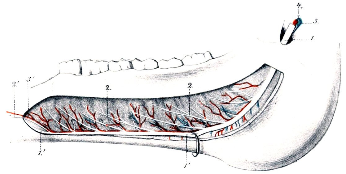
Fig. 516.—Part of Branch of Lower Jaw of Horse, Internal View. The bone has been removed to show the vessels and nerves. 1, 1′, Inferior alveolar or dental nerve; 2, 2, branches to cheek teeth and gums; 2′, branch to canine and incisor teeth; 3, 3′, inferior alveolar or dental artery; 4, satellite vein. (After Leisering’s Atlas.)
THE SIXTH OR ABDUCENT NERVE
The abducent nerve (N. abducens) emerges from the brain behind the pons and just external to the pyramid (Fig. 499). It passes forward across the pons, pierces the dura mater, and accompanies the third and ophthalmic nerves, below which it emerges through the foramen lacerum orbitale. In the orbit it divides into two branches; the larger of these enters the external rectus, the smaller supplies the superior and external parts of the retractor muscle of the eyeball. Within the cranium it receives filaments from the carotid plexus of the sympathetic.
The fibers of the abducent nerve are axones of the large multipolar cells of the abducent nucleus which is situated beneath the eminentia teres of the floor of the fourth ventricle. The nucleus lies within the loop formed by the fibers of origin of the facial nerve. It is connected with (a) the anterior olive; (b) the oculomotor nucleus of the opposite side; (c) the motor area of the cerebral cortex by means of the pyramidal tract of the opposite side.
684
THE SEVENTH OR FACIAL NERVE
The facial nerve (N. facialis) has its superficial origin at the lateral part of the corpus trapezoideum, immediately behind the pons (Fig. 499). It passes outward in front of the eighth nerve and enters the internal auditory meatus. At the bottom of the meatus the two nerves part company, the facial coursing in the facial canal of the petrous temporal bone. The canal and nerve are at first directed outward between the vestibule and the cochlea, then curve backward and downward in the posterior wall of the tympanum to end at the stylo-mastoid foramen. The bend formed by the nerve is called the knee (Geniculum n. facialis) and bears at its highest point the round geniculate ganglion (G. geniculi).
The nerve consists of two parts, motor and sensory. The motor part constitutes the bulk of the nerve. Its deep origin is from the cells of the facial nucleus, which is situated in the medulla above the facial tubercle. On leaving the nucleus the root-fibers pass upward and inward, incline forward close to the median plane, and then bend sharply downward to the point of emergence. The highest point of the bend is subjacent to the gray matter of the eminentia teres in the floor of the fourth ventricle, and the abducent nucleus lies in the concavity of the curve. The small sensory part (N. intermedius) consists of axones of cells of the geniculate ganglion, which is interposed on the facial nerve as it bends downward in the facial canal. The fibers of this part after entering the medulla, pass to the nucleus of termination which it shares with the ninth and tenth nerves. The peripheral fibers from the geniculate ganglion constitute the chorda tympani.
After its emergence through the stylo-mastoid foramen (Fig. 514) the nerve passes downward, forward, and outward on the guttural pouch under cover of the parotid gland, and crosses between the origin of the superficial temporal and internal maxillary arteries internally and the superficial temporal vein externally. It then crosses the posterior border of the ramus of the mandible ventral to the transverse facial artery and about an inch and a half (ca. 3.5 to 4 cm.) below the articulation of the jaw. Emerging from beneath the parotid gland upon the masseter muscle, it receives the lower branch of the superficial temporal nerve, and divides into superior and inferior buccal branches. The following collateral branches are given off, the first five being detached within the facial canal, and the others between the stylo-mastoid foramen and the border of the jaw.
1. The great superficial petrosal nerve (N. petrosus superficialis major) arises from the geniculate ganglion.[203] It passes through the petrosal canal, contributes a filament to the tympanic plexus, receives the great deep petrosal nerve from the carotid plexus of the sympathetic, emerges through the foramen lacerum, and is continued as the Vidian nerve to the sphenopalatine plexus and ganglia (Fig. 515).
2. A delicate branch (R. anastomoticus cum plexu tympanico) emerges from the geniculate ganglion and unites with a filament issuing from the tympanic plexus to form the small superficial petrosal nerve (N. petrosus superficialis minor); this ends in the otic ganglion.
3. The stapedial nerve (N. stapedius) (Fig. 515) is a short filament detached from the facial nerve as it turns down in the facial canal. It innervates the stapedius muscle.
4. The chorda tympani (Fig. 515) is a small nerve which arises a little below the preceding and pursues a recurrent course in a small canal in the mastoid part of the temporal bone to reach the tympanic cavity. It traverses the latter, passing between the handle of the malleus and the long branch of the incus. Emerging through the petro-tympanic fissure, the nerve passes downward and forward, crosses beneath the internal maxillary artery, and joins the lingual nerve. It sends twigs to the submaxillary and sublingual glands, and through its incorporation with the lingual nerve furnishes fibers to the mucous membrane of the anterior two-thirds of the tongue which are believed to mediate the sense of taste.
6855. Anastomotic filaments unite with the auricular branch of the vagus near the stylo-mastoid foramen.
6. The posterior auricular nerve (N. auricularis posterior) arises from the facial at its emergence from the facial canal (Fig. 514). It runs upward and backward with the posterior auricular artery under cover of the parotid gland and supplies the posterior auricular muscles and the skin of the convex surface of the external ear. It anastomoses with branches of the first and second cervical nerves.
7. The internal auricular nerve (N. auricularis internus) springs from the facial close to or in common with the preceding (Fig. 514). It ascends in the parotid gland just behind the styloid process of the conchal cartilage, passes through an opening in the cartilage, and ramifies in the skin of the concave surface of the ear.
8. The digastric branch (R. digastricus) (Fig. 514) arises from the facial below the auricular nerves. Its branches innervate the posterior belly of the digastricus, the stylo-hyoideus, and the occipito-hyoideus. At its origin it gives off a small branch which forms a loop around the great auricular artery or its posterior branch and rejoins the trunk.
9. The auriculo-palpebral nerve (N. auriculo-palpebralis) (Fig. 514) arises from the upper edge of the facial near the posterior border of the ramus. It ascends in the parotid gland behind the superficial temporal artery, and terminates in anterior auricular and temporal branches. The anterior auricular branches form with branches of the trigeminus the anterior auricular plexus. They innervate the anterior auricular and parotido-auricularis muscles. The temporal branch runs forward and inward over the temporal muscle to the inner canthus of the eye, forms a plexus with the terminal branches of the ophthalmic nerve, and is distributed to the orbicularis oculi, corrugator supercilii, and levator nasolabialis.
10. The cervical branch (R. colli) (Fig. 435) arises from the ventral border of the facial opposite to the preceding nerve. It emerges obliquely through the parotid gland, passes downward and backward on or near the jugular vein, and anastomoses with the cutaneous branches of the cervical nerves. It gives branches to the parotido-auricularis and the cervical panniculus. In its course along the neck the nerve is reinforced by twigs from the cutaneous branches of the second to the sixth cervical nerves.
11. Small branches are detached to the guttural pouch and the parotid gland. The latter (Rami parotidei) concur with branches of the superficial temporal nerve in forming the parotid plexus.
The facial nerve usually terminates after a short course on the surface of the masseter by dividing into two buccal branches (Figs. 435, 461).
1. The superior buccal nerve (N. buccalis dorsalis) passes forward on the upper part of the masseter, dips under the zygomaticus, and continues along the lower border of the dilatator naris lateralis. It then runs under the last-named muscle and anastomoses with branches of the infraorbital nerve, and is distributed to the muscles of the cheek, upper lip, and nostril.
2. The inferior buccal nerve (N. buccalis ventralis) crosses the masseter obliquely and continues forward along the depressor labii inferioris. It is connected by variable anastomotic branches with the superior nerve. It gives collateral branches to the panniculus, buccinator, and depressor labii inferioris, and ramifies with the terminal branches of the inferior alveolar nerve in the lower lip.
The buccal nerves are subject to much variation in regard to their course, anastomoses, and relations to the sensory components derived from the superficial temporal nerve. Their distribution is constant. The point at which the branch of the superficial temporal nerve joins the facial is variable.
686
THE EIGHTH OR AUDITORY NERVE
The auditory nerve (N. acusticus) is connected with the lateral aspect of the medulla just behind and external to the facial (Fig. 499). It has two roots, vestibular and cochlear (Radix vestibularis et cochlearis).
The auditory nerve consists of two distinct parts which might well be regarded as separate nerves. The cochlear part mediates the sense of hearing, while the vestibular part is not auditory in function, but is concerned in the sense of the position of the body and the mechanism of equilibration.
The nerve passes outward to the internal auditory meatus, which it enters behind the facial nerve. In the meatus it divides into two nerves, of which the upper is the vestibular and the lower is the cochlear nerve.
1. The vestibular nerve (N. vestibuli) is distributed to the utriculus, the sacculus, and to the ampullæ of the semicircular canals, of the internal ear. In the internal auditory meatus the nerve is connected by filaments with the geniculate ganglion of the facial nerve. At the bottom of the meatus it bears the vestibular ganglion (G. vestibulare), from the cells of which the fibers of the nerve arise.
2. The cochlear nerve (N. cochleæ) detaches a filament to the sacculus, passes through the lamina cribrosa to the labyrinth, and is distributed to the organ of Corti in the cochlea.
The fibers of the vestibular nerve arise from the vestibular ganglion as central processes (axones) of the bipolar cells of the ganglion. The peripheral processes (dendrites) of the cells form arborizations about the deep ends of the hair cells of the maculæ and cristæ acusticæ of the utriculus, sacculus, and semicircular canals. The fibers enter the medulla, pass between the restiform body and the spinal tract of the trigeminus, and spread out to end in the vestibular nucleus in the floor of the fourth ventricle. Among the central connections of the vestibular nerve are: (1) fibers which connect its nucleus with centers in the cerebellum (chiefly of the opposite side); (2) the vestibulo-spinal tract, which conveys impulses to the motor cells of the ventral columns of the spinal cord; (3) fibers which connect the nucleus with those of the abducent nerve of the same side, the third and fourth nerves, and the motor part of the trigeminus of both sides.
The fibers of the cochlear nerve are the central processes of the bipolar cells of the spiral ganglion of the cochlea. The peripheral processes of these cells end in relation to the hair cells of the organ of Corti. Some of the nerve-fibers enter the ventral cochlear nucleus in the medulla close to the superficial origin of the nerve; others end in the dorsal nucleus of the tuberculum acusticum at the lateral angle of the floor of the fourth ventricle. From the ventral nucleus fibers pass in the corpus trapezoideum to the anterior olivary nucleus of the same and of the opposite side. Thence tracts pass to the nuclei of the motor nerves of the eye, and through the lateral fillet to the posterior quadrigeminal body and the internal geniculate body. The axones of the cells of the dorsal nucleus pass largely (as the striæ acusticæ) over the restiform body and across the floor of the fourth ventricle toward the median plane. They then turn ventrally, cross to the opposite side, and are continued by the lateral fillet. From the mid-brain a tract proceeds to the cortex of the temporal lobe of the hemisphere.
THE NINTH OR GLOSSO-PHARYNGEAL NERVE
The glosso-pharyngeal nerve (N. glossopharyngeus) is attached to the anterior part of the lateral aspect of the medulla by several filaments (Fig. 499). The root-bundles enter the furrow ventral to the restiform body; they are separated by a short interval from the origin of the facial nerve, but are not marked off behind from the roots of the vagus. The bundles converge laterally to form a nerve which perforates the dura mater and emerges through the foramen lacerum posterius just in front of the tenth nerve (Fig. 515). As it issues from the cranium the nerve bears a considerable ovoid gray enlargement, the petrous ganglion (G. petrosum).[204] It then curves downward and forward over the guttural pouch and behind the great cornu of the hyoid bone, crosses the deep face of the external carotid artery, and divides into pharyngeal and lingual branches (Fig. 437). The collateral branches are as follows:
1. The tympanic nerve (N. tympanicus) (Fig. 515) arises from the petrous 687ganglion and passes upward between the petrous and tympanic parts of the temporal bone to reach the cavity of the tympanum. Here it breaks up into branches to form, along with branches from the carotid plexus of the sympathetic, the tympanic plexus. From the plexus branches pass to the mucous membrane of the tympanum and the Eustachian tube. The continuation of the nerve issues from the plexus and unites with a filament from the geniculate ganglion of the facial to form the small superficial petrosal nerve; this runs forward and ends in the otic ganglion.
Filaments also connect the petrous ganglion with the jugular ganglion of the vagus nerve and with the superior cervical ganglion of the sympathetic.
2. A considerable branch runs backward on the guttural pouch, contributes filaments to the pharyngeal plexus, and concurs with twigs from the vagus and the sympathetic in forming the carotid plexus on the terminal part of the carotid artery and on its chief branches. In this plexus is the small ganglion intercaroticum.
The pharyngeal branch (R. pharyngeus) (Fig. 437) is the smaller of the two terminal branches. It runs forward across the deep face of the great cornu of the hyoid bone and concurs with the pharyngeal branches of the vagus and with sympathetic filaments in forming the pharyngeal plexus; from this branches pass to the muscles and mucous membrane of the pharynx.
The lingual branch (R. lingualis) is the continuation of the trunk (Fig. 437). It runs along the posterior border of the great cornu of the hyoid bone in front of the external maxillary artery and dips under the hyo-glossus muscle. It gives collateral branches to the soft palate, isthmus faucium, and tonsil, and ends in the mucous membrane of the posterior part of the tongue, where it supplies gustatory fibers to the vallate papillæ. A considerable branch unites with a twig from the lingual nerve.
The glosso-pharyngeal is a mixed nerve, containing both motor and sensory fibers. The latter constitute the bulk of the nerve and include those which mediate the special sense of taste. They are processes of the cells of the petrous ganglion. The central processes of the ganglion cells enter the medulla, pass upward and inward through the formatio reticularis, and end in the nucleus of termination in the floor of the fourth ventricle. The motor fibers arise from dorsal and ventral efferent nuclei in the medulla. The glosso-pharyngeal shares these nuclei with the vagus and has practically the same central connections as that nerve (q. v.).
THE TENTH, VAGUS, OR PNEUMOGASTRIC NERVE
The vagus or pneumogastric (N. vagus) is the longest and most widely distributed of the cranial nerves; it is also remarkable for the connections which it forms with adjacent nerves and with the sympathetic. It is attached to the lateral aspect of the medulla by several filaments which are in series with those of the ninth nerve in front and the eleventh nerve behind (Fig. 499). The bundles converge to form a trunk which passes outward, pierces the dura mater, and emerges from the cranium through the foramen lacerum posterius (Fig. 515). In the foramen the nerve bears on its lateral aspect the elongated flattened jugular ganglion (G. jugulare).
The ganglion communicates with (a) the tympanic nerve, (b) the petrous ganglion of the ninth nerve, (c) the spinal accessory, and (d) the hypoglossal. It also gives off the auricular branch (R. auricularis), which runs forward below the petrous ganglion and passes through a small canal in the petrous temporal bone to gain the facial canal. Here it gives filaments to the facial and emerges with that nerve through the stylo-mastoid foramen. It ascends behind the external auditory meatus, dips under the rotator longus muscle, and passes through a foramen in the conchal cartilage to ramify in the integument which lines the meatus and the adjacent part of the ear.
Beyond the ganglion the vagus runs backward and downward with the spinal accessory in a fold of the guttural pouch (Fig. 437). Then the two nerves separate, allowing the hypoglossal to pass between them, and the vagus descends with the 688internal carotid artery and crosses the inner face of the origin of the occipital artery. Here it is joined by the cervical trunk of the sympathetic, and the two nerves continue along the dorsal aspect of the common carotid artery in a common sheath (Fig. 433). At the root of the neck the vagus separates from the sympathetic, and from this point backward the relations of the right and left vagi differ somewhat and must be described separately.
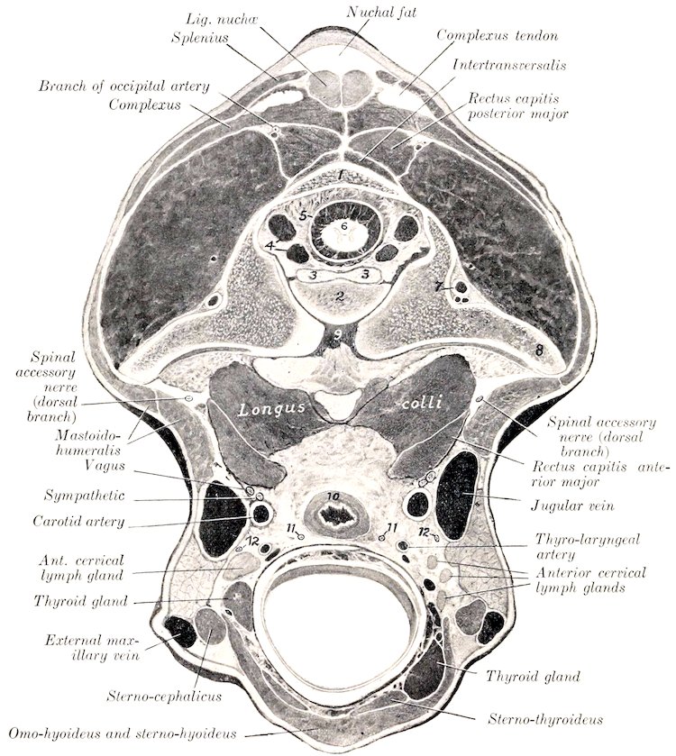
Fig. 517.—Cross-section of Neck of Horse, Passing through Posterior Part of Atlas. The head and neck were extended. 1, Dorsal arch of atlas; 2, odontoid process; 3, odontoid ligament; 4, vertebral sinuses; 5, dura mater; 6, spinal cord; 7, vertebral artery; 8, wing of atlas; 9, atlanto-axial joint cavity; 10, œsophagus; 11, 11, recurrent nerves; 12, 12, ventral branches of spinal accessory nerves. By an oversight the obliquus cap. post. (above wing of atlas) is unmarked.
The right vagus (Fig. 429) enters the thorax in the angle of divergence of the right brachial artery and the truncus bicaroticus. It then passes backward and slightly upward, crossing obliquely the outer surface of the brachiocephalic artery and the right face of the trachea. Reaching the dorsal surface of the latter near the bifurcation, it divides into dorsal and ventral branches.
689The left vagus (Fig. 428) enters the thorax on the ventral face of the œsophagus, crosses obliquely under the left brachial artery, and passes back on the external surface of that vessel in company with a large cardiac nerve.[205] Separating from the latter, the vagus continues backward on the left face of the aorta, inclines to the upper surface of the left bronchus, and divides into dorsal and ventral branches.
The dorsal and ventral branches unite with the corresponding branches of the opposite nerve, thus forming dorsal and ventral œsophageal trunks (Truncus (œsophageus dorsalis, ventralis). These run backward in the posterior mediastinum, above and below the œsophagus respectively, and enter the abdominal cavity through the hiatus œsophageus; they supply branches to the œsophagus and anastomose with each other. The dorsal trunk receives the major part of its fibers from the right vagus. After entering the abdomen it passes to the left of the cardia, gives branches to the visceral surface of the stomach, and ends in the cœliac and subsidiary plexuses. The ventral trunk passes to the lesser curvature of the stomach and ramifies on the parietal surface of the stomach; it forms here the anterior gastric plexus, from which branches are supplied also to the first part of the duodenum and to the liver.
The collateral branches of the vagus are as follows:
1. The pharyngeal branch (R. pharyngeus) is given off in relation to the superior cervical ganglion, turns around the internal carotid artery, and runs downward and forward on the guttural pouch to the dorsal wall of the pharynx (Fig. 437). Here its branches concur with the pharyngeal branch of the ninth nerve and with filaments from the spinal accessory and the sympathetic in forming the pharyngeal plexus. This supplies numerous twigs to the pharynx, and a larger branch which passes along the side of the œsophagus and ramifies in its cervical part.
According to Ellenberger and Baum the pharyngeal plexus receives filaments also from the digastric, superior laryngeal, hypoglossal, and first cervical nerves. The branches of the plexus form secondary intermuscular and submucous plexuses, in which there are numerous minute ganglia.
2. The superior or anterior laryngeal nerve (N. laryngeus cranialis) is larger than the preceding and arises a little behind it (Fig. 437). It crosses the deep face of the origin of the external carotid artery, runs downward and forward over the lateral wall of the pharynx behind the hypoglossal nerve, and passes through the foramen below the anterior cornu of the thyroid cartilage. Its terminal branches ramify in the mucous membrane of the larynx, the floor of the pharynx, and the entrance to the œsophagus; they anastomose with those of the recurrent. At its origin the nerve gives off its small external branch (R. externus); this descends to the crico-thyroid muscle, which it supplies, and sends filaments to the crico-pharyngeus also. It may arise from the trunk of the vagus or from the pharyngeal branch.
At the point of origin of the superior laryngeal nerve there is a plexiform widening which is regarded by some authors as the homologue of the ganglion nodosum of man. From it a filament arises which, after a short course, rejoins the vagus or enters the sympathetic trunk. Stimulation of its central end causes a reduction of the blood-pressure, and it is therefore termed the depressor nerve (N. depressor).
3. The recurrent nerve (N. recurrens), also termed the inferior or posterior laryngeal nerve, differs on the two sides in its point of origin and in the first part of its course. The right nerve (Fig. 429) is given off opposite the second rib, turns around the dorso-cervical artery from without inward, runs forward on the lower part of the lateral surface of the trachea, and ascends in the neck on the ventral face of the common carotid artery. The left nerve (Fig. 428) arises from the vagus where the latter begins to cross the aortic arch. It passes back over the ligamentum 690arteriosum, winds around the concavity of the aortic arch from without inward, runs forward on the lower part of the left face of the trachea, and continues in the neck in a similar position to the right nerve.
It is worthy of note that the left nerve passes beneath the bronchial lymph glands as it winds around the aorta; also that in the next part of its course it lies between the left surface of the trachea and the deep face of the aorta, and is then related to lymph glands which lie along the ventral aspect of the trachea. The left recurrent is often incorporated in part of its course in the anterior mediastinum with a deep cardiac nerve. Further, the left nerve lies at first ventral to, and then upon, the œsophagus in the neck. The right recurrent is given off from or in common with a considerable trunk which connects the vagus with the first thoracic ganglion of the sympathetic. The arrangement here is commonly more or less plexiform, and from it one or two cardiac nerves arise.
The terminal part of each nerve (Fig. 517) lies on the dorsal surface of the trachea, in relation to the œsophagus internally and the carotid artery above.[206] It passes between the crico-arytenoideus posterior and the crico-pharyngeus; the terminal branches supply all the muscles of the larynx except the crico-thyroid, and communicate with branches of the superior laryngeal nerve. Collateral branches are given off to the cardiac plexus (Rr. cardiaci), to the trachea (Rr. tracheales), to the œsophagus (Rr. œsophagei), and to the inferior cervical ganglion of the sympathetic.
4. Cardiac branches (Rr. cardiaci), usually two or three in number, are given off from each vagus within the thorax (Figs. 428, 429). These concur with the cardiac branches of the sympathetic and recurrent nerves to form the cardiac plexus, which innervates the heart and great vessels.
5. Small tracheal and œsophageal branches (Rr. tracheales et œsophagei) are given off from both vagi in the thorax. These concur with branches from the recurrent nerves and the inferior cervical and anterior thoracic ganglia of the sympathetic in forming the posterior tracheal and œsophageal plexuses, from which twigs go to the trachea, œsophagus, heart, and large vessels.
6. Bronchial branches (Rr. bronchiales) are detached at the roots of the lungs and unite with sympathetic filaments in forming the pulmonary plexuses. From the latter numerous branches proceed in a plexiform manner along the bronchi and vessels into the substance of the lungs.
The vagus and glosso-pharyngeal nerves are so closely associated in origin and central connections that they may be described together in this respect.
The sensory fibers arise from the petrous and jugular ganglia, and their central parts enter the lateral aspect of the medulla and divide into anterior and posterior branches like the fibers of the dorsal roots of the spinal nerves. Most of the fibers end in arborizations about the cells of the vago-glosso-pharyngeal nucleus of termination, which consists of two parts. Of these the dorsal sensory nucleus (Nucleus alæ cinereæ) is situated in the posterior part of the floor of the fourth ventricle and in the adjacent part of the closed portion of the medulla near the median plane. The other part is termed the nucleus of the solitary tract, and is so named because its cells are grouped about the bundle (Tractus solitarius) formed by the posterior divisions of the afferent nerve-fibers. It ends about the level of the pyramidal decussation. The secondary central connections are similar to those of the sensory part of the trigeminus.
The motor fibers (and those of the medullary part of the accessory) arise from the dorsal motor nucleus and the ventral motor nucleus. The cells of the former lie in groups along the ventro-medial side of the dorsal sensory nucleus. The latter, also termed the nucleus ambiguus, is situated more deeply in the lateral part of the formatio reticularis.
THE ELEVENTH OR SPINAL ACCESSORY NERVE
The spinal accessory nerve (N. accessorius) is purely motor. It consists of two parts which differ in origin and function.
The medullary part arises from the lateral aspect of the medulla by several rootlets which are behind and in series with those of the vagus (Fig. 499). The spinal part arises from the cervical part of the spinal cord by a series of fasciculi 691which emerge between the dorsal and ventral roots. The bundles unite to form a trunk which is very small at its origin at the fifth segment of the cord, but increases in size when traced toward the brain, since it continually receives accessions of fibers. It passes through the foramen magnum and joins the medullary part. The trunk thus formed sends its medullary fibers to the tenth and ninth nerves and emerges through the foramen lacerum posterius. It then runs backward and downward with the vagus in a fold of the guttural pouch, separates from that nerve, crosses the deep face of the submaxillary gland and the occipital artery, and divides in the recessus atlantis into dorsal and ventral branches.
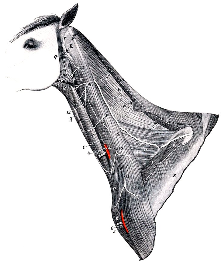
Fig. 518.—Neck of Horse, after Removal of Part of Panniculus and Trapezius. a, a′, Mastoido-humeralis; b, anterior superficial pectoral muscle; c, cervical panniculus; d, sterno-cephalicus; e, omo-hyoideus; f, sterno-thyro-hyoideus; g, trachea; h, h′, h″, tendons of splenius, mastoido-humeralis, and trachelo-mastoideus; i, trapezius cervicalis; k, supraspinatus; l, anterior deep pectoral muscle; m, rhomboideus cervicalis; n, serratus cervicis; o, splenius, upper and lower borders of which are indicated by dotted lines; p, parotid gland; q, parotido-auricularis muscle; r, wing of atlas; s, spine of scapula; 1, external maxillary vein; 2, 3, jugular vein; 4, carotid artery; 5, descending branch of inferior cervical artery; 6, cephalic vein; 7–11, ventral branches of second to seventh cervical nerves; 12, cutaneous branch of second cervical nerve; 13, cervical branch of facial nerve; 14, terminal branches of dorsal divisions of cervical nerves; 15, dorsal branch of spinal accessory nerve. (After Ellenberger-Baum, Top. Anat. d. Pferdes.)
It is connected by anastomotic branches with the vagus and hypoglossal nerves and the superior cervical ganglion of the sympathetic, and contributes a branch to the pharyngeal plexus.
The dorsal branch (R. dorsalis) (Figs. 433, 517, 518) receives a twig from the second and third cervical nerves and turns around the atlantal tendon of the splenius 692under cover of the mastoido-humeralis. It then passes obliquely through the latter muscle and continues backward on the splenius and the cervical part of the serratus, inclines upward across the anterior deep pectoral and the supraspinatus, and enters the deep face of the trapezius, in which it ramifies.
The ventral branch (R. ventralis) (Fig. 436) is smaller. It enters the sterno-cephalicus muscle behind the cervical angle of the parotid gland.
The fibers of the spinal part of the accessory arise from the ventro-lateral cells of the ventral gray column of the cord as far back as the fifth cervical segment. The fibers of the medullary part come chiefly from the nucleus ambiguus in common with the motor fibers of the vagus.
THE TWELFTH OR HYPOGLOSSAL NERVE
The hypoglossal nerve (N. hypoglossus) is purely motor and innervates the muscles of the tongue (Fig. 437). Its root-fibers arise from the ventral face of the medulla in linear series about 3 to 4 mm. lateral to the posterior half of the pyramid (Fig. 499). The filaments converge to three or four bundles which perforate the dura mater and unite to form the trunk. The latter emerges through the hypoglossal foramen (Fig. 515) and runs downward and backward between the guttural pouch and the capsule of the atlanto-occipital articulation for a distance of a little less than an inch (ca. 2 cm.). It then passes between the tenth and eleventh nerves, turns downward and forward, crosses the external face of the external carotid artery, and continues over the pharynx parallel with the great cornu of the hyoid bone and behind the external maxillary artery. It then crosses beneath the artery, runs forward on the external face of the hyo-glossus muscle, and divides into its terminal branches (Rami linguales). The smaller branch supplies the stylo-glossus, hyo-glossus, and lingualis. The larger branch ramifies on the genio-glossus and supplies the remaining muscles. Anastomoses occur with branches of the lingual nerve.
In the first part of its course the nerve communicates with the superior cervical ganglion and with the ventral branch of the first cervical nerve, and gives filaments to the pharyngeal branch of the vagus and the pharyngeal plexus.
The fibers of the nerve arise from the hypoglossal nucleus, an elongated group of large multipolar cells situated chiefly under the posterior part of the floor of the fourth ventricle close to the median plane. The two nuclei are connected by commissural fibers. The other central connections include: (a) communications by the medial longitudinal fasciculus with the nuclei of termination of other cranial nerves; (b) cortico-nuclear fibers which come from the cortex by way of the internal capsule and the pyramids and go largely to the nucleus of the opposite side; (c) fibers which join the dorsal longitudinal bundle of Schütz, a tract which underlies the floor of the fourth ventricle and is traceable forward below the cerebral aqueduct.
The Spinal Nerves
The spinal nerves (Nervi spinales) are arranged in pairs, of which there are usually forty-two in the horse. They are designated according to their relations to the vertebral column as cervical (8), thoracic (18), lumbar (6), sacral (5), and coccygeal (5). Each nerve is connected with the spinal cord by two roots, dorsal and ventral (Fig. 497).
The dorsal (or superior) root (Radix dorsalis) is the larger of the two. Its fibers (Fila radicularia) spread out in fan shape and join the cord in a linear series along the dorso-lateral groove. Laterally the fibers converge to form a compact bundle, on which is a gray nodular enlargement, the spinal ganglion (Ganglion spinale). Beyond the ganglion the dorsal root joins the ventral root to constitute the nerve. The ganglia are external to the dura mater, and are situated in the intervertebral foramina, except in the case of the sacral and coccygeal nerves, the ganglia of which lie within the vertebral canal. Those of the coccygeal nerves are intradural.
693The ganglia vary greatly in size; that of the first cervical nerve is scarcely as large as a hemp seed, while that of the eighth cervical is about 2 cm. long and 1 cm. wide. On the large roots connected with the cervical and lumbar enlargement of the cord there are multiple ganglia of varying sizes interposed in the course of the root-bundles. The fibers of the dorsal roots arise from the cells of the spinal ganglia; each cell gives off a fiber which enters the spinal cord and another which passes into the nerve.
The ventral (or inferior) root (Radix ventralis) contains fewer fibers than the dorsal root, except in the case of the first cervical nerve. It arises from the ventral surface of the spinal cord (Fig. 495) by means of numerous small bundles of fibers which do not form a linear series but emerge from the cord over an area three to five millimeters in width (ventral root zone). The fibers are processes of the large cells of the ventral gray columns of the spinal cord. There is no ganglion on the ventral root.
In the cervical, thoracic, and anterior lumbar regions the bundles of both roots pass through separate openings in linear series in the dura mater before uniting into a root proper. Further back the bundles of each root unite within the dura. In the anterior part of the cervical region and in the thoracic part of the cord there are intervals of varying length between adjacent roots, but in some places the fibers of adjacent roots overlap and an exchange of fibers may be observed. Many of the roots are directed almost straight outward or incline slightly backward, but the posterior lumbar, sacral, and coccygeal roots and nerves run backward to reach the foramina through which they emerge. The distance thus to be traversed increases from before backward, so that these nerves form a tapering sheaf around the conus medullaris and filum terminale in the last lumbar vertebra and the sacrum which is known as the cauda equina.
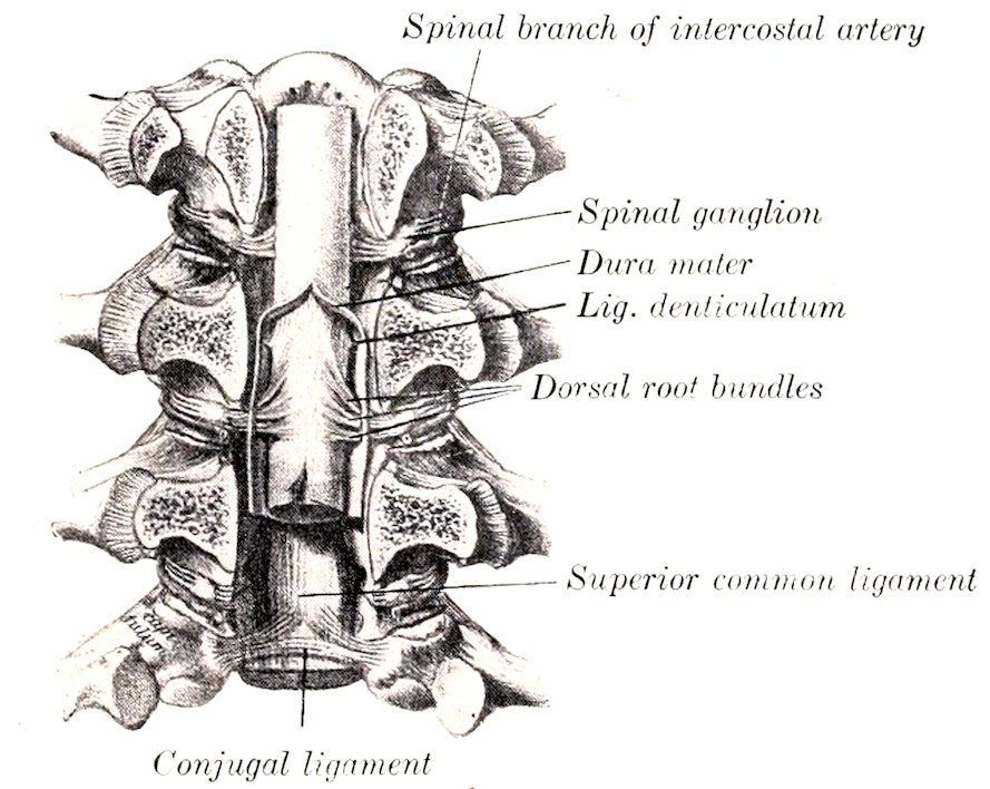
Fig. 519.—Vertebral Canal Opened by Sawing off the Arches. (After Schmaltz. Atlas d. Anat. a. Pferdes.)
The size of the spinal nerves varies greatly. The largest are connected with the cervical and lumbar enlargements.
In or immediately after its emergence from the intervertebral foramen each spinal nerve gives off a small meningeal or recurrent branch (Ramus meningeus). This is joined by a bundle of fibers from the ramus communicans and enters the vertebral canal, in which it is distributed. Each nerve then divides into two primary branches, dorsal and ventral (Ramus dorsalis, ventralis). The dorsal branches are smaller than the ventral, except in the cervical region. They are distributed chiefly to the muscles and skin of the dorsal part of the body. The ventral branches supply in general the muscles and skin of the ventral parts of the body, including the limbs. Each nerve or its ventral branch is connected with an adjacent ganglion of the sympathetic system by at least one small short branch known as a ramus communicans. Many nerves have two and some have three 694such rami. A nerve may be connected with two ganglia, and a ganglion may be connected with two nerves.
The dorsal root is sensory or afferent, i. e., it conveys impulses to the central system. Its fibers are axones of the cells of the spinal ganglion. The ventral root is motor or efferent, and conveys impulses toward the periphery. Its fibers are axones of the large cells in the ventral gray columns of the spinal cord. The common trunk or nerve formed by the union of the two roots contains both kinds of fibers, as do also their primary divisions. In addition to these fibers, which are distributed to the skeletal muscles and the skin, the spinal nerves contain fibers derived from the sympathetic system through the rami communicantes; these go to the glands and unstriped muscle and are designated secretory and vasomotor fibers.
THE CERVICAL NERVES
The cervical nerves (Nervi cervicales) (Figs. 431, 433, 518, 523) number eight pairs. Of these the first emerges through the intervertebral foramen of the atlas, the second through that of the axis, and the eighth between the last cervical and the first thoracic vertebræ.
The dorsal branches are distributed to the dorso-lateral muscles and skin of the neck. They divide usually into inner and outer branches. The internal branches (Rami mediales) run in general across the multifidus and the lamellar part of the ligamentum nuchæ to the skin of the upper border of the neck; they supply the deep lateral muscles and the skin. The external branches (Rami laterales) are chiefly muscular in their distribution. The dorsal branches of the third to the sixth nerves are connected by anastomotic branches to form the dorsal cervical plexus.
The ventral branches are smaller than the dorsal ones—an exception to the general rule. They increase in size from first to last. They supply in general the muscles and skin over the lateral and ventral aspect of the vertebræ, but the last three enter into the formation of the brachial plexus, and the three preceding the last give off the roots of the phrenic nerve. An irregular ventral cervical plexus is formed by anastomoses established between the ventral branches. The following special features may be noted:
The first cervical nerve emerges through the intervertebral (antero-internal) foramen of the atlas. Its dorsal branch (N. occipitalis) passes upward and outward between the obliqui capitis and the recti capitis dorsales and supplies branches to these muscles, the scutularis and posterior auricular muscles, and the skin of the poll. The ventral branch descends through the alar (antero-external) foramen of the atlas, crosses over the ventral straight muscles and the carotid artery under cover of the parotid gland, and divides into two branches. The anterior branch enters the omo-hyoideus muscle. The posterior branch passes downward and backward under cover of that muscle, unites with a branch of the ventral division of the second cervical nerve, and continues its course on the ventro-lateral surface of the trachea to enter the sterno-thyro-hyoideus behind the intermediate tendon. In the recessus atlantis the ventral branch is connected by one or more twigs with the superior cervical ganglion of the sympathetic, and a little lower with the hypoglossal nerve. It also sends branches to the ventral straight muscles of the head and the thyro-hyoideus. Below the atlas the ventral branch is crossed superficially by the spinal accessory nerve, the occipital artery, and the inferior cerebral vein.
The second cervical nerve is larger than the first. It emerges from the vertebral canal through the intervertebral foramen of the anterior part of the arch of the axis. Its dorsal branch ascends between the complexus and the ligamentum nuchæ and ramifies in the skin of the poll. The ventral branch gives off muscular branches to the rectus capitis anterior major, and anastomotic branches to the spinal accessory and the ventral divisions of the first and third cervical nerves; 695one of these crosses over the carotid artery and concurs in the formation of the nerve to the sterno-thyro-hyoideus mentioned above. The ventral branch then becomes superficial by passing between the two parts of the mastoido-humeralis, and divides into posterior auricular and cutaneous branches. The posterior auricular nerve passes upward and forward on the parotid gland parallel with the posterior border of the parotido-auricularis to ramify on the convex face of the external ear. The cutaneous nerve of the neck (N. cutaneus colli) crosses the mastoido-humeralis muscle and turns backward along the course of the jugular vein. On the lower part of the parotid gland it is connected by a twig with the cervical cutaneous branch of the facial nerve. It gives off twigs to the subcutaneous muscles and the skin of the parotid and laryngeal regions and a long branch which passes forward in the submaxillary space.
The third cervical nerve leaves the vertebral canal through the foramen between the second and third cervical vertebræ. Its dorsal branch emerges between two bundles of the intertransversalis muscle accompanied by a branch of the vertebral artery, turns upward on the multifidus, and divides into several branches which radiate on the deep face of the complexus. It gives branches to these muscles and to the skin, and a twig which joins the corresponding branch of the fourth nerve. The ventral branch emerges through the intertransversalis below the bundle above which the dorsal branch appears. It gives branches to the trachelo-mastoideus, rectus capitis anterior major, longus colli, splenius, and mastoido-humeralis. It also gives off a large cutaneous nerve which passes out between the two parts of the mastoido-humeralis and divides into several divergent branches.
The fourth and fifth cervical nerves are distributed in general like the third. Their dorsal branches are united by anastomotic twigs with each other and with those of the third and sixth nerves to form the dorsal cervical plexus. The ventral branch of the fifth nerve often contributes a small twig to the phrenic nerve.
The sixth cervical nerve has a smaller dorsal branch than the fifth. Its ventral branch is larger and goes in part to the brachial plexus; it supplies twigs to the intertransversales, the longus colli, the mastoido-humeralis, and the cervical parts of the serratus and rhomboideus, furnishes a root of the phrenic nerve, and gives off several considerable subcutaneous branches. One of the latter ramifies on the thick part of the cervical panniculus, to which it gives branches; another and larger branch (N. supraclavicularis) sends twigs to the skin over the shoulder joint, and descends to the skin over the superficial pectoral muscles (Fig. 466).
The seventh and eighth cervical nerves have small dorsal branches, which ascend between the longissimus and multifidus, giving twigs to these muscles, the spinalis and semispinalis, the rhomboideus, and the skin. Their ventral branches are very large and concur in the formation of the brachial plexus; that of the seventh nerve contributes the posterior root of the phrenic nerve.
Phrenic Nerve
The phrenic nerve (N. phrenicus) (Figs. 428, 429, 433), the motor nerve to the diaphragm, is formed by the union of two or three roots which cross the superficial face of the scalenus muscle obliquely downward and backward. The chief roots come from the ventral branches of the sixth and seventh cervical nerves. The root derived from the fifth nerve is small and inconstant. The root from the seventh cervical comes by way of the brachial plexus. The course of the nerve is not the same on both sides. On the right side the nerve enters the thorax by passing between the brachial artery and the anterior vena cava. It then courses backward and somewhat downward over the right face of the anterior vena cava, crosses the pericardium, and continues along the posterior vena cava to the diaphragm. In the latter part of its course it is inclosed in a special fold of the right pleura and inclines 696gradually to the ventral face of the vein. On the left side the nerve enters the thorax between the left brachial and inferior cervical arteries, and then runs its entire course in the mediastinum. In the anterior mediastinum it lies along the brachiocephalic artery ventral to the left vagus and cardiac nerves, and crosses over the dorso-cervical vein. It then passes over the upper part of the pericardium and runs backward in the posterior mediastinum to reach the tendinous center of the diaphragm considerably to the left of the median plane. Each nerve is usually connected near its origin with the first thoracic ganglion of the sympathetic by a ramus communicans, and each terminates by dividing into several branches which are distributed to the corresponding part of the diaphragm.
The Brachial Plexus
The brachial plexus (Plexus brachialis) (Fig. 433) results from anastomoses established between the ventral branches of the last three cervical and first two thoracic nerves. It appears as a thick, wide band between the two parts of the scalenus muscle, and is covered by the anterior deep pectoral and subscapularis muscles. The largest root of the plexus is derived from the first thoracic nerve, while the root supplied by the sixth cervical nerve is quite small. Each of the three chief roots, i. e., those from the last two cervical and the first thoracic nerve, is connected with the sympathetic by a ramus communicans.
The branches emanating from the plexus go for the most part to the thoracic limb, but some are distributed on the chest-wall. The names of the branches, and their arrangement so far as they can be conveniently examined before removal of the fore limb, are as follows:[207]
1. The large suprascapular nerve (N. suprascapularis) turns outward and forward and disappears between the supraspinatus and subscapularis.
2. The much smaller subscapular nerves (Nn. subscapulares), usually two primary trunks, arise close behind the suprascapular, run backward a short distance, and divide into several branches which enter the lower third of the subscapularis.
3. The anterior thoracic or pectoral nerves (N. pectorales craniales), three or four in number, arise from the anterior part of the plexus and from the loop formed by the musculo-cutaneous and median nerves. One enters the anterior deep pectoral muscle. Another passes out between the divisions of the deep pectoral to supply the superficial pectoral, giving a twig usually to the posterior deep muscle. The latter receives one or two other nerves.
4. The musculo-cutaneous nerve (N. musculocutaneus) arises from the anterior part of the plexus and passes over the outer face of the brachial artery, below which it is connected by a large but short branch with the median nerve, thus forming a loop in which the artery lies. One or two branches to the pectoral muscles are given off from the nerve or the loop.
5. The median nerve (N. medianus) is usually the largest branch of the brachial plexus. It lies at first above the brachial artery, then passes between the artery and vein to reach the anterior border of the former. It is easily recognized by its large size and the loop which it forms with the musculo-cutaneous nerve.
6. The ulnar nerve (N. ulnaris) arises with the median by a short common trunk. It descends behind the brachial artery and is accompanied a short distance by the radial nerve.
7. The radial nerve (N. radialis) arises from the posterior part of the plexus and is sometimes the largest branch. It descends behind the ulnar nerve over the origin of the subscapular artery and the lower part of the teres major, and 697dips into the interstice between that muscle and the long and internal heads of the triceps.
8. The axillary or circumflex nerve (N. axillaris) arises behind the musculo-cutaneous. It passes downward and backward on the inner face of the subscapularis and disappears between that muscle and the subscapular artery.
9. The long thoracic nerve (N. thoracalis longus) is wide and thin. It passes backward across the surface of the serratus magnus, to which it is distributed. The branches which enter the muscle are given off both upward and downward in fairly regular fashion.
10. The thoraco-dorsal nerve (N. thoracodorsalis) passes upward and backward across the subscapularis muscle to ramify in the teres major and the latissimus dorsi.
11. The external (or subcutaneous) thoracic nerve arises by a common trunk with the ulnar. It runs backward and downward across the inner face of the tensor fasciæ antibrachii, communicates with the anterior pectoral branches, and gives twigs to the deep pectoral muscle. It then runs backward in company with the external thoracic or “spur” vein, gives branches to the latissimus dorsi and the deep pectoral, and ramifies in the panniculus and the skin of the abdominal wall. It communicates with perforating branches of the intercostal nerves. A branch from it, accompanied by a large perforating intercostal branch, winds around the lower border of the latissimus dorsi and ramifies in the panniculus on the outer surface of the arm.
The term posterior thoracic or pectoral nerves (Nn. pectorales caudales) may be used to include 8, 9, and 10.
Suprascapular Nerve
The suprascapular (Fig. 441) is a large nerve derived chiefly, if not exclusively, from the sixth and seventh cervical components of the brachial plexus. It passes between the supraspinatus and subscapularis muscles and turns around the distal fourth of the anterior border of the scapula to reach the supraspinous fossa. It gives branches to the supraspinatus and continues backward and upward into the infraspinous fossa, where it supplies the infraspinatus, deltoid, and teres minor muscles.
The direct relation of this nerve to the scapula renders it liable to injury, the result of which may be paralysis and atrophy of the muscles supplied by it.
Musculo-cutaneous Nerve
The musculo-cutaneous nerve (Fig. 441) arises close behind the suprascapular, and is derived chiefly from the part of the brachial plexus which is supplied by the seventh and eighth cervical nerves. It descends across the outer surface of the brachial artery, below which a great part of the nerve unites with the median to form the loop previously mentioned. It gives off a branch which enters the upper part of the belly of the coraco-brachialis, passes downward and forward between the two parts of that muscle or between the muscle and the bone, and divides into branches which enter the biceps brachii. It contributes one of the nerves to the pectoral muscles. In some cases this nerve sends a branch to join the cutaneous branch of the median.
Axillary Nerve
The axillary nerve (Figs. 441, 520), also termed the circumflex, derives its fibers from the eighth cervical and first thoracic roots of the brachial plexus. It runs downward and backward across the lower part of the subscapularis and dips in between that muscle and the subscapular artery at the level of the shoulder joint. Continuing outward in the interval between the teres minor and the long and external heads of the triceps, it reaches the deep face of the deltoid and divides into 698several divergent branches. The muscular branches supply the teres major, capsularis, teres minor, infraspinatus, deltoid, and mastoido-humeralis. The cutaneous branch (N. cutaneus brachii lateralis) runs downward and a little forward across the external head of the triceps and ramifies on the fascia on the front of the forearm and on the superficial pectoral muscle.
Radial Nerve
The radial nerve (Figs. 441, 443, 520), also called the musculo-spiral, is sometimes the largest branch of the brachial plexus. Its fibers are derived from the seventh and eighth cervical and first thoracic roots of the plexus. It passes downward and backward over the inner surface of the subscapular artery and the teres major. In this part of its course it is related in front to the ulnar nerve, which separates it from the brachial vein. It detaches a branch to the tensor fasciæ antibrachii, passes outward in the interval between the teres major and the long and internal heads of the triceps, and gains the musculo-spiral groove of the humerus. Accompanied by a branch of the deep brachial artery, it runs obliquely downward and outward in the groove, covered externally by the external head of the triceps and the extensor carpi radialis, and reaches the flexion surface of the elbow joint. In this part of its course it gives off a muscular branch which ramifies in the long and external heads of the triceps and in the anconeus, and a cutaneous nerve (N. cutaneus antibrachii dorsalis); branches of the latter emerge below or through the external head of the triceps and ramify on the dorso-lateral surface of the forearm. At the elbow the nerve descends with the anterior radial vessels on the joint capsule between the brachialis and extensor carpi radialis, and supplies branches to the extensor carpi and the common or anterior extensor of the digit and (inconstantly) to the brachialis. Below the elbow joint the nerve detaches a large branch which passes back to the flexor carpi externus, and terminates by small branches which descend on the radius to enter the radial and ulnar heads of the common extensor, the lateral extensor, and the oblique extensor of the carpus. Thus the radial nerve innervates the extensors of the elbow, carpal and digital joints, and supplies also the flexor carpi externus.[208]
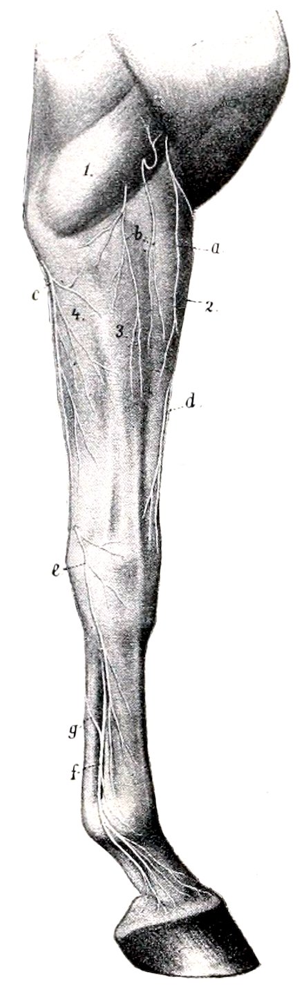
Fig. 520.—Cutaneous Nerves of Right Fore Limb of Horse, External Face. a, Cutaneous branch of axillary nerve; b, cutaneous branches of radial nerve; c, posterior cutaneous branch of ulnar nerve; d, cutaneous branch of median nerve; e, superficial branch of ulnar nerve; f, external metacarpal nerve; g, anastomotic branch connecting internal and external metacarpal nerves; 1, external head of triceps; 2, extensor carpi radialis; 3, anterior extensor; 4, flexor carpi externus. (After Ellenberger, in Leisering’s Atlas.)
Ulnar Nerve
The ulnar nerve (Figs. 441, 442, 443, 520) arises with the median from the thoracic components of the brachial plexus. It descends between the brachial artery and vein, accompanied for a short distance by the radial nerve. It then crosses the vein and continues behind the latter along the anterior border of the tensor fasciæ antibrachii and dips under that muscle near the elbow. Here it is joined by the ulnar vessels and passes downward and backward over the internal 699epicondyle of the humerus. In the forearm it crosses obliquely the deep face of the ulnar head of the flexor carpi medius and descends under the deep fascia with the vessels, at first on the ulnar head of the perforans, and then between the middle and external flexors of the carpus. Near the level of the accessory carpal bone it divides into two terminal branches, superficial and deep. It gives off two principal collateral branches. The cutaneous branch (Ramus cutaneus palmaris) is detached just before the nerve passes under the tensor fasciæ antibrachii; it runs downward and backward on that muscle under cover of the posterior superficial pectoral, becomes superficial below the elbow and ramifies on the posterior surface and both sides of the forearm. The muscular branch is given off at the elbow joint and divides to supply the flexors of the digit and the middle flexor of the carpus. Of the two terminals, the superficial branch (Ramus superficialis) emerges between the tendons of insertion of the external and middle flexors of the carpus and ramifies on the dorso-lateral aspect of the carpus and metacarpus. The deep branch (Ramus profundus), after a very short course, unites under cover of the tendon of the flexor carpi medius with the outer branch of the median nerve to form the external metacarpal or volar nerve.
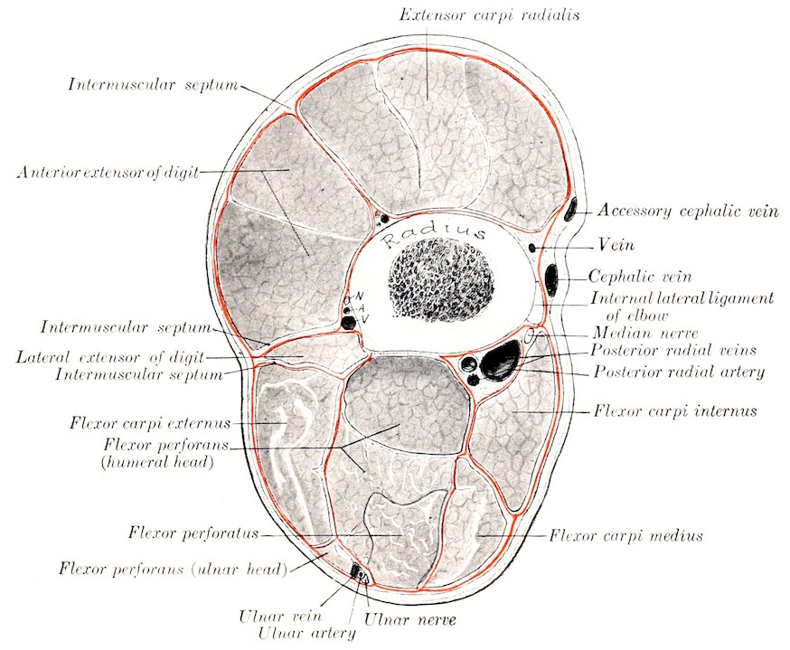
Fig. 521.—Cross-section of Forearm of Horse about Three Inches (ca. 8 cm.) below the Elbow Joint. The deep fascia is designated by red line. N, A, V, Interosseous vessels and nerve.
Median Nerve
The median nerve (Figs. 441, 442, 447, 520, 521) derives its fibers chiefly from the first thoracic root of the brachial plexus. It is usually the largest branch and it accompanies the chief arterial trunks to the distal part of the limb. It descends over the inner face of the brachial artery, which it crosses obliquely, and continues 700down the arm in front of the artery. Near its origin it is joined by a large branch with the musculo-cutaneous nerve, thus forming a loop in which the artery appears to be suspended. Near the elbow it crosses obliquely over the artery (posterior radial) and lies behind it on the internal lateral ligament. Below the joint it again crosses the artery and lies behind the radius and the lower part of the long internal lateral ligament.
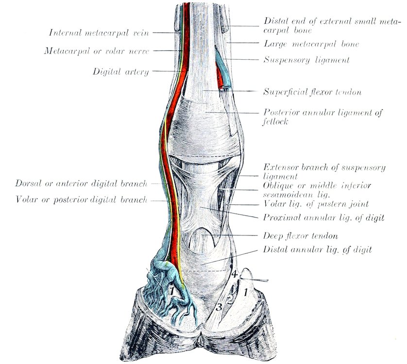
Fig. 522.—Dissection of Distal Part of Right Fore Limb of Horse, Viewed from Behind. 1, Lateral cartilage; 2, 3, 4, ligaments from first phalanx to third sesamoid, bulb of heel, and lateral cartilage. (After Schmaltz, Atlas d. Anat. d. Pferdes.)
At the elbow the nerve is covered by the posterior superficial pectoral muscle and is crossed by the large oblique connection between the cephalic and brachial veins. The variable and often plexiform venous arrangement renders approach to the nerve here undesirable. It can be reached best by an incision just behind the lower part of the long internal lateral ligament; here the nerve lies under the deep fascia in the furrow between the radius and the anterior border of the flexor carpi internus, overlapped by the latter. In some cases the nerve retains its position in front of the artery at the elbow, and in others it crosses the deep face of the artery.
Passing beneath the internal flexor of the carpus, the nerve continues downward in the forearm with the posterior radial vessels, and divides at a variable distance above the carpus into two branches; these are the internal volar or metacarpal nerve and the branch which unites with the ulnar to form the corresponding external nerve.
At the proximal part of the forearm the nerve runs almost straight downward along the posterior border of the long internal lateral ligament, while the artery here inclines somewhat backward. Thus the nerve is superficial to the artery for a short distance, then lies in front of the latter to about the middle of the region, where it inclines a little backward and arrives at the 701interval between the internal and middle flexors of the carpus. The division may occur about the middle of the region, but commonly takes place in the distal third or fourth.
The collateral branches are as follows:
1. The musculo-cutaneous branch is in reality the continuation of the nerve of that name. It is given off about the middle of the arm, passes beneath the lower part of the biceps, and divides into muscular and cutaneous branches. The muscular branch enters the brachialis. The cutaneous branch emerges between the mastoido-humeralis and the biceps and divides into two branches; these descend on the fascia of the forearm with the cephalic vein and its accessory, and ramify on the front and inner face of the forearm, carpus, and metacarpus.
2. Muscular branches to the internal flexor of the carpus and the flexors of the digit are given off just as the nerve passes under the first named muscle.
3. The very small interosseous nerve passes through the interosseous space and is distributed chiefly to the periosteum, but in some cases twigs may go to the extensor muscles.
The metacarpal or volar nerves,[209] internal and external, are the continuations of the median and ulnar nerves in the distal part of the limb.
The internal metacarpal or volar nerve (N. volaris medialis) arises as the inner terminal branch of the median nerve at a variable distance above the carpus (Fig. 442). It descends through the carpal canal along the inner border of the superficial flexor tendon, and lies at first in front of the large metacarpal or common digital artery. It then passes behind the artery to the distal third of the metacarpus, where it lies behind the vein, the artery here becoming deeper in position. In addition to cutaneous twigs, the nerve gives off about the middle of the metacarpus a considerable anastomotic branch which winds obliquely downward and outward over the flexor tendons and joins the external nerve at the lower third of the metacarpus (Fig. 447). Near the fetlock the nerve divides into two digital branches.
1. The dorsal or anterior digital branch (Ramus dorsalis) descends at first between the digital artery and vein, then crosses over the vein and ramifies in the skin and the matrix of the hoof on the dorsal (anterior) face of the digit.
2. The volar or posterior digital branch (Ramus volaris) is the direct continuation of the trunk. It descends behind the digital artery, which it accompanies in its ramification. A middle digital branch is sometimes described as descending behind the vein. In some cases this branch is distinct, but usually there are instead several small twigs derived from the posterior branch, which cross very obliquely over the artery and anastomose in a variable manner with each other and with the anterior branch.
The external metacarpal or velar nerve (N. volaris lateralis) is formed by the union of the external terminal branch of the median with the deep branch of the ulnar nerve (Fig. 442). It descends with the internal volar or small metacarpal artery in the texture of the posterior annular ligament of the carpus. In the metacarpus it descends along the outer border of the deep flexor tendon behind the external metacarpal vein, and is accompanied by a small artery from the volar subcarpal arch. Toward the distal end of the metacarpus it is joined by the oblique branch from the internal nerve, and beyond this is arranged like the latter. Below the carpus it detaches a deep branch to the suspensory ligament and the interossei, and also supplies twigs to the skin.
Anastomoses are established between the digital branches, and the areas innervated by them are not well defined, but really overlap each other. In certain diseased conditions, however, in which the lesions are confined to the volar structures, relief from pain may be afforded by section of the volar branches only.
702
THE THORACIC NERVES
The thoracic nerves (Nn. thoracales) number eighteen on either side in the horse. They are designated numerically according to the vertebræ behind which they emerge. Most of them are arranged in a very similar manner and therefore do not require separate description. Each divides into a dorsal and a ventral branch, the latter being the larger.
The dorsal branches (Rami dorsales) emerge behind the levatores costarum and divide into internal and external branches. The internal branches ascend on the multifidus and supply the dorsal spinal muscles. The external branches run outward under the longissimus and emerge between that muscle and the transversalis costarum; after giving twigs to these muscles they pass through the latissimus dorsi and the lumbo-dorsal fascia and ramify as dorsal cutaneous nerves under the skin of the back (Fig. 523). In the region of the withers they give branches to the serratus anticus and rhomboideus, and their cutaneous terminals pass through these muscles and the dorso-scapular ligament to supply the skin over the ligamentum nuchæ and the scapular cartilage.
The ventral branches or intercostal nerves (Nn. intercostales) are much larger than the preceding, and are connected with the sympathetic by rami communicantes. The first goes almost entirely to the brachial plexus, but sends a fine branch downward in the first intercostal space which is expended in the muscle there without reaching the lower end of the space. The second ventral branch furnishes a considerable root to the brachial plexus, but its intercostal continuation is typical. The intercostal nerves (Fig. 184) descend in the intercostal spaces with the vessels of like name, at first between the intercostal muscles, and lower down chiefly between the pleura and the internal intercostal muscle. In the anterior spaces the artery lies along the posterior border of the rib, with the nerve in front of it; further back the nerve lies behind the border of the rib, with the artery in front of it. They supply the intercostal muscles, give off lateral perforating branches, and terminate in the following manner: The second to the sixth inclusive emerge through the spaces between the costal cartilages and concur in supplying the pectoral muscles. The second to the eighth give branches to the transversus thoracis. The succeeding ones give branches to the diaphragm, pass between the transverse and internal oblique muscles, give twigs to these, and end in the rectus abdominis. There are three series of cutaneous nerves given off by the intercostal nerves. The upper nerves emerge through the serratus magnus and the external intercostals about parallel with the digitations of the external oblique. The middle set perforate the origin of the latter muscle. The lower ones appear through the abdominal tunic. They supply branches to the abdominal muscles, the panniculus, and the skin. Some of the anterior ones anastomose with the posterior thoracic branches of the brachial plexus. The posterior three supply in part the skin of the flank. The ventral branch of the last thoracic nerve runs outward behind the last rib across the dorsal surface of the psoas major and divides into superficial and deep branches. The superficial branch passes between the obliquus internus and transversus abdominis, perforates the obliquus externus, and ramifies under the skin of the flank (Fig. 525). The deep branch descends on the inner face of the internal oblique to the rectus abdominis, in which it ends.
THE LUMBAR NERVES
There are six pairs of lumbar nerves (Nn. lumbales) in the horse, the last of which emerge between the last lumbar vertebra and the sacrum. The anterior two or three are about the same size as the thoracic nerves, but the others are much larger.
703
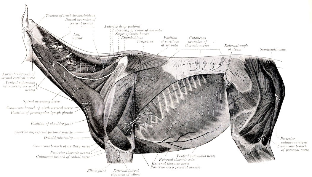
Fig. 523.—Superficial Nerves of Neck and Trunk of Horse. (After Schmaltz, Atlas d. Anat. d. Pferdes.)
704Their dorsal branches are small in comparison with the ventral ones. They are distributed to the muscles and skin of the loins and croup in a fashion similar to those of the thoracic nerves.
The ventral branches are connected with the sympathetic by small rami communicantes, and give branches to the sublumbar muscles. Those of the first two nerves are arranged in a manner analogous to the corresponding branch of the last thoracic nerve.
The ventral branch of the first lumbar nerve is termed the ilio-hypogastric nerve (N. iliohypogastricus). It passes outward between the quadratus lumborum and the psoas major, and divides at the outer border of the latter into an anterior or superficial and a posterior or deep branch. The anterior or superficial branch passes over the upper edge of the internal oblique, descends between that muscle and the external oblique, perforates the latter, and runs downward and backward and ramifies under the skin of the posterior part of the flank and the outer surface of the thigh. It gives branches to the transversus and obliquus externus abdominis. The posterior or deep branch is smaller; it runs downward and backward beneath the peritoneum to the outer border of the rectus abdominis, gives branches to the internal oblique, and terminates in the rectus abdominis.