THE MALE GENITAL ORGANS
Reading edition
The male genital organs (Organa genitalia masculina) are: (1) The two testicles, the essential reproductive glands, with their coverings and appendages; (2) the vasa deferentia, the ducts of the testicles; (3) the vesiculæ seminales; (4) the prostate, a musculo-glandular organ; (5) the two bulbo-urethral (or Cowper’s) glands; (6) the male urethra, a canal which transmits the generative and urinary secretions; (7) the penis, the male copulatory organ. The vesiculæ seminales, the prostate, and the bulbo-urethral glands discharge their secretions into the urethra, where they mix with the seminal fluid secreted by the testicles; hence they are often termed the accessory sexual glands.
MALE GENITAL ORGANS OF THE HORSE
THE TESTICLES
The testicles (Testes) are situated in the inguinal region, inclosed in a diverticulum of the abdomen termed the scrotum. Their long axes are nearly longitudinal. They are ovoid in form, but considerably compressed from side to side. Each presents two surfaces, two borders, and two extremities. The internal and external surfaces (Facies medialis, lateralis) are convex and smooth. The free or ventral border (Margo liber) is convex in both directions. The attached or dorsal border (Margo epididymidis) is nearly straight, and is the one by which the gland is suspended in the scrotum by the spermatic cord; the epididymis is attached to this border and overlies it externally. The anterior and posterior extremities (Extremitas capitata, caudalis) are rounded.
At the anterior extremity there is often a sessile or pedunculated sac which contains a clear fluid; this is the appendix testis or hydatid of Morgagni, from which a thread-like process extends backward toward the vas deferens. It is regarded as a remnant of the Müllerian duct of the embryo.
A testicle of average size of an adult stallion is about four or five inches (ca. 10 to 12 cm.) long, two and a half inches (ca. 6 to 7 cm.) high, and one and a half to two inches (ca. 4 to 5 cm.) wide; it weighs about ten and one-half ounces (ca. 300 grams). They vary much in size in different subjects, and are commonly of unequal size, the left one being more often the larger.
The epididymis is adherent to the attached border of the testicle, and overlaps somewhat the external surface. Its anterior enlarged end is termed the head or globus major (Caput epididymidis), and its posterior, slightly enlarged end is the tail or globus minor (Cauda epididymidis); while the intermediate narrow part is the body (Corpus epididymidis). The head is closely connected with the testicle by the efferent ducts of the latter, by connective tissue, and by the serous membrane. The body is less closely attached by the serous covering, which forms externally a pocket beneath the epididymis termed the digital fossa (Sinus epididymidis). The tail is firmly attached to the posterior extremity of the testicle by a short ligament (Lig. epididymidis), formed by a thick fold of the tunica vaginalis, which contains smooth muscle-fibers; it is continued by the vas deferens.
Structure of the Testicle and Epididymis. The greater part of the surface of the testicle is covered by a serous membrane, the tunica vaginalis propria, which 486is the visceral layer of the serous envelop of the cord and testicle; this is reflected from the attached border of the gland, leaving an uncovered area at which the vessels and nerves in the spermatic cord reach the testicle. Beneath this serous covering is the tunica albuginea, a strong capsule composed of dense white inelastic fibrous tissue and unstriped muscle-fibers. From the deep surface of this tunic delicate membranous septa (Septula testis) pass into the gland, dividing it into pyramidal spaces, the bases of which are peripheral. At the anterior part of the attached border of the testicle connective tissue and smooth muscle-fibers pass into the substance of the gland, forming the mediastinum testis; from this the septa radiate. The mediastinum contains a network of fine canals, the rete testis; it is traversed by blood-vessels, which radiate on the septa to the deep face of the tunica albuginea, forming a delicate network—the so-called tunica vasculosa. The spaces imperfectly marked off by the septa contain the parenchyma testis, which consists of seminiferous tubules (Tubuli seminiferi), supported by loose intertubular connective tissue. The tubules are collected into small masses or lobules (Lobuli testis). They are at first very tortuous (Tubuli contorti); toward the apex of the lobule they unite with other tubules, forming larger straight tubules (Tubuli recti); these pass into the mediastinum testis, where they form the rete testis. From the latter a number (ca. 15 to 20) of larger efferent tubules (Vasa efferentia) pierce the albuginea and enter the head of the epididymis.
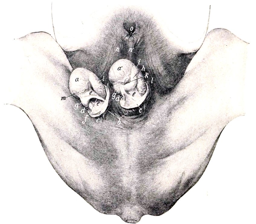
Fig. 390.—Inguinal Region of Stallion, with Testicles Exposed. a, a′, Testicles; b, scrotum, opened and reflected; c, tunica vaginalis communis, opened and reflected; d, ligament of epididymis; e, tunica vaginalis propria (mesorchium); f, vas deferens; g, g′, tail of epididymis; h, body of same; i, head of same; k, digital fossa or sinus epididymidis; l, spermatic vessels showing through tunica vaginalis propria; m, spermatic artery; n, prepuce; n′, raphé; o, preputial orifice. (After Ellenberger-Baum, Top. Anat. d. Pferdes.)
The epididymis is covered by the tunica vaginalis propria and a thin albuginea. 487Its head consists of lobules (Lobuli epididymidis), each of which consists of a coiled tubule or conus vasculosus, which succeeds a vas efferens. By the union of these tubules is formed a single tube, the duct of the epididymis (Ductus epididymidis), which, by its complex coils, forms the body and tail of the epididymis and terminates in the vas deferens. The tubules and the coils of the duct of the epididymis are held together by connective tissue and unstriped muscle-fibers. The tubules and duct are lined with ciliated epithelium, and the duct has a muscular coat which consists of longitudinal and circular fibers.
Vessels and Nerves.—The testicle is richly supplied with blood by the spermatic artery, a branch of the posterior aorta. The artery descends in the anterior part of the spermatic cord, and is very tortuous near the testicle; on reaching the attached border of the gland it passes backward in a flexuous manner, giving branches to the testicle and epididymis, turns around the posterior extremity, and runs forward on the free border to the anterior extremity. It is partially embedded in the tunica albuginea, and detaches lateral branches which ascend in a tortuous fashion on each surface of the testicle; these give off small branches which enter the gland on the septa.
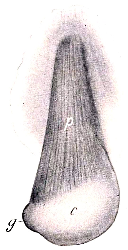
Fig. 391.—Right Testicle and Spermatic Cord of Horse, Inclosed in Tunica Vaginalis. c, Tunica vaginalis communis; g, prominence caused by tail of epididymis; p, cremaster externus muscle. (After Ellenberger-Baum, Top. Anat. d. Pferdes.)
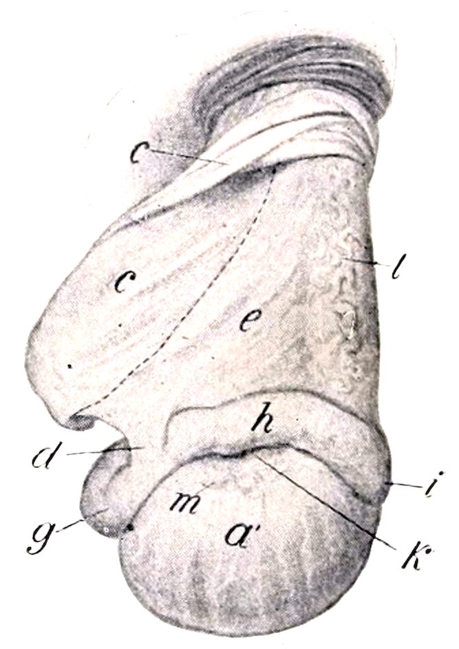
Fig. 392.—Right Testicle and Spermatic Cord of Horse, Exposed. a′, External surface of testicle; c, tunica vaginalis, cut and reflected; d, ligament of epididymis; e, mesorchium; g, tail, h, body, i, head of epididymis; k, digital fossa or sinus epididymidis; l, spermatic vessels showing through tunica vaginalis propria; m, end of spermatic artery. Dotted line indicates position of vas deferens on other side of mesorchium. (After Ellenberger-Baum, Top. Anat. d. Pferdes.)
The spermatic veins on leaving the testicle, form the pampiniform plexus around the artery in the spermatic cord. The vein which issues from this plexus usually joins the posterior vena cava on the right side, the left renal vein on the left side.
The nerves, derived from the renal and posterior mesenteric plexuses, form the spermatic plexus around the vessels.
THE SCROTUM
The scrotum, in which the testicles and the lower parts of the spermatic cords are situated, is somewhat globular in form, but is commonly asymmetrical, since 488one testicle—more often the left—is the larger and more dependent. It varies in form and appearance in the same subject, according to the condition of its subcutaneous muscular tissue. The latter contracts on exposure to cold, so that the scrotum is drawn up and becomes thicker and wrinkled; when relaxed under the influence of heat or fatigue, or from debility, it becomes smooth and pendulous, with a constriction or neck superiorly. It consists of layers which correspond with those of the abdominal wall; considered from without inward these are:
(1) The skin, which is thin, elastic, usually black in color, and smooth and oily to the touch. It presents scattered short fine hairs, and is abundantly supplied with very large sebaceous and sweat glands. It is marked centrally by a longitudinal furrow, in which is a line, the raphé scroti; this is continued forward on the prepuce and behind on the perineum.
(2) The dartos (Tunica dartos) is reddish in color and is closely adherent to the skin except superiorly. It consists of fibro-elastic tissue and unstriped muscle. Along the raphé it forms a median partition, the septum scroti, which divides the scrotum into two pouches. Dorsally the septum divides into two layers which diverge on either side of the penis to join the abdominal tunic. At the bottom of the scrotum fibers connect the dartos closely with the tunica vaginalis and the tail of the epididymis, constituting the scrotal ligament.[143] Elsewhere the dartos is loosely connected with the underlying tunic by areolar tissue which contains no fat.
(3) The scrotal fascia, which is apparently derived from the oblique abdominal muscles.
It has been customary to describe three layers of fascia, in conformity with the accounts given in text-books of human anatomy. These are: (1) the intercolumnar or spermatic fascia, derived from the margin of the external inguinal ring; (2) the cremasteric fascia, derived from the internal oblique muscle; (3) the infundibuliform fascia, derived from the fascia transversalis. The first two cannot be distinguished by dissection and the third is (in the scrotum) fused with the parietal peritoneum of the tunica vaginalis.
(4) The parietal layer of the tunica vaginalis.—This is a fibro-serous sac which is continuous with the parietal peritoneum of the abdomen at the internal inguinal ring. It is thin above, but is thick in its scrotal part, where it is strengthened by fibrous tissue (Lamina fibrosa) derived from the transversalis fascia. It will be described further under the caption tunica vaginalis.[144]
THE VAS DEFERENS
This tube (Ductus deferens) extends from the tail of the epididymis to the ejaculatory duct. It passes upward in the inguinal canal, inclosed in a fold detached from the inner surface of the mesorchium, near the posterior (attached) border of the latter. At the vaginal ring it separates from the other constituents of the spermatic cord, and turns backward and inward into the pelvic cavity (Fig. 272). For some distance it lies in the free edge of the urogenital fold, by which it is attached to the lower part of the lateral wall of the pelvis. In its further course (over the dorsal surface of the bladder) it leaves the edge of the fold and inclines inward between its layers, coming in contact with the inner face of the vesicula seminalis. Over the neck of the bladder the two vasa lie very close together, flanked laterally by the necks of the vesiculæ seminales, and having the uterus masculinus between them. They then disappear under the isthmus of the prostate, and are continued through the wall of the urethra by the ejaculatory ducts. The latter are formed in the urethral wall by the union of the 489vas deferens with the duct (or neck) of the corresponding vesicula seminalis; they are thin-walled tubes, about 2 to 3 mm. long and 6 to 7 mm. wide, which open into the urethra on either side of the colliculus seminalis. In about 15 per cent. of subjects the vas deferens and duct of the seminal vesicle do not unite, but open side by side into the urethra. From its origin until it reaches the dorsal surface of the bladder the vas deferens has a uniform diameter of about a quarter of an inch (ca. 6 mm.). It then forms a fusiform dilatation, the ampulla ductus deferentis (Fig. 394); this part is about six to eight inches (ca. 15 to 20 cm.) long, and in its largest part nearly an inch (ca. 2 cm.) in diameter in the stallion; in geldings the dilatation is usually not very pronounced. Beyond the ampulla the duct abruptly contracts.
Structure.—The wall of the vas deferens is thick and the lumen relatively small, so that the tube has a firm and cord-like character. It is covered with peritoneum, except in the last few inches of its course. The loose adventitia contains numerous vessels and nerves. The thick muscular coat consists of longitudinal and circular layers. The mucous membrane has an epithelium of short columnar cells. In the posterior part of the tube, and especially in the ampulla, there are numerous tubulo-alveolar glands.
Blood-supply.—Spermatic and umbilical arteries.
Nerve-supply.—Spermatic and pelvic plexuses.
THE SPERMATIC CORD
The spermatic cord (Funiculus spermaticus) consists of the structures carried down by the testicle in its migration through the inguinal canal from the abdominal cavity to the scrotum. It begins at the internal inguinal ring, where its constituent parts come together, extends obliquely downward through the inguinal canal, passes over the side of the penis, and ends at the attached border of the testicle. It consists of the following structures:
(1) The spermatic artery.
(2) The spermatic veins, which form the pampiniform plexus around the artery.
(3) The lymphatics, which accompany the veins.
(4) Sympathetic nerves, which run with the artery.
(5) The vas deferens.
(6) The internal cremaster muscle, which consists of bundles of unstriped muscular tissue.
(7) The visceral layer of the tunica vaginalis or mesorchium.
The first four of these constituents are gathered into a rounded mass which forms the anterior part of the cord; they are united by connective tissue, interspersed with which are bundles of the cremaster internus. The vas deferens is situated posteriorly, inclosed in a special fold detached from the inner surface of the mesorchium; hence it is not visible externally.
The term spermatic cord is to a certain extent misleading as applied to most animals, while in man the structure is distinctly cord-like. In the horse, when the tunica vaginalis is slit open and the “cord” stretched out, the latter is seen to have the form of a wide sheet, the mesorchium, which has a thick, rounded anterior edge, the so-called “vascular part” of the cord. The posterior edge of the mesorchium is continuous with the parietal layer of the tunic; its inner surface presents posteriorly the deferential fold (Plica ductus deferentis). Between the two layers of the mesorchium are bundles of unstriped muscle (cremaster internus) and small vessels.
THE TUNICA VAGINALIS
The tunica vaginalis is a somewhat pyriform serous sac which extends from the internal inguinal ring through the inguinal canal to the bottom of the scrotum. 490Like the abdominal peritoneum, of which it is an evagination, it consists of two layers—parietal and visceral. The parietal layer, or tunica vaginalis communis (s. reflexa), lines the scrotum below; its narrow, tubular part lies in the inguinal canal and is directly continuous with the parietal peritoneum of the abdomen at the internal inguinal ring. The cavity of the tunica vaginalis (Cavum vaginale) is a diverticulum of the general peritoneal cavity, with which it communicates through the vaginal ring (Annulus vaginalis). It contains normally a small quantity of serous fluid. The parietal layer is reflected from the posterior wall of the inguinal canal around the structures of the cord, forming the mesorchium, a fold analogous to the mesentery of the intestine. The visceral layer, or tunica vaginalis propria, covers the spermatic cord, testicle, and epididymis.
The external cremaster muscle (M. cremaster externus) lies on the outer and posterior part of the tunic, to the scrotal part of which it is attached.
Confusion has arisen from the use of the term internal abdominal or inguinal ring in two senses. The term is used to designate the upper or abdominal opening of the inguinal canal, but it is also applied to the upper opening of the cavity of the tunica vaginalis. It should only be applied to the abdominal opening of the canal, the subperitoneal ring. The peritoneal ring at which the cavity of the tunica vaginalis opens into the general peritoneal sac is distinguished by the name vaginal ring. It is placed about four or five inches (ca. 10 to 12 cm.) from the linea alba, and about two or three inches (ca. 6 to 8 cm.) in front of the ilio-pectineal eminence. In stallions it will usually admit the end of the finger readily, but it may be abnormally large and allow a loop of bowel to enter the cavity of the tunica vaginalis. In the gelding it is smaller and sometimes partially occluded. In man the cavity is almost always obliterated early, except in its scrotal portion, thus abolishing the vaginal ring.
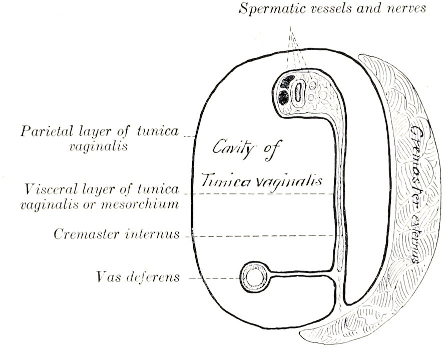
Fig. 393.—Diagram of Cross-section of Spermatic Cord and Tunica Vaginalis; Latter Represented as Distended.
DESCENT OF THE TESTICLES
During early fœtal life the testicle is situated against the dorsal wall of the abdominal cavity, in contact with the ventral surface of the corresponding kidney. As growth proceeds it gradually migrates from this primitive position, and finally passes down the inguinal canal into the scrotum. Previous to its descent through the abdominal wall the testicle is suspended by a fold of peritoneum, termed the mesorchium. This fold contains the vessels and nerves of the testicle in its anterior border. In its posterior edge is the elongated tail of the epididymis, and two cords of fibrous tissue and unstriped muscle. One of these cords is short and connects the tail of the epididymis with the testicle; later it becomes shorter, 491and is termed the ligament of the epididymis. The other cord, the gubernaculum testis, extends from the tail of the epididymis to the subperitoneal tissue in the vicinity of the future vaginal ring. The deferential fold (Plica ductus deferentis) is given off from the inner face of the mesorchium, and joins the urogenital fold posteriorly. The body of the epididymis at this time lies in the edge of an oblique fold formed by the outer layer of the mesorchium. After the middle of fœtal life, a pouch or diverticulum of the peritoneum, the processus vaginalis, grows downward through the inguinal canal, carrying with it cremaster fibers derived from the internal oblique muscle and a layer from the transversalis fascia. It is accompanied by an inguinal extension of the gubernaculum testis. The latter blends below with the subcutaneous tissue which later becomes the dartos. The tail of the epididymis first enters the processus vaginalis, followed by the testicle with its mesorchium, which descends within this diverticulum of the peritoneum until it reaches the scrotum. The vas deferens and its fold descend synchronously with the epididymis and testicle.
The mechanical factors concerned in the migration of the testicle are matters on which much uncertainty still exists. That the gubernaculum exerts sufficient traction to guide the epididymis and testicle to the inguinal canal seems plausible. The internal inguinal ring may constitute a locus minoris resistentiæ in the abdominal wall, especially after the descent of the processus vaginalis. Progressive shortening of the gubernaculum was formerly considered to be the chief cause of the descent through the abdominal wall. Increase in the intra-abdominal pressure is probably an important factor.
In the foal the descent of the testicles is often complete at birth, but it frequently happens that one testicle or both may be retained in the inguinal canal or in the abdomen for some months. In other cases the testicle may return into the canal or abdomen, since in the young foal the vaginal ring is large and the testicle small and soft, and not yet closely anchored by the scrotal ligament. In rare cases the descent may be completed as late as the fourth year (personal observation).
Indefinite retention of one testicle or both in the abdominal cavity or inguinal canal is not rare in horses; this condition is termed cryptorchism. Abdominal retention is the more usual form of cryptorchism in adult horses, inguinal retention being usually temporary. The retained testicle is usually, but not always, small, thin, soft, and flabby, and is non-spermiogenic. The processus vaginalis and the inguinal part of the gubernaculum are usually present, but may be rudimentary. The ligament of the epididymis and the corresponding part of the mesorechium are often so long that the tail of the epididymis may be several inches distant from the testicle. The abdominal part of the gubernaculum may be 8 to 10 inches (ca. 20 to 25 cm.) long, and the ligament of the epididymis much elongated (10 to 15 cm. in length according to Vennerholm); thus the testicle may have a wide range. The vaginal ring is sometimes closed.
In many mammals the testicles normally remain in the abdominal cavity; such animals are termed testiconda, and include the elephant, some insectivora, hyrax, sloths, ant-eaters, armadillos, and cetacea. In others the testicles descend periodically during the period of œstrum, and then return into the abdomen, or they may be extruded and retracted voluntarily; this is true of most rodents, many insectivora (moles, shrews, hedgehog), and bats.
THE VESICULAE SEMINALES
The vesiculæ seminales (Fig. 394) are two elongated and somewhat pyriform sacs, which lie on either side of the posterior part of the dorsal surface of the bladder. They are partly inclosed in the urogenital fold, and are related to the rectum dorsally. Their long axes are parallel with the vas deferentia and converge posteriorly. Each consists of a rounded blind end, the fundus, a middle, slightly narrower part, the body, and a posterior constricted part, the neck or duct.
In the stallion they are about six to eight inches (ca. 15 to 20 cm.) long, and their greatest diameter is about two inches (ca. 5 cm.); in the gelding they are usually much smaller.[145]
492The vesiculæ are, for the most part, retroperitoneal, but the fundus extends forward into the urogenital fold and hence has a serous covering. The neck or duct (Ductus excretorius) dips under the prostate, and usually unites with the end of the vas deferens to form the ejaculatory duct.
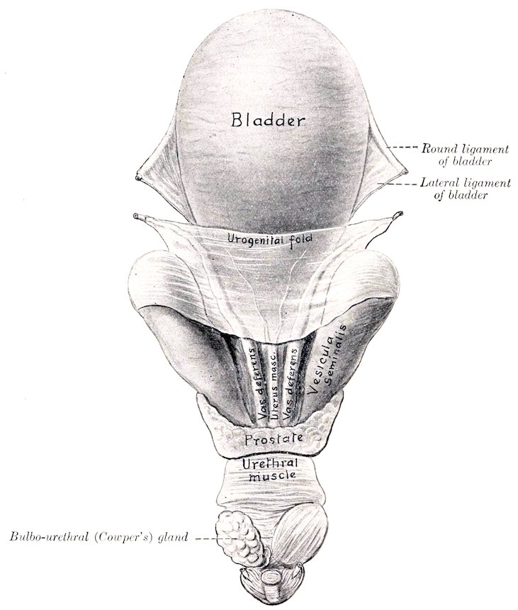
Fig. 394.—Internal Genital Organs of Stallion, Dorsal View. On left side urethral muscle has been removed over bulbo-urethral gland. Cornua of uterus masculinus are indicated in urogenital fold.
Structure.—The wall, exclusive of the partial serous coat, consists of a fibrous adventitia, a middle muscular coat, and a mucous lining. The muscular coat is thickest at the fundus, and consists of two planes of longitudinal fibers with a circular layer between them. The mucous membrane is thin, and is arranged in numerous folds which form a network; the spaces so inclosed present the openings of tubulo-alveolar glands. The epithelium is columnar. The blood-supply is derived from the internal pudic artery.
493
THE PROSTATE
The prostate (Prostata) is a musculo-glandular organ which lies on the neck of the bladder and the beginning of the urethra, beneath the rectum. It consists of two lateral lobes and a connecting isthmus.
The lateral lobes, right and left (Lobus dexter et sinister), are somewhat prismatic in form, and are directed forward, outward, and somewhat upward. The antero-internal surface of each lobe is concave and partly embraces the corresponding vesicula seminalis. The dorsal surface is concave and is in relation with the rectum. The ventral surface is convex and lies on the obturator internus muscle and fat. The apex is pointed and lies near the posterior end of the superior ischiatic spine.
The isthmus is a thin, transverse band, about four-fifths of an inch (ca. 2 cm.) wide. It lies over the junction of the bladder with the urethra, the uterus masculinus, the terminal parts of the vasa deferentia, and the necks of the vesiculæ seminales. Dorsally it is partly covered by transverse fibers of the urethral (Wilson’s) muscle.
Structure.—The prostate is inclosed in a fibro-muscular capsule, which sends thick, somewhat radially arranged trabeculæ into the gland substance, dividing it into spheroidal or ovoid lobules. The lobules contain a central space in which the prostatic secretion (Succus prostaticus) collects. The walls of these spaces are pouched out to form tubular diverticula, and the latter present saccular dilatations. These cavities are all lined by cubical epithelium. There are 15 to 20 prostatic ducts (Ductus prostatici) on either side, which perforate the urethra and open lateral to the colliculus seminalis. The blood-supply is derived from the internal pudic artery.
The surface of the prostate is commonly tuberculate in old subjects, and amyloid bodies and calcareous concretions may be found in it.
THE UTERUS MASCULINUS
The uterus masculinus or prostatic utricle is a rudimentary structure of variable size and form, situated centrally on the posterior part of the dorsal surface of the bladder. When well developed, it consists of a median flattened tube, some three or four inches (ca. 7.5 to 10 cm.) long, and about half an inch (ca. 1 to 1.5 cm.) wide, the anterior part of which lies in the urogenital fold and gives off two slender processes or cornua; the latter curve forward and outward in the fold a variable distance, being sometimes traceable as far as the anterior end of the ampulla of the vas deferens. The posterior extremity of the tube passes under the isthmus of the prostate, and opens into the urethra on the summit of the colliculus or joins an ejaculatory duct. It has a muscular coat and a mucous lining. In many cases it consists merely of a very small central tubule with a blind anterior end, or a band, not at all sharply marked off from the adjacent tissue; in other cases it cannot be recognized. It is interesting chiefly as being a remnant of the ducts of Müller and the homologue of the uterus and vagina.
THE BULBO-URETHRAL GLANDS
The bulbo-urethral (or Cowper’s) glands (Glandulæ bulbourethrales) are two in number, and are situated on either side of the pelvic part of the urethra close to the ischial arch (Fig. 452). They are covered by the urethral (Wilson’s) muscle. They are ovoid in form, somewhat depressed dorso-ventrally, and their long axes are directed obliquely forward and outward. In the stallion they may measure 494about two inches (ca. 5 cm.) in length, and an inch or more (ca. 3 cm.) in width. In the gelding they are about the size of an average hazel nut.
Structure.—They resemble the prostate in general structure, but the interstitial tissue is much less abundant, especially in regard to its muscular elements. In the peripheral part there are striped muscle-fibers. Each gland has six to eight excretory ducts (Ductus excretorii) which open into the urethra on a series of small papillæ behind the prostatic ducts and close to the median plane. The blood-supply comes from the internal pudic artery.
THE PENIS
The penis, the male organ of copulation, is composed essentially of erectile tissue, and incloses the extrapelvic part of the urethra. It extends from the ischial arch forward between the thighs on to the umbilical region of the abdominal wall. It is supported by the fascia penis and the skin, and its prescrotal portion is situated in a cutaneous pouch, the prepuce or sheath. It is cylindrical in form, but much compressed laterally in the greater part of its extent.
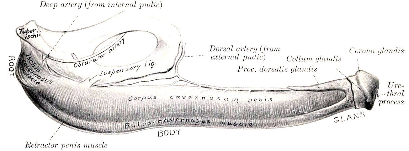
Fig. 395.—Penis of Horse, Lateral View.
In the quiescent state it is about 20 inches (ca. 50 cm.) long; of this, about 6 to 8 inches (ca. 15 to 20 cm.) is free in the prepuce. In erection it increases 50 per cent. or more in length.
It may be divided into a root, a body, and a terminal enlargement, the glans.
The root (Radix penis) is attached to the lateral parts of the ischial arch by two crura, which converge and unite below the arch (Fig. 452). The urethra passes over the ischial arch between the crura, and curves forward to become incorporated with the penis.
The body (Corpus penis) begins at the junction of the crura and constitutes the bulk of the organ. At its origin it is attached to the symphysis ischii by two strong flat bands, the suspensory ligaments of the penis (Ligamenta suspensoria penis), which blend with the tendon of origin of the graciles muscles (Figs. 451, 456). This part of the penis is flattened laterally for the most part, but becomes rounded and smaller anteriorly. It presents four surfaces. The dorsal surface or dorsum penis, is narrow and rounded; on it are the dorsal arteries and nerves of the penis and a rich venous plexus. The ventral or urethral surface (Facies urethralis) is rounded, and along it runs the urethra, embedded in the deep urethral groove of the corpus cavernosum. The lateral surfaces are high and flattened, except anteriorly, where they are lower and rounded.
495The glans penis is the enlarged free end of the organ. It is marked off from the body by a faintly marked neck (Collum glandis). In front of this is a prominent circular ridge, the corona glandis, which is notched below. The base of the glans is rounded and extends further backward dorsally than ventrally; it is marked in its lower part by a deep depression, the fossa glandis, in which the urethra protrudes for about an inch (ca. 2.5 cm.) as a free tube, the urethral process (Processus urethræ), covered by a thin integument. The urethra is thus surrounded by a circular fossa, which opens superiorly into the urethral sinus, a bilocular diverticulum lined by thin black skin. This diverticulum is filled sometimes with a caseous mass of sebaceous matter and epithelial débris.
Structure.—The penis consists essentially of two erectile bodies, the corpus cavernosum penis and the corpus spongiosum.
The corpus cavernosum penis forms the greater part of the bulk of the penis except at its free extremity. It arises from each side of the ischial arch by a crus, which is embedded in the ischio-cavernosus muscle. Below the ischial arch the crura fuse completely to form the laterally compressed body of the corpus cavernosum; this presents ventrally the urethral groove (Sulcus urethralis), which contains the urethra and corpus spongiosum. Anteriorly, the corpus cavernosum divides into three processes, a long central one, which is capped by the glans penis, and two short blunt lateral ones. The corpus cavernosum is inclosed by the tunica albuginea, a thick capsule of fibrous tissue which contains many elastic fibers and some unstriped muscle. Externally the fibers are chiefly longitudinal; internally they are mainly circular and are looser in arrangement. Numerous trabeculæ pass inward from the tunic and form a sponge-like framework in the interior of the corpus cavernosum, which is thus divided into numerous spaces (Cavernæ). These spaces may be regarded as greatly enlarged capillaries; they contain blood, are lined with flat endothelial cells, and are directly continuous with the veins of the penis. Their walls are composed very largely of unstriped muscle. Erection is produced by distention of these spaces with blood; at other times the spaces are mere slits.
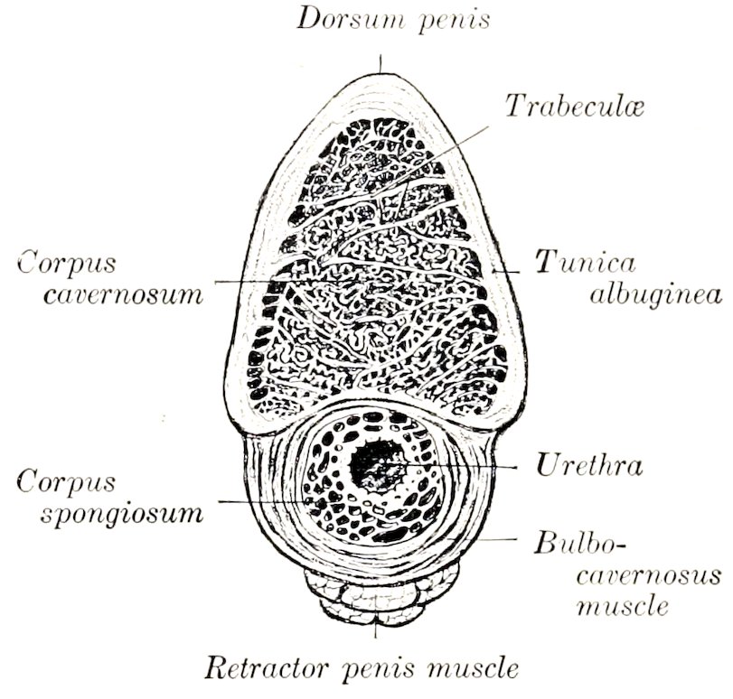
Fig. 396.—Cross-section of Body of Penis of Horse.
In man there are two distinct corpora cavernosa, separated by a complete septum penis, except in the middle part of the organ, where the septum is composed of vertical trabeculæ, between which are slit-like intervals; through the latter the blood-spaces of the two corpora cavernosa communicate. In the horse no distinct septum exists except near the root, but in the proximal and distal parts of the corpus cavernosum there are vertical trabeculæ.
The corpus spongiosum or corpus cavernosum urethræ forms a tube around the urethra, and is much enlarged at its free end to form the bulk of the glans penis; throughout the remainder of its course it is practically uniform in size, and does not form any very distinct bulb (Bulbus urethræ) at its origin, as in man and many animals. The glans has been described in part; it should be noted that the corpus spongiosum extends backward dorsally a distance of about four inches (ca. 10 cm.) on the middle process of the corpus cavernosum penis, forming the processus dorsalis glandis. The urethral process is covered by a thin layer of corpus spongiosum.
496In structure the corpus spongiosum has a general resemblance to the corpus cavernosum, but the trabeculæ are finer and more elastic. In the glans the trabeculæ are highly elastic, and the spaces are large and very extensible; the latter are continuous at the extremity of the processus dorsalis with large veins on the dorsum penis. There is a partial septum glandis. The skin covering the glans is thin, destitute of glands, and richly supplied with nerves and special nerve-endings.
Vessels and Nerves.—The penis is supplied with blood by three arteries, viz., the internal pudic, obturator, and external pudic. The veins form a rich plexus on the dorsum and sides of the penis, which is drained by the external pudic and obturator veins; from the root the blood is carried by the internal pudic veins. The lymphatics run with the veins and go to the inguinal and lumbar glands. The nerves are derived chiefly from the pudic nerves and the pelvic plexus of the sympathetic.
MUSCLES OF THE PENIS (Figs. 183, 451, 452, 456)
1. The ischio-cavernosus (or erector penis) is a short but strong paired muscle, which arises from the tuber ischii and the adjacent part of the sacro-sciatic ligament, and is inserted on the crus and adjacent part of the body of the penis. It is somewhat fusiform, incloses the crus as in a sheath, and is situated in a deep depression in the semimembranosus muscle. It pulls the penis against the pelvis, and assists in producing and maintaining erection by compressing the dorsal veins of the penis. Its blood-supply is derived from the obturator artery, and the nerve-supply from the pudic nerve.
2. The retractor penis is an unstriped muscle which arises on the ventral surface of the first two or three coccygeal vertebræ. It divides into two flat bands, half an inch or more (ca. 1.5 cm.) in width, which pass downward over the sides of the rectum to meet below the anus. Here there is a decussation of fibers, thus forming a sort of suspensory apparatus for the posterior part of the rectum and the anus. From the decussation the muscle passes for a short distance between superficial and deep layers of the bulbo-cavernosus, and then along the ventral surface of the penis, to which it is loosely attached. Near the glans penis it splits up into bundles which pass through the bulbo-cavernosus and are attached to the tunica albuginea. Below the anus the muscle is attached to the sphincter ani externus. On the penis the two muscles are intimately united to each other. Their action is to withdraw the penis into the sheath after erection or protrusion. The proximal part would draw the anus upward and forward and support it during defecation.
THE PREPUCE
The prepuce (Præputium), popularly called the “sheath,” is a double invagination of the skin which contains and covers the free or prescrotal portion of the penis when not erect. It consists of two parts, external and internal. The external part or sheath extends from the scrotum forward close to the umbilicus, where the external layer is reflected ventrally and laterally, forming the thick margin of the preputial orifice (Ostium præputiale); dorsally it is directly continuous with the integument of the abdominal wall. It is marked by a median raphé præputii, a continuation of the scrotal raphé. At the lower margin of the preputial orifice there are often in the stallion two papillæ, which are rudimentary teats or nipples. The internal layer passes backward from the preputial orifice a distance of about six to eight inches (ca. 15 to 20 cm.), lining the cavity of the external prepuce, and is then reflected forward until it approaches the orifice, where it is again reflected backward. It thus forms within the cavity of the sheath a secondary tubular invagination, the prepuce proper, in which the anterior part of 497the penis lies. This tubular cavity is closed behind by the reflection of the internal layer on to the penis to form the penile layer of the prepuce. Its orifice is surrounded by a thick margin, the preputial ring (Annulus præputialis), which is connected ventrally with the outer part of the sheath by the preputial frenum (Frenulum præputii).
The arrangement differs from that found in man in the fact that the inner part of the prepuce as described above is equivalent to the entire human prepuce. This part, the prepuce proper, is well seen on sagittal sections, and can be demonstrated by pulling the penis, inclosed in this prepuce, out of the cavity of the sheath; the arrangement of the free part of the penis and prepuce is then like that in man. (In paraphimosis the penis is strangulated by the preputial ring.)
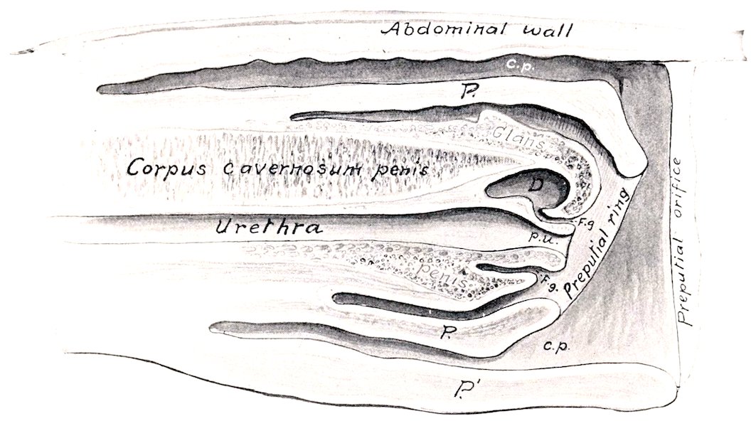
Fig. 397.—Sagittal Section of Prepuce and Part of Penis of Horse. P, Internal part of prepuce or prepuce proper; P′, external part of prepuce or sheath; c.p., preputial cavity; F.g., fossa glandis; D, diverticulum of fossa glandis; p.u., processus urethræ.
Structure.—The external skin of the prepuce resembles that of the scrotum. The inner layers of skin are hairless, variable in color, and often irregularly pigmented; they form irregular folds, and are supplied with numerous large sebaceous glands and coil glands, which reach their greatest size at the preputial ring. Beyond this the glands are absent, and the skin resembles a non-glandular mucous membrane. The secretion of the preputial glands (Glandulæ præputiales), together with desquamated epithelial cells, forms the fatty smegma præputii, which has a strong unpleasant odor, and often accumulates in considerable amount. Beneath the skin is a large amount of loose connective tissue, except over the glans penis, where the skin is closely attached to the tunic of the corpus spongiosum. The external part of the prepuce is strengthened by a layer of elastic tissue, derived from the abdominal tunic, and termed the suspensory ligament.
Vessels and Nerves.—The arteries are branches of the external pudic artery, and the veins go chiefly to the external pudic vein. The lymphatics go to the superficial inguinal and lumbar lymph glands. The nerves are derived from the pudic, ilio-hypogastric, and ilio-inguinal nerves.
THE MALE URETHRA
The urethra of the male (Urethra masculina) is the long mucous tube which extends from the bladder to the glans penis. It passes backward on the floor of 498the pelvis, turns around the ischial arch, forming an acute angle, and passes forward inclosed in the corpus spongiosum. It may, therefore, be divided into two segments, pelvic and extrapelvic.
The pelvic part (Pars pelvina) is four or five inches (ca. 10 to 12 cm.) long. At its origin it is not distinguishable from the neck of the bladder in size or structure; in fact no line of demarcation exists between the two. Behind the prostate the tube dilates to a potential width of two inches or more (ca. 5 to 6 cm.). Near the ischial arch, between the bulbo-urethral (Cowper’s) glands, it contracts again, forming the isthmus urethræ. It is related dorsally to the rectum and the prostate, ventrally to the internal obturator muscles, and laterally to the bulbo-urethral glands. It is inclosed, except at its origin, by the urethral (Wilson’s) muscle. Beneath this is a rich venous plexus, forming a sort of erectile tissue.[146]
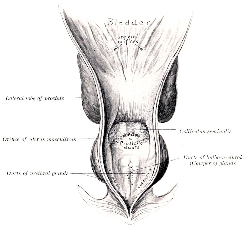
Fig. 398.—Pelvic Urethra and Posterior Part of Bladder of Horse Slit Ventrally and Laid Open. e.d., Ejaculatory ducts.
The extrapelvic part (Pars cavernosa) passes between the two crura of the penis and runs along the groove on the ventral surface of the corpus cavernosum penis, inclosed by the corpus spongiosum and the bulbo-cavernosus muscle. It passes through the glans penis and projects forward about an inch in the fossa glandis as a free tube, the processus urethræ; this part is covered by a delicate integument, under which there is a thin layer of erectile tissue.
499The lumen of the tube is largely obliterated in the inactive condition of the parts. When moderately distended its dimensions in a horse of medium size are as follows: At its origin the diameter is about half an inch (1 to 1.5 cm.). The pelvic dilatation at its widest part measures one and a half to two inches (ca. 3.5 to 5 cm.) transversely, and about an inch (2 to 3 cm.) vertically; it is elliptical in cross-section when fully distended. The isthmus at the ischial arch is a little smaller than the initial part. Beyond this the lumen is about one-half to three-fourths of an inch (ca. 1.5 cm.) in diameter, and is fairly uniform to the glans penis. Here there is a slight fusiform dilatation (Fossa navicularis), beyond which the tube contracts.
The opening from the bladder into the urethra is termed the internal urethral orifice (Orificium urethræ internum); it is closed except during urination. The terminal opening is the external urethral orifice (Orificium urethræ externum) or meatus urinarius. The two orifices of the ejaculatory ducts are situated dorsally on either side of the colliculus seminalis, about two inches (ca. 5 cm.) behind the internal urethral orifice; they are slit-like and close together. The small orifice of the uterus masculinus is placed centrally on the colliculus; it is inconstant. The orifices of the prostatic ducts are on two groups of small papillæ, placed lateral to the openings of the ejaculatory ducts. The ducts of the bulbo-urethral (Cowper’s) glands open on two lateral series of small papillæ, about an inch (ca. 2.5 cm.) further back and close to the median line. The small orifices of the urethral glands are situated laterally in the wide pelvic portion.
Structure.—The mucous membrane contains an unusually large amount of fine elastic fibers, and in its pelvic part there are alveolar glands (Glandulæ urethrales). The epithelium is at first like that of the bladder, then becomes cylindrical, and in the terminal part is stratified squamous. In the dorsal wall the membrane forms a median ridge, the urethral crest (Crista urethralis); this terminates about two inches from the internal urethral orifice in a rounded elevation, the colliculus seminalis, on the sides of which the ejaculatory ducts open.
At the origin of the urethra there is a layer of circular unstriped muscle-fibers outside of the mucous coat. Beyond this the latter is inclosed by a layer of erectile tissue, composed of plexuses of veins supported by trabeculæ of elastic and smooth muscular tissue. This is continued by the corpus spongiosum. Outside of the erectile tissue there is a continuation of the intrinsic unstriped muscular coat, consisting of external and internal longitudinal strata, with a layer of circular fibers between them.
Except at its origin and termination the urethra is provided with a continuous layer of striped muscle, placed outside of the erectile tissue. This is described as consisting of two parts or muscles.
(1) The urethral (or Wilson’s) muscle (M. urethralis) consists of dorsal and ventral layers of transverse fibers on the wide pelvic part of the urethra, and covers the bulbo-urethral (Cowper’s) glands. It is a compressor of the pelvic part of the urethra and the bulbo-urethral glands. By its forcible contraction it plays an important rôle in the ejaculation of the seminal fluid, and also in evacuating the last of the urine in micturition.[147]
(2) The bulbo-cavernosus muscle (or accelerator urinæ) is the continuation of the urethral muscle on the extrapelvic part of the urethra; it extends from the ischial arch to the glans penis. At the root of the penis it is the thickest, and forms a complete layer of circular fibers which inclose the corpus spongiosum and the urethra. Beyond this it diminishes very gradually in thickness, and consists of fibers which arise on a median ventral raphé and curve around the corpus spongiosum to end on the tunica albuginea of the corpus cavernosum. Its action is to empty the extrapelvic part of the urethra.
The ischio-urethral muscles (Fig. 452) are small bands which arise on the ischial arch and crura of the penis and pass forward to become lost on the ventral layer of the urethral muscle. They may assist in erection of the penis by exerting pressure on the dorsal veins.
500
MALE GENITAL ORGANS OF THE OX
The scrotum is situated somewhat further forward than in the horse. It is long, pendulous, and has a well-marked neck. The skin here is usually flesh-colored, but in some breeds is more or less pigmented; it is sparsely covered with short hairs. Just in front of it are four (sometimes only two) rudimentary teats.
The testicles are relatively larger than those of the horse, and have an elongated, oval outline. The long axis is vertical. The inner surface is somewhat flattened.
A testicle of an adult bull measures on the average about four or five inches (10 to 12 cm.) in length, exclusive of the epididymis; with the latter the length is about six inches (15 cm.). The width is about two and a half to three inches (ca. 6 to 8 cm.), and the antero-posterior diameter about the same. The weight is about nine or ten ounces (ca. 250 to 300 grams).
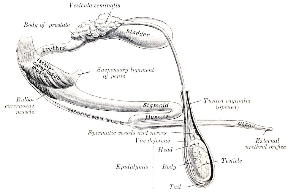
Fig. 399.—General View of Genital Organs of Bull.
The tunica albuginea is thin. The parenchyma is yellowish in color. The mediastinum testis descends from the anterior part of the upper extremity deeply into the gland as an axial strand about 5 mm. in thickness. The vasa efferentia leave at the upper end of the mediastinum.
The epididymis is very closely attached to the testicle. The head is long; it curves over the upper extremity and nearly half-way down the anterior border of the testicle. The body is very narrow, and lies along the outer part of the posterior border of the testicle, to which it is attached by a narrow peritoneal fold. The tail is large and is closely attached to the lower extremity of the testicle.
The spermatic cord and the tunica vaginalis are much longer than in the horse; the extra-inguinal part of the cord is about eight to ten inches (20 to 25 cm.) in length. The cremaster externus is well developed, and almost completely incloses the tunic to the nook of the scrotum; it does not extend so far down as in the horse, but is inserted about at the level of the upper pole of the testicle. The vaginal ring is 501relatively very small, and is about four inches (ca. 10 cm.) from the linea alba. The cremaster internus is feeble.
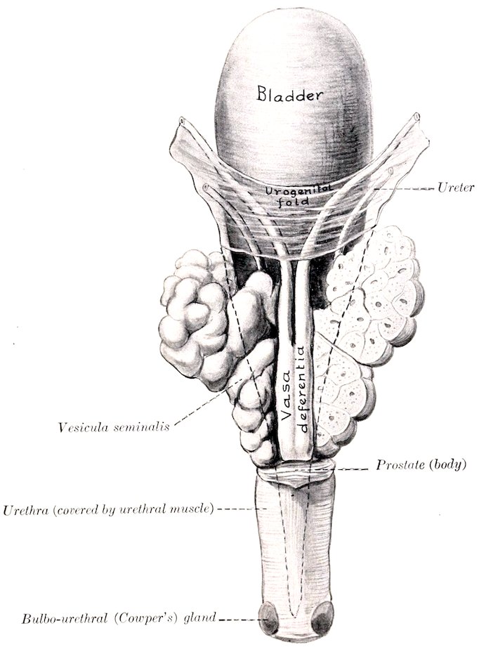
Fig. 400.—Internal Genital Organs of Bull; Dorsal View. The right vesicula is sectioned frontally. The dotted line indicates the backward extension of the peritoneum.
The spermatic cords emerge through the outer angles of the external inguinal rings, and curve downward and inward to the neck of the scrotum. The external rings are about four inches (ca. 10 cm.) long, and are two and a half to three inches (6 to 8 cm.) apart. In animals in good condition there is a large mass of fat above the neck of the scrotum, between the spermatic cords. The mesorchium extends to the bottom of the tunica vaginalis forming a narrow fold (ca. 1 cm. wide) which attaches the epididymis to the posterior part of the tunic.
The inguinal canal presents several special features in the bull. The internal ring is very long. Its anterior border, formed by the edge of the internal oblique, is decidedly concave and is tendinous in its inner part. The long axis of the external ring is directed outward, forward, and downward. The spermatic cord lies in its outer part, the external pudic vessels in its middle. A muscular band about an inch wide, detached from the internal oblique muscle, crosses the outer side of the vaginal ring.
The vas deferens is small in caliber. The urogenital fold is narrow, so that the vasa are closer together in it than in the horse. On reaching the posterior part of the bladder they are in apposition for a distance of about four inches 502(10 cm.), flanked and overlapped by the vesiculæ seminales. They form ampullæ about four or five inches (10 to 12 cm.) long and half an inch (1.2 to 1.5 cm.) wide, the mucous lining of which is plicated. They then pass under the body of the prostate and unite with the ducts of the vesiculæ seminales to form the ejaculatory ducts. The latter have slit-like openings on either side of the colliculus seminalis.
The vesiculæ seminales are not bladder-like sacs, as in the horse, but are compact glandular organs with a lobulated surface. In the adult they measure about four or five inches (ca. 10 to 12 cm.) in length, two inches (5 cm.) in width in their largest part, and an inch or more (ca. 3 cm.) in thickness. The dorsal surface of each faces upward and inward and is partially covered with peritoneum. The ventral surface faces in the opposite direction and is non-peritoneal. Each may be regarded as consisting of a very thick-walled, sacculated tube, bent on itself in a tortuous manner. This tube, if straightened out, would be about ten inches (25 cm.) in length. They are commonly unsymmetrical in size and shape. Short branches are often given off from the chief tube. The excretory duct or neck joins the outer side of the vas deferens at an acute angle to form the ejaculatory duct.
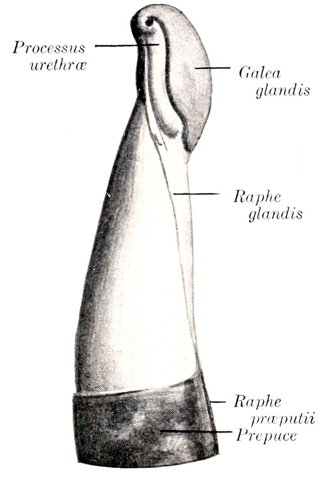
Fig. 401.—Anterior Extremity of Penis of Bull, Left View. (After Böhm.)
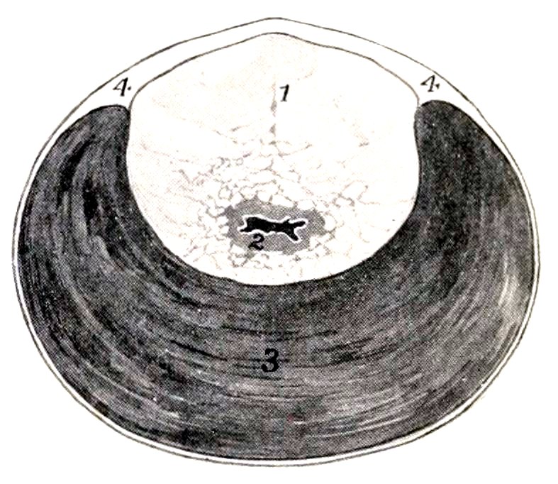
Fig. 402.—Cross-section of Pelvic Urethra of Bull. 1, Prostate gland (pars disseminata); 2, urethra; 3, urethral muscle; 4, aponeurosis. The lumen of the urethra is black.
The structure of the vesicula is masked by a thick capsule of fibrous tissue and unstriped muscle, which maintains it in its bent condition and also sends trabeculæ between the alveoli. There is a central canal into which the secretion formed in the alveoli passes. The cavities (central canal and alveoli) are lined with columnar epithelium. On account of this structure the term glandulæ vesiculares is preferred by some anatomists.
The prostate is pale yellow in color, and consists of two parts. The body (Corpus prostatæ) is a mass which stretches across the dorsal surface of the neck of the bladder and the origin of the urethra. It measures about an inch and a half (ca. 3.5 to 4 cm.) transversely, and about half an inch (ca. 1 to 1.5 cm.) in width and thickness. The pars disseminata surrounds the pelvic part of the urethra; dorsally it forms a layer about half an inch (ca. 10 to 12 mm.) thick, but ventrally it is quite thin. It is concealed by the urethral (Wilson’s) muscle; hence it often escapes notice, but is very evident on cross-section (fig. 402). The prostatic ducts open into the urethra in rows between two folds of the mucous membrane which proceed backward from the colliculus seminalis.
503The uterus masculinus appears to be absent frequently. Ellenberger states that it opens between the ejaculatory ducts, while Martin says that it usually has two orifices at the colliculus, but that it often unites with the vasa deferentia.
The bulbo-urethral (Cowper’s) glands are somewhat smaller than in the stallion. They are liable to escape notice, since they are covered by a thick layer of dense fibrous tissue and also partially by the bulbo-cavernosus muscle. Each has a single duct which opens into the urethra under cover of a fold of the mucous membrane.
The penis is cylindrical, and is longer and of very much smaller diameter than in the horse. Just behind the scrotum it forms an S-shaped curve, the sigmoid flexure; thus about one foot is folded up when the penis is fully retracted. The flexure is effaced during erection. The glans penis is about three inches (ca. 8 cm.) in length. It is flattened dorso-ventrally, and its extremity is pointed and twisted. The external urethral orifice is situated at the end of a groove formed by this twist; it is only large enough to admit a probe of medium size. Even in the non-erect state the penis is remarkably dense and firm. The tunica albuginea is very thick, and incloses the urethra. In the first part, as far as the first curve, there is a thick median septum penis. Beyond this there is a central axial rod of dense fibrous tissue from which numerous strong trabeculæ radiate. The erectile tissue is small in amount, so that the organ undergoes very little enlargement in erection.
The length of the penis in the adult is about three feet (ca. 90 cm.). The crura are flattened laterally. The suspensory ligaments are attached to the ventral ridge on the symphysis. The body is somewhat flattened dorso-ventrally beyond the first curve. The extremity of the glans is unsymmetrical, the urethral orifice being situated ventro-laterally. The glans contains little erectile tissue, and hence undergoes little enlargement in erection.
The bulbo-cavernosus muscle presents several remarkable features. It is for the greater part an inch or more (ca. 3 cm.) in thickness, but its length is only six to eight inches (15 to 20 cm.). It is covered by a strong aponeurosis, and is divided by a median raphé into two lateral halves except at its origin. It diminishes in size from behind forward, and its anterior extremity is pointed.
The ischio-cavernosus muscle resembles in general that of the horse, but is flattened laterally. It is covered by a close aponeurosis.
The retractor penis muscle resembles that of the horse in origin. Its two parts are about an inch (ca. 2 to 5 cm.) apart on the root of the penis, where they lie in a groove on either side of the bulbo-cavernosus. They then come close together and pass on either side of the ventral curve of the sigmoid flexure. Further forward they lie along the ventral aspect of the penis, and end about five or six inches (ca. 12 to 15 cm.) behind the glans.
The prepuce is very long and narrow. Its orifice is about two inches (ca. 5 cm.) behind the umbilicus; it is only large enough to admit a finger readily, and is surrounded by long hairs. The cavity of the sheath is about fifteen inches (35 to 40 cm.) long, and a little over an inch (ca. 3 cm.) in diameter. The lining membrane forms longitudinal folds; it is covered with squamous stratified epithelium, and has coiled tubular glands. The penile layer is glandless, and is reddish in color. It presents lymph nodules in its posterior part.
There are two pairs of preputial muscles which are derivatives of the panniculus carnosus. The anterior pair, the protractors of the prepuce (Mm. præputiales craniales), are two flat bands, two inches or more (5 to 6 cm.) in width, which arise close together in the xiphoid region, about eight inches (20 cm.) in front of the preputial orifice. Traced backward they diverge, leaving the umbilicus and an area about one and a half inches wide free, and then unite behind the preputial orifice. They draw the prepuce forward. The posterior pair, the retractors of 504the prepuce (Mm. præputiales caudales), arise in the inguinal region and converge on the anterior part of the prepuce. They draw the prepuce backward.
These muscles are subject to a good deal of variation. The retractor may be absent. Many fibers come in from the panniculus on either side, dip under the protractor, and are inserted into the skin just behind the preputial opening. The homologue of the protractor is present in the cow.
The urethra in its pelvic part is about five inches (12 cm.) long, and is of relatively small and uniform caliber. The urethral (Wilson’s) muscle incloses the tube ventrally and laterally; it is very thick, crescentic in cross-section, and is covered by an aponeurosis. Dorsally, the aponeurosis is thick and the muscle absent. Inside of these the pars disseminata of the prostate surrounds the urethra as far back as the bulbo-urethral glands. The colliculus seminalis is short, but prominent; from it the urethral crest extends forward to the trigonum vesicæ. The extrapelvic part has an enlargement, the bulb, at its origin; it then gradually diminishes in diameter, and is relatively very small at its termination. Other features have been mentioned in preceding paragraphs.
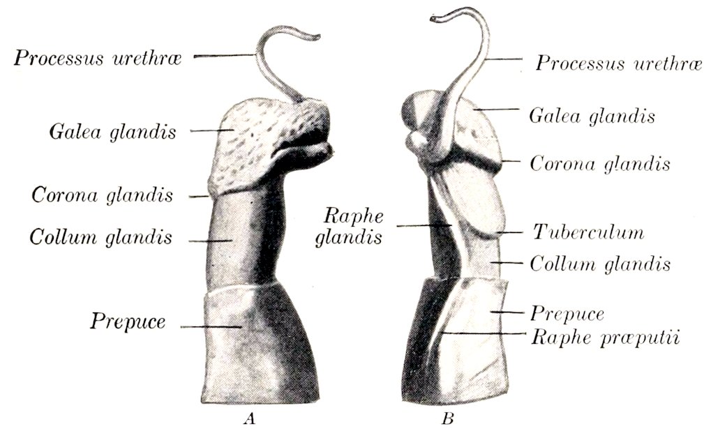
Fig. 403.—Anterior Part of Penis of Ram. A, Right side; B, left side. (After Böhm.)
In the ram the urethra lies in a groove on the ventral surface of the corpus cavernosum. Its terminal part projects commonly 3 to 4 cm. beyond the glans penis, forming a twisted processus urethræ.
MALE GENITAL ORGANS OF THE PIG
The scrotum is situated a short distance from the anus, and is not so sharply defined from the surrounding parts as in the other animals.
The testicles are very large and are soft in texture. Their long axis is almost vertical, the free border being posterior and the tail of the epididymis dorsal. The mediastinum testis is well developed. The epididymis is closely attached to the testicle and the tunica vaginalis; the tail is specially large.
The spermatic cord is necessarily very long (20 to 25 cm. in a boar of medium size). The vas deferens in its testicular part is flexuous, and is closely attached to the tunica vaginalis; it forms no ampullæ. The cremaster externus is well developed, and extends to about the middle of the scrotal part of the tunic.
505The vesiculæ seminales are exceedingly large, and extend into the abdominal cavity. They are three-sided pyramidal masses, are in apposition with each other medially, and cover the posterior part of the bladder and the ureters, the vasa deferentia, the body of the prostate, and the anterior part of the urethra. They are pale pink in color, distinctly lobulated and glandular in structure, and contain large alveoli, but no central cavity. There are two excretory ducts which open into the urethra close to the vasa deferentia; the two ducts frequently unite.
In the adult boar the vesiculæ are about five to six inches (ca. 12 to 15 cm.) long, two to three inches (ca. 5 to 8 cm.) wide, and one and a half to two inches (ca. 4 to 5 cm.) thick; they weigh about 6 to 8 ounces each.
The prostate consists of two parts, as in the ox. The body is about an inch (2.5 cm.) wide, and embraces the neck of the bladder and the urethra at their junction dorsally and laterally. It is concealed by the vesiculæ seminales. The pars disseminata forms a layer which surrounds the pelvic part of the urethra, and is covered by the urethral muscle.
The uterus masculinus is small.
The bulbo-urethral (Cowper’s) glands are very large and dense. They are somewhat cylindrical, and lie on either side of and upon the posterior two-thirds of the pelvic urethra. In a large boar they are about five inches (ca. 12 cm.) in length and an inch or more (ca. 2.5 to 3 cm.) in diameter. They are partially covered with a layer of striated muscle (M. ischio-glandularis), and have a lobulated surface. Each gland has a large excretory duct which leaves at the posterior extremity, perforates the dorsal wall of the urethra at the ischial arch, and opens in a cul-de-sac covered by a fold of mucous membrane.
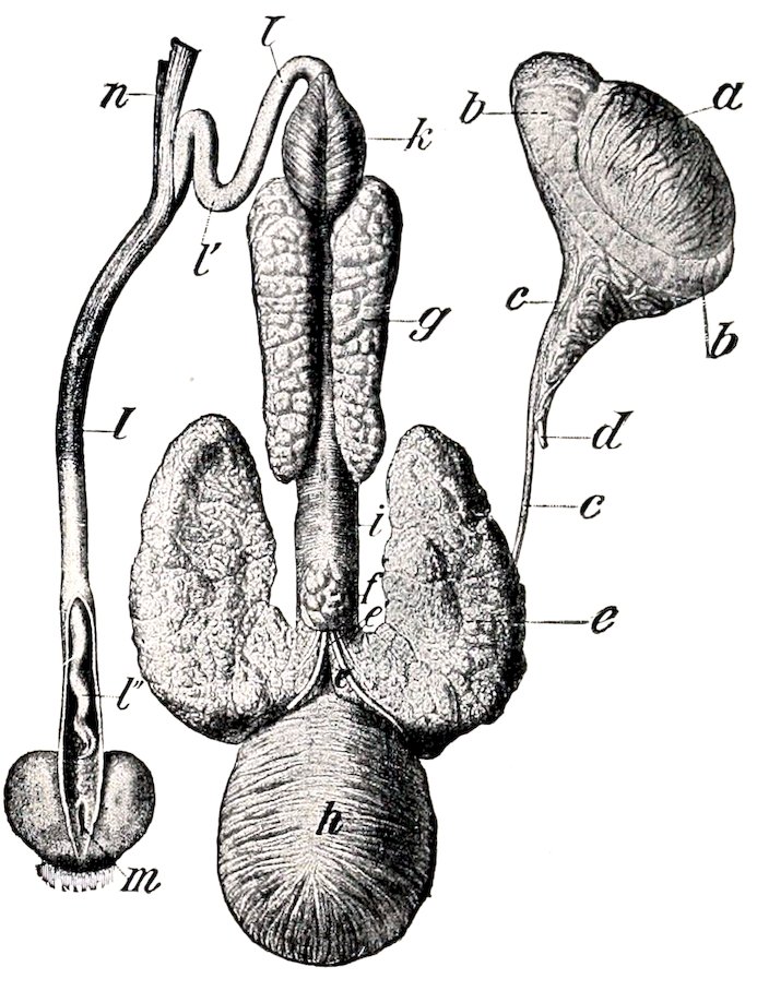
Fig. 404.—Genital Organs of Boar. a, Testicle; b, epididymis; c, vas deferens; d, spermatic artery; e, vesicula seminalis reflected to show the body of the prostate, f; e′, excretory ducts of vesiculæ; g, bulbo-urethral (Cowper’s) gland; h, urinary bladder; i, urethral muscle; k, bulbo-cavernosus muscle; l, penis; l′, sigmoid flexure of penis; l″, spiral anterior part of penis, exposed by slitting open prepuce; m, orifice of preputial pouch; n, retractor penis muscle. (Ellenberger-Baum, Anat. d. Haustiere.)
It is to be noted that these accessory glands are very small in animals which have been castrated early.
The penis resembles in general that of the ox. The sigmoid flexure is, however, prescrotal. The anterior portion has no glans, and is spirally twisted, especially in erection. The urethral orifice is slit-like and is situated ventro-laterally, close to the pointed extremity. The penis in the adult boar measures about 18 to 20 inches (ca. 45 to 50 cm.) in length. Its muscles resemble those of the bull; the bulbo-cavernosus is very strong but short.
506The prepuce has a narrow orifice, around which there are a number of stiff hairs. The cavity is partially divided by a circular fold into a posterior narrow portion and a much wider anterior portion. The lining membrane of the posterior part is papillated, and is in close contact with the penis. In the dorsal wall of the wide part there is a circular opening which leads into a cul-de-sac, the preputial diverticulum. This pouch is ovoid in form (when distended), and varies greatly in size in different subjects. It extends for the most part backward over the narrow part of the prepuce. Its cavity is partially divided by a narrow median septum. It contains usually decomposing urine and macerated epithelium, which have a characteristic and very unpleasant odor. Concrements have been found in it.
Oehmke found that a cast of the pouch in a Yorkshire boar weighing about 500 pounds measured 9 cm. in length, 12½ cm. in breadth, and 6 cm. in height. The opening into the prepuce will admit two fingers in the adult, but is ordinarily closed by folds of the lining membrane. The sac is much smaller in animals which were castrated young, and the opening is vertical and further back; in them it is often empty or contains only a little clear urine. The pouch is covered by a layer of striped muscle which is mainly derived from the homologue of the protractor of the prepuce of ruminants.
The urethra has a very long pelvic portion (ca. 15 to 18 cm. long in the adult); it is inclosed (with the pars disseminata of the prostate) by a thick urethral muscle. There is a distinct bulb at the root of the penis. The penile part is of small caliber, and is surrounded by erectile tissue which, however, does not extend to the extremity of the penis.
MALE GENITAL ORGANS OF THE DOG
The scrotum is situated about half-way between the inguinal region and the anus. The skin covering it is pigmented and is covered sparsely with fine hairs. The raphé is not very distinct.
The testicles are relatively small, and have a round-oval form. The long axis is oblique, and is directed upward and backward. The mediastinum testis is central and is well developed. The epididymis is large, and is closely attached along the outer part of the dorsal border of the testicle.
The spermatic cord and the tunica vaginalis are long; they cross the side of the penis very obliquely. The upper end of the tunic is sometimes closed, so that there is then no vaginal ring. The vasa deferentia have very narrow ampullæ.
The vesiculæ seminales are absent; hence there are no ejaculatory ducts.
The prostate is relatively large; it is yellowish in color, dense in structure, and lies at the anterior border of the pubis. It is globular, and surrounds the neck of the bladder and the urethra at their junction. A faint median furrow indicates a division into two lateral lobes. The capsule and stroma contain a large amount of unstriped muscle. The ducts are numerous. Lobules of prostatic tissue (pars disseminata) are also found on the urethra further back. The gland is subject to much variation in size, and is often enlarged, especially in old subjects.
The uterus masculinus is a very small saccule (Utriculus prostaticus), often difficult to find.
The bulbo-urethral (Cowper’s) glands are absent.
The penis presents several special features. In its posterior part there are two distinct corpora cavernosa, separated by a median septum penis. In its anterior part there is a bone, the os penis, which in large dogs reaches a length of four inches (ca. 10 cm.) or more. It is regarded as a continuation forward of the corpus cavernosum. Ventrally it is grooved for the urethra; dorsally it is convex, and anteriorly it becomes smaller and has a curved fibrous prolongation. The glans penis is very long, extending over the entire length of the os penis; posteriorly it 507forms a rounded enlargement, the bulbus glandis; anteriorly it is cylindrical, smaller, and pointed at the extremity. Just in front of the bulbus glandis there is a thick circular layer of glands, forming the annulus glandularis (Schmaltz). The two dorsal veins arise from the bulbus glandis, pass backward on the dorsum penis, and unite at the ischial arch. A small muscle (M. compressor venæ dorsalis penis) arises from the tuber ischii on either side; the two converge on the dorsum penis near the bulbus glandis. They compress the dorsal veins, and may also tend to elevate the penis and thus assist in copulation. The other muscles offer no features worthy of special description.
The prepuce forms a complete sheath around the anterior part of the penis. The outer layer is ordinary integument. The inner layer has the character of a glandular mucous membrane; it is thin and reddish in color. The penile layer is glandless. In the fundus of the præputial cavity there are numerous lymph follicles. The protractor muscles arise in the xiphoid region and decussate posteriorly around the extremity of the penis.
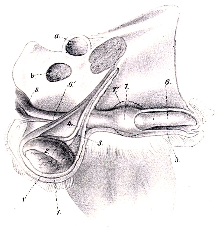
Fig. 405.—External Genital Organs of Male Dog, Ventro-lateral View of Preparation. 1, Scrotum; 1′, tunica vaginalis; 2, epididymis; 3, vascular part of spermatic cord; 4, vas deferens; 5, prepuce; 6, glans penis; 6′, corpus cavernosum penis; 7, bulbus glandis; 7′, contour of bulbus in erection; 8, ischio-cavernosus muscle; a, acetabulum; b, obturator foramen. (After Ellenberger, in Leisering’s Atlas.)
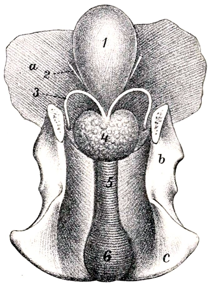
Fig. 406.—Internal Genital Organs of Male Dog. 1, Urinary bladder; 2, ureter; 3, vas deferens; 4, prostate; 5, urethral muscle; 6, bulb of urethra; a, abdominal wall; b, ilium; c, ischium. (After Ellenberger, in Leisering’s Atlas.)
The pelvic part of the urethra is relatively long. Its first part is inclosed in the prostate.[148] At the ischial arch the urethra has a well-developed bilobed bulb, formed by an enlargement here of the corpus spongiosum. It is covered by the strong but short bulbo-cavernosus muscle. The other erectile bodies have been described.
The urethral (Wilson’s) muscle is very strong; it encircles the urethra and has a median raphé dorsally.
508
THE FEMALE GENITAL ORGANS
The female genital organs (Organa genitalia feminina) are: (1) The two ovaries, the essential reproductive glands, in which the ova are produced; (2) the uterine or Fallopian tubes, which convey the ova to the uterus; (3) the uterus, in which the ovum develops; (4) the vagina, a dilatable passage through which the fœtus is expelled from the uterus; (5) the vulva, the terminal segment of the genital tract, which serves also for the expulsion of the urine; (6) the mammary glands, which are glands of the skin, but are so closely associated functionally with the generative organs proper as to be described with them.
GENITAL ORGANS OF THE MARE
THE OVARIES
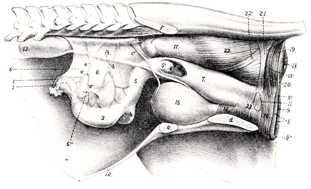
Fig. 407.—Lateral View of Genital Organs and Adjacent Structures of Mare. The removal of the other abdominal viscera has allowed the ovaries and uterus to sink down, whereas they are normally in contact with the roof of the cavity in the non-pregnant subject. 1, Left ovary; 2, Fallopian tube; 3, left cornu uteri; 4, right cornu uteri; 5, corpus uteri; 5′, portio vaginalis uteri, and 5″, os uteri, seen through window cut in vagina; 6, broad ligament of uterus; 6″, round ligament; 7, vagina; 8, labia vulvæ; 9, rima vulvæ; 9′ superior commissure, and 9′, inferior commissure of vulva; 10, constrictor vulvæ; 11, position of vestibular bulb; 12, ventral wall of abdomen; 13, left kidney; 14, left ureter; 15, urinary bladder; 16, urethra; 17, rectum; 18, anus; 19, 19′, unpaired and paired parts of sphincter ani externus; 20, retractor ani cut at disappearance under sphincter; 21, suspensory ligament of anus; 22, longitudinal muscular layer of rectum; 22′, recto-coccygeus; 23, constrictor vaginæ; a, utero-ovarian artery, with ovarian (a′) and uterine (a″) branches; b, uterine artery; c, umbilical artery; d, ischium; e, pubis; f, ilium. (After Ellenberger, in Leisering’s Atlas.)
The ovaries (Ovaria) of the mare are bean-shaped, and are much smaller than the testicles. Their size varies much in different subjects, and they are normally larger in young than in old animals; one ovary is often larger than the other. They are about three inches (ca. 7 to 8 cm.) long and an inch to an inch 509and a half (ca. 3 to 4 cm.) thick. The weight is about two and a half to three ounces (ca. 70 to 80 grams).
Each presents for description two surfaces, two borders, and two extremities or poles. The surfaces are termed internal and external (Facies medialis, lateralis); they are both smooth and rounded.[149] The attached or mesovarial border (Margo mesovaricus) is convex. It is inclosed in a part of the broad ligament termed the mesovarium; the vessels and nerves reach the gland at this border. The free border (Margo liber) is marked by a notch which leads into a depression, the ovulation fossa. The tubal or anterior extremity (Extremitas tubaria) is rounded, and is related to the fimbriated end of the uterine or Fallopian tube. The uterine or posterior extremity (Extremitas uterina) is also round, and is connected with the horn of the uterus by the ovarian ligament.
The ovaries are situated in the sublumbar region of the abdominal cavity, about a handbreadth behind the corresponding kidney. They are in contact with the lumbar wall of the abdomen, and are commonly about two or three inches (ca. 5 to 7.5 cm.) from the median plane. The distance from the ovaries to the vulvar orifice is about twenty inches (ca. 50 cm.) in a mare of medium size. The attached (mesovarial) border is commonly directed toward the median plane, the free border laterally and somewhat ventrally.
The position of the ovaries is not constant, but is subject to less variation than might be expected from their mode of attachment. The normal variations in the non-pregnant state are chiefly in a transverse direction, and do not usually exceed two or three inches (ca. 5 to 7 cm.). It is not at all rare to find the ovary deflected medially, so that the external surface faces ventrally. In the longitudinal direction little variation appears to occur. The right ovary is usually about four or five inches (ca. 10 to 12 cm.), and the left one about three or four inches (ca. 8 to 10 cm.), behind the corresponding kidney. Thus they lie about in a transverse plane tangent to the external angles of the ilia, i. e., through the fifth lumbar vertebra. They may be about two inches (ca. 5 cm.) from the extremity of the corresponding cornu of the uterus or in contact with it. The right ovary commonly lies a little (ca. 2.5 cm.) further forward than the left one. Except during pregnancy, they are normally in contact with the lumbar abdominal wall, and do not hang down among the adjacent viscera.
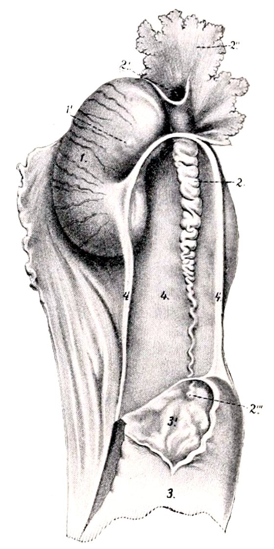
Fig. 408.—Right Ovary of Mare, with Adjacent Structures. The extremity of the cornu uteri is opened. 1, Ovary; 2, Fallopian tube, with its ostium abdominale (2′), fimbriæ (2″), and ostium uterinum (2‴); 3, cornu uteri, with its mucous lining (3′); 4, ovarian ligament; 4′, mesosalpinx. (After Ellenberger, in Leisering’s Atlas, reduced.)
The ovary is attached to the sublumbar region by the anterior part of the broad ligament; this part of the ligament, the mesovarium, is about three or four inches (ca. 8 to 10 cm.) wide, measured from the ovary directly to the parietal attachment. The uterine or posterior extremity of the ovary, is connected with the extremity of the cornu of the uterus by the ovarian ligament (Ligamentum ovarii proprium); this is a band of unstriped muscle inclosed between the layers of the broad ligament.
Structure.—The greater part of the surface of the ovary has a covering of peritoneum. The peritoneal investment is absent at the attached border where 510the vessels and nerves enter; this area is termed the hilus. The ovulation fossa is covered by a layer of columnar cells, a remnant of the primitive germinal epithelium. The stroma of the ovary (Stroma ovarii) is a network of connective tissue and unstriped muscular fibers. In the deeper or medullary portion of the stroma (Zona vasculosa) the meshes are largely occupied by the numerous blood-vessels. In the peripheral or cortical portion (Zona parenchymatosa) are numerous ova (Ovula) in various stages of development. The immature ova are surrounded by follicle cells; those more advanced in development are inclosed also by a condensation of the stroma termed the theca folliculi; within the theca is a quantity of fluid, the liquor folliculi. These constitute the Graafian follicles (Folliculi oöphori), which enlarge as they mature, becoming visible to the naked eye as vesicles with a diameter of a centimeter or more. When fully developed, the follicles are superficially situated, and often project slightly from the surface of the ovary. At intervals follicles rupture and their contents escape. This process, which sets free the ovum, is termed ovulation; it takes place in the mare only at the ovulation fossa, and occurs during the periods of œstrum.
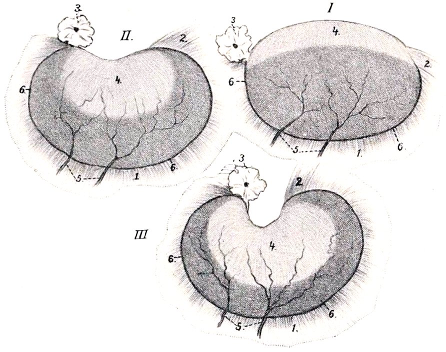
Fig. 409.—Schematic Representation of Changes in Ovary of Mare from Fœtal to Adult State. (After Born.) The changes affect chiefly the free border (upper in figure) and the extent and form of the area covered by germinal epithelium (4). 1, Peritoneum (broad ligament); 2, ligament of ovary; 3, fimbria of Fallopian tube; 4, germinal epithelium; 5, vessels, which reach the ovary at the attached border (6). The latter represents the hilus, while the deep depression at the free border is the ovulation fossa. (Ellenberger, in Leisering’s Atlas.)
After rupture of a follicle its cavity is partly occupied by a blood-clot, constituting what may be termed a corpus rubrum. By proliferation and enlargement and fatty changes, the follicle cells are transformed into lutein cells, forming a yellow mass known as a corpus luteum. If impregnation takes place, the accompanying increase in vascularity of the organs may cause the corpus luteum to reach a large size; if impregnation does not occur, it is much smaller, and is sooner replaced by scar tissue, the corpus albicans.
In the young foal the ovaries are large and oval in form. The free border is convex and is covered by germinal epithelium, which extends over part of the surfaces also. This area is distinguishable by its dull gray appearance from the peritoneal surface, which has the usual smooth glistening character. As growth proceeds the ovary gradually becomes bent until it assumes its definitive curved shape. The germinal epithelium is then limited to the ovulation fossa. The ovary migrates somewhat during development from its primitive position, which is the same as that of the testicle.
In old animals the ovaries commonly consist largely of fibrous tissue, in which there are often cysts of various sizes. The ova, present in enormous number at birth, have then been extruded, or destroyed by phagocytic action or degeneration.
Vessels and Nerves.—The arteries of the ovary are derived from the ovarian artery. The artery is relatively large and is flexuous; it reaches the attached border of the ovary by passing between the layers of the mesovarium. The veins are large and numerous. They form a plexus somewhat like that of the spermatic cord. The lymph vessels pass to the lumbar glands. The nerves are derived from the sympathetic system through the renal and aortic plexuses. They accompany the arterial branches.
511
THE UTERINE OR FALLOPIAN TUBES
The uterine or Fallopian tubes (Tubæ uterinæ) act as excretory ducts of the ovaries, since they convey the ova from the reproductive glands to the uterus. They are not, however, in direct continuity with the glands, but rather partly in contiguity with, and partly attached to, them.[150] They are two flexuous tubes, about ten to twelve inches (ca. 25 to 30 cm.) long, which extend from the extremities of the uterine cornua to the ovaries. Each is inclosed in a peritoneal fold, derived from the external layer of the broad ligament, and termed the mesosalpinx. This largely covers the outer aspect of the ovary, and forms with it and the broad ligament a pouch called the bursa ovarica. The uterine extremity of the tube communicates with the cavity of the cornu by a minute orifice, the ostium uterinum. The ovarian or fimbriated extremity is expanded into a funnel-shaped infundibulum. The margin of the latter is slit into irregular processes or fimbriæ, some of which, the fimbriæ ovaricæ, are attached in the ovulation fossa. In the center of the fimbriated extremity is a small opening, the ostium abdominale, by which the tube communicates with the peritoneal cavity. The tube appears normally to be applied to the ovary, so that the extruded ova pass into it and are conveyed to the uterus.[151]
Pedunculated cysts, the hydatids of Morgagni, are often found on one or more of the fimbriæ. In the mesosalpinx are blind flexuous tubules, which constitute the paroöphoron, a remnant of the Wolffian body. They are most evident in the young adult, and tend to disappear with increasing age. Not uncommonly they give rise to cysts.
Structure.—The tube is covered externally by a serous coat formed by the mesosalpinx. The serous membrane is continued on the fimbriæ, and meets the mucous lining on them. The fibrous coat or adventitia is continuous with the fibrous lamina of the broad ligament. The muscular coat consists chiefly of circular fibers, although longitudinal fibers occur externally. The mucous coat is thin and is much plicated. The folds (Plicæ tubariæ) are chiefly longitudinal, but in the wide part of the tube (Ampullæ tubæ) they are very complex, so that on cross-sections the spaces between the folds may be mistaken for branched tubular glands. The folds are continued on to the fimbriæ. The epithelium is a single layer of columnar ciliated cells, the cilia producing a current directed toward the uterus. At the fimbriated extremity this epithelium passes gradually into the squamous type of the serous coat.
Vessels and Nerves.—The arteries are derived from the utero-ovarian artery. The veins are satellites of the arteries. The lymphatics pass with the ovarian vessels to the lumbar glands. The nerves have a similar origin to those of the ovary.
THE UTERUS
The uterus is a hollow muscular organ, which receives the Fallopian tubes anteriorly and opens into the vagina posteriorly. It is situated chiefly in the abdominal cavity, but extends a short distance into the pelvic cavity. It is attached to the lumbar wall of the abdomen and the lateral walls of the pelvic cavity by two folds of peritoneum termed the broad ligaments. It consists of two cornua or horns, the body, and the neck.
The cornua or horns of the uterus (Cornua uteri) are situated entirely in the abdomen. They appear to vary considerably in position; commonly they 512are pressed up against the sublumbar muscles by the intestine (left portions of large colon, small colon, and small intestine). They are cylindrical, and are about ten inches (ca. 25 cm.) in length. The anterior extremity of each forms a blunt point which receives the Fallopian tube. Posteriorly they increase somewhat in caliber, converge, and unite with the body. The dorsal (or attached) border is slightly concave and is attached to the broad ligament. The ventral border is convex and free.
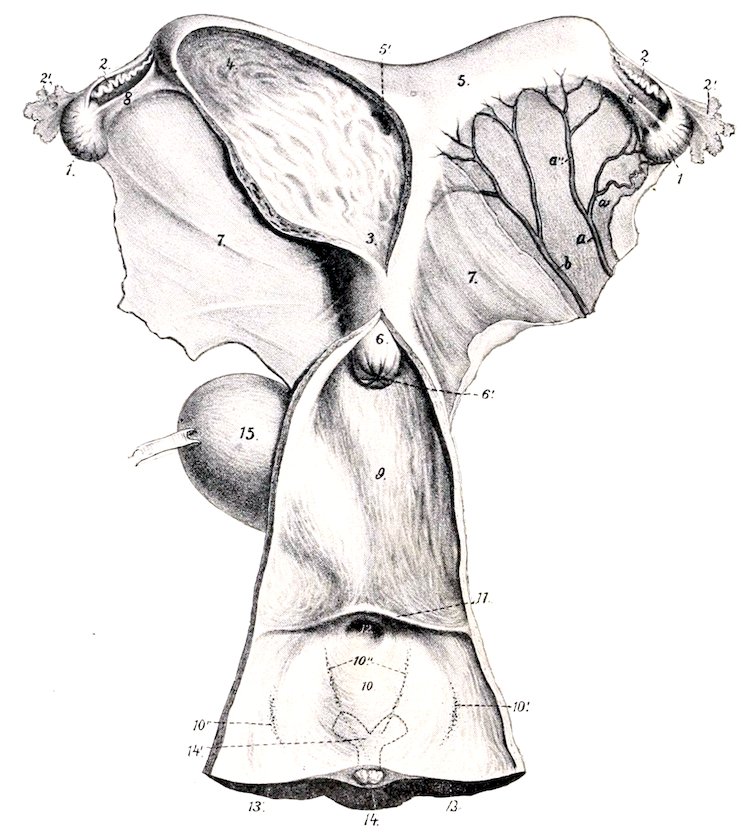
Fig. 410.—Genital Organs of Mare, Dorsal View. The left cornu and adjacent part of the body of the uterus and the vagina and vulva are opened up. 1, Ovary; 2, Fallopian tube, with 2′, its fimbriated end; 3, cavity of corpus uteri; 4, cavity of left cornu; 5, right cornu, with 5′, its communication with the body; 6, portio vaginalis uteri; 6′, os uteri; 7, broad ligament of uterus; 8, ligament of ovary; 9, vagina; 10, vulva; 10′, glandulæ vestibulares majores; 10″, glandulæ vestibulares minores; 11, transverse fold; 12, external urethral opening; 13, 13′, labia vulvæ; 14, glans clitoridis; 14′, junction of crura to form corpus clitoridis indicated by dotted lines; 15, urinary bladder; a, utero-ovarian artery with ovarian (a′) and uterine (a″) branches; b, uterine artery. (After Ellenberger, in Leisering’s Atlas.)
When a soft uterus is distended, the cornua are strongly curved, but this does not represent the natural form. When fixed in situ the horns are either slightly curved or nearly straight. The two horns are commonly unsymmetrical in length and diameter in mares which have borne young.
The body of the uterus (Corpus uteri) is situated partly in the abdomen, partly in the pelvic cavity. It is cylindrical, but considerably flattened dorso-ventrally, so that in cross-section it is elliptical. Its average length is about six inches (15 cm.), and its diameter, when moderately distended, about four inches (10 cm.). Its dorsal surface (Facies rectalis) is related to the small colon and rectum. 513Its ventral surface (Facies vesicalis) is in contact with the bladder, and has inconstant relations with various parts of the intestine. The term fundus uteri is applied to the anterior wide part from which the cornua diverge.
The position of the body of the uterus is variable, especially in regard to its anterior part. It is often pressed up against the rectum, and may be deflected to either side—most frequently to the left—by the pelvic flexure of the colon or coils of the small colon.
The cervix or neck of the uterus (Cervix uteri) is the constricted posterior part which joins the vagina. Part of it (Portio vaginalis uteri) projects into the cavity of the vagina; it is therefore not visible externally, but may be felt through the vaginal wall. It is about two inches (5 cm.) in length, and an inch and a half (3.5 to 4 cm.) in diameter.
Attachments.—The body and cornua are attached to the abdominal and pelvic walls by two extensive peritoneal folds, the broad ligaments (Ligamenta lata). These extend on either side from the sublumbar region and the lateral pelvic walls to the dorsal border of the cornua and the lateral margins of the body of the uterus. They contain the vessels and nerves of the uterus and ovaries, connective tissue, and unstriped muscular fibers. The ureters are situated along their parietal margins. The outer layer of each forms a fold, the round ligament of the uterus (Ligamentum teres uteri), which blends with the parietal peritoneum over the internal inguinal ring; its anterior extremity is situated above the extremity of the cornu and is rounded and free. It contains muscular tissue, vessels, and nerves, and is the homologue of the gubernaculum testis. The anterior part of the neck is continuous with the vagina, and thus has a more fixed position than the rest of the organ.
The cavity of the uterus (Cavum uteri) is largely obliterated in the non-pregnant state by the contraction of the wall and by folds of the mucous lining. At the extremity of each cornu it communicates by a minute opening with the Fallopian tube. The cavity of the neck is termed the cervical canal (Canalis cervicis); it is closed ordinarily by mucous folds and a plug of mucus. It opens into the vagina by the os uteri externum, and into the body by the os uteri internum.
Structure.—The wall of the uterus consists of three coats—an external serous, a middle muscular, and an internal mucous. The serous coat (Perimetrium) is, for the most part, closely adherent to the muscular coat. It is continuous with the broad ligaments. The muscular coat consists essentially of two layers, a thin external stratum of longitudinal fibers, and a thick layer of circular fibers. Between these is a very vascular layer (Stratum vasculare) of connective tissue with circular and oblique muscular fibers in it. The circular coat is very thick in the neck, where it forms a sphincter half an inch or more (ca. 1.5 cm.) in thickness. The mucous membrane rests directly on the muscular coat. It is thick, and is covered by columnar epithelium. It contains numerous long tubular uterine glands (Glandulæ uterinæ). It is brownish-red in color, except in the neck, where it is pale.
Vessels and Nerves.—The chief arteries are the uterine and the uterine branch of the utero-ovarian, which have a flexuous course in the broad ligaments; there is also a branch from the internal pudic artery. The veins form pampiniform plexuses and accompany the arteries. The lymphatics are numerous and go to the internal iliac and lumbar glands. The nerves are derived from the sympathetic through the uterine and pelvic plexuses.
The foregoing statements refer to the non-gravid uterus. In the pregnant state it undergoes important changes in size, position, and structure. The increase in size affects chiefly the gravid cornu (except in the case of twins) and the body. The cornu attains a length of about two and a half to three feet (ca. 80 to 90 cm.), and a corresponding diameter; in this process it extends much beyond the ovary, and the broad ligament. The gravid uterus is entirely abdominal in position, and extends along the ventral wall, chiefly to the left of the median plane. It weighs about nine pounds (ca. 4 kg.), according to Ellenberger. The broad ligaments increase greatly 514in size and contain more muscular tissue. The vessels are greatly enlarged and form new branches. The muscular coat, in spite of the increase in size and number of the fibers, is somewhat thinner, except in the neck. The mucous membrane is thicker and more vascular; the epithelium becomes non-ciliated and stratified.
THE VAGINA
The vagina is the passage which extends horizontally through the pelvic cavity from the neck of the uterus to the vulva. It is tubular, is about six to eight inches (ca. 15 to 20 cm.) in length, and, when slightly distended, about four or five inches (ca. 10 to 12 cm.) in diameter. Its dilatability appears to be limited only by the pelvic wall. There is no external line of demarcation between the vagina and the uterus or the vulva.
It is related dorsally to the rectum, ventrally to the bladder and urethra, and laterally to the pelvic wall. The recto-genital pouch of the peritoneum extends between the vagina and rectum for a distance of about two inches (5 cm.), and ventrally the vesico-genital pouch passes backward somewhat less between the vagina and bladder. Thus most of the vagina is retroperitoneal and is surrounded by a quantity of loose connective tissue, a venous plexus, and a variable amount of fat.
Structure.—With the exception of the short peritoneal part, as indicated above, the proper wall of the vagina is composed of muscular and mucous coats. The muscular coat is composed of a thin layer of longitudinal fibers, and a thicker layer of circular fibers; it contains also many elastic fibers. The mucous coat is highly elastic, and is covered with a stratified epithelium. It has no glands.
Under usual conditions the cavity is practically obliterated by apposition of the walls, so that the lumen is a transverse slit; this condition is pronounced when the ampulla of the rectum is full. The anterior end of the vagina is largely occupied by the intravaginal part of the neck of the uterus, so that the cavity is here reduced to an annular recess termed the fornix vaginæ. The posterior part is directly continuous with the vulva without any line of demarcation except the valvular fold, which covers the external urethral orifice; in very young subjects this fold is continued on either side, forming the hymen, which narrows the entrance to the vagina (Introitus vaginæ).[152]
Vessels and Nerves.—The blood-supply is derived from the branches of the internal pudic arteries. The veins form a plexus which is drained by the internal pudic veins. The lymphatics pass to the internal iliac lymph glands. The nerves are derived from the sympathetic through the pelvic plexus.
THE VULVA
The vulva or urogenital sinus is the terminal part of the genital tract.[153] It is continuous in front with the vagina, and opens externally at the vulvar cleft below the anus. There is no external line of demarcation between the vagina and vulva. The tube is about five inches (ca. 12 cm.) in length, measured from the external urethral orifice to the inferior commissure; dorsally it is considerably shorter. It is related dorsally to the rectum and anus, ventrally to the pelvic floor, and laterally to the sacro-sciatic ligament, the semimembranosus muscle, and the internal pudic artery. The external orifice or vulvar cleft (Rima vulvæ) has the form of a vertical slit, about four to six inches (ca. 10 to 15 cm.) high, and is margined 515by two prominent rounded folds, the labia. The labia of the vulva (Labia vulvæ) meet above at an acute angle, forming the superior commissure (Commissura dorsalis), which is about two inches (ca. 5 cm.) below the anus. Below they unite to form the thick rounded inferior commissure (Commissura ventralis), which lies about two inches (ca. 5 cm.) behind and below the ischial arch. When the labia are drawn apart, a rounded body, about an inch (ca. 2.5 cm.) wide, is seen in the inferior commissure; this is the glans clitoridis, the homologue of the glans penis. Overlying it is a thin fold, the prepuce of the clitoris (Præputium clitoridis). At the anterior extremity of the ventral wall of the vulva, i. e., about four or five inches (ca. 10 to 12 cm.) from the inferior commissure, is the external urethral orifice or meatus urinarius (Orificium urethræ externum). It readily admits the finger and is very dilatable. It is covered by a fold of mucous membrane, the free edge of which is directed backward.
Structure.—The labia are covered by thin, pigmented, smooth skin, which is richly supplied with sebaceous and sweat glands. This is continuous at a distance of about half an inch (ca. 1 to 1.5 cm.) from the free edge with a thin glandless mucous membrane. Between these is a layer of striped muscle, the constrictor vulvæ; this fuses above with the sphincter ani, and embraces the clitoris below, spreading out laterally at the inferior commissure. It constricts the vulvar orifice and elevates the clitoris. The constrictor vestibuli muscle embraces the vulva in front of the preceding; it is joined on either side by a band of unstriped muscle which arises from the first and second coccygeal vertebræ, and is homologous with the proximal part of the retractor penis. It constricts the vulva. Under this muscle in the lateral wall is a flattened, oval body, the bulbus vestibuli (Fig. 453); this is an erectile structure, homologous with the corpus spongiosum urethræ of the male. It is about two and a half to three inches (ca. 6 to 8 cm.) long, and an inch or more (ca. 3 cm.) wide. It consists of a venous network inclosed in a fibrous capsule, and is supplied with blood by a large branch of the internal pudic artery. The mucous membrane of the vulva is reddish in color, and forms longitudinal and transverse folds. It presents ventrally two linear series of small papillæ which converge toward the inferior commissure; these mark the orifices of the ducts of the glandulæ vestibulares minores. On either side of the dorsal wall is a group of eight to ten larger prominences on which the ducts of the glandulæ vestibulares majores (of Bartholin) open.
Quite exceptionally there may be found on either side of the urethral orifice the opening of the canals of Gartner (Ductus epoöphori longitudinales).
The clitoris is the homologue of the penis. It arises from the ischial arch by two crura, which unite to form a body as large as one’s little finger. This projects backward into the inferior commissure of the vulva, capped by a small rounded glans. The organ is composed of erectile tissue similar to the corpus cavernosum penis. The ischio-cavernosus (or erector clitoridis) is a very feeble muscle which depresses the clitoris. The veins of the clitoris communicate by an intermediate plexus on either side with the bulbus vestibuli.
THE FEMALE URETHRA
The female urethra (Urethra feminina) represents only that part of the canal of the male which lies between the internal urethral orifice and the openings of the ejaculatory ducts. Its length is about two inches (5 cm.), and its lumen is sufficient to admit of the introduction of the finger; it is, however, capable of remarkable dilatation if sufficient care and patience are exercised in the process. It lies centrally on the pelvic floor, and is related and attached dorsally to the vagina.
516Structure.—The intrinsic muscular coat consists of longitudinal and circular layers of unstriped fibers. Between these is a layer of areolar tissue which contains a rich venous plexus. The mucous membrane is thrown into longitudinal folds when the canal is closed; it is highly elastic, and is covered with stratified epithelium.
The urethral muscle (M. compressor urethræ) embraces the urethra and is continued backward for some distance under the constrictor vestibuli. It is covered by a fibro-elastic membrane.
THE MAMMARY GLANDS
The mammary glands (Glandulæ lactiferæ) are modified cutaneous glands which are so closely associated functionally with the genital organs as to be considered accessory to them.
In the mare they are two in number, and are placed on either side of the median plane in the inguinal region. Each gland has the form of a short cone, much compressed transversely, and having a flat inner surface. It consists of the glandular mass or body of the gland (Corpus mammæ) and the teat or nipple (Papilla mammæ). The base is related to the abdominal wall, to which it is attached by areolar tissue, which contains a venous plexus, the superficial inguinal lymph glands, and a variable amount of fat. The apex is constituted by the teat, which is also flattened transversely and varies in length from one to two inches (ca. 2.5 to 5 cm.). Between the bases of the teats is the intermammary groove. On the apex of each teat two or three small orifices are placed close together; these are the openings of the lactiferous ducts.
Structure.—The skin over the glands is thin, pigmented, chiefly hairless, and supplied with numerous large sebaceous and sweat glands. Under this are two layers of fascia except on the teats. The superficial fascia presents no special features. The deep fascia consists of two laminæ detached from the abdominal tunic which descend on either side of the median plane, forming a septum between the two glands, and constituting their ligamentum suspensorium.
These laminæ are separated by a layer of areolar tissue, so that it is possible to remove one gland if diseased by careful dissection between the layers of the septum.
The gland substance or parenchyma is pinkish gray in color, and of firmer consistence than the fat which is found around and within the gland. It is inclosed by a fibro-elastic capsule which sends inward numerous trabeculæ; these form the interstitial tissue, and divide the gland into lobes and lobules. In the latter are the secretory tubules and alveoli, which unite to form the larger ducts. Each lobe has a duct, which opens at the base of the teat into a space called the lactiferous sinus (Sinus lactiferus); this is lined with mucous membrane, and from it two (or three) lactiferous ducts (Ductus lactiferi) pass through the extremity of the nipple. These ducts are lined with a non-glandular mucous membrane, which is covered with stratified squamous epithelium. They are surrounded by unstriped muscular tissue, the bulk of the fibers being arranged in a circular manner to form a sphincter.
The size and form of the mammary glands are subject to much variation. In the young subject, before pregnancy, they are small and contain little gland tissue. During the latter part of gestation, and especially during lactation, they increase greatly in size, and the gland tissue is highly developed. After lactation the secretory structures undergo marked involution, and the gland is much reduced in size. The relative amounts of gland substance and interstitial tissue vary greatly; in some cases a gland of considerable size contains little secretory tissue and is consequently functionally deficient.