ÆSTHESIOLOGY
Reading edition
THE SENSE ORGANS AND COMMON INTEGUMENT
The organs of the senses (Organa sensuum) receive external stimuli and conduct impulses to the brain which result in sensations of sight, hearing, taste, smell, and touch. They consist essentially of specially differentiated cells, the neuro-epithelium, and a conduction path which is simple in the more generalized sense organs, elaborate in those which are highly specialized—the eye and the ear.
THE SENSE ORGANS AND SKIN OF THE HORSE
The Eye
The eye or organ of vision (Organon visus) in the broader sense of the term comprises the eyeball or globe of the eye, the optic nerve, and certain accessory organs associated therewith. The accessory organs (Organa oculi accessoria) are the orbital fasciæ and muscles, the eyelids and conjunctiva, and the lacrimal apparatus. These structures will be considered in the order in which they may be most conveniently examined, taking the horse as a type. The bony walls of the orbit have been described in connection with the skull; the periorbita, a fibrous membrane which incloses the eyeball together with its muscles, vessels, and nerves, may be appropriately included in the account of the fasciæ.
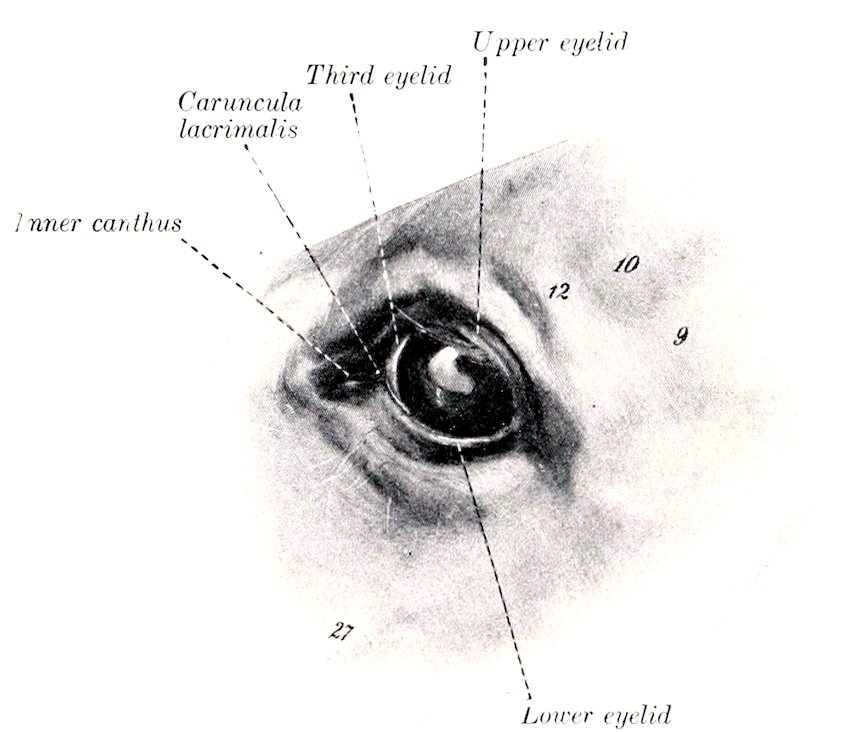
Fig. 551.—Left Eye of Horse. 9, Zygomatic arch; 10, supraorbital depression; 12, supraorbital process; 27, facial crest. (After Ellenberger-Baum, Anat. für Künstler.)
735
THE EYELIDS AND CONJUNCTIVA
The eyelids, upper and lower (Palpebra superior et inferior), are movable folds of integument situated in front of the eyeball. When closed, they cover the entrance to the orbit and the anterior surface of the eyeball. The upper lid is much more extensive and more movable than the lower one, and its free edge is more concave. The interval between the lids is termed the palpebral fissure (Rima palpebrarum). When the eye is closed, it is an oblique slit about two inches (ca. 5 cm.) in length; when open, it is biconvex in outline. The ends of the fissure are the angles or canthi, and are distinguished as internal or nasal, and external or temporal (Angulus oculi medialis, lateralis). The external angle is rounded when the eye is open, but the internal angle is narrowed to form a ᑐ-shaped bay or recess, termed the lacrimal lake (Lacus lacrimalis). In this there is a rounded, pigmented prominence known as the lacrimal caruncle (Caruncula lacrimalis); it is about the size of a small pea and is covered with modified skin, connected with that of the inner commissure, from which project a number of hairs provided with sebaceous glands. The lids unite on either side and form the commissures, internal and external (Commissura palpebrarum medialis, lateralis). The anterior surface of the lids (Facies anterior palpebrarum) is convex and presents a variable number of long tactile hairs in addition to the ordinary but very short hairs. A considerable number of tactile hairs are scattered over the lower part of the lower lid, but on the upper lid they are very scanty. The infrapalpebral depression (Sulcus infrapalpebralis) indicates somewhat indistinctly the limit of the lower lid. The upper lid is marked by two furrows when raised. The posterior surface (Facies posterior palpebrarum) is adapted to the free surface of the eyeball and is covered by the palpebral conjunctiva. The free border of the lid is smooth and usually black. It has a well defined posterior margin (Limbus palpebralis posterior), along which the ducts of the tarsal glands open. The anterior margin (Limbus palpebralis anterior) bears stiff hairs termed the cilia or eyelashes. On the upper lid the cilia are long and numerous except at its inner third, where they are very small or absent. On the lower lid the cilia are often scarcely distinguishable from the ordinary hairs; in other cases they may be clearly seen except near the external canthus, and are much finer and shorter than those of the upper lid. The edge of each lid is pierced near the internal angle by a minute, slit-like opening, the punctum lacrimalis, which is the entrance to the lacrimal duct or canaliculus.
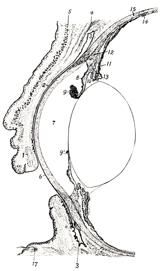
Fig. 552.—Vertical, Section of Anterior Part of Eye of Horse, with Lids Half Closed. 1, Tarsal gland of upper lid; 2, palpebral conjunctiva; 3, fornix conjunctivæ; 4, levator palpebræ superioris; 5, orbicularis oculi; 6, cornea; 7, anterior chamber; 8, iris; 9, 9′, granula iridis; 10, posterior chamber; 11, ciliary process; 12, ciliary muscle; 13, ciliary zone or suspensory lig. of lens; 14, chorioid; 15, sclera; 16, lens; 17, root of tactile hair. (After Bayer, Augenheilkunde.)
736Structure of the Eyelids.—The skin of the eyelids is thin and freely movable, except near the free edge, where it is more firmly attached. The underlying subcutaneous tissue is destitute of fat. The muscular layer consists chiefly of the elliptical bundles of the orbicularis oculi, with which are associated fibers of the corrugator supercilii in the upper lid and the malaris in the lower lid. At the inner side there is a fibrous band, the palpebral ligament, which is attached to the lacrimal tubercle and furnishes origin to some fibers of the orbicularis. At the inner commissure a bundle detached from the orbicularis passes inward behind the lacrimal sac, and is known as the pars lacrimalis or Horner’s muscle. At the outer side an indistinct palpebral raphé occurs where fibers of the orbicularis decussate. The fibrous layer is thicker and denser along the free edge of the lid, forming here the tarsus. The tarsus furnishes insertion to a layer of unstriped muscle known as the tarsal muscle (of Müller). The tarsal glands (Glandulæ tarsales) are partly embedded in the deep face of the tarsus, and are visible when the lid is everted if the conjunctiva is not too strongly pigmented. They are arranged in a linear series, close together, and with their long axes perpendicular to the free edge of each lid. In the upper lid they number forty-five to fifty; in the lower, thirty to thirty-five. Each consists of a tubular duct beset with numerous alveoli, in which a fatty substance, the palpebral sebum, is secreted. The palpebral conjunctiva lines the posterior surface of the eyelids.
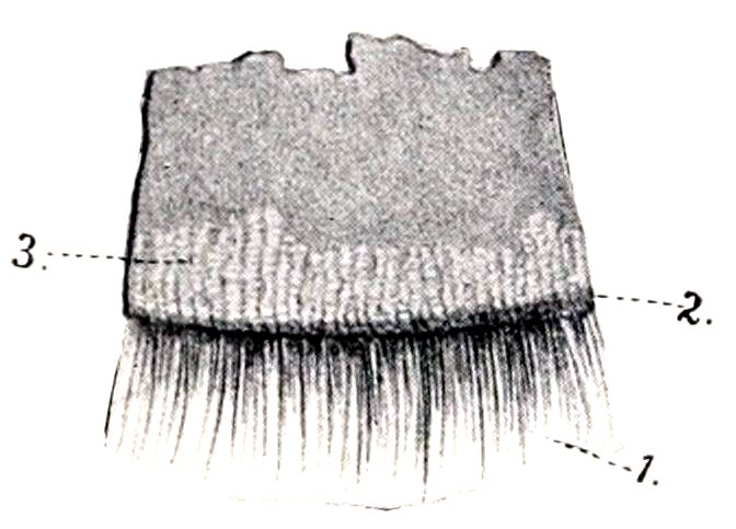
Fig. 553.—Piece of Upper Eyelid, Inner Surface. 1, Cilia; 2, limbus palp. posterior; 3, tarsal glands. (After Ellenberger, in Leisering’s Atlas.)
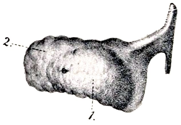
Fig. 554.—Cartilage of Third Eyelid of Horse, Convex Surface. 1, Gland; 2, fat surrounding deep part of cartilage. (After Ellenberger, in Leisering’s Atlas.)
The conjunctiva is the mucous membrane which lines the lids as palpebral conjunctiva (C. palpebrarum) and is reflected upon the anterior part of the eyeball as bulbar conjunctiva (C. bulbi); the line of reflection is termed the fornix conjunctivæ. The palpebral part is closely adherent to the tarsus, but is loosely attached further back. It is papillated and is covered with stratified cylindrical epithelium in which many goblet cells are present. In the fornix and its vicinity there are tubular glands (of Henle). Near the inner angle there are numerous lymph nodules. The conjunctiva of the outer part of the upper lid is pierced near the fornix by the orifices of the excretory ducts of the lacrimal gland. The bulbar conjunctiva is loosely attached to the anterior part of the sclera and is pigmented in the vicinity of the corneo-scleral junction. On the cornea it is represented by a stratified epithelium.
When the lids are in apposition, the conjunctiva (including the epithelium of the cornea) forms a complete conjunctival sac, as the capillary space between the lids and the eyeball is termed.
The third eyelid (Palpebra tertia) is situated at the inner angle of the eye (Figs. 551, 556). It consists of a semilunar fold of the conjunctiva, known as the membrana nictitans, which covers and partly incloses a curved plate of hyaline cartilage. Its marginal part is thin and usually more or less pigmented. The 737cartilage has an elongated quadrilateral outline. The part of it which lies in the membrana is wide and thin. The deep part is narrower and thicker and is embedded in fat at the inner side of the eyeball. Numerous minute lymph nodules occur in the membrana nictitans, and the deep part of the cartilage is surrounded by a gland which resembles the lacrimal gland in structure (Glandula superficialis palpebæ tertiæ).
Ordinarily the third eyelid extends very little over the inner end of the cornea, but when the eyeball is strongly retracted (as in tetanus) the membrana is protruded over it so as to measure about an inch (ca. 2 to 3 cm.) in its middle. This effect results from the pressure of the eyeball and its muscles on the fat which surrounds the deep part of the cartilage.
Vessels and Nerves.—The arteries which chiefly supply the eyelids and conjunctiva are branches of the ophthalmic and facial arteries, and the blood is drained away by corresponding veins. The sensory nerves are branches of the ophthalmic and maxillary divisions of the trigeminus. The motor nerves to the orbicularis oculi, corrugator supercilii, and malaris come from the facial nerve, the levator palpebræ superioris is innervated by the oculomotor, and the unstriped muscle of the lids by the sympathetic.
THE LACRIMAL APPARATUS
The lacrimal apparatus (Apparatus lacrimalis) comprises: (1) the lacrimal gland, which secretes the clear lacrimal fluid; (2) the excretory ducts of the gland; (3) the two lacrimal ducts or canaliculi, lacrimal sac, and naso-lacrimal duct, which receive the fluid and convey it to the nostril.
The lacrimal gland (Glandula lacrimalis) is situated between the supraorbital process and the dorso-lateral surface of the eyeball (Fig. 438). It is flattened, oval in outline, and measures about two inches (ca. 5 cm.) transversely and an inch or more (2.5 to 3 cm.) in the sagittal direction. Its upper face is convex and is related to the concave lower surface of the supraorbital process. The lower face is concave in adaptation to the eyeball, from which it is separated by the periorbita. The excretory ducts (Ductuli excretorii) are very small and are twelve to sixteen in number; they open into the outer part of the conjunctival sac along a line a little in front of the fornix conjunctivæ superior. In appearance and structure the gland resembles the parotid. It receives its blood-supply chiefly from the lacrimal artery. The sensory nerve is the lacrimal, and the secretory fibers are derived from the sympathetic.
The puncta lacrimalia are the entrances to the two lacrimal ducts. Each is a fine slit-like opening (about 2 mm. long), situated close behind the free edge of the lid and about a third of an inch (ca. 8 mm.) from the internal canthus. The lacrimal ducts (Ductus lacrimales), upper and lower, begin at the puncta and converge at the inner commissure to open into the lacrimal sac. The latter (Saccus lacrimalis) may be regarded as the dilated origin of the naso-lacrimal duct. It occupies the funnel-like origin of the bony lacrimal canal, and leads to the naso-lacrimal duct (Ductus naso-lacrimalis), which passes forward and a little downward along the outer wall of the frontal sinus and the nasal cavity and opens near the lower commissure of the nostril. Its length is about ten to twelve inches (ca. 25 to 30 cm.). In the first part of its course it is inclosed in the osseous lacrimal canal; further forward it lies in the lacrimal groove of the maxilla, covered at first by a plate of cartilage and then by the mucous membrane of the middle meatus. The terminal part lies in the inferior turbinal fold and opens on the skin of the floor of the nostril near the transition to mucous membrane. Accessory openings may occur a little further back.
The first part of the duct, about 6 to 7 mm. in diameter, extends in a gentle curve, convex dorsally, from the internal commissure toward a point just above the level of the infraorbital foramen. The second part (isthmus) is narrower (ca. 3 to 4 mm.); it extends forward and a 738little ventrally about to a transverse plane through the first cheek tooth and lies in the groove above the inferior turbinal crest. Beyond this the duct inclines upward and widens very considerably, crosses the nasal process of the premaxilla obliquely, and contracts at its termination. The mucous membrane may present valvular folds, the most distinct of which is situated at the origin.
THE PERIORBITA
The periorbita or ocular sheath is a conical fibrous membrane which incloses the eyeball with its muscles, vessels, nerves, etc. Its apex is attached around the optic and lacerated foramina, and its base is in part attached to the bony rim of the orbit, in part continuous with the fibrous layer of the lids. Its inner part, which is in contact with the orbital wall, is thin; incorporated with it beneath the root of the supraorbital process is the bar of cartilage around which the superior oblique muscle is reflected. The outer part is thicker, and is strengthened by an elastic band which is attached to the pterygoid crest and furnishes origin to the thin unstriped orbital muscle. A quantity of fat (Corpus adiposum extraorbitale) lies about the periorbita, and within it is the intraorbital adipose tissue (Corpus adiposum intraorbitale) which fills the interstices between the eyeball, muscles, etc.
THE ORBITAL FASCIÆ AND OCULAR MUSCLES (Figs. 438, 439, 555)
The straight muscles of the eyeball and the oblique muscles in part are inclosed in fibrous sheaths (Fasciæ musculares), formed by superficial and deep layers of fascia, which are united by intermuscular septa in the interstices between the muscles. The superficial fascia is thin; it blends in front with the fibrous layer of the eyelids and is attached behind around the optic foramen. The deep fascia consists anteriorly of two layers, one of which is continuous with the fibrous tissue of the lids, while the other is attached at the corneo-scleral junction.
The posterior part of the eyeball is covered by the bulbar fascia or capsule of Tenon (Fascia bulbi), so that between them a lymph space (Spatium interfasciale) is inclosed which communicates with the subdural space along the course of the optic nerve.
The levator palpebræ superioris muscle[217] is a thin band about half an inch in width which lies above the rectus superior. It is narrow at its origin above and behind the ethmoidal foramen and ends by an expanded tendon in the upper lid. Its action is to raise the upper lid.
The recti oculi (Mm. recti bulbi) are four in number and are designated according to their positions as superior, inferior, internal, and external. They are all band-like, arise close together around the optic foramen, and diverge as they pass forward to the eyeball. On reaching the latter they end in thin tendons which are inserted into the sclera in front of the equator of the eyeball.
The retractor oculi (M. retractor bulbi) surrounds the optic nerve, and is incompletely divided into four parts which alternate with the recti. They arise around the optic nerve and are inserted into the sclera behind the recti.
The obliquus oculi superior (M. obliquus bulbi dorsalis s. superior) is the longest and narrowest of the ocular muscles. It arises near the ethmoidal foramen and passes forward internal to the rectus internus. Under the root of the supraorbital process it is reflected almost at a right angle around a cartilaginous pulley (trochlea), which is attached to the anterior part of the inner wall of the orbit, a bursa being interposed here. The muscle is then directed outward and somewhat forward, and ends in a thin tendon which passes between the rectus superior and the eyeball, and is inserted into the sclera between the superior and external recti, about half an inch behind the margin of the cornea.
The obliquus oculi inferior (M. obliquus bulbi ventralis s. inferior) is wide and 739much shorter than the recti. It arises from the inner wall of the orbit in the small depression (Fossa muscularis) behind the lacrimal fossa. It curves around the rectus inferior and is inserted into the sclera near and partly beneath the rectus externus.
Actions.—The superior and inferior recti rotate the eyeball about a transverse axis, moving the vertex of the cornea upward and downward respectively. Similarly the internal and external recti rotate the eyeball about a vertical axis, turning the vertex of the cornea inward and outward respectively. The oblique muscles rotate the eyeball about a longitudinal axis; the superior oblique raises the outer end of the pupil, while the inferior oblique lowers it. The retractor as a whole draws the eyeball backward, and its parts may separately reinforce the corresponding recti. Also the four recti acting together will retract the eyeball.
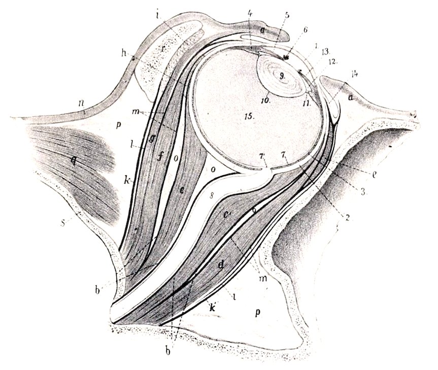
Fig. 555.—Vertical, Axial Section of Orbit of Horse. a, Eyelids; b, bulbar fascia (Tenon’s capsule); c, c′, retractor bulbi; d, rectus oculi inferior; e, obliquus oculi inferior (in cross-section); f, rectus oculi superior; g, levator palpebræ superioris; h, obliquus oculi superior (in cross-section); i, lacrimal gland; k, k′, periorbita; l, superficial fascia; m, deep fascia; n, skin; o, retrobulbar fat; p, extraorbital fat; q, temporalis muscle; r, supraorbital process; s, cranial wall; 1, cornea; 2, sclera; 3, chorioidea; 4, ciliary muscle; 5, iris; 6, granula iridis; 7, retina; 7′, optic papilla; 8, optic nerve; 9, crystalline lens; 10, capsule of lens; 11, ciliary zone; 12, posterior chamber; 13, anterior chamber; 14, conjunctiva bulbi; 15, vitreous body. (After Ellenberger, in Leisering’s Atlas.)
The actual movements of the eyeball are by no means so simple as might be inferred from the foregoing general statements. Practically all movements are produced by the coördinated actions of several muscles, involving combinations which are quite complex and difficult to analyze accurately. Further complication is caused by the fact that the recti are not inserted at equal distances from the equator and the axes of rotation of the oblique muscles do not correspond to the longitudinal axis of the eyeball.
Nerve-supply.—The oculomotor nerve supplies the foregoing muscles, with the exception of the rectus externus and obliquus superior, which are innervated by the abducens and trochlearis respectively.
THE EYEBALL
The eyeball (Bulbus oculi) is situated in the anterior part of the orbital cavity, protected in front by the eyelids and conjunctiva, in its middle by the complete orbital ring, and related behind to the bulbar fascia, fat, and ocular muscles.
740It has the form approximately of an oblate spheroid, but is composed of the segments of two spheres of different sizes. The anterior, transparent, corneal segment has a radius of curvature of about 17 mm., and the posterior, opaque, scleral segment a radius of about 25 mm. The anterior segment therefore projects more strongly, and the junction of the two segments is marked externally by a broad, shallow groove, the sulcus scleræ. The central points of the anterior and posterior curvatures of the eyeball are termed respectively the anterior and posterior poles (Polus anterior, posterior), and the line connecting the poles is the optic axis (Axis oculi externa). The angle of divergence of the optic axes is about 137 degrees. The equator (Æquator) is an imaginary line drawn around the eyeball midway between its poles, and meridians (Meridiani) are lines drawn around it through the poles.
The average transverse diameter of the eyeball is about 5 cm., the vertical about 4.5 cm., and the axial about 4.25 cm. The distance from the anterior pole to the point of entrance of the optic nerve is about 3 cm.
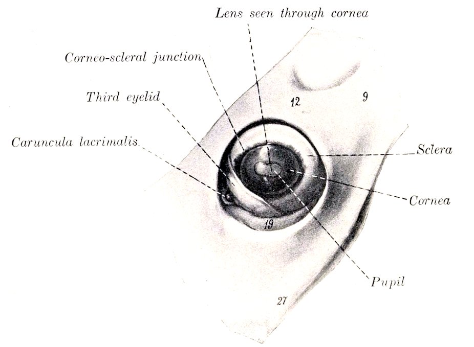
Fig. 556.—Left Eyeball of Horse, in situ, after Removal of Upper and Lower Lids. 9, Zygomatic arch; 12, supraorbital process; 19, orbital fat; 27, facial crest. (After Ellenberger-Baum, Anat. für Künstler.)
The eyeball consists of three concentric tunics or coats, within which three refractive media are inclosed.
The Fibrous Tunic
The fibrous tunic (Tunica fibrosa oculi) is the external coat and is composed of a posterior opaque part, the sclera, and a transparent anterior part, the cornea.
The sclera, popularly termed the “white of the eye,” is a dense fibrous membrane which forms about four-fifths of the fibrous tunic. Thickest in the vicinity of the posterior pole (ca. 2 mm.), it thins at the equator (ca. 0.4 mm.), and increases in thickness toward the junction with the cornea (ca. 1.3 mm.). It is in general white, but may have a bluish tinge in its thinnest parts. Its external surface furnishes insertion to the ocular muscles and is covered by the conjunctiva scleræ in its anterior part. The episcleral tissue, which is richly supplied with vessels and nerves, attaches the conjunctiva to the sclera; it is abundant and loosely meshed except at the junction with the cornea. The inner surface is attached to the chorioid coat by a layer of delicate, pigmented, connective tissue, the lamina 741fusca. The anterior border is oval, the long axis being transverse, and is continuous with the cornea. The transition from the opaque scleral tissue to the transparent corneal substance occurs in such manner that the sclera appears to form a groove (Rima cornealis), into which the cornea fits somewhat as a watch-glass in the case. Near the corneo-scleral junction there is a circular venous plexus, the plexus s. sinus venosus scleræ, formerly called the canal of Schlemm. The optic nerve passes through the posterior part of the sclera a little below and external to the posterior pole. The opening for the nerve is crossed by interlacing fibrous strands, forming the lamina cribrosa scleræ.
The sclera consists of interlacing bundles of white fibrous tissue, associated with which there are a few elastic fibers. The bundles are arranged chiefly in meridional and equatorial layers. The very limited blood-supply is derived from the ciliary arteries, and the veins open into the venæ vorticosæ and ciliary veins. The lymphatics are represented by intercommunicating cell spaces. The nerves are derived from the ciliary nerves.
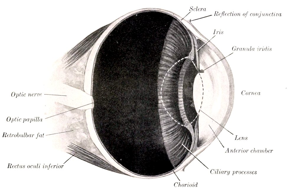
Fig. 557.—Vertical Section of Eyeball of Horse, about ³⁄₂. The contour of the crystalline lens is dotted.
The cornea forms the anterior fifth of the fibrous tunic. It is transparent, colorless, and non-vascular. Viewed from in front it is oval in outline, the long axis being transverse and the broad end internal; it appears more nearly circular when viewed from behind. Its anterior surface (Facies anterior) is convex and is more strongly curved than the sclera; its central part is termed the vertex corneæ. The posterior surface (Facies posterior) is concave; it forms the anterior boundary of the anterior chamber, and is in contact with the aqueous humor. The margin (limbus corneæ) joins the sclera; the latter overlaps the cornea more in front than behind, and more above and below than at the sides, thus explaining the difference in outline of the two surfaces. The cornea is thinnest at the vertex.
The cornea consists, from before backward, of the following layers: (1) The epithelium corneæ is continuous with that of the conjunctiva scleræ, and is of the stratified squamous type. (2) The lamina limitans anterior is merely a condensation 742of the next layer. (3) The substantia propria forms the bulk of the cornea and is composed of interlacing bundles of connective tissue, arranged in part in lamellæ disposed parallel with the surface. In the amorphous cement substance between the lamellæ are flattened connective-tissue cells, the corneal corpuscles. These have branching processes which unite with those of other cells, thus forming a protoplasmic network.[218] (4) The lamina elastica posterior, also termed the membrane of Descemet or Demours, is a thin and practically homogeneous membrane which is less intimately attached to the substantia propria than the anterior lamina. It is clear, glistening, and elastic. At the periphery the lamina divides into three sets of fibers. The anterior fibers join the sclera, the middle give attachment to the ciliary muscle, while the posterior pass into the iris and form the ligamentum pectinatum iridis. (5) The endothelium (cameræ anterioris) consists of a layer of flattened polygonal cells, and is reflected on to the anterior surface of the iris.
The cornea is without blood-vessels except at its periphery, where the terminal twigs of the vessels of the sclera and conjunctiva terminate in loops. The nerves are derived from the ciliary nerves. They form a plexus around the periphery (Plexus annularis), from which fibers pass into the substantia propria, become non-medullated, and form the fundamental or stroma plexus. From this perforating branches pass through the anterior limiting layer and form a subepithelial plexus, from which filaments ascend between the epithelial cells. Other branches from the plexuses in the substantia propria end as fibrils which are in close relation with the corneal corpuscles.
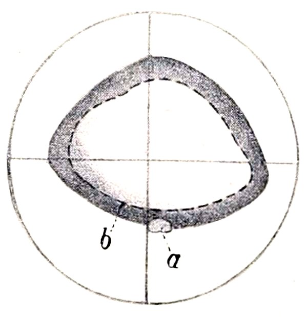
Fig. 558.—Tapetum of Horse. a, Optic papilla; b, lower border of tapetum. (After Ellenberger, in Leisering’s Atlas.)
The Vascular Tunic
The vascular tunic (Tunica vasculosa oculi) lies internal to the fibrous coat; it comprises three parts—the chorioid, the ciliary body, and the iris.
1. The chorioid (Chorioidea) is a thin membrane which lies between the sclera and retina. It is in general rather loosely attached to the sclera by the lamina fusca, but is intimately adherent at the point of entrance of the optic nerve and less closely in places where the ciliary vessels and nerves pass through. The inner surface is in contact with the layer of pigmented cells of the retina, which adhere so closely to the chorioid that they were formerly regarded as a part of the latter. The general color of the chorioid is dark brown, but an extensive semilunar area a little above the level of the optic papilla has a remarkable metallic luster, and is termed the tapetum of the chorioid (Tapetum chorioideæ). The appearance here varies in different individuals, but the prevailing colors in most cases are iridescent blue and green in various nuances shading into yellow. Posteriorly the chorioid is perforated by the optic nerve, and anteriorly it is continuous with the ciliary body.
The chorioid consists of four layers, which from without inward are as follows: (1) The lamina suprachorioidea consists of interlacing fine lamellæ of fibrous tissue, each containing a network of elastic tissue. Among these are large-branched, pigmented, connective-tissue cells. The spaces between the lamellæ are lined with endothelium, and form a system of lymph-clefts which together form the perichorioid space (Spatium perichorioideale). (2) The lamina vasculosa is the outer part 743of the proper tissue of the chorioid. It contains the larger blood-vessels, which are supported by connective-tissue and elastic fibers. (3) The lamina choriocapillaris consists of an extremely rich network of capillaries embedded in an almost homogeneous matrix. Between it and the lamina vasculosa is a layer of fibro-elastic tissue, the tapetum fibrosum, which causes the metallic luster mentioned above. (4) The lamina basalis is very thin and transparent. It is composed of an inner homogeneous part and an outer elastic part.
2. The ciliary body (Corpus ciliare), the middle part of the vascular coat, connects the chorioid with the periphery of the iris. In meridional section it has the form of a narrow triangle, the base of which is next to the iris. On its inner side are the ciliary processes and on its outer side the ciliary muscle. It consists of three parts—the ciliary ring, ciliary processes, and ciliary muscle. The ciliary ring (Orbicularis ciliaris) is the posterior zone, which is distinguished from the chorioid mainly by its greater thickness and the absence of the choriocapillaris. Its inner face presents numerous fine meridional ridges, by the union of which the ciliary processes are formed. The ciliary processes (Processus ciliares), more than a hundred in number, form a circle of radial folds which surround the lens and furnish attachment to the zonula ciliaris or suspensory ligament of the latter. They are small at their origin on the ciliary ring and become much thicker and higher toward their inner ends. The width of the circle formed by them is narrower at the inner side than elsewhere. Their bases extend forward to the periphery of the iris, and their inner ends are close to the margin of the lens. They bear numerous secondary folds. Their inner surface is covered by a continuation of the lamina basalis of the chorioid, on which there are two layers of epithelial cells which constitute the pars ciliaris retinæ. They consist of a rich network of tortuous vessels supported in pigmented connective tissue. The ciliary muscle (M. ciliaris) (Figs. 552, 555, 559) constitutes the outer part of the ciliary body, and lies between the sclera and the ciliary processes. It forms a circular band of unstriped muscle, the fibers of which are for the most part directed meridionally. They arise from the inner surface of the sclera and from the ligamentum pectinatum iridis close to the corneo-scleral junction, and run backward along the sclera to be inserted into the ciliary processes and ring. When the muscle contracts, it pulls the processes and ring forward, thus slackening the ciliary zone or suspensory ligament of the lens, and allowing the latter to become more convex. This is the mechanism of accommodation for near objects.
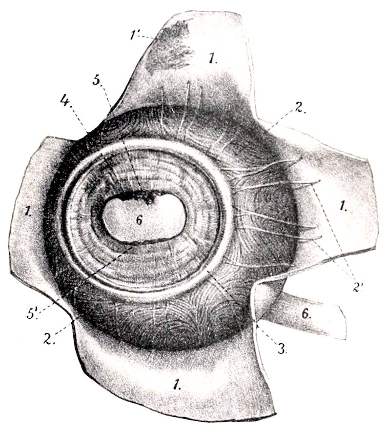
Fig. 559.—Vascular Tunic of Eyeball of Horse, Front View. The cornea is removed and the sclera is reflected in flaps. 1, Sclera; 1′, lamina fusca; 2, chorioidea; 2′, ciliary veins; 3, ciliary muscle; 4, iris; 5, 5′, granula iridis; 6, pupil, through which the lens is visible. (After Ellenberger, in Leisering’s Atlas.)
In man the muscle has the form of a prismatic ring which is triangular in meridional section, the base being directed toward the periphery of the iris. It consists chiefly of meridional fibers, 744but a ring of circular fibers forms the inner angle of its base. In the horse the muscle is much less developed, and has the form of a flat band; it does not contain circular fibers, but the arrangement is rendered more or less plexiform by the existence of oblique and equatorial fibers.
3. The iris (Figs. 552, 557, 559) is a muscular diaphragm placed in front of the lens, and is visible through the cornea. It is pierced centrally by an elliptical opening, the pupil (Pupilla), which varies in size during life and determines the amount of light admitted to act on the retina. In strong light the vertical diameter of the pupil is very short, but the opening is almost circular when the pupil is fully dilated. The ciliary border (Margo ciliaris) or periphery of the iris is continuous with the ciliary body and is connected with the corneo-scleral junction by the ligamentum pectinatum iridis. The ligament consists of strands of connective tissue which are attached externally to the corneo-scleral junction. Its bundles interlace and inclose spaces (Spatia anguli iridis) which are lined with endothelium and communicate with the anterior chamber. The pupillary border (Margo pupillaris) surrounds the pupil. Its upper part bears in its middle several black masses of variable size, termed the granula iridis or corpora nigra; similar, but much smaller, projections may be seen on the lower margin of the pupil. The anterior surface (Facies anterior) determines the color of the eye, which is dark brown usually. It is marked by delicate concentric lines (Plicæ iridis), which fade out near the pupil. The smooth, narrow, central part is termed the pupillary zone or annulus iridis minor, while the much broader plicated part is the ciliary zone or annulus iridis major. The posterior surface (Facies posterior) is usually black. It bears numerous fine radial lines except at the pupillary margin. Its central part is in contact with the anterior surface of the lens, but peripherally the two are separated by a narrow space termed the posterior chamber. The iris consists chiefly of the stroma iridis, a delicate framework of connective tissue, which supports numerous blood-vessels, and contains branched pigmented cells. The muscular tissue (unstriped) consists of a sphincter and a dilator of the pupil. The sphincter pupillæ lies in the posterior part around the pupil, with which the fibers are largely concentric. The dilatator pupillæ consists of fibers which radiate outward from the sphincter to the ciliary border. The anterior surface of the iris is covered by a continuation of the endothelium of the cornea. Beneath this is a condensation of the stroma, in which the cells are close together and are full of pigmented granules.[219] There appear to be minute clefts here by which the lymph-spaces of the stroma communicate with the anterior chamber. The posterior surface is covered by a pigmented epithelium, the stratum pigmenti iridis, which is a continuation of that of the ciliary body.
The arteries of the vascular tunic come from the ciliary branches of the ophthalmic artery. The arteries of the chorioidea are derived chiefly from the short posterior ciliary arteries. These perforate the sclera around the posterior pole, run forward in the lamina vasculosa, and form the rich capillary network of the choriocapillaris. The two long ciliary arteries perforate the sclera obliquely near the optic nerve; they run forward in the lamina suprachorioidea in the horizontal meridian, one on the inner, the other on the outer, side of the eyeball. On reaching the ciliary body each divides into diverging branches; the subdivisions of these unite with each other and with twigs of the anterior ciliary arteries to form a circular anastomosis, the circulus arteriosus major. From this branches go to the ciliary muscle and processes and to the iris. The two anterior ciliary arteries, dorsal and ventral, form an episcleral plexus around the corneo-scleral junction, and give off branches which perforate the sclera. These supply twigs to the ciliary muscle and recurrent branches to the chorioid, and assist in forming the circulus arteriosus major.
The blood is carried away from the vascular tunic chiefly by four or five venous 745trunks, the venæ vorticosæ, which are formed by the convergence in whorls of numerous veins, coming not only from the chorioid, but also from the ciliary body and iris. The venæ vorticosæ perforate the sclera about at the equator and join the veins of the ocular muscles.
The nerves come from the long and short ciliary nerves. They form a plexus in the lamina suprachorioidea, which contains ganglion cells, and sends numerous non-medullated fibers chiefly to the blood-vessels of the chorioid. At the ciliary muscle a second plexus (P. gangliosus ciliaris) is formed, which supplies the muscle and sends fibers to the iris. The sphincter pupillæ is supplied by fibers derived from the oculomotor nerve, while the dilatator pupillæ is innervated by the sympathetic.
THE RETINA
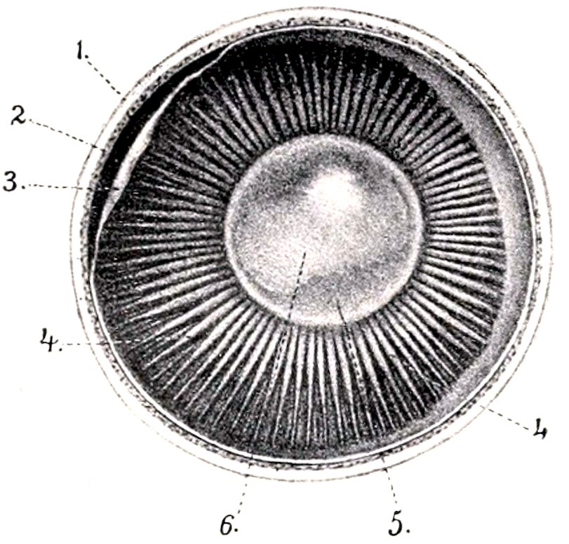
Fig. 560.—Inner Surface of Anterior Part of Eyeball of Horse (Equatorial Section). 1, Sclera; 2, chorioidea; 3, retina (drawn away from chorioidea); 4, ciliary processes; 5, crystalline lens, through which the pupil (6) is seen. (After Ellenberger, in Leisering’s Atlas.)
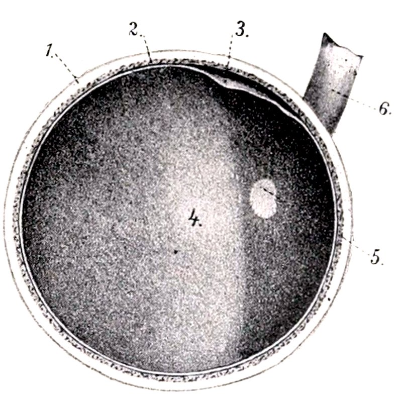
Fig. 561.—Fundus Oculi, seen on Equatorial Section of Eyeball of Horse. 1, Sclera; 2, chorioidea; 3, retina (loosened); 4, tapetum; 5, optic papilla; 6, optic nerve. (After Ellenberger, in Leisering’s Atlas.)
The retina or nervous tunic of the eyeball is a delicate membrane which extends from the entrance of the optic nerve to the margin of the pupil. It consists of three parts. The large posterior part, which alone contains the nervous elements, including the special neuro-epithelium and the optic nerve-fibers, is termed the pars optica retinæ. It extends forward to the ciliary body, where it terminates at an irregular line called the ora serrata. Here the retina rapidly loses its nervous elements, becomes much thinner, and is continued over the ciliary body and the posterior surface of the iris by two layers of epithelial cells. In the pars ciliaris retinæ the inner stratum is non-pigmented, while the outer layer is a direct continuation of the stratum pigmenti of the pars optica. In the pars iridica retinæ both are pigmented. In the dead subject the pars optica is an opaque, gray, soft membrane which can be stripped of the chorioid, leaving most of its outer pigmented layer on the latter. During life it is transparent, except as to its pigmented epithelium, and the reddish appearance of the fundus as viewed by the ophthalmoscope is caused by the blood in the network of the choriocapillaris. The entrance of the optic nerve forms a sharply defined, oval, light area, the optic papilla (Papilla nervi optici), situated about 15 mm. below the horizontal meridian and 3 to 4 mm. external to the vertical meridian. The central part of the papilla is slightly depressed (Excavatio papillæ n. optici).
746The transverse diameter of the papilla is about 6 to 7 mm., and the vertical about 4 to 5 mm. It is commonly situated a little below the margin of the tapetum, but the latter may extend down somewhat on either side of the papilla. The lower margin is often indented a little. In inspection of the fundus with the ophthalmoscope numerous fine branches of the arteria centralis retinæ are seen radiating from the periphery of the papilla.
The optic nerve-fibers converge from all parts of the pars optica to the papilla, where they collect into bundles which traverse the lamina cribrosa of the chorioidea and sclera, and constitute the optic nerve. The area centralis retinæ is a round spot, 2 to 3 mm. in diameter, situated above and external to the optic papilla; it corresponds to the macula lutea of man, which is histologically more highly differentiated than the rest of the retina and is the area of most acute vision.
The structure of the retina is very complex. It consists of nervous elements which are supported in a peculiar sustentacular tissue, and are covered externally by a layer of pigmented epithelium (Stratum pigmenti retinæ). The nervous elements comprise a highly specialized neuro-epithelium, the rods and cones; ganglion cells, the axones of which form the optic nerve; and intermediate neurones. Ten layers may be recognized in sections microscopically.[220]
The arteries of the retina are derived from the arteria centralis retinæ and anastomotic branches from the short ciliary arteries. The arteria centralis enters the optic nerve a short distance behind the eyeball and runs in the axis of the nerve. The artery divides 2 to 3 mm. before reaching the papilla, and gives off thirty to forty branches which radiate in the posterior part of the retina and divide dichotomously into end-arteries. The anterior part of the retina is non-vascular. The veins accompany the arteries except in the capillary plexuses; their walls consist merely of a layer of endothelial cells, around which are a lymph-channel and sheath.
CHAMBERS OF THE EYE AND AQUEOUS HUMOR
The anterior chamber of the eye (Camera oculi anterior) is inclosed in front by the cornea and behind by the iris and lens (Figs. 552, 555, 557). It communicates through the pupil with the posterior chamber (Camera oculi posterior); this is a small annular space, triangular in cross-section, which is bounded in front by the iris, behind by the peripheral part of the lens and its ligament, and externally by the ciliary processes. The chambers are filled by the aqueous humor (Humor aqueus), a clear fluid which consists of about 98 per cent. of water, with a little sodium chlorid and traces of albumin and extractives. It is carried off chiefly through the spaces (of Fontana) in the zonula ciliaris or suspensory ligament of the lens into the plexus venosus scleræ.
REFRACTIVE MEDIA OF THE EYEBALL
The vitreous body (Corpus vitreum) is a semifluid transparent substance situated between the crystalline lens and the retina. In front it presents a deep cavity, the fossa hyaloidea, which receives the posterior surface of the lens. It consists of a framework of delicate fibrils, the stroma vitreum, the meshes of which are filled by the fluid humor vitreus. The surface is covered by a condensation of the stroma known as the membrana hyaloidea.
The crystalline lens (Lens crystallina) is a biconvex, transparent body, situated in front of the vitreous body and in partial contact with the posterior surface of the iris. Its periphery, the æquator lentis, is almost circular and is closely surrounded by the ciliary processes. The anterior surface (Facies anterior) is convex; it is bathed by the aqueous humor and is in contact with the iris to an extent which varies with the state of the pupil. The posterior surface (Facies posterior) is much 747more strongly curved than the anterior. It rests in the fossa of the vitreous body. The central points of the surfaces are the anterior and posterior poles (Polus anterior et posterior lentis), and the line which connects them is the axis of the lens (Axis lentis).
The transverse diameter of the lens is about 2 cm., the vertical diameter is slightly smaller, and the axis measures about 13 mm. The radius of curvature of the anterior surface is 13.5 mm., and of the posterior surface 9.5 to 10 mm. But the curvatures of its surfaces—especially that of the anterior—vary during life according as the eye is accommodated for near or far vision.
The zonula ciliaris (of Zinn) or suspensory ligament of the lens (Fig. 552) consists of delicate fibers (Fibræ zonulares) which pass in a meridional direction from the ciliary processes to the capsule of the equator of the lens. Many fibers cross each other, and the spaces between the fibers (Spatia zonularia) are filled with aqueous humor; they communicate with each other and with the posterior chamber.
The substance of the lens (Substantia lentis) is inclosed by a structureless, highly elastic membrane, the capsule of the lens (Capsula lentis), and consists of a softer substantia corticalis, and a dense central part, the nucleus lentis. The capsule is thickest on the anterior surface, and here it is lined by a layer of flat polygonal cells, the epithelium of the lens capsule. The lens substance, when hardened, is seen to consist of concentric laminæ arranged somewhat like the layers of an onion, and united by an amorphous cement substance. The laminæ consist of lens fibers, hexagonal in section, and of very different lengths. Faint lines radiate from the poles and indicate the edges of layers of cement substance which unite the groups of lens fibers. These lines, the radii lentis, are three in number in the fœtus and new-born, and form with each other angles of 120 degrees. On the anterior surface one is directed upward from the pole and the other two diverge downward; on the posterior surface one is directed downward and the others diverge upward. The developed lens has neither vessels nor nerves.
In the fœtus the lens is nearly globular, and is soft and pinkish in color. During part of fœtal life it is surrounded by a vascular network, the tunica vasculosa lentis. This is derived chiefly from a temporary vessel, the hyaloid artery, which is a continuation forward of the arteria centralis retinæ through the hyaloid canal that traverses the vitreous body. In old age the lens tends to lose its elasticity and transparency; it also becomes flatter and the nucleus especially grows denser.
The Ear
The ear or organ of hearing (Organon auditus) consists of three natural divisions—external, middle, and internal.
THE EXTERNAL EAR
The external ear (Auris externa) comprises (1) the auricula, a funnel-like organ which collects the sound waves, together with its muscles, and (2) the external auditory canal, which conveys these waves to the tympanic membrane, the partition which separates the canal from the cavity of the middle ear.
The auricula or pinna is attached by its base around the external auditory canal in such a manner as to be freely movable. In the following description it will be assumed that the opening is directed outward and that the long axis is practically vertical. It has two surfaces, two borders, a base, and an apex. The convex surface or dorsum (Dorsum auriculæ) faces inward and is widest in its middle part; its lower part is almost circular in curvature, while above it narrows and flattens. The concave surface (Scapha) is the reverse of the dorsum; it presents several ridges which subside toward the apex. The anterior border is sinuous; it is largely convex, but becomes concave near the apex. It divides below into two diverging 748parts (crura helicis). The posterior border is convex. The apex is flattened, pointed, and curved a little forward. The base is strongly convex. It is attached to the external auditory process of the petrous temporal bone, and around this there is a quantity of fat. The parotid gland overlaps it below and externally. The structure of the external ear comprises a framework of cartilages (which are chiefly elastic), the integument, and a complicated arrangement of muscles.
The conchal or auricular cartilage (Cartilago auriculæ) determines the shape of the ear; its form can be made out without dissection, except below, where it is concealed by the muscles and the parotid gland. The basal part is coiled to form a tube, which incloses the cavity of the concha (Cavum conchæ). This part is funnel-shaped and curves outward and a little backward. Its internal (medial) surface is strongly convex, forming a prominence termed the eminentia conchæ. The lowest part of the internal margin bears a narrow, pointed prolongation, the styloid process. This process is about an inch long and projects downward externally over the annular cartilage; the guttural pouch is attached to its free end. Behind its base there is a foramen through which the auricular branch of the vagus passes.
The basal part of the posterior border is cut into by a notch, which separates two irregular quadrilateral plates. The upper plate (Tragus) is overlapped by the anterior border, and is separated from the adjacent part of the posterior border (Antitragus) by a notch (Incisura intertragica). The lower plate is curved to form a half ring and partly overlaps the anterior border and the annular cartilage. Behind the notch there is a foramen, which transmits the internal auricular artery and internal auricular branch of the facial nerve.
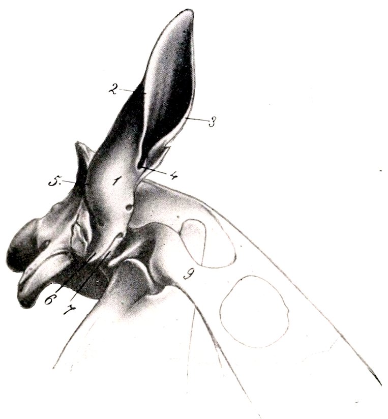
Fig. 562.—Conchal and Annular Cartilages of Ear of Horse, External View. 1, Base of concha; 2, posterior border, 3, anterior border of concha; 4, intertragic notch; 5, eminentia conchæ; 6, styloid process; 7, annular cartilage; 9, zygomatic arch. (After Ellenberger-Baum, Anat. für Künstler.)
The annular cartilage (Cartilago annularis) is a quadrilateral plate, curved to form about three-fourths of a ring; its ends are a little less than half an inch (ca. 1 cm.) apart internally and are united by elastic tissue. It embraces the external auditory process and forms with the lower part of the conchal cartilage the cartilaginous part of the external auditory canal.
The scutiform cartilage (Cartilago scutiformis s. scutulum) is an irregular quadrilateral plate which lies on the temporal muscle in front of the base of the conchal cartilage. Its superficial face is slightly convex from side to side and its deep face is correspondingly concave. The anterior end is thin and rounded; the posterior part or base is wider and thicker, and its inner angle is prolonged by a pointed process half an inch or more in length. The cartilage moves very freely over the underlying parts.
The external auditory canal or meatus leads from the cavum conchæ to the tympanic membrane. It does not continue the general direction of the cavity of the concha, but extends inward, downward, and slightly forward. It consists of a cartilaginous part formed by the lower part of the conchal cartilage and the annular cartilage, and an osseous part formed by the external auditory process of the temporal 749bone. These are united by elastic membranes to form a complete tube. Its caliber diminishes from without inward, so that the lumen of the inner end is about half of that of the outer end.
The skin on the convex surface of the concha presents no special features; it is attached to the cartilage by a considerable amount of subcutaneous tissue except at the apex. The integument which lines the concave surface is intimately adherent to the cartilage and is relatively dark in color. There are three or four cutaneous ridges which run about parallel with the borders of the conchal cartilage, but do not extend to the apex or the cavum conchæ. The upper and marginal parts and the ridges are covered with long hairs, but between the ridges and below it is thin, covered sparsely with very fine hairs, and supplied with numerous sebaceous glands. In the external auditory canal the skin becomes thinner; in the cartilaginous part it is supplied with numerous large, coiled, ceruminous glands (Glandulæ ceruminosæ) and is sparsely covered with very fine hairs; in the osseous part the glands are small and few or absent and there are no hairs.
THE AURICULAR MUSCLES
The auricular muscles may be subdivided into two sets, viz., (a) extrinsic muscles, which arise on the head and adjacent part of the neck, and move the external ear as a whole, and (b) intrinsic muscles, which are confined to the auricula. In this connection the scutiform cartilage may be regarded as a sesamoid cartilage intercalated in the course of some of the muscles.
The extrinsic muscles are as follows:
1. The scutularis is a thin muscular sheet situated subcutaneously over the temporalis muscle. Its fibers arise from the zygomatic arch and the frontal and sagittal crests, and converge to the scutiform cartilage. It consists of three parts.
(a) The fronto-scutularis comprises temporal and frontal parts, which arise from the zygomatic arch and the frontal crest, and are inserted into the outer and anterior borders of the scutiform cartilage respectively.
(b) The interscutularis arises from the sagittal crest, over which it is in part continuous with the muscle of the opposite side. Its fibers converge to the inner border of the scutiform cartilage.
(c) The cervico-scutularis is not well defined from the preceding muscle. It arises from the occipital crest and is inserted into the inner border of the scutiform cartilage.
2. The anterior auricular muscles (Mm. auriculares nasales) are four in number.
(a) The zygomatico-auricularis arises from the zygomatic arch and the parotid fascia, and is inserted into the outer face of the base of the conchal cartilage under cover of the parotido-auricularis.
(b) The scutulo-auricularis superficialis inferior arises on the outer part of the superficial face of the scutiform cartilage and ends on the base of the conchal cartilage with the preceding muscle.
(c) The scutulo-auricularis superficialis medius arises from the posterior part of the deep surface of the scutiform cartilage and is inserted into the dorsum of the conchal cartilage, close to the lower part of its anterior border and above and behind the preceding muscle. It receives a slip from the cervico-scutularis.
(d) The scutulo-auricularis superficialis superior is a thin slip which is detached from the interscutularis over the inner border of the scutiform cartilage. It ends on the anterior aspect of the lower part of the dorsum of the conchal cartilage.
3. The superior auricular muscles (Mm. auriculares dorsales) are two in number.
(a) The scutulo-auricularis superficialis accessorius is a narrow band which is 750largely covered by the preceding muscle. It arises from the posterior prolongation and the adjacent part of the superficial face of the scutiform cartilage, and is inserted into the convex surface of the conchal cartilage internal to the preceding muscle, the two crossing each other at an acute angle.
(b) The parieto-auricularis is flat and triangular; it arises from the sagittal crest under cover of the cervico-scutularis, runs outward and a little backward, and is inserted by a flat tendon into the lower part of the convex surface of the concha under cover of the cervico-auricularis superficialis.
4. The posterior auricular muscles (Mm. auriculares caudales) are three in number.
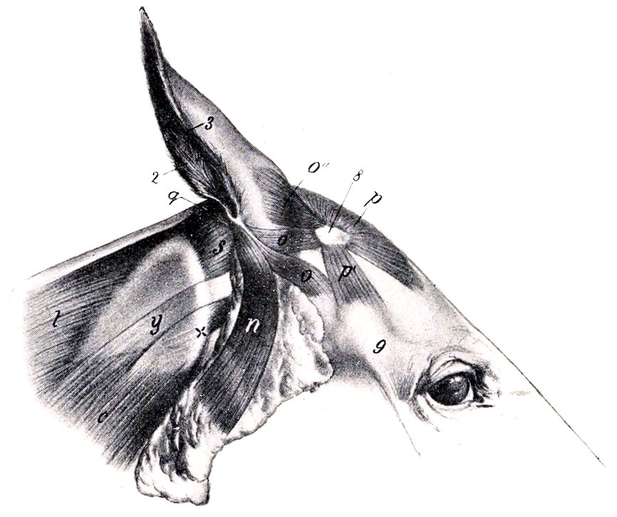
Fig. 563.—External Ear of Horse and its Muscles, Lateral View. 2, Posterior border; 3, anterior border of auricula; 8, scutiform cartilage; 9, zygomatic arch; n, parotido-auricularis; o, zygomatico-auricularis; o′, scutulo-auricularis superficialis inferior; o″, scutulo-auricularis superficialis medius et superior; p, interscutularis; p′, fronto-scutularis (pars temporalis); q, cervico-auricularis profundus major; s, small (or anterior) oblique muscle of head; t, splenius; y, tendon of mastoido-humeralis; c, mastoido-humeralis; x, wing of atlas. (After Ellenberger-Baum, Anat. für Künstler.)
(a) The cervico-auricularis superficialis is a thin triangular sheet. It is wide at its origin from the occipital crest and the adjacent part of the ligamentum nuchæ, and becomes narrower as it passes outward to be inserted into the inner side of the convex surface of the concha.
(b) The cervico-auricularis profundus major arises from the ligamentum nuchæ, partly beneath and partly behind the preceding muscle. It is directed outward and is inserted into the postero-external aspect of the base of the ear, partly under cover of the parotido-auricularis.
(c) The cervico-auricularis profundus minor arises under the preceding muscle and passes downward and outward to be inserted into the lowest part of the convex surface of the concha, partly under cover of the parotid gland.
5. The parotido-auricularis (s. M. auricularis inferior) is a ribbon-like muscle which lies on the parotid gland. It is thin and wide at its origin from the fascia on the lower part of the parotid gland and becomes somewhat narrower and thicker as it passes upward. It is inserted into the conchal cartilage just below the angle of junction of its borders.
7516. The deep auricular or rotator muscles (M. auriculares profundis, rotatores auriculæ), two in number, are situated under the scutiform cartilage and the base of the concha and cross each other.
(a) The scutulo-auricularis profundus major is the strongest of the auricular muscles. It is flat and is about an inch wide. It arises from the deep face of the scutiform cartilage and passes backward to end on and below the most prominent part of the base of the concha (eminentia conchæ).
(b) The scutulo-auricularis profundus minor lies between the base of the concha and the preceding muscle, and is best seen when the latter is cut and reflected. It is flat and about an inch in length. It arises from the posterior part of the deep face of the scutiform cartilage and from the cervico-scutularis, and passes downward, backward, and outward to be inserted into the base of the concha under cover of the preceding muscle.
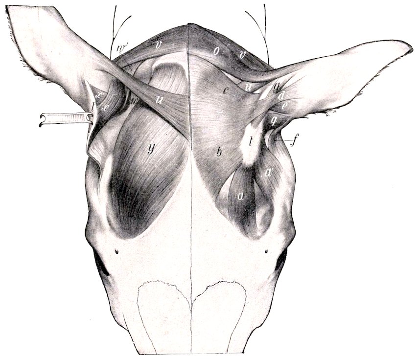
Fig. 564.—Muscles of External Ear of Horse, Viewed from Above. On the right side parts of the superficial muscles have been removed and the scutiform cartilage turned back to display the deeper muscles. a, Frontal part, a′, temporal part of fronto-scutularis; b, interscutularis; c, cervico-scutularis; d, e, scutulo-auricularis superficialis superior and medius; f, zygomatico-auricularis; g, scutulo-auricularis superficialis accessorius; o, cervico-auricularis superficialis; q, scutulo-auricularis superficialis inferior; t, scutiform cartilage; u, parieto-auricularis; v, cervico-auricularis profundus major; w, cervico-auricularis prof. minor; x, x′, scutulo-auricularis prof. major et minor; y, temporalis. (Ellenberger-Baum, Anat. d. Haustiere.)
7. The tragicus or mastoido-auricularis is a small muscle which arises from the temporal bone just behind the external auditory process and from the annular cartilage; it passes upward to be inserted into the lower part of the anterior border of the conchal cartilage.
The intrinsic muscles are very small and of little importance. They are as follows:
1. The antitragicus consists of a few bundles which are attached to the conchal 752cartilage behind the junction of its two borders, and are partially blended with the insertion of the parotido-auricularis.
2. The helicis is a small muscle attached in a position opposite to the preceding on the anterior border of the conchal cartilage; it extends also into the depression between the two divisions of the border. It is in part continuous with the insertion of the parotido-auricularis.
3. The verticalis auriculæ[221] is a thin stratum of muscular and tendinous fibers which extends upward on the posterior surface of the concha from the eminentia conchæ.
Actions.—The base of the concha is rounded and rests in a pad of fat (Corpus adiposum auriculæ), so that the movements of the external ear resemble those of a ball-and-socket joint. It is noticeable that movements about the longitudinal and transverse axes are accompanied by rotation. When the ear is vertical or drawn forward (“pricked up”), the opening is usually directed forward; conversely, when the ears are “laid back,” the opening faces backward. These movements evidently result from the coördinated actions of several muscles which are very complex and cannot be discussed in detail here. The scutularis acting as a whole fixes the scutiform cartilage, so that the muscles which arise on the latter act efficiently on the concha. The anterior auricular muscles in general erect the ear and turn the opening forward. The interscutularis concurs in this action, causing adduction and a symmetrical position of the ears; it also acts directly on the conchal cartilage, since the scutulo-auricularis superficialis superior is in reality a conchal insertion of the interscutularis. The scutulo-auricularis superficialis accessorius draws the concha forward and turns the opening outward. The parieto-auricularis adducts the concha and inclines it forward. The cervico-auricularis superficialis is chiefly an adductor of the conchal cartilage, and directs the opening outward. The cervico-auricularis profundus major turns the opening outward and, acting with the parotido-auricularis, inclines the ear toward the poll. The cervico-auricularis profundus minor tends to direct the opening downward and outward. The parotido-auricularis draws the ear downward and backward, and acts with the cervico-auricularis profundus major in “laying back the ears”; the deep auricular muscles concur in these actions. The tragicus shortens the external auditory canal. The actions of the intrinsic muscles are inappreciable.
The arteries of the external ear are derived from the anterior auricular branch of the superficial temporal, the posterior or great auricular branch of the internal maxillary, and the anterior branch of the occipital artery.
The motor nerves to the muscles come from the auricular and auriculo-palpebral branches of the facial nerve and from the first and second cervical nerves. The sensory nerves are supplied by the superficial temporal branch of the mandibular nerve and the auricular branch of the vagus.
THE MIDDLE EAR
The middle ear (Auris media) or tympanic cavity (Cavum tympani) is a space in the tympanic and petrous parts of the temporal bone situated between the membrana tympani and the internal ear. It is a pneumatic cavity, which is lined by mucous membrane, and communicates with the pharynx and the guttural pouch by the Eustachian tube. It contains a chain of auditory ossicles by which the vibrations of the membrana tympani are transmitted to the internal ear.
The cavity consists of: (1) A main part or atrium, which lies immediately to the inner side of the membrana tympani; (2) the recessus epitympanicus, situated above the level of the membrane and containing the upper part of the malleus 753and the greater part of the incus; (3) a relatively large ventral recess in the bulla ossea.
The external wall (Paries membranacea) is formed largely by the thin membrana tympani, which closes the inner end of the external auditory canal, and thus forms the septum between the external and middle parts of the ear. The membrane is an oval disc, which slopes obliquely downward and inward at an angle of about 30 degrees with the lower wall of the external auditory canal. The circumference is attached in a groove (Sulcus tympanicus) in the thin ring of bone (Annulus tympanicus) which almost completely surrounds it. The handle of the malleus (the outermost of the auditory ossicles) is attached to the inner surface of the membrane and draws the central part inward, producing a slight concavity of the outer surface. The periphery is thickened, forming the annulus fibrosus. The membrana tympani consists of three layers. The external cutaneous layer (Stratum cutaneum) is a prolongation of the lining of the external auditory meatus. The middle fibrous layer or membrana propria includes two chief sets of fibers; the outer stratum (Stratum radiatum) consists of fibers which radiate from the handle of the malleus, while the inner stratum (Stratum circulare) is composed of circular fibers which are best developed peripherally. There are also branched or dendritic fibers in part of the membrane. The internal mucous layer (Stratum mucosum) is a part of the general mucous membrane which lines the tympanic cavity.
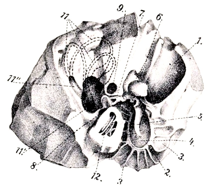
Fig. 565.—Right Petrous Temporal Bone of Horse, Antero-internal View. 1, External auditory meatus; 2, annulus tympanicus; 3, laminæ; radiating from 2; 4, membrana tympani; 5, malleus; 6, incus; 7, stapes; 8, fenestra cochleæ (s. rotundum); 9, facial canal; 11, 11″, semicircular canals; 11′, vestibule; 12, cochlea. (After Ellenberger, in Leisering’s Atlas)
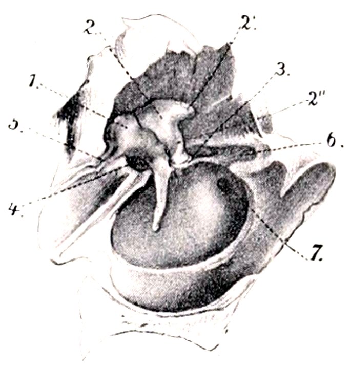
Fig. 566.—Right Auditory Ossicles and Membrana Tympani, Enlarged and Viewed from Inner Side and Below. 1, Malleus; 2, incus; 2′, 2″, short and long processes of 2; 3, stapes; 4, tensor tympani; 5, ligament attaching long process of malleus; 6, stapedius muscle; 7, fenestra cochleæ (s. rotundum). (After Ellenberger, in Leisering’s Atlas.)
The internal wall of the tympanic cavity (Paries labyrinthica) separates it from the internal ear; it presents a number of special features. The promontory (Promontorium) is a distinct eminence near the center which corresponds to the first coil of the cochlea, and is marked by a faint groove for the superficial petrosal nerve. Above this is the fenestra vestibuli (s. ovalis), a reniform opening which is closed by the foot-plate of the stapes and its annular ligament. The fenestra cochleæ (s. rotundum) is situated below and behind the preceding; it is an irregularly oval opening and is closed by a thin membrane (Membrana tympani secundaria), which separates the tympanic cavity from the scala tympani of the cochlea.
The anterior wall (Paries tubaria) is narrow, and is pierced by the slit-like 754tympanic opening of the Eustachian tube. Above this and incompletely separated from it by a thin plate of bone is the semicanal for the tensor tympani muscle.
The superior wall or roof (Paries tegmentalis) is crossed in its inner part by the facial nerve; here the facial canal is more or less deficient ventrally, and the nerve is covered by the mucous membrane of the tympanum.
The posterior wall (Paries mastoidea) presents nothing of importance; a tympanic antrum and mastoid cells, such as are found behind the tympanic cavity proper in man and many animals, are not present in the horse.
The inferior wall or floor (Paries tympanica) is concave and thin. It is crossed by delicate curved ridges, which radiate from the greater part of the annulus tympanicus.
The auditory ossicles (Ossicula auditus) form a chain which extends from the outer to the inner wall of the cavity. They are named, from without inward, the malleus, the incus, the os lenticulare, and the stapes. The first is attached to the inner surface of the tympanic membrane and the last is fixed in the fenestra vestibuli.
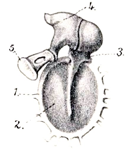
Fig. 567.—Auditory Ossicles and Membrani Tympani, Inner View, Enlarged. 1, Annulus tympanicus; 2, membrana tympani; 3, malleus; 4, incus; 5, stapes. (After Ellenberger, in Leisering’s Atlas.)
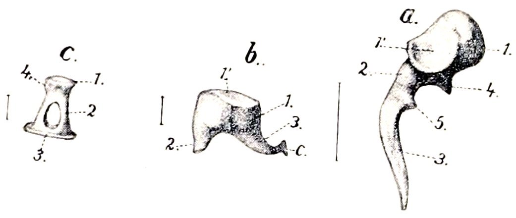
Fig. 568.—Right Auditory Ossicles, Enlarged as Indicated by Lines Giving their Actual Length. a, Malleus; 1, head; 2, neck; 3, handle; 4, long process; 5, muscular process. b, Incus; 1, body; 2, short branch; 3, long branch; c, os lenticulare. c, Stapes; 1, head; 2, crus; 3, base; 4, attachment of stapedius. (After Ellenberger, in Leisering’s Atlas.)
The malleus or hammer, the largest of the ossicles, consists of a head, neck, handle, and two processes. The head (Capitulum mallei) is situated in the epitympanic recess. It is smooth and convex above and in front, and presents on its postero-internal aspect a concave facet for articulation with the body of the incus. The neck (Collum mallei) is the constricted part below the head; its inner surface is crossed by the chorda tympani. The handle (Manubrium mallei) is directed downward, inward, and a little forward from the neck, and is attached along its entire length to the membrana tympani. On its inner surface, near the upper end, there is a slight projection to which the tendon of the tensor tympani muscle is attached. The long process (Processus longus) is a pointed spicule which projects forward from the neck toward the petro-tympanic fissure. The short process (Processus brevis) is a slight projection of the outer side of the neck, and is attached to the upper part of the membrana tympani.
The incus or anvil is situated chiefly in the epitympanic recess. It may be said to resemble a bicuspid tooth with two divergent roots, and consists of a body and two processes. The body (Corpus incudis) articulates with the head of the hammer. The long process (Crus longum) projects downward from the body and then curves inward; its extremity has attached to it a small nodule of bone, the os lenticulare, which articulates with the head of the stapes. The short process (Crus breve) projects chiefly backward, and is attached to the wall of the recess by a small ligament.
The stapes or stirrup consists of a head, two crura, and a base. The head 755(Capitulum stapedis) is directed outward and articulates with the os lenticulare. The crura, anterior and posterior (Crus anterius, posterius), are directed inward from the head, and join the ends of the base. The base or foot-plate (Basis stapedis) occupies the fenestra vestibuli, to which it is attached. The space between the crura and the base is closed by a membrane.
The articulations and ligaments of the auditory ossicles comprise: (1) A diarthrodial joint between the head of the malleus and the body of the incus (Articulatio incudomalleolaris), inclosed by a capsule. (2) An enarthrosis between the os lenticulare and the head of the stapes (Articulatio incudostapedia), also surrounded by a capsule. (3) The base of the stapes is attached to the margin of the fenestra vestibuli by a ring of elastic fibers (Lig. annulare baseos stapedis). (4) Small ligaments attach the head of the malleus and the short crus of the incus to the roof of the epitympanic recess. (5) The axial ligament (of Helmholtz) attaches the neck of the malleus to a small projection (Spina tympanica anterior) above and in front of the annulus tympanicus.
The muscles of the auditory ossicles are two in number, viz., the tensor tympani and the stapedius. The tensor tympani arises from the upper wall of the osseous Eustachian tube, and ends in a delicate tendon which bends outward and is inserted into the handle of the malleus near its upper end. When it contracts, it draws the handle of the malleus inward and tenses the membrana tympani; it probably also rotates the malleus around its long axis. It is innervated by the motor part of the trigeminus through the otic ganglion. The stapedius arises from a small prominence (Eminentia pyramidalis) of the posterior wall of the tympanum, runs forward on the facial nerve, and is inserted into the neck of the stapes. Its action is to draw the head of the stapes backward and rotate the anterior end of the base outward, thus tensing the annular ligament. It is innervated by the facial nerve.
The tympanic mucous membrane (Tunica mucosa tympanica) is continuous with that of the pharynx and the guttural pouch through the Eustachian tube. It is thin, closely united with the underlying periosteum, and is reflected over the ossicles, ligaments, and muscles, the chorda tympani, and the facial nerve in the open part of the facial canal. It contains minute lymph nodules and small mucous glands (Glandulæ tympanicæ). The epithelium is in general columnar ciliated, but over the membrana tympani, ossicles, and promontory it is flattened.
The artery of the tympanum is the stylo-mastoid, a small vessel which arises from the posterior auricular branch of the internal maxillary artery. It enters the tympanum through the stylo-mastoid foramen, and forms a circle around the tympanic membrane. The nerves of the mucous membrane come from the tympanic plexus.
The Eustachian Tube
The Eustachian or auditory tube (Tuba auditiva [Eustachii]) extends from the tympanic cavity to the pharynx; it transmits air to the former and equalizes the pressure on the two surfaces of the membrana tympani. It is directed forward, downward, and slightly inward, and is four to five inches (ca. 10 to 12 cm.) in length. Its posterior extremity lies at the inner side of the root of the muscular process of the petrous temporal, and communicates with the anterior part of the tympanic cavity by the small slit-like tympanic opening (Ostium tympanicum tubæ auditivæ). For a distance of about a quarter of an inch (ca. 6 to 7 mm.) in front of this opening it is a complete tube, with a curved lumen which is little more than a capillary space. Further forward it has the form of a plate which widens anteriorly and is curved to inclose a narrow groove, that opens ventrally into an extensive diverticulum termed the guttural pouch. The pharyngeal opening (Ostium pharyngeum tubæ auditivæ) is situated on the postero-superior part of the lateral wall of the pharynx just 756below the level of the posterior nares. It is a slit, an inch or more (ca. 3 cm.) in length, which slopes downward and backward. It is bounded internally by the thin free edge of the tube, from the lower part of which a fold of mucous membrane (Plica salpingo-pharyngea) extends in the same direction on the lateral wall of the pharynx for a distance of a little more than an inch usually. The outer boundary of the opening is the lateral wall of the pharynx. The basis of the tube is a plate of fibro-cartilage (Cartilago tubæ auditivæ) which is firmly attached dorsally to the fibrous tissue which closes the foramen lacerum basis cranii, the temporal wing of the sphenoid, and the pterygoid bone.[222] On cross-section the cartilage (except at its tympanic end) is seen to consist of two laminæ which are continuous with each other above. The internal lamina (Lamina medialis) gradually widens toward the pharyngeal end, where it forms a broad valvular flap. This is convex internally, and its thin anterior edge forms the inner margin of the pharyngeal opening. Behind this the lamina has a thick free edge which projects ventrally from the roof of the guttural pouch. The external lamina (Lamina lateralis) is narrow and thin, and is related externally to the levator and tensor palati muscles, which are in part attached to it; it does not exist at the pharyngeal end of the tube. The mucous membrane of the tube is continuous behind with that of the tympanum and in front with that of the pharynx. On either side it is reflected to form a large diverticulum, the guttural pouch. It is covered with ciliated epithelium, and contains mucous glands and lymph nodules.
The pharyngeal opening appears to be closed ordinarily, but the tube opens during deglutition. This action is apparently produced by the part of the palato-pharyngeus muscle which is attached to the tube.
The Guttural Pouches
The guttural pouches, right and left (Figs. 255, 256, 569), are large mucous sacs, each of which is a ventral diverticulum of the Eustachian tube (Diverticulum tubæ auditivæ); they are not present in the domesticated animals other than the equidæ. They are situated between the base of the cranium and the atlas above and the pharynx below. Medially they are in apposition in great part, but are to some extent separated by the intervening ventral straight muscles of the head. The anterior end is a small cul-de-sac which lies below the body of the presphenoid between the Eustachian tube and the median recess of the pharynx. The posterior extremity lies near or below the atlantal attachment of the longus colli. The pouch is related dorsally to the base of the cranium, the atlanto-occipital joint capsule, and the ventral straight muscles. Ventrally it lies on the pharynx and the origin of the œsophagus. Laterally the relations are numerous and complex. They comprise the pterygoid, levator palati, tensor palati, stylo-hyoideus, and digastricus muscles; the parotid and submaxillary glands; the external carotid, internal maxillary, and external maxillary arteries; the internal maxillary and jugular veins; the pharyngeal or guttural lymph glands; the glosso-pharyngeal, hypoglossal, and superior laryngeal nerves. The vagus, accessory, and sympathetic nerves, the superior cervical ganglion, the internal carotid artery, and the inferior cerebral vein are situated in a fold of the upper wall of the pouch. The pouch is reflected around the anterior border of the great cornu of the hyoid bone so as to clothe both surfaces of the upper part of the latter. It thus forms an outer compartment, which extends backward external to the great cornu and the occipito-hyoideus muscle; it is related externally to the parotid gland, the articulation of the jaw, the ascending part of the internal maxillary artery, the superficial temporal artery, and the facial nerve; dorsally it covers the mandibular nerve and its chief branches, and is attached to the styloid process of the conchal cartilage.
757Each pouch communicates with the pharynx through the pharyngeal orifice of the Eustachian tube, and is in direct continuity with the mucous membrane of the latter.
The wall of the pouch is a delicate mucous membrane which is in general rather loosely attached to the surrounding structures. It is covered with ciliated epithelium and is supplied with mucous glands.
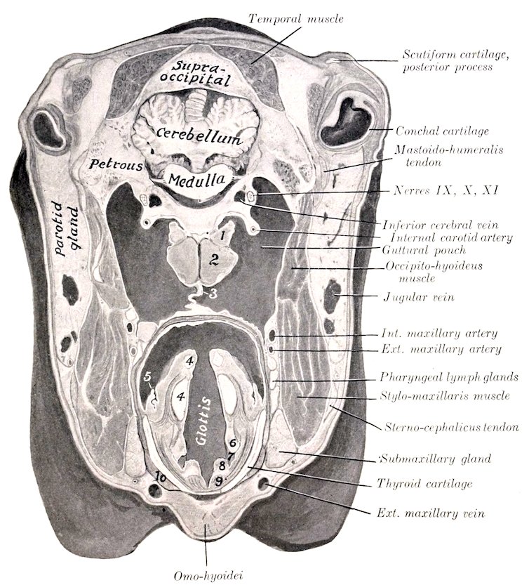
Fig. 569.—Cross-section of Head of Horse. The section passes through the base of the external ear and just behind the posterior border of the lower jaw. 1, Rectus capitis anterior minor; 2, rectus capitis anterior major; 3, inner walls of guttural pouches in apposition; 4, 4, arytenoid cartilage, upper piece apex; 5, posterior pillar of soft palate; 6, false vocal cord; 7, lateral ventricle of larynx; 8, true vocal cord; 9, vocal muscle; 10, thyro-hyoideus muscle.
It is worthy of note that the pharyngeal orifice of the Eustachian tube is at such a level as to provide (in the ordinary position of the head) only an overflow outlet for the escape of fluid which may accumulate in the pouch. The two pouches are often unequal in size, and variations in regard to the distance which they extend backward are not uncommon. In one case, a small aged horse, the right pouch extended along the œsophagus about five inches (ca. 12 cm.) behind the ventral tubercle of the atlas, and the left one a little more than two inches (ca. 6 cm.). No pathological changes were apparent, and the condition was not recognizable externally. Cases of extreme size—so-called tympanites—of the pouches occur, and are apparently congenital defects. In a case in a yearling colt, the head of which was 24 inches long, the left pouch extended about 12 inches (ca. 30 cm.) behind the tubercle of the atlas, and had a capacity of six quarts. 758The anterior end formed a cul-de-sac about two inches (ca. 5 cm.) long between the Eustachian tube and levator palati internally and the internal pterygoid muscle externally.
THE INTERNAL EAR
The internal ear or labyrinth (Auris interna s. Labyrinthus) consists of two parts, viz.: (1) a complex membranous sac, which supports the auditory cells and the peripheral ramifications of the auditory nerve; (2) a series of cavities in the petrous temporal bone, which inclose the membranous part. The first is called the membranous labyrinth, and contains a fluid, the endolymph. The second is the osseous labyrinth. The two are separated by the perilymphatic space, which is occupied by a fluid termed the perilymph.
The Osseous Labyrinth
The osseous labyrinth (Labyrinthus osseus) (Fig. 565) is excavated in the petrous temporal bone to the inner side of the tympanic cavity. It consists of three divisions: (1) a middle part, the vestibule; (2) an anterior one, the cochlea; and (3) a posterior one, the semicircular canals.
1. The vestibule (Vestibulum) is the central part of the osseous labyrinth, and communicates in front with the cochlea, behind with the semicircular canals. It is a small, irregularly ovoid cavity, which is about 5 to 6 mm. in length. Its outer wall separates it from the tympanic cavity, and in it is the fenestra vestibuli, which is occupied by the base of the stapes. The inner wall corresponds to the fundus of the internal auditory meatus. It is crossed by an oblique ridge, the crista vestibuli, which separates two recesses. The anterior and smaller of these is the recessus sphæricus, which lodges the saccule of the membranous labyrinth. In its lower part there are about a dozen minute foramina which transmit filaments of the vestibular nerve to the saccule. The posterior and larger depression is the recessus ellipticus, which lodges the utricle of the membranous labyrinth. The crista vestibuli divides below into two divergent branches, which include between them the small recessus cochlearis; this is perforated by small foramina, through which nerve-bundles reach the ductus cochlearis. Similar foramina in the recessus ellipticus and the crista vestibuli transmit nerve filaments to the utricle and the ampullæ of the superior and external semicircular ducts. The anterior wall is pierced by an opening which leads into the scala vestibuli of the cochlea. The posterior part of the vestibule presents the four openings of the semicircular canals. The inner opening of the aquæductus vestibuli is a small slit behind the lower part of the crista vestibuli. The aquæductus passes backward in the petrous temporal bone, and opens on the inner surface of the latter near the middle of its posterior border; it contains the ductus endolymphaticus.
2. The osseous semicircular canals (Canales semicirculares ossei), three in number, are situated behind and above the vestibule. They are at right angles to each other, and are designated according to their positions as superior, posterior, and external. They communicate with the vestibule by four openings only, since the inner end of the superior and the upper end of the posterior canal unite to form a common canal (Crus commune), and the ampullate ends of the superior and external canals have a common orifice. Each canal forms about two-thirds of a circle, one end of which is enlarged and termed the ampulla. The superior canal (Canalis semicircularis superior) is nearly vertical and is placed obliquely with regard to a sagittal plane, so that its outer limb is further forward than the inner one. The antero-external end is the ampulla and opens into the vestibule with that of the external canal. The opposite non-dilated end joins the adjacent end of the posterior canal to form the crus commune, which opens into the supero-internal part of the vestibule. The posterior canal (Canalis semicircularis posterior) is 759also nearly vertical. Its ampulla is below, and opens into the vestibule directly, while the non-dilated end joins that of the superior canal. The external canal (Canalis semicircularis lateralis) is nearly horizontal. Its ampulla is external and opens into the vestibule with that of the superior canal.
3. The cochlea is the anterior part of the bony labyrinth. It has the form of a short blunt cone, the base of which (Basis cochleæ) corresponds to the anterior part of the fundus of the internal auditory meatus, while the apex or cupola (Cupula) is directed outward, forward, and downward. It measures about 7 mm. across the base and about 4 mm. from base to apex. It consists of a spiral canal (Canalis spiralis cochleæ), which forms two and a half turns around a central column termed the modiolus. The modiolus diminishes rapidly in diameter from base to apex. Its base (Basis modioli) corresponds to the area cochleæ of the fundus of the internal auditory meatus, and its apex extends nearly to the cupola. Projecting from the modiolus like the thread of a screw is a thin plate of bone, the lamina spiralis ossea. This begins between the two fenestræ and ends near the cupola as a hook-like process (Hamulus laminæ spiralis). The lamina extends about half-way to the periphery of the cochlea and partially divides the cavity into two passages; of these, the upper one is termed the scala vestibuli, and the lower the scala tympani. The membrana basilaris extends from the free margin of the lamina to the outer wall of the cochlea and completes the septum between the two scalæ, but they communicate through the opening (Helicotrema) at the cupola. The modiolus is traversed by an axial canal which transmits the nerves to the apical coil, and by a spiral canal (canalis spiralis modioli), which follows the attached border of the lamina spiralis, and contains the spiral ganglion and vein. Close to the beginning of the scala tympani is the inner orifice of the aquæductus cochleæ, a small canal which opens behind the internal auditory meatus, and establishes a communication between the scala tympani and the subarachnoid space.
The internal auditory meatus has been described in part (vide Osteology). The fundus of the meatus is divided by a ridge (Crista transversa) into upper and lower parts. The anterior part of the upper depression (Area n. facialis) presents the cranial opening of the facial canal; and the posterior part (Area vestibularis superior) is perforated by foramina for the passage of nerves to the utricle and the ampullæ of the superior and external membranous semicircular canals. The anterior part of the inferior depression (Area cochleæ) presents a central foramen and a spiral tract of minute foramina (Tractus spiralis foraminosus) for the passage of nerves to the cochlea. Behind these is an area of small openings which transmit nerves to the saccule (Area vestibularis inferior), and a single foramen (F. singulare) for the passage of a nerve to the ampulla of the posterior semicircular canal.
The Membranous Labyrinth
The membranous labyrinth (Labyrinthus membranaceus) lies within, but does not fill, the osseous labyrinth. It is attached to the latter by delicate trabeculæ which traverse the perilymphatic space. It conforms more or less closely to the bony labyrinth, but consists of four divisions, since the vestibule contains two membranous sacs—the utricle and saccule.
1. The utricle (Utriculus), the larger of the two sacs, lies in the postero-superior part of the vestibule, largely in the recessus ellipticus. It receives the openings of the membranous semicircular canals, and the small ductus utriculo-saccularis leads from its lower part to the ductus endolymphaticus.
2. The saccule (Sacculus) is situated in the recessus sphæricus of the vestibule. From its lower part the ductus reuniens proceeds to open into the ductus cochlearis, a little in front of the blind end of the latter. A second narrow tube, the ductus endolymphaticus, passes from the posterior part of the saccule, and is joined by the ductus utriculo-saccularis; it then traverses the aquæductus vestibuli, and terminates 760under the dura mater on the posterior part of the internal surface of the petrous temporal bone in a dilated blind end, the saccus endolymphaticus.
3. The membranous semicircular canals (Ductus semicirculares) correspond in general to the osseous canals already described, but it may be noted that while the ampullæ of the membranous canals nearly fill those of the osseous canals, the other parts of the membranous canals only occupy about one-fourth of the bony cavities.
4. The cochlear duct (Ductus cochlearis) is a spiral tube situated within the cochlea. It begins by a blind end (Cæcum vestibulare) in the cochlear recess of the vestibule, and ends by a second blind end (Cæcum cupulare), which is attached to the cupola of the cochlea. The vestibular part is connected with the saccule by the ductus reuniens. The duct is triangular in cross-section, and it is usual to regard it as having three walls. The vestibular wall or roof, which separates the cochlear duct from the scala vestibuli, is formed by the very delicate membrana vestibularis (of Reissner), which extends obliquely from the lamina spiralis ossea to the outer wall of the cochlea. The tympanic wall or floor intervenes between the cochlear duct and the scala tympani; it is formed by the periosteum of the marginal part of the lamina spiralis and the membrana basilaris, which stretches between the free edge of the lamina and the outer wall of the cochlea. The outer wall consists of the fibrous lining of the cochlea, which is greatly thickened to form the ligamentum spirale cochleæ.
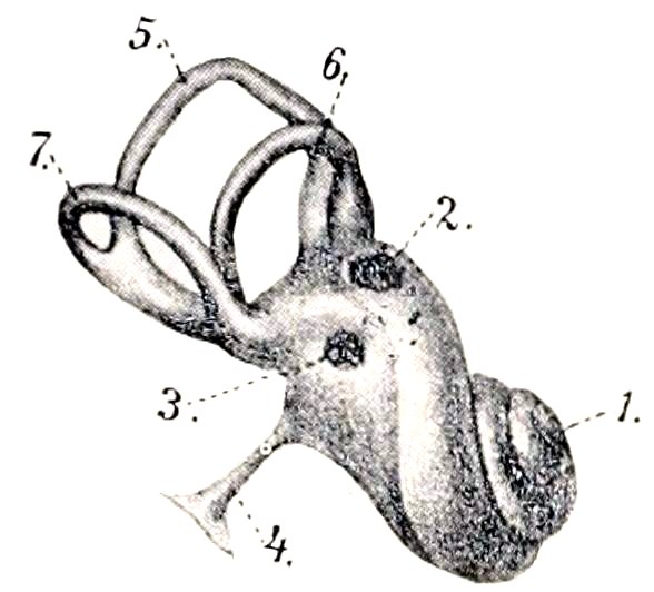
Fig. 570.—Left Membranous Labyrinth (Enlarged). 1, Cochlea; 2, fenestra vestibuli; 3, fenestra cochleæ; 4, ductus endolymphaticus; 5, superior, 6, external, 7, inferior semicircular canal. (After Ellenberger, in Leisering’s Atlas.)
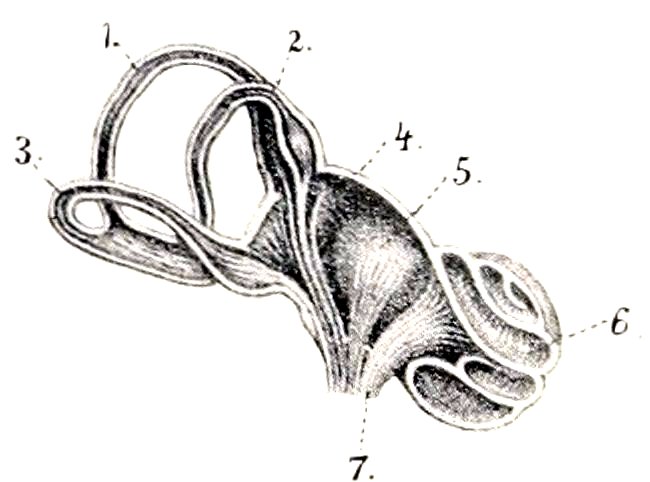
Fig. 571.—Schematic Sectional View of Labyrinth (Enlarged). 1, 2, 3, Superior, external, and inferior semicircular canals; 4, utricle; 5, saccule; 6, cochlea; 7, auditory nerve. (After Ellenberger, in Leisering’s Atlas.)
Structure.[223]—The membranous labyrinth consists in general of an outer thin fibrous layer, a middle transparent tunic, and an internal epithelium, composed of flattened cells. But in certain situations special and remarkable structures occur, among which are the following: (1) The maculæ acusticæ appear as small whitish thickenings of the inner walls of the saccule and utricle. The epithelium here consists of two kinds of cells, viz., supporting cells and hair cells. The latter are flask-shaped and are surrounded by the fusiform supporting cells. The free end of each hair cell bears a stiff, hair-like process composed of a bundle of cilia. Fibers of the saccular and utricular branches of the vestibular nerve form arborizations about the basal parts of the hair cells. Adherent to the surface of the maculæ are fine crystals of lime salts, embedded in a mucoid substance, and termed otoconia. (2) The cristæ acusticæ are linear thickenings of the wall of each ampulla of the membranous semicircular canals. Their structure is similar to that of the maculæ. (3) The spiral organ of Corti (Organon spirale) is an epithelial elevation which is situated upon the inner part of the membrana basilaris, and extends the entire length of the ductus cochlearis. It is very complicated in structure, but consists 761essentially of remarkable supporting cells and hair cells. Fibers of the cochlear nerve ramify about the basal parts of the hair cells.
Vessels and Nerves.—The artery of the internal ear is the internal auditory artery, a very small vessel which usually arises from the posterior cerebellar artery, and enters the internal auditory meatus. The veins go to the inferior petrosal sinus. The vestibular nerve is distributed to the utricle, saccule, and membranous semicircular canals, and mediates equilibration. The cochlear nerve gives a branch to the saccule and enters the central canal of the modiolus. Along its course it gives off fibers which radiate outward between the two plates of the lamina spiralis ossea, and ramify about the hair cells of the organ of Corti. The ganglion spirale or ganglion of Corti is situated in the spiral canal of the modiolus near the fixed border of the lamina spiralis. The cochlear nerve mediates the sense of hearing.
The Skin
The skin or common integument (Integumentum commune) is the protective covering of the body, and is continuous at the natural openings with the mucous membranes of the digestive, respiratory, and urogenital tracts. It contains peripheral ramifications of the sensory nerves, and is thus an important sense organ. It is the principal factor in the regulation of the temperature of the body, and by means of its glands it plays an important part in secretion and excretion. Some of its special horny modifications or appendages are used as organs of prehension or as weapons.
The thickness of the skin varies in the different species, on different parts of the body of the same animal, and also with the breed, sex, and age. The color also varies greatly, but this is masked in most places by the covering of hair or wool. The skin is in general highly elastic and resistant.
Permanent folds of the skin (Plicæ cutis) occur in certain situations, and in some places there are cutaneous pouches or diverticula (Sinus cutis).
The skin is attached to the underlying parts by the subcutis or superficial fascia (Tela subcutanea). This consists of connective tissue containing elastic fibers and fat. When the fat forms a layer of considerable thickness, it is termed the panniculus adiposus. Over a considerable part of the body the subcutis contains striped muscle, the panniculus carnosus (M. cutaneus); in some regions the fibers of the muscle are inserted into the skin, and their contraction twitches the skin or produces temporary folds.[224] The amount of subcutaneous tissue varies widely; in some places it is abundant, so that the skin can be raised considerably; in other situations it is practically absent and the skin is closely adherent to the subjacent structures. Subcutaneous bursæ often develop over prominent parts of the skeleton where there is much pressure or friction.
Structure.—The integument consists of the cutis or skin proper and its epidermal appendages, e. g., hairs, hoofs, claws, horns, etc. The cutis consists of two distinct strata, viz., a superficial epithelial layer, the epidermis, and a deep connective-tissue layer, the corium.
The epidermis is a non-vascular, stratified epithelium of varying thickness. It presents the openings of the cutaneous glands and the hair-follicles, and its deep surface is adapted to the corium.[225] It is divisible into a superficial, harder, drier part, the stratum corneum, and a deeper, softer, moister part, the stratum germinativum. The cells of the latter contain pigment, and by their proliferation compensate the loss by desquamation of the superficial part of the stratum corneum. 762In many places further subdivision into strata is evident on properly prepared cross-sections.
The corium consists essentially of a feltwork of connective-tissue and elastic fibers. It is well supplied with vessels and nerves, and contains the cutaneous glands, the hair-follicles, and unstriped muscle. The deeper part of the corium, the tunica propria, consists of a relatively loose network of coarse bundles of fibers, and in most places there is no clear line of demarcation between it and the subcutis. The superficial part, the corpus papillare, is of finer texture and is free from fat. Its superficial face is thickly beset with blunt conical prominences, the papillæ, which are received into corresponding depressions of the epidermis. They contain vascular loops and nerves, or, in certain situations, special nerve-endings.
The papillæ are best developed where the epidermis is thick and hairs are small or absent. On thickly-haired regions they are small or even absent. On certain parts of the body (anus, vulva, prepuce, scrotum, eyelids, etc.) the corium contains pigment in its connective-tissue cells.
The glands of the skin (Glandulæ cutis) are chiefly of two kinds, sudoriferous and sebaceous.
The sudoriferous or sweat glands (Glandulæ sudoriferæ) consist of a tube, the lower, secretory part of which is coiled in the deep part of the corium or in the subcutis to form a round or oval ball (Corpus glandulæ sudoriferæ). The excretory duct (Ductus sudoriferus) passes almost straight up through the corium, but pursues a more or less flexuous course through the epidermis, and opens into a hair-follicle or by a funnel-shaped pore (Porus sudoriferus) on the surface of the skin.
The sebaceous glands (Glandulæ sebaceæ) are in great part associated with the hairs, into the follicles of which they open. Their size varies widely, and is in general in inverse ratio to that of the hair. The larger ones are easily seen with the naked eye, and appear as small, pale yellow or brownish bodies. In certain situations (e. g., the labia vulvæ, anus, prepuce) they are independent of the hairs and are well developed. In form they may be branched alveolar, simple alveolar, or even tubular in type. They secrete a fatty substance, the sebum cutaneum, which serves as a protective against moisture, and may also (by its aromatic constituents) play an important part in the sexual life of animals.
The two kinds of glands described above are those which are most widely distributed, but many special types occur. Some of these are to be regarded as modified sweat glands, e. g., the naso-labial glands of the ox, the glands of the snout of the pig, and the glands of the plantar cushion of the horse. Others, e. g., the tarsal (or Meibomian) glands of the eyelids, are modified sebaceous. Still others are not yet classified satisfactorily. Some of these special types have been referred to in previous chapters, and others will receive attention in the special descriptions which follow. The mammary glands are highly modified cutaneous glands, which are intimately associated in function with the genital organs, and have been described with the latter.
Vessels and Nerves.—The arteries of the skin enter from the subcutis, where they communicate freely. In the deeper part of the corium they form a plexus, and another network is formed under the papillæ. Small vessels from the deep plexus go to the fat and sweat glands, and the subpapillary plexus sends fine branches to the papillæ, hair-follicles, and sebaceous glands. The veins form two plexuses, one beneath the papillæ, and another at the junction of the corium and subcutis. The lymph vessels form subpapillary and subcutaneous plexuses.
The nerves vary widely in number in different parts of the skin. The terminal fibers either end free in the epidermis and in certain parts of the corium, or form special microscopic corpuscles of several kinds.
THE APPENDAGES OF THE SKIN
The appendages of the skin are modifications of the epidermis, and comprise the hairs, hoofs, claws, horns, etc.
The hairs (Pili) cover almost the entire surface of the body in the domesticated 763mammals, and some parts which appear at first sight to be bare are found on close inspection to be provided with sparse and very fine hair. The hairs are constantly being shed and replaced, but at certain periods in the horse, for example, they fall out in great numbers, constituting the shedding of the coat. It is customary to distinguish the ordinary hairs (the coat), which determine the color of the animal, from the special varieties found in certain places. Among the latter are the long tactile hairs about the lips, nostrils, and eyes; the eyelashes or cilia; the tragi of the external ear; and the vibrissæ of the nostrils. Other special features will be noted in the discussion of the skin of the various species. The hairs are directed in such a way as to form more or less definite hair-streams (Flumina pilorum), and at certain points these converge to form vortices (Vortices pilorum).
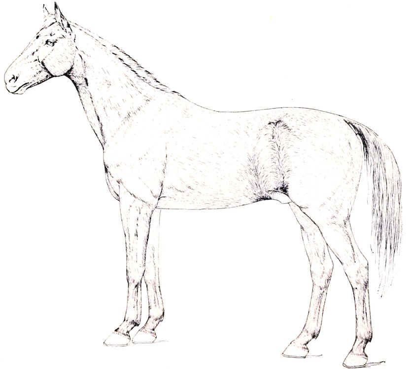
Fig. 572.—Lateral View of Horse to Show Hair-streams and Vortices. (After Ellenberger-Baum, Anat. für Künstler.)
The part of the hair above the surface of the skin is the shaft (Scapus pili), while the root (Radix pili) is embedded in a depression termed the hair-follicle (Folliculus pili). A vascular papilla (Papilla pili) projects up in the fundus of the follicle and is capped by the expanded end of the root, the bulb of the hair (Bulbus pili). The hair-follicles extend obliquely into the corium to a varying depth; in the case of the long tactile hairs they reach to the underlying muscle. Most of the follicles have attached to them small unstriped muscles known as the arrectores pilorum; these are attached at an acute angle to the under side of the deep part of the follicle, and their contraction causes erection of the hair and compression of the sebaceous glands, one or more of which open into the follicle.
The hairs are composed of epidermal cells, and consist from without inward of three parts. The cuticle is composed of horny, scale-like cells which overlap like slates on a roof. The cortex consists of horny fusiform cells which are packed close 764together and contain pigment. The medulla is the central core of softer, cubical or polyhedral cells; it contains some pigment and air-spaces.
The hair-follicles, being invaginations of the skin, are composed of a central epidermal part, and a peripheral layer which corresponds in structure to the corium. The follicles of the tactile hairs have remarkably thick walls which contain blood-sinuses between their outer and inner layers; in ungulates the sinuses are crossed by trabeculæ and assume the character of cavernous or erectile tissue.
The hoofs, claws, horns, and other horny structures consist of closely packed epidermal cells which have undergone cornification. In structure they might be compared to hairs matted together by intervening epidermal cells. They cover a specialized corium known as their matrix, from which the stratum germinativum derives its nutrition.
THE SKIN OF THE HORSE
The thickness of the skin of the horse varies from 1 to 5 mm. in different regions, and is greatest at the attachment of the mane and the ventral surface of the tail.
The glands are numerous and are larger than those of the other domesticated animals. The sebaceous glands are specially developed on the lips, the prepuce, mammary glands, perineum, and labia of the vulva. The sweat glands are yellow or brown in color. They occur in almost all parts of the skin, but are largest and most numerous on the outer wing of the nostril, the flank, mammary glands, and free part of the penis.