THE DIGESTIVE SYSTEM
Reading edition
This system consists of the organs directly concerned in the reception and digestion of the food, its passage through the body, and the expulsion of the unabsorbed portion. These organs are conveniently grouped under two heads, viz.: (1) the alimentary canal; (2) the accessory organs.
The alimentary canal (Tractus alimentarius) is a tube, about 100 feet (ca. 30 m.) in length in the horse, and extends from the lips to the anus. It has a complete lining of mucous membrane, external to which is an almost continuous muscular coat. The abdominal portion of the tube is largely covered with a serous membrane—the visceral peritoneum. The canal consists of the following consecutive segments:
1.
Mouth.
2.
Pharynx.
3.
Œsophagus.
4.
Stomach.
5.
Small intestine.
6.
Large intestine.
The accessory organs are the teeth, tongue, salivary glands, liver, and pancreas.
DIGESTIVE SYSTEM OF THE HORSE
THE MOUTH
The mouth[77] (Cavum oris) is the first part of the alimentary canal. In the horse it is a long cylindrical cavity, and when closed, it is almost entirely filled up by the contained structures. The entrance to it (Rima oris) is closed by the lips. Laterally it is bounded by the cheeks; above, by the hard palate; below, by the body of the mandible and the mylo-hyoid muscles; behind, by the soft palate.
The cavity of the mouth is subdivided into two parts by the teeth and alveolar processes. The space external to these and inclosed externally by the lips and cheeks is termed the vestibule of the mouth (Vestibulum oris). In the resting state of the parts the walls of this cavity are in contact, and the space is practically obliterated. Its existence becomes very evident in facial paralysis, when the food tends to collect in it laterally, pouching out the cheeks. The space within the teeth and alveolar processes is termed the mouth cavity proper (Cavum oris proprium). When the teeth are in contact, it communicates with the vestibule only by the interdental spaces and the intervals behind the last molar teeth. Posteriorly it communicates with the pharynx through the isthmus of the fauces.
The mucous membrane lining the mouth (Tunica mucosa oris) is continuous at the margin of the lips with the common integument, and behind with the mucous lining of the pharynx. During life it is chiefly of a pink color.
The lips (Labia oris) are two musculo-membranous folds which surround the orifice of the mouth. Their angles of union (Anguli oris s. commissuræ labiorum) are situated near the first cheek tooth and are rounded. Each lip presents two surfaces and two borders. The outer surface is covered by the skin, which presents long tactile hairs in addition to the ordinary fine hair. The upper lip shows 331a shallow median furrow (Philtrum), the lower a rounded prominence, the chin (Mentum). The inner or oral surface is covered with mucous membrane which is commonly more or less pigmented. The small papillæ on this surface show on their summits the openings of the ducts of the labial glands. Small folds of mucous membrane which pass from the lip to the gum represent the frænula labii (superioris, inferioris). The free border of the lip is dense and presents short, very stiff hairs. The attached border is continuous with the surrounding structures, and is adherent to the alveolar borders of the bones of the jaws.
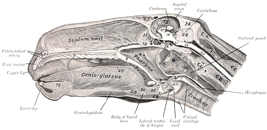
Fig. 238.—Sagittal Section of Head of Horse. The Section is about 1 cm. to the Left of the Median Plane. 1, Hard palate; 2, soft palate; 3, posterior pillar of soft palate; 4, Eustachian opening; 5, great cornu of hyoid bone; 6, stylo-pharyngeus muscle; 9, glosso-pharyngeal nerve; 10, hypoglossal nerve; 11, perpendicular plate of ethmoid; 12, presphenoid; 13, sphenoidal sinus; 14, occipital bone; 15, median surface of cerebral hemisphere; 16, corpus callosum; 17, septum lucidum; 18, fornix; 19, thalamus opticus; 20, corpora quadrigemina; 21, pons; 22, medulla oblongata; 23, spinal cord; 24, cerebellum; 25, pituitary body; 26, hyo-epiglottic muscle; 27, superior longitudinal muscle of tongue; 28, omo-hyoideus; 29, hyo-glossus; 30, hyoideus transversus; 31, thyroid cartilage (body); S. f., frontal sinus; L. n., lig. nuchæ; C I, atlas; C II, axis; R. c. m., rectus cap. ant. major and minor; E p., epiglottis; A r., arytenoid cartilage; P m., premaxilla; M., mandible. The leader line for the guttural pouch points to the external carotid artery.
332Structure.—The lips are covered externally by the skin, and are lined by mucous membrane; between these are muscular tissue and glands. The skin lies directly on the muscles, many fibers of which are inserted into the former. The muscles have been described (page 214). The labial glands (Glandulæ labiales) form a compact mass near the angles; they are numerous in the upper lip, fewer in the lower. The mucous membrane is often pigmented, and is reflected upon the bones of the jaws to form the gums.
Blood-vessels and Nerves.—The arteries are derived from the superior and inferior labial and palato-labial arteries. The sensory nerves come from the trigeminus, and the motor from the facial nerve.
The cheeks (Buccæ) form the sides of the mouth, and are continuous in front with the lips. They are attached above and below to the alveolar borders of the bones of the jaws.
Structure.—This comprises: (1) The skin; (2) the muscular and glandular layer; (3) the mucous membrane. The skin offers nothing in particular to notice. The muscular tissue is formed mainly by the buccinator, but also by parts of the panniculus, zygomaticus, dilatator naris lateralis, levator nasolabialis, and depressor labii inferioris. The buccal glands (Glandulæ buccales) are arranged in two rows. The upper row (superior buccal or molar glands) is found on the outer surface of the buccinator muscle, near its upper border. The anterior part of the row consists of scattered lobules; the posterior part, which lies under cover of the masseter muscle, is more developed and compact. The lower row (inferior buccal or molar glands), less voluminous than the upper, is situated in the submucous tissue at the lower border of the buccinator muscle. The mucous membrane is reflected above and below upon the gums, and is continuous behind with that of the soft palate. It is reddish in color and frequently shows pigmented areas. Opposite the third upper cheek tooth is the opening of the parotid (Stenson’s) duct, surrounded by a circular fold of the mucous membrane. A linear series of small papillæ above and below indicates the orifices of the small ducts from the buccal glands.
Blood-vessels and Nerves.—The blood-supply is derived from the facial and buccinator arteries. The sensory nerves come from the trigeminus and the motor from the facial nerve.
The gums (Gingivæ) are composed of a dense fibrous tissue intimately united with the periosteum of the alveolar processes, and blending at the edges of the alveoli with the alveolar periosteum, which fixes the teeth in their cavities. They are covered by a smooth mucous membrane, destitute of glands, and of a low degree of sensibility.
The hard palate (Palatum durum) is bounded in front and on the sides by the alveolar arches; behind it is continuous with the soft palate. Its osseous basis is formed by the premaxilla, maxilla, and palate bones. The mucous membrane is smooth, and is attached to the bones by a submucosa which contains in its anterior part an exceedingly rich venous plexus, constituting an erectile tissue. A central raphé (Raphé palati) divides the surface into two equal portions. Each of these presents about eighteen transverse curved ridges (Rugæ palatini) which have their concavity and their free edges directed backward. They are further apart and more prominent anteriorly. There are no glands in the submucosa.
Vessels and Nerves.—The blood-supply is derived chiefly from the palatine arteries and the nerves from the trigeminus.
The soft palate (Palatum molle) is a musculo-membranous curtain which separates the cavity of the mouth from that of the pharynx. It slopes downward and backward from its junction with the hard palate. The oral surface faces 333downward and forward, and is covered with a mucous membrane continuous with that of the hard palate. It is corrugated and presents numerous small orifices (of gland-ducts) and two sagittal ridges. On each side a short, thick fold passes to join the base of the tongue; this is the anterior pillar of the soft palate (Arcus glossopalatinus). The pharyngeal surface looks upward and backward and is covered by a mucous membrane continuous with that of the nasal cavity. The free border (Arcus palatinus) is concave and thin; it is in contact (except during deglutition) with the epiglottis. It is continuous with a fold of the mucous membrane, which passes on each side along the lateral wall of the pharynx and unites with its fellow over the beginning of the œsophagus; this fold is termed the posterior pillar of the soft palate (Arcus pharyngopalatinus). The space between the diverging anterior and posterior pillars (Sinus tonsillaris) is occupied by the faucial tonsil. In the horse, however, there is not a compact tonsil, as in man, dog, etc., but a somewhat extended group of mucous glands and masses of lymphoid tissue. These cause elevations of the surface, on which are seen numerous depressions (crypts) in which the gland-ducts open. The soft palate is greatly developed in equidæ, its length being five to six inches (12 to 15 cm.). Its length and contact with the epiglottis account for the fact that in these animals mouth-breathing does not occur under normal conditions, and that in vomiting the ejected matter escapes usually through the nasal cavity.[78]
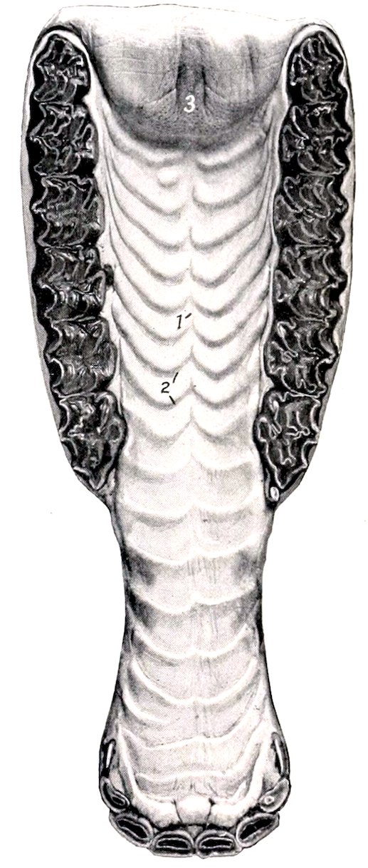
Fig. 239.—Hard Palate and Part of Soft Palate of Horse. 1, Raphé palati; 2, ridges of palate; 3, anterior end of soft palate, showing orifices of ducts of palatine glands.
Structure.—The soft palate consists of: (1) The oral mucous membrane, continuous with that of the hard palate; (2) the palatine glands (Glandulæ palatinæ), which form a layer about half an inch in thickness; (3) the aponeurotic and muscular layer; (4) the pharyngeal mucous membrane, continuous with that of the nasal cavity.
The muscles proper to the soft palate are the azygos uvulæ, the levator palati, and the tensor palati.
The azygos uvulæ (M. palatinus) consists of two small muscular bundles which lie together at the median line. It is attached through the medium of the palatine aponeurosis to the palatine arch, and terminates near the free edge of the soft palate. Its action is to shorten and raise the soft palate.
The levator palati (M. levator veli palatini) arises from the muscular process of the petrous temporal bone and from the Eustachian tube, and passes forward and downward externally to the latter, to spread out on the pharyngeal surface of the soft palate. It raises the soft palate, thus closing the posterior nares during deglutition.
The tensor palati (M. tensor veli palatini) is larger than the levator, and is fusiform and flattened. It arises from the muscular process of the petrous temporal bone and the Eustachian tube, and passes forward external to the levator. 334Its tendon is then reflected around the hamulus of the pterygoid bone, being held in position by a fibrous band and lubricated by a bursa. It turns inward and expands in the aponeurosis of the soft palate. It tenses the soft palate.
Vessels and Nerves.—The blood-supply of the soft palate is derived from the internal and external maxillary arteries. The nerves come from the trigeminus, vagus, and glosso-pharyngeal nerves.
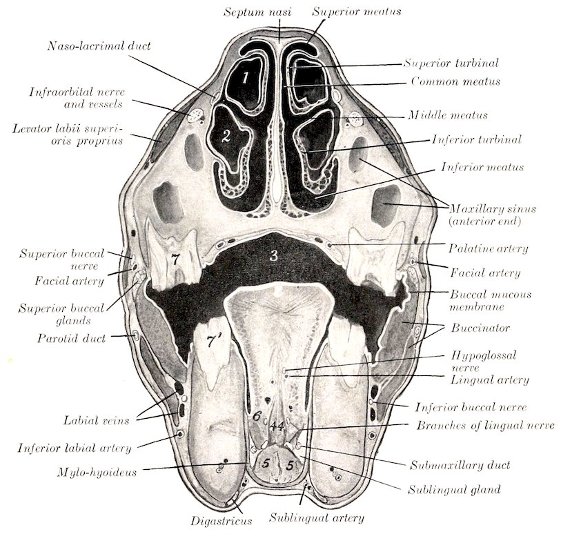
Fig. 240.—Cross-section of Head of Horse just in Front of Facial Crest. 1, Cavity of superior turbinal; 2, cavity of inferior turbinal; 3, cavum oris; 4, 4, genio-glossi; 5, 5, genio-hyoidei; 6, hyo-glossus; 7, upper, 7′, lower fourth cheek tooth. Line to facial artery crosses zygomaticus.
The floor of the mouth in its anterior part is free and is formed by the body of the mandible, covered by mucous membrane. The remainder is concealed by the attached portion of the tongue, with the exception of a narrow space on each side of the latter. About opposite the canine tooth on each side is a papilla, the caruncula sublingualis, through which the duct of the submaxillary gland opens. Just behind these papillæ is a median fold of mucous membrane which passes to the under surface of the tongue, constituting the frenum linguæ. On either side are the sublingual crests, which extend from the frenum to the level of the fourth cheek tooth. The crest presents numerous small papillæ, through which open the ducts from the subjacent sublingual gland.[79] Behind the last tooth a vertical fold 335of the mucous membrane passes from upper to lower jaw. This is termed the plica pterygomandibularis: it contains a ligament of like name.
The isthmus faucium is the orifice of communication between the mouth and the pharynx. It is bounded above by the soft palate, below by the root of the tongue, and laterally by the anterior pillars of the soft palate. It is long, relatively small, and not very dilatable in the horse, and is closed by the soft palate under normal conditions, except during deglutition.
THE TONGUE
The tongue (Lingua) is situated on the floor of the mouth, between the rami of the mandible, and is supported mainly in a sort of sling formed by the mylo-hyoid muscles. Its posterior portion, the root (Radix linguæ), is attached to the hyoid bone, soft palate, and pharynx. Only the upper surface of this part is free, and slopes downward and backward. The middle portion, the body (Corpus linguæ), has three free surfaces. The upper surface or dorsum (Dorsum linguæ) is rounded. The lateral surfaces are nearly flat for the most part, but anteriorly become rounded and narrower. The lower surface is attached to the mandible. The apex or tip (Apex linguæ) is free, spatula-shaped, and presents superior and inferior surfaces and a rounded border.
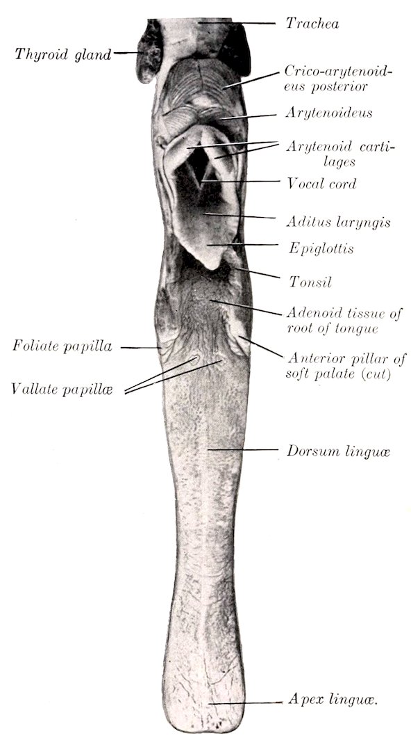
Fig. 241.—Tongue of Horse, Dorsal Aspect.
Structure.—The tongue consists of: (1) The mucous membrane; (2) the glands; (3) the muscles.
The mucous membrane (Tunica mucosa linguæ) adheres intimately to the subjacent tissue, except on the lower part of the lateral surfaces of the body and the under surface of the tip. It varies considerably in thickness. On the dorsum it is very thick and dense. Underneath this portion there is a dense fibrous cord, which extends medially a distance of five or six inches forward from the vallate papillæ. On the sides and under surface of the tongue the membrane is much thinner and smooth, and can more readily be dissected off the muscular tissue. From the under surface of the free part of the tongue a fold of the mucous membrane passes to the floor of the mouth, forming the frenum linguæ. This contains the anterior edges of the genio-glossi muscles. Posteriorly a fold passes on each side 336from the edge of the dorsum to join the soft palate, forming the anterior pillars of the latter. A central glosso-epiglottic fold (Plica glossoepiglottica) passes from the root to the base of the epiglottis. The mucous membrane presents numerous papillæ, which are of four kinds—filiform, fungiform, vallate, and foliate. The filiform papillæ (Papillæ filiformes) are fine, pointed projections. They cover the upper surface of the body and tip, to which they give a distinct pile. The fungiform papillæ (Papillæ fungiformes) are rounded at the free end, which is supported by a neck. They occur principally on the lateral part of the tongue, but are also found scattered over the dorsum and upper surface of the free portion. The vallate papillæ (Papillæ vallatæ) are usually two or three in number. The two constant ones have a diameter of about 7 mm., and are found on the posterior part of the dorsum, one on each side of the median plane, about an inch (ca. 3 cm.) apart. The third, when present, is behind these, is centrally situated, and is always smaller. Rarely a fourth may be seen. They are rounded, broader at their exposed than at their attached surfaces, and are situated in a cup-shaped cavity. The foliate papillæ (Papillæ foliatæ) are situated just in front of the anterior pillars of the soft palate, where they form a rounded eminence about an inch (ca. 2 to 3 cm.) in length, marked by transverse fissures. The last three varieties are covered with microscopic secondary papillæ and are furnished with taste buds. The mucous membrane of the root of the tongue presents numerous folds and depressions. Into the latter open the ducts from the lingual glands (Glandulæ linguales), which constitute a thick layer in the loose submucous tissue. Mucous glands are found also on the dorsum and sides of the tongue. In the submucosa of the root is found also a large quantity of lymph follicles (Folliculi tonsillares) and diffuse lymphoid tissue.
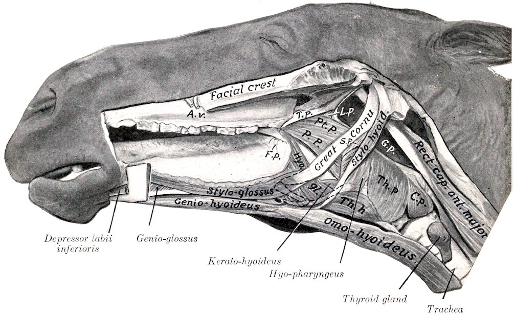
Fig. 242.—Muscles of Tongue, Hyoid Bone, Pharynx, etc., of Horse. T. p., Tensor palati; L. p., levator palati; Pt. p., pterygo-pharyngeus; P. p., palato-pharyngeus; S. p., stylo-pharyngeus; Th. p., thyro-pharyngeus; C. p., crico-pharyngeus; Th. h., thyro-hyoideus; Hyo. gl., hyo-glossus; G. p., guttural pouch; F. p., foliate papilla; A. v., facial artery and vein. The concealed parts of the hyoid bone are indicated by dotted line.
337The muscular tissue may be divided into intrinsic and extrinsic. The intrinsic musculature consists, not of distinct muscles, but rather of systems of fibers which run longitudinally, vertically, and transversely, blending with the extrinsic muscles, which are now to be described.
1. Stylo-glossus.—This is a long, thin muscle, which lies on the lateral part of the tongue. It arises by a thin tendon from the outer surface of the great cornu of the hyoid bone, near the articulation with the small cornu. It terminates near the tip of the tongue by blending with its fellow of the opposite side and with the intrinsic musculature. The action is to retract the tongue. Unilateral contraction would also draw the tongue toward the side of the muscle acting.
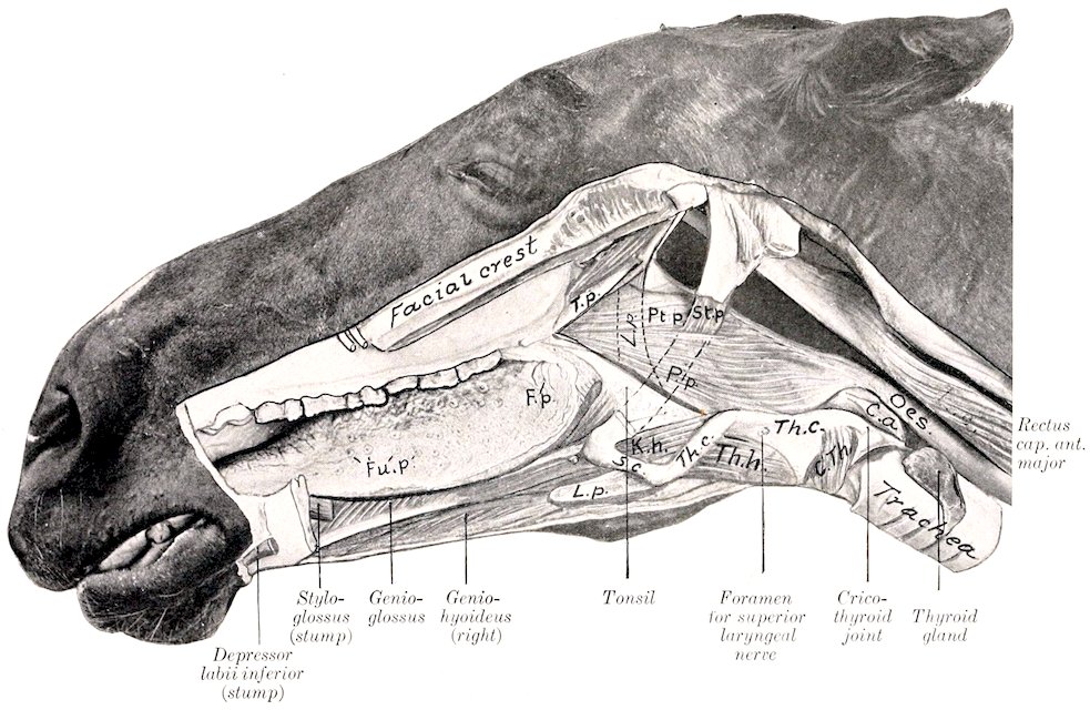
Fig. 243.—Muscles of Tongue, Pharynx, Larynx, etc., Deep Dissection. T. p., Tensor palati; L. p., levator palati, concealed part indicated by dotted line; Pt. p., pterygo-pharyngeus; P. p., palato-pharyngeus; St. p., stylo-pharyngeus; C. a., crico-arytenoideus post.; C. th., crico-thyroideus; Th. c., thyroid cartilage (wing); Th. h., thyro-hyoideus; K. h., kerato-hyoideus; Th. c., thyroid cornu; S. c., small cornu; L. p., lingual process; F. p., foliate papilla; Fu. p., fungiform papillæ. Part of great cornu is removed and indicated by dotted line.
3382. Hyo-glossus.—This is a wide, flat muscle, somewhat thicker than the preceding. It lies on the lateral part of the root and body of the tongue, partly under cover of the preceding muscle. Its deep face is related to the genio-glossus. It arises from the lateral aspect of the hyoid bone, from the lingual process to the oral extremity of the great cornu, and from the thyroid cornu. The fibers pass obliquely forward and upward, and for the most part turn toward the median plane of the dorsum of the tongue. Its action is to retract and depress the tongue.
(It is usually possible to recognize in this muscle three portions, which would correspond to the baseo-, kerato-, and chondro-glossus of human anatomy.)
3. Genio-glossus.—This is a fan-shaped muscle, which lies parallel to the median plane of the tongue. It is separated from the muscle of the opposite side by a quantity of fat and connective tissue. It arises from the inner surface of the ramus of the mandible near the symphysis. From the tendon the fibers pass in a radiating manner, some toward the tip, others toward the dorsum, and others toward the root of the tongue; some fibers pass from the posterior end of the tendon to the body and small cornu of the hyoid bone. The muscle as a whole is a depressor of the tongue, and especially of its middle portion; when both muscles act, a median groove is formed on the dorsum. The posterior fibers protrude the tongue, the middle fibers depress the tongue, and the anterior fibers retract the tip of the tongue.
Vessels and Nerves.—The arteries of the tongue are the lingual and sublingual branches of the external maxillary artery. The sensory nerves are the lingual and glosso-pharyngeal, and the muscles are innervated by the hypoglossal nerve.
THE TEETH
The teeth are hard white or yellowish-white structures, implanted in the alveoli of the bones of the jaws—premaxilla, maxilla, and mandible. Morphologically they are large calcified papillæ. Functionally they are organs of prehension and mastication, and may serve as weapons of offense and defense. They are classified according to form and position as follows:
1. The incisor teeth (Dentes incisivi) are situated in front and are implanted in the premaxilla and mandible.
2. The canine teeth (Dentes canini) are situated a little further back, at or near the premaxillary suture in the upper jaw; in the lower jaw they are nearer the incisors.
3. The cheek teeth (Dentes premolares et molares), the remaining teeth, occupy the sides of the dental arch. The space between the incisors and cheek teeth is termed the interdental or interalveolar space. It is customary to divide the cheek teeth into an anterior series, termed premolars, which appear as temporary, deciduous, or milk teeth, and are replaced by permanent successors, and a posterior series, the molars, which appear only as permanent teeth without deciduous predecessors.
As the teeth of the two sides of the jaws are alike in number and character (in normal cases), the complete dentition may be briefly indicated by a dental formula such as the following:
( 2 1 2 3 )
2 ( I C P M ) = 32.
( 2 1 2 3 )
In this formula the letters indicate the kinds of teeth, and the figures above and below the lines give the number of teeth of one side in the upper and lower jaw respectively in man.
The individual teeth of each group are designated numerically, the starting-point being the middle line; thus the incisor on either side of the middle line is the 339first incisor, and may be conveniently indicated by the notation I1. The temporary or deciduous teeth may be designated in a similar manner, prefixing D (for deciduous) to the letter indicating the kind of tooth. In addition to the above systematic method of notation other terms have received the sanction of popular usage. Thus the first incisors are commonly called “pinchers,” or “nippers”; the second, intermediate; and the third, corner teeth. The canines, when highly developed, may be termed tusks or fangs. The vestigial and inconstant first premolar of the horse is popularly termed the “wolf tooth.”
Each tooth presents for description a portion coated with enamel, termed the crown (Corona dentis), and a portion covered with cement, termed the root (Radix dentis). The line of union of these parts is the neck (Collum dentis).[80] In many teeth the neck is distinct and is embraced by the gum, e. g., the teeth of the dog and the temporary incisors of the horse. In other teeth no constriction is seen, as in the permanent incisors of the horse. Between these extremes may be noted the molars of the horse, in which the neck is seen only in advanced age.
The surface of a tooth directed toward the lips is termed labial; toward the cheek, buccal; and toward the tongue, lingual (Facies labialis, buccalis, lingualis). The surface opposed to a neighboring tooth of the same dental arch is termed the contact surface (Facies contactus). The grinding or masticatory or “table” surface (Facies masticatoria) is that which comes in contact with a tooth or teeth of the opposite jaw.
Structure.—Teeth are composed of four tissues, which are considered here from within outward. The pulp (Pulpa dentis) is a soft gelatinous tissue, which occupies a space in the central part of the tooth termed the pulp cavity (Cavum dentis). The pulp is well supplied with blood-vessels and nerves. It occupies a relatively large space in young growing teeth, but later the dentine deposited on its surface gradually encroaches on it until, in advanced age, the cavity is obliterated or much reduced. The dentine (Substantia eburnea) forms the bulk of most teeth, covering the surface of the pulp. It is very hard, and is yellowish-white in color. The enamel (Substantia adamantina) constitutes a layer of varying thickness covering the dentine of the crown of the tooth. It is easily distinguished by its clear bluish-white appearance and its extreme density. The cement (Substantia ossea) is always the outermost tooth substance. In simple teeth it forms usually a thin layer on the surface of the dentine of the root only, but in complex teeth it exists in considerable quantity, tending to fill in the spaces between the enamel folds of the crown also. Its structure is practically the same as that of bone without Haversian canals, and even these occur where the cement forms a very thick layer. The embedded part of the tooth is attached to the alveolus by a vascular layer of connective tissue, the alveolar periosteum (Periosteum alveolare), which constitutes the periosteum at once of tooth and alveolus.
The blood-supply to the pulp is derived from the alveolar or dental branches of the internal artery; the nerve-supply comes from branches of the trigeminus.
340
The Teeth of the Horse
The Permanent Teeth
The formula of the permanent teeth of the horse is:
( 3 1 3 or 4 3 )
2 ( I C P M ) = 40 or 42
( 3 1 3 3 )
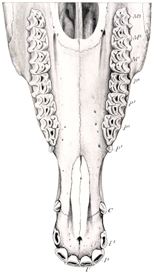
Fig. 244.—Upper Teeth of Horse, About Four and One-half Years Old. I1, I2, I3, Incisors; C, canine; P1, P2, P3, P4, premolars; M1, M2, M3, molars.
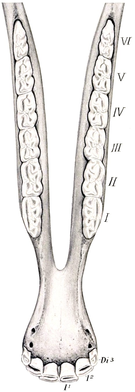
Fig. 245.—Lower Teeth of Horse, Four Years of Age. I1, I2, First and second permanent incisors; Di3, third deciduous incisor. The cheek teeth are numbered according to popular usage.
In the mare the canines usually are very small and do not erupt, reducing the number to 36 or 38.[81]
341Incisor Teeth.—These are twelve in number. The six in each jaw are placed close together, so that their labial edges form almost a semicircle. They have the peculiarity (not found in existing mammals other than the equidæ) of presenting, instead of the simple cap of enamel on the crown, a deep invagination, the infundibulum, which becomes partly filled up with cement. Hence as the tooth wears the table surface has a central ring of enamel surrounding this cavity in addition to the peripheral enamel. The cavity becomes darkened by deposits from the food, and is commonly termed the “cup” or “mark.” Each tooth is curved so that the labial surface is convex and the roots converge. The average length of the incisors at five or six years of age is about two and a half to three inches (ca. 7 cm.). They taper regularly from crown to root, without any constriction, and in such a manner that in young horses the exposed crown is broad transversely; toward the middle, the two diameters of a cross-section are about equal; near the root the antero-posterior diameter is considerably greater than the transverse.
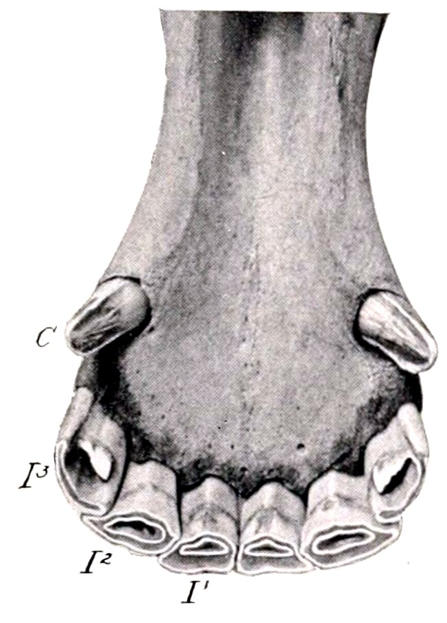
Fig. 246.—Lower Incisor and Canine Teeth of Horse, Five Years Old.
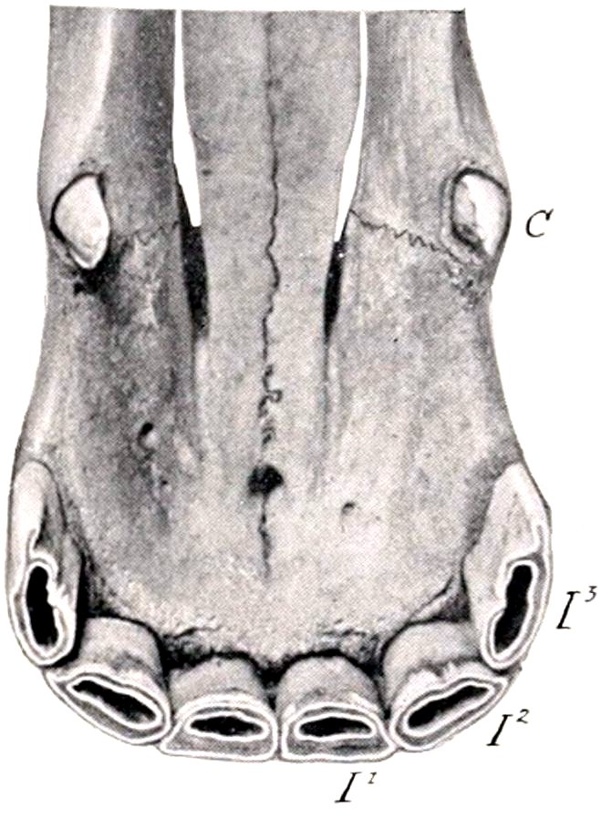
Fig. 247.—Upper Incisor and Canine Teeth of Horse, Five Years Old. I1, I2, I3, Incisors; C, canine.
This fact is of value in the determination of age by the teeth, since the table surface at different ages represents a series of such cross-sections. As the exposed crown wears down the embedded part (reserve crown) pushes up out of the alveolus, so that the tables of the first and second lower incisors are at first oval, with the long diameter transverse; later—at about fourteen years usually for the first lower incisors—the tables are triangular, with the base at the labial edge. At the same time the infundibulum or cup becomes smaller, approaches the lingual border, and finally disappears; it remains longer on the upper incisors, as it is deeper in them. Another marked feature in old age is the progressive approach to a horizontal direction as seen in profile; at the same time the teeth become parallel and finally convergent.
Canine Teeth.—These are four in number in the male; in the mare they are usually absent or rudimentary.[82] They interrupt the interdental space, dividing it into two unequal parts. The upper canine is situated at the junction of the premaxilla and the maxilla; the lower canine is placed nearer the corner incisor. The canines are simple teeth, smaller than the incisors, and are curved with the concavity directed backward. The crown is compressed, convex, and smooth 342externally; concave with a median ridge internally; its edges are sharp, and the apex is pointed in the unworn tooth. The root is round and the pulp cavity is large, persisting to advanced age.
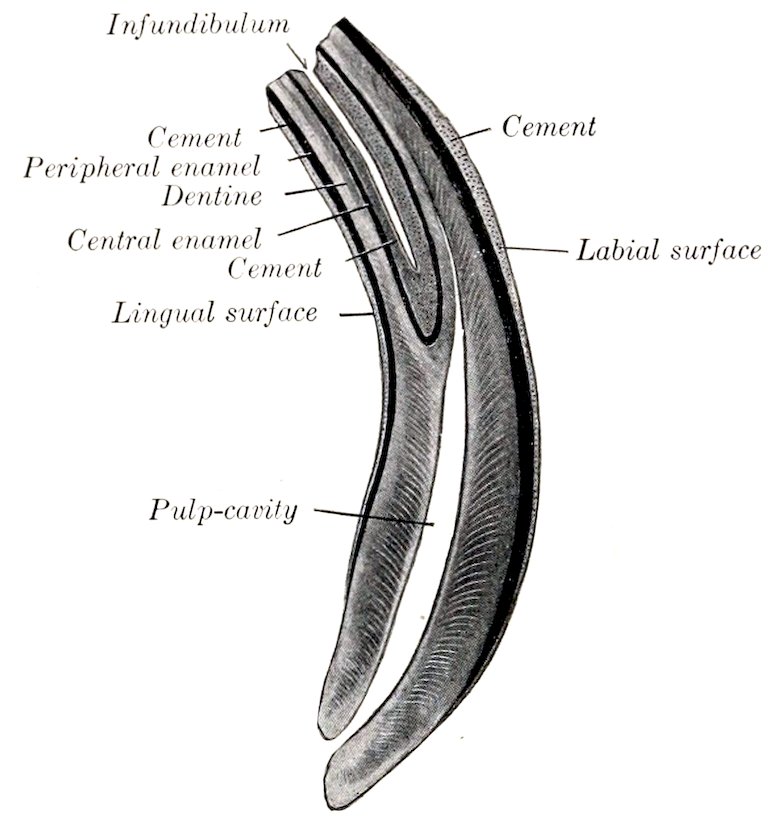
Fig. 248.—Longitudinal Section of Permanent Incisor Tooth of Horse, About Natural Size.
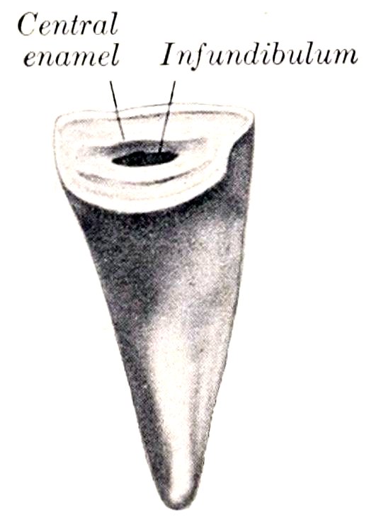
Fig. 249.—Incisor Tooth of Horse, Lingual Aspect. (After Ellenberger-Baum, Anat. für Künstler.)
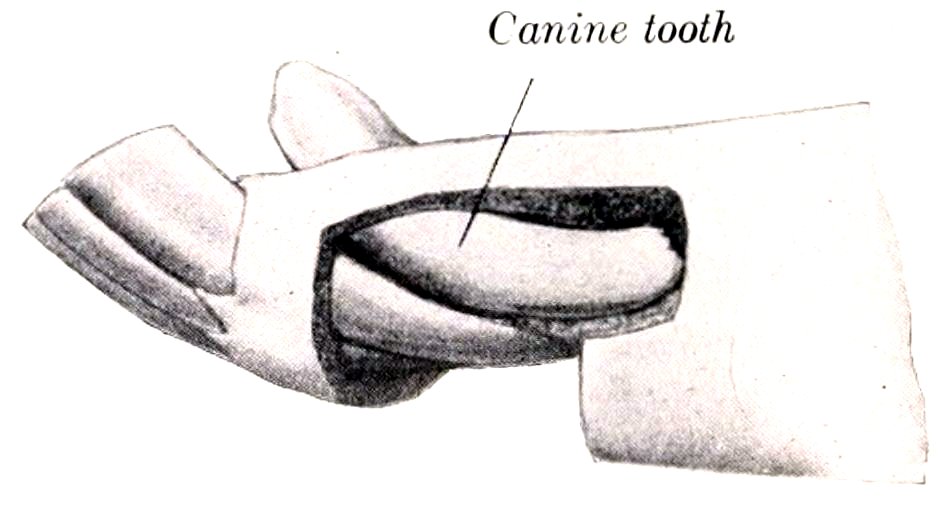
Fig. 250.—Canine and Incisor Teeth of Horse. The bone has been removed to show the embedded parts of the teeth. (After Ellenberger-Baum, Anat. für Künstler.)
Cheek Teeth (Premolars and Molars).[83]—The constant number of these is twenty-four—twelve in each jaw. Quite commonly, however, the number is increased by the presence in the upper jaw of the so-called wolf tooth. This tooth is usually situated just in front of the first well-developed tooth; it is a much reduced vestige, not often more than one-half or three-fourths of an inch (ca. 1 to 2 cm.) in length. (It is interesting as being the remnant of a tooth which was well developed in the Eocene ancestors of the horse.) It may erupt during the first six months, and is often shed about the same time as the milk-tooth behind it, but may remain indefinitely. The occurrence of a similar tooth in the lower jaw—which rarely erupts—increases the dental formula to 44, which is considered the typical number for mammals. They may be regarded as belonging to the permanent set, having no predecessors. The cheek teeth are very large, prismatic in form, and quadrilateral in cross-section, except the first and last of the series, which are triangular. The crown is remarkably long, most of it being embedded in the bone or situated in the maxillary sinus in the young horse. As the exposed part wears down the embedded part pushes up to replace it, so that a functional crown of about four-fifths of an inch (ca. 2 cm.) 343is maintained. The root begins to grow at four or five years of age, and is complete at twelve to fourteen, although the deposition of cement may continue indefinitely.
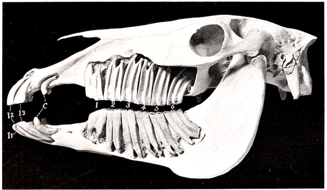
Fig. 251.—Skull of Adult Horse Sculptured to Embedded Parts of Teeth. The bone has been removed to show the embedded parts of the teeth I1, I2, I3, Incisors; C, canines. The cheek teeth are numbered.
The upper or maxillary teeth are embedded in the alveolar processes of the maxilla. The exposed parts of the crowns are normally in close contact, forming a continuous row which is slightly curved, with the convexity toward the cheek. The embedded parts diverge in the manner shown in the annexed figures (Figs. 251, 253). Thus the long axis of the first is directed upward and a little forward, 344that of the second is almost vertical, while in the remainder it is curved backward in an increasing degree. The average length at six years of age is about three to three and a half inches (ca. 7 to 9 cm.). The buccal (or outer) surface presents a central ridge running lengthwise, and separating two grooves; the first tooth has, in addition, a less prominent ridge in front of the primary one. The lingual (or inner) surface is marked by a wide, rounded ridge, the accessory pillar or column, which separates two very shallow grooves. The masticatory or table surface presents two infundibula, an anterior and a posterior. It slopes obliquely downward and outward, so that the outer edge is prominent and sharp. The first and last teeth have three roots, the remainder four or three.
The position of the embedded crowns and roots of the last four varies at different ages and in different subjects. Two factors in this variation may be noted. All these teeth are developed in the maxillary sinus close to the orbital fossa. As growth proceeds the teeth move forward, so that commonly only the last three, but sometimes also the third, remain (except as to their free crowns) in the sinus. The second cause of variation is the fact that the anterior limit of the maxillary sinus may be at the extremity of the facial ridge, or more than an inch beyond it. In the latter case the third tooth projects into the sinus.[84]
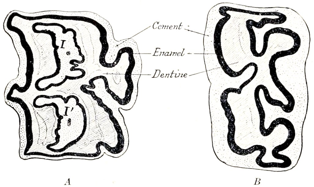
Fig. 252.—Cross-sections of Cheek Teeth of Horse. Buccal (outer) surfaces face to left. A, Upper tooth, B, lower tooth; I, anterior, I′, posterior infundibulum, both almost filled up with cement.
The structure is quite complex. Two infundibula run vertically through the entire length of the crown; these become filled with cement. There are five main divisions of the pulp-cavity and five enamel folds, four of which are arranged symmetrically, while the fifth is an outgrowth from the inner side of the antero-internal fold. On the exposed crown of the unworn tooth the enamel folds form rounded ridges covered with a thin layer of cement. After the tooth comes into wear the enamel on the masticatory surface stands out in the form of sharp prominent ridges. Progressive cementation of the periphery of the tooth takes place, thus leveling up the irregularities of surface to a considerable extent.
The lower or mandibular cheek teeth are implanted in the rami of the mandible, forming two straight rows which diverge behind. The space between the rows is considerably less than that separating the upper teeth, especially in the middle of the series. The length of the lower teeth is about the same as that of the upper set. Their direction is also similar, but the embedded portions diverge even more, with the exception of the first and second. The long axis of the first is vertical; the remainder project downward and backward in a gradually increasing obliquity. The buccal (outer) surface has a longitudinal furrow; the last molar has a secondary, 345shallower furrow in addition. The lingual (inner) surface is uneven, but the grooves are not regular; there are usually three on the first and last tooth. The masticatory or table surface is oblique, sloping upward and inward in correspondence with the opposing tooth; thus the inner edge is prominent. The first five have two roots, while the sixth commonly has three. The width of the lower molars is a little more than half that of the upper. There are two infundibula, which are open along the inner face of the tooth until closed by cement. The pulp-cavity has two principal divisions, and four or three secondary diverticula. The enamel folds correspond, forming a pattern even more complicated than on the upper teeth.
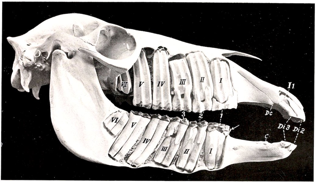
Fig. 253.—Skull of Two-year-old Colt, Sculptured to Show the Embedded Parts of the Teeth. Both permanent and temporary cheek teeth are shown. The upper first premolar (“wolf tooth”) is present, but not visible; the lower one is indicated by the arrow. Temporary chief premolars are numbered 1, 2, 3; permanent premolars and molars are designated by Roman numerals; Dc., upper temporary canine; C, lower permanent canine, which was not ready to erupt; Di 2, Di 3, second and third temporary incisors; I 1, first permanent incisor, not quite ready to erupt.
346
The Temporary Teeth
The deciduous, temporary, or “milk” teeth (Dentes decidui) are smaller and fewer than those of the permanent set. The formula is:
( 3 0 3 )
2 ( Di Dc Dp - ) = 24.
( 3 0 3 )
The deciduous incisors are much smaller than the permanent ones. They have a distinct neck at the junction of the crown and root. The crown is short, white in color, and its labial surface is smooth. The infundibulum is shallow. The root is flattened; it undergoes absorption as the permanent tooth develops behind it.
The deciduous canines are quite vestigial. They occur in both sexes as slender spiculæ about a quarter of an inch in length, but do not erupt. The lower one develops close to the corner incisor. They are not usually included in the formula, as they are never functional.
The deciduous cheek teeth differ from the permanent set chiefly in that they have much shorter crowns than the latter. The roots form early, so that a distinct neck occurs.
The subjoined table indicates the average periods of the eruption of the teeth:
Teeth Eruption
A. Deciduous:
1st incisor (Di 1) Birth or first week.
2nd „ (Di 2) 4–6 weeks.
3rd „ (Di 3) 6–9 months.
Canine (Dc )
1st premolar (Dp 2) Birth or first two weeks.
2nd „ (Dp 3)
3rd „ (Dp 4)
B. Permanent:
1st incisor (I 1) 2½ years.
2nd „ (I 2) 3½ years.
3rd „ (I 3) 4½ years.
Canine. (C ) 4–5 years.
1st premolar (or wolf tooth) (P 1) 5–6 months.
2nd „ (P 2) 2½ years.
3rd „ (P 3) 3 years.
4th „ (P 4) 4 years.
1st molar (M 1) 10–12 months.
2nd „ (M 2) 2 years.
3rd „ (M 3) 3½–4 years.
(The periods given for P 3 and 4 refer to the upper teeth; the lower ones may erupt about six months earlier.)
THE SALIVARY GLANDS
This term is usually restricted to the three pairs of large glands situated on the sides of the face and the adjacent part of the neck—the parotid, submaxillary, and sublingual. Their ducts open into the mouth.
The parotid gland (Glandula parotis) (Fig. 172)—so named from its proximity to the ear—is the largest of the salivary glands in the horse. It is situated chiefly in the space between the ramus of the mandible and the wing of the atlas. It is somewhat triangular in shape, the apex partially embracing the base of the external ear. Its length is about eight to ten inches (ca. 20 to 25 cm.), and its average thickness nearly an inch (ca. 2 cm.). Its average weight is about seven ounces (ca. 200 to 225 g.).
347It presents for description two surfaces, two borders, a base, and an apex. The external (or superficial) surface is covered by the parotid fascia, the panniculus, and the parotido-auricularis muscle. It is crossed obliquely by the jugular vein, which is embedded in the gland tissue to a varying extent. It is also related to the great auricular vein, the cervical branch of the facial nerve, and branches of the second cervical nerve. The internal (or deep) surface is very uneven, and has numerous important relations. Some of these are: the guttural pouch, and the great cornu of the hyoid bone; the masseter, stylo-maxillaris, digastricus, and occipito-hyoideus muscles; the tendons of the mastoido-humeralis and sterno-cephalicus (which separate the parotid from the underlying submaxillary gland); the external carotid artery and some of its branches; the facial nerve; the pharyngeal lymph glands. The anterior (or facial) border is closely attached to the ramus of the mandible and the masseter muscle; it overlaps the latter to a varying extent. (In some cases there is a well-marked triangular facial process, which covers the temporo-mandibular joint, the facial nerve, and the transverse facial vessels.) The posterior (or cervical) border is somewhat concave, and is loosely attached to the underlying muscles. The base or ventral border is related to the external maxillary vein. The apex is attached to the base of the external ear, which it partially embraces.
The gland has a yellowish-gray color and is distinctly lobulated. It is inclosed in a capsule formed by the parotid fascia. The parotid duct (Ductus parotideus Stenonis) is formed at the lower part of the gland, near the facial edge, by the union of three or four radicles. It leaves the gland about an inch (ca. 2 to 3 cm.) above the external maxillary vein, crosses the tendon of the sterno-cephalicus, and gains the inner face of the pterygoideus internus. It then runs forward in the submaxillary space below the external maxillary vein and winds around the lower border of the mandible behind the vein, passes upward between the vein and the masseter muscle for about two inches (ca. 5 cm.), turns forward underneath the facial vessels, and perforates the cheek obliquely opposite the third upper cheek tooth. Before piercing the cheek it is somewhat dilated, but its termination is small, and is surrounded by a circular mucous fold (Papilla salivalis).
Blood-supply.—Branches of the carotid and maxillary arteries.
Nerve-supply.—Trigeminal, facial, and sympathetic nerves.
The submaxillary gland (Glandula submaxillaris) is much smaller than the parotid. It is long, narrow, and curved, the dorsal edge being concave. It extends from the fossa below the wing of the atlas to the body of the hyoid bone, so that it is covered partly by the parotid gland, partly by the lower jaw. Its length is about eight to ten inches (ca. 20 to 25 cm.), its width an inch to an inch and a half (ca. 2.5 to 3 cm.), and its thickness about half an inch (ca. 1 cm.). It weighs about one and a half to two ounces (ca. 45 to 60 g.). It is often divisible into two parts.
It presents for description two surfaces, two borders, and two extremities. The external surface is covered by the parotid gland, the stylo-maxillaris, digastricus, and pterygoideus internus muscles. The tendon of the sterno-cephalicus crosses this surface, and is a useful guide in separating the parotid gland from it. The internal surface is related chiefly to the flexor muscles of the head; the guttural pouch; the larynx; the division of the carotid artery; and the tenth, eleventh, and sympathetic nerves. The superior border is concave and thin. It is related to the guttural pouch and the duct of the gland. The inferior border is convex and thicker. It is related to the thyroid gland and the external maxillary vein. The posterior extremity is loosely attached in the fossa atlantis. The anterior extremity lies at the side of the root of the tongue, and is crossed externally by the external maxillary artery.
348The submaxillary duct (Ductus submaxillaris Whartoni) is formed by the union of small radicles which emerge along the concave edge. It runs forward along this border, and, after leaving the anterior extremity, crosses the intermediate tendon of the digastricus, passes between the hyo-glossus and mylo-hyoideus, and gains the inner surface of the sublingual gland. Its terminal part lies on the body of the mandible, under the mucous membrane, which it pierces opposite the canine tooth. The orifice is at the end of a flattened papilla (Caruncula sublingualis).
Blood-supply.—Occipital, external carotid, and external maxillary arteries.
Nerve-supply.—Chorda tympani and sympathetic nerves.
The sublingual gland (Glandula sublingualis) is situated beneath the mucous membrane of the mouth, between the body of the tongue and the ramus of the mandible. It extends from the symphysis to the third or fourth lower cheek tooth. Its length is about five or six inches (ca. 12 to 15 cm.) and its weight about half an ounce (ca. 15 to 16 g.).
It is flattened laterally, and has a thin upper border which causes an elevation of the mucous membrane of the floor of the mouth, termed the sublingual crest (Plica sublingualis). The external surface is related to the mylo-hyoideus muscle, and the internal surface to the genio-glossus and stylo-glossus, the submaxillary duct, and branches of the lingual nerve. The inferior border is related to the genio-hyoid muscle.
The sublingual ducts (Ductus sublinguales minores), about thirty in number, are small, short, and twisted; they open on small papillæ on the sublingual crest.
Blood-supply.—Sublingual artery.
Nerve-supply.—Trigeminal and sympathetic nerves.
THE PHARYNX
The pharynx is a musculo-membranous sac which belongs to the digestive and respiratory tracts in common. It is funnel-shaped, the base joining the mouth and nasal cavity, while the apex is continued by the œsophagus. Its long axis is directed obliquely downward and backward, and has a length of about six inches (ca. 15 cm.).
The pharynx is attached by its muscles to the palate, pterygoid, and hyoid bones, and to the cricoid and thyroid cartilages of the larynx.
Its principal relations are: dorsally, the base of the cranium and the guttural pouches; ventrally, the larynx; laterally, the internal pterygoid muscle, the great cornu of the hyoid bone, the internal and external maxillary arteries, the glosso-pharyngeal, superior laryngeal, and hypoglossal nerves, the submaxillary salivary gland, and the pharyngeal lymph glands.
It presents seven openings. The posterior nares (Choanæ) communicate dorsally with the nasal chambers. The pharyngeal orifices (Ostia pharyngea) of the two Eustachian tubes are situated on the lateral wall behind the nares and a little below the level of the inferior nasal meatus. They are slit-like openings, slightly oblique downward and backward, and are a little more than an inch (ca. 3 cm.) in length. They are bounded internally by a valvular flap formed by the expanded extremity of the cartilaginous Eustachian tube. The isthmus faucium is situated below and in front. It is closed by the soft palate except during swallowing. The laryngeal orifice (Aditus laryngis) occupies the greater part of the ventral wall or floor of the pharynx. Behind this is the œsophageal opening.
The wall of the pharynx comprises from without inward: the muscles, the pharyngeal aponeurosis, and the mucous membrane.
The muscles (Figs. 243, 244) are covered by the pharyngeal fascia, which is 349attached to the base of the skull, the great cornu of the hyoid bone, and the thyroid cartilage of the larynx. They are as follows:
1. The stylo-pharyngeus arises from the inner surface of the dorsal third of the great cornu of the hyoid bone, passes downward and inward, and enters the wall of the pharynx by passing between the pterygo-pharyngeus and palato-pharyngeus. Its fibers radiate, many passing forward, others inward beneath the hyo-pharyngeus. It raises and dilates the pharynx to receive the bolus in swallowing.
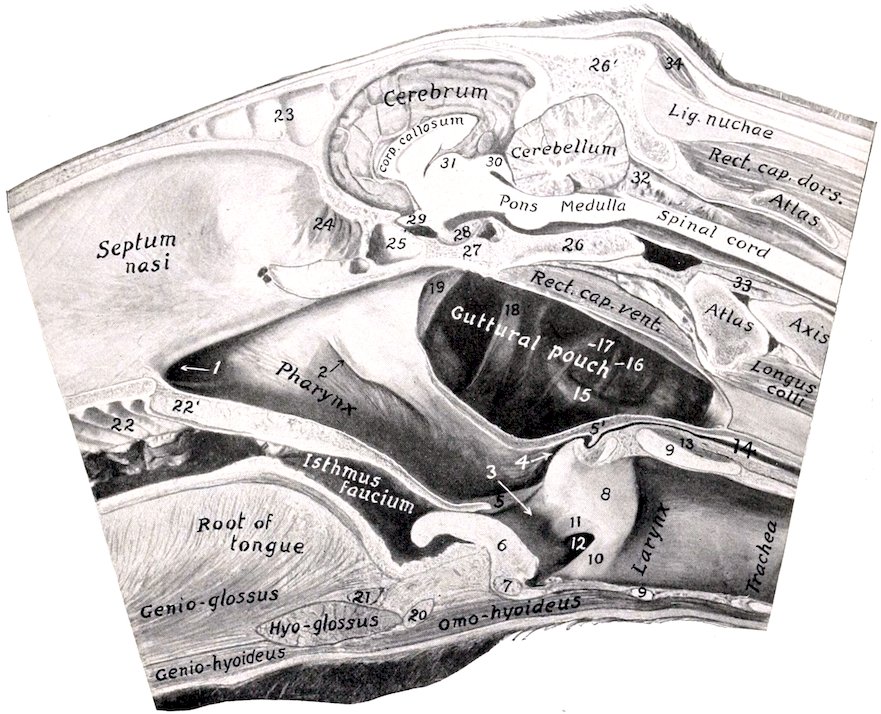
Fig. 254.—Posterior Part of a Sagittal Section of Head of Horse, Cut about 1 cm. to the Left of the Median Plane. 1, Posterior nares; 2, pharyngeal orifice of Eustachian tube; 3, aditus laryngis; 4, entrance to œsophagus; 5, posterior pillar of soft palate; 5′, junction of 5 with its fellow over entrance to œsophagus; 6, epiglottis; 7, body of thyroid cartilage; 8, arytenoid cartilage; 9, 9, cricoid cartilage; 10, true vocal cord; 11, false vocal cord; 12, lateral ventricle of larynx; 13, crico-arytenoideus post. s. dorsalis; 14, œsophagus; 15, external carotid artery; 16, hypoglossal nerve; 17, glosso-pharyngeal nerve; 18, great cornu of hyoid bone; 19, Eustachian tube; 20, body of hyoid bone; 21, hyoideus transversus; 22, ridges of hard palate; 22′, soft palate; 23, septum between frontal sinuses; 24, olfactory mucous membrane; 25, sphenoidal sinus; 26, basilar part of occipital bone; 26′, supraoccipital; 27, body of sphenoid bone; 28, pituitary body; 29, chiasma opticum; 30, corpora quadrigemina; 31, thalamus; 32, arachnoid; 33, odontoid ligament; 34, posterior auricular muscles.
2. The palato-pharyngeus arises by means of the aponeurosis of the soft palate from the palate and pterygoid bones. Its fibers pass backward on the lateral wall of the pharynx, and are inserted in part into the upper edge of the thyroid cartilage, in part turn inward to end at the median fibrous raphé. Its action is to shorten the pharynx, and to draw the larynx and œsophagus toward the root of the tongue in swallowing.
3. The pterygo-pharyngeus is flat and triangular. It lies on the anterior part of the lateral wall of the pharynx. It arises from the pterygoid bone above the 350preceding muscle—from which it is not distinctly separated—crosses the levator palati, and is inserted into the median raphé. Its action is similar to the preceding.
4. The hyo-pharyngeus may consist of two portions:
(a) The kerato-pharyngeus is a small and inconstant muscle which arises from the inner surface of the great cornu of the hyoid bone near its lower end. It passes upward and backward, turns inward toward the raphé, and spreads out under the next muscle.
(b) The chondro-pharyngeus, broad and fleshy, arises from the thyroid cornu of the hyoid bone and by a thin fasciculus from the wing of the thyroid cartilage and ends at the median raphé.
5. The thyro-pharyngeus arises from the lateral surface of the wing of the thyroid cartilage. Its fibers pass forward and inward to the median raphé.
6. The crico-pharyngeus arises from the cricoid cartilage and ends at the raphé. The fibers are directed upward, forward, and inward; they blend behind with the longitudinal fibers of the œsophagus.
The last three muscles are constrictors of the pharynx.
The pharyngeal aponeurosis is attached to the base of the cranium. It is well developed on the inner face of the palato-pharyngeus muscle and forms a median raphé (Raphé pharyngis) dorsally, which is wide in its posterior part.
The mucous membrane of the pharynx is continuous with that of the several cavities which open into it. It is thin and closely adherent to the base of the skull in the vicinity of the posterior nares, where the muscular wall is absent. Behind the Eustachian openings is a median cul-de-sac, the pharyngeal recess. The recess is somewhat variable, but is usually about an inch in depth and will admit the end of the finger. In the ass and mule it is much deeper. Here also the muscular wall is absent and the mucous membrane lies against the guttural pouches. From the Eustachian opening a fold of the mucous membrane (Plica salpingo-pharyngea) passes toward but does not reach the laryngeal opening. Below, a horizontal fold, the posterior pillar of the soft palate (Arcus pharyngopalatinus), passes along the lateral wall and unites with its fellow over the entrance to the œsophagus. The upper part of the cavity (the naso-pharynx) is lined with a ciliated epithelium, while the lower part (oro-pharynx) has a stratified squamous epithelium. The communication between the two is oval and is bounded by the free edge of the soft palate and its posterior pillars; it is termed the pharyngeal isthmus. On either side of the laryngeal opening is a narrow deep depression, the pyriform sinus (Recessus piriformis).
The submucous tissue contains numerous mucous glands (Glandulæ pharyngeæ). In the young subject the lymph follicles are numerous and form a collection dorsally and between the Eustachian openings, known as the pharyngeal tonsil.
Blood-supply.—External carotid, external maxillary, and thyro-laryngeal arteries.
Nerve-supply.—Glosso-pharyngeal, vagus, and sympathetic nerves.
THE ŒSOPHAGUS
The œsophagus is a musculo-membranous tube, about 50 to 60 inches (ca. 125 to 150 cm.) in length, which extends from the pharynx to the stomach. It begins in the median plane above the cricoid cartilage of the larynx. In its course it shows several changes of direction. At the level of the fourth cervical vertebra it inclines to the left side of the trachea, and continues this relation to the level of the third thoracic vertebra. Here it again gains the dorsal surface of the trachea, and passing backward, crosses the left bronchus, being here almost in the median plane. It continues in the mediastinum between the lungs backward, upward, and a little to the left, to reach the hiatus œsophageus of the diaphragm. Passing 351through this it terminates at once at the cardiac orifice of the stomach, at the level of the fourteenth thoracic vertebra, a little to the left of the median plane, and about four or five inches (ca. 10 to 12 cm.) ventral to the vertebral column.
Viewed with reference to the frontal plane, its course is downward and backward till it enters the thorax and passes upward to gain the dorsal face of the trachea. For a short distance (i. e., to the root of the lung) its direction is almost horizontal; behind this it passes somewhat upward to its termination. The cervical part of the tube is about four to six inches (10 to 15 cm.) longer than the thoracic part, while the so-called abdominal part is about an inch (2 to 3 cm.) long.[85]
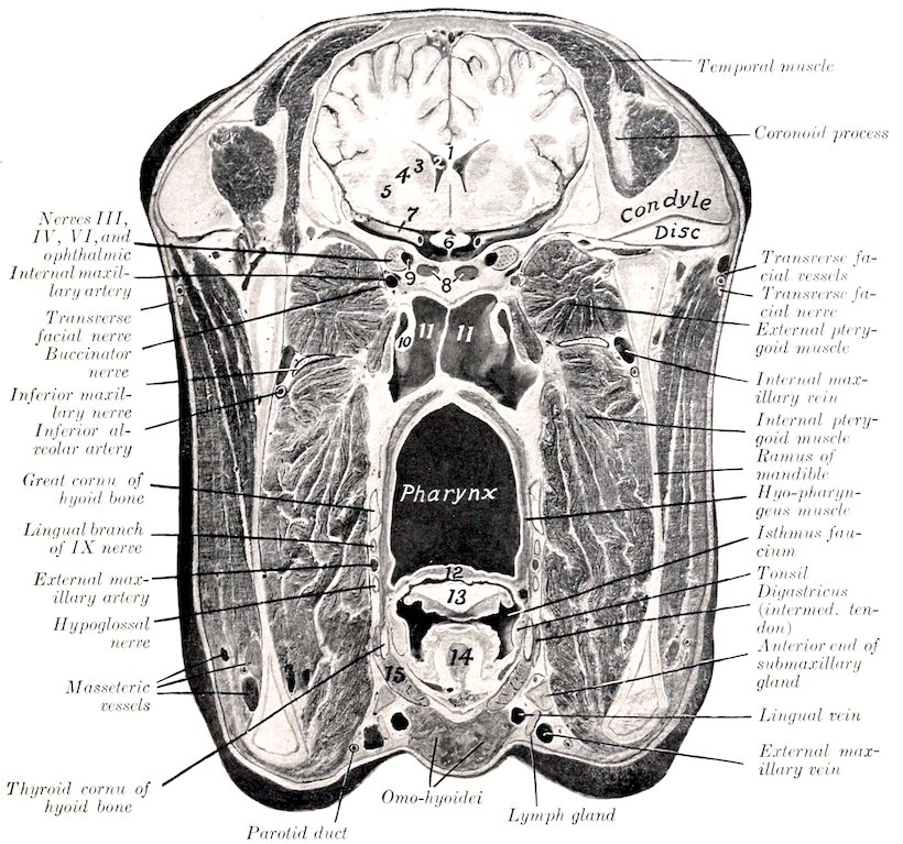
Fig. 255.—Cross-section of Head of Horse. The section passes through the temporo-mandibular articulation, but is slightly oblique. 1, Corpus callosum; 2, lateral ventricle of brain; 3, caudate nucleus; 4, internal capsule; 5, lenticular nucleus; 6, optic chiasma; 7, middle cerebral artery; 8, sphenoidal sinuses; 9, cavernous sinus; 10, Eustachian tube, inner lamina; 11, 11, guttural pouches; 12, soft palate; 13, epiglottis; 14, hyo-epiglottic muscle; 15, thyro-hyoid muscle.
352The principal relations of the œsophagus at its origin are: to the cricoid cartilage below; to the guttural pouches and the ventral straight muscles above; and to the carotid arteries laterally. In the middle of the neck the relations are: to the left longus colli muscle above; to the trachea internally; to the left carotid artery, vagus, sympathetic, and recurrent nerves externally. At its entrance into the thorax it has the trachea on its inner side; the first rib, the roots of the brachial plexus of nerves and the inferior cervical ganglion externally. After gaining the upper surface of the trachea, it has the aorta on its left and the vena azygos and right vagus nerve on its right side. In its course through the posterior mediastinum the œsophageal trunks of the vagus nerves lie above and below it, and the œsophageal artery is dorsal to it.
Structure.—The wall is composed of four coats: (1) A fibrous sheath; (2) the muscular coat; (3) a submucous layer; (4) the mucous membrane. The muscular coat is of the striped variety as far as the base of the heart, where it rapidly changes to the unstriped type. In addition to this change, the muscular coat becomes much thicker and firmer, while the lumen is diminished.[86] The outer fibers are arranged longitudinally, beginning in two bundles attached in the interval between the arytenoid and cricoid cartilages. The inner fibers run in two spiral strata to the terminal part of the tube, where the arrangement is an outer longitudinal and an inner circular layer.[87] The mucous membrane is pale, and is covered with squamous stratified epithelium. It is loosely attached to the muscular coat by an abundant submucosa, and lies in longitudinal folds which obliterate the lumen except during deglutition.
Blood-supply.—Carotid, broncho-œsophageal, and gastric arteries.
Nerve-supply.—Vagus, glosso-pharyngeal, and sympathetic nerves.
THE ABDOMINAL CAVITY
The abdominal cavity (Cavum abdominis) is the largest of the body cavities. It is separated from the thoracic cavity by the diaphragm and is continuous behind with the pelvic cavity.
It is ovoid in form but somewhat compressed laterally. Its long axis extends obliquely from the center of the pelvic inlet to the sternal part of the diaphragm. Its dorso-ventral diameter is greatest at the first lumbar vertebra, while its greatest transverse diameter is a little nearer the pelvis.
The dorsal wall or roof is formed by the lumbar vertebræ, the lumbar muscles, and the lumbar part of the diaphragm.
The lateral walls are formed by the oblique and transverse abdominal muscles, the abdominal tunic, the anterior parts of the ilia, the cartilages of the asternal ribs, and the parts of the posterior ribs which are below the attachment of the diaphragm.
The ventral wall or floor consists of the two recti, the aponeuroses of the oblique and transverse muscles, the abdominal tunic, and the xiphoid cartilage.
The anterior wall is formed by the diaphragm, which is very deeply concave, thus greatly increasing the size of the abdomen at the expense of the thorax.
It should be noted that the diaphragm also concurs practically in the formation of a considerable part of the lateral walls, since its costal portion even during ordinary inspiration lies directly on the ribs over a width of four or five inches (ca. 10 to 12 cm.); in expiration this area 353of contact would be about twice as wide, including about all of the fleshy rim. This fact is of clinical importance, with reference to auscultation and percussion, and penetrating wounds. The cupola of the diaphragm extends as far forward as a plane through the sixth intercostal space to the right of the heart.
There is no wall between the abdominal and pelvic cavities. The line of demarcation here is the terminal line (Linea terminalis) or brim of the pelvis; it is formed by the base of the sacrum, the ilio-pectineal lines, and the anterior borders of the pubic bones.
The muscular walls are lined by a layer of fascia, distinguished in different parts as: (1) the diaphragmatic fascia; (2) the transversalis fascia; (3) the iliac fascia; (4) the deep layer of the lumbo-dorsal fascia.
The subperitoneal or extraperitoneal connective tissue (Tela subserosa) unites the fascia and peritoneum. It is composed of areolar tissue, more or less loaded with fat according to the condition of the subject, except over the diaphragm. It sends laminæ into the various peritoneal folds.
The peritoneum, the serous membrane which lines the cavity, will be described later.
The abdominal walls are pierced in the adult by five apertures. These are: the three openings in the diaphragm which transmit the aorta, posterior vena cava, and the œsophagus; the inguinal canals, which contain the spermatic cord or the round ligament (in female carnivora). In the fœtus there is the umbilical opening also.
The cavity contains the greater part of the digestive and urinary organs, part of the internal generative organs, numerous nerves, blood-vessels, lymph vessels and glands, ductless glands (spleen and adrenal bodies), and certain fœtal remains.
For topographic purposes the abdomen is divided into nine regions by imaginary planes.[88] Two of these planes are sagittal, and two are transverse. The sagittal planes cut the middles of the inguinal (Poupart’s) ligaments; the transverse planes pass through the last thoracic and fifth lumbar vertebræ, or the lower end of the fifteenth rib and the external angle of the ilium respectively. The transverse planes divide the abdomen into three zones, one behind the other, viz., epigastric, mesogastric, and hypogastric: these are subdivided by the sagittal planes as indicated in the subjoined table.
Left parachondriac Xiphoid Right parachondriac
Left lumbar Umbilical Right lumbar
Left iliac Prepubic Right iliac.
Other useful regional terms are: sublumbar, diaphragmatic, inguinal. The first two require no explanation. The inguinal regions (right and left) lie in front of the inguinal (Poupart’s) ligament. The flank is that part of the lateral wall which is formed only of soft structures. The depression on its upper part is termed the paralumbar fossa.
THE PERITONEUM[89]
The peritoneum is the thin serous membrane which lines the abdominal and (in part) the pelvic cavity, and covers to a greater or less extent the viscera contained therein. In the male it is a completely closed sac, but in the female there are two small openings in it; these are the abdominal orifices of the Fallopian tubes, which at their other ends communicate with the uterus, and so indirectly with the exterior. The peritoneal cavity is only a potential one, since its opposing walls 354are normally separated only by the thin film of serous fluid (secreted by the membrane) which acts as a lubricant.
The free surface of the membrane has a glistening appearance and is very smooth. This is due to the fact that this surface is formed by a layer of flat endothelial cells, and is moistened by the peritoneal fluid. Friction is thus reduced to a minimum during the movements of the viscera. The outer surface of the peritoneum is related to the subperitoneal tissue, which attaches it to the abdominal wall or the viscera.
In order to understand the general disposition of the peritoneum, we may imagine the abdominal cavity to be empty and lined by a simple layer of peritoneum, termed the parietal layer (Lamina parietalis). We may regard the organs as beginning to develop in the subperitoneal tissue, enlarging, and migrating into the cavity to a varying extent. In doing so they carry the peritoneum before them, producing introversion of the simple sac, and forming folds which connect them with the wall or with each other. The viscera thus receive a more or less complete covering of peritoneum, termed the visceral layer (Lamina visceralis). The connecting folds are termed omenta, mesenteries, ligaments, etc. They contain a varying quantity of connective tissue, fat and lymph glands, and furnish a path for the vessels and nerves of the viscera. Some contain unstriped muscular tissue. An omentum is a fold which passes from the stomach to other viscera. There are three of these, namely: (1) the small or gastro-hepatic omentum (Omentum minus), which passes from the lesser curvature of the stomach to the liver; (2) the gastro-splenic omentum (Ligamentum gastrolienale), which extends from the greater curvature of the stomach to the spleen; (3) the great omentum (Omentum majus), which passes from the greater curvature of the stomach and from the spleen to the terminal part of the great colon and the origin of the small colon. It does not pass directly from one organ to the other, but forms an extensive loose sac (Figs. 278, 279). A mesentery (Mesenterium) is a fold which attaches the intestine to the dorsal wall of the abdomen. There are two mesenteries, namely: (1) the great mesentery which connects the greater part of the small intestine with the dorsal abdominal wall; (2) the colic mesentery, which attaches the small colon to the dorsal abdominal wall. Ligaments are folds which pass between viscera other than parts of the digestive tube, or connect them with the abdominal wall. The term is also applied to folds which attach parts of the digestive tract to the abdominal wall, but do not contain their blood-vessels and nerves. In some cases (e. g., the lateral and coronary ligaments of the liver) they are strengthened by fibrous tissue; in other cases (e. g., the broad ligaments of the uterus) they contain also unstriped muscular tissue.
THE PELVIC CAVITY
The pelvis is the posterior part of the trunk. It incloses the pelvic cavity (Cavum pelvis), which communicates in front with the abdominal cavity, the line of demarcation being the pelvic brim or terminal line.
The dorsal wall or roof is formed by the sacrum and first three coccygeal vertebræ. The lateral walls are formed by the parts of the ilia behind the ilio-pectineal lines and the sacro-sciatic ligaments. The ventral wall or floor is formed by the pubic and ischial bones. The boundary of the outlet is formed by the third coccygeal vertebra dorsally, the ischial arch ventrally, and the posterior edges of the sacro-sciatic ligaments and the semimembranosus muscles laterally. The outlet is closed by the perineal fascia; this consists of superficial and deep layers, which are attached around the margin of the outlet and centrally to the organs at the outlet—the anus and its muscles, the vulva (in the female), and the root of the penis (in the male).
The cavity contains the rectum, parts of the internal generative and urinary 355organs, some fœtal remnants, muscles, vessels, and nerves. It is lined by the fascia pelvis, and in part by the peritoneum.
The pelvic peritoneum is continuous in front with that of the abdomen. It lines the cavity as far back as the third or fourth sacral vertebra in the horse, where it is reflected on to the viscera, and from one organ to another. We may therefore distinguish an anterior, peritoneal, and a posterior, retroperitoneal part of the cavity. Along the mid-dorsal line it forms a continuation of the colic mesentery, the mesorectum, which attaches the first or peritoneal part of the rectum to the roof. In animals in fair condition a considerable quantity of subperitoneal and retroperitoneal fat is found on the walls and in the various interstices.
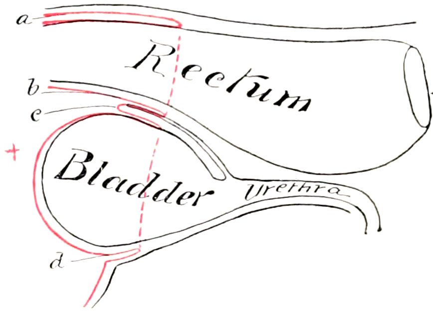
Fig. 256.—Diagram of Sagittal Section of Male Pelvis to show Disposition of Peritoneum. a, Pouch between rectum and roof of cavity, continuous laterally with b, recto-genital pouch; c, vesico-genital pouch; d, pouch below bladder and its lateral ligaments. The lateral line of reflection of the peritoneum is dotted. The area of rectum covered by peritoneum varies widely. When the rectum is empty, the reflection dorsally may be at the posterior end of the sacrum; when the rectum is very full, the reflection may occur a short distance behind the promontory.
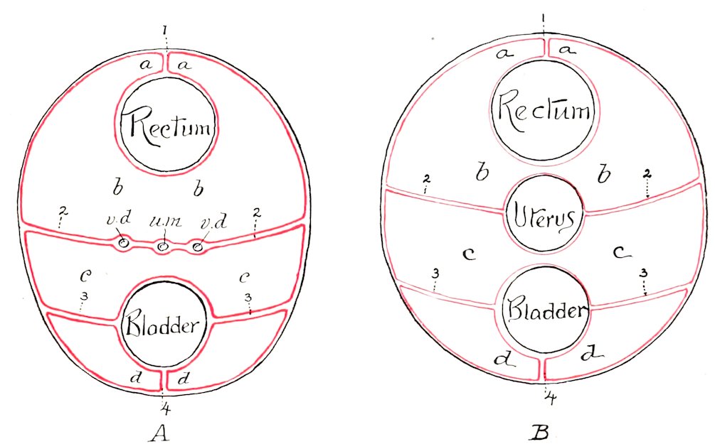
Fig. 257.—Schematic Cross-sections to show Arrangement of Pelvic Peritoneum of Horse: A, in Male; B, in Female. A: a, b, Recto-genital pouch, c, c, vesico-genital pouch; d, d, pouch below bladder and its lateral ligaments; 1, mesorectum; 2, 2, urogenital fold; 3, 3, lateral, 4, median ligaments of bladder; v. d., vas deferens; u. m., uterus masculinus. B: a, b, recto-genital pouch; c, c, vesico-genital pouch; d, d, pouch below bladder and its lateral ligaments; 1, mesorectum; 2, 2, broad ligaments of uterus; 3, 3, lateral, 4, median ligaments of bladder.
In the male the general disposition of the peritoneum here is as follows. If traced along the dorsal wall, it is reflected at the third or fourth sacral vertebra on to the rectum, forming the visceral peritoneum for the first part of that tube. Laterally it is reflected in a similar fashion. If the rectum be raised, it will be seen that the peritoneum passes from its ventral surface and forms a transverse fold which lies on the dorsal surface of the bladder (Fig. 272). This is the urogenital 356fold (Plica urogenitalis). Its concave free edge passes on either side into the inguinal canal. The ventral layer of this fold is reflected on to the dorsal surface of the bladder near its neck. Thus there is formed a pouch between the rectum and bladder—the recto-vesical pouch (Excavatio recto-vesicalis), which is partially subdivided by the urogenital fold into recto-genital and vesico-genital cavities. The fold contains the vasa deferentia, part of the vesiculæ seminales, and the uterus masculinus (a fœtal remnant). The space on either side of the rectum is occupied by coils of the small colon and the pelvic flexure of the great colon usually. If the bladder is now raised, it is seen that the peritoneum passes from its ventral surface on to the pelvic floor, forming a median fold, the so-called middle ligament (Plica umbilicalis media). It also forms on each side a lateral fold, the lateral ligament (Plica umbilicalis lateralis), which contains in its edge the so-called round ligament (Ligamentum teres)—the partially occluded umbilical artery, which is a large vessel in the fœtus.
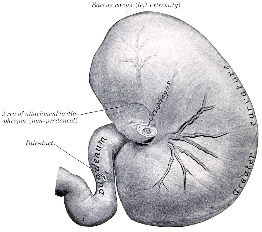
Fig. 258.—Stomach of Horse, Parietal Surface, with First Part of Duodenum. Fixed in situ when full but not distended. The larger branches of the anterior gastric artery with two satellite veins are shown.
In the female the arrangement is modified by the presence of the uterus; the urogenital fold is very large, so as to inclose the uterus and a small part of the vagina. It forms two extensive folds, the broad ligaments of the uterus (Ligamenta lata uteri), which attach that organ to the sides of the pelvic cavity and the lumbar part of the abdominal wall (Fig. 271). It thus divides the recto-vesical pouch completely into dorsal and ventral compartments—the recto-genital pouch (Excavatio recto-uterina), and the vesico-genital pouch (Excavatio vesico-uterina).
Further details will be given in the description of the pelvic viscera.
357
THE STOMACH
The stomach (Ventriculus) is the large dilatation of the alimentary canal between the œsophagus and the small intestine. It is a sharply curved, U-shaped sac, the right branch being, however, much shorter than the left one. The convexity is directed ventrally. When moderately distended, there is often a slight constriction which indicates the division into right and left sacs. It is relatively small, and is situated in the dorsal part of the abdominal cavity behind the diaphragm and liver, mainly to the left of the median plane.
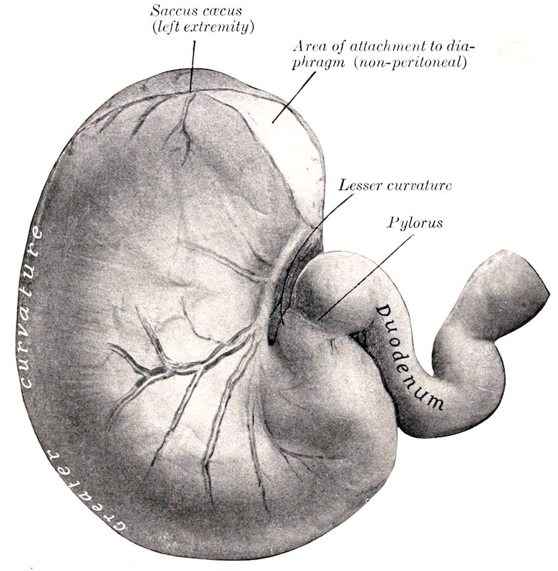
Fig. 259.—Stomach of Horse, Visceral Surface, with First Part of Duodenum. Fixed in situ when full but not distended. The posterior gastric artery and its larger branches with two satellite veins are shown.
It presents for description two surfaces, two curvatures, and two extremities. The parietal surface (Facies parietalis) is convex and is directed forward, upward, and toward the left; it lies against the diaphragm and liver. The visceral surface (Facies visceralis), also convex, faces in the opposite direction; it is related to the terminal part of the large colon, the pancreas, the small colon, and the small intestine. The lesser curvature (Curvatura minor) is very short, extending from the termination of the œsophagus to the junction with the small intestine. When the stomach is in situ, its walls are here in contact, and the cardia and pylorus close together. The greater curvature (Curvatura major) is very extensive. From the cardia it is first directed upward and curves over the left extremity; it then descends, passes to the right, crosses the median plane, and curves upward to end at the pylorus. Its left portion is related to the spleen, while its ventral portion rests on the left divisions of the great colon. The left extremity or saccus cæcus is a rounded cul-de-sac which lies under the upper ends of the fourteenth, fifteenth, 358and sixteenth ribs and the diaphragm.[90] It is related to the pancreas behind and the base of the spleen externally. The right or pyloric extremity is much smaller and is continuous with the duodenum, the junction being indicated by a marked constriction. It lies on the liver, a little to the right of the median plane, and a little lower than the cardiac opening. About two or three inches (ca. 5 to 8 cm.) from the pylorus there is a constriction which marks off the antrum pylori from the rest of the right sac. The œsophageal orifice or cardia is situated at the left extremity of the lesser curvature, but about eight to ten inches (ca. 20 to 25 cm.) from the left extremity. The œsophagus joins the stomach very obliquely. The opening is closed by the sphincter cardiæ and numerous folds of mucous membrane. The pyloric orifice communicates with the duodenum. Its position is indicated externally by a distinct constriction. Internally it presents a circular ridge produced by a ring of muscular tissue—the sphincter pylori.
The stomach is held in position mainly by the pressure of the surrounding viscera and by the œsophagus. The following peritoneal folds connect it with the adjacent parts:
1. The gastro-phrenic ligament (Lig. gastrophrenicum) connects the great curvature, from the cardia to the left extremity, with the crura of the diaphragm. This leaves a narrow area uncovered with peritoneum, and here the stomach is attached to the diaphragm by loose connective tissue.
2. The small or gastro-hepatic omentum (Omentum minus) connects the lesser curvature and the first part of the duodenum with the liver below the œsophageal notch and the portal fissure.
3. The gastro-splenic omentum (Lig. gastrolienale) passes from the left part of the great curvature to the hilus of the spleen.
4. The great or gastro-colic omentum (Omentum majus) connects the ventral part of the great curvature and the first curve of the duodenum with the terminal part of the great colon and the initial part of the small colon.
5. The gastro-pancreatic fold (Plica gastro-pancreatica) extends from the left sac above the cardia to the duodenum. It is attached dorsally to the liver and vena cava, ventrally to the pancreas.
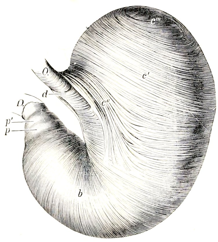
Fig. 260.—Everted Stomach of Horse from which the Mucous Membrane has been Removed. O, Œsophagus; D, duodenum; b, circular layer; c′, internal oblique fibers; c″, loop around cardia; c‴, transition of internal to external oblique fibers; d, fibers connecting the two branches of the cardiac loop; p, antral sphincter; p′, pyloric sphincter. (Ellenberger-Baum, Anat. d. Haustiere.)
The stomach of the equidæ is relatively small, its capacity varying from two to four gallons (ca. 8 to 15 liters).
The size, form, and position of the stomach are subject to considerable variation. When the stomach is nearly empty the saccus cæcus contains only gas and is strongly contracted; the middle portion (physiological fundus) contains the ingesta and preserves its rounded character, while the pyloric portion is contracted. When distended the middle portion settles down some four or five inches, pushing back coils of the small colon and small intestine which may lie between the great curvature and the large colon, and also pushing to the left or right the left dorsal part of 359the great colon; the spleen, small colon, and small intestines are pushed back by the distention of the left sac.
Structure.—The wall is composed of four coats—serous, muscular, submucous, and mucous.
The serous coat (Tunica serosa) covers the greater part of the organ and is closely adherent to the muscular coat except at the curvatures. It partially bridges over the lesser curvature, and covers here elastic tissue which assists in retaining the bent form of the stomach. The peritoneal folds have been described.
The muscular coat consists of three incomplete layers, an external of longitudinal, a middle of circular, and an internal of oblique fibers. The layer of longitudinal fibers (Stratum longitudinale) is very thin and exists only along the curvatures and at the antrum. At the lesser curvature it is continuous with the longitudinal fibers of the œsophagus. On the antrum pylori it forms a well developed complete layer. The layer of circular fibers (Stratum circulare) exists only on the right sac. At the pyloric orifice it forms a thick ring—the pyloric sphincter. Another ring, the antral sphincter, is found at the left end of the antrum pylori. The oblique fibers (Fibræ obliquæ) are arranged in two layers; the external stratum covers the left sac and is a continuation (in part) of the longitudinal fibers of the œsophagus; the internal stratum is found also on the left sac, and exchanges fibers with the circular and external oblique layers. It forms a remarkable loop around the cardiac orifice, constituting a powerful cardiac sphincter (Sphincter cardiæ).
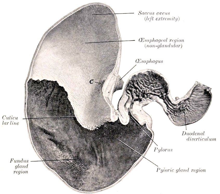
Fig. 261.—Frontal Section of Stomach and First Part of Duodenum of Horse. C, Cardiac orifice. Photograph of specimen fixed in situ.
The submucous coat is a layer of loose connective tissue which connects the muscular and mucous coats; in it the vessels and nerves ramify before entering the mucosa.
360The mucous coat is clearly divided into two parts. That which lines the left sac resembles the œsophageal mucous membrane, and is termed œsophageal or cuticular. It is white in color, destitute of glands, and covered with a thick, squamous, stratified epithelium. At the cardiac orifice it presents numerous folds which occlude the opening.[91] It terminates abruptly at an elevated, denticulated, sinuous line, termed the cuticular ridge (Margo plicatus). Below and to the right of this line the mucous membrane has a totally different character, being soft and velvety to the touch, and covered by a mucoid secretion. It is glandular, and three zones may be recognized, although no sharp line of demarcation exists. A narrow zone next to the cuticular ridge has a yellowish-gray color, and contains short tubular cardiac glands (Cardiac gland region). Next to this is a large area which has a mottled reddish-brown color, and contains fundus glands (fundus gland region). This part of the mucous membrane is thick and very vascular, and corresponds to the fundus of the stomach in man and the dog. The remainder of the mucous membrane has a reddish-gray color and contains branched, tubular, pyloric glands (pyloric gland region); it corresponds to the pyloric portion of man and the dog.
The folding of the stomach wall at the lesser curvature produces a prominent ridge which projects into the cavity of the stomach. Circular ridges occur at the antral and pyloric sphincters.
Blood-vessels and Nerves.—The stomach receives blood from all the branches of the cœliac artery. The gastric veins drain into the portal vein. The nerves are derived from the vagus and sympathetic nerves.
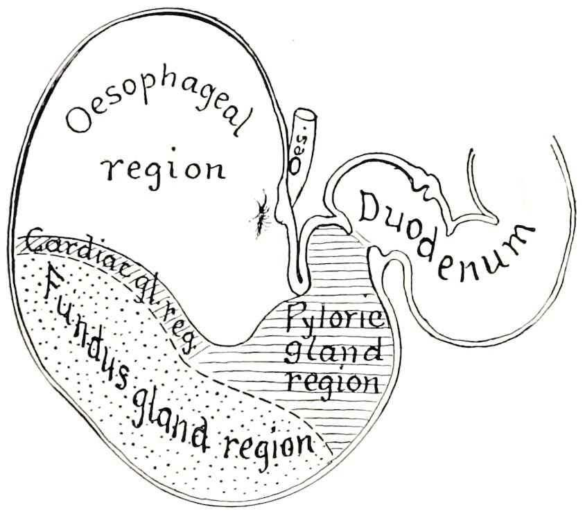
Fig. 262.—Diagram of Zones of Mucous Membrane of Stomach of Horse.
THE SMALL INTESTINE
The small intestine (Intestinum tenue) is the tube which connects the stomach with the large intestine. It begins at the pylorus and terminates at the lesser curvature of the cæcum. Its average length is about seventy feet (ca. 22 meters). When distended its diameter varies from two to four inches (5 to 10 cm.). Its capacity is about twelve gallons (40 to 50 liters).
It is clearly divisible into a fixed and a mesenteric or floating portion. The fixed portion is termed the duodenum, while the mesenteric portion (Intestinum tenue mesenteriale) is arbitrarily divided into parts termed the jejunum and ileum.[92]
The duodenum is about three to four feet (ca. 1 to 1.25 m.) long. Its shape is somewhat like a horseshoe, the convexity being directed toward the right. The first part is directed to the right and forms an ᔕ-shaped curve. The convexity of the first part of the curve is dorsal, of the second ventral. It lies on the middle and right lobes of the liver, and presents two dilatations (Ampullæ) with a constriction between them. The duodenal angle or head of the pancreas lies in the concavity 361of the second curve, and here, five to six inches (ca. 12 to 15 cm.) from the pylorus, the pancreatic duct and the bile-duct pierce the bowel wall. The second part passes upward and backward on the right lobe of the liver and, on reaching the right kidney and the base of the cæcum, it curves toward the median plane, opposite the last rib. The third part passes almost transversely from right to left behind the base of the cæcum, crosses the median plane under the third and fourth lumbar vertebræ, and turns forward to become continuous with the jejunum under the left kidney. The sacculations of the first part have a diameter of three to four inches (ca. 7.5 to 10 cm.).
It is attached by a short peritoneal fold termed the mesoduodenum. This fixes the first part of the duodenum closely to the liver and the right dorsal part of the colon; the remainder is somewhat less closely attached by it to the cæcum and right kidney, the sublumbar muscles, and (more closely) to the terminal part of the great colon and the first part of the small colon.[93]
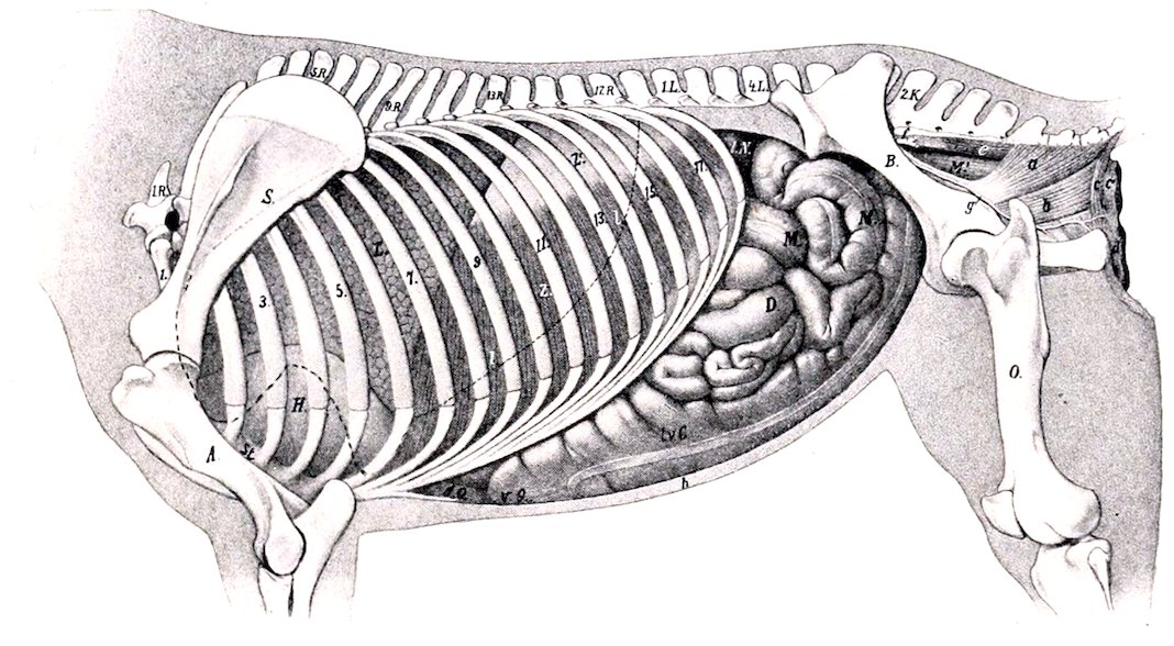
Fig. 263.—Topography of Viscera of Horse, Left View. 1.R., First thoracic vertebra; 1.L., first lumbar vertebra; 2.K., second sacral spine; S., scapula; A., humerus; St., sternum; B., ilium; O., femur; L., lung in complete expiration; l., dotted line indicating contour of lung in inspiration; H, pericardium; Z, costal part of diaphragm; Z′, tendinous center of diaphragm; l.N., left kidney; l.v.C., left ventral colon; v.Q., sternal flexure of colon; d.Q., diaphragmatic flexure of colon; D., small intestine; M., small colon; M′, rectum; a, coccygeus muscle; b, retractor ani; c, c, sphincter ani externus; d, constrictor vulvæ; e, sacro-coccygeus inferior; f, anterior gluteal artery; g, internal pudic artery; h, abdominal wall in section. (After Ellenberger, in Leisering’s Atlas, reduced.)
The jejunum and ileum together (Jejuno-ileum) constitute the mesenteric or floating portion of the small intestine. No distinct point exists at which to make the demarcation. With the exception of the last two or three feet, the mesenteric part of the intestine varies so much in position that only a general statement can be made. It lies in numerous coils (Ansæ) mingled with those of the small colon, chiefly in the dorsal part of the left half of the abdomen, from the visceral surface of the stomach to the pelvis. It may insinuate itself between the left portions of the colon and the abdominal wall; also between the ventral portions of the colon, reaching the floor of the abdomen. The terminal part of the intestine (ileum) ascends almost vertically, a little to the right of the median plane, to open into the 362cæcum at the lesser curvature of its base. The average diameter of the jejuno-ileum is about two and a half to three inches (ca. 6 to 7 cm.). In the cadaver one finds most of the tube presenting irregular constricted and dilated parts. The last three or four feet (ca. 1 meter) are usually tightly contracted, resembling somewhat the terminal part of the œsophagus. This part may be termed the ileum.
The mesenteric part is connected with the dorsal abdominal wall by the great mesentery. This is a wide fan-shaped fold, consisting of two layers of peritoneum, between which the vessels and nerves reach the bowel; it also contains the mesenteric lymph glands and some fat. The visceral border of the mesentery contains the intestine, while the parietal border or root (Radix mesenterii) is attached to a small area around the great mesenteric trunk under the first and second lumbar vertebræ. The root is thick, as it contains a large number of vessels and nerves placed close together. The mesentery is short at first, but soon reaches a length of one and a half to two feet (ca. 50 cm.)—sufficient to allow coils of the intestine to reach the abdominal floor, the pelvic cavity, or even to descend into the scrotum through the inguinal canal. Near its termination the intestine (ileum) leaves the border of the mesentery, so that the latter has a free edge which passes to the cæcum.
Structure.—The wall consists of four coats—serous, muscular, submucous, and mucous, enumerated from without inward.
The serous coat is complete except at the mesenteric edge, where the vessels and nerves reach the bowel.
The muscular coat consists of an outer longitudinal and an inner circular layer, the latter being the thicker. In the last few feet of the intestine the muscular coat is very thick, and being usually firmly contracted in the dead subject, gives the impression that this part of the bowel is of smaller caliber; such, however, is not the case during life.
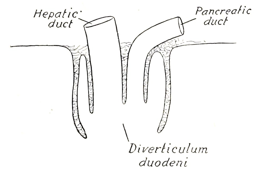
Fig. 264.—Diagram of Section of Diverticulum Duodeni of Horse. Solid line indicates mucous membrane.
The submucous coat is a layer of areolar tissue in which the vessels and nerves ramify. It contains also the duodenal glands and the bases of the solitary glands and Peyer’s patches.
The mucous membrane is soft and velvety. It has a grayish or yellowish-red color and is very vascular. About five or six inches from the pylorus it forms a pouch, the diverticulum duodeni, in which the pancreatic and hepatic ducts open. On a small papilla opposite this is the termination of the accessory pancreatic duct. At the ileo-cæcal opening the mucous membrane projects slightly into the cavity of the cæcum, forming the ileo-cæcal valve. The free surface is thickly beset with villi, small projections of the mucous membrane which can be seen well by placing a piece of the membrane in water. They are relatively short and thick and have a distinct neck in the horse. Each contains a central lymph vessel (lacteal), and around this a plexus of capillaries, lymphoid tissue, and unstriped muscle-fibers. They are important agents in absorption from the contents of the intestine. The epithelium is columnar, with many goblet cells. Underneath the basement membrane is a layer of unstriped muscle-fibers, the muscularis mucosæ.
The glands of the small intestine are of three kinds:
1. The intestinal glands (Glandulæ intestinales Lieberkuehni) are found throughout. They are simple tubular glands which open between the villi.
3632. The duodenal glands (Glandulæ duodenales Brunneri) are found in the first twenty feet (ca. 6 meters) of the bowel. They are racemose glands, and are situated in the submucosa, so that their ducts perforate the muscularis mucosæ and the mucous membrane.
3. The lymph follicles (Noduli lymphatici) are found either scattered or in groups. In the former case they are termed solitary glands (Noduli lymphatici solitarii), in the latter Peyer’s patches (Noduli lymphatici aggregati). The solitary glands are about the size of a millet-seed or a small sago grain. Peyer’s patches are situated chiefly along the surface opposite to the mesenteric attachment and begin about three or four feet from the pylorus. They number one to two hundred, and are usually one to two inches (2 to 5 cm.) long and a quarter of an inch to one-half inch (ca. 2 to 14 mm.) wide. Larger ones occur in the terminal part, where one patch may have a length of seven to fifteen inches (ca. 17 to 38 cm.) and a width of half an inch to one inch (ca. 5 to 25 mm.) in young horses (Ellenberger). They undergo atrophy in old subjects.
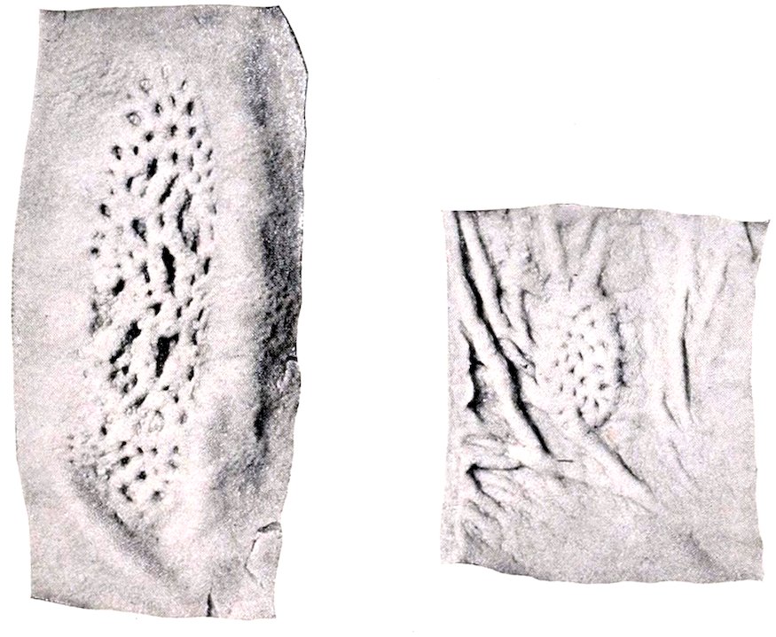
Fig. 265.—Peyer’s Patches of Small Intestine of Horse.
Vessels and Nerves.—The arteries of the small intestine come from the cœliac and anterior mesenteric arteries. The veins go to the portal vein. The lymph vessels are numerous and go to the mesenteric lymph glands. The nerves are derived from the vagus and sympathetic through the solar plexus.
THE LARGE INTESTINE
The large intestine (Intestinum crassum) extends from the termination of the ileum to the anus. It is about twenty-five feet (ca. 7.5 to 8 in.) in length. It differs from the small intestine in its greater size, in being sacculated, for the most part, possessing longitudinal bands, and having a more fixed position. It is divided into cæcum, great colon, small colon, and rectum.
The Cæcum
The cæcum (Intestinum cæcum) is a great cul-de-sac intercalated between the small intestine and the colon. It has a remarkable size, shape, and position in the horse. Its length is three to four feet (ca. 1 to 1.25 m.), and its capacity about 364seven to eight gallons (ca. 25 to 30 liters). It is conical in form, and is curved somewhat like a reversed comma. It is situated almost entirely to the right of the median plane, extending from the right iliac and sublumbar regions to the abdominal floor behind the xiphoid cartilage. Both extremities are blind, and the two orifices are placed close together on the concave curvature. It presents for description a base, a body, and an apex.
The base (Saccus cæcus) extends from about the thirteenth intercostal space backward almost to the pelvic inlet. Its greater curvature is dorsal, its lesser ventral; connected with the latter are the termination of the ileum and the origin of the colon. The body (Corpus cæci) extends downward and forward from the base and rests largely on the ventral wall of the abdomen. Its lesser curvature is about parallel with the costal arch and about five to six inches (10 to 15 cm.) below it. The apex (Apex cæci) lies usually on the abdominal floor about a hand’s length behind the xiphoid cartilage.
The base is attached dorsally by connective tissue and peritoneum on the ventral surface of the pancreas and right kidney, the psoas muscles, and the iliac fascia; internally, it is attached to the terminal part of the great colon, and ventrally to the origin of the great colon. The body is attached dorsally to the first part of the colon by the cæco-colic fold. The apex is free, and consequently may vary in position.
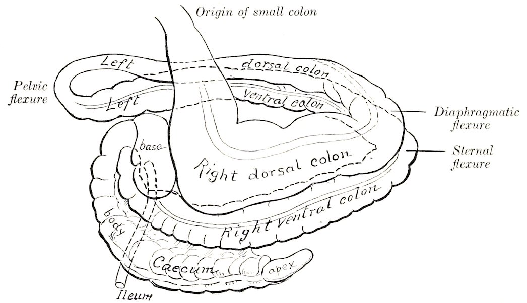
Fig. 266.—Diagram of Cæcum and Large Colon of Horse.
The cæcum has four longitudinal bands (Tæniæ), situated on the dorsal, ventral, right, and left surfaces; these cause four rows of sacculations (Haustra). The ventral band is entirely exposed or free (Tænia libera); the dorsal band is free on the apex. The cæcal arteries are placed on the other two. The right or parietal surface of the cæcum is related chiefly to the right abdominal wall, the diaphragm, duodenum, and liver. The left or visceral surface lies against the left divisions of the colon, the root of the great mesentery, and the small intestine.
The ileo-cæcal orifice (Ostium ileocæcale) is situated in the lesser curvature of the base, about four or five inches (ca. 10 to 12 cm.) to the right of the median plane and about opposite the lower end of the last rib. The end of the ileum is partially telescoped into the cæcum, so that the orifice is surrounded by a fold of mucous membrane, forming the ileo-cæcal valve (Valvula ileocæcalis). The peritoneum and longitudinal muscle-fibers do not take part in its formation.
The cæco-colic orifice (Ostium cæcocolicum) is placed above and external to 365the preceding one; the interval between them is only about two inches (ca. 5 cm.), and they are separated by a distinct ridge which projects into the interior of the cæcum. The orifice is slit-like and is small in relation to the size of the cæcum and colon. It has a valvular fold (Valvula cæcocolica) at its lower margin and a muscular ring (Sphincter cæci). Large crescentic or semilunar folds (Plicæ cæci) project into the cavity of the bowel, and between these are large pouches (Cellulæ cæci).
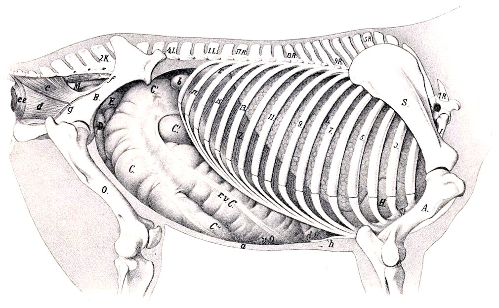
Fig. 267.—Topography of Viscera of Horse, Right View. 1 R., First thoracic vertebra; 1 L., first lumbar vertebra; 2 K., second sacral spine; S., scapula; A., humerus; St., sternum; B., ilium; O., femur; L., right lung; H., pericardium; Z., diaphragm (pars costalis); r.v.C., right ventral colon; v.Q., sternal flexure of colon; d.Q., diaphragmatic flexure of colon; C., body of cæcum; C′., base of cæcum; C″., apex of cæcum; D., small intestine; F., pelvic flexure of colon; M., rectum; a., abdominal wall in section; b, duodenum; c, coccygeus muscle; d, retractor ani; e, e′, sphincter ani externus; f, anterior gluteal artery; g, internal pudic artery; h, xiphoid cartilage. (After Ellenberger, in Leisering’s Atlas.)
Vessels and Nerves.—The cæcal arteries come from the great mesenteric artery. The veins go to the portal vein. The nerves are derived from the great mesenteric plexus of the sympathetic.
The Great Colon
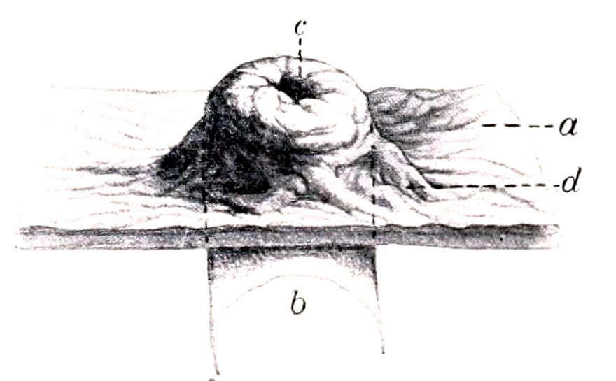
Fig. 268.—Ileo-cæcal Orifice of Horse. a, Mucous membrane of cæcum; b, ileum; c. ileo-cæcal orifice; d, fold of mucous membrane. (After P. Schumann.)
The great colon (Colon crassum) begins at the cæco-colic orifice, and terminates by joining the small colon behind the saccus cæcus of the stomach. It is ten to twelve feet (ca. 3 to 3.7 m.) long, and its average diameter (exclusive of its narrowest part) is about eight to ten inches (ca. 20 to 25 cm.). Its capacity is more than double that of the cæcum. When removed from the abdomen, it consists of two parallel portions, which are connected by peritoneum and partially by areolar tissue also. In situ, it is folded so that it consists of four parts, which are designated according to their position or numerically. The three bent connecting parts are termed 366the flexures. The first, right ventral part (Colon ventrale dextrum), begins at the cæco-colic orifice about opposite the middle of the last rib, passes downward and forward along the right costal arch, and reaches the floor of the abdomen. Over the xiphoid cartilage it bends sharply to the left and backward, forming the sternal flexure (Flexura diaphragmatica ventralis). The second, left ventral part (Colon ventrale sinistrum), passes backward on the left part of the abdominal floor, and, on reaching the pelvic inlet, bends sharply dorsally and forward, forming the pelvic flexure (Flexura pelvina). This is continued by the third, left dorsal part (Colon dorsale sinistrum), which passes forward above the left ventral portion. On reaching the stomach, diaphragm, and left lobe of the liver, it turns to the right, forming the diaphragmatic or gastro-hepatic flexure (Flexura diaphragmatica dorsalis). The fourth or right dorsal part (Colon dorsale dextrum) passes backward above the first portion, and on reaching the inner or left surface of the base of the cæcum it turns upward and to the left behind the left sac of the stomach; here it becomes constricted, and joins the small colon below the left kidney.
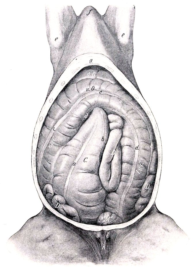
Fig. 269.—Abdominal Viscera of Horse, Ventral View. The ventral wall and part of the lateral walls of the abdomen are removed. C, Cæcum; r.v.C., right ventral part of colon; v.Q., sternal flexure of colon; l.v.C., left ventral part of colon; d.Q., diaphragmatic flexure of colon; D, small intestine; M, small colon; a, ventral free band of cæcum; b, inner band of cæcum; c, lateral band of ventral part of colon; d, ventral band of ventral part of colon; e, point of elbow; f, anterior end of sternal region; g, xiphoid cartilage; h, teats. (After Ellenberger-Baum, Top. Anat. d. Pferdes.)
367The caliber of the great colon varies greatly at different points. At its origin it is only about two to three inches (ca. 5 to 7.5 cm.) in diameter. This soon increases to about eight to ten inches (ca. 20 to 25 cm.) for the ventral portions. Beyond the pelvic flexure the diameter is reduced to about three or four inches (ca. 8 to 9 cm.). Near the diaphragmatic flexure the caliber rapidly increases, and reaches its maximum in the last division, where it may be about twenty inches (50 cm.) in its widest part. This is succeeded by a somewhat funnel-shaped terminal contraction.
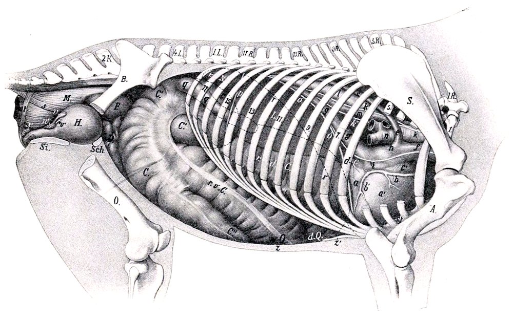
Fig. 270.—Topography of Viscera of Horse, Right Side, Deeper View. 1.R., First thoracic vertebra; 1.L., first lumbar vertebra; 2. K., second sacral spine; S, scapula; A, humerus; B, ilium; O., femur; Sch., pubis; Si., ischium; St., sternum; L., right lobe of liver; r.N., right kidney; C., body, C′., base, C″., apex of cæcum; r.v.C., right ventral colon; v.Q., sternal flexure of colon; d.Q., diaphragmatic flexure of colon; r.d.C., right dorsal colon; F., pelvic flexure of colon; D., small intestine; H., urinary bladder; M., rectum; a, left ventricle; a′, right ventricle; b, b′, right coronary artery; c, left atrium; c′, right atrium; d, small (left) coronary vein; e, vena azygos; f, anterior vena cava; g, posterior vena cava; h, sinus venosus; i, right phrenic nerve; k, right vagus, with its dorsal (k′) and ventral (k″) divisions; l, aorta; m, œsophagus; n, trachea; o, diaphragm (median section); p, right lateral ligament of liver; q, duodenum; r, dotted line indicating position in median section of diaphragm in inspiratory phase; s, recto-coccygeus; t, suspensory ligament of rectum; u, sphincter ani; v, vesicula seminalis; w, prostate; x, bulbo-urethral (Cowper’s) gland; y, urethra; z, abdominal wall; z′, xiphoid cartilage. (After Ellenberger, in Leisering’s Atlas, reduced.)
The first part of the great colon is attached to the lesser curvature of the cæcum by two layers of peritoneum which form the cæco-colic fold. The ventral parts of the colon are similarly connected to the dorsal parts of the same side by the mesocolon. The right portions are united also by areolar tissue and muscular fibers, the surface of contact being about four or five inches (ca. 10 to 12 cm.) wide; the left portions are attached to each other by a peritoneal fold wide enough to allow them to be drawn apart five or six inches (ca. 12 to 15 cm.) near the pelvic flexure. The terminal part of the colon is attached by peritoneum and areolar tissue to the ventral surface of the pancreas dorsally and to the base of the cæcum externally. It is connected indirectly with the diaphragm and liver by means of a fold derived from the right lateral ligament of the liver.
368The relations are complex, but the more important facts are as follows: The ventral portions (first and second) have extensive contact with the abdominal wall ventrally and laterally. On the right side the colon is almost entirely excluded from contact with the flank by the cæcum; on the left side it has no contact with the upper part of the flank, being excluded here by coils of the small colon and small intestine. Dorsally the chief relations are to the stomach, duodenum, liver, pancreas, small colon, small intestine, aorta, vena cava, and portal vein. Since there are no transverse attachments of the right and left portions, and the latter have no attachment to the wall, they are subject to considerable displacement.[94] The pelvic flexure is variable in position, but usually it is directed to the right across the pelvic inlet.
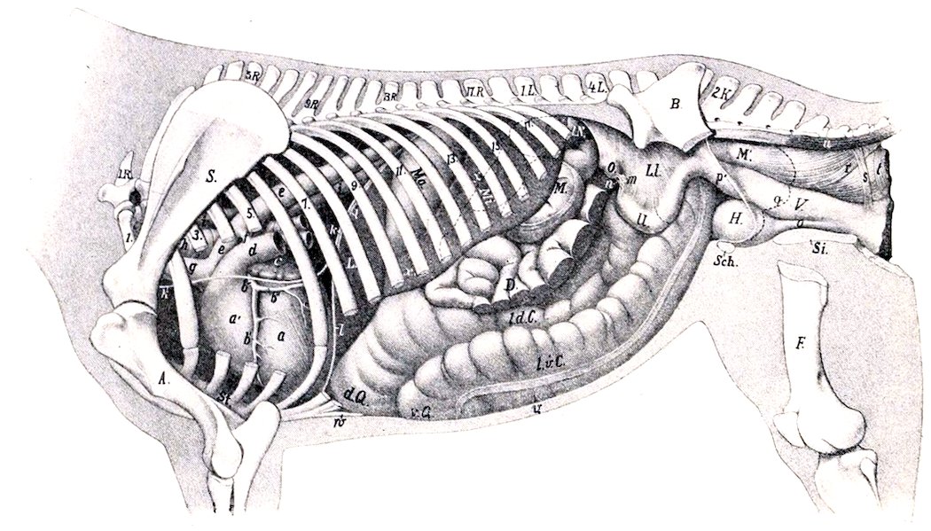
Fig. 271.—Topography of Viscera of Horse, Left Deep View. 1R., First thoracic vertebra; 1L., first lumbar vertebra; 2K., second sacral spine; S., scapula; A., humerus; B., ilium; F., femur; Sch., pubis; Si., ischium; L., liver (left lobe); Ma., stomach, the posterior contour of which is indicated by dotted line x; Mi., spleen; l.N., left kidney, concealed part indicated by dotted line; M., small colon; D., small intestine, parts of which have been removed; l. d. C., left dorsal colon; l.v.C., left ventral colon; v.Q., sternal flexure; d.Q., diaphragmatic flexure; O., left ovary; U., cornu uteri; L.1., broad ligament; M′., rectum; V., vagina; H., bladder; a, left ventricle; a′, right ventricle; b, left coronary artery with descending (b′) and circumflex (b″) branches; c, left auricle; d, pulmonary artery (cut); e, aorta; f, ligamentum arteriosum; g, brachiocephalic trunk (anterior aorta); h, trachea; i, œsophagus; k, left phrenic nerve; l, diaphragm in median section; m, Fallopian tube; n, bursa ovarica; o, urethra; p, cut edge of broad ligament; q, line of reflection of pelvic peritoneum; r, recto-coccygeus; s, so-called suspensory ligament of rectum; t, sphincter ani internus; u, sacro-coccygeus inferior; v, abdominal wall in section; w, xiphoid cartilage. (After Ellenberger, in Leisering’s Atlas.)