OSTEOLOGY
Reading edition
THE SKELETON
The term skeleton is applied to the framework of hard structures which supports and protects the soft tissues of animals. In the descriptive anatomy of the higher animals it is usually restricted to the bones and cartilages, although the ligaments which bind these together might well be included.
In zoölogy the term is used in a much more comprehensive sense, and includes all the harder supporting and protecting structures. When the latter are situated externally, they form an exoskeleton, derived from the ectoderm. Examples of this are the shells and chitinous coverings of many invertebrates, the scales of fishes, the shields of turtles, and the feathers, hair, and hoofs of the higher vertebrates. The endoskeleton (with which we have to deal at present) is embedded in the soft tissues. It is derived chiefly from the mesoderm, but includes the notochord or primitive axial skeleton, which is of entodermal origin.
The skeleton may be divided primarily into three parts: (1) axial; (2) appendicular; (3) splanchnic.
The axial skeleton comprises the vertebral column, ribs, sternum, and skull.
The appendicular skeleton includes the bones of the limbs.
The splanchnic skeleton consists of certain bones developed in the substance of some of the viscera or soft organs, e. g., the os penis of the dog and the os cordis of the ox.
The number of the bones of the skeleton of an animal varies with age, owing to the fusion during growth of skeletal elements which are separate in the fœtus or the young subject. Even in adults of the same species numerical variations occur, e. g., the tarsus of the horse may consist of six or seven bones, and the carpus of seven or eight; in all the domestic mammals the number of coccygeal vertebræ varies considerably.
The bones are commonly divided into four classes according to their shape and function.
(1) Long bones (Ossa longa) are typically of elongated cylindrical form with enlarged extremities. They occur in the limbs, where they act as supporting columns and as levers. The cylindrical part, termed the shaft or body (Corpus), is tubular, and incloses the medullary cavity, which contains the medulla or marrow.
(2) Flat bones (Ossa plana) are expanded in two directions. They furnish sufficient area for the attachment of muscles and afford protection to the organs which they cover.
(3) Short bones (Ossa brevia), such as those of the carpus and tarsus, present somewhat similar dimensions in length, breadth, and thickness. Their chief function appears to be that of diffusing concussion. Sesamoid bones, which are developed in the capsules of some joints or in tendons, may be included in this group. They diminish friction or change the direction of tendons.
(4) Irregular bones. This group would include bones of irregular shape, such as the vertebræ and the bones of the cranial base; they are median and unpaired. Their functions are various and not so clearly specialized as those of the preceding classes.
This classification is not entirely satisfactory; some bones, e. g., the ribs, are not clearly provided for, and others might be variously placed.
20
STRUCTURE OF BONES[1]
Bones consist chiefly of bone tissue, but considered as organs they present also an enveloping membrane, termed the periosteum, the medulla or marrow, vessels, and nerves.
The architecture of bone can be studied best by means of longitudinal and cross-sections. These show that the bone consists of an external shell of dense compact substance, within which is the more loosely arranged spongy substance. In typical long bones the shaft is hollowed to form the medullary cavity (Cavum medullare).
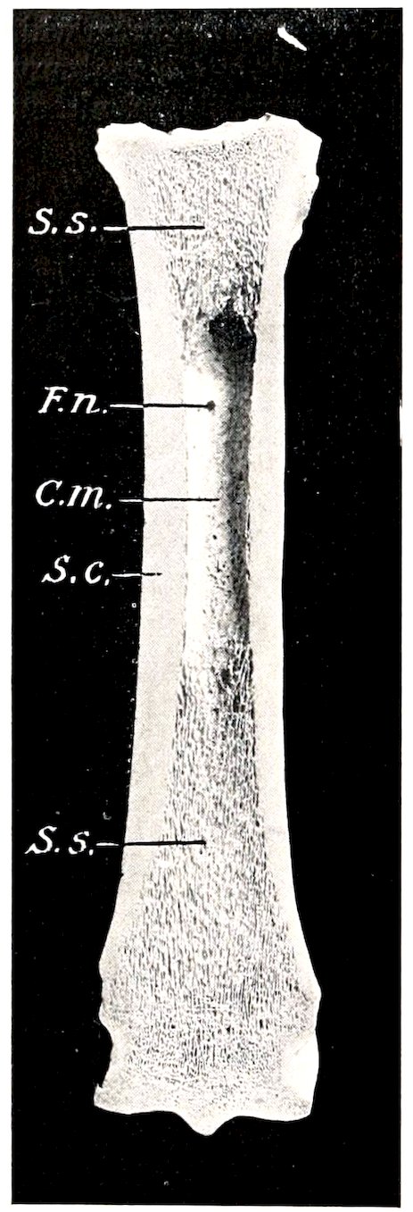
Fig. 1.—Frontal Section of Large Metatarsal Bone of Horse, Posterior Part.
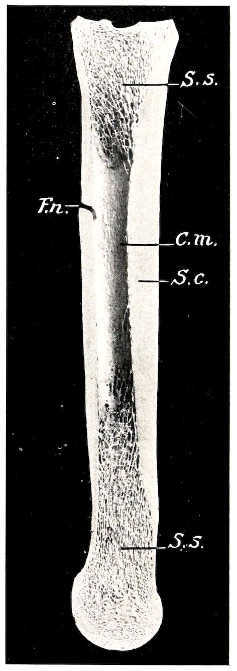
Fig. 2.—Sagittal Section of Large Metatarsal Bone of Horse. S.C., Compact substance; S.s., spongy substance; C.m., medullary cavity; F.n., nutrient foramen. Note the greater thickness of the compact substance of the inner and anterior parts of the shaft.
The compact substance (Substantia compacta) differs greatly in thickness in various situations, in conformity with the stresses and strains to which the bone is subjected. In the long bones it is thickest in the middle part of the shaft and thins out toward the extremities. On the latter the layer is very thin, and is especially dense and smooth on joint surfaces.
21The spongy substance (Substantia spongiosa) consists of delicate bony plates and spicules which run in various directions and intercross. These plates are definitely arranged with regard to mechanical requirements, so that systems of pressure and tension plates can be recognized, in conformity with the lines of pressure and the pull of tendons and ligaments respectively. The intervals (marrow spaces) between the plates are occupied by marrow. The spongy substance forms the bulk of short bones and of the extremities of long bones; in the latter it is not confined to the ends, but extends a variable distance along the shaft also. Some bones (Ossa pneumatica) contain air-spaces or sinuses within the compact substance instead of spongy bone and marrow. In certain situations the two compact layers of flat bones are not separated by spongy bone, but fuse with each other; in some cases of this kind the bone is so thin as to be translucent, or may even undergo absorption, producing an actual deficiency.
The flat bones of the cranial vault and sides are composed of an outer layer of ordinary compact substance, an inner layer of very dense bone, the tabula vitrea, and between these a variable amount of spongy bone, here termed diploë.
The periosteum is the membrane which invests the outer surface of bone, except where it is covered with cartilage. It consists of an outer protective fibrous layer, and an inner cellular osteogenic layer. During active growth the osteogenic layer is well developed, but later it becomes much reduced. The fibrous layer varies much in thickness, being in general thickest in exposed situations. The adhesion of the periosteum to the bone also differs greatly in various places; it is usually very thin and easily detached where it is thickly covered with muscular tissue which has little or no attachment. The degree of vascularity conforms to the activity of the periosteum.
The marrow (Medulla ossium) occupies the interstices of the spongy bone and the medullary cavity of the long bones. There are two varieties in the adult—red and yellow. In the young subject there is only red marrow (Medulla ossium rubra), but later this is replaced in the medullary cavity by yellow marrow (Medulla ossium flava). The red marrow contains several types of characteristic cells and is a blood-forming substance, while the yellow is practically ordinary adipose tissue.
Since yellow marrow is formed by regressive changes in red marrow, including fatty infiltration and degeneration of the characteristic cells, we find transitional forms or stages in the process. In aged or badly nourished subjects the marrow may undergo gelatinous degeneration, resulting in the formation of gelatinous marrow.
Vessels and Nerves.—It is customary to recognize two sets of arteries—the periosteal and the medullary. The former ramify in the periosteum and give off innumerable small branches which enter minute openings (Volkmann’s canals) on the surface and reach the Haversian canals of the compact substance. Other branches enter the extremities of the long bones and supply the spongy bone and marrow in them. In the case of the larger bones—and especially the long bones—the large medullary or nutrient artery enters at the so-called nutrient foramen (Foramen nutricium), passes in a canal (Canalis nutricius) through the compact substance, and ramifies in the marrow; its branches anastomose with the central branches of the periosteal set. The larger veins of the spongy bone do not, as a rule, accompany the arteries, but emerge chiefly near the articular surfaces. Within the bone they are destitute of valves.
The lymph vessels form perivascular channels in the periosteum and the Haversian canals of the compact substance. Lymph-spaces exist at the periphery of the marrow.
The nerves appear to be distributed chiefly to the blood-vessels. Special nerve-endings (Vater-Pacini corpuscles) in the periosteum are to be regarded as sensory, and probably are concerned in mediating the muscle sense (Kopsch).
22
DEVELOPMENT AND GROWTH OF BONE[2]
The primitive embryonal skeleton consists of cartilage and fibrous tissue, in which the bones develop. The process is termed ossification or osteogenesis, and is effected essentially by bone-producing cells, called osteoblasts. It is customary, therefore, to designate as membrane bones those which are developed in fibrous tissue, and as cartilage bones those which are preformed in cartilage. The principal membrane bones are those of the roof and sides of the cranium and most of the bones of the face. The cartilage bones comprise, therefore, most of the skeleton. Correspondingly we distinguish intramembranous and endochondral ossification.
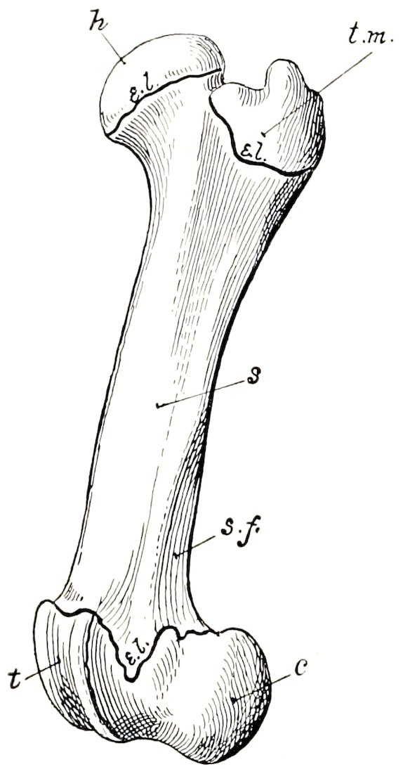
Fig. 3.—Left Femur of Young Pig, External View, to Show Division of a Long Bone into Shaft (s) and Extremities. Proximal extremity consists of two parts, head (h) and trochanter major (t. m.), which have separate centers of ossification. Distal extremity consists of trochlea (t) and condyles (c); e.l., epiphyseal cartilages; s.f., supracondyloid fossa.
In intramembranous development the process begins at a definite center of ossification where the cells (osteoblasts) surround themselves with a deposit of bone. The process extends from this center to the periphery of the future bone, thus producing a network of bony trabeculæ. The trabeculæ rapidly thicken and coalesce, forming a bony plate which is separated from the adjacent bones by persistent fibrous tissue. The superficial part of the original tissue becomes periosteum, and on the deep face of this successive layers of periosteal bone are formed by osteoblasts until the bone attains its definitive thickness.
In endochondral ossification the process is fundamentally the same, but not quite so simple. Osteoblasts emigrate from the deep face of the perichondrium or primitive periosteum into the cartilage and cause calcification of the matrix or ground-substance of the latter. Vessels extend into the calcifying area, the cartilage cells shrink and disappear, forming primary marrow cavities which are occupied by processes of the osteogenic tissue. There is thus formed a sort of scaffolding of calcareous trabeculæ on which the bone is constructed by the osteoblasts. At the same time perichondral bone is formed by the osteoblasts of the primitive periosteum. The calcified cartilage is broken down and absorbed through the agency of large cells called osteoclasts, and is replaced by bone deposited by the osteoblasts. The osteoclasts also cause absorption of the primitive bone, producing the marrow cavities; thus in the case of the long bones the primitive central spongy bone is largely absorbed to form the medullary cavity of the shaft, and persists chiefly in the extremities. Destruction of the central part and formation of subperiosteal bone continue until the shaft of the bone has completed its growth.
A typical long bone is developed from three primary centers of ossification, one for the diaphysis or shaft and one for each epiphysis or extremity. Many bones have secondary centers from which processes or apophyses develop.
The foregoing outline accounts for the growth of bones except in regard to length. Increase in length may be explained briefly as follows: Provision for continued ossification at either end of the diaphysis is made by a layer of actively growing cartilage—the epiphyseal cartilage—which intervenes between the diaphysis 23and the epiphysis. It is evident that so long as this cartilage persists and grows, new bone may continue to be formed at its expense, and increase of length is possible. When the epiphyseal cartilage ceases to grow, it undergoes ossification, the bone is consolidated, and no further increase in length is possible. This fusion takes place at fairly definite periods in the various bones, and it is of value to know the usual times at which it occurs in the larger bones of the limbs at least.
After the bones have reached their full size, the periosteum becomes relatively reduced and inactive so far as its osteogenic layer is concerned; the bone-forming function may be stimulated by various causes, as is well seen in the healing of fractures and the occurrence of bony enlargements.
CHEMICAL COMPOSITION OF BONE
Dried bone consists of organic and inorganic matter in the ratio of 1 ∶ 2 approximately. The animal matter gives toughness and elasticity, the mineral matter hardness, to the bone tissue. Removal of the organic matter by heat does not change the general form of a bone, but reduces the weight by about one-third, and makes it very fragile. Conversely, decalcification, while not affecting the form and size of the bone, renders it soft and pliable. The animal matter when boiled yields gelatin. The following table represents the composition in 100 parts of ox bone of average quality:
Gelatin 33.30
Phosphate of lime 57.35
Carbonate of lime 3.85
Phosphate of magnesia 2.05
Carbonate and chlorid of sodium 3.45
100.00
PHYSICAL PROPERTIES OF BONE
Fresh dead bone has a yellowish-white color; when macerated or boiled and bleached, it is white. The specific gravity of fresh compact bone is a little over 1.93. It is very hard and resistant to pressure; a 5-millimeter cube of compact bone of the ox will resist pressure up to 852 pounds, if the pressure be applied in the line of the lamellæ (Rauber). Its tensile strength is estimated to be nearly twice that of oak.
DESCRIPTIVE TERMS
The surfaces of the bones present a great variety of eminences and depressions, as well as perforations. The prominences and cavities may be articular, or non-articular, furnishing attachment to muscles, tendons, ligaments or fascia. A number of descriptive terms are used to designate these features, and the following are some of those in general use:
Process (Processus) is a general term for a prominence.
A tuberosity (Tuber, Tuberositas) is a large, rounded projection; a tubercle (Tuberculum) is a smaller one.
The term trochanter is applied to a few prominences, e. g., the trochanters of the femur.
A spine (Spina) or spinous process (Processus spinosus) is a pointed projection.
A crest (Crista) is a sharp ridge.
A line (Linea) is a very small ridge.
A head (Caput) is a rounded articular enlargement at the end of a bone; it may be joined to the shaft by a constricted part, the neck (Collum).
A condyle (Condylus) is an articular eminence which is somewhat cylindrical; a non-articular projection in connection with a condyle may be termed an epicondyle.
24A trochlea is a pulley-like articular mass.
A glenoid cavity (Cavitas glenoidalis) is a shallow articular depression, and a cotyloid cavity or acetabulum is a deeper one.
The term facet is commonly applied to articular surfaces of small extent, especially when they are not strongly concave or convex.
The terms fossa, fovea, groove or sulcus, and impression are applied to various forms of depressions.
A foramen is a perforation for the transmission of vessels, nerves, etc.
A sinus or antrum is an air-cavity.
Other terms, such as canal, fissure, notch, etc., require no explanation.[3]
VERTEBRAL COLUMN
The vertebral column (Columna vertebralis) is the fundamental part of the skeleton. It consists of a chain of median, unpaired, irregular bones which extends from the skull to the end of the tail. In the adult certain vertebræ have become fused to form a single bony mass with which the pelvic girdle articulates. Vertebræ so fused are termed fixed or “false” vertebræ (Vertebræ immobiles), as distinguished from the movable or “true” vertebræ (Vertebræ mobiles).
The column is subdivided for description into five regions, which are named according to the part of the body in which they are placed. Thus the vertebræ are designated as cervical, thoracic (or dorsal), lumbar, sacral, and coccygeal or caudal (Vertebræ cervicales, thoracales, lumbales, sacrales, coccygeæ). The number of vertebræ in a given species is fairly constant in each region except the last, so that the vertebral formula may be expressed (for the horse, for example) as follows:
C7T18L6S5Cy15–21.
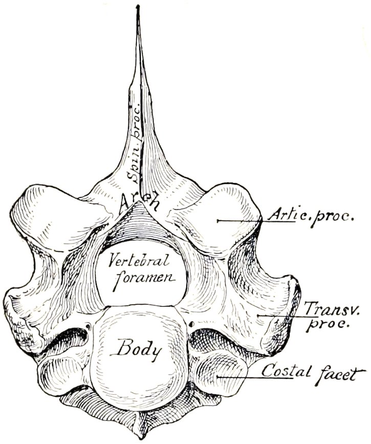
Fig. 4.—First Thoracic Vertebræ of Horse. To illustrate plan of structure of vertebræ.
The vertebræ in a given region have special characters by which they may be distinguished from those of other regions, and individual vertebræ have characters which are more or less clearly recognizable. All typical vertebræ have a common plan of structure, which must first be understood. The parts of which a vertebra consists are the body or centrum, the arch, and the processes.
The body (Corpus vertebræ) is the more or less cylindrical mass on which the other parts are constructed. The anterior and posterior extremities of the body are attached to the adjacent vertebræ by intervertebral fibro-cartilages, and are usually convex and concave respectively. The dorsal surface is flattened and enters into the formation of the vertebral canal, while the ventral aspect is rounded laterally, and is in relation to various muscles and viscera. In the thoracic region the body presents two pairs of demifacets (Foveæ costales) at the extremities for articulation with the heads of two pairs of ribs.
The arch (Arcus vertebræ) is constructed on the dorsal aspect of the body. It consists originally of two lateral halves, each of which is considered to consist of a pedicle and a lamina. The pedicles form the lateral parts of the arch, and are 25cut into in front and behind by the vertebral notches (Incisura vertebralis cranialis, caudalis). The notches of two adjacent vertebræ form intervertebral foramina for the passage of the spinal nerves and vessels; in some vertebræ, however, these are complete foramina instead of notches. The laminæ are plates which complete the arch dorsally, uniting with each other medially at the root of the spinous process.
The body and the arch form a bony ring which incloses the vertebral foramen (Foramen vertebrale); the series of vertebral rings, together with the ligaments which unite them, inclose the vertebral canal (Canalis vertebralis), which contains the spinal cord and its coverings and vessels.
The articular processes, two anterior and two posterior (Processus articulares craniales, caudales), project from the borders of the arch on either side. They present joint surfaces adapted to those of adjacent vertebræ, and the remaining surface is roughened for muscular and ligamentous attachment.
The spinous process (Processus spinosus) is single, and projects dorsally from the middle of the arch. It varies greatly in form, size, and direction in different vertebræ. It furnishes attachment to muscles and ligaments.
The transverse processes (Processus transversi) are two in number and project laterally from the side of the arch, or from the junction of the arch and body. In the thoracic region each has a facet for articulation with the tubercle of a rib (Fovea costalis transversalis). They also give attachment to muscles and ligaments.
Some vertebræ have also a ventral or hæmal spine.
Mammillary processes (Processus mammillares) are found in most animals on the last thoracic and anterior lumbar vertebræ between the transverse and anterior articular processes or on the latter.
Accessory processes (Processus accessorii), when present, are situated between the transverse and posterior articular processes.
Development.—The vertebræ are developed by ossification in the cartilage which surrounds the notochord and forms the sides of the neural canal. There are three primary centers of ossification, one for the body and one for each side of the arch. Secondary centers appear later for the summit of the spinous process (except in the cervical region), the extremities of the transverse processes, and the thin epiphyseal plates at the extremities of the body.
Sometimes there are at first two centers for the body which soon fuse. The process of ossification extends from the lateral centers to form not only the corresponding part of the arch, but also the processes and a part of the body next to the root of the arch (Radix arcus). In the horse and ox the body and arch are usually fused at birth, but the epiphyses do not fuse till growth is complete. In the pig, sheep, and dog the body and arch are united at birth by cartilage (neurocentral synchondrosis), but fuse in the first few months.
THE RIBS
The ribs (Costæ) are elongated curved bones which form the skeleton of the lateral thoracic walls. They are arranged serially in pairs which usually correspond in number to the thoracic vertebræ. Each articulates dorsally with the spine and is continued ventrally by a costal cartilage. Those which articulate with the sternum by means of their cartilages are termed sternal or “true” ribs (Costæ sternales s. veræ); the remainder are asternal or “false” ribs (Costæ asternales s. spuriæ). Ribs at the end of the series which have their ventral ends free in the abdominal wall are named floating ribs (Costæ fluctuantes). The intervals between the ribs are termed intercostal spaces (Spatia intercostalia).
A typical rib[4] consists of a shaft and two extremities. The shaft (Corpus costæ) 26is band-like and varies much in length, breadth, and curvature. In the case of some ribs the curvature is not uniform, but is most accentuated at a certain point, termed the angle of the rib (Angulus costæ); this occurs at a variable distance from the vertebral end, and is usually marked by a rough ridge. The direction also varies; the first rib is almost vertical, while the remainder slope backward in increasing degree. The external surface is convex, and the internal flattened from edge to edge; on the latter, close to the posterior border, is the costal groove (Sulcus costalis), which fades out ventrally. It contains the intercostal vein. The anterior and posterior borders are thin and sharp on some ribs, rounded on others.
The vertebral extremity (Extremitas vertebralis) consists of the head, neck, and tubercle. The head (Capitulum costæ) is the actual end of the rib, and is rounded and somewhat enlarged. It presents two facets (Facies articularis capituli costæ) for articulation with the bodies of two adjacent thoracic vertebræ; these surfaces are separated by a groove in which the conjugal ligament is attached. The neck (Collum costæ) joins the head to the shaft. It varies in length and diameter. Its outer surface is rough, its inner smooth. The tubercle (Tuberculum costæ) projects backward at the junction of the neck and shaft. It has a facet (Facies articularis tuberculi costæ) for articulation with the transverse process of the posterior vertebra of the two with which the head articulates. The tubercle gradually approaches the head in the posterior ribs, and eventually fuses with it.
The sternal extremity (Extremitas sternalis) is commonly slightly enlarged, and has a rough depression in which the costal cartilage is embedded.
Development.—The ribs are ossified in cartilage from three centers—one each for the shaft (and sternal end), head, and tubercle; the third center does not occur in the last two ribs.
THE COSTAL CARTILAGES
These (Cartilagines costales) are bars of hyaline cartilage which continue the ribs. Those of the sternal ribs articulate with the sternum, while the remainder overlap and are attached to each other to form the costal arch (Arcus costalis).
THE STERNUM
The sternum or breast-bone is a median segmental bone which completes the skeleton of the thorax ventrally, and articulates with the cartilages of the sternal ribs laterally. It consists of six to eight bony segments (Sternebræ) connected by intervening cartilage in the young subject. Its form varies with that of the thorax in general and with the development of the clavicles in animals in which they are present. Its anterior extremity, the manubrium sterni or presternum, is specially affected by the latter factor, being broad and strong when the clavicles are well developed and articulate with it (as in man), relatively small and laterally compressed when they are absent (as in the horse) or rudimentary (as in the dog). The cartilages of the first pair of ribs articulate with it. The body or mesosternum (Corpus sterni) presents laterally, at the junction of the segments, concave facets (Incisuræ costales) for articulation with the cartilages of the sternal ribs. The posterior extremity or metasternum presents the xiphoid (or ensiform) cartilage (Processus xiphoideus); this is thin and plate-like, as in the horse and ox, or narrow and short, as in the pig and dog.
Development.—The cartilaginous sternum is formed by the fusion medially of two lateral bars which unite the ventral ends of the first eight or nine costal cartilages, and is primitively unsegmented. The manubrium ossifies from a single center, but the centers for the other segments appear to be primitively paired. The sternum never becomes completely ossified; details in regard to persisting 27cartilage will be given in the special descriptions. The layer of compact tissue is for the greater part very thin and the spongy substance is open-meshed and very vascular.
THE THORAX
The skeleton of the thorax comprises the thoracic vertebræ dorsally, the ribs and their cartilages laterally, and the sternum ventrally. The thoracic cavity (Cavum thoracis) resembles in shape an irregular truncated cone; it is compressed laterally, especially in front, and the dorsal wall or roof is much longer than the ventral wall or floor. The anterior aperture (Apertura thoracis cranialis) is bounded by the first thoracic vertebra dorsally, the first pair of ribs and their cartilages laterally, and the manubrium sterni ventrally. The posterior aperture (Apertura thoracis caudalis) is bounded by the last thoracic vertebra, the last pair of ribs, the costal arches, and the anterior part of the xiphoid cartilage.
It may be noted here that the diaphragm (which forms the partition between the thoracic and abdominal cavities) does not follow the costal arches in its posterior attachment, so that the posterior ribs enter also into the formation of the abdominal wall.
THE SKULL
The term skull is usually understood to include all of the bones of the head. The head consists of the cranium and the face, and it is therefore convenient to divide the bones into cranial and facial groups.
The cranial bones (Ossa cranii) inclose the brain with its membranes and vessels and the essential organs of hearing. They concur with the facial bones in forming the orbital and nasal cavities, in which the peripheral organs of sight and of smell are situated.
The facial bones (Ossa faciei) form the skeleton of the oral and nasal cavities, and also support the larynx and the root of the tongue.
Most of the bones of the skull are flat bones, developed in membrane; those of the cranial base may be classed as irregular, and are cartilage bones. Only two form permanent movable joints with other parts of the skull. The mandible or lower jaw-bone forms diarthrodial joints with the temporal bones, and the hyoid bone is attached to the latter by bars of cartilage. The other bones form immovable joints, most of which disappear with age.
In order to study the separate bones, skulls of young subjects are necessary, since later most of the lines of demarcation become effaced. The relations of each bone to its surroundings should be specially noted, since the final object is to understand the skull as a whole. In the descriptions which follow the skull is considered with its long axis horizontal, and that of the horse will serve as a type.
THE BONES OF THE THORACIC LIMB
The thoracic limb consists of four chief segments, viz., the shoulder girdle, the arm, the forearm, and the forefoot or manus.
The shoulder girdle (Cingulum extremitatis thoracicæ), when fully developed, consists of three bones—the scapula or shoulder-blade, the coracoid, and the clavicle or collar-bone. In the domesticated mammals only the scapula, a large, flat bone, is well developed, and the small coracoid element has fused with it, while the clavicle is either absent or is a small rudiment embedded in the mastoido-humeralis muscle. There is therefore no articulation of the shoulder with the axial skeleton.
The shoulder girdle is fully developed in birds and the lower mammals (monotremata). In the higher mammals the coracoid is reduced to the coracoid process of the scapula, and the development of the clavicle is in conformity with the function of the limb. Thus in typical quadrupeds, 28such as the horse and ox, in which the forelimbs are used only for support and locomotion, the clavicle is absent. Other animals which use these limbs for grasping, burrowing, climbing, etc. (e.g., man, apes, moles), have well-developed clavicles which connect the scapula with the sternum.
The arm (Brachium) contains a single long bone, the humerus or arm bone.
In the forearm (Antibrachium) are two long bones, the radius and ulna. These vary in relative size and mobility. In the horse and ox the two bones are fused, and the lower part of the limb is fixed in the position of pronation. The radius is placed in front and supports the weight. The ulna is well developed only in its upper part, which forms a lever for the extensor muscles of the elbow. In the pig the ulna is the larger and longer of the two bones, but is closely attached to the back of the radius. In the dog the ulna is also well developed and a small amount of movement is possible between the two bones.
The forefoot or hand (Manus) consists of three subdivisions, viz., the carpus, metacarpus, and digit or digits.
The carpus, popularly termed the “knee” in animals, and homologous with the wrist of man, contains a group of short bones (Ossa carpi). These are typically eight in number and are arranged in two transverse rows—a proximal or antibrachial, and a distal or metacarpal. The bones of the proximal row, named from the radial to the ulnar side (i. e., from within outward), are the radial, intermediate, ulnar, and accessory carpal bones. The bones of the distal row are designated numerically, in the same direction, as first, second, third, and fourth carpal bones.
This nomenclature, introduced by Gegenbaur, and now used largely by comparative anatomists, seems decidedly preferable to the variety of terms borrowed from human anatomy and based on the form of the bones in man. The following table of synonyms in common use is appended for comparison. The Latin terms and abbreviated notations are given in parenthesis.
Radial (Os carpi radiale, Cr) Scaphoid
Intermediate (Os carpi intermedium, Ci) Semilunar
Ulnar (Os carpi ulnare, Cu) Cuneiform
Accessory (Os carpi accessorium, Ca) Pisiform
First carpal (Os carpale primum, C1) Trapezium
Second carpal (Os carpale secundum, C2) Trapezoid
Third carpal (Os carpale tertium, C3) Os magnum
Fourth carpal (Os carpale quartum, C4) Unciform
The central carpal bone (Os carpi centrale) is omitted, since it is not a separate element in the animals under consideration here.
The metacarpus contains typically five metacarpal bones (Ossa metacarpalia I–V), one for each digit; they are long bones and are designated numerically from within outward. This arrangement occurs in the dog, although the first metacarpal is much smaller than the others, and the second and fifth are somewhat reduced. Further reduction has taken place in the other animals, resulting in the perissodactyl and artiodactyl forms. In the horse the first and fifth metacarpals are absent, the third is the large supporting metacarpal bone and carries the single digit, while the second and fourth are much reduced. In artiodactyls (e. g., ox, sheep, pig) the third and fourth are the chief metacarpals and carry the well developed digits; they are fused in the ox and sheep. The others are variously reduced or absent as noted in the special descriptions to follow.
The fossil remains of the ancestors of the existing Equidæ illustrate in a most complete manner the reduction which has occurred in this respect. The earliest known ancestor of the horse, Eohippus or Hyracotherium of the Lower Eocene, had four well developed metacarpal bones, each of which carried a digit; the first metacarpal bone was small. Eleven intermediate stages show the gradual evolution of the race from this primitive animal, which was about the size of the domestic cat. There is reason to believe that earlier forms had five digits.
The digits (Digiti manus) are homologous with the fingers of man, and are typically five in number. They are designated numerically from the radial to the ulnar side, in correspondence with the metacarpus. The full number is present in the dog. In the ox and pig the third and fourth are well developed and support 29the weight, while the second and fifth are reduced. The existing horse has a single digit, the third of his polydactyl ancestors. The skeleton of each fully developed digit consists of three phalanges and certain sesamoid bones. The first or proximal phalanx (Phalanx prima) articulates with the corresponding metacarpal bone above and with the second or middle phalanx (Phalanx secunda) below. The third or distal phalanx (Phalanx tertia) is inclosed in the hoof or claw, and is modified to conform to the latter. The sesamoid bones (Ossa sesamoidea) are developed along the course of the flexor tendons or in the joint capsules. Two proximal sesamoids (Ossa sesamoidea phalangis primæ) occur at the flexor side of the metacarpo-phalangeal joint and form a pulley for the flexor tendon. The distal sesamoids (Ossa sesamoidea phalangis tertiæ) are similarly placed between the deep flexor tendon and the joint between the second and third phalanx; they are absent in the dog, which has a small sesamoid on the extensor side of the metacarpo-phalangeal joints, and often at the proximal interphalangeal joint also.
Numerous cases are recorded of the occurrence of supernumerary digits (hyperdactylism) in the horse and other animals. In some pigs, on the other hand, the two chief digits are fused, and the condition appears to be inherited.
THE BONES OF THE PELVIC LIMB
The pelvic limb, like the thoracic, consists of four segments, viz., the pelvic girdle, thigh, leg, and the hind foot or pes; the last is subdivided into tarsus, metatarsus, and digits.
The pelvic girdle (Cingulum extremitatis pelvinæ) consists of the os coxæ or hip bone, which joins its fellow of the opposite side ventrally at the symphysis pelvis, and articulates very firmly with the sacrum dorsally. The two coxal bones, together with the sacrum and the first two or three coccygeal vertebræ, constitute the bony pelvis. The os coxæ consists originally of three flat bones, the ilium, ischium, and pubis, which meet at the acetabulum, a large cotyloid cavity that articulates with the head of the femur. These three parts are fused before growth is complete, but are considered separately for convenience of description. The ilium (Os ilium) is situated in the lateral wall of the pelvis, the pubis (Os pubis) in the anterior part, and the ischium (Os ischii) in the posterior part of the ventral wall.
The thigh (Femur), like the arm, contains a single large, long bone, the femur or thigh bone (Os femoris). This articulates with the acetabulum above and the tibia and patella below.
The skeleton of the leg (Crus) comprises three bones (Ossa cruris), viz., the tibia, fibula, and patella. The tibia is a large prismatic long bone which supports the weight, and articulates below with the tibial tarsal bone. The fibula is situated along the outer border of the tibia, from which it is separated by the interosseous space of the leg. It is much more slender than the tibia and does not articulate with the femur. In the pig and dog it has a complete shaft and two extremities, but in the horse and ox it is much reduced and otherwise modified. The patella or “knee-cap” is a short bone which articulates with the trochlea of the distal end of the femur; it is to be regarded as a large sesamoid bone intercalated in the tendon of the quadriceps femoris muscle.
The tarsus or “hock” consists of a group of short bones (Ossa tarsi) numbering five to seven in the different animals. The proximal or crural row consists of two bones, the tibial and fibular tarsals; the former is situated at the inner or tibial side, and has a trochlea for articulation with the distal end of the tibia; the latter, situated externally, has a process, the tuber calcis, which projects upward and backward and constitutes a lever for the muscles which extend the hock joint. The distal or metatarsal row consists of four bones when seven tarsal elements are 30present, as in the pig and dog. They are best designated numerically as first tarsal, second tarsal, etc. The central tarsal is interposed between the rows.
The preceding terms are anglicized abbreviations of those introduced by Gegenbaur into comparative anatomy. The Latin names and synonyms are given in the following table.
Tibial (Os tarsi tibiale, Tt.) Astragalus or Talus
Fibular (Os tarsi fibulare, Tf.) Calcaneus or Os calcis
Central (Os tarsi centrale, Tc.) Scaphoid or Navicular
First Tarsal (Os tarsale primum, T1) First or internal cuneiform
Second Tarsal (Os tarsale secundum, T2) Second or middle cuneiform
Third Tarsal (Os tarsale tertium, T3) Third or external cuneiform
Fourth Tarsal (Os tarsale quartum, T4) Cuboid.
The metatarsal and digital bones resemble in general those of the corresponding regions of the thoracic limb; the differential features will be noted in the special descriptions.
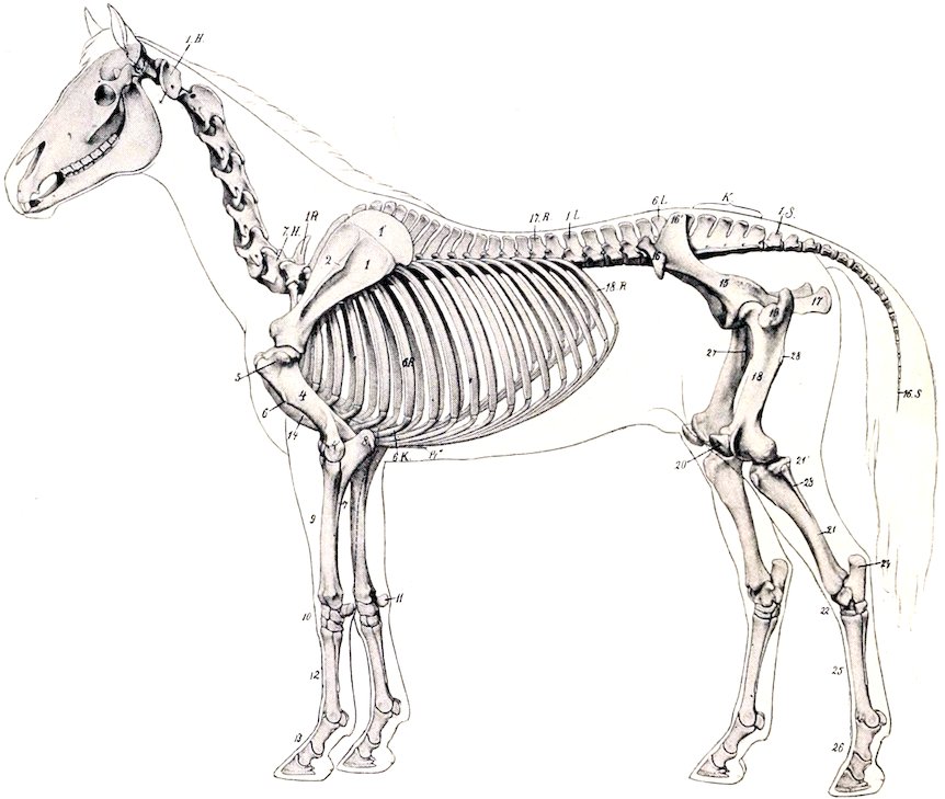
Fig. 5.—Skeleton of Horse, with Outline of Contour of Body. 1.H., Atlas; 7.H., seventh cervical vertebra; 1.R., first thoracic vertebra; 17.R., seventeenth thoracic vertebra; 1.L., first lumbar vertebra; 6.L., sixth lumbar vertebra; K, sacrum; 1.S., first coccygeal vertebra; 16.S., sixteenth coccygeal vertebra; 6.R., sixth rib; 6.K., costal cartilage; 18.R., last rib; 1, scapula; 1′, cartilage of scapula; 2, spine of scapula; 4, humerus; 4′ external epicondyle of humerus; 5, external tuberosity of humerus; 6, deltoid tuberosity; 7, shaft of ulna; 8, olecranon; 9, radius; 10, carpus; 11, accessory carpal bone; 12, metacarpus; 13, digit; 14, sternum; 14″, xiphoid cartilage; 15, ilium; 16, 16′, external and internal angles of ilium; 17, ischium; 18, femur (shaft); 19, trochanter major; 27, trochanter minor; 28, trochanter tertius; 20, patella; 21, tibia (shaft); 21′, external condyle of tibia; 23, fibula; 22, tarsus; 24, tuber calcis; 25, metatarsus; 26, digit. (After Ellenberger-Baum, Anat. für Künstler.)
31
THE SKELETON OF THE HORSE
The skeleton of the horse consists of 205 bones, as shown in the following table.
Vertebral column 54
Ribs 36
Sternum 1
Skull (including auditory ossicles) 34
Thoracic limbs 40
Pelvic limbs 40
205
In this enumeration the average number of coccygeal vertebræ is taken to be 18, the temporal and os coxæ are not divided into parts, the usual number of carpal and tarsal elements is taken, and the sesamoids are included.
The Vertebral Column
The vertebral formula of the horse is C7T18L6S5Cy15–21.
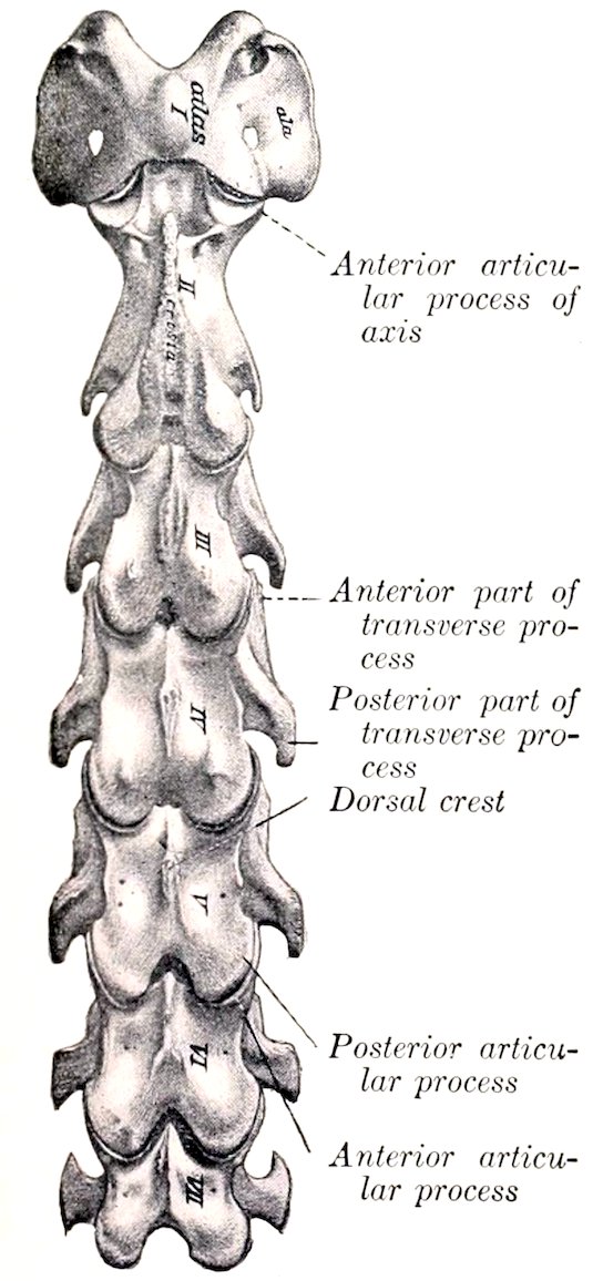
Fig. 6.—Cervical Vertebræ of Horse, Dorsal View. (After Schmaltz, Atlas d. Anat. d. Pferdes.)
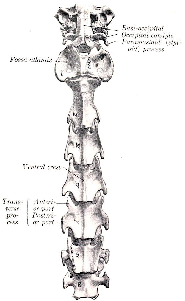
Fig. 7.—Cervical Vertebræ of Horse, Ventral View. (After Schmaltz, Atlas d. Anat. d. Pferdes.)
32
THE CERVICAL VERTEBRÆ
These are quadrangular, massive, and longer than the vertebræ of other regions; they decrease in length from the second to the last. The third, fourth, and fifth are typical, and have the following characters:
1. The bodies are long as compared with those of other vertebræ. Each presents a median ventral spine or crest, which becomes more prominent as it is traced backward, and is tuberculate at its posterior end. The lateral aspect is concave. The dorsal surface has a flat central area which is narrow in the middle of the vertebræ, and wide at either end; it gives attachment to the superior common ligament. On either side of this area is a groove which lodges the longitudinal spinal vein. These lateral grooves are connected at the middle of the surface by a transverse furrow, in which there are several foramina through which veins emerge from the spongy substance of the body. The anterior extremity presents a head which has an oval articular surface, strongly convex, and wider above than below. The posterior extremity is larger and has a nearly circular cotyloid cavity.
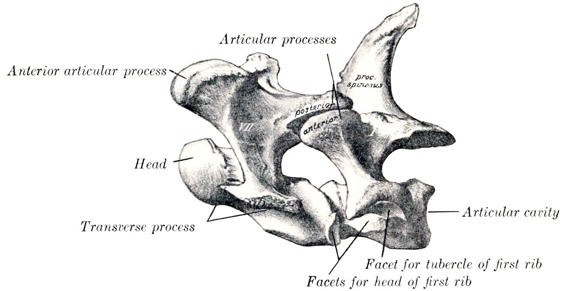
Fig. 8.—Last Cervical and First Thoracic Vertebræ of Horse, Lateral View. (After Schmaltz, Atlas d. Anat. d. Pferdes.)
2. The arches are large and strong. They are perforated on either side by a foramen which communicates with the foramen transversarium. The vertebral notches are large.
3. The articular processes are large. Their articular surfaces are extensive, oval in outline, and slightly concave; the anterior ones are directed upward and inward, the posterior downward and outward. The remaining surface is mainly roughened for ligamentous and muscular attachment. A crest connects the articular processes of the same side on the fourth and fifth; on the third it does not reach the anterior process.
4. The transverse processes are large and plate-like. Each arises by two roots, one from the arch and one from the body; between these is the foramen transversarium, through which the vertebral artery passes. The process divides externally into anterior and posterior branches, which are thickened and rough for muscular attachment.
5. The spinous process is represented by a crest (Crista spinosa), which widens behind, and is connected by ridges with the posterior articular processes.
The sixth cervical vertebra has the following distinctive features: It is shorter and wider than the fifth. The arch is large, especially posteriorly. The posterior 33articular processes are shorter, thicker and further apart; they are connected with the anterior ones by a thick ridge. The spinous process is less rudimentary; it is half an inch or more (ca. 1.5 cm.) in height. The transverse processes have three branches; the third part is a thick, almost sagittal plate, which forms with its fellow and the body a wide ventral groove on the posterior part of the vertebra; the other branches correspond to those of the typical vertebræ, but are short and thicker. The foramen transversarium is large; below its posterior end is a fossa. The ventral crest is small and is less prominent posteriorly.
The third branch of the transverse process and the fossa are sometimes absent on one side.
The seventh cervical vertebra is readily distinguished by the following characters: It is shorter and wider than the others. The body is flattened dorso-ventrally and wide, especially behind; here it has a demifacet on either side for articulation with part of the head of the first rib. The arch and its notches are large. The anterior articular processes are wider and longer than the posterior pair. The spinous process is an inch or more (ca. 3 cm.) in height. The transverse process is undivided, and has no foramen transversarium. The ventral crest is replaced by a pair of tubercles.
In some specimens a large foramen transversarium is present on one side or (rarely) on both.
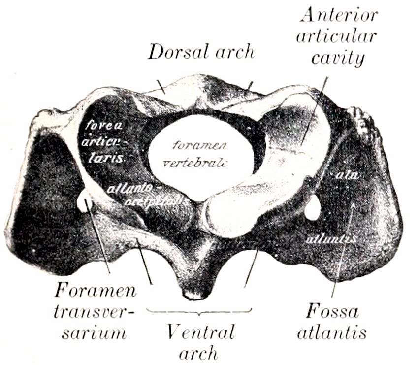
Fig. 9.—Atlas of Horse, Anterior View. (After Schmaltz, Atlas d. Anat. d. Pferdes.)
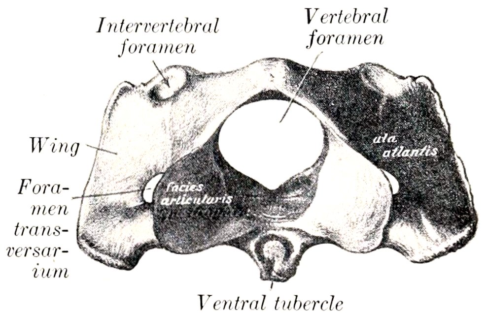
Fig. 10.—Atlas of Horse, Posterior View. (After Schmaltz, Atlas d. Anat. d. Pferdes.)
The Atlas
This vertebra is decidedly atypical in form and structure. The body and spinous process are absent. It has the form of a strong ring, from which two curved plates, the wings, project laterally. The ring incloses a very large vertebral foramen, and consists of two lateral masses connected by dorsal and ventral arches.
The lateral masses (Massæ laterales) present two deep oval anterior articular cavities (Foveæ articulares craniales) which receive the occipital condyles; they are separated by a wide notch above and a narrow one below. The outer margin is also notched, and a triangular non-articular depression cuts into the inner part of each cavity. The posterior articular surfaces (Facies articulares caudales) are somewhat saddle-shaped; they are confluent on the ventral arch below, but are widely separated above, and do not conform in shape to the corresponding surfaces of the axis.
The dorsal arch (Arcus dorsalis) presents a median dorsal tubercle (Tuberculum dorsale) and is concave below. It is perforated on either side near its anterior margin by the intervertebral foramen. The anterior border is deeply notched, and the posterior is thin and concave.
The ventral arch (Arcus ventralis) is thicker, narrower, and less curved than 34the dorsal. On its lower surface is the ventral tubercle (Tuberculum ventrale), into which the terminal tendon of the longus colli muscle is inserted. The upper face has posteriorly a transversely concave articular surface (Fovea dentis), on which the dens or odontoid process of the axis rests. In front of this is a transverse rough excavation for the attachment of the odontoid ligament.
The wings (Alæ) are modified transverse processes. They are extensive curved plates which project outward, downward, and backward from the lateral masses. The dorsal surface is concave. Between the ventral aspect of the wing and the lateral mass is a cavity, the fossa atlantis; in this there is a foramen which opens into the vertebral canal. The border is thick and rough. Two foramina perforate each wing. The anterior one, the foramen alare, is connected with the intervertebral foramen by a short groove. The posterior one is the foramen transversarium.
Development.—The atlas ossifies from three or four centers, one or two for the ventral arch, and one on either side for each lateral mass, wing, and half of the dorsal arch. At birth the bone consists of three pieces—the ventral arch and two lateral parts, which are separated by a layer of cartilage in the dorsal median line. These parts are usually fused at about six months.
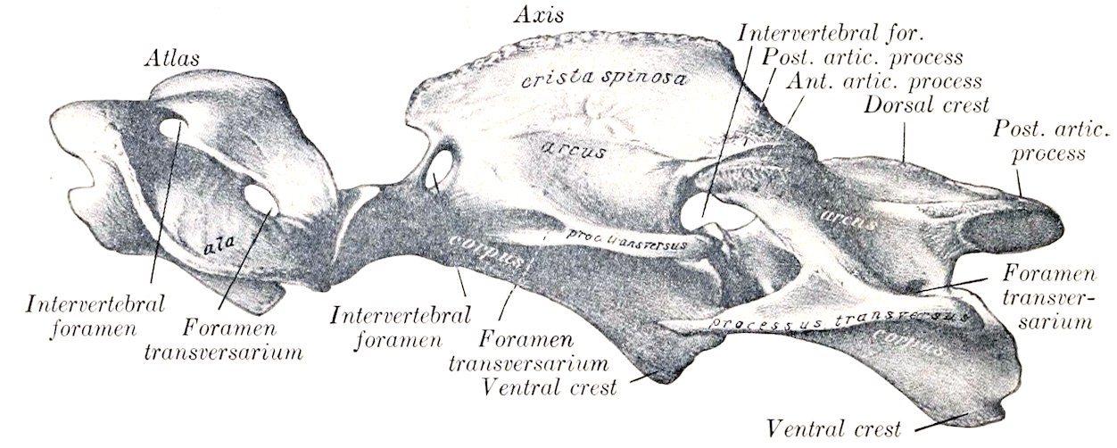
Fig. 11.—First Three Cervical Vertebræ of Horse, Lateral View. (After Schmaltz, Atlas d. Anat. d. Pferdes.)
The Axis
The axis (Epistropheus) is the longest of the vertebræ, and is characterized by the presence of the odontoid process, which projects from the anterior part of the body.
The body has a median ventral crest which terminates in a tubercle behind. The anterior extremity presents centrally the odontoid process (Dens); this has a convex articular surface ventrally for articulation with the ventral arch of the atlas, and two rough depressions for the attachment of the odontoid ligament dorsally. Flanking this on either side are the modified anterior articular processes, which have saddle-shaped articular surfaces confluent below with that of the dens. The posterior extremity has the usual cavity.
The arch presents in the young subject a notch on each side of its anterior border; this is converted into a foramen by a ligament which usually ossifies later. The posterior border has the usual notches.
The posterior articular processes are typical.
The transverse processes are small, single, and project backward. The foramen transversarium is small.
The spinous process is very large and strong. Its free border is rough, thickens posteriorly, and is continued to the articular processes by two ridges. The lateral surfaces are concave and rough for muscular attachment.
35Development.—The axis has six or seven centers of ossification. In addition to the usual five, one or two appear for the dens, which is regarded as the displaced body of the atlas. A nucleus behind the dens, which remains distinct to three or four years of age, is considered by Lesbre to be the head of the axis.
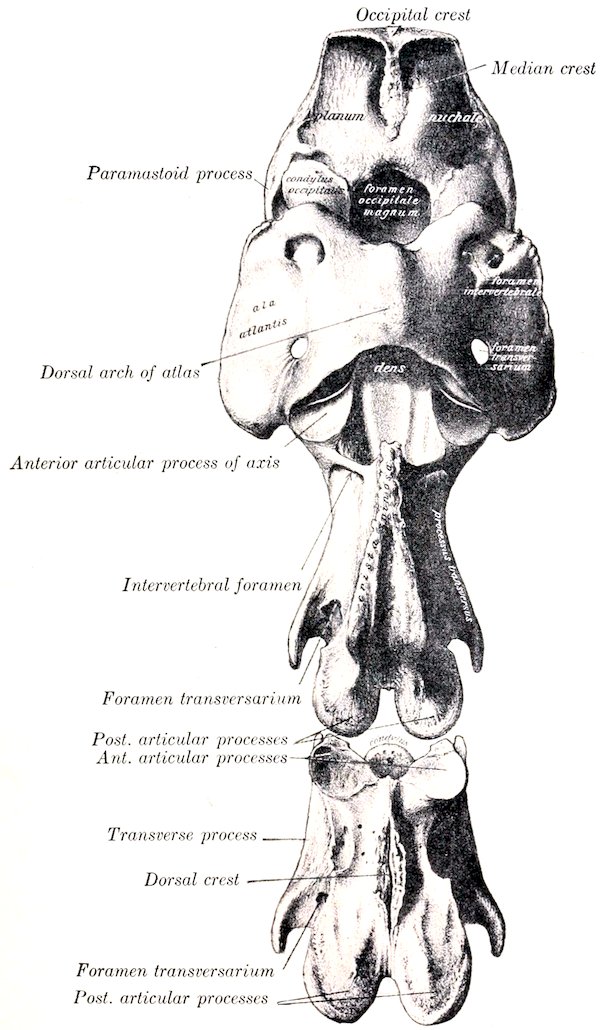
Fig. 12.—Occipital Bone and First Three Cervical Vertebræ of Horse, Dorsal View. (After Schmaltz, Atlas d. Anat. d. Pferdes.)
THE THORACIC VERTEBRÆ
These (Vertebræ thoracales) are usually eighteen in number in the horse, but there are sometimes nineteen, rarely seventeen. As regional characters we note the surfaces for articulation with the ribs and the length and form of the spinous 36processes. Those in the middle of the series are the most typical and present the following features:
1. The bodies are short and constricted in the middle. The ends are expanded and have articular surfaces which are not strongly curved. On the upper part of each side are anterior and posterior costal facets (Fovea costalis cranialis, caudalis), which, with those of adjacent vertebræ, form sockets for the heads of the ribs.
2. The arches are small. Their posterior notches are relatively large and are often converted into foramina.
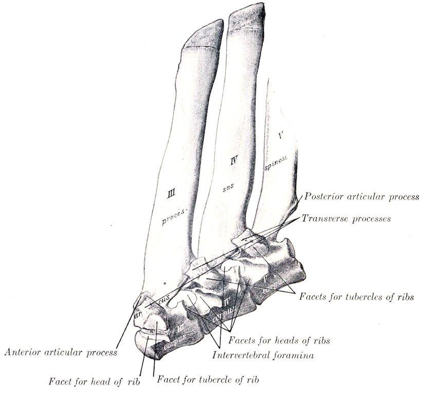
Fig. 13.—Third, Fourth, and Fifth Thoracic Vertebræ of Horse, Lateral View. (After Schmaltz, Atlas d. Anat. d. Pferdes.)
3. The articular processes are small. The anterior pair are in fact represented only by two oval facets on the anterior part of the laminæ which face almost directly upward. The posterior pair spring from the base of the spinous process; their facets face almost directly downward.
4. The transverse processes are short, thick, and tuberous at the free end. Each has a facet (Fovea transversaria) for articulation with the tuberosity of the rib which has the same serial number.
5. The spinous process is large, narrow, and slopes upward and backward. The anterior border is thin, the posterior wider and furrowed. The summit is expanded and rough.
The first thoracic vertebra is easily recognized by the following specific characters: The body is wide and flattened dorso-ventrally. In front it has a head like 37the cervical vertebræ, and behind a cavity somewhat deeper than any other thoracic vertebra. Two costal facets are found on either side, and a well-marked spine ventrally. The arch is large and strong, and has large notches. The articular processes are much larger than those of other thoracic vertebræ, and resemble a good deal those of the seventh cervical in form. The transverse processes are short and thick, and each has on its ventral aspect a large concave facet for articulation with the tubercle of the first rib. The spinous process is curved backward and tapers to a point. Its length is usually about three or four inches (ca. 8 to 10 cm.). It may be mistaken at first glance for the last cervical, but is promptly identified by the three costal facets on each side and the length of the spine.
The last thoracic vertebra is distinguished by the absence of the posterior pair of costal facets, and the confluence of the anterior pair with those on the transverse processes.
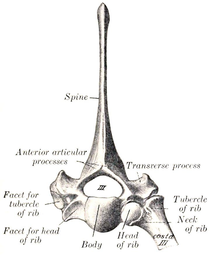
Fig. 14.—Third Thoracic Vertebræ and Upper Part of Rib of Horse, Anterior View. (After Schmaltz, Atlas d. Anat. d. Pferdes.)
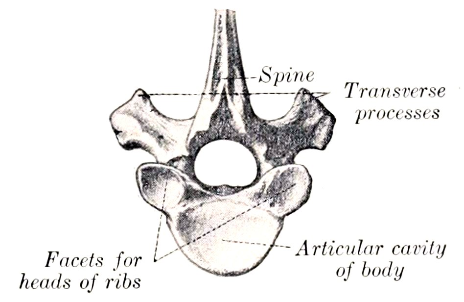
Fig. 15.—Lower Part of Ninth Thoracic Vertebra of Horse, Posterior View. (After Schmaltz, Atlas d. Anat. d. Pferdes.)
The serial position of others may be determined at least approximately by the following data: (1) The bodies gradually diminish in length and width to the middle of the region and then increase slightly. Their costal facets become smaller and less concave from first to last. The ventral crest is distinct on three or four vertebræ at either end of the region. (2) The transverse processes diminish in size and are placed lower down as they are traced backward. Their costal facets become smaller and lower in position; on the last (and sometimes on its predecessor also) it fuses with the costal facet of the body. The upper non-articular part of the process gradually becomes more sharply defined, and in the last four or five separates to form the mammillary process. (3) The spinous processes increase in length to the third and fourth, and then gradually diminish to the fifteenth, beyond which they have about the same length. The backward inclination is most pronounced in the second, the sixteenth is vertical, and the last two are directed a little forward. The longest spines (i. e., those of the withers) are the thickest and have expanded summits which remain more or less cartilaginous; the others are more plate-like, and are surmounted by a thick lip. The second spine is more than twice as large as the first. The summits of the fourth and fifth usually form the highest point of the withers.
Development.—There are six or seven centers, three for the body, two for the arch, and one for the spinous process; some of the latter have an additional center for the summit.
38
THE LUMBAR VERTEBRÆ
The lumbar vertebræ (Vertebræ lumbales) are usually six in number in the horse. They are characterized by the size and form of their transverse processes.
The bodies of the first three are triangular on cross-section, and present a distinct ventral crest. From the fourth backward they become wider and flatter and the ventral crest fades out.
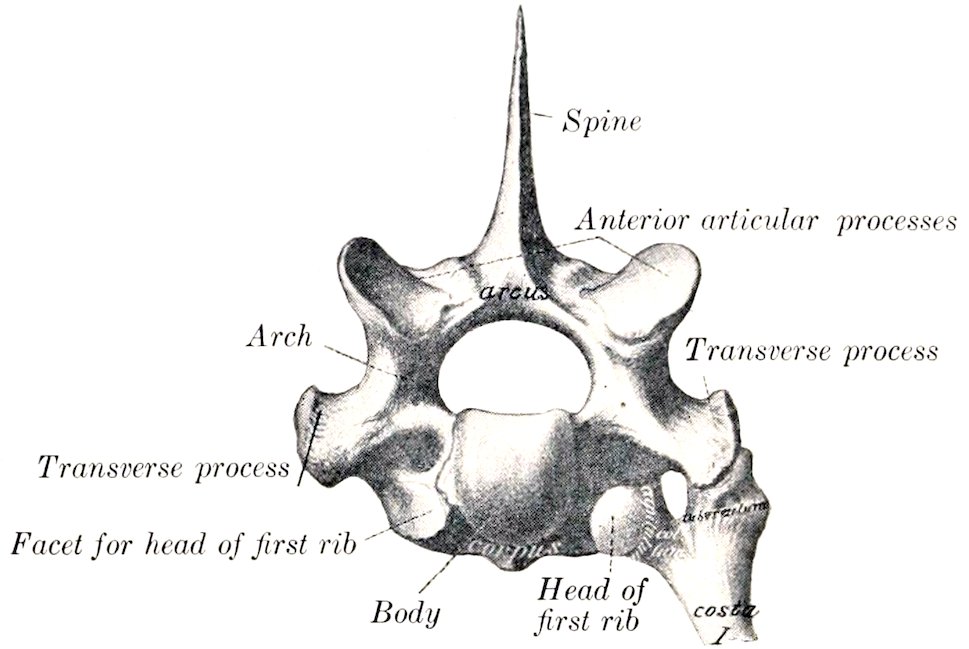
Fig. 16.—First Thoracic Vertebra and Upper Part of First Rib of Horse, Anterior View. (After Schmaltz, Atlas d. Anat. d. Pferdes.)
The arches of the first two or three are about equal in size and similar to that of the last thoracic; from the fourth they decrease noticeably in breadth and height.
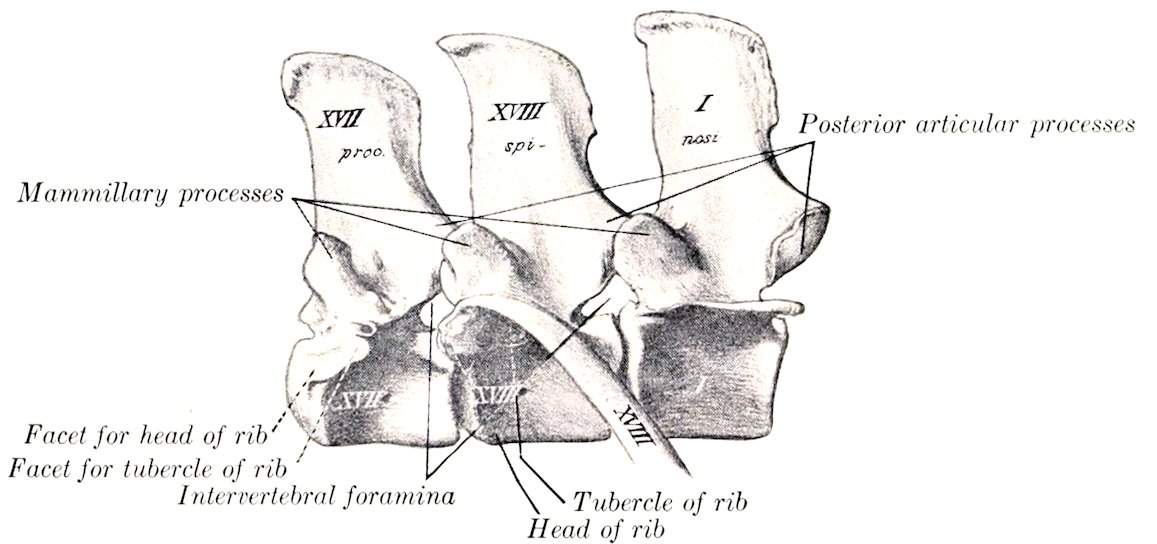
Fig. 17.—Last Two Thoracic and First Lumbar Vertebræ of Horse, Lateral View. (After Schmaltz, Atlas d. Anat. d. Pferdes.)
The anterior articular processes are fused with the mammillary processes, and present superiorly concave surfaces for articulation with the posterior pair of the preceding vertebra. The posterior articular processes project distinctly from the arch at the base of the spinous process, and have ventrally convex articular surfaces, which fit into the grooved surfaces of the anterior pair of the next vertebra.
The transverse processes are large plates, flattened dorso-ventrally, which project outward and usually curve slightly downward; their length increases to 39the third and fourth, and then diminishes to the last, which is the shortest. The first one or two usually curve somewhat backward, the last two decidedly forward. Those of the fifth have an oval concave facet on the inner part of the posterior border for articulation with the sixth process; the latter has a corresponding convex facet on the anterior border, and a larger concave surface on the posterior border for articulation with the wing of the sacrum. Sometimes the fifth process has a small surface for articulation with the fourth. The inner part of the sixth process is thick, the outer part thinner, narrower, and curved forward. The inner part of the fifth is also somewhat thickened.
The spinous processes resemble those of the last two thoracic vertebræ. They are usually about equal in height, but minor differences are common, and the width diminishes in the last three.
Development.—This is similar to that of the thoracic vertebræ. The extremities of the transverse processes remain cartilaginous for some time after ossification is otherwise complete.
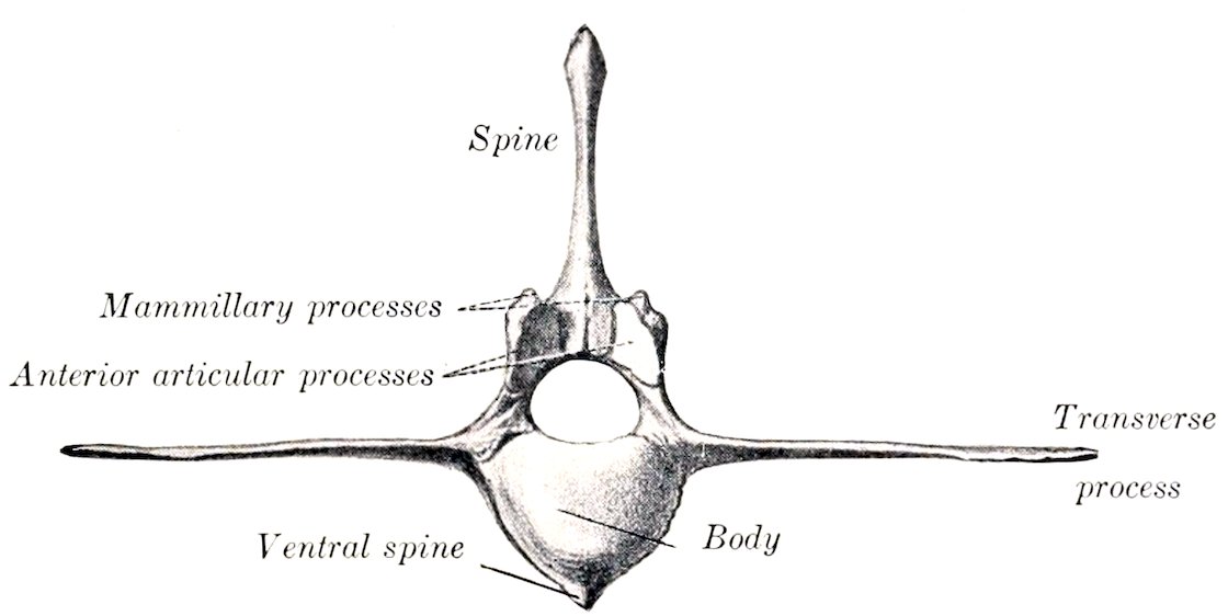
Fig. 18.—Second Lumbar Vertebra of Horse, Anterior View. (After Schmaltz, Atlas d. Anat. d. Pferdes.)
The transverse processes of this region are considered equivalent to the proper transverse process + the costal element, and hence the distinctive term processus lateralis (s. costarius) has been proposed. The occurrence of a lumbar rib in connection with the transverse process of the first lumbar is not rare. Reduction of the number to five has been observed frequently, and may or may not be compensated by an additional thoracic vertebra. This variation is not more common in certain races as Sanson and others have maintained. Very few cases are recorded of seven lumbar vertebræ—especially with the normal thoracic number. An anomalous vertebra with mixed thoracic and lumbar characters sometimes occurs at the junction of the two regions.
THE SACRUM
The sacrum (Os sacrum) is formed by the fusion of five vertebræ usually, and is conveniently described as a single bone. It is triangular in form and is wedged in between the ilia, with which it articulates very firmly on each side. Its long axis is gently curved, and slightly oblique, so that the posterior end is a little higher than the anterior. It presents two surfaces, two borders, a base, and an apex.
The dorsal surface presents centrally the five sacral spines, which are directed upward and backward, and have (with the exception of the first) tuberous summits which are sometimes bifid.
The first spine is relatively thin and narrow, and is not so high as the internal angle of the ilium. The second is the longest, and the height diminishes rapidly to the last. The bases of the spines are often fused in old subjects.
On either side of the spines is a groove, in which are the four dorsal sacral 40foramina (Foramina sacralia dorsalia); the dorsal branches of the sacral nerves emerge through them.
The ventral or pelvic surface (Facies pelvina) is concave in its length, wide in front, narrow behind. It is marked by four more or less distinct transverse lines (Linæ transversæ), which indicate the demarcation of the bodies of the vertebræ. At the ends of these lines are the ventral sacral foramina (Foramina sacralia ventralia), which are larger than the dorsal series and diminish in size from first to last; they transmit the ventral divisions of the sacral nerves.
The dorsal and ventral foramina communicate with the sacral canal and are together equivalent to the usual intervertebral foramina.
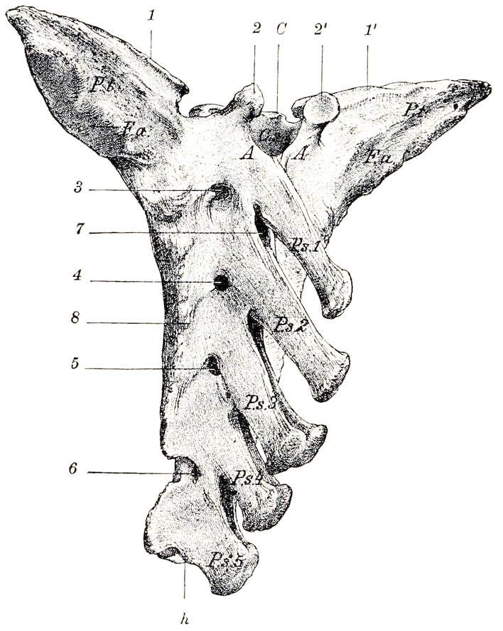
Fig. 19.—Sacrum of Horse, Dorso-lateral View. C, Body of first sacral vertebra; A, arch of first vertebra; Cs, sacral canal; P.s. 1–5, sacral spines; P.t., wings of sacrum; 1, 1′, surfaces for articulation with transverse processes of last lumbar vertebra; F.a., auricular surface; 2, 2′, articular processes; 3–6, dorsal sacral foramina; 7, interarcuate space; 8, lateral border; h, apex. (Struska, Anat. d. Haustiere.)
The lateral borders are rough, thick in front, thin behind.
The base (Basis ossis sacri) is directed forward, and is relatively very wide. It presents centrally the body of the first sacral segment, which is wide transversely, flattened dorso-ventrally, and has a rounded surface which articulates with the last lumbar vertebra through the medium of an intervertebral fibro-cartilage. The ventral margin projects slightly, forming the promontory (Promontorium). Above the body is the entrance to the sacral canal, flanked by a pair of articular processes, which project upward and forward from the arch, and have concave surfaces internally for articulation with those of the last lumbar vertebra. On each side of these is a smooth notch which is converted into a foramen by apposition with the last lumbar. The lateral parts of the base, the wings or alæ (Alæ sacrales), 41are strong prismatic masses with pointed ends, which result from the fusion of the first with part of the second transverse process. Each has in front a large, oval, slightly convex surface for articulation with the transverse process of the last lumbar. Posteriorly there is an elongated oval area which faces upward, backward, and outward. This is the auricular surface (Facies auricularis), which articulates with the ilium; it is slightly concave in its length, and somewhat rough and irregular. The rest of the dorsal surface of the wing is roughened for ligamentous attachment, while the ventral surface is smooth.
The apex (Apex ossis sacri) is the posterior aspect of the last sacral vertebra and is quite small. It presents the elliptical flattened surface of the body, above which is the triangular posterior opening of the sacral canal, surmounted by the last sacral spine. There is a pair of narrow notches between the arch and body, above which rudiments of articular processes may occur.
The name sacral canal (Canalis sacralis) is applied to that part of the vertebral canal which traverses the sacrum. Its anterior part is large and has the form of a triangle with the angles rounded off; its width is about twice its height. Traced backward it is seen to diminish in size rapidly, and the posterior opening is quite small and triangular.
The term lateral part (Pars lateralis) designates the portion external to the foramina, which results from the fusion of the transverse processes.
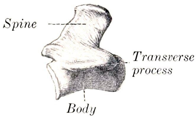
Fig. 20.—First Coccygeal Vertebra of Horse, Left View. (After Schmaltz, Atlas d. Anat. d. Pferdes.)
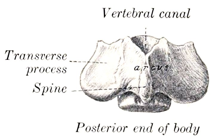
Fig. 21.—First Coccygeal Vertebra of Horse, Dorsal View. (After Schmaltz, Atlas d. Anat. d. Pferdes.)
Development.—The several sacral vertebræ ossify in the typical manner. Separate centers for costal elements in the lateral parts have not yet been found in the domesticated animals. Fusion begins in front, and is usually not complete behind till near adult age. The lateral parts unite before the bodies. It is rather curious that the epiphyseal plates of adjacent segments unite with each other before they fuse with the main portion of the bodies.
THE COCCYGEAL VERTEBRÆ
These (Vertebræ coccygeæ) vary considerably in number, but eighteen may be taken as an average. From first to last they become reduced in size and, with the exception of a few at the beginning of the series, consist of bodies only. The first three have bodies which are somewhat flattened dorso-ventrally, constricted in the middle, and have at the ends slightly convex, elliptical, articular surfaces. The ventral surface has a median groove for the coccygeal artery. The arch is small and triangular; it is formed of two flat plates which are prolonged to form a short spinous process with a thickened and often double summit. The anterior notches are absent. Functional articular processes are not present, but small rudiments of the anterior pair commonly occur. The transverse processes are relatively large plates which project horizontally outward. Further back the arch becomes incomplete, open above, and soon disappears; the transverse processes 42gradually fade out, and the vertebræ are reduced to cylindrical rods of diminishing size. The last one has a pointed end.
Variations.—The number is said by good observers to vary between fourteen and twenty-one. In old age the first is often fused with the sacrum, and sometimes with the second. The arch of the third may be open.
THE VERTEBRAL COLUMN AS A WHOLE
In the mid-dorsal line is the series of spinous processes, which are low ridges in the cervical region with the exception of the second and seventh, reach their maximum height at the fourth and fifth thoracic vertebræ, and diminish to the fifteenth or sixteenth thoracic. Behind this they are about equal in height as far as the last lumbar and first sacral, which are somewhat lower. The second sacral spine is about as high as the middle lumbar; behind this they diminish rather rapidly in height and fade out about the third coccygeal. Their inclination backward is most decided at the second thoracic, diminishes from the sixth or seventh to the fifteenth or sixteenth, which is vertical and is termed the anticlinal or diaphragmatic vertebra. Behind this they are inclined a little forward until the sacrum is reached; here there is an abrupt change to the backward inclination, so that a considerable interspinous angle is formed.
On either side of the spinous processes is a vertebral groove which contains the deep muscles of the spine. The floor of the groove is formed by the laminæ and articular processes. It is wide in the neck and narrows progressively in the back.
Viewed from the side, the column presents a series of curves. When the head and neck are in the ordinary neutral position, the anterior part of the cervical spine forms a gentle curve, concave ventrally. The posterior cervical and first thoracic vertebræ form a more pronounced curve in the opposite direction. At the junction of the cervical and thoracic regions there is a marked change of direction, forming a ventral projection or angle. At the second thoracic vertebra a gentle curve, concave ventrally, begins. This is continued to the lumbo-sacral junction, where there is a change of direction, and hence a promontory. The sacrum has a variable, but never very pronounced, ventral concave curvature, which is continued in a much accentuated form in the coccygeal region. It may be remarked that a line through the summits of the spines does not correspond to these curves formed by the bodies.
The vertebral canal, of course, corresponds in curvature to the bodies. Its caliber varies greatly at different points. The greatest diameter (ca. 5 cm.) is in the atlas, where it contains the dens of the axis in addition to the spinal cord, and provision must be made for extensive movement. It is very much smaller in the axis (ca. 2.5 cm. wide, 3 cm. high). It widens considerably at the junction of the cervical and thoracic regions to accommodate the cervical enlargement of the spinal cord. Beyond this it diminishes, and is smaller in the middle of the back than at any preceding point; this is correlated with the small size of the spinal cord and the very limited movement of the spine. At the middle of the lumbar region it again widens considerably to contain the lumbar enlargement of the spinal cord. The caliber diminishes very rapidly from the second sacral segment backward, and the canal ceases to be complete at the fourth coccygeal vertebra.
The articular processes are very large and wide apart in the neck, greatly reduced and much closer together in the back, larger and interlocking in the lumbar region.
The transverse processes are large and outstanding in the neck, where they form the outer boundary of a ventral groove occupied by the longus colli muscle. In the back they are short and stout, and are characterized by the facets for the tubercles of the ribs. On the first thoracic vertebra this facet is large, deeply concave, 43and situated almost directly outward from the cavity for the head of the rib; traced backward it becomes smaller and flatter, and gradually comes to lie behind the cavity for the head of the rib, with which it is fused on the last and often also on the next to the last thoracic vertebra. The processes in the lumbar region have a characteristic elongated plate-like form. In the sacral region they are fused to form the wings and lateral parts of the sacrum. In the coccygeal region they are at first of considerable size relatively, but undergo rapid reduction, and disappear at the fifth or sixth vertebra.
The cavities for the heads of the ribs diminish progressively in size and depth from first to last.
The mammillary processes are usually distinct on the fourteenth to the seventeenth thoracic vertebræ. In front of these they blend with the transverse, behind with the anterior articular processes.
The length of the vertebral column (including the intervertebral fibro-cartilages) in a horse of medium size is about 260 to 265 cm. (ca. 8 feet 8 inches to 8 feet 10 inches). The relative lengths of the various regions appear to vary most in the neck and back.
The table below gives the measurements in centimeters in a trotting stallion of medium size and in an adult Percheron stallion. The percentages are in round numbers.
Trotter Percheron
Cervical 60.0 22.7% 74.0 25.4%
Thoracic 98.0 37.2% 100.0 34.4%
Lumbar 31.5 12.0% 36.5 12.5%
Sacral 20.0 7.6% 22.5 7.7%
Coccygeal 54.0 20.5% 58.0 20.0%
263.5 100.0 291.0 100.0
The Ribs
There are usually eighteen pairs of ribs in the horse, but a nineteenth rib on one side or both is not at all rare. Eight are sternal or “true” ribs, the remainder asternal or “false.” Ribs from different parts of the series vary much in length, curvature, and other characters. We will therefore consider as a type a rib from the middle of the series first, and afterward note the chief serial differences. A typical rib has the following characters:
The shaft or body (Corpus costæ) is elongated, relatively very narrow, and strongly curved; the curvature is most pronounced in the dorsal third, and the ventral part is twisted and inclined inward, so that when a rib is laid with its outer surface on the table, the ventral end is raised. The external surface is convex in its length and also transversely; its anterior part is, however, grooved longitudinally. A distinct angle, i. e., a point at which the curve of the rib changes rather suddenly, as in man, can scarcely be said to exist in the horse. The term is often applied, however, to a corresponding rough elevation which gives attachment to the ilio-costalis muscle; it is most distinct on the fourth to the eighth inclusive. The internal surface is smooth, concave in its length, and rounded from side to side; the costal groove, situated posteriorly, is very distinct above and fades out about the middle. The anterior border is concave, the posterior convex.
The vertebral extremity (Extremitas vertebralis) consists of the head, neck, and tubercle. The head (Capitulum costæ) has an articular surface (Facies articularis capituli costæ), composed of two convex facets, anterior and posterior, separated by a groove for the attachment of the conjugal ligament. It articulates with the cavity formed by facets on the bodies of two adjacent thoracic vertebræ 44and the intervertebral fibro-cartilage. The neck (Collum costæ) is roughened above and in front. The tubercle (Tuberculum costæ) is placed above and behind the junction of neck and shaft; it has a small surface (Facies articularis tuberculi costæ) for articulation with the transverse process of the corresponding thoracic vertebra.
The sternal extremity (Extremitas sternalis) is somewhat expanded, and is continued by the costal cartilage.
The first rib is easily distinguished. It is the shortest and least curved. At the lower part of the anterior border there is a smooth impression where the brachial vein curves around it; above this is usually the scalene tubercle. The costal groove is absent. The head is large and has two facets of unequal extent which meet at an acute angle in front; the smaller one faces forward and articulates with the last cervical vertebra; the larger one is directed inward and articulates with the first thoracic vertebra. The neck is thick and very short. The tubercle is larger than that of any other rib and has an extensive articular surface which is convex in its length. The sternal end is larger than that of any other rib; it is thick and very wide, and is turned a little forward.
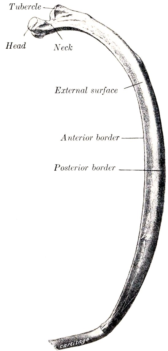
Fig. 22.—Left Seventh Rib of Horse, Antero-external View. (After Schmaltz, Atlas d. Anat. d. Pferdes.)
The last rib is the most slender and regularly curved. It is usually but little longer than the second. The facet on the tubercle is confluent with that of the head. (This feature, however, is usual on the seventeenth also, and not rare on the sixteenth.)
The serial position of the other ribs may be determined approximately by the following considerations: The length increases from the first to the tenth and eleventh and then diminishes. The width increases somewhat to the sixth and then diminishes. The anterior border is thin and sharp from the second to the eighth, and behind this becomes thick and rounded. The external groove is distinct on the fourth to the eighth inclusive. The curvature increases in degree rapidly from the first to the seventh, remains about the same to the sixteenth, and then decreases very noticeably. In regard to dorso-ventral direction, the first rib inclines a little forward, the second is about vertical, while behind this they slope backward in increasing degree, so that a plane tangent to the ventral ends of the last pair cuts the third lumbar vertebra. The head and tubercle diminish in size from first to last. Their relative positions change in that the tubercle of the first rib lies almost directly outward from the head, while further back it gradually comes to lie behind it. The neck is longest on the longest ribs, and is absent on the last two or three. A costo-transverse foramen is formed between the neck and the transverse process.
Development.—The ribs ossify in cartilage from three centers, one each for the shaft, head, and tubercle; the third center is absent in some of the posterior ribs.
45Variations.—A nineteenth rib on one side or both is not rare. It is usually imperfectly developed and quite variable. In some cases it is a mere strip of cartilage connected by ligament with the first lumbar transverse process; in other cases it is ossified, and may be fused with the process; in others again it is connected with an additional presacral vertebra which may be thoracic or lumbar or ambiguous in character. It is commonly floating, but may be attached to the eighteenth. Reduction in number is uncommon. Fusion of adjacent ribs sometimes occurs.
THE COSTAL CARTILAGES
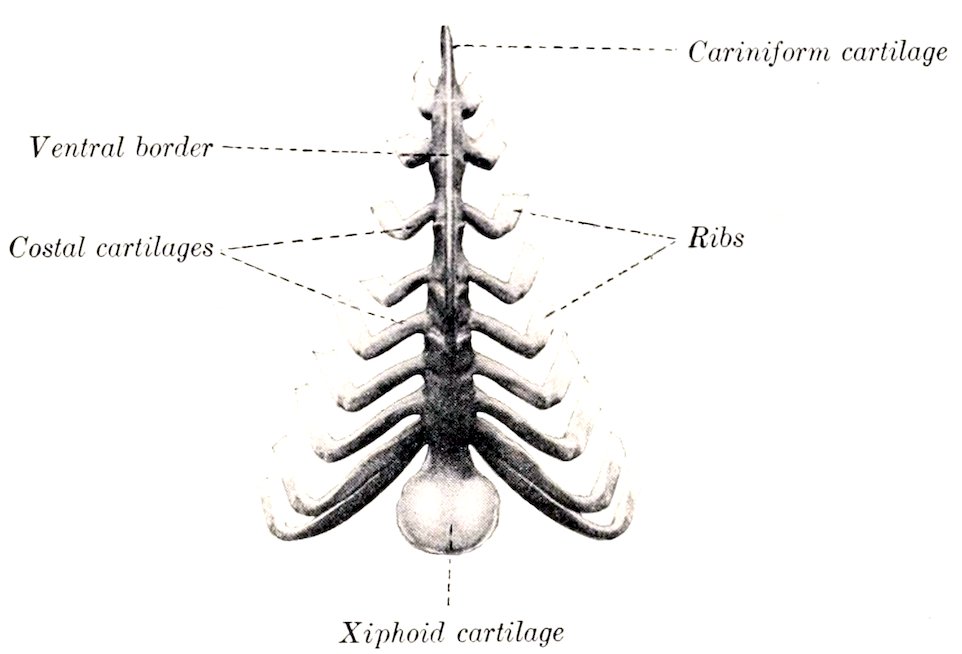
Fig. 23.—Sternum and Costal Cartilages of Horse, Ventral View. (After Ellenberger-Baum, Anat. f. Künstler.)
The first costal cartilage is an inch or more (2.5 to 3 cm.) in length. The upper part is very wide and thick. The sternal end is small. The two articulate with each other as well as with the sternum. The cartilages of the other sternal ribs increase progressively in length and become more rounded. The sternal end is expanded and has an elliptical convex facet for articulation with the sternum. The cartilages of the asternal ribs are long, slender, and pointed. The ninth is the longest, and is very firmly attached to the eighth; behind this they diminish progressively in size, and are attached to each other by elastic tissue. Except in the case of the first, the cartilage does not continue the direction of the rib, but forms with the latter an angle which is open in front, and decreases in obtuseness from second to last.
More or less extensive ossification is to be regarded as a normal occurrence, especially in the cartilages of the sternal ribs.
The Sternum
The sternum of the horse is shaped somewhat like a canoe; it is compressed laterally, except in its posterior part, which is flattened dorso-ventrally. It is inclined obliquely so that the posterior end is about six to eight inches (15 to 20 cm.) lower than the anterior.
The dorsal surface has the form of a very narrow isosceles triangle with the apex in front. It is concave longitudinally, flattened transversely.
The lateral surfaces are convex above, slightly concave below, and diminish in extent behind. Each presents on its upper part seven articular cavities (Foveæ costales), which receive the sternal ends of the second to the eighth costal cartilages inclusive. These cavities are situated in series at the intersternebral junctions. 46The first four are elliptical in outline with the long diameter vertical, and are separated by considerable regular intervals. The others are progressively smaller, more circular, and closer together. The area below these cavities gives attachment to the pectoral muscles.
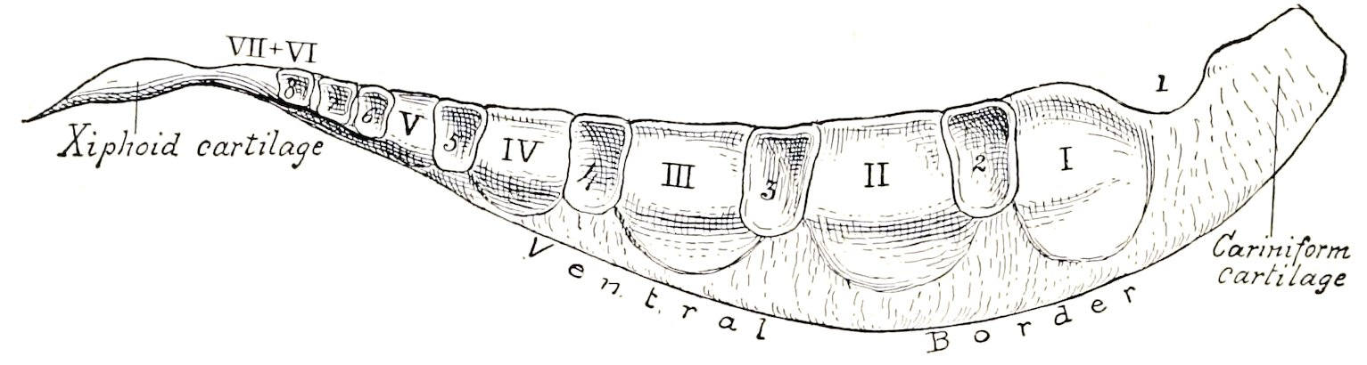
Fig. 24.—Sternum of Horse, Lateral View. The sternebræ are designated by Roman numerals and the costal facets by ordinary figures.
The dorso-lateral borders separate the dorsal and lateral surfaces. They give attachment to the lateral branches of the sternal ligament.
The ventral border forms the prominent keel-like crest of the sternum (Crista sterni) which may be felt in the living animal; it fades out behind.
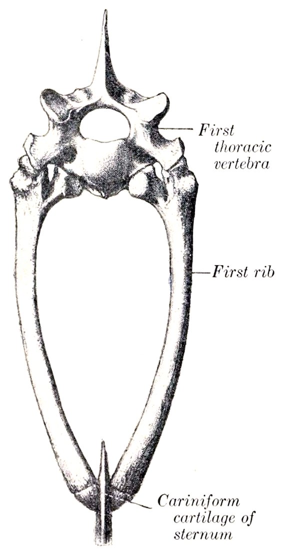
Fig. 25.—Anterior Aperture of Thorax of Horse. (After Schmaltz, Atlas d. Anat. d. Pferdes.)
The anterior extremity or manubrium sterni[5] can be distinctly felt in the central furrow of the breast. It consists of a laterally compressed cartilaginous prolongation, commonly called the cariniform cartilage. Its lateral surfaces are flat and furnish attachment to muscles of the breast and neck. The ventral border is rounded, and is continued backward on the body of the bone. The dorsal border is concave and has an articular cavity for the first pair of costal cartilages.
The posterior extremity is formed by the xiphoid or ensiform cartilage (Processus xiphoideus). This is a thin plate, connected in front with the last bony segment by a relatively thick, narrow neck, and expanding in nearly circular form behind and laterally. Its dorsal surface is concave, and gives attachment to the diaphragm. The ventral surface is convex. The free margin is very thin.
Development.—At birth the sternum of the horse consists of seven bony segments or sternebræ united by intersternebral cartilages. The last two sternebræ fuse in the second month, but the others do not usually unite completely even in old age. The sternebræ consist of very vascular spongy bone covered by a very thin layer of compact substance. The adult sternum thus consists to a very considerable extent of persisting cartilage, viz., the intersternebral cartilages, the ventral keel, and the extremities; in old age these undergo partial ossification.
47
THE THORAX
The bony thorax of the horse is remarkably compressed laterally in its anterior part, but widens greatly behind. The anterior aperture is oval and very narrow below; in a horse of medium size its greatest width is about 4 inches (10 cm.), and its height 7 to 8 inches (ca. 18 to 20 cm.). The ventral wall or floor is about 10 inches (40 cm.) long, and the dorsal wall or roof about 38 to 40 inches (95 to 100 cm.) long. The height from the last segment of the sternum to the seventh or eighth thoracic vertebra is about twice that of the anterior aperture; this is due to the obliquity and divergence of the roof and floor. The greatest width of the posterior aperture is about 20 to 24 inches (50 to 60 cm.). The intercostal spaces increase in width from the first to the seventh or eighth, and then diminish. Their average width is about 1¼ to 1½ inches (3 to 3.5 cm.).
The Bones of the Skull
(A) BONES OF THE CRANIUM
The bones of the cranium (Ossa cranii) are the Occipital, Sphenoid, Ethmoid, Interparietal, Parietal, Frontal, and Temporal. The first four are single, the others paired.
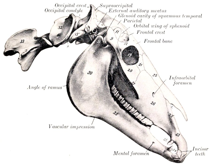
Fig. 26.—Skull, Atlas, and Axis of Horse, Lateral View. 8, Body of mandible; 28′, horizontal (molar) part of ramus; 30, vertical part of ramus; 9, zygomatic process of squamous temporal; 11, coronoid process of mandible; 12, supraorbital process; 13″, paramastoid (styloid) process of occipital; 19, orbit; 20, malar bone; 21, lacrimal bone; 22, nasal bone; 23, premaxilla; 23′, nasal process of premaxilla; 25, 29, canine teeth; 26, maxilla; 27, facial crest; 31, condyle of mandible; 32, atlas; 33, axis; x, wing of atlas; e, naso-maxillary notch. (After Ellenberger-Baum, Anat. für Künstler.)
The Occipital Bone
The occipital bone (Os occipitale) is situated at the posterior part of the cranium, of which it forms the posterior wall and part of the ventral wall or base.[6]
48Its lower part is perforated centrally by a large, almost circular opening, the foramen magnum (Foramen occipitale magnum), at which the brain and spinal cord join. The foramen is bounded laterally and dorsally by the lateral parts of the bone, and ventrally by the basilar part or process. Above the lateral parts—but not entering into the formation of the foramen magnum—is the squamous part.
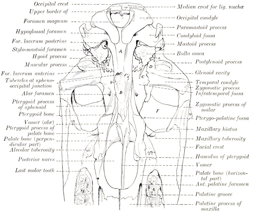
Fig. 27.—Line Drawing of Posterior Half of Base of Skull of Horse, Without Mandible. (Key to Fig. 28.) 1, Incisura carotica; 2, incisura ovalis; 3, incisura spinosa; 4, external orifice of parieto-temporal canal; 5, Eustachian canal; 6, petro-tympanic fissure; 7, external auditory canal; 8, hyoid process; 9, Vidian groove; 10, supraorbital process; A, basilar part of occipital; B, body of sphenoid; C, temporal wing of sphenoid; D, squamous temporal bone; E, petrous temporal bone; F, orbital part of frontal bone.
The lateral parts (Partes laterales) bear the occipital condyles (Condyli occipitales), which articulate with the atlas. The condyles are obliquely placed, wide apart dorsally, and separated by a small interval ventrally. The articular surface is curved so sharply in the dorso-ventral direction as to form a blunt ridge externally. The cranial surface is concave and smooth. External to the condyle is the paramastoid or styloid process (Processus jugularis), a strong flattened bar of bone which projects downward and backward; its external surface is convex and roughened for muscular attachment. Between the root of this process and the condyle is a smooth depression, the condyloid fossa (Fossa condyloidea inferior); in this is the hypoglossal foramen (Foramen hypoglossi), which transmits the nerve of like name.
The basilar part or process (Pars basilaris) is a strong, somewhat prismatic bar which extends forward from the ventral margin of the foramen magnum. It 49is much narrower in front than behind. The ventral surface is rounded. The cranial surface is concave and smooth; its posterior part, supports the medulla, and its anterior part has a shallow cavity on which the pons rests. The lateral borders are thin and sharp, and form the inner margin of the foramen lacerum (Foramen lacerum et jugulare). The anterior end has, in the young subject, a semicircular, flat, pitted surface which is attached to the body of the sphenoid bone by a layer of cartilage; in the adult there is complete fusion. On the ventral aspect of the junction are tubercles for the attachment of the ventral straight muscles of the head.
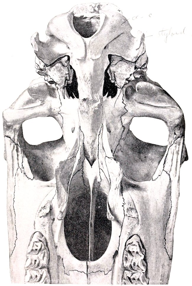
Fig. 28.—Ventral Surface of Skull of Horse, Posterior Half Without Mandible. The skull is inclined slightly. (Notation on key Fig. 27.)
The squamous part (Squama occipitalis) is the somewhat quadrilateral mass 50situated above the lateral portions, from which it remains distinct till the second year. The outer surface is crossed by a very prominent ridge, the occipital crest; the middle part of this is thick, transverse in direction, and forms the highest point of the skull when the head is in the ordinary position; laterally it becomes thinner and runs downward and forward to join the temporal crest.[7] The crest divides the surface into two very unequal parts; the small anterior area (Planum parietale) presents a median ridge which is the posterior part of the external sagittal crest; the large area below the crest (Planum nuchale) also has a central eminence, the external occipital protuberance, on the sides of which the funicular part of the ligamentum nuchæ is attached. The internal surface is concave and presents a deep central depression and two shallower lateral ones which adapt it to the surface of the cerebellum.
The occipital bone is connected by suture with the interparietal, two parietals, and two temporals, and by synchondrosis with the sphenoid; the condyles articulate with the atlas.
Development.—The occipital ossifies in cartilage from four centers, and consists at birth of four pieces as described above.[8] The lateral parts unite with the basilar at three to four months, and with the squama in the second year, when the bone is consolidated.
The parieto-occipital suture and the spheno-occipital synchondrosis are obliterated about the fifth year usually. The temporo-occipital suture partially ossifies in old subjects.
The Sphenoid Bone
The sphenoid bone (Os sphenoidale) is situated in the base of the cranium, its central part lying in front of the basilar part of the occipital. It consists of a central part, the body, two pairs of wings, and two pterygoid processes.
The body (Corpus) is a cylindrical bar, flattened dorso-ventrally, and wider in front than behind. Its ventral surface (Facies externa) is convex in the transverse direction; and its anterior part is concealed to a large extent by the vomer and pterygoid bones. The cerebral surface (Facies cerebralis) presents the following features: (1) In front is a raised, flattened part which is subdivided by a median elevation into two slightly concave lateral areas; this part has a posterior, thin, free margin (Limbus sphenoidalis), which overlies the entrance to the optic foramina. (2) Just behind this and at a lower level is a smooth transverse depression, the optic groove (Sulcus chiasmatis), on which the optic chiasma rests. (3) From each end of this groove the optic foramen passes forward and outward to terminate in the posterior part of the orbital fossa.[9] (4) Near the posterior end is a central depression, the hypophyseal or pituitary fossa (Fossa hypophysea), which lodges the hypophysis cerebri or pituitary body. On each side of this is an ill-defined groove for the internal carotid artery and the cavernous sinus. The anterior end is expanded, and is excavated to form the sphenoidal sinuses. These cavities extend back as far as the optic groove, and are usually continuous in front with the cavities in the vertical parts of the palate bones; they are separated by a complete septum which is not always median.[10] The posterior end is flat and is 51joined to the basilar part of the occipital; at the line of junction there is dorsally a transverse elevation, the spheno-occipital crest (Crista sphenooccipitalis).
The orbital wings (Alæ orbitales) curve upward and somewhat outward from the sides of the body of the presphenoid. Their inner or cerebral surfaces are concave, and are marked by digital impressions (Impressiones digitatæ) for the gyri of the cerebrum. The external surface is convex and is largely concealed by the overlapping temporal wing and the squamous temporal and frontal bones; a narrow part of it (Facies orbitalis) is uncovered on the inner wail of the orbital cavity at the sphenoidal notch of the frontal bone. The dorsal border unites with the frontal bone at the spheno-frontal suture. The anterior border joins the ethmoid at the spheno-ethmoidal suture; at its lower part it concurs with the frontal in the formation of the ethmoidal (or internal orbital) foramen. The posterior border is overlapped by the temporal wing and the squamous temporal. The root of the wing is perforated by the optic foramen. Immediately below and behind the latter (i. e., beneath the root) is the foramen lacerum orbitale or orbital fissure. Below this, and separated from it usually by a thin plate, is a larger opening, the foramen rotundum, which is bounded externally by the root of the pterygoid process. Behind these foramina is the pterygoid crest (Crista pterygoidea), which is continued downward and forward on the pterygoid process; on its upper part may be found the small and inconstant trochlear (or pathetic) foramen. Just behind the crest is the temporal foramen (For. alare parvum), through which the anterior deep temporal artery emerges from the alar canal of the pterygoid process.
The temporal wings (Alæ temporales) extend outward and somewhat upward from the body of the postsphenoid; they are irregularly quadrilateral in outline. The external surface (Facies temporalis) enters into the formation of the infratemporal fossa, and bears the pterygoid process on its anterior part; at the junction with the body there is a small groove for the pterygoid nerve. The internal surface (Facies cerebralis) presents, at the junction with the body, two longitudinal grooves (Sulci nervorum). The outer groove is the larger, and leads forward to the foramen rotundum; it contains the maxillary nerve. The inner groove conducts to the orbital fissure, and contains the third, sixth, and ophthalmic nerves. The outer groove is bounded externally by a thin overhanging crest, on which is a small groove for the fourth nerve. The remainder of the surface is concave and supports the pyriform lobe of the brain. The dorsal border joins the squamous temporal at the spheno-squamous suture. The anterior border joins the orbital wing. The posterior border forms the anterior boundary of the foramen lacerum; it presents three notches, which are (from within outward) the carotid, oval, and spinous (Incisura carotica, ovalis, spinosa). The angle of junction of the dorsal and posterior borders articulates with the parietal bone.
The pterygoid processes (Processus pterygoidei) arise from the temporal wings and the body. They project downward and forward, and curve outward at the lower part. The root is perforated by the alar canal (Canalis alaris),[11] which transmits the internal maxillary artery. From this canal a branch leads upward and forward to open at the temporal foramen. The external surface is concave, and is marked by lines for muscular attachment. The internal surface is largely concealed by the overlapping palate and pterygoid bones, with which it concurs in the formation of the pterygoid or Vidian canal.
Development.—The sphenoid is ossified in cartilage, and consists in early life of two distinct parts, the presphenoid and postsphenoid. The former develops from two centers, one in each wing; the latter has three centers, one for the body and one for each wing. The pterygoid processes ossify from the centers of the temporal wings.
52Variation.—The dorsal border of the orbital wing may come to the surface through a defect in the frontal bone at the place where the horn process is situated in animals which have frontal horns.
The Ethmoid Bone
The ethmoid (Os ethmoidale) is situated in front of the body and orbital wings of the sphenoid. It projects forward between the orbital plates of the frontal bones and enters into the formation of the cranial, nasal, and paranasal cavities.[12] It consists of four parts—the cribriform plate, two lateral masses, and the perpendicular plate.
The cribriform plate (Lamina cribrosa) is a sieve-like partition between the cranial and nasal cavities. Its margin joins the orbital wings of the sphenoid laterally, and the cranial plate of the frontal bones dorsally. Its cranial surface is divided into two parts by a median ridge, the crista galli, which is the intracranial portion of the perpendicular plate. Each half forms a deep oval cavity, the ethmoidal or olfactory fossa, which lodges the olfactory bulb. The plate is perforated by numerous small foramina for the passage of the olfactory nerve filaments, and on either side is the much larger ethmoidal foramen. The nasal surface is convex, and has the lateral masses attached to it.
The lateral masses or labyrinth project forward from the cribriform plate into the posterior part of the nasal cavity, which they nearly fill. Each mass is somewhat conical in shape, with the base attached to half of the cribriform plate. The inner surface is separated by a narrow space from the perpendicular plate. The outer surface is convex and faces chiefly into the frontal and maxillary sinuses, but is attached behind to the inner wall of the orbital cavity; it is covered by a very thin layer of bone, the lamina papyracea. The mass consists of a large number of delicate, scroll-like plates of bone, termed ethmoturbinals or ethmoidal cells. These are attached to the lamina papyracea, and are separated by narrow intervals termed ethmoidal meatuses, which communicate with the nasal cavity. In the living animal the ethmoturbinals are covered with mucous membrane.
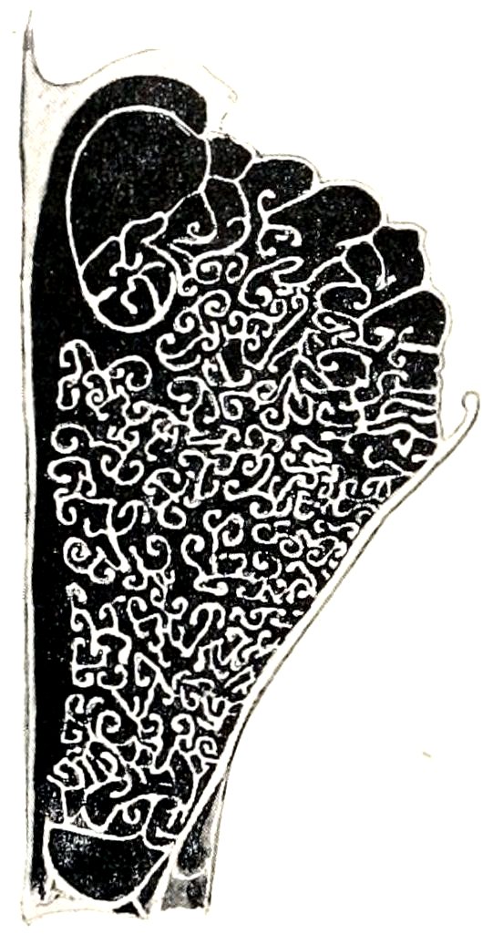
Fig. 29.—Cross-section of Lateral Mass of Ethmoid Bone of Horse.
The lateral mass is a very complex structure, the arrangement of which may be studied on cross-sections of decalcified specimens with the mucous membrane retained. Each mass consists of six turbinals which extend almost to the perpendicular plate and are termed endoturbinals. These diminish in size from above downward; the largest is attached to the nasal bone, and is hence usually called the naso-turbinal or superior turbinal; the second is much smaller, and is very commonly termed the great ethmoid cell. Between the endoturbinals are twenty-one small ectoturbinals, and all are beset with secondary and tertiary coiled lamellæ.
The perpendicular plate or mesethmoid (Lamina perpendicularis) is median, and forms the posterior part of the septum nasi. Its lateral surfaces are nearly plane, but are marked below by some grooves and ridges; they are covered by the nasal mucous membrane. The anterior border is irregular and is continuous with the septal cartilage. The posterior border projects into the cranial cavity as a ridge, the crista galli. The dorsal border joins the frontal bones at their line of junction—the frontal suture. The ventral border is received into the groove of the vomer.
Development.—The ethmoid develops in cartilage from five centers, two for 53each lateral mass, and one for the perpendicular plate; from the latter ossification extends into the cribriform plate. At birth the perpendicular and cribriform plates are entirely cartilaginous. By the time ossification is complete the ethmoid has united with surrounding bones to such an extent that it cannot be separated intact for study.
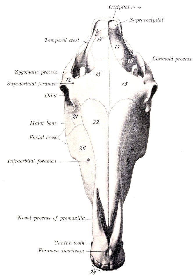
Fig. 30.—Skull of Horse, Dorsal View. 12, Supraorbital process; 14, parietal bone; 14′, external sagittal crest; 15, frontal bone; 15′, frontal crest; 21, lacrimal bone; 22, nasal bone; 26, maxilla; 24, incisor teeth. (After Ellenberger-Baum, Anat. für Künstler.)
The Interparietal Bone
This bone (Os interparietale) is centrally placed between the squamous part of the occipital and the parietal bones. It is usually described as a single bone, although it ossifies from two chief lateral centers, and is sometimes distinctly paired in skulls of young foals.
The external surface (Facies externa) is quadrilateral and is flat and 54smooth in the very young foal; later it presents the median external sagittal crest.
The internal surface (Facies cerebralis) presents the internal occipital protuberance, a three-sided process which projects downward and forward into the cranial cavity between the cerebral hemispheres and the cerebellum; it has three concave surfaces and three sharp borders which form part of the tentorium osseum.
The posterior border is thick; it joins the squamous part of the occipital bone. The lateral and anterior borders are united by suture with the parietal bones.
Development.—The interparietal ossifies in membrane from two chief lateral centers.[13] It fuses first with the parietals, somewhat later with the occipital, but the period at which this union takes place is quite variable.
The Parietal Bones
The two parietal bones (Ossa parietalia) form the greater part of the roof of the cranium; they unite in the median line, forming the sagittal suture. Each is quadrilateral in outline and has two surfaces and four borders.
The external surface (Facies parietalis) is convex, and is marked by a more or less prominent curved line, the external sagittal crest; this is median in its posterior part, and is continuous with the crest of like name on the occipital bone; in front it curves outward and is continuous with the frontal crest. The surface external to the crest (Planum temporale) enters into the formation of the temporal fossa, and is roughened for the attachment of the temporal muscle.
The internal or cerebral surface (Facies cerebralis) is concave. It presents numerous digital impressions (Impressiones digitatæ) which correspond to the gyri of the cerebrum. There are also furrows (Sulci vasculosi) for the meningeal arteries. Along the inner border there is a sagittal groove (Sulcus sagittalis) for the superior longitudinal sinus.
The anterior border joins the frontal bone at the parieto-frontal suture (Sutura coronalis).
The posterior border meets the occipital bone at the parieto-occipital suture (Sutura lambdoidea). Below this junction it curves inward and concurs with the temporal bone in the formation of the parieto-temporal canal (Meatus temporalis). A transverse groove (Sulcus transversus) connects this canal with the sagittal sulcus.
The internal border is thick and serrated. It joins its fellow at the sagittal suture, and (in the young subject) meets the interparietal at the interparietal suture. The line of junction is marked internally by the internal sagittal crest (Crista sagittalis interna).
The external border is beveled and is overlapped by the squamous temporal bone, forming the parieto-temporal suture (Sutura parieto-squamosa). The angle of junction of the external and posterior borders articulates with the posterior angle of the temporal wing of the sphenoid.
Development.—Each parietal bone ossifies in membrane from a single center. In the young foal the central part of the bone is much more convex than in the adult and forms a prominence similar to the pronounced tuber parietale of the young child; the external sagittal crest is not present, and the external surface is smooth.
The sagittal suture is usually closed at four years, the parieto-occipital at five years, and the parieto-temporal at twelve to fifteen years.
55
The Frontal Bones
The frontal bones (Ossa frontalia) are situated on the limits of the cranium and face, between the parietals behind and the nasal bones in front. Each is irregularly quadrilateral, and consists of frontal, orbital, and temporal parts.
The frontal part (Pars naso-frontalis) forms the basis of the forehead. Its external surface (Facies frontalis) is nearly flat, and is smooth and subcutaneous; it is separated from the temporal part by the external frontal crest (Crista frontalis externa). At the junction with the orbital part the supraorbital or zygomatic process (Proc. zygomaticus) curves outward and downward to join the zygomatic arch. The process partially separates the orbit from the temporal fossa; its root is perforated by the supraorbital foramen, or presents instead a notch on its anterior border; its upper surface is convex, while the lower or orbital surface is concave and smooth, forming a shallow fossa for the lacrimal gland (Fossa glandulæ lacrimalis). The internal surface enters into the formation of the cranial cavity and the frontal sinus. The cranial surface presents digital impressions for the cerebral gyri. The two plates of the bone separate and diverge in front, and thus inclose a large air-space which is part of the frontal sinus. The cranial plate curves downward and articulates with the cribriform plate of the ethmoid bone; the facial plate extends forward and joins the nasal and lacrimal bones.
The orbital part (Pars orbitalis) forms the major part of the inner wall of the orbital cavity. It is separated from the frontal part by a prominent ridge which is part of the orbital margin. Its external or orbital surface is concave and smooth, and presents superiorly a small depression (Fovea trochlearis), which is bridged by a small bar of cartilage, around which the superior oblique muscle of the eye is reflected. The lower border concurs with the orbital wing of the sphenoid in the formation of the ethmoidal or internal orbital foramen. The internal surface faces into the frontal sinus and gives attachment to the lateral mass of the ethmoid.