ANGIOLOGY: THE VASCULAR SYSTEM
Reading edition
Angiology is the description of the organs of circulation of the blood and lymph—the heart and vessels. The heart is the central hollow muscular organ which functions as a suction and force pump; the differences in pressure caused by its contraction and relaxation determine the circulation of the blood and lymph. It is situated in the middle mediastinal space of the thorax, between the two lungs, and is inclosed in a fibro-serous sac—the pericardium. The vessels are tubular and run through almost all parts of the body. They are designated according to their contents as blood- and lymph vessels.
The blood-vascular system consists of: (1) the arteries, which convey blood from the heart to the tissues; (2) the capillaries, microscopic tubes in the tissues which permit of the necessary interchange between the blood and the tissues; (3) the veins, which convey the blood back to the heart.
The blood-vessels are divided into the pulmonary and the systemic. The pulmonary artery conveys the blood from the right ventricle of the heart to the lungs, where it is arterialized, and is returned by the pulmonary veins to the left atrium of the heart, and passes into the left ventricle. The systemic arteries convey the blood from the left ventricle all over the body, whence it is returned by the venæ cavæ to the right atrium, and passes into the right ventricle.
It should be noted, however, that the lungs receive arterial blood through the systemic bronchial arteries. This blood is returned in part by the bronchial veins to the right atrium (indirectly), in part by the pulmonary veins to the left atrium.
The term portal system is often applied to the portal vein and its tributaries which come from the stomach, intestine, pancreas, and spleen. The vein enters the liver, where it branches like an artery, so that the blood in this subsidiary system passes through a second set of capillaries before being conveyed to the heart by the hepatic veins and the posterior vena cava.
The arteries (Arteriæ), as a rule, divide at an acute angle, giving off finer and finer branches. In some cases branches come off at a right angle, and others are recurrent, i. e., run in a direction opposite to that of the parent stem. The intercommunication of branches of adjacent arteries is termed anastomosis. Most commonly the connections are made by a network of numerous fine branches (Plexus vasculosus). Relatively large communicating branches (Rami communicantes) occur in certain places; they may be transverse or in the form of arches. Wide-meshed networks of vessels are termed Retia vasculosa. Terminal or end arteries are such as form isolated networks, i. e., do not anastomose with adjacent arteries. A rete mirabile is a network intercalated in the course of an artery. A collateral vessel (Vas collaterale) is one which pursues a course near and similar to that of a larger vessel.
The veins (Venæ) are in general arranged like the arteries, but are usually of greater caliber. When a vein accompanies an artery, it is termed a vena comitans or satellite vein; in many places two veins accompany a single artery. The primitive venous trunks do not run with the arteries, and most of the superficial veins (Venæ cutaneæ) pursue independent courses. They anastomose even more freely than the arteries, and large communicating branches are very common. The veins form very rich plexuses (Plexus venosi) in many places. Some veins which are without independent walls and are inclosed by dense membranes and run usually 525in bony grooves are termed (venous) sinuses; examples of this are the sinuses of the dura mater of the brain.
Structure of Arteries.—The wall consists of three coats. The external coat (Tunica externa) consists chiefly of fibrous connective tissue. In the deeper part are some elastic fibers, and in some arteries there are also longitudinal unstriped muscle-fibers. The middle coat (Tunica media) is composed of unstriped muscle and elastic tissue in medium-sized arteries. In small vessels there is only the muscular tissue, and in the largest trunks only elastic tissue. The internal coat or intima (Tunica intima) consists of a layer of endothelial cells, resting on an elastic membrane. The sheath (Vagina vasis) is a condensation of the surrounding connective tissue, and is attached more or less closely to the external coat.
Structure of Veins.—The walls of veins are similar in structure to those of the arteries, but are very much thinner, so that veins collapse more or less completely when empty, while arteries do not. The middle coat is very thin and consists to a large extent of ordinary connective tissue. The intima is also less elastic than in the arteries. In many veins this coat forms semilunar valves, the free edges of which are directed toward the heart. They are most numerous in the veins of the skin and the deep veins of the extremities (except the foot), while in most veins of the body cavities and viscera they are absent or occur only where the veins open into larger ones or where two veins join.
The walls of the vessels are supplied with blood by numerous small arteries, called vasa vasorum. These arise from branches of the artery which they supply or from adjacent arteries, ramify in the external coat, and enter the middle coat also.
The nerves of the vessels consist of both medullated and non-medullated fibers. They form plexuses around the vessels, from which fibers pass mainly to the muscular tissue of the middle coat.
BLOOD-VASCULAR SYSTEM OF THE HORSE
THE PERICARDIUM[155]
The pericardium is the fibro-serous sac which surrounds the heart, and also the great vessels in connection with it to a greater or less extent. Its form is in general similar to that of the heart. The fibrous layer is relatively thin, but strong and inelastic. It is attached dorsally to the large vessels at the base of the heart, and is continued in part up to the longus colli muscle. Ventrally it is firmly attached to the middle of the posterior half of the thoracic surface of the sternum. The serous layer is a closed sac, surrounded by the fibrous pericardium, and invaginated by the heart. It is smooth and glistening, and contains a small amount of clear serous fluid, the liquor pericardii. Like other serous membranes, it may be regarded as consisting of two parts, parietal and visceral. The parietal part lines the fibrous layer, to which it is closely attached. The visceral layer covers the heart and parts of the great vessels, and is therefore also termed the epicardium. The serous pericardium is composed of a connective-tissue membrane, rich in elastic fibers, and covered on its free surface by a layer of flat endothelial cells.
The pericardium is covered by the mediastinal pleura (Pleura pericardica) and is crossed laterally by the phrenic nerves. Its lateral surfaces are related chiefly to the lungs, but the lower part is in partial contact with the chest-wall. On the left side the area of contact is from the third to the fifth intercostal space inclusive. On the right side the contact is smaller and is at the third and fourth 526ribs. The anterior border of the base is opposite to the second intercostal space, and the posterior border is opposite to the sixth rib or space. The base is related to the great vessels, the trachea and its bifurcation, the bronchial lymph glands, the œsophagus, and the vagus, left recurrent, and cardiac nerves.
The two parts of the serous pericardium are, of course, continuous with each other at the line of reflection on to the great vessels. The latter are covered in varying degree by the visceral layer. The aorta and pulmonary artery are inclosed in a complete common sheath as far as the bifurcation of the latter. A pouch passes inward between the pulmonary artery and the left auricle, and is connected with another pouch which passes backward between the right auricle and the aorta, thus forming the great transverse sinus of the pericardium. The posterior vena cava is covered on the right and below for a distance of an inch or a little more (ca. 3 cm.). The pulmonary veins receive practically no serous covering. The epicardium is closely adherent to the muscular tissue of the heart, but is attached to the vessels by loose tissue and fat, and hence is easily dissected off them.
THE HEART
The heart (Cor) occupies the greater part of the middle mediastinal space. Its shape is that of an irregular and somewhat flattened cone. It is attached at its base by the great vessels, but is otherwise entirely free in the pericardium. It presents two surfaces, two borders, a base, and an apex for description.
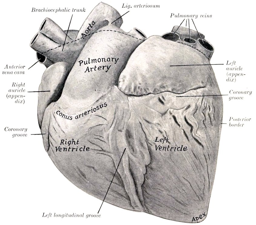
Fig. 419.—Heart of Horse, Left View. Hardened in situ. The dotted line indicates the line of reflection of the serous pericardium. The epicardium and subepicardial fat have not been removed from the heart.
In position the heart is decidedly asymmetrical, about two-fifths being to the 527right of the median plane and three-fifths to the left. The long axis (from the middle of the base to the apex) is directed downward, backward, and somewhat to the left. The base (Basis cordis) is directed dorsally and lies a little above the middle of the dorso-ventral diameter of the thorax. It is opposite to the ribs from the third to the sixth inclusive. The apex (Apex cordis) lies above the last segment of the sternum, and is separated by a very short interval from the sternal part of the diaphragm. The anterior border (Margo cranialis) is strongly convex and is directed obliquely downward and backward; its lower part is parallel with the sternum. The posterior border (Margo caudalis) is much shorter, nearly vertical, and is opposite to the sixth rib or intercostal space. The surfaces, right and left (Facies dextra, sinistra), are convex and are marked by grooves which indicate the division of the heart into four chambers, the two atria above and two ventricles below. The left surface (covered by the pericardium) is in contact with the lower third of the chest-wall from the third to the fifth intercostal space. On the right side the cardiac notch of the lung is smaller and further forward, so that the area of contact is at the third and fourth ribs.
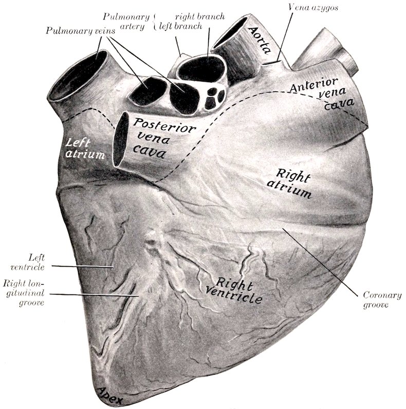
Fig. 420.—Heart of Horse, Right View. Hardened in situ. Line of reflection of serous pericardium dotted. Epicardium and subepicardial fat have not been removed from heart. Left ventricle is considerably contracted.
The coronary or atrio-ventricular groove (Sulcus coronarius) indicates the division between the atria and the ventricles. It almost completely encircles the 528heart, but is interrupted by the origin of the pulmonary artery. The longitudinal or interventricular grooves, right and left (Sulci longitudinales sinister et dexter), correspond to the septum between the ventricles. The left groove is left-anterior in position. It begins at the coronary groove behind the origin of the pulmonary artery, and passes downward almost parallel to the posterior border, but does not reach the apex. The right groove is right posterior in position. It begins at the coronary groove below the termination of the posterior vena cava and passes downward and a little backward, ending about an inch and a half (ca. 3 to 4 cm.) above the apex. The grooves are occupied by the coronary vessel and a variable quantity of fat.
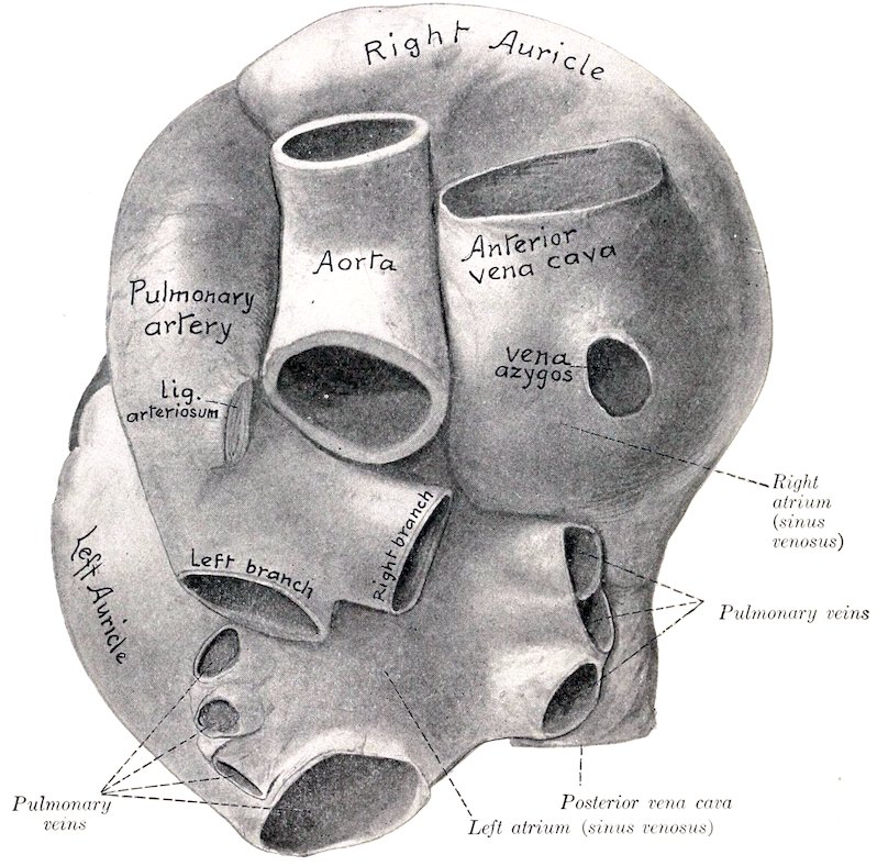
Fig. 421.—Base of Heart of Horse with Large Vessels, Dorsal View. Specimen Hardened in situ.
Size and Weight.—In horses of medium size the heart usually weighs about seven or eight pounds (ca. 3.5 kg.), or about 0.7 per cent. of the body-weight. There is, however, great range of variation in apparently normal specimens.
The following measurements were obtained in medium-sized hearts in diastole:
Sagittal diameter of base 25 cm.
Greatest width of base 18 to 20 cm.
Circumference at coronary groove 65 to 70 cm.
Distance between origin of pulmonary artery and apex 25 cm.
Distance between termination of posterior vena cava and apex 18 to 20 cm.
529
The Right Atrium
The right atrium (Atrium dextrum) or auricle forms the right-anterior part of the base of the heart, and lies above the right ventricle. It consists of a sinus venosus, into which the veins open, and an auricle or auricular appendix. The latter is a conical diverticulum which curves around the right and anterior surfaces of the aorta, its blind end appearing on the left side in front of the origin of the pulmonary artery.
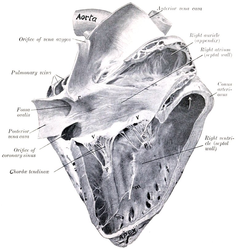
Fig. 422.—Right Side of Heart of Horse Opened up by Removal of Greater Part of Right Wall. Organ Hardened in situ. The right ventricle was in diastole. r, Intervenous crest; v, v, tricuspid valve; p, p, papillary muscles; m, m, moderator bands. Arrow points into origin of pulmonary artery.
There are five chief openings in the right atrium. The opening of the anterior vena cava (Ostium venæ cavæ cranialis) is in the upper and fore part. The opening of the posterior vena cava (Ostium venæ cavæ caudalis) is at the lower posterior part. Between the two the wall pouches upward somewhat, forming what is sometimes termed the sinus venosus or sac of Lower. In the anterior part of this the vena azygos opens. The coronary sinus opens immediately below the 530posterior vena cava; the orifice is provided with a small semilunar valve or valve of Thebesius (Valvula sinus coronarii). The middle coronary vein has a separate opening in some cases close to that of the coronary sinus. The atrio-ventricular opening is in the lower part, and leads into the right ventricle. In addition to the foregoing there are several small orifices of the venæ cordis parvæ; these are concealed in the depressions between the musculi pectinati.
The atrium is lined with a glistening membrane, the endocardium. Its walls are smooth except on the right and in the auricle (or appendix), where it is crossed in various directions by muscular ridges, the musculi pectinati. Small bands extend across some of the spaces inclosed by the musculi pectinati. The latter terminate above on a curved crest, the crista terminales, which indicates the junction of the primitive sinus venosus of the embryo with the atrium proper, and corresponds with the sulcus terminalis externally. The openings of the venæ cavæ are without valves. A ridge, the intervenous crest,[156] projects downward and forward from the roof just in front of the opening of the posterior vena cava; it tends to direct the flow of blood from the anterior vena cava to the atrio-ventricular opening. The fossa ovalis is an oval depression in the septal wall at the point of entrance of the posterior vena cava, bounded internally by a thick margin (Limbus fossæ ovalis). The fossa is the remains of an opening, the foramen ovale, through which the two atria communicate in the fœtus.
The Left Atrium
The left atrium (Atrium sinistrum) or auricle forms the posterior part of the base of the heart. It lies behind the pulmonary artery and the aorta and above the left ventricle. The auricle (or appendix) extends outward and forward on the left side, and its blind end is behind the origin of the pulmonary artery. The pulmonary veins, usually seven or eight in number, open into the atrium behind and on the right side. The cavity of the atrium is smooth, with the exception of the auricle (or appendix), in which the musculi pectinati are present. In some cases there is a depression on the septal wall opposite the fossa ovalis, bounded above by a fold which is the remnant of the valve of the foramen ovale of the fœtus. The atrio-ventricular opening is situated below and in front; it usually appears smaller than the right one on account of the contraction of the left ventricle in the dead subject. The apertures of small veins of the heart are found in the spaces inclosed by the musculi pectinati.
The number and the arrangement of the pulmonary veins are variable. They may be five to nine in number. The largest orifice is posterior. Usually three veins of considerable size enter close together on the right above the posterior vena cava, and three or four open close to the ridge which projects from the roof at the base of the auricle (appendix).
The Right Ventricle
The right ventricle (Ventriculus dexter) constitutes the right-anterior part of the ventricular mass. It forms almost all of the anterior border of the heart, but does not reach the apex, which is formed entirely by the left ventricle. It extends from the third rib to the fourth intercostal space on the left side, to the fifth rib and space on the right side. It is somewhat triangular in outline, and is semilunar in cross-section. Its base faces upward and a little to the right and is connected largely with the right atrium, with which it communicates through the atrio-ventricular orifice; but its left part projects higher and forms the conus arteriosus, from which the pulmonary artery arises. Its apex is two inches or more (ca. 5 to 6 cm.) above the apex of the heart. On opening the cavity it is seen that the two openings are separated by a thick rounded ridge (Crista supraventricularis). 531The axis of the cavity, taken from this ridge to the apex forms a spiral curve downward and to the right. The septal wall is convex and faces obliquely forward and to the right.
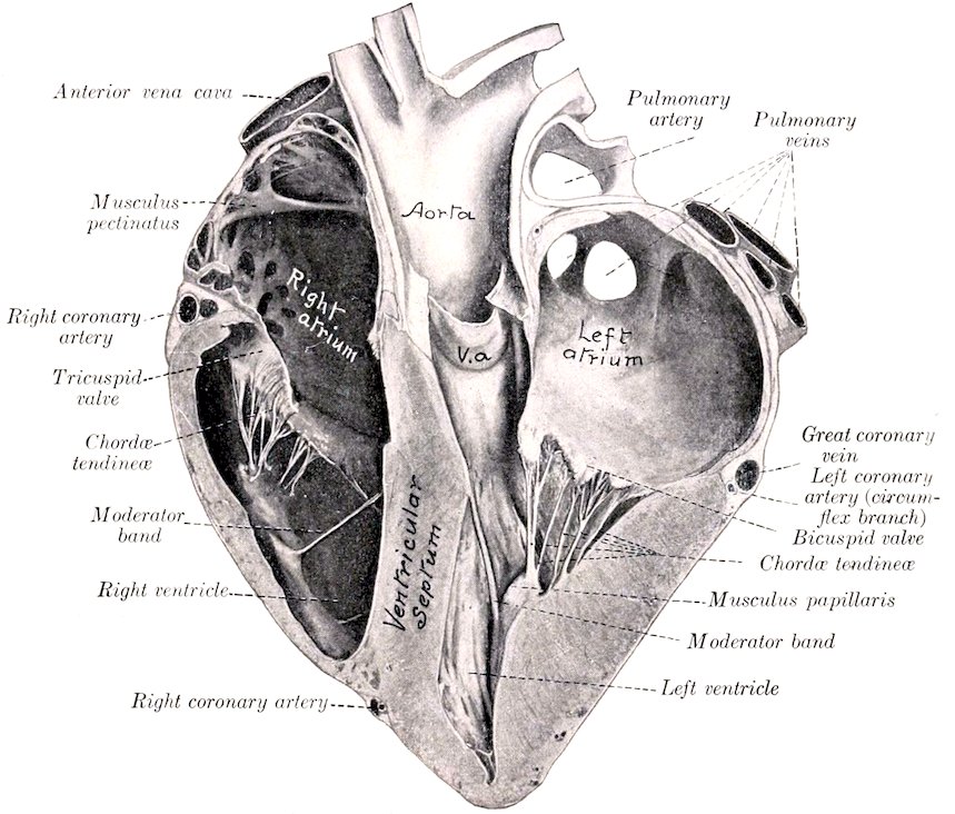
Fig. 423.—Section of Heart of Horse. Specimen hardened in situ and cut nearly at right angles to the ventricular septum. The left ventricle is contracted, but not ad maximum. V. a., Segment of aortic valve.
The right atrio-ventricular orifice (Ostium venosum dextrum) is oval and is situated opposite to the lower parts of the third and fourth ribs. It is guarded by a tricuspid valve (Valvula tricuspidalis); of the three large cusps of this valve, one is between the atrio-ventricular opening and the conus arteriosus, one is septal, and the third is on the right margin. Small intermediate cusps intervene between the large ones. The peripheral edges of the cusps are attached to the fibrous ring at the atrio-ventricular opening. The central edges are irregular and hang down into the ventricle; they give attachment to chordæ tendineæ. The auricular surfaces are smooth. The ventricular surfaces are rough and furnish attachment to interlacing branches of the chordæ tendineæ. The valves are folds of the endocardium, strengthened by fibrous tissue and at the periphery by muscular fibers also. The chordæ tendineæ are attached below to the three musculi papillares, which project from the ventricular wall; superiorly they divide into branches which are inserted into the ventricular surfaces and the free edges of the valves. Each segment of the valve receives chordæ tendineæ from two papillary muscles. Of the latter, two are on the septum and the third and largest springs from the anterior wall.
The pulmonary orifice (Ostium arteriæ pulmonalis) is circular and is situated 532at the summit of the conus arteriosus, opposite to the lower part of the third intercostal space. It is guarded by the pulmonary valve, composed of three semilunar cusps (Valvulæ semilunares arteriæ pulmonalis); of these, one is internal, one external, and the third posterior. The convex peripheral border of each cusp is attached to the fibrous ring at the junction of the pulmonary artery and the conus arteriosus. The central border is free and slightly concave. Each cusp consists of a layer of endocardium on its ventricular surface, a continuation of the inner coat of the artery on its arterial surface, and an intermediate layer of fibrous tissue. The upper edge of the conus arteriosus forms three arches with intermediate projecting angles or horns, to all of which the cusps are attached; and the artery forms opposite each cusp a pouch, the sinus of the pulmonary artery (or of Valsalva).
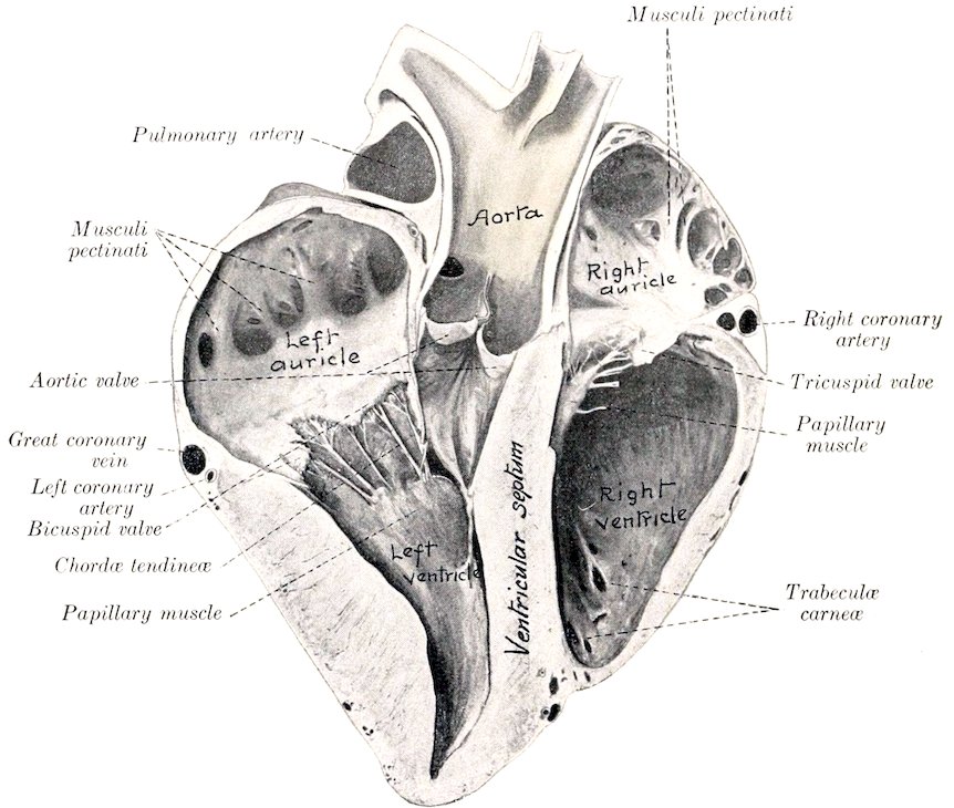
Fig. 424.—Section of Heart of Horse. Specimen Hardened in situ. The section is cut nearly at right angles to the ventricular septum, and is viewed from the right and posteriorly.
The walls of the ventricle (except in the conus arteriosus) bear muscular ridges and bands, termed trabeculæ carneæ. These are of three kinds, viz., (1) ridges or columns in relief; (2) musculi papillares, somewhat conical flattened projections, continuous at the base with the wall and giving off the chordæ tendineæ to the tricuspid valve; (3) moderator bands (Musculi transversi cordis) which extend from the septum to the opposite wall. The latter are partly muscular, partly tendinous, and vary in different subjects. The strongest one is usually about midway between the base and apex and extends from the septum to the base of the anterior musculus papillaris. It is considered that they tend to prevent overdistention.
533
The Left Ventricle
The left ventricle (Ventriculus sinister) forms the left posterior part of the ventricular mass. It is more regularly conical than the right ventricle and its wall is much thicker except at the apex. It forms all of the posterior border of the ventricular part and the apex of the heart. Its base is largely continuous with the left atrium, with which it communicates through the left atrio-ventricular opening, but in front it opens into the aorta. The cavity usually appears smaller than that of the right ventricle in the dead subject, on account of the greater contraction of its wall. It is almost circular in cross-section.
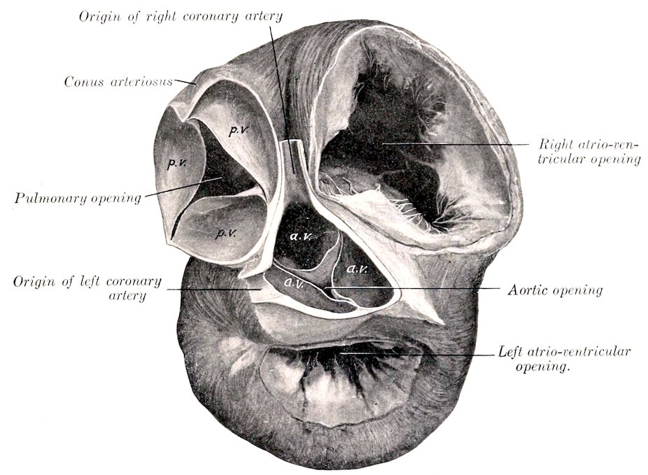
Fig. 425.—The Bases of the Ventricles of the Heart of the Horse. The atria have been removed and the aorta and pulmonary artery cut off short. The left ventricle is contracted. p.v., Pulmonary valve; a.v., aortic valve.
The left atrio-ventricular orifice (Ostium venosum sinistrum) is opposite to the fifth rib and intercostal space. It is almost circular and is guarded by the bicuspid or mitral valve (Valvula bicuspidalis). The two segments of this valve are larger and thicker than those of the right side of the heart. One is placed in front and separates the atrio-ventricular and aortic openings. The other is placed behind and laterally and is usually divided into two or three flaps.
The aortic orifice (Ostium aorticum) is directed upward and slightly forward. It is situated opposite to the fourth rib. It is guarded by the aortic valve, composed of three semilunar cusps (Valvulæ semilunares aortæ); one cusp is anterior, the others right and left posterior. They are similar to those of the pulmonary valve, but are much stronger. The free edge of each contains a central nodule of fibrous tissue, the corpus Arantii (Nodulus valvulæ semilunaris).
The chordæ tendineæ are fewer but larger than those of the right ventricle.
There are two large musculi papillares, one on each side; they are usually 534compound. The moderator bands are variable. Commonly two or three larger ones (which are often branched) extend from the musculi papillares to the septum. Smaller ones may be found in various places, especially at the apex. The other trabeculæ are fewer and less prominent than in the right ventricle.
The interventricular septum (Septum ventriculorum) is the musculo-membranous partition which separates the cavities of the two ventricles. It is placed obliquely. One surface is convex, faces forward and to the right, and bulges into the right ventricle. The other surface, which faces into the left ventricle, is concave and looks backward and to the left. The greater part of the septum is thick and muscular (Septum musculare), but the upper part is thin and membranous (Septum membranaceum). The latter intervenes between the left ventricle, on the one hand, and the right ventricle and atrium on the other.
Structure of the Heart
The heart-wall consists mainly of peculiar striped muscle, the myocardium, which is covered externally by the visceral part of the serous pericardium or epicardium, and is lined by the endocardium.
The epicardium is in general closely attached to the muscular wall, but is loosely attached over the coronary vessels and the associated subepicardial fat. It consists of a layer of flat polygonal cells, resting on a membrane of white and elastic fibers.
The myocardium consists of planes of fibers arranged in a somewhat complicated manner. The muscular tissue of the atria is almost completely separated from that of the ventricles by the fibrous rings around the atrio-ventricular orifices.
In the atria the muscle bands fall naturally into two groups—superficial and deep. The former are common to both atria, the latter special to each. The superficial or common fibers for the most part begin and end at the atrio-ventricular rings, but some enter the interatrial septum. The deep or special bundles also form two sets. Looped fibers pass over the atria from ring to ring, while annular or spiral fibers surround the ends of the veins which open into the atria, the auricles, and the fossa ovalis.
The muscular wall of the ventricles is much stronger than that of the atria. That of the left ventricle is in general about three times as thick as that of the right one, but is thin at the apex. The superficial fibers are attached above to the atrio-ventricular fibrous rings and pass in a spiral toward the apex. Here they bend upon themselves and pass deeply upward to terminate in a papillary muscle of the ventricle opposite to that in which they arose. The loops so formed at the apex constitute a whorl, the vortex cordis. The deep fibers, although they appear to be proper to each ventricle, have been shown by MacCallum to be in reality almost all common to both. Their arrangement is scroll-like. They begin on one side, curve around in the wall of that ventricle, then pass in the septum to the opposite side, and curve around the other ventricle. There is a layer of deep fibers which is confined to the basal part of the left ventricle; it is attached to the left atrio-ventricular ring.
Four fibrous rings (Annuli fibrosi) surround the orifices at the bases of the ventricles. The atrio-ventricular rings separate the musculature of the atria from that of the ventricles. Those which surround the origins of the pulmonary artery and aorta are festooned in conformity with the attached borders of the valves. The aortic ring contains on the right side a plate of cartilage (Cartilago cordis), which frequently becomes more or less calcified in old animals. Sometimes a smaller plate is present on the left side.
The endocardium lines the cavities of the heart and is continuous with the internal coat of the vessels which enter and leave the organ. Its free surface is 535smooth and glistening and is formed by a layer of endothelial cells. The latter rest on a thin layer of connective tissue, which is connected with the myocardium by a subendocardial elastic tissue containing vessels and nerves.
Vessels and Nerves.—The heart receives its blood-supply through the two coronary arteries which arise from the aorta opposite to the anterior and left cusps of the aortic valve. Most of the blood is returned by the coronary veins, which open into the right atrium by the coronary sinus.[157] A few small veins open directly into the right atrium, and others are said to open into the left atrium and the ventricles. The lymph vessels form a subepicardial network which communicates through stomata with the cavity of the pericardium. There is a less distinct subendocardial network. The vessels converge usually to two trunks which accompany the blood-vessels in the atrio-ventricular grooves and enter the glands at the bifurcation of the trachea. The nerves are derived from the vagus and sympathetic through the cardiac plexus.
The Arteries
THE PULMONARY ARTERY
The pulmonary artery (A. pulmonalis) springs from the conus arteriosus at the left side of the base of the right ventricle. It curves upward, backward, and inward, and divides behind the arch of the aorta into right and left branches. It is related in front to the right auricle (appendix), behind to the left auricle (appendix), and internally to the aorta. It is enveloped with the latter in a common sheath of the visceral layer of the serous pericardium. Near the bifurcation it is connected with the arch of the aorta by a fibrous band about half an inch (ca. 1.2 cm.) in width; this is the ligamentum arteriosum, a remnant of the large ductus arteriosus, which conducts most of the blood from the pulmonary artery to the aorta in the fœtus. The artery is bulbous at its origin, and forms three sinuses or pouches, which correspond to the cusps of the semilunar valve. Beyond this it gradually diminishes in caliber.
In a horse of medium size the artery is about seven inches (ca. 17 to 18 cm.) long. At the origin it is about two and a half inches (ca. 6 to 6.5 cm.) in width; at the bifurcation its caliber is about one and a half inches (ca. 3.5 to 4 cm.). The wall is relatively thin, especially at the origin.
The right branch (Ramus dexter) of the pulmonary artery is a little longer and wider than the left one. It passes over the fore part of the left atrium and below the bifurcation of the trachea to the hilus of the right lung, and enters the latter below the right bronchus. In the lung it passes to the outer and lower side of the stem bronchus and accompanies it to the base of the organ. The branches correspond to the ramification of the bronchi. The left branch (Ramus sinister) is very short. It passes backward and enters the lung below the left bronchus. Its branches within the lung are arranged like that of the right one.
THE SYSTEMIC ARTERIES
The aorta is the main systemic arterial trunk. It begins at the base of the left ventricle and is practically median at its origin. It passes upward and slightly forward between the pulmonary artery on the left and right atrium on the right. It then curves sharply backward and upward and inclines somewhat to the left, forming the arch of the aorta (Arcus aortæ), and reaches the ventral surface of the spine at the eighth or ninth thoracic vertebra. After passing backward along the ventral aspect of the bodies of the vertebræ between the lungs it traverses the hiatus 536aorticus and enters the abdominal cavity, where it lies below the vertebral bodies and the psoas minor, just to the left of the median plane. It divides under the fifth lumbar vertebra into the two internal iliac or hypogastric arteries.
From the bifurcation a small vessel, the middle sacral artery (A. sacralis media), sometimes passes backward on the pelvic surface of the sacrum. It becomes lost in the periosteum or joins the coccygeal artery, or in exceptional cases is traceable to the sphincter ani externus.
The caliber of the aorta is greatest at its origin, which is termed the bulbus aortæ. Here it forms three pouch-like dilatations, the sinuses of the aorta (or of Valsalva). These correspond to the cusps of the aortic valve, and the coronary arteries arise from the left posterior and anterior sinuses. At the arch the diameter is about two inches (ca. 5 cm.), and beyond this it diminishes gradually in width.
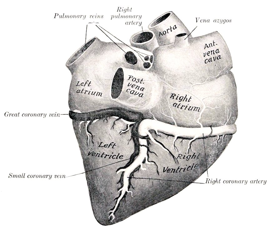
Fig. 426.—Cardiac Vessels of Horse, Right Side. Veins are black, arteries white.
It is convenient to divide the aorta into thoracic and abdominal parts. The thoracic aorta (Aorta thoracica) lies within the pericardium to the point of attachment of the ligamentum arteriosum, and is inclosed with the pulmonary artery in a prolongation of the epicardium. Beyond this it is between the two pleural sacs. It is crossed on the right by the œsophagus and trachea, on the left by the left vagus nerve. The left recurrent nerve winds around the concavity of the arch from left to right, and the vena azygos and thoracic duct lie along the dorsal part of its right face. The trachea causes it to deviate to the left; but beyond this it becomes median. The abdominal aorta (Aorta abdominalis) (Fig. 450) is related above to the lumbar vertebræ, the inferior common ligament, and the left psoas minor muscle; in the hiatus aorticus it is related to the cisterna chyli. On its right is the posterior vena cava, and on its left the left kidney and ureter.
537
BRANCHES OF THE THORACIC AORTA
1. CORONARY ARTERIES
The two coronary arteries, right and left, are distributed almost entirely to the heart, but send some small twigs to the origins of the great vessels.
The right coronary artery (A. coronaria dextra) arises from the anterior sinus of the aorta. It passes forward between the conus arteriosus and the right auricle (appendix) to the coronary groove, in which it curves around to the right and backward. It then descends in the right ventricular groove almost to the apex of the heart.
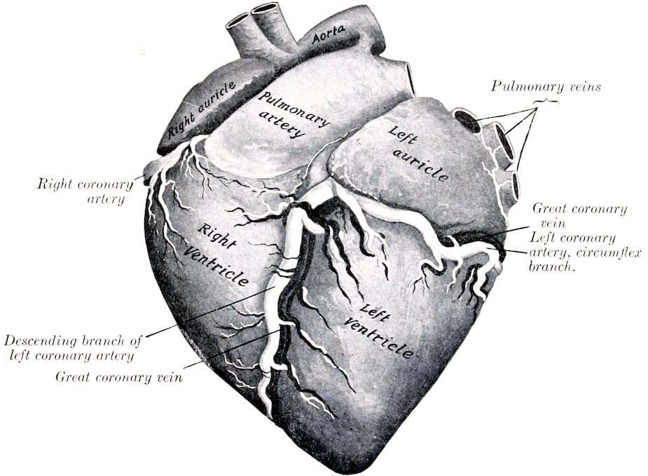
Fig. 427.—Cardiac Vessels of Horse, Left Side. The dotted lines indicate part of the left coronary artery which is concealed by the left auricle.
The left coronary artery (A. coronaria sinistra) arises from the left posterior sinus of the aorta, passes to the left behind the origin of the pulmonary artery, and divides into two branches. The descending branch (Ramus descendens) passes down the left ventricular groove toward the apex. The circumflex branch (Ramus circumflexus) runs backward in the coronary groove, in which it winds around to the right side.
2. COMMON BRACHIOCEPHALIC TRUNK OR ANTERIOR AORTA
The common brachiocephalic trunk or anterior aorta (Truncus brachiocephalicus communis) is a very large vessel which arises from the convexity of the arch of the aorta within the pericardium. It is directed forward and upward. Its length in horses of medium size is usually about two inches (ca. 5 to 6 cm.), but it is sometimes only half an inch or less (ca. 1 cm.). It is crossed on the left by the left vagus and cardiac nerves, and the left recurrent nerve runs between it and 538the trachea. It divides opposite to the second intercostal space or third rib into the brachiocephalic and left brachial arteries.
The brachiocephalic artery (A. brachiocephalica) is directed forward and a little upward in the anterior mediastinum, beneath the trachea. Opposite the first rib it gives off the bicarotid trunk and is continued as the right brachial artery. The latter (A. subclavia dextra) turns downward and bends around the anterior border of the first rib and the insertion of the scalenus muscle above the brachial vein. Its course and branches beyond this point will be described with the vessels of the thoracic limb.
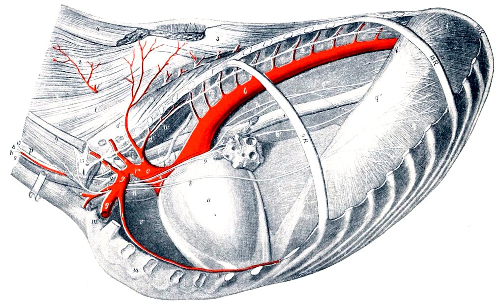
Fig. 428.—Topography of Thoracic Cavity of Horse, Left Side, after Removal of Left Lung. The ninth, fifteenth, and eighteenth ribs are retained. a, Pericardium; b, thoracic aorta; c, brachiocephalic trunk; d, d″, dorsal artery; d′, subcostal artery; e, e′, superior or deep cervical artery; f, vertebral artery; g, g′, left brachial artery; h, inferior cervical artery; i, internal thoracic artery; k, common carotid artery; l, jugular vein; m, brachial vein; m′, inferior cervical vein; n, anterior vena cava; o, thoracic duct; p, œsophagus; q, costal part of diaphragm; q′, tendinous center of same; q″, left crus of same; r, anterior mediastinal lymph glands; s, trachea; t, bronchial lymph glands; u, root of left lung; v, anterior mediastinum; v′, posterior mediastinum; w, longus colli; x, sterno-cephalicus; y, scalenus; z, transversalis costarum; 1, longissimus; 2, splenius; 3, spinalis et semispinalis; 4, 4′, rhomboideus; 5, left phrenic nerve; 6, left vagus; 6′, 6″, œsophageal continuations of vagi; 7, dorsal branch of right vagus; 8, cardiac nerve; 9, left recurrent nerve; 10, cervical trunk of sympathetic; 11, first thoracic ganglion of sympathetic; 12, thoracic trunk of sympathetic; 13, brachial plexus. (After Ellenberger-Baum, Top. Anat. d. Pferdes.)
The left brachial artery (A. subclavia sinistra) is longer than the right one and rises to a higher level. It forms an almost semicircular curve, the concavity being ventral. It is related internally to the œsophagus, trachea, and thoracic duct, and the left vagus, phrenic, and cardiac nerves cross under its origin. It emerges from the thorax like the artery of the right side. There is thus a difference at first between the trunks of opposite sides, but beyond this their course and distribution are similar.
The brachial and brachiocephalic arteries within the thorax give off the dorsal, superior cervical, vertebral, and internal thoracic arteries. At the first rib they give off the external thoracic and inferior cervical arteries.
1. The left dorsal or dorso-intercostal artery (A. costo-cervicalis) passes dorsally across the left face of the trachea and œsophagus toward the second intercostal space. The right artery arises usually by a common trunk with the 539superior cervical, crosses the right face of the trachea and has no contact with the œsophagus. Both detach small branches to the trachea, lymph glands, and pleura, and divide on reaching the longus colli into two branches. Of these the subcostal artery (A. intercostalis suprema) is the smaller. It passes backward under the costo-vertebral joints with the sympathetic trunk. It gives off the second, third, and fourth intercostal arteries, and ends at the fifth space, where it anastomoses with the first aortic intercostal artery, or constitutes the fifth intercostal artery, or dips into the longissimus muscle. It also gives off spinal branches and twigs to the longus colli and the pleura. The other branch (A. transversa colli) is the direct continuation of the trunk. It emerges through the upper end of the second intercostal space, passes across the transversalis costarum and longissimus toward the withers, and divides into several diverging branches. An anterior branch passes upward and forward between the splenius and complexus and anastomoses with branches of the superior cervical artery; the others are directed upward under the serratus magnus and rhomboideus to the withers, supplying the muscles and skin of this region.
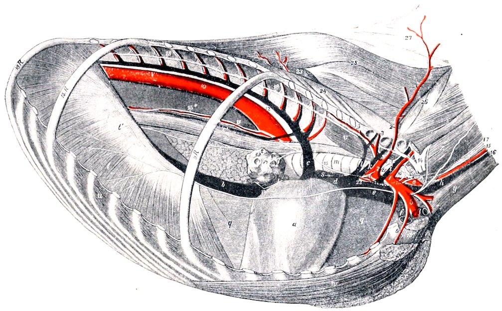
Fig. 429.—Topography of Thoracic Cavity of Horse, Right View. The ninth, fifteenth, and eighteenth ribs are retained. a, Pericardium; b, posterior vena cava; c, vena azygos; c′, œsophageal vein; d, e, anterior vena cava; f, brachial vein; g, inferior cervical vein; h, jugular vein; i, vertebral vein; k, superior cervical vein; l, dorsal vein; m, trachea; n, root of right lung; o, œsophagus; p, mediastinal lobe of right lung; q, mediastinum; r, bronchial lymph glands; s, mediastinal lymph glands; t, costal part of diaphragm; t′, tendinous center of same; u, intercostal muscle; v, longus colli; w, posterior deep pectoral muscle; w′, anterior deep pectoral muscle; x, superficial pectoral muscle; y, sterno-cephalicus; z, scalenus; 1, 5, right brachial artery; 2, dorso-cervical trunk; 2′, superior or deep cervical artery; 3, dorsal artery (cut); 3′, subcostal artery; 4, internal thoracic artery; 6, external thoracic artery; 7, inferior cervical artery; 8, bicarotid trunk; 10, aorta; 11, œsophageal artery; 12, thoracic duct; 13, right phrenic nerve; 14, brachial plexus; 15, right vagus; 15′, 15″, œsophageal continuations of vagi; 16, right recurrent nerve; 17, cervical trunk of sympathetic; 18, thoracic trunk of sympathetic; 20, inferior cervical ganglion; 21, first thoracic ganglion of sympathetic; 22, right cardiac nerve; 23, longissimus; 24, transversalis costarum; 25, spinalis; 26, multifidus; 27, ligamentum nuchæ. (After Ellenberger-Baum, Top. Anat. d. Pferdes.)
The left dorsal artery sometimes arises with the superior cervical by a common trunk. On the right side there may be a common stem for the dorsal, superior cervical, and vertebral arteries. Occasionally the artery arises from the anterior aorta. Sometimes it emerges through the third space. The subcostal may arise independently behind the dorsal or from the superior cervical artery.
5402. The superior or deep cervical artery (A. cervicalis profunda) arises in front of the dorsal or by a common trunk with it. It crosses the œsophagus (left side), the trachea (right side), and the longus colli, and emerges from the thoracic cavity by passing through the space behind the first costo-transverse articulation. In the thorax it gives off a small branch (A. mediastini cranialis) to the mediastinum and the pericardium; also the first intercostal artery (A. intercostalis prima), a very small vessel which passes down in the first intercostal space. After leaving the thorax the artery passes upward and forward on the spinalis muscle and the lamellar part of the ligamentum nuchæ, covered by the complexus. Its terminal branches anastomose with branches of the occipital and vertebral arteries in the region of the axis. Numerous collateral branches are detached to the extensor muscles of the head and neck, the ligamentum nuchæ, and the skin, and anastomoses occur with the dorsal artery above and the vertebral artery below.
The artery sometimes emerges through the second intercostal space.
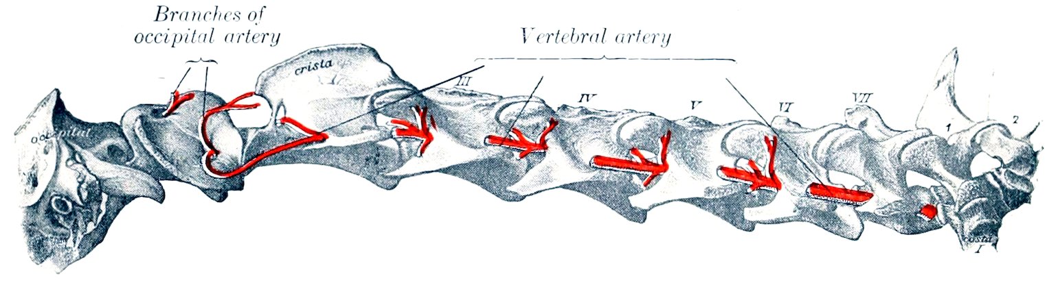
Fig. 430.—Vertebral Artery of Horse. (After Schmaltz, Atlas d. Anat. d. Pferdes.)
3. The vertebral artery (A. vertebralis) arises from the brachial (or brachiocephalic) opposite the first intercostal space and passes upward and forward. On the left side it crosses the œsophagus, on the right the trachea. Emerging from the thorax it passes between the longus colli internally and the scalenus externally, under the transverse process of the seventh cervical vertebra, and continues along the neck through the series of foramina transversaria, between which it is covered by the intertransversales colli. Emerging from the foramen of the axis, it crosses the capsule of the atlanto-axial joint, and joins the retrograde branch of the occipital artery under cover of the great oblique muscle of the head. At each intervertebral foramen a spinal branch (Ramus spinalis) is given off which enters the vertebral canal and reinforces the ventral spinal artery. It also gives off series of dorsal and ventral muscular branches (Rami musculares). The dorsal branches are the larger; they supply the deep extensor muscles of the head and neck, and anastomose with the deep cervical and occipital arteries. The ventral branches supply chiefly the scalenus, longus colli, intertransversales, and rectus capitis anterior major. The artery is accompanied by the vertebral vein and a sympathetic nerve-trunk.
In some cases the last cervical transverse process has a foramen transversarium, through which the artery passes.
4. The internal thoracic artery (A. thoracica s. mammaria interna) is a large vessel which arises from the ventral side of the brachial opposite the first rib. It curves downward and backward, being at first on the inner surface of the rib, and then crosses the lower part of the first intercostal space and passes under the transversus thoracis muscle. It runs backward under cover of that muscle over the chondro-sternal joints to the eighth costal cartilage, where it divides into asternal and anterior abdominal branches. At each intercostal space two collateral 541branches are detached. The intercostal branches (Rami intercostales) pass upward in the intercostal spaces and anastomose with homonymous descending arteries. The lower branches detach small twigs to the transversus thoracis, pleura, and pericardium, and pass out between the costal cartilages as perforating branches (Rami perforantes) to supply the pectoral muscles and skin, anastomosing with the external thoracic artery. A very small pericardiaco-phrenic artery ascends in the mediastinum on the left side, in the caval fold of pleura on the right side; it supplies fine twigs to the pericardium and pleura and accompanies the phrenic nerve to the diaphragm. In the young subject it gives small branches (Aa. thymicæ) to the thymus gland. The asternal artery (A. musculophrenica) passes along the ninth costal cartilage and continues along the costal attachment of the transversus abdominis (Fig. 185). It gives off intercostal branches which anastomose with those descending from the thoracic aorta, and twigs to the diaphragm and transversus abdominis. The anterior abdominal artery (A. epigastrica cranialis) is the direct continuation of the internal thoracic. It passes between the ninth costal cartilage and the xiphoid cartilage, runs backward on the abdominal surface of the rectus abdominis and then becomes embedded in it (Fig. 466). It supplies the ventral wall of the abdomen and anastomoses with the posterior abdominal artery.
5. The external thoracic artery (A. thoracica externa) is given off from the ventral aspect of the brachial, usually at the inner surface or anterior border of the first rib. It turns around the first rib below or behind the brachial vein (when given off within the thorax) and passes backward under the deep pectoral muscle; it is continued as a small vessel in the panniculus carnosus, where it accompanies the external thoracic (“spur”) vein. It sends branches to the pectoral muscles and the axillary lymph glands.
This artery varies in origin and size. Not rarely it arises from the internal thoracic or from the brachial outside of the thorax. In other cases it arises by a common trunk with the inferior cervical. It may be very small or even absent, in which case the perforating branches of the internal thoracic compensate.
6. The inferior cervical artery (Truncus omo-cervicalis) arises usually from the dorsal surface of the brachial opposite the first rib or where that vessel winds around the rib. It is directed downward and a little forward across the external surface of the jugular vein and the deep face of the scalenus among the lymph glands at the thoracic inlet, and divides into ascending and descending branches. The ascending branch (A. cervicalis ascendens) passes upward and forward along the external surface of the jugular vein, then turns sharply backward and runs upward along the anterior border of the anterior deep pectoral muscle, between the omo-hyoideus and mastoido-humeralis and in relation to the prescapular lymph glands; it gives branches to these muscles and the prepectoral and prescapular lymph glands. The descending branch (A. transversa scapulæ) passes downward and outward across the surface of the anterior deep pectoral and then runs in the groove between that muscle and the mastoido-humeralis in company with the cephalic vein. It supplies branches to these muscles and the skin of the breast.
THE COMMON CAROTID ARTERY
These two vessels (Arteriæ carotides communes) arise from the brachiocephalic artery by a common trunk. This stem, the truncus bicaroticus or cephalic artery, is detached from the inner face of the brachiocephalic opposite the first rib and passes forward in the median plane beneath the trachea. It is related ventrally to the prepectoral lymph glands, the terminal parts of the jugular veins, and the anterior vena cava, and laterally to the vagus and recurrent nerves. It is commonly 542two or three inches (ca. 5 to 7 cm.) in length, but it may vary between one and eight inches (ca. 2.5 to 20 cm.).
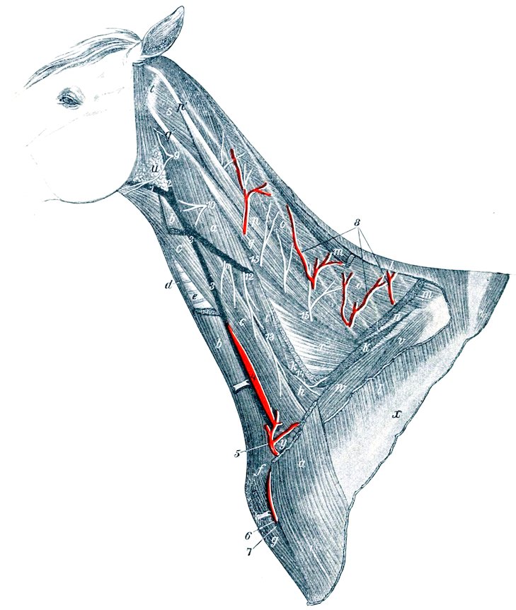
Fig. 431.—Vessels and Nerves of Neck of Horse. a, Mastoido-humeralis; b, sterno-cephalicus; c, omo-hyoideus; d, sterno-thyro-hyoideus; e, trachea; f, position of cariniform cartilage; g, anterior superficial pectoral muscle; h, scalenus; i, intertransversales; k, insertion of serratus cervicis; l, origin of trapezius; m, rhomboideus; n, splenius; o, complexus; p, q, trachelo-mastoideus; p′, q′, tendons of same; r, longissimus; s, obliquus capitis posterior; t, wing of atlas; u, parotid gland; v, supraspinatus; w, anterior deep pectoral; x, spine of scapula; y, prescapular lymph glands; 1, external maxillary vein; 2, 3, jugular vein; 4, carotid artery, exposed by drawing jugular vein aside; 5, 6, ascending and descending branches of inferior cervical artery; 7, cephalic vein; 8, branches of superior or deep cervical artery; 9–14, ventral branches of second to seventh cervical nerves; 15, branches of dorsal divisions of cervical nerves. (Ellenberger-Baum, Top. Anat. d. Pferdes.)
The right common carotid artery passes obliquely from the ventral face of the trachea to its right side. In this position it runs upward and forward and divides at the crico-pharyngeus muscle and under the submaxillary gland into external carotid, internal carotid, and occipital arteries. It is inclosed in a fibrous sheath, and is accompanied dorsally by the vagus and sympathetic nerves, ventrally by the recurrent nerve. At the last two cervical vertebræ it is in contact superficially with the jugular vein, but further forward the omo-hyoideus muscle intervenes between the artery and vein. Near its termination the artery becomes more deeply placed and is related externally to the submaxillary and parotid glands, internally to the œsophagus. In some cases it is in contact ventrally with the thyroid gland, especially when the latter is larger than usual.
543The left common carotid artery differs from the right one in that it is related deeply to the œsophagus, which separates it from the trachea in the greater part of its course.
The collateral branches of the common carotids are in the main small. They comprise:
1. Muscular branches (Rami musculares) of variable size, which go to the ventral muscles of the neck and the skin.
2. Œsophageal and tracheal branches (Rami œsophagei et tracheales). Small twigs go to the cervical lymph glands also.
3. The parotid artery. This comes off near the termination and enters the ventral part of the parotid gland. It also supplies the subparotid lymph glands, and sometimes sends a branch to the submaxillary gland. It is inconstant.
4. The thyro-laryngeal artery (A. thyreoidea cranialis). This, the largest collateral branch of the carotid, arises from the latter two or three inches before it divides. It curves over the anterior end of the thyroid gland, into which it sends several branches. It gives off a laryngeal branch (A. laryngea), which sends twigs to the external muscles of the larynx and the constrictors of the pharynx, passes between the cricoid and thyroid cartilages, and supplies the internal muscles and the mucous membrane of the larynx. A small pharyngeal branch (A. pharyngea ascendens) runs upward and forward to the crico-pharyngeus, and supplies twigs to the posterior part of the pharynx and the origin of the œsophagus. Small innominate twigs are given off to the trachea, the œsophagus, and the sterno-thyro-hyoideus and omo-hyoideus muscles.
In some cases the thyroid and laryngeal arteries arise from the carotid separately or by a short common stem. A laryngeal branch is often detached from the carotid in front of the thyro-laryngeal and enters the larynx with the superior laryngeal nerve. The pharyngeal branch frequently comes directly from the carotid.
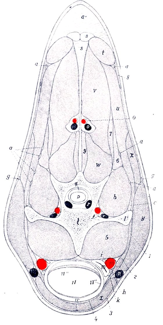
Fig. 432.—Cross-section of Neck of Horse, Passing through Fifth Cervical Vertebra; Anterior View. a, Branches of cervical nerves; a′, nuchal fat; b, intertransversalis muscle; c, longissimus muscle; d, vertebral artery; e, vertebral vein; f, vertebral nerve; g, spinal accessory nerve (upper division); h, recurrent nerve; i, vago-sympathetic trunk; k, tracheal lymph duct; l, body of fifth cervical vertebra; l′, transverse process of same; m, carotid artery; n, jugular vein; o, superior cervical artery; o′, satellite vein of o; p, spinal cord; q, dura mater; r, spinal vein; s, ligamentum nuchæ; t, rhomboideus muscle; u, splenius; v, complexus; w, multifidus; x, serratus cervicis; y, mastoido-humeralis; z, sterno-cephalicus; 1, rectus capitis ant. major; 2, omo-hyoideus; 3, panniculus; 4, sterno-thyro-hyoideus; 5, longus colli; 6, 7, trachelo-mastoideus; 8, trapezius; 9, spinalis; 10, œsophagus; 11, trachea, with cartilaginous ring (11′), mucous membrane (11″), and muscular layer (11‴). (After Ellenberger, in Leisering’s Atlas.)
5. The accessory thyroid artery (A. thyreoidea caudalis) is an inconstant vessel which arises from the carotid at a variable distance behind the thyro-laryngeal—sometimes from the latter or from the parotid artery. It sends branches into the posterior part of the thyroid gland and detaches small tracheal and muscular twigs. In some cases it is distributed chiefly or entirely to the adjacent muscles.
544
THE OCCIPITAL ARTERY
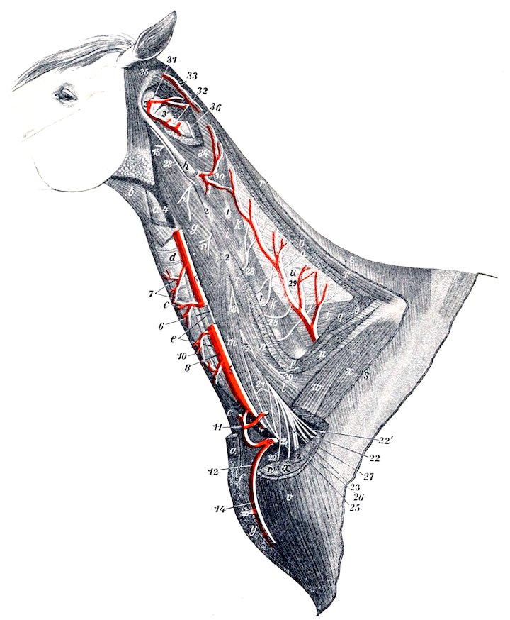
Fig. 433.—Deep Dissection of Neck of Horse. a, a, Ends of sterno-cephalicus; b, anterior part of omo-hyoideus; c, sterno-thyro-hyoideus; d, trachea; e, œsophagus; f, cariniform cartilage; g, rectus cap. ant. major; h, stump of trachelo-mastoideus; i, intertransversales; k, multifidus; l, m, scalenus; n, serratus cervicis; o, o, stumps of splenius, p, longissimus, q, complexus (most of which is removed); r, rhomboideus; s, trapezius; t, spinalis et semispinalis; u, lamellar part of lig. nuchæ; v, mastoido-humeralis; w, anterior deep pectoral; x, supraspinatus; y, anterior superficial pectoral; z, scapular tuberosity; 1, 1, articular processes of cervical vertebræ; 2, 2, transverse processes of same; 3, atlas; 3′, axis; 4, 4, jugular vein (remainder removed); 5, common carotid artery, from which a piece is removed to show the accompanying nerves; 6, vago-sympathetic trunk; 7, 7, tracheal and muscular branches of carotid artery; 8, recurrent nerve; 10, left tracheal lymph duct; 11, 12, ascending and descending branches of inferior cervical artery (13); 14, cephalic vein; 15–20, ventral branches of second to seventh cervical nerves; 21, roots of phrenic nerve; 22, thoracic or pectoral nerves; 22′, nerve to serratus magnus; 23, musculo-cutaneous nerve; 24, median nerve; 25, ulnar nerve; 26, radial nerve; 27, axillary nerve; 28, dorsal branches of cervical nerves; 28′, accessory nerve (cut); 29, superior or deep cervical artery; 30, muscular branch of vertebral artery; 31, posterior (anastomotic) branch of occipital artery; 32, vertebral artery; 33, muscular branches of occipital artery; 34, obliquus capitis post.; 35, obi. cap. ant.; 36, twig from dorsal branch of third cervical nerve. (After Ellenberger-Baum, Top. Anat. des Pferdes.)
The occipital artery (A. occipitalis) is the second in size of the terminals of the carotid. It arises usually just in front of the internal carotid, but in some cases with that artery by a common trunk of variable length. It pursues a somewhat flexuous course to the fossa atlantis, where it divides into anterior and posterior branches. It is related superficially to the submaxillary gland and the mastoido-humeralis, and deeply to the guttural pouch and the rectus capitis anterior 545major.[158] The internal carotid artery, the inferior cerebral vein, and the accessory, vagus, and sympathetic nerves cross its deep face. It gives twigs to the submaxillary gland, the anterior straight muscles, the guttural pouch and the adjacent lymph glands, and two named collateral branches. The condyloid or prevertebral artery (A. condyloidea) is a small vessel which passes upward and forward on the guttural pouch, and divides into muscular and meningeal branches. The latter enter the cranium through the foramen lacerum and hypoglossal foramen and are distributed to the dura mater. This artery is very variable in its origin. The posterior meningeal or mastoid artery (A. meningea caudalis) is a much larger vessel which runs upward and forward between the small oblique muscle and the paramastoid process, passes through the mastoid foramen into the parieto-temporal canal, enters the cranial cavity, and is distributed to the dura mater.
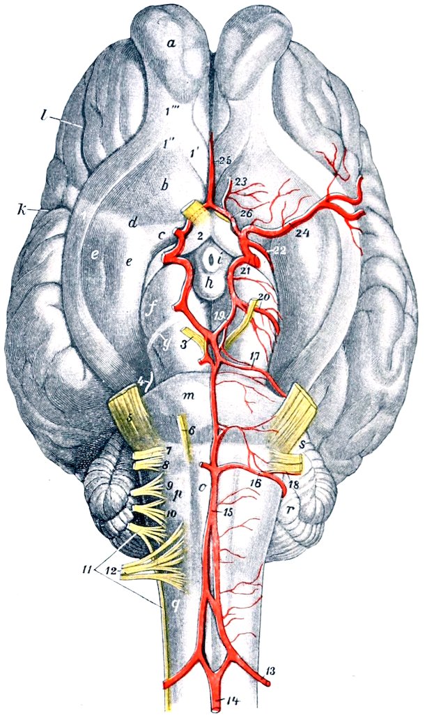
Fig. 434.—Vessels and Nerves of Base of Brain of Horse. 13, Cerebro-spinal artery; 14, middle spinal artery; 15, basilar artery; 16, posterior cerebellar artery; 17, anterior cerebellar artery; 18, internal auditory artery; 19, posterior cerebral artery; 20, deep cerebral artery; 21, stump of internal carotid artery; 22, anterior choroid artery; 23, anterior meningeal artery; 24, middle cerebral artery; 25, artery of corpus callosum; 26, anterior communicating artery; 1, 1′, 1″, olfactory tracts; 1‴, olfactory peduncle; 2–12, cranial nerves; a, olfactory bulb; b, trigonum olfactorium; c, lamina perforata anterior; d, fossa transversa; e, pyriform lobe; f, cerebral peduncle; g, tractus transversus; h, corpus mammillare; i, tuber cinereum; k, lateral fissure (of Sylvius); l, presylvian fissure; m, pons; o, pyramid; p, facial eminence; q, corpus restiforme; r, cerebellum; s, middle peduncle of cerebellum. (After Ellenberger Baum, Top. Anat. d. Pferdes.)
The posterior or retrograde branch (Ramus descendens) of the occipital passes up through the foramen transversarium of the atlas and joins the vertebral artery. It gives branches to the great oblique muscle of the head, which covers it.
The anterior or occipital branch (Ramus occipitalis) passes through the alar (antero-external) foramen of the atlas and supplies the muscles and skin of the poll, anastomosing with the deep cervical artery and its fellow of the opposite side. In the alar furrow it gives off the cerebrospinal artery, which enters the spinal canal through the intervertebral (antero-internal) foramen of the atlas, perforates the dura mater, and divides into cerebral and spinal branches. The cerebral branch unites with that of the opposite side to form the basilar 546artery, and the spinal branch similarly forms by union with its fellow the middle spinal artery.
The basilar artery (A. basilaris cerebri) passes forward in the median groove on the ventral surface of the medulla and pons and divides into the two posterior cerebral arteries. The collateral branches of the basilar are:
1. Medullary branches (Rami medullares), ten or twelve in number, distributed to the medulla oblongata.
2. Posterior cerebellar arteries (Aa. cerebelli caudales) which pass outward around the medulla behind the pons to the cerebellum, to which they are distributed after giving twigs to the medulla and pons.
3. The small auditory artery (A. auditiva interna) accompanies the eighth nerve to the internal ear. It often arises from the posterior cerebellar.
4. Anterior cerebellar arteries (Aa. cerebelli nasales). These are very variable in number and origin. There are often two or three on either side and they frequently arise from the posterior cerebrals. They pass outward in front of the pons and supply the anterior part of the cerebellum.
The posterior cerebral arteries (Aa. communicantes caudales) diverge at an acute angle and join the posterior communicating branches of the internal carotid arteries on the inferior surface of the cerebral peduncles. They are connected by a transverse branch and by a network of fine twigs which form often a rete mirabile.
THE INTERNAL CAROTID ARTERY (Figs. 434, 436, 437)
This artery (A. carotis interna) is somewhat smaller than the occipital. It usually arises just behind that artery, crosses its deep face, and runs upward and forward on the guttural pouch to the foramen lacerum. It is closely related to the vagus nerve and the superior cervical ganglion of the sympathetic nerve, fibers from which accompany it. It is crossed externally by the ninth and twelfth cranial nerves and the pharyngeal branch of the vagus. It passes through the inferior petrosal sinus and enters the cavernous sinus, within which it forms an S-shaped curve. It is connected with the opposite artery by a transverse branch (A. intercarotica), which lies behind the pituitary body in the intercavernous sinus. A branch (A. caroticobasilaris) sometimes connects it with the basilar artery. It then perforates the dura mater, gives off the posterior communicating branch, and passes forward and divides at the side of the optic chiasma into anterior and middle cerebral arteries.
The posterior communicating artery (A. communicans posterior) turns backward and joins the posterior cerebral. It gives off the deep cerebral artery (A. cerebri profunda) which winds around the cerebral peduncle and is distributed chiefly to the mid-brain. A smaller collateral branch is the anterior choroid artery (A. chorioidea nasalis) which passes along the optic tract and is distributed in the choroid plexus of the lateral ventricle.
The anterior cerebral artery (A. cerebri anterior) unites with the corresponding branch of the opposite artery above the optic chiasma. From this junction proceeds the artery of the corpus callosum (A. corporis callosi), which turns around the genu of the corpus callosum, enters the great longitudinal fissure, divides into two branches, and is distributed to the inner aspect of the cerebral hemispheres. A small anterior meningeal branch (A. meningea nasalis) of the anterior cerebral is distributed to the anterior part of the dura, and assists in forming a network in the ethmoidal fossa (Rete ethmoidale), anastomosing with the ethmoidal branch of the ophthalmic artery.
The middle cerebral artery (A. cerebri media) passes outward in the lateral fissure (of Sylvius) and divides into branches on the outer surface of the hemisphere.
The arterial circle of Willis (Circulus arteriosus) (Fig. 434) is formed at the 547interpeduncular space of the base of the brain by the union of the anterior cerebral arteries in front, by the diverging posterior cerebral arteries behind, and is completed laterally by the junction of the latter with the posterior communicating arteries and by the internal carotid. It is irregularly polygonal in outline.
The cerebral arteries are very variable in arrangement, and the foregoing account is a brief statement of the more usual disposition of the larger vessels. The internal carotid artery often arises with the occipital by a common trunk of variable length.
THE EXTERNAL CAROTID ARTERY (Figs. 436, 437)
This artery (A. carotis externa) by its size and direction constitutes the continuation of the common carotid. It passes forward on the lateral wall of the pharynx at the lower border of the guttural pouch, covered by the submaxillary gland and the stylo-maxillaris, digastricus, and stylo-hyoideus muscles. It then emerges between the stylo-hyoideus and the great cornu of the hyoid bone, passes upward on the latter parallel with the posterior border of the lower jaw, and terminates about two inches (ca. 5 cm.) below the temporo-maxillary articulation by dividing into superficial temporal and internal maxillary branches. It is crossed deeply near its origin by the superior laryngeal and pharyngeal branches of the vagus nerve. Just before its emergence its superficial face is crossed by the hypoglossal nerve, and the glosso-pharyngeal nerve passes over its inner surface at the ventral border of the great cornu. The chief collateral branches are the masseteric, external maxillary, and posterior auricular. It also furnishes variable branches to the submaxillary and parotid glands, the guttural pouch, and the pharyngeal lymph glands, as well as twigs to some adjacent muscles.
1. The inferior masseteric or maxillo-muscular artery (A. masseterica inferior) is given off from the external carotid at its emergence from beneath the stylo-hyoideus. It passes downward and slightly forward under cover of the parotid gland and over the tendon of insertion of the sterno-cephalicus to the posterior border of the mandible and appears on the masseter muscle, into which it plunges after a short course on its surface. It gives branches also to the internal pterygoid and stylo-maxillaris muscles and the parotid gland.
2. The external maxillary, facial, or submaxillary artery (A. maxillaris externa)[159] arises from the external carotid on the inner surface of the posterior belly of the digastricus (Fig. 436). It runs downward and forward on the lateral wall of the pharynx across the deep face of the stylo-hyoideus toward the great cornu of the hyoid bone, accompanied by the glosso-pharyngeal nerve in front and the hypoglossal nerve behind. After giving off the lingual artery at the posterior border of the great cornu, it inclines more ventrally on the inner surface of the internal pterygoid muscle, crosses over the hyo-glossus muscle, the hypoglossal nerve, the submaxillary duct, and the intermediate tendon of the digastricus, and turns forward in the submaxillary space. Here it lies on the lower part of the internal pterygoid muscle, and is related internally to the submaxillary lymph glands, above to the anterior belly of the digastricus, and below to the homonymous vein. At the anterior border of the masseter it turns around the lower border of the jaw and runs upward on the face in front of that muscle.[160] At the turn the artery is in front, the vein in the middle, and the parotid duct posterior. The artery and vein pass upward along the anterior border of the masseter, under cover of the facial panniculus and the zygomaticus, and are crossed superficially by branches 548of the facial nerve and deeply by the parotid duct. The artery terminates over the levator labii superioris proprius by dividing into the dorsal nasal and the angular artery of the eye. The chief branches of the external maxillary are as follows:
(1) The pharyngeal artery (A. palatina ascendens) arises usually behind the stylo-pharyngeus, passes between that muscle and the great cornu, and runs forward on the lateral wall of the pharynx under the elastic pharyngeal fascia. It is distributed to the soft palate, pharynx, and tonsil.
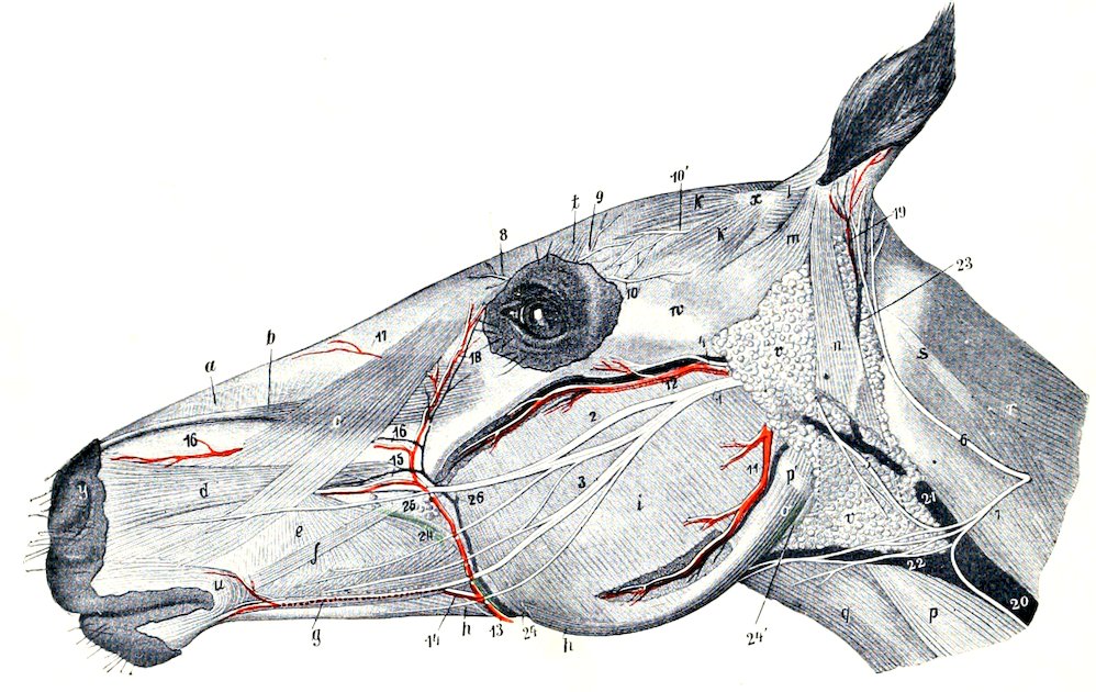
Fig. 435.—Superficial Dissection of Head of Horse. Most of the Panniculus is Removed. a, Dilatator naris superior; b, levator labii superioris proprius; c, levator nasolabialis; d, dilatator naris lateralis; e, buccinator; f, zygomaticus; g, depressor labii inferioris; h, stump of retractor anguli oris; i, masseter; k, k, scutularis; l, scutulo-auriculares superficiales; m, zygomatico-auricularis; n, parotido-auricularis; o, stylo-maxillaris; p, sterno-cephalicus; p′, tendon of p; q, omo-hyoideus; r, splenius; s, tendon of splenius and trachelo-mastoideus; t, corrugator supercilii; u, orbicularis oris; v, parotid gland; w, zygomatic arch; x, scutiform cartilage; y, upper commissure of nostril; 1, facial nerve; 2, superior buccal nerve; 3, inferior buccal nerve; 4, transverse facial nerve; 5, cervical branch of facial nerve; 6, posterior auricular branch of second cervical nerve; 7, cutaneous cervical branch of same; 8, infratrochlear nerve; 9, frontal nerve; 10, lacrimal nerve; 10′, end of auriculo-palpebral nerve; 11, inferior masseteric artery and vein; 12, transverse facial artery and vein; 13, facial artery; 14, inferior labial artery; 15, superior labial artery; 16, lateral nasal artery; 17, dorsal nasal artery; 18, angular artery of the eye; 19, posterior auricular artery; 20, 21, jugular vein; 22, external maxillary vein; 23, great auricular vein; 24, parotid duct; 24′, origin of same. (After Ellenberger-Baum, Top. Anat. d. Pferdes.)
(2) The lingual artery (A. lingualis) is a large branch which diverges from the parent trunk at an acute angle, runs along the ventral border of the great cornu of the hyoid bone, and dips under the hyo-glossus muscle. It then passes across the kerato-hyoideus, turns inward under the intercornual joint of the hyoid bone, and runs forward in the tongue between the hyo-glossus and genio-glossus. This part (A. profunda linguæ) is flexuous and is accompanied by branches of the hypoglossal and lingual nerves. It is the chief artery of the tongue, and anastomoses with the opposite artery and the sublingual.
(3) The sublingual artery (A. sublingualis) is a smaller vessel which arises at the anterior extremity of the submaxillary gland (Fig. 436). It passes forward on the anterior belly of the digastricus between the ramus of the mandible and the mylo-hyoideus, perforates the latter, runs along the lower border of the sublingual gland, and ramifies in the mucous membrane of the anterior part of the floor of the mouth. 549It detaches branches to the muscles and skin in the submaxillary space, the submaxillary lymph glands, and the sublingual gland. It also gives off the small submental artery, which runs forward superficially toward the lower lip, supplying twigs to the skin and the mylo-hyoideus.
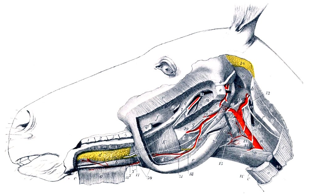
Fig. 436.—Parotid, Masseteric, and Lingual Regions of Horse: Deep Dissection, Third Layer. a, Mylo-hyoideus, anterior part, reflected; b, genio-hyoideus; c, genio-glossus; d, sublingual gland; e, ramus of mandible, the greater part of which is removed; e′, stump of masseter; f, maxillary tuberosity; g, great cornu of hyoid bone; h, wing of atlas; i, intermediate tendon of digastricus; i′, anterior belly, i″, posterior belly of digastricus; k, posterior part of mylo-hyoideus; l, hyo-glossus; m, pterygoideus internus (cut); n, stylo-hyoideus; o, stylo-maxillaris (cut); p, crico-pharyngeus; q, obliquus capitis ant.; r, tendon of trachelo-mastoideus; s, rectus cap. ant. major; t, mastoido-humeralis (cut); u, sterno-cephalicus (cut); v, sterno-thyroideus (cut); w, sterno-hyoideus (cut); x, omo-hyoideus; y, obliquus cap. post.; z, splenius (cut); 1–4, upper cheek teeth; 4′, last cheek tooth; 5, stump of facial nerve; 6, stump of buccinator nerve; 7, lingual nerve; 7′, superficial branch, 7″, deep branch of lingual nerve; 8, stumps of inferior alveolar artery, vein and nerve; 9, mylo-hyoid nerve (cut); 10, glosso-pharyngeal nerve; 11, hypoglossal nerve; 12, superior laryngeal nerve; 13, ventral branch of first cervical nerve; 14, vagus and sympathetic; 15, dorsal branch of spinal accessory nerve; 16, ventral branch of same; 17, inferior cerebral vein; 18, submaxillary duct; 19, common carotid artery; 20, parotid branch; 21, thyro-laryngeal artery; 22, pharyngeal artery; 23, laryngeal artery; 24, internal carotid artery; 25, occipital artery; 26, external carotid artery; 27–31, external maxillary artery; 28, posterior palatine artery; 29, lingual artery; 30, sublingual artery; 32, internal maxillary artery; 33, internal maxillary vein (origin); 34, remnant of parotid gland; 35, thyroid gland; 36, jugular vein; 37, pharyngeal lymph glands. (After Ellenberger-Baum, Top. Anat. d. Pferdes.)
In some cases the sublingual artery arises from the lingual and the submental from the external maxillary. Sometimes the sublingual remains on the external face of the mylo-hyoideus—thus resembling the submental of man—and the sublingual gland is supplied by a special branch of the lingual.
Not uncommonly a considerable branch, given off in the submaxillary space, turns round the lower border of the jaw and enters the middle of the lower part of the masseter muscle. In some cases this artery is of large size and its pulsation can be felt. It is accompanied by a vein.
(4) The inferior labial artery (A. labialis inferior) arises from the facial a little before it reaches the depressor labii inferioris (Fig. 435). It passes forward, dips under the depressor muscle, and continues to the lower lip. It supplies branches to the muscles and skin in this region, to the inferior buccal glands, the mucous membrane of the cheek, and the lower lip, anastomosing with the mental artery and the corresponding vessels of the opposite side. It detaches a branch (A. anguli oris) to the angle of the mouth, which anastomoses with the superior labial.
550(5) The superior labial artery (A. labialis superior) arises from the facial in front of the facial crest (Fig. 435). It passes forward under the dilatator naris lateralis and levator nasolabialis to the upper lip, gives twigs to the upper part of the cheek and the lateral nasal region, and ramifies in the upper lip, anastomosing with the opposite artery and the palato-labial.
(6) The lateral nasal artery (A. lateralis nasi) arises usually a little above the preceding one, and runs forward parallel with it and under the levator nasolabialis to the nostril (Fig. 435). It supplies branches to the lateral nasal region and the nostril.
The vessel is often double. It may arise from the superior labial at the point of bifurcation of the facial or with the dorsal nasal from the infraorbital artery (as in the ox). In some cases it gives off a dorsal nasal branch.
(7) The dorsal nasal artery (A. dorsalis nasi) arises on the levator labii superioris proprius and passes forward under the levator nasolabialis to the dorsum nasi (Fig. 435).
(8) The angular artery of the eye (A. angularis oculi) runs toward the internal canthus of the eye, where it anastomoses with the orbital branch (Ramus malaris) of the infraorbital artery (Fig. 435).
In addition to the preceding, unnamed branches are supplied to the submaxillary salivary gland and the submaxillary lymph glands.
3. The posterior auricular artery (A. auricularis posterior) arises at an acute angle from the external carotid just above the origin of the masseteric. It passes upward under cover of the parotid gland, to which it gives branches, and divides into several branches which supply the skin and muscles of the external ear (Fig. 435). The posterior branch passes to the posterior part of the base of the ear, where it divides into two branches; of these, one (Ramus intermedius) passes up the convex surface of the external ear to the apex, while the other (Ramus medialis) winds around to the anterior (inner) border, and forms an arch with the intermediate branch. The external branch (Ramus lateralis) passes up the posterior (external) border of the ear and forms an arch with the intermediate branch. The deep branch (A. auricularis profunda) enters the interval between the external auditory canal and mastoid process, and passes through an opening into the interior of the external ear and ramifies in the skin which lines it. It gives off the stylo-mastoid artery, which passes through the stylo-mastoid foramen into the tympanum, forms an arch around the membrana tympani, and supplies the middle ear and its muscles.
THE SUPERFICIAL TEMPORAL ARTERY
This artery (A. temporalis superficialis) is much the smaller of the two terminal branches of the external carotid. It passes upward behind the posterior border of the ramus of the mandible under cover of the parotid gland, and divides below the level of the condyle into the anterior auricular and transverse facial arteries. It is crossed superficially by the facial nerve.
The anterior auricular artery (A. auricularis anterior) passes upward behind the temporo-mandibular articulation under cover of the parotid gland, crosses the zygomatic arch, and reaches the temporalis muscle. It is crossed deeply at its origin by the superficial temporal nerve and is accompanied by a satellite vein and the auriculo-palpebral branch of the facial nerve. It is distributed to the skin and the temporal and anterior auricular muscles, and sends a branch through the conchal cartilage to the skin which lines it. Collateral twigs are detached to the parotid gland, and an anterior branch anastomoses with the supraorbital artery. A branch sometimes passes into the parieto-temporal canal and anastomoses with the posterior meningeal (mastoid) artery.
The transverse facial artery (A. transversa faciei) is larger than the preceding. 551It turns around the neck below the condyle of the mandible and emerges from beneath the parotid gland (Fig. 435). It then passes forward a short distance on the masseter about half an inch below the zygomatic arch and plunges into the muscle, in which it commonly divides into two chief branches. It is accompanied by a vein and a branch of the superficial temporal nerve. It supplies the masseter and the skin of this region, and anastomoses with the external maxillary and posterior deep temporal arteries.
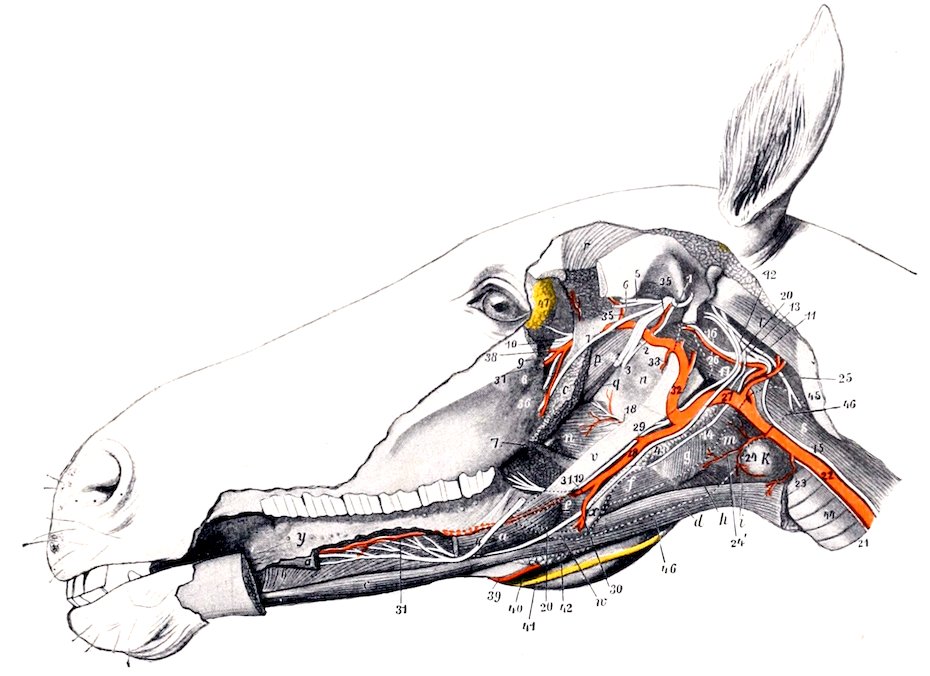
Fig. 437.—Deep Dissection of Head of Horse. The left ramus of the mandible and structures connected with it have been removed. a, a, Stumps of stylo-glossus; b, genio-glossus; c, genio-hyoideus; d, omo-hyoideus; e, kerato-hyoideus; f, thyro-hyoideus; g, thyro-pharyngeus; h, crico-thyroideus; i, sterno-thyroideus; k, thyroid gland; m, crico-pharyngeus; n, palatinus and palato-pharyngeus; o, pterygoideus externus; p, tensor palati; q, levator palati; r, temporalis; s, rectus cap. ant. major; t, obliquus cap. ant.; u, guttural pouch; v, great cornu of hyoid bone, posterior extremity of which is removed and indicated by dotted line; w, position of small cornu, dotted line; x, thyroid cornu; y, tongue; z, anterior pillar of soft palate; 1, superficial temporal nerve; 2, chorda tympani; 3, stump of inferior alveolar nerve; 4, 4, lingual nerve, intermediate part removed; 5, deep temporal nerve; 6, masseteric nerve; 7, buccinator nerve; 8, great palatine nerve; 9, infraorbital nerve; 10, sphenopalatine and posterior nasal nerves; 11, spinal accessory nerve; 12, vagus; 13, pharyngeal branch of vagus; 14, superior laryngeal nerve; 15, vago-sympathetic trunk; 16, sympathetic, with superior cervical ganglion a little further back; 17, glosso-pharyngeal nerve; 18, pharyngeal and 19, lingual branches of glosso-pharyngeal; 20, hypoglossal nerve; 21, left recurrent nerve; 22, common carotid artery; 23, parotid branch; 24, thyro-laryngeal artery; 24′, laryngeal artery; 25, occipital artery; 26, internal carotid artery; 27, external carotid artery; 28, 30, external maxillary artery; 29, pharyngeal artery; 31, lingual artery; 32, internal maxillary artery; 33, stump of inferior alveolar artery; 34, middle meningeal artery; 35, deep temporal artery; 36, buccinator artery; 37, palatine artery; 38, end of internal maxillary artery; 39, right external maxillary artery; 40, satellite vein of 39; 41, right parotid duct; 42, submaxillary lymph glands; 43, pharyngeal lymph glands; 44, trachea; 45, wing of atlas; 46, dotted line indicating outline of submaxillary gland; 47, lacrimal gland. (After Ellenberger-Baum, Top. Anat. d. Pferdes.)
The pulse may usually be felt in this artery as it passes below the articulation of the jaw.
THE INTERNAL MAXILLARY ARTERY (Figs. 437, 438, 439)
This artery (A. maxillaris interna) is much the larger of the two terminal branches of the external carotid. It begins at the inner side of the posterior border 552of the mandible about two inches (ca. 5 cm.) below the articulation of the jaw and ends in the anterior part of the pterygo-palatine fossa. On account of its complex course and the large number of branches given off it is convenient to divide it into three parts.
The first part is much the longest, forms an S-shaped curve, and is in great part in contact with the guttural pouch. It passes upward and forward on the inner surface of the mandible a distance of about an inch (ca. 2 to 3 cm.) and is related here to the vein, which is ventral. It then turns inward below the external pterygoid muscle and the inferior maxillary nerve, passes between that muscle and the tensor palati, and runs forward to enter the alar (pterygoid) foramen. This part gives off the following branches:
1. The inferior alveolar or dental artery (A. alveolaris mandibulæ s. inferior) passes downward and forward with the homonymous vein and nerve, being at first between the external and internal pterygoid muscles, then between the latter and the ramus of the mandible. It enters the mandibular foramen, passes downward and forward in the mandibular canal, and terminates at the mental foramen by dividing into mental and incisor branches. The mental branch (A. mentalis) emerges through the mental foramen and anastomoses in the lower lip with the opposite artery and the inferior labial. The incisor branch continues forward in the bone, supplies twigs to the canine and incisor teeth, and anastomoses with its fellow of the opposite side. Collateral branches are detached to the pterygoid and mylo-hyoid muscles, and within the bone to the teeth, alveolar periosteum, the gums, and the spongy substance of the mandible.
2. The pterygoid arteries, two or three in number, are distributed to the pterygoid and tensor and levator palati muscles.
3. The tympanic artery is a very small vessel which passes along the Eustachian tube to the petro-tympanic fissure (of Glaser) and enters the middle ear.
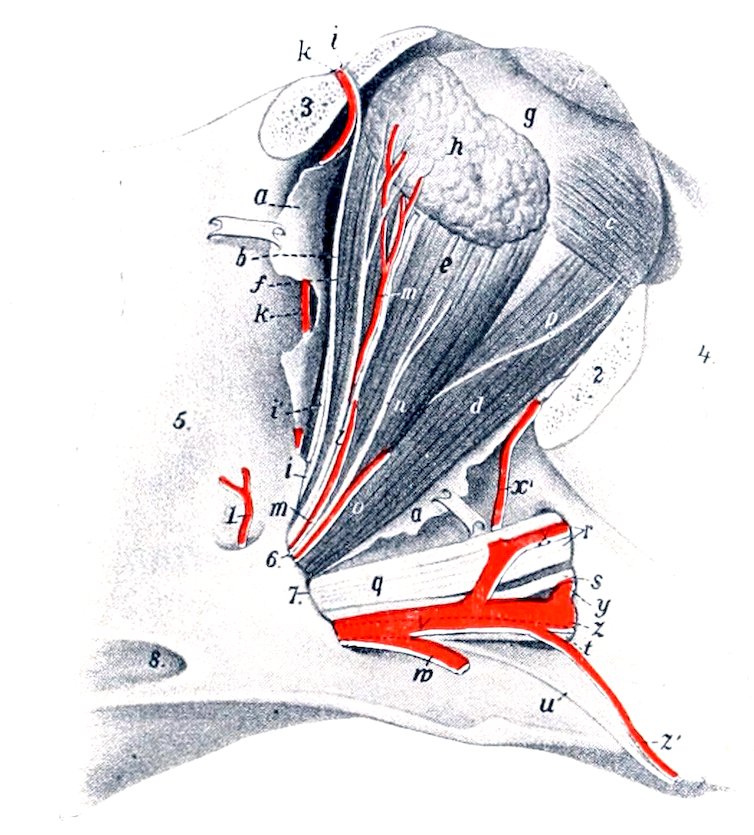
Fig. 438.—Right Eye of Horse. a, Remnants of periorbita; b, levator palpebræ superioris; c, obliquus oculi inferior; d, rectus oculi inferior; e, rectus oculi externus; f, rectus oculi superior; g, sclera; g′, cornea; h, lacrimal gland; i, frontal nerve; k, frontal artery; l, branch of lacrimal nerve to gland; m, lacrimal artery; n, zygomatic nerve; o, branch of ophthalmic artery; p, branch of oculomotor nerve to obliquus oculi inferior; q, maxillary nerve; r, infraorbital nerve; s, posterior nasal nerve; t, great palatine nerve; u, small palatine nerve; v, internal maxillary artery; w, buccinator artery (cut); x, infraorbital artery; x′, malar artery; y, sphenopalatine artery; z, great palatine artery; z′, small palatine (or staphyline) artery; 1, posterior deep temporal artery; 2, 3, stumps of orbital margin; 4, facial crest; 5, temporal fossa; 6, foramen lacerum orbitale; 7, anterior end of alar canal; 8, posterior opening of same. (After Ellenberger, in Leisering’s Atlas.)
4. The middle meningeal artery (A. meningea media) arises beneath the buccinator nerve where the internal maxillary turns forward. It passes backward across the temporal wing of the sphenoid to the antero-external part (foramen spinosum) of the foramen lacerum. Entering the cranium, it divides into branches which course in the grooves on the temporal and parietal bones and supply the dura mater. It anastomoses with the posterior meningeal.
The size of this artery is variable, and is in inverse ratio to that of the posterior meningeal.
5. The posterior deep temporal artery (A. temporalis profunda aboralis) 553arises from the internal maxillary just before the latter enters the canal of the pterygoid process. It passes upward and backward in the temporal fossa on the deep face of the temporalis muscle, in which it ramifies. It sends a branch outward to the masseter, and anastomoses with the superficial temporal and middle meningeal arteries.
In some cases the tympanic and middle meningeal arise from this artery.
The second part lies in the alar canal and is about an inch (ca. 2 to 3 cm.) in length. It gives off two branches—the anterior deep temporal and the ophthalmic.
1. The anterior deep temporal artery (A. temporalis profunda oralis) emerges from the canal through the temporal or small alar foramen, and passes upward in the anterior part of the temporal fossa on the deep face of the temporalis muscle, in which it is chiefly distributed. It gives twigs to the orbital fat and the skin of the frontal region.
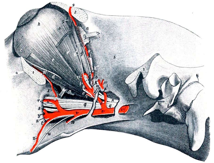
Fig. 439.—Left Eye of Horse, Deeper Dissection. The outer plate of bone has been removed behind the pterygoid crest to expose the vessels and nerves. a, a, Remnants of periorbita; b, stump of rectus oculi superior; c, obliquus oculi inferior; d, rectus oculi inferior; e, rectus oculi lateralis; e′, retractor oculi; f, rectus oculi internus; g, g, obliquus oculi superior; h, eyeball; i, trochlear nerve; k, ophthalmic nerve; k′, nasal nerve; k″, infratrochlear nerve; k‴, ethmoidal nerve; l, optic nerve; m, frontal nerve; n, lacrimal nerve; o, zygomatic nerve; p, nerve to obliquus inferior (from oculomotor); q, maxillary nerve; r, infraorbital nerve; s, sphenopalatine nerve; t, great palatine nerve; u, small palatine nerve; v, internal maxillary artery; w, buccinator artery (cut off); x, infraorbital artery; x′, malar artery; y, sphenopalatine artery; z, great palatine artery; z′, small palatine (or staphyline) artery; 1, 2, stumps of zygomatic arch; 3, stump of supraorbital process; 4, facial crest; 5, temporal fossa; 6, ophthalmic artery; 7, muscular branch of 6; 8, lacrimal artery (cut); 9, frontal artery; 10, anterior deep temporal artery; 11, ethmoidal artery. (After Ellenberger-Baum, Top. Anat. d. Pferdes.)
2. The ophthalmic artery (A. ophthalmica externa) emerges from the anterior opening of the alar canal, and enters the apex of the periorbita or ocular sheath. Within this it forms a semicircular bend under the rectus oculi superior and is continued by the ethmoidal artery. Its branches are as follows:
(a) The supraorbital or frontal artery (A. supraorbitalis)[161] is a small vessel which often arises from the anterior deep temporal or the internal maxillary. It passes along the inner wall of the orbit in company with the nerve of the same name to the supraorbital foramen, through which it emerges. It is distributed to the orbicularis oculi, the corrugator supercilii, and the skin of the supraorbital region.
(b) The lacrimal artery (A. lacrimalis) runs upward and forward within the 554periorbita along the outer edge of the levator palpebræ superioris to the lacrimal gland, in which it is chiefly distributed. It also sends twigs to the upper eyelid.
(c) Muscular branches (Rami musculares) supply the orbital muscles, the periorbita, the third eyelid, and the conjunctiva.
(d) Ciliary branches (Aa. ciliares), two sets of very slender arteries, arise from the ophthalmic direct and from the muscular branches. The anterior ciliary arteries (Aa. ciliares anteriores) pierce the sclera in front of the equator and ramify chiefly in the ciliary body and the iris. The posterior ciliary arteries (Aa. ciliares posteriores) pierce the posterior part of the sclera; most of them ramify in the choroid coat as the short ciliary arteries, but two of larger size, the long ciliary arteries, run forward, one on each side, between the sclera and choroid to the periphery of the iris. Here they divide into branches which anastomose and form a circle (Circulus iridis major). From this secondary branches are detached which form a second circle around the pupil (Circulus iridis minor).
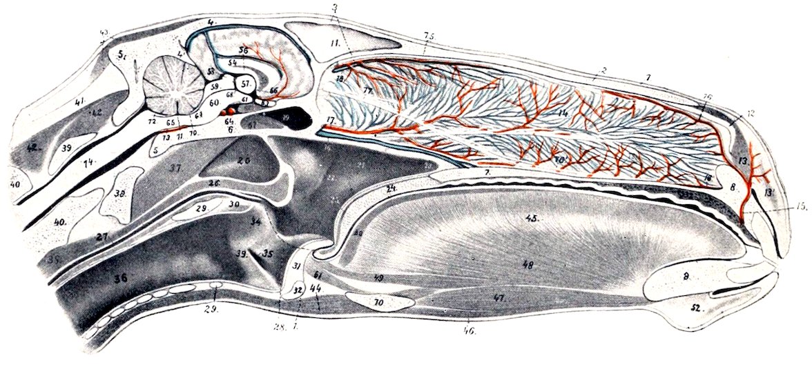
Fig. 440.—Sagittal Section of Head of Horse, Cut a Little to the Right of the Median Plane. 1, Skin; 2, nasal bone; 3, frontal bone; 4, parietal bone; 4′, tentorium osseum; 5, occipital bone; 6, sphenoid bone; 7, hard palate; 8, premaxilla; 9, mandible; 10, hyoid bone; 11, septum between frontal sinuses; 12, alar cartilage; 13, dilatator naris transversus; 14, septum nasi with venous plexuses; 15, palato-labial artery; 16, 16′, upper and lower septal branches of 15; 17, septal branch of sphenopalatine artery and satellite vein; 18, septal branch of ethmoidal artery; 19, sphenoidal sinus; 20, guttural pouch; 21, pharynx; 22, pharyngeal orifice of Eustachian tube; 23, posterior naris; 24, soft palate; 25, palatinus muscle; 26, pharyngeal muscles; 27, œsophagus; 28, dotted line indicating position of posterior pillar of soft palate; 29, lamina; 29′, arch of cricoid cartilage; 30, arytenoideus transversus muscle; 31, epiglottis; 32, body of thyroid cartilage; 33, vocal process and cord; 34, arytenoid cartilage; 35, lateral ventricle of larynx; 36, trachea; 37, ventral straight muscles of head; 38, longus colli; 39, atlas; 40, axis; 41, lig. nuchæ; 42, dorsal spinal muscles; 43, muscles of external ear; 44, omo- and sterno-hyoideus; 45, tongue; 46, mylo-hyoideus; 47, genio-hyoideus; 48, genio-glossus; 49, longitudinalis inferior; 50, longitud. superior; 51, hyo-epiglotticus; 52, chin and mentalis muscle; 53, venous plexus of hard palate; 54, corpus callosum; 55, septum pellucidum; 56, fornix; 57, thalamus; 58, pineal body; 59, corpora quadrigemina; 60, cerebral peduncle; 61, corpus mammillare; 62, pituitary body; 63, chiasma opticum; 64, intercarotid artery; 65, medulla oblongata; 66, interventricular foramen; 67, infundibulum; 68, third ventricle; 69, cerebral aqueduct; 70, anterior medullary velum; 71, fourth ventricle; 72, posterior medullary velum; 73, basilar artery; 74, spinal cord; 75, ethmoidal nerve; 76, septal branch of posterior nasal nerve; 76′, branch of same to organ of Jacobson; 77, olfactory nerve to organ of Jacobson; 78, nasal branches of palatine artery. (After Ellenberger, in Leisering’s Atlas.)
(e) The central artery of the retina (Arteria centralis retinæ) is a small vessel which arises from the ophthalmic or from a posterior ciliary artery. It pierces the optic nerve a short distance behind the sclera and runs in its center to the lamina cribrosa, where it breaks up in thirty to forty fine branches. These appear in the fundus of the eye at the margin of the optic papilla and radiate in the posterior part of the retina.
(f) The ethmoidal artery (A. ethmoidalis) is the continuation of the ophthalmic. 555It enters the cranial cavity through the ethmoidal foramen, passes inward on the cribriform plate, and divides into meningeal and nasal branches. The former ramify in the anterior part of the dura mater and anastomose with branches of the artery of the corpus callosum. The nasal branch passes through the cribriform plate, gives branches to the mucous membrane of the lateral mass of the ethmoid and the adjacent part of the septum nasi, and runs forward on the superior turbinal.
The third part passes forward in the pterygo-palatine fossa, accompanied by branches of the maxillary nerve. On reaching the posterior palatine foramen it is continued by the palatine artery. Its branches are as follows:
(1) The buccinator artery (A. buccinatoria) arises from the lower aspect of the internal maxillary shortly after its emergence (Fig. 437). It turns around the maxillary tuberosity, accompanied by the buccinator nerve, and under the masseter muscle, enters the cheek, and runs forward in it. It supplies branches to the cheek, the superior buccal glands, and the masseter and pterygoid muscles. Near its origin it gives off a branch to the orbital fat behind the periorbita.
(2) The infraorbital or superior dental artery (A. infraorbitalis) arises from the upper aspect of the internal maxillary a little in front of the preceding vessel. It passes upward and forward to the maxillary foramen, runs in the infraorbital canal in company with the nerve of the same name, and is continued forward within the jaw to the incisor teeth. It gives branches to the teeth and gums, and detaches a branch through the infraorbital foramen which anastomoses with the lateral nasal and superior labial. About midway between its origin and the maxillary foramen it gives off the malar or orbital branch (Ramus malaris), which passes along the floor of the orbit to end in the lower lid and anastomose with the angularis oculi. It gives twigs to the inferior oblique muscle and the lacrimal sac.
The infraorbital artery is usually small at its emergence upon the face, but in some cases it is rather large and may partially replace the superior labial and lateral nasal arteries.
(3) The staphyline or small palatine artery (A. palatina minor) is a small vessel which passes forward in the groove at the inner side of the maxillary tuberosity to the soft palate. In the groove it is accompanied by the nerve of the same name and the palatine vein.
(4) The sphenopalatine artery (A. sphenopalatina) arises in the extreme anterior part of the pterygo-palatine fossa and passes into the nasal cavity, where it divides into internal and external branches. The internal branch is distributed to the mucous membrane of the septum nasi; the external one goes to the inferior turbinal, the inferior meatus, the posterior nares, and the maxillary and frontal sinuses. It may arise from the infraorbital.
(5) The palatine or palato-labial artery (A. palatina major) is the direct continuation of the internal maxillary. It passes through the palatine canal to the roof of the mouth, accompanied by the palatine nerve, and runs forward in the palatine groove, where it is joined by the vein. A little behind the plane of the corner incisor teeth it curves inward over a bar of cartilage to the foramen incisivum, where it unites with its fellow of the opposite side. The single artery thus formed passes up through the foramen and divides under the transverse dilator of the nostril into two branches. These ramify in the upper lip and anastomose with the lateral nasal and superior labial arteries. In its course in the roof of the mouth the palatine artery gives off branches to the hard and soft palate and the gums, and others which pass through the accessory palatine foramina to be distributed in the mucous membrane of the lower part of the nasal cavity. Commonly two branches, right and left, are detached from the convexity of the arch formed by the union of the two arteries; these run forward in the anterior part of the hard palate.
556
Arteries of the Thoracic Limb
THE BRACHIAL ARTERY
The brachial artery, after turning around the lower border of the scalenus at the first rib, passes backward and a little downward across the origin of the coraco-brachialis and the insertion of the subscapularis muscle at the inner side of the shoulder joint. At the posterior border of the subscapularis it gives off the subscapular artery, and turns downward on the inner surface of the arm. In its course in the arm it inclines a little forward, crosses the humerus very obliquely, and divides at the antero-internal aspect of the distal third of the shaft of the bone into the anterior and posterior radial arteries.[162]
It is related internally to the serratus magnus and the posterior deep pectoral muscles, and the cubital lymph glands. Externally it lies on the subscapularis, the brachialis, the insertion of the teres major and latissimus dorsi, and the inner head of the triceps. The coraco-brachialis muscle lies along the front of the artery, which, however, overlaps the muscle distally. The vein crosses the inner face of the artery and runs down behind it, accompanied part way by the ulnar and radial nerves. The musculo-cutaneous and median nerves form a loop around the artery close to its origin, and the latter nerve then passes down in front of the artery. The chief branches are as follows:
1. The suprascapular artery (A. thoracico-acromialis) is a small and somewhat flexuous vessel, which arises near the anterior border of the subscapularis, runs upward, and dips in between that muscle and the supraspinatus. It gives branches to these muscles, the anterior deep pectoral, and the mastoido-humeralis. A branch passes in front of the coraco-brachialis to the shoulder joint and the proximal end of the humerus.
This vessel may be double, or be represented by one or more branches of the inferior cervical artery.
2. The subscapular artery (A. subscapularis) is a very large vessel which arises at the posterior border of the subscapularis muscle. It passes upward in the interstice between that muscle and the teres major on the inner surface of the long head of the triceps, turns around the posterior border of the scapula below the posterior angle, and ends in the infraspinatus and deltoid. Besides collaterals (Rami musculares) to the subscapularis, teres major, triceps, and tensor fasciæ antibrachii it gives off the following named branches:
(a) The thoracico-dorsal (A. thoracicodorsalis) is a long artery which is usually given off about an inch from the origin of the subscapular, crosses the inner face of the teres major, and runs upward and backward on the latissimus dorsi. It gives branches to these muscles, the abdominal panniculus and the axillary lymph glands.
(b) The posterior circumflex artery (A. circumflexa humeri posterior) arises a little above the preceding vessel and passes outward behind the shoulder joint between the long and external heads of the triceps with the axillary (or circumflex) nerve. It gives branches to these muscles, the joint capsule, and the muscles and skin of the outer side of the shoulder, anastomosing with the anterior circumflex artery.
(c) The circumflex artery of the scapula (A. circumflexa scapulæ) arises about two or three inches (ca. 5 to 7.5 cm.) above the shoulder joint, passes forward to the posterior border of the scapula, and divides into two branches. The outer one runs forward on the external surface of the scapula below the spine and gives branches to the supraspinatus, infraspinatus, and teres minor. The inner branch passes forward in a similar fashion on the costal surface of the scapula and supplies branches to the subscapularis.
557
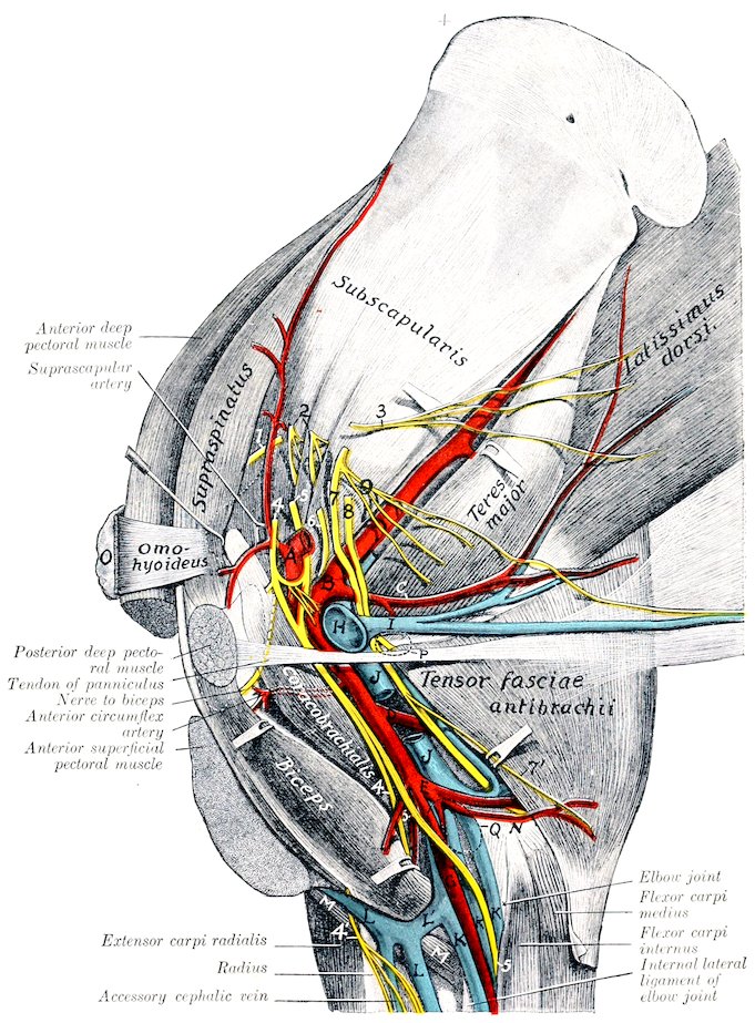
Fig. 441.—Dissection of Shoulder and Arm of Horse, Inner Surface. A, Brachial artery; B, subscapular artery; C, thoracico-dorsal artery; D, deep brachial artery; E, ulnar artery; F, anterior radial artery; G, posterior radial (or median) artery; H, brachial vein; I, external thoracic (or “spur”) vein; J, brachial vein; K, K, K, posterior radial veins; L, cephalic vein; L′, communicating vein; M, brachialis muscle; N, internal epicondyle of humerus; O, prescapular lymph glands; P, position of axillary lymph glands; Q, position of cubital lymph glands; 1, suprascapular nerve; 2, subscapular nerves; 3, thoraco-dorsal nerve; 4, musculo-cutaneous nerve; 4′, cutaneous branch of musculo-cutaneous nerve; 5, median nerve; 6, axillary nerve; 7, ulnar nerve; 8, radial nerve; 9, thoracic nerves. (After Schmaltz, Atlas d. Anat. d. Pferdes.)
558
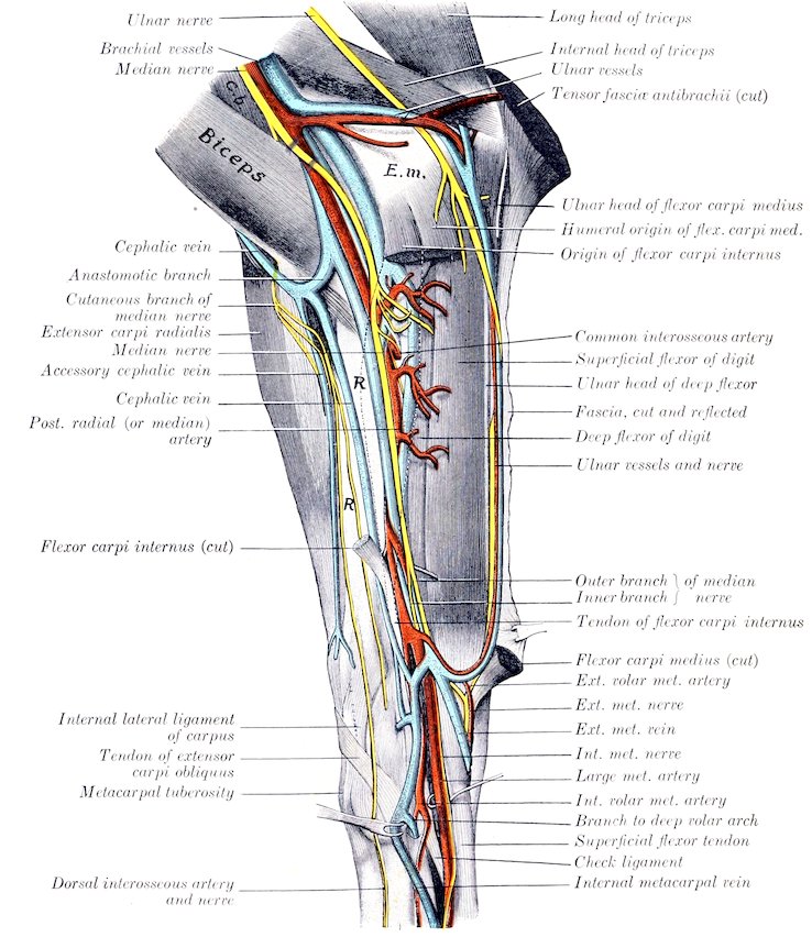
Fig. 442.—Dissection of Right Forearm and Carpus of Horse, Internal Surface. C.b., Coraco-brachialis muscle; E.m., internal epicondyle of humerus; R, subcutaneous surface of radius. (After Schmaltz, Atlas d. Anat. d. Pferdes.)
3. The anterior circumflex (or prehumeral) artery (A. circumflexa humeri anterior) arises usually at the upper border of the teres major. It passes forward and outward between the two parts of the coraco-brachialis or between the latter and the humerus. It gives branches to the coraco-brachialis and deep pectoral and ends in the upper part of the biceps and in the mastoido-humeralis. It anastomoses with the posterior circumflex.
559Not rarely it arises below the middle of the humerus and passes upward between the biceps and coraco-brachialis. In these cases a small branch for the coraco-brachialis is usually given off at the usual point of origin of the anterior circumflex.
4. The deep brachial artery (A. profunda brachii) is a large but short trunk which arises usually about the middle of the humerus. It passes backward and divides into several branches which supply the triceps, tensor fasciæ antibrachii, anconeus, and brachialis. A branch runs in the musculo-spiral groove with the radial nerve to the front of the elbow joint and anastomoses with the anterior radial. A slender branch passes down along the external border of the extensor carpi and supplies cutaneous twigs. Anastomoses occur with the ulnar and recurrent interosseous arteries.
The point of origin is inconstant and it is not uncommon to find two arteries instead. Often a large branch for the posterior deep pectoral muscle is detached close to the origin or arises from the brachial directly.
5. Muscular branches (Rami musculares) are distributed to the teres major, deep pectoral, coraco-brachialis, and biceps. The largest and least variable of these supplies the lower part of the biceps.
6. The ulnar artery (A. collateralis ulnaris superior) arises a little below the nutrient foramen of the humerus and passes downward and backward along the lower edge of the internal head of the triceps under cover of the brachial vein and the tensor fascia antibrachii. It gives branches to these muscles, the posterior superficial pectoral, the cubital lymph glands, panniculus, and skin. At the internal epicondyle of the humerus it is joined by the ulnar nerve and turns downward under the ulnar head of the flexor carpi medius. It continues with the vein and nerve under the deep fascia of the forearm between the ulnar and humeral heads of the perforans, and in the distal half of the region between the external and middle flexors of the carpus. It unites just above the carpus (under cover of the flexor carpi externus) with a branch of the radial artery, with which it forms the supracarpal arch. It detaches small collaterals to the muscles along which it passes and terminal twigs to the outer surface of the carpus.