THE MUSCULAR SYSTEM (continued 2)
Reading edition
The Prepubic Tendon.—The prepubic tendon is essentially the tendon of insertion of the two recti abdominis, but also furnishes attachment to the obliqui, the graciles, and the pectinei. It is attached to the anterior borders of the pubic bones, including the ilio-pectineal eminences. It has the form of a very strong thick band, with concave lateral borders which form the inner boundaries of the external inguinal rings. Its direction is oblique upward and backward.[54] Its structure is somewhat complex. Most of the fibers of the posterior part extend from one ilio-pectineal eminence to the other. The fibers which belong to the recti curve in to the median line. The aponeuroses of the internal oblique muscles are inserted into its abdominal surface, and the inguinal ligaments are attached to and continue across it in arciform fashion. The anterior part of the tendon of origin of the gracilis is fused with it ventrally, and many of the fibers of the pectineus arise from it. It gives off on either side a strong round band, the so-called pubo-femoral or accessory ligament, which is inserted into the fossa of the head of the femur with the round ligament (vide hip joint).
Muscles of the Thoracic Limb
I. THE MUSCLES OF THE SHOULDER GIRDLE (Figs. 177, 178, 179, 186)
This group consists of those muscles which connect the thoracic limb with the head, neck, and trunk. The group naturally falls into two divisions—dorsal and ventral.[55]
A. Dorsal Division
This division consists of two layers which overlie the proper muscles of the neck and back.
First Layer
1. Trapezius.—This is a flat, triangular muscle, the base of the triangle corresponding with the spine. It is divided by an aponeurotic portion into two divisions:
(a) Trapezius cervicalis.—Origin.—The funicular portion of the ligamentum nuchæ, from the second cervical to the third thoracic vertebra.
Insertion.—The spine of the scapula and the fascia of the shoulder and arm.
251(b) Trapezius thoracalis s. dorsalis.—Origin.—The supraspinous ligament, from the third to the tenth thoracic vertebra.
Insertion.—The tubercle of the spine of the scapula.
Action.—Acting as a whole, to elevate the shoulder; the cervical portion draws the scapula forward and upward and the thoracic portion draws it backward and upward.
Structure.—The muscle arises by a short, thin aponeurosis, from which the fibers of the flat fleshy portion converge to the spine of the scapula and the aponeurosis which separates the two portions. The cervical fascia joins the ventral edge of the cervical portion to the mastoido-humeralis, or the two muscles may unite here.
Relations.—Superficially, the skin and fascia; deeply, the rhomboideus, latissimus dorsi, supraspinatus, infraspinatus, deltoid, splenius, serratus magnus, and anterior deep pectoral muscles, and the cartilage of the scapula.
Blood-supply.—Deep cervical and intercostal arteries.
Nerve-supply.—Spinal accessory nerve.
Second Layer
This consists of two muscles—the rhomboideus and the latissimus dorsi.
2. Rhomboideus.—This consists of two portions:
(a) Rhomboideus cervicalis s. cervicis.—Origin.—The funicular portion of the ligamentum nuchæ, from the second cervical to the second thoracic vertebra.
Insertion.—The internal surface of the cartilage of the scapula.
(b) Rhomboideus thoracalis s. dorsalis.—Origin.—The spinous processes of the second to the seventh thoracic vertebra by means of the dorso-scapular ligament.
Insertion.—The inner surface of the cartilage of the scapula.
Action.—To draw the scapula upward and forward. When the limb is fixed the cervical portion will elevate the neck.
Structure.—The cervical portion is narrow, pointed at its anterior extremity, and lies along the funicular part of the ligamentum nuchæ, to which it is attached by short tendon bundles. The fibers are directed for the most part longitudinally. The thoracic portion is quadrilateral in shape, and its fibers are nearly vertical. Its deep face is intimately attached to the dorso-scapular ligament.
Relations.—Superficially, the skin and fascia (over a small area), the trapezius, and the cartilage of the scapula; deeply, the dorso-scapular ligament, the splenius, complexus, longissimus dorsi, and serratus anticus.
Blood-supply.—Dorsal and superior cervical arteries.
Nerve-supply.—Sixth cervical nerve.
3. Latissimus Dorsi.—This is a wide muscle which has the form of a right-angled triangle. It lies for the most part under the skin and panniculus, on the lateral wall of the thorax, from the spine to the arm.
Origin.—The lumbo-dorsal fascia—and by this means from the lumbar and thoracic spines as far forward as the highest point of the withers.
Insertion.—The internal tubercle of the humerus, in common with the teres major.
Action.—To draw the humerus upward and backward and flex the shoulder joint. If the limb is advanced and fixed, it draws the trunk forward.
Structure.—The muscle arises by a wide aponeurosis, which fuses with that of the serratus posticus and with the lumbo-dorsal fascia. The muscular portion is at first rather thin, but by the convergence of its fibers becomes thicker as it approaches the arm. The anterior fibers pass almost vertically downward over the dorsal angle of the scapula and its cartilage. The posterior fibers are directed 252downward and forward. The thick belly formed by the convergence of these passes under the triceps to end on the flat tendon of insertion, which is common to this muscle and the teres major.
Blood-supply.—Subscapular, intercostal, and lumbar arteries.
Nerve-supply.—Brachial plexus (eighth cervical and dorsal roots).
B. Ventral Division
1. Mastoido-humeralis (M. brachiocephalicus; levator humeri).—This muscle extends along the side of the neck from the head to the arm. It is incompletely divisible into two portions.
Origin.—(1) The mastoid process of the petrous temporal bone and the occipital crest; (2) the wing of the atlas and the transverse processes of the second, third, and fourth cervical vertebræ.
Insertion.—The deltoid tuberosity and the curved rough line which extends from this to the distal extremity of the humerus.
Action.—When the head and neck are fixed, to draw the limb forward, extending the shoulder joint. When the limb is fixed, to extend the head and neck, if the muscles act together; acting separately, to incline the head and neck to the same side.
Structure.—As already mentioned, the muscle is capable of incomplete division into two parts, the line of division being indicated by the emergence of superficial branches of the ventral divisions of the cervical nerves. The mastoid portion (M. cleido-mastoideus) partly overlaps the other portion (M. cleido-transversarius), which lies dorsal to it. The former is attached to the mastoid process and the occipital bone by a broad tendon which fuses with that of the splenius and trachelo-mastoideus; it is also attached to the tendon of insertion of the sterno-cephalicus by aponeurosis. The dorsal portion is attached to the transverse processes by four fleshy digitations. The belly of the muscle is adherent superficially to the cervical fascia and the panniculus, and deeply to the subscapulo-hyoideus. In front of the shoulder its deep face is marked by a tendinous intersection of variable development.[56] Here the muscle becomes wider, covers the shoulder joint, passes between the brachialis and biceps, and is inserted by means of a wide tendon which it shares with the superficial pectoral muscle.
Relations.—Superficially, the skin, cervical fascia, the parotid gland, the panniculus, brachialis, and branches of the cervical nerves; deeply, the splenius, trachelo-mastoideus, rectus capitis anterior major, omo-hyoideus, serratus magnus, anterior deep pectoral and biceps muscles, the inferior cervical artery, the prescapular lymph glands, and branches of the cervical nerves. The ventral edge of the muscle forms the dorsal boundary of the jugular furrow. The dorsal border may be in contact with the cervical trapezius, or separated from it by a variable interval.
Blood-supply.—Inferior cervical, carotid, and vertebral arteries.
Nerve-supply.—Spinal accessory and cervical nerves.
The pectoral fascia is a thin membrane covering the surface of the pectoral muscles, to which it is, for the most part, pretty intimately attached. It detaches a layer which passes between the superficial and deep pectorals. At the posterior edge of the triceps another layer is given off, which passes on the outer surface of this muscle to blend with the scapular fascia; the deeper layer becomes continuous with the subscapular and cervical fasciæ.
The pectoral muscles form a large fleshy mass which occupies the space 253between the ventral part of the chest-wall and the shoulder and arm. They are clearly divisible into a superficial and a deep layer. The superficial layer may be subdivided into two portions by careful dissection; the deep layer is clearly made up of two muscles.
2. Superficial pectoral (M. pectoralis superficialis).
(a) Anterior superficial pectoral (portio clavicularis s. descendens; pectoralis anticus).—This is a short, thick, somewhat rounded muscle, which extends between the anterior part of the sternum and the front of the arm. It forms a distinct prominence on the front of the breast, which is easily recognized in the living animal.
Origin.—The cariniform cartilage of the sternum.
Insertion.—(1) The curved line of the humerus with the mastoido-humeralis; (2) the fascia of the arm.
Action.—To adduct and advance the limb.
Structure.—The belly of the muscle is convex on its superficial face, but deeply it is flattened where it overlaps the posterior superficial pectoral. Here the two muscles are usually pretty intimately attached to each other, and care must be exercised in making the separation. The tendon of insertion blends with that of the mastoido-humeralis and with the fascia of the arm. At the middle line of the breast a furrow occurs between the two muscles; laterally, another furrow, containing the cephalic vein, lies between the muscle and the mastoido-humeralis.
Relations.—Superficially, the skin, fascia, and panniculus; deeply, the posterior division, the deep pectoral, and the biceps. The cephalic vein lies in the groove between this muscle and the mastoido-humeralis.
(b) Posterior superficial pectoral (portio sternocostalis; pectoralis transversus).—This is a wide muscular sheet which extends from the ventral edge of the sternum to the fascia on the inner surface of the forearm.
Origin.—(1) The ventral edge of the sternum as far back as the sixth cartilage; (2) a fibrous raphé common to the two muscles.
Insertion.—(1) The fascia on the proximal third of the forearm; (2) the curved line of the humerus with the preceding muscle.
Action.—To adduct the limb and to tense the fascia of the forearm.
Structure.—It is thin and pale, and mixed with a good deal of fibrous tissue. The right and left muscles fuse at a median fibrous raphé. The tendon of insertion unites with the fascia on the inner side of the forearm for the most part; only a small part in front, about an inch in width, is attached to the humerus.
Relations.—Superficially, the skin, fascia, and the preceding muscle; deeply, the deep pectoral, the biceps, and the brachialis; at the elbow, the posterior radial vessels, the median nerve, and the inner and middle flexors of the carpus.
3. Deep pectoral (M. pectoralis profundus).—This muscle is much thicker and more extensive in the horse than the superficial pectoral. It consists of two distinct portions.
(a) Anterior deep pectoral (portio prescapularis; pectoralis parvus).—This division is prismatic and extends from the anterior part of the lateral surface of the sternum to the cervical angle of the scapula.
Origin.—The anterior half of the lateral surface of the sternum and the cartilages of the first four ribs.
Insertion.—The aponeurosis which covers the supraspinatus at its dorsal end, and the scapular fascia.
Action.—To adduct and retract the limb; when the limb is advanced and fixed, to draw the trunk forward.
Structure.—The muscle is almost entirely fleshy. It describes a curve (convex anteriorly), passing at first forward, then upward over the front of the shoulder, a little to its inner side, and finally upward and backward along the anterior 254border of the supraspinatus. It is loosely attached to the latter muscle, and terminates in a pointed end which becomes more firmly attached near the cervical angle of the scapula.
Relations.—Superficially, the skin and fascia, the panniculus, superficial pectoral, trapezius, and mastoido-humeralis muscles, the cephalic vein, and the inferior cervical artery; deeply, the posterior deep pectoral, biceps, supraspinatus, omo-hyoideus, and serratus magnus muscles, the brachial vessels, and the branches of the brachial plexus of nerves.
(b) Posterior deep pectoral (portio humeralis s. ascendens; pectoralis magnus).—This is much the largest of the pectoral group in the horse. It is somewhat triangular or fan-shaped.
Origin.—(1) The abdominal tunic; (2) the xiphoid cartilage and ventral aspect of the sternum; (3) the cartilages of the fourth to the ninth ribs.
Insertion.—(1) The internal tuberosity of the humerus; (2) the external lip of the bicipital groove; (3) the tendon of origin of the coraco-brachialis.
Action.—To adduct and retract the limb; if the limb is advanced and fixed, to draw the trunk forward.
Structure.—This muscle is also almost entirely fleshy. Its posterior part is wide and thin, but as the muscle is traced forward, it becomes narrower and much thicker. It passes forward and slightly upward in a gentle curve to its insertion. The humeral insertion is just below that of the inner division of the supraspinatus. Part of the fibers are inserted by means of a tendinous band which binds down the tendon of the biceps and is attached to the external lip of the bicipital groove, and a small part is attached to the tendon of origin of the coraco-brachialis.
Relations.—Superficially, the skin, panniculus, and superficial pectoral; deeply, the abdominal tunic, the external oblique, the rectus abdominis et thoracis, the brachial vessels, and branches of the brachial plexus of nerves. The external thoracic vein lies along the outer or upper border.
Blood-supply.—Internal and external thoracic, inferior cervical, anterior circumflex, and intercostal arteries.
Nerve-supply.—Pectoral (or thoracic) nerves, from the brachial plexus.
4. Serratus magnus (M. serratus ventralis).—This is a large, fan-shaped muscle, situated on the lateral surface of the neck and thorax. It derives its name from the serrated ventral edge of its thoracic portion. It consists of a cervical and a thoracic portion.
(a) Cervical part (M. serratus cervicis; levator scapulæ hominis).
Origin.—The transverse processes of the last four or five cervical vertebræ.
Insertion.—The anterior triangular area on the costal surface of the scapula and the adjacent part of the cartilage.
(b) Thoracic part (M. serratus thoracis; serratus anterior hominis).
Origin.—The external surfaces of the first eight or nine ribs.
Insertion.—The posterior triangular area on the costal surface of the scapula and the adjacent part of the cartilage.
Action.—The two muscles form a sort of sling in which the trunk is suspended. Contracting together, they raise the thorax; contracting singly, the weight is shifted to the limb on the side of the muscle acting. The two parts can contract separately and are antagonistic in their action on the scapula. The cervical part draws the base of the scapula toward the neck, while the thoracic part has the opposite action; these effects concur in the backward and forward swing of the limb respectively. With the limb fixed, the cervical part extends (raises) the neck or inclines it laterally. The thoracic part may act as a muscle of forced inspiration.
Structure.—In the domesticated animals there is no such clear division of the muscle as is found in man and the apes. On account of the difference in 255action, however, it seems desirable to distinguish the two portions. The cervical part is thick and almost entirely fleshy. The thoracic part has on its superficial face a thick, tendinous layer which may sustain the weight of the trunk when the muscle substance relaxes. The ventral edge presents distinct digitations, the last four of which alternate with those of the obliquus externus abdominis, and are covered by the abdominal tunic. The fourth, fifth, and sixth digitations extend nearly to the distal ends of the ribs. The last digitation is small and may be absent. Exceptionally additional digitations may be attached to the tenth or eleventh rib or to the fascia over the intercostal muscles. The fibers converge to the insertion, which is thick and is intersected by elastic lamellæ derived from the dorso-scapular ligament.
Relations.—Superficially, the mastoido-humeralis, trapezius, deep pectoral, subscapularis, teres major, latissimus dorsi, panniculus carnosus, the abdominal tunic, the brachial vessels, and the long thoracic nerve; deeply, the splenius, complexus, longissimus, transversalis costarum, the ribs and external intercostal muscles, and branches of the superior cervical and dorsal arteries.
Blood-supply.—Superior cervical, dorsal, vertebral, and intercostal arteries.
Nerve-supply.—Brachial plexus.
II. THE MUSCLES OF THE SHOULDER
Under this head will be described those muscles which arise on the scapula and end on the arm; they may be divided into two groups—one covering the dorsum, the other the venter of the scapula.
The superficial fascia of the shoulder and arm contains the panniculus carnosus of this region, and may be considered to be continued on the inner side of the limb by the subscapular fascia.
The thoracic or scapulo-humeral portion of the panniculus (M. cutaneus scapulæ et humeri) arises by a thin aponeurosis from the ligamentum nuchæ at the withers. The fleshy portion begins over the upper part of the scapula and extends to the elbow. Its fibers have in general a dorso-ventral direction. It is continuous behind with the abdominal portion.
The deep fascia of the shoulder and arm (Fascia omobrachialis) is much more developed and important. It is strong and tendinous, and is intimately adherent to the muscles on the outer surface of the scapula, between which it detaches intermuscular septa, which are attached to the spine and borders of the scapula. The brachial portion is, for the most part, only loosely attached to the underlying muscles, for which it forms sheaths; it is attached to the humerus, especially to the lips of the bicipital groove and the deltoid tuberosity. It blends distally with the tendon of insertion of the biceps, and is continued by the antibrachial fascia.
A. External Group (Figs. 178, 179)
1. Deltoid (M. deltoideus; long abductor of the arm; scapular portion of the deltoid of man).—This lies partly on the triceps in the angle between the scapula and humerus, partly on the infraspinatus and teres minor.
Origin.—(1) The upper part of the posterior border of the scapula; (2) the spine of the scapula, by means of the strong aponeurosis which covers the infraspinatus.
Insertion.—The deltoid tuberosity of the humerus.
Action.—To flex the shoulder joint and abduct the arm.
Structure.—The origin of the muscle is partly aponeurotic, partly fleshy. The aponeurosis fuses with that of the infraspinatus; the posterior part is attached to the scapula immediately in front of the origin of the long head of the 256triceps. The belly of the muscle lies for the most part in a cavity formed in the triceps. It is widest about its middle.
Relations.—Superficially, the skin, fascia, panniculus, and mastoido-humeralis; deeply, the infraspinatus, teres minor, triceps, and brachialis muscles, and branches of the posterior circumflex artery and axillary nerve.
Blood-supply.—Subscapular artery (chiefly through the posterior circumflex).
Nerve-supply.—Suprascapular and axillary nerves.
2. Supraspinatus.—This muscle occupies the supraspinous fossa, which it fills, and beyond which it extends, thus coming in contact with the subscapularis.
Origin.—The supraspinous fossa, the spine, and the lower part of the cartilage of the scapula.
Insertion.—The inner and outer lips of the bicipital groove.
Action.—To extend the shoulder joint. It also assists in preventing dislocation.
Structure.—The surface of the muscle is covered by a strong aponeurosis, from the deep face of which many fibers arise. The muscle is thin at its origin from the cartilage, but becomes considerably thicker below. At the neck of the scapula it divides into two branches, between which the tendon of origin of the biceps emerges. These branches, fleshy superficially, tendinous deeply, are inserted into the lips of the bicipital groove. They are united by a fibrous membrane already mentioned in connection with the deep pectoral muscle; some fibers are attached to this membrane and the capsule of the shoulder joint.
Relations.—Superficially, the skin, fascia, panniculus, trapezius, and mastoido-humeralis; deeply, the scapula and its cartilage, the subscapularis muscle, and the suprascapular vessels and nerve; in front, the anterior deep pectoral muscle; behind, the spine of the scapula and infraspinatus muscle.
Blood-supply.—Suprascapular and posterior circumflex arteries.
Nerve-supply.—Suprascapular nerve.
3. Infraspinatus.—This muscle occupies the greater part of the infraspinous fossa.
Origin.—The infraspinous fossa and the scapular cartilage.
Insertion.—(1) The outer tuberosity of the humerus, distal to the outer insertion of the supraspinatus; (2) the posterior eminence of the outer tuberosity.
Action.—To abduct the arm and rotate it outward.[57] It also plays the part of a lateral ligament.
Structure.—This muscle is also covered by a strong aponeurosis, from which many fibers arise, and by means of which the deltoid is attached to the spine of the scapula. A thick tendinous layer partially divides the muscle into two strata, and, coming to the surface at the shoulder joint, constitutes the chief means of insertion. This tendon, an inch or more (3 cm.) in width, passes over the posterior eminence of the external tuberosity of the humerus; it is bound down by a fibrous sheet, and a synovial bursa is interposed between the tendon and the bone. When the long insertion is cut and reflected, the short insertion, partly tendinous, partly fleshy, is exposed.
Relations.—Superficially, the skin, fascia, panniculus, trapezius, and deltoid; deeply, the scapula and its cartilage, the shoulder joint and capsule, the long head of the triceps, the teres minor, and the nutrient artery of the scapula.
Blood-supply.—Subscapular artery.
Nerve-supply.—Suprascapular and axillary nerves.
4. Teres minor.—This is a much smaller muscle than the foregoing. It lies chiefly on the triceps, under cover of the deltoid and infraspinatus.
Origin.—(1) The rough lines on the distal and posterior part of the infraspinous 257fossa; (2) a small part of the posterior border of the scapula, about its middle; (3) a tubercle near the rim of the glenoid artery.
Insertion.—The deltoid tuberosity and a small area just above it.
Action.—To flex the shoulder joint and to abduct the arm; also to assist in outward rotation.
Structure.—The muscle is not rounded in the horse and ox, but flat and triangular. Its origin from the posterior border of the scapula is by means of an aponeurosis which also gives origin to fibers of the infraspinatus and triceps. A bursa is commonly found between the terminal part of the muscle and the capsule of the shoulder joint, and is often continuous with that of the infraspinatus.
Relations.—Superficially, the deltoid and infraspinatus muscles; deeply, the scapula, the shoulder joint, and the triceps muscle.
Blood-supply.—Subscapular artery (circumflex branches).
Nerve-supply.—Axillary nerve.
B. Internal Group
1. Subscapularis.—This muscle occupies the subscapular fossa, beyond which, however, it extends both before and behind.
Origin.—The subscapular fossa.
Insertion.—The posterior eminence of the internal tuberosity of the humerus.
Action.—To adduct the humerus.
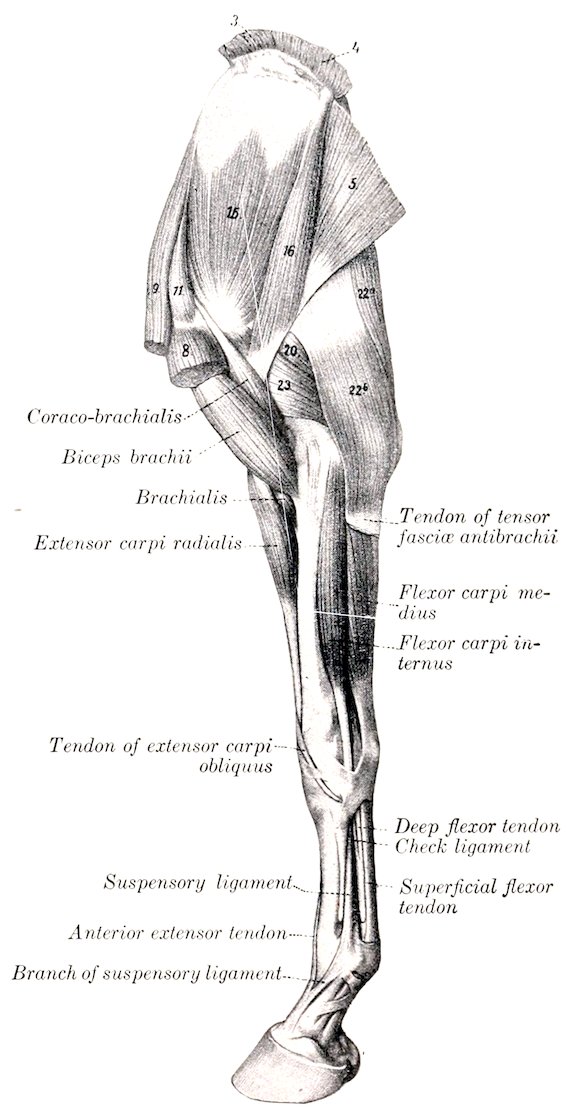
Fig. 187.—Muscles of Thoracic Limb of Horse, Internal View. 3, 4, Rhomboideus; 5, latissimus dorsi; 8, posterior deep pectoral; 9, anterior deep pectoral; 11, supraspinatus; 15, subscapularis; 16, teres major; 20, long head of triceps; 22a, 22b, tensor fasciæ antibrachii; 23, internal head of triceps. (After Ellenberger, in Leisering’s Atlas.)
Structure.—The muscle is flat and triangular, with the base upward. The latter is thin and interdigitates with the scapular attachments of the serratus. Below this the belly thickens and becomes narrower. It is covered by an aponeurosis, and contains a considerable amount of tendinous tissue. The tendon of insertion is crossed by the tendon of origin of the coraco-brachialis; it is intimately adherent to the capsule of the shoulder joint, and 258may be regarded as replacing the internal ligament of the latter. A small bursa may occur here.
Relations.—Superficially, the scapula and shoulder joint, the supraspinatus, triceps, and teres major muscles; deeply, the serratus magnus muscle, the brachial vessels, and the chief branches of the brachial plexus. The subscapular vessels run along or near the posterior edge of the muscle.
Blood-supply.—Subscapular artery.
Nerve-supply.—Subscapular nerves (from the brachial plexus).
2. Teres major (Teres internus; adductor of the arm).—This muscle is flat, widest about its middle, and lies chiefly on the deep face of the triceps.
Origin.—The dorsal angle and the adjacent part of the posterior border of the scapula.
Insertion.—The tubercle on the inner surface of the shaft of the humerus, in common with the latissimus dorsi.
Action.—To flex the shoulder joint and adduct the arm.
Structure.—It is for the most part fleshy, but the origin consists of an aponeurosis which blends with that of the tensor fasciæ antibrachii. The insertion is by a flat tendon which fuses with that of the latissimus dorsi.
Relations.—Superficially, the triceps, infraspinatus, and deltoid muscles; deeply, the serratus magnus muscle. The subscapular vessels lie in a groove between the anterior edge of this muscle and the posterior border of the subscapularis; near the shoulder joint the posterior circumflex artery and the axillary nerve emerge between the two muscles. The deep face of the muscle is crossed by the thoracic branches of the brachial plexus, and by the branch of the subscapular artery which supplies the latissimus dorsi.
Blood-supply.—Subscapular artery.
Nerve-supply.—Axillary nerve.
3. Coraco-brachialis (Coraco-humeralis).—This muscle lies on the inner surface of the shoulder joint and the arm.
Origin.—The coracoid process of the scapula.
Insertion.—(1) A small area above the internal tubercle of the humerus; (2) the middle third of the anterior surface of the humerus.
Action.—To adduct the arm and to flex the shoulder joint.
Structure.—The long tendon of origin emerges between the subscapularis and the inner branch of the supraspinatus. It passes over the terminal part of the subscapularis and is provided with a synovial sheath. The muscular part spreads out and divides into two portions. The smaller and shorter portion is inserted into the proximal third of the shaft of the humerus, close to the origin of the lateral head of the triceps; the larger and longer portion is inserted into the middle third of the humerus, in front of the internal tubercle and the inner head of the triceps.
Relations.—Externally, the subscapularis muscle and the humerus; internally, the deep pectoral and brachialis muscles. The anterior circumflex artery and the nerve to the biceps usually emerge between the two insertions, and the brachial vessels lie along the posterior border of the muscle.
Blood-supply.—Anterior circumflex artery.
Nerve-supply.—Musculo-cutaneous nerve.
4. Capsularis (Scapulo-humeralis posticus s. gracilis).—This is a very small muscle, which lies on the back of the capsule of the shoulder joint.
Origin.—The scapula, close to the rim of the glenoid cavity.
Insertion.—The posterior surface of the shaft of the humerus, a short distance below the head.
Action.—It may perhaps tense the capsule of the shoulder joint and prevent its being pinched during flexion.
259Structure.—It is fleshy and usually about the breadth of a finger. It may, however, consist of only a few bundles of fibers; sometimes it is double. Its attachment to the joint capsule is slight. It passes through the brachialis muscle to reach its insertion.
Relations.—Superficially, the teres minor and triceps muscles; deeply, the teres major and subscapularis muscles, and the capsule of the joint.
Blood-supply.—Posterior circumflex artery.
Nerve-supply.—Axillary nerve.
III. THE MUSCLES OF THE ARM
This group consists of five muscles which are grouped around the humerus. They arise from the scapula and the humerus, and are inserted into the forearm. They act on the elbow joint and the fascia of the forearm.
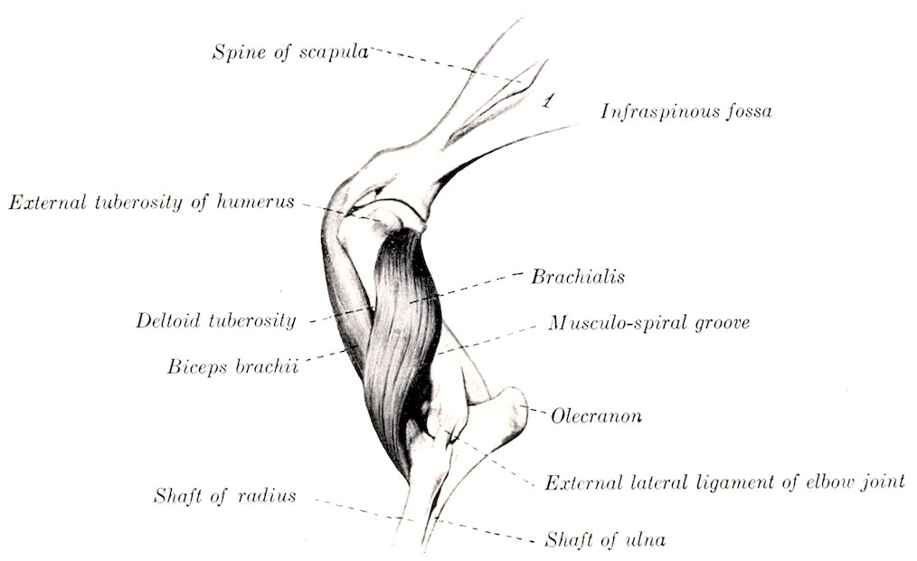
Fig. 188.—Biceps and Brachialis Muscles of Horse. (After Ellenberger-Baum, Anat. für Künstler.)
1. Biceps brachii (Coraco-radialis; flexor brachii).—This is a strong, somewhat rounded muscle, which lies on the anterior surface of the humerus.
Origin.—The tuberosity of the scapula.
Insertion.—(1) The bicipital tuberosity of the radius; (2) the internal lateral ligament of the elbow joint; (3) the fascia of the forearm and the tendon of the extensor carpi radialis.
Action.—To flex the elbow joint, to fix the shoulder, elbow, and carpus in standing, and to tense the fascia of the forearm.
Structure.—The muscle is inclosed in a double sheath of fascia, which is attached to the lips of the bicipital groove and the deltoid ridge of the humerus. The tendon of origin is moulded on the bicipital groove; it is very strong and dense and is partly cartilaginous. Its play over the groove is facilitated by the large bicipital bursa (Bursa intertubercularis). The synovial membrane covers not only the deep face of the tendon, but extends somewhat over the edges to the superficial face. A well-marked tendinous intersection runs through the belly of the muscle and divides distally into two portions. Of these, the short, thick one is inserted into the bicipital tuberosity and detaches fibers to the internal 260lateral ligament. The long tendon (Lacertus fibrosus) is thinner, blends with the fascia of the forearm, and ends by fusing with the tendon of the extensor carpi radialis.
Relations.—Externally, the mastoido-humeralis and brachialis muscles; internally, the posterior deep pectoral and the superficial pectoral muscles; in front, the anterior deep pectoral muscle; behind, the humerus, the coraco-brachialis muscle, the anterior circumflex and anterior radial vessels, and the musculo-cutaneous nerve.
Blood-supply.—Branches of the brachial and anterior radial arteries.
Nerve-supply.—Musculo-cutaneous nerve.
2. Brachialis (Humeralis obliquus s. externus; brachialis anticus).—This muscle occupies the musculo-spiral groove of the humerus.
Origin.—The proximal third of the posterior surface of the humerus.
Insertion.—The inner surface of the neck of the radius (under cover of the lateral ligament) and the arciform ligament.
Action.—To flex the elbow joint.
Structure.—The peculiar spiral course of this muscle gave rise to the name often applied to it—humeralis obliquus. Beginning on the posterior surface of the shaft, close to the head of the humerus, it winds over the lateral surface, crosses the biceps very obliquely, and finally reaches the inner side of the forearm by passing between the biceps and the extensor carpi. It is entirely fleshy, with the exception of its relatively slender tendon of insertion.
Relations.—Externally the skin and fascia, the teres minor, deltoid, triceps (lateral head), biceps, and mastoido-humeralis muscles. The anterior radial artery crosses the deep face of the muscle in its distal third, and the radial nerve accompanies the muscle in the distal half of the musculo-spiral groove.
Blood-supply.—Brachial artery.
Nerve-supply.—Radial nerve.
3. Tensor fasciæ antibrachii (Fig. 187)[58] (Scapulo-ulnaris; long extensor of the forearm; accessory anconeus of the latissimus dorsi).—This is a thin muscle which lies on the inner surface of the triceps.
Origin.—The tendon of insertion of the latissimus dorsi and the posterior border of the scapula.
Insertion.—(1) The fascia of the forearm; (2) a small eminence on the posterior border of the olecranon.
Action.—(1) To tense the fascia of the forearm and to extend the elbow joint.
Structure.—The origin consists of a very thin aponeurosis which blends with those of the caput longum and the latissimus dorsi. The muscular portion is quite thin in its anterior part, somewhat thicker behind, and is narrower than the aponeurotic origin. It is succeeded by an aponeurotic insertion, which ends chiefly by blending with the fascia of the forearm a little below the elbow. There is, however, a small but constant tendinous attachment to the olecranon.
Relations.—Externally, the panniculus, triceps (long and internal heads), the inner and middle flexors of the carpus, and the ulnar vessels and nerve; internally, the latissimus dorsi, serratus magnus, and posterior pectoral muscles.
Blood-supply.—Subscapular, ulnar, and deep brachial arteries.
Nerve-supply.—Radial nerve.
4. Triceps brachii (Figs. 178, 179, 187) (Brachial triceps; triceps extensor cubiti).—This, together with the preceding muscle, constitutes the large muscular 261mass which fills the angle between the posterior border of the scapula and the humerus. It is clearly divisible into three heads, as described below.
(a) Long head (Caput longum tricipitis; anconeus longus; caput magnum).—This, the largest and longest of the three heads, is a powerful, thick, triangular muscle, which extends from the posterior border of the scapula to the olecranon.
Origin.—The posterior border of the scapula.
Insertion.—The outer and posterior part of the summit of the olecranon.
Action.—(1) To extend the elbow joint; (2) to flex the shoulder joint.
Structure.—The muscle arises by a wide, strong aponeurosis from the posterior border of the scapula. From this the bundles of the fleshy portion converge to the short, strong tendon of insertion. A careful examination will show that the muscle is penetrated by a tendinous intersection from which many fibers take origin obliquely. The superficial face is covered by an aponeurosis which is specially developed at its distal part. A small bursa occurs under the tendon of insertion.
Relations.—Externally, the panniculus, deltoid, infraspinatus, teres minor, and the external head; internally, the tensor fasciæ antibrachii, teres major, latissimus dorsi, and posterior deep pectoral muscles, and the subscapular vessels; in front, the brachialis, and the inner head, the deep brachial and posterior circumflex vessels, and the axillary and radial nerves; behind, the skin and fascia.
Blood-supply.—Subscapular and deep brachial arteries.
Nerve-supply.—Radial nerve.
(b) External head (Caput laterale tricipitis s. anconeus lateralis s. externus; caput medium).—This is a strong, quadrilateral muscle, which lies on the outer surface of the arm. Its proximal third is covered by the deltoid and teres minor muscles; the remainder only by the thin panniculus and the skin.
Origin.—The deltoid tuberosity and the curved rough line which extends from it to the neck of the humerus.
Insertion.—(1) A small prominent area on the outer surface of the olecranon; (2) the tendon of the long head.
Action.—To extend the elbow joint.
Structure.—The origin consists of short tendinous fibers. The belly is thick, and is composed of parallel bundles which are directed obliquely downward and backward. They are inserted partly into the tendon of the long head and partly into the olecranon below and in front of that tendon.
Relations.—Externally, the deltoid, teres minor, and panniculus muscles; internally, the long and inner heads and the brachialis muscle. Branches of the circumflex vessels and axillary nerve emerge between the posterior edge of the muscle and the long head. The deep face of the muscle is related to the branches of the deep brachial artery and of the radial nerve.
Blood-supply.—Posterior circumflex and deep brachial arteries.
Nerve-supply.—Radial nerve.
(c) Internal head (Fig. 193) (Caput mediale tricipitis; anconeus medialis s. internus; caput parvum).—This is much the smallest of the three heads. It is situated on the inner surface of the arm, and extends from the middle third of the humerus to the olecranon.
Origin.—The middle third of the inner surface of the shaft of the humerus, behind and below the internal tubercle.
Insertion.—The inner and fore part of the summit of the olecranon, between the insertion of the long head and the origin of the ulnar head of the flexor perforans.
Action.—To extend the elbow joint.
Structure.—The muscle is fleshy except at its insertion, where it has a flat tendon, under which a small bursa may be found.
Relations.—Externally, the humerus, brachialis, anconeus, and the external head; internally, the posterior deep pectoral, coraco-brachialis, teres major, 262latissimus dorsi, and tensor fasciæ antibrachii muscles, the brachial and deep brachial vessels, and the median and ulnar nerves; behind, the long head, branches of the deep brachial vessels, and the radial nerve.
Blood-supply.—Deep brachial and ulnar arteries.
Nerve-supply.—Radial nerve.
5. Anconeus (M. anconeus parvus s. subanconeus).—This is a small fleshy muscle which covers the olecranon fossa and is covered by the triceps. It is somewhat difficult to separate from the outer head.
Origin.—The distal third of the posterior surface of the humerus.
Insertion.—The outer surface of the olecranon.
Action.—To extend the elbow joint, and to raise the capsule of the joint and prevent its being pinched during extension.
Structure.—It is almost entirely fleshy, and is adherent by its deep face to the joint capsule.
Relations.—Superficially, the triceps muscle; deeply, the humerus and the elbow joint.
Blood-supply.—Deep brachial artery.
Nerve-supply.—Radial nerve.
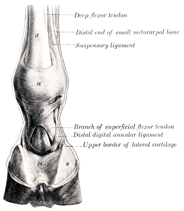
Fig. 189.—Digit of Horse, Posterior View. 14, Deep flexor tendon; 15, superficial flexor tendon; 16, posterior annular ligament of fetlock; 17, proximal annular or vaginal ligament of digit; 11, lateral cartilage; 24, plantar cushion. (After Ellenberger-Baum, Anat. für Künstler.)
IV. FASCIÆ AND MUSCLES OF THE FOREARM AND MANUS
The forearm is covered on three sides by the muscles of this group, leaving the inner surface of the radius for the most part subcutaneous. The extensors of the carpus and digit lie on the anterior (dorsal) and external part of the region, while the flexors occupy the posterior (volar) surface.
The fascia of the forearm (Fascia antibrachii) forms a very strong and complete investment for all the muscles of the region. The superficial fascia is thin, and disappears at the carpus by fusing with the deep fascia. The deep fascia is very strong and tendinous in character. It furnishes insertion at its upper and inner part to the tensor fasciæ antibrachii and posterior superficial pectoral muscles; at its upper anterior and outer part, to aponeuroses from the mastoido-humeralis and biceps. It is attached at the elbow by its deep face to the outer tuberosities of the humerus and radius, to the ulna, and to the lateral ligaments. On the inner surface of the forearm it blends with the periosteum on the subcutaneous surface of the radius. It is closely adherent to the surface of the extensor muscles, but is rather loosely attached to the flexors. From its deep face are detached intermuscular septa, which form sheaths for the muscles and are attached to the underlying bones. The principal septa are: (a) One which passes between the common 263extensor (in front) and the lateral extensor and flexor carpi externus (behind); (b) one between the common extensor and the extensor carpi radialis; (c) one between the inner and middle flexors of the carpus.
The carpal fascia (Fascia carpi) is a direct continuation of that of the forearm. It is attached chiefly to the tuberosities at the distal end of the radius, to the accessory carpal (pisiform) bone, and to the lateral ligaments. In front it forms the so-called anterior annular ligament (Ligamentum carpi dorsale), bridging over the grooves and binding down the extensor tendons and their synovial sheaths. Behind it is greatly thickened and forms the posterior annular or transverse ligament of the carpus (Ligamentum carpi transversum). This stretches across from the accessory carpal bone to the internal lateral ligament and the proximal extremity of the inner metacarpal bone. It thus completes the carpal canal, in which lie the flexor tendons, their synovial sheath, and the principal vessels and nerves of the region.
The fascia of the metacarpus and digit (Fascia metacarpea et digitalis) is, in general, thinner than the preceding. It is attached to the tendons, ligaments, and the exposed bony prominences—especially to the small metacarpal bones. On the flexion surface of the fetlock joint it is much thickened by fibers passing transversely from one sesamoid bone to the other, forming an annular ligament which binds down the flexor tendons in the sesamoid groove or canal. Distal to this is a second thick quadrilateral sheet (Ligamentum vaginale) which covers and is adherent to the tendon of the flexor perforatus. It is attached on either side by two bands to the borders of the first phalanx, thus firmly binding down the flexor tendons. A little further down a crescentic fibro-elastic sheet covers the terminal expansion of the deep flexor tendon. It is attached on either side by a strong band to the side of the first phalanx about its middle.
A. Extensor Division
1. Extensor carpi radialis (M. extensor carpi radialis s. radialis dorsalis; extensor metacarpi magnus; anterior extensor of the metacarpus).—This is the largest muscle of the extensor division, and lies on the anterior (dorsal) surface of the radius.
Origin.—(1) The external condyloid crest of the humerus; (2) the coronoid fossa.
Insertion.—The tuberosity on the anterior (dorsal) surface of the proximal extremity of the large (third) metacarpal bone.
Action.—To extend and fix the carpal joint and to flex the elbow joint.
Structure.—The belly of the muscle is rounded, and runs out to a point at the distal third of the forearm. The tendon, which runs nearly the whole length of the fleshy portion, appears on the surface of the latter about its middle; here the muscle shows a distinctly pennate arrangement. The tendon passes through the middle groove at the distal extremity of the radius and over the capsule of the carpal joint, bound down by the anterior annular ligament and invested with a synovial sheath. The latter begins three to four inches (ca. 8 to 10 cm.) above the carpus and extends almost to the insertion of the tendon. In the lower half of the forearm the deep fascia blends with the tendon, and here the latter is joined by the long tendon of the biceps.
Relations.—Superficially, the skin, fascia, and the oblique extensor; deeply, the capsule of the elbow joint, the biceps tendon, the radius, the carpal joint capsule, the anterior radial artery, and the radial nerve; externally, the anterior or common extensor; internally, at the elbow, the brachialis and biceps.
Blood-supply.—Anterior radial artery.
Nerve-supply.—Radial nerve.
2. Anterior or common digital extensor (M. extensor digitalis communis; 264anterior extensor of the phalanges; extensor pedis).—This muscle lies external to the foregoing, which it resembles in general form, although less bulky.
Origin.—(1) The front of the distal extremity of the humerus, just external to the coronoid fossa; (2) the external tuberosity on the proximal extremity of the radius, the external lateral ligament of the elbow, and the external border of the radius at the junction of its proximal and middle thirds; (3) the external surface of the shaft of the ulna; (4) the fascia of the forearm.
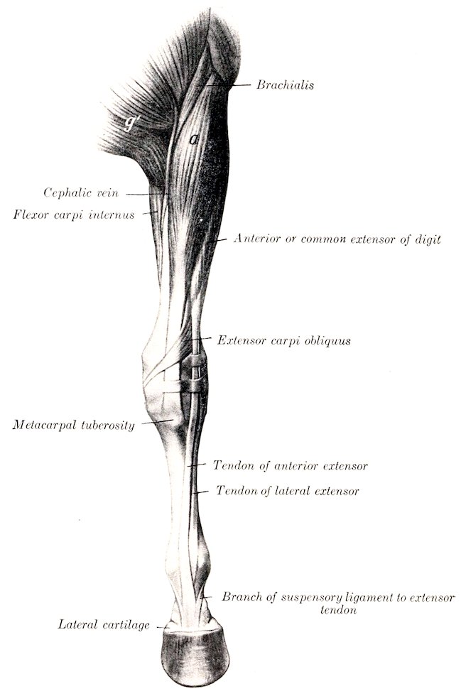
Fig. 190.—Muscles of Left Thoracic Limb of Horse, from Elbow downward, Anterior View. a, Extensor carpi radialis; g′, superficial pectoral muscle. (After Ellenberger-Baum, Anat. für Künstler.)
Insertion.—(1) The extensor (or pyramidal) process of the third phalanx; (2) the anterior surface of the proximal extremities of the first and second phalanges.
Action.—To extend the digital and carpal joints, and to flex the elbow joint.
Structure.—The muscle is a compound one, representing the common extensor, 265together with vestiges of the proper extensors of the digits. Usually at least two heads may be distinguished. The principal or humeral head (Caput humerale) arises from the front of the extensor epicondyle of the humerus in common with the extensor carpi. Its belly is fusiform, and terminates in a point near the distal third of the radius. The tendon appears on the surface of the muscle about the middle of the belly, the arrangement being pennate. The tendon passes downward through the outer of the two large grooves on the front of the distal end of the radius, and over the capsule of the carpal joint. Passing down over the front of the metacarpus, it inclines gradually inward, reaching the middle line of the limb near the fetlock. A little below the middle of the first phalanx it is joined by the branches of the suspensory ligament, and the tendon thus becomes much wider. Two synovial membranes facilitate the play of the tendon. The proximal one is a sheath which begins about three inches (ca. 7 to 8 cm.) above the carpus, and terminates at the proximal end of the metacarpus. At the fetlock a bursa occurs between the tendon and the joint capsule, but otherwise the two are adherent. The smaller head, arising chiefly from the radius and ulna, is often divisible into two parts (Fig. 443). The larger of these (Caput radiale, muscle of Phillips)[59] arises from the outer tuberosity and outer border of the radius, and from the lateral ligament of the elbow joint. The flat belly is succeeded by a delicate tendon, which accompanies the principal tendon over the carpus (included in the same sheath), and then passes outward to fuse with the tendon of the lateral extensor. Usually a slip is detached which is inserted on the proximal extremity of the first phalanx, or ends in the fascia here. The smaller and deeper division (Caput ulnare, muscle of Thiernesse)[60] is usually somewhat difficult to isolate. It arises from the ulna close to the interosseous space. It has a small rounded belly and is provided with a delicate tendon which may fuse with the principal tendon or may be inserted into the capsule and the fascia in front of the fetlock joint.
Relations.—The chief relations of the belly of the muscle are: superficially, the skin and fascia; deeply, the elbow joint, the radius and ulna, the extensor carpi obliquus, and the anterior radial vessels and radial nerve; in front and internally, the extensor carpi radialis; behind, the lateral extensor and the interosseous vessels.
Blood-supply.—Radial and interosseous arteries.
Nerve-supply.—Radial nerve.
3. Lateral digital extensor (M. extensor digitalis lateralis s. digiti quinti proprius; lateral extensor of the phalanges; extensor suffraginis).—This muscle is much smaller than the preceding, behind which it is situated.
Origin.—The external tuberosity of the radius and the lateral ligament of the elbow joint, the shaft of the ulna, the outer border of the radius, and the intermuscular septum.
Insertion.—An eminence on the front of the proximal extremity of the first phalanx.
Action.—To extend the digit and carpus.
Structure.—The muscle is pennate, and is inclosed in a sheath formed by the deep fascia, from which many fibers arise. The belly is thin and fusiform and terminates at the lower third of the forearm. From here the tendon (at first small and round) passes downward through the groove on the outer tuberosity of the distal end of the radius, then over the carpus, and, gradually inclining toward the front, but not reaching the middle line of the limb, it passes over the metacarpus and fetlock to its insertion. Two synovial membranes occur in connection 266with the tendon. A sheath envelops the tendon, beginning about three inches (ca. 6 to 8 cm.) above the carpus, and reaching to the proximal end of the metacarpus. At the fetlock a small bursa lies between the tendon and the joint capsule, but otherwise the tendon is adherent to the capsule. The tendon becomes flat and much larger below the carpus, having received the tendon of the radial head of the anterior extensor and a strong band from the accessory carpal bone.
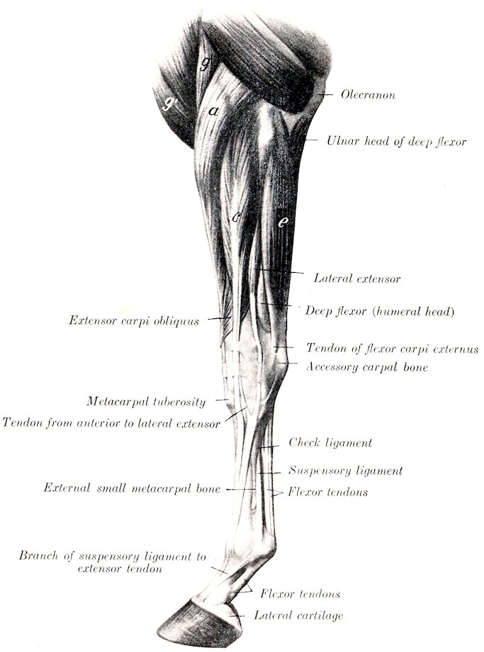
Fig. 191.—Muscles of Left Thoracic Limb of Horse from Elbow Downward; External View. a, Extensor carpi radialis; g, brachialis; g′, anterior superficial pectoral; c, anterior or common digital extensor; e, flexor carpi externus. (After Ellenberger-Baum, Anat. für Künstler.)
Relations.—Superficially, the skin and fascia; deeply, the outer face of the radius and ulna; in front, the common extensor, the oblique extensor, and the interosseous artery; behind, the external flexor of the carpus and the deep flexor of the digit.
Blood-supply.—Interosseous artery.
267Nerve-supply.—Radial nerve.
4. Extensor carpi obliquus (M. abductor pollicis longus et extensor pollicis brevis; oblique extensor of the metacarpus; extensor metacarpi obliquus).—This is a small muscle which curves obliquely over the distal half of the radius and the carpus.
Origin.—The external border and adjacent part of the anterior surface of the radius (the attachment area beginning at a point above the middle of the bone and extending down to its lower fourth).
Insertion.—The head of the inner (second) metacarpal bone.
Action.—To extend the carpal joint.
Structure.—The muscle is pennate and has a flat belly which curves downward, forward, and inward over the distal part of the radius. The tendon continues the direction of the muscle, crossing obliquely over the tendon of the extensor carpi radialis; it then occupies the oblique groove at the distal end of the radius, and crosses the inner face of the carpus. It is provided with a synovial sheath.
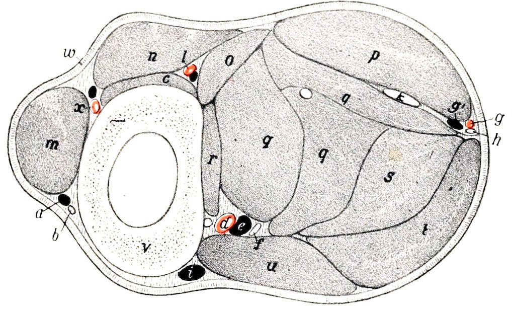
Fig. 192.—Cross-section of Middle of Right Forearm of Horse. a, Accessory cephalic vein; b, cutaneous branch of median nerve; c, extensor carpi obliquus; d, posterior radial artery; e, satellite vein; f, median nerve; g, g′, ulnar vessels; h, ulnar nerve; i, cephalic vein; k, tendon of ulnar head of deep flexor; l, interosseous artery; m, extensor carpi radialis; n, anterior digital extensor; o, lateral digital extensor; p, flexor carpi externus; q, q, q, humeral heads of deep digital flexor; r, radial head of same; s, superficial digital flexor; t, flexor carpi medius; u, flexor carpi internus; v, radius; w, skin; x, anterior radial artery. (After Ellenberger, in Leisering’s Atlas.)
Relations.—Superficially, the skin and fascia, the lateral extensor, and the common extensor; deeply, the radius, the extensor carpi radialis, the carpal capsule, and the internal lateral ligament.
Blood-supply.—Interosseous and anterior radial arteries.
Nerve-supply.—Radial nerve.
B. Flexor Division
1. Flexor carpi internus (M. flexor carpi radialis s. radialis volaris; internal flexor of the metacarpus).—This muscle lies on the inner surface of the forearm, immediately behind the inner border of the radius.
Origin.—The flexor (internal) epicondyle of the humerus, below and behind the lateral ligament.
Insertion.—The proximal end of the internal (second) metacarpal bone.
Action.—To flex the carpal joint and to extend the elbow.
Structure.—The muscle has a short tendon of origin, which is succeeded by a somewhat flattened, fusiform belly. The tendon of insertion begins near the distal fourth of the radius. It is provided with a synovial sheath which begins two or 268three inches (ca. 5 to 8 cm.) above the carpus and extends almost to the insertion of the tendon.
Relations.—Superficially, the skin and fascia, the posterior superficial pectoral and the tensor fasciæ antibrachii; deeply, the elbow joint, the radius, the deep flexor, the flexor carpi medius, the posterior radial vessels, and the median nerve. At the elbow the artery and nerve lie in front of the muscle, but below they dip beneath it.
Blood-supply.—Posterior radial artery.
Nerve-supply.—Median nerve.
On removing the deep fascia on the inner surface of the elbow the student may notice a small muscle lying along the lateral ligament. This is the pronator teres, which is usually not present or a mere vestige in the horse. It arises by a small, flat tendon from the flexor epicondyle of the humerus, and is inserted into the internal lateral ligament of the elbow. On account of its small size and the fact that the forearm is fixed in the position of pronation, the muscle can have no appreciable function. It is usually represented by the long portion of the internal lateral ligament.
2. Flexor carpi medius (M. flexor carpi ulnaris s. ulnaris medialis; oblique or middle flexor of the metacarpus).—This muscle lies on the inner and posterior aspect of the forearm partly under, partly behind, the preceding muscle. It arises by two heads—humeral and ulnar.
Origin.—(1) The flexor (internal) epicondyle of the humerus just behind the preceding muscle; (2) the inner surface and posterior border of the olecranon.
Insertion.—The upper edge of the accessory carpal (pisiform) bone.
Action.—To flex the carpal joint, and to extend the elbow.
Structure.—The humeral head is much the larger, constituting, in fact, the bulk of the muscle. It is flattened, curved, and tapers at both ends. The ulnar head, much smaller and very thin, is covered by an aponeurosis from which many of its fibers arise. It joins the large head a little above the middle of the forearm. The tendon of insertion is short and strong; it blends with the posterior annular ligament of the carpus.
Relations.—Superficially, the tensor fasciæ antibrachii, superficial pectoral, and flexor carpi internus, the skin and fascia, and cutaneous branches of the ulnar nerve; deeply, the superficial and deep flexors of the digit. In the distal half of the forearm the ulnar vessels and nerve lie between the outer edge of this muscle and the external flexor of the carpus.
Blood-supply.—Ulnar and posterior radial arteries.
Nerve-supply.—Ulnar and median nerves.
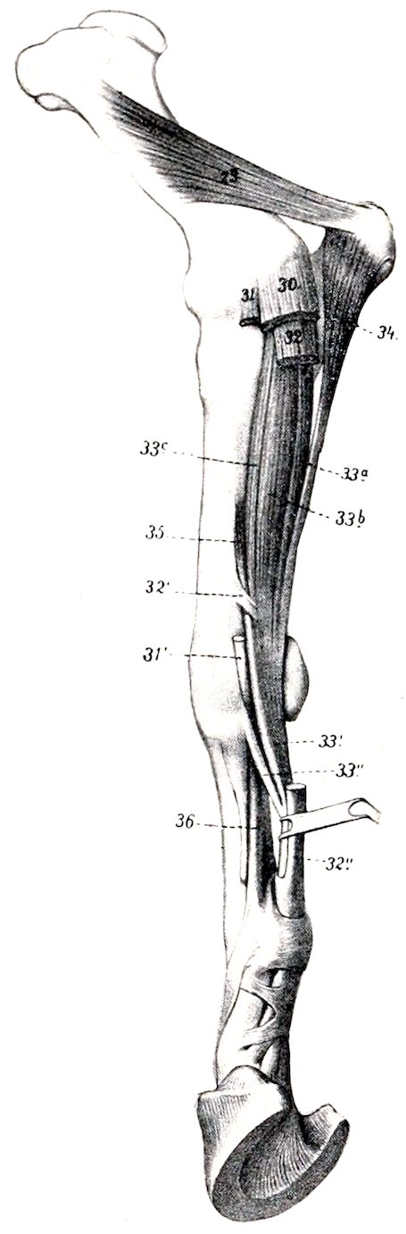
Fig. 193.—Deeper Muscles of Forearm of Horse, Internal View. 23, Internal head of triceps; 30, flexor carpi medius; 31, flexor carpi internus; 31′, tendon of 31; 32, superficial digital flexor; 32′, radial check ligament; 32″, tendon of superficial flexor; 33a, 33b, 33c, humeral heads of deep flexor; 33′, deep flexor tendon; 33″, subcarpal check ligament; 34, ulnar head of deep flexor; 35, radial head of deep flexor; 36, suspensory ligament. (After Ellenberger, in Leisering’s Atlas.)
3. Flexor carpi externus (M. extensor carpi ulnaris[61] s. ulnaris lateralis; external flexor of the metacarpus).—This muscle lies on the outer face of the forearm, behind the lateral extensor of the digit.
269Origin.—The extensor (external) epicondyle of the humerus, behind and below the lateral ligament.
Insertion.—(1) The outer surface and upper edge of the accessory carpal bone; (2) the proximal extremity of the outer (fourth) metacarpal bone.
Action.—To flex the carpal joint and to extend the elbow.
Structure.—The belly of the muscle is flattened and is intersected by a good deal of tendinous tissue. There are two tendons of insertion. The short tendon is inserted into the accessory carpal bone. The long tendon is detached just above the carpus; it is smaller and rounded; it passes downward and a little forward through a groove on the outer surface of the accessory carpal bone, enveloped by a synovial sheath, to reach its insertion on the outer metacarpal bone. A synovial pouch lies under the origin of the muscle at the elbow joint, with the cavity of which it communicates.
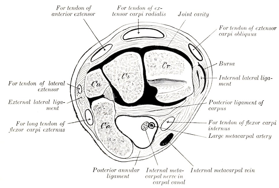
Fig. 194.—Cross-section of Proximal Part of Left Carpus of Horse. The tendons have been removed to show the canals in which they lie. The joint cavity is black. By an oversight the carpal canal for the two flexor tendons behind the posterior ligament of the carpus is not marked. Cr, Radial, Ci, intermediate, Cu, ulnar, Ca, accessory carpal bones.
Relations.—Superficially, the skin, fascia, and cutaneous branches of the ulnar nerve; deeply, the elbow joint, the ulna, and the flexors of the digit; in front, the lateral extensor of the digit; behind, the middle flexor of the carpus, the ulnar head of the deep flexor, and the ulnar vessels and nerve.
Blood-supply.—Interosseous, ulnar, and median arteries.
Nerve-supply.—Radial nerve.
4. Superficial digital flexor or flexor perforatus (M. flexor digitalis sublimis; superficial flexor of the phalanges).—This muscle is situated in the middle of the flexor group, chiefly under cover of the middle flexor of the carpus.
Origin.—(1) The flexor epicondyle of the humerus, between the flexor carpi internus and the deep flexor of the digit; (2) a ridge on the posterior surface of the radius, below its middle and near the internal border.
Insertion.—(1) The eminences on the proximal extremity of the second phalanx 270behind the lateral ligaments; (2) the distal extremity of the first phalanx, also behind the lateral ligaments.
Action.—To flex the digit and carpus, and to extend the elbow.
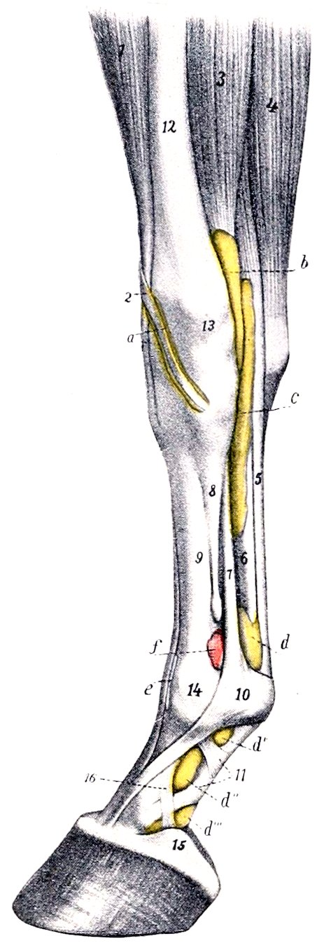
Fig. 195.—Synovial Sheaths and Bursæ of Lower Part of Right Fore Limb of Horse, Internal View. a, Sheath of extensor carpi obliquus; b, sheath of flexor carpi internus; c, carpal sheath; d, d′ d″, d‴, digital sheath; e, bursa under anterior extensor tendon; f, capsule of fetlock joint; 1, extensor carpi radialis; 2, tendon of extensor carpi obliquus; 3, flexor carpi internus; 4, flexor carpi medius; 5, superficial flexor tendon; 6, deep flexor tendon; 7, suspensory ligament; 8, small metacarpal bone; 9, large metacarpal bone; 10, posterior annular ligament of fetlock; 11, proximal digital annular ligament; 12, radius; 13, radio-carpal joint; 14, fetlock joint; 15, lateral cartilage; 16, band from first phalanx to lateral cartilage. (After Ellenberger, in Leisering’s Atlas.) The synovial sheaths (colored yellow) and the joint capsules (colored pink) are injected.
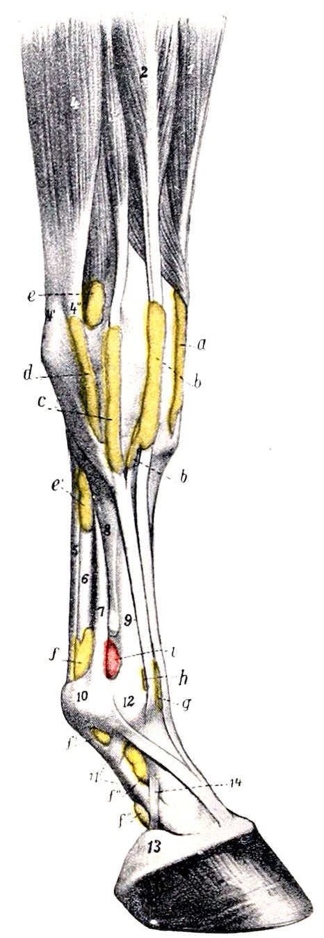
Fig. 196.—Synovial Sheaths and Bursæ of Lower Part of Right Fore Limb of Horse, External View. a, Sheath of extensor carpi radialis; b, sheath of anterior extensor; c, sheath of lateral extensor; d, sheath of outer tendon of flexor carpi externus; e, e′, carpal sheath; f, f′ f″, digital sheath; g, bursa under anterior extensor tendon; h, bursa under lateral extensor tendon; i, capsule of fetlock joint; 1, extensor carpi radialis; 2, anterior digital extensor; 3, lateral digital extensor; 4, flexor carpi externus; 4′ 4″, tendons of 4; 5, superficial flexor tendon; 6, deep flexor tendon; 7, suspensory ligament; 8, external metacarpal bone; 9, large metacarpal bone; 10, posterior annular ligament of fetlock; 11, digital annular ligament; 12, fetlock joint; 13, lateral cartilage; 14, band from first phalanx to lateral cartilage. (After Ellenberger, in Leisering’s Atlas.)
Structure.—The fleshy portion of the muscle or humeral head takes origin from the humerus. The radial head (Caput tendineum) consists of a strong fibrous band, usually termed the radial or superior check ligament, which fuses with the 271tendon near the carpus. The belly of the muscle is intersected by tendinous strands, and fuses more or less with that of the deep flexor. Near the carpus it is succeeded by a strong, thick tendon which passes down through the carpal canal, formed by the posterior ligament of the carpus, the accessory carpal bone, and the posterior annular ligament (Ligamentum carpi transversum). Here it is enveloped by a synovial sheath, in common with the deep flexor. This carpal sheath (Vagina carpea) begins three or four inches (8 to 10 cm.) above the carpus, and extends downward nearly to the middle of the metacarpus. Below the carpus the tendon becomes flattened and broader. Near the fetlock it forms a ring through which the tendon of the deep flexor passes (Fig. 150). Here the two tendons are bound down in the sesamoid groove by the posterior annular ligament, which fuses more or less with the superficial flexor tendon. At the distal end of the first phalanx the tendon divides into two branches which diverge to reach their points of insertion, and between these branches the tendon of the deep flexor emerges (Fig. 189). A second synovial sheath, the digital or sesamoidean (Vagina digitalis), begins at the distal third of the metacarpus, three to four inches (8 to 10 cm.) above the fetlock, and extends to the middle of the second phalanx.
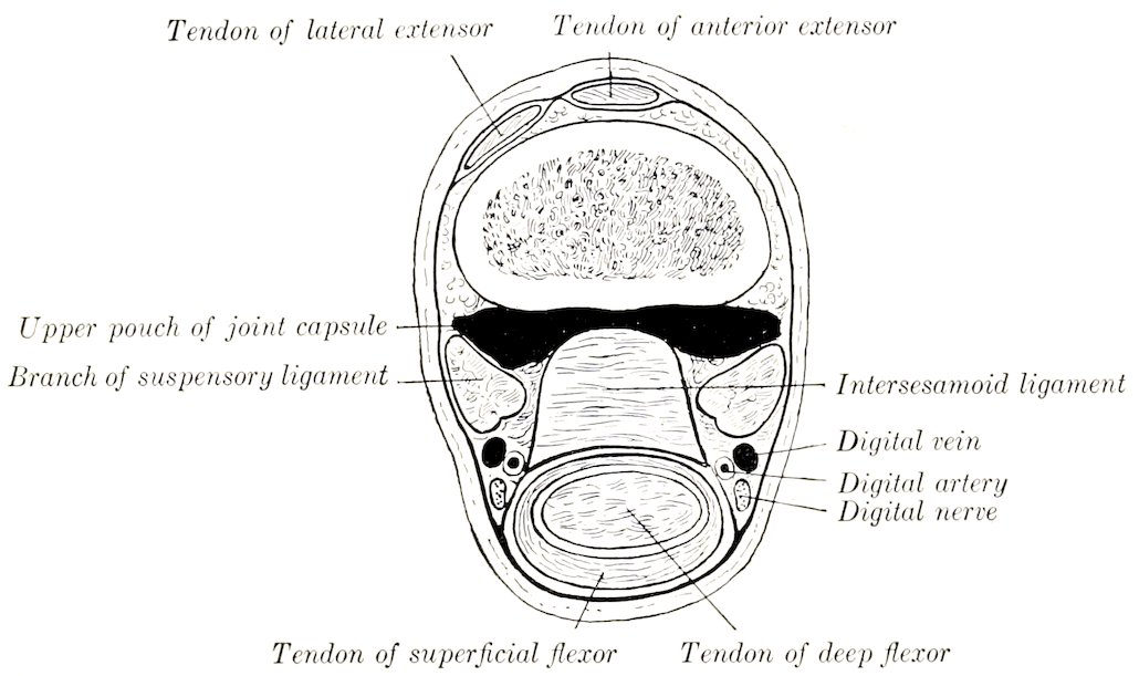
Fig. 197.—Cross-section of Distal Part of Left Metacarpus of Horse, Just Above Sesamoids.
Relations.—The belly of the muscle is related superficially to the ulnar head of the deep flexor, the flexor carpi medius, and, at its origin, to the ulnar vessels and nerve; deeply to the humeral head of the deep flexor. The tendon is related superficially to the skin and fascia; deeply to the deep flexor tendon.
Blood-supply.—Posterior radial artery.
Nerve-supply.—Median nerve.
5. Deep digital flexor or flexor perforans (M. flexor digitalis profundus; deep flexor of the phalanges).—The fleshy part of this muscle lies on the posterior surface of the radius, under cover of the preceding muscles. It is the largest muscle of the flexor group.
Origin.—(1) The flexor (internal) epicondyle of the humerus; (2) the inner surface of the olecranon; (3) the middle of the posterior surface of the radius and a small adjacent area of the ulna.
Insertion.—The semilunar crest of the third phalanx, and the adjacent surface of the lateral cartilage.
Action.—To flex the digit and carpus, and to extend the elbow.
272Structure.—This muscle consists of three principal heads. The humeral head (Caput humerale) constitutes the bulk of the muscle. It is marked by tendinous intersections, and is separable into three secondary heads. The ulnar head (Caput ulnare, ulnaris accessorius) is much smaller, and is at first superficially situated between the outer and middle flexors of the carpus, the radial head (Caput radiale, radialis accessorius) is the smallest, and is not always present; it is situated on the distal two-thirds of the posterior surface of the radius, under the humeral head. Each of these heads is provided with a tendon. The principal tendon—that of the humeral head—begins about three or four inches (8 to 10 cm.) above the carpus. It is soon joined by the small tendon of the ulnar head, which begins about the middle of the forearm. The tendon of the radial head fuses with the principal tendon close to the carpus. The conjoined tendon passes downward through the carpal canal, being included in the carpal synovial sheath with the superficial flexor tendon, as previously described. Continuing downward, it is joined about the middle of the metacarpus by a strong fibrous band, the so-called inferior or subcarpal check ligament (Caput tendineum). This is a direct continuation of the posterior ligament of the carpus. Below this the tendon passes through the ring formed by the perforatus, then in succession over the sesamoid groove, the inferior sesamoidean ligaments, and the tendon surface of the third sesamoid, to its insertion (Figs. 150 and 151). Its terminal part is much widened. From the distal third of the metacarpus to the distal end of the second phalanx it is inclosed in the digital synovial sheath described in connection with the perforatus. The navicular bursa or bursa podotrochlearis is found between the tendon and the third sesamoid or navicular bone. The terminal part of the tendon is bound down by the fibrous sheet described with the fascia.
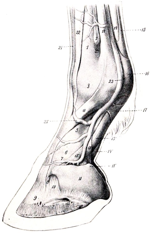
Fig. 198.—Digit of Horse, Lateral View. 1, Large metacarpal bone; 2, distal end of small metacarpal bone; 3, fetlock joint; 5, first phalanx; 6, pastern joint; 7, second phalanx; 8, coffin joint; 9, third phalanx; 11, lateral cartilage; 11′, upper (subcutaneous) border of cartilage; 12, suspensory ligament; 13, branch of 12 to extensor tendon; 14, deep flexor tendon; 15, superficial flexor tendon; 15′, branch of 15; 16, posterior annular ligament of fetlock; 17, proximal annular or vaginal ligament of digit; 18, distal annular ligament or reinforcing sheath of deep flexor tendon; 19, lateral ligament of coffin joint; 21, tendon of common or anterior extensor; 22, tendon of lateral extensor; 23, digital vein. (After Ellenberger-Baum, Anat. für Künstler.)
Relations.—The belly of the muscle is related posteriorly to the perforatus and the middle flexor of the carpus; internally, to the internal carpal flexor, the radial check ligament, and the posterior radial vessels and median nerve; externally, to the external flexor of the carpus; anteriorly, to the radius and ulna and branches of the posterior radial artery and median nerve. Below the carpus the tendon is accompanied by the vessels and nerves of the digit. It may also be noted that the muscle is not entirely covered by the other flexors; it comes in contact with the skin and fascia on the postero-external aspect of the proximal half of the forearm, and also on the outer aspect of the distal fourth.
Blood-supply.—Posterior radial and ulnar arteries.
Nerve-supply.—Median and ulnar nerves.
273
METACARPAL MUSCLES
The five muscles of this group are either reduced to vestiges or modified greatly in structure.
1, 2. Lumbricales (internus et externus).—These are two small muscles which lie on either side of the flexor tendons above the fetlock. They arise from the deep flexor tendon, and are lost in the fibrous tissue which lies under the nodule of horn or ergot at the fetlock (Fig. 447). Their action is inappreciable. The size of these muscles is subject to much variation. Often very little muscular tissue can be found, but the small tendon is constantly present.
Blood-supply.—Metacarpal arteries.
Nerve-supply.—Median and ulnar nerves.
3, 4, 5. Interossei.—These are three in number in the horse, and are situated chiefly in the metacarpal groove. Two, the internus and externus, are very small muscles, each of which arises from the corresponding small metacarpal bone near its proximal extremity, and is provided with a delicate tendon which is usually lost in the fascia at the fetlock (Fig. 150). They have no appreciable action. Their blood- and nerve-supply is the same as that of the preceding muscles.
The interosseus medius is so much modified that it is usually termed the suspensory, or superior sesamoidean ligament. It contains little muscular tissue, being transformed very largely into a strong tendinous band, bifurcate below, and having for its chief function the supporting of the fetlock. It has been described, in deference to common usage, with the ligaments.
Fasciæ and Muscles of the Pelvic Limb
The Fasciæ
The iliac fascia (Fascia iliaca) covers the ventral surface of the iliacus and psoas muscles, over which it is tightly stretched (Fig. 450). It is attached internally to the tendon of the psoas minor, externally to the inguinal (Poupart’s) ligament and the external angle of the ilium. Its anterior part is thin. Posteriorly, it is in part attached to the ilium, in part becomes continuous with the pelvic fascia. It furnishes surfaces of origin for the sartorius, cremaster externus, and transversus abdominis muscles.
The pelvic fascia (Fascia pelvis) lines the cavity (parietal layer) and is reflected on the viscera at the pelvic outlet (visceral layer). Laminæ are detached from it to strengthen the various peritoneal folds.
The superficial fascia of the gluteal region is continuous with the aponeurosis of the panniculus. It covers and partly blends with the deep fascia. The gluteal fascia (Fascia glutea) covers the superficial muscles of the region, and detaches intermuscular septa, which pass between the muscles. It is attached to the sacral spines, the dorsal sacro-iliac ligament, and the angles of the ilium, and is continuous in front with the lumbo-dorsal fascia, behind with the coccygeal fascia. Its deep face gives origin to fibers of the superficial and middle glutei, the biceps femoris, and the semitendinosus, so that care is necessary in dissecting it off these muscles. The chief intermuscular septa are: (1) One which passes between the superficial gluteus and the biceps femoris; (2) one between the biceps and semitendinosus, from which a lamella is detached which passes between the middle and posterior portions of the biceps and is attached to the tuber ischii; (3) one between the semitendinosus and semimembranosus, which is attached to the sacro-sciatic ligament and tuber ischii; it furnishes origin for fibers of the long head of the semimembranosus.
The fascia lata is continuous with the preceding, and covers the muscles on the 274outer surface and front of the thigh. It is tendinous and very strong, and for the most part easily separable from the underlying muscles. It furnishes insertion to the tensor fasciæ latæ and to the biceps femoris (in part), by both of which it is tensed. At the stifle it is attached to the patella and the inner and outer straight ligaments. Internally it is continuous with the internal femoral fascia. It furnishes the following intermuscular septa: (1) One which passes between the vastus externus and biceps femoris to be attached to the external trochanter of the femur; (2) two which pass between the three branches of the biceps femoris; (3) a fourth between the biceps femoris and semitendinosus.
The internal femoral fascia (Fascia femoralis medialis) covers the superficial muscles on the inner surface of the thigh. At its upper part it is joined by part of the aponeurosis of the external oblique muscle (Lamina femoralis) (Fig. 450). The posterior part is thin. It is continuous with the fascia lata in front and the crural fascia below. At the stifle it fuses with the tendons of the sartorius and gracilis.
The crural fascia, or fascia of the leg (Fascia cruris), consists of three layers. Two of these invest the entire region and may, therefore, be termed the common fasciæ. The superficial layer is a continuation of the fasciæ of the thigh, while the second layer may be regarded chiefly as a continuation of the tendons of the superficial muscles of the hip and thigh (biceps femoris, semitendinosus, tensor fasciæ latæ, sartorius, and gracilis). The two layers frequently fuse, and are attached chiefly to the patellar ligaments and the crest and internal surface of the tibia. About the middle of the leg the two layers unite behind the deep flexor of the digit and form a strong band which passes downward in front of the tendons of the gastrocnemius and superficial flexor, to be attached with the latter to the anterior and inner part of the tuber calcis. This may be regarded as an accessory or tarsal tendon of insertion of the biceps femoris and semitendinosus. The third layer forms sheaths for the muscles, furnishing origin in part to their fibers. Two important intermuscular septa are detached, viz.: (1) One which passes between the anterior and lateral digital extensors to be attached to the fibula and the external border of the tibia; (2) one between the lateral extensor and the deep flexor.
The tarsal fascia (Fascia tarsi) fuses with the ligaments and bony prominences of the region. It is strong and tendinous in front, and joins the tendon of the anterior extensor below the joint. Laterally, it is thin and fuses with the ligaments. Posteriorly, it is very thick and strong, forming an annular ligament which stretches from the internal lateral ligament to the fibular tarsal bone and the plantar ligament. This converts the groove at the back of the hock into a canal, in which are the deep flexor tendon with its synovial sheath and the plantar nerves. In front of the tarsus and above and below it there are three annular ligaments (Ligamenta transversa). The proximal one binds down the tendons of the anterior extensor, peroneus tertius, and tibialis anterior on the distal end of the tibia. The middle one stretches from the fibular tarsal bone to the outer tendon of the peroneus tertius, forming a loop around the tendon of the anterior extensor. The distal band stretches across the proximal extremity of the large metatarsal bone and incloses the tendons (and sheaths) of the two extensors of the digit.
The metatarsal and digital fasciæ do not differ materially from those of the corresponding regions of the thoracic limb.
The Muscles
I. THE SUBLUMBAR MUSCLES (Figs. 450, 456)
The muscles of this group are not confined to the sublumbar region, but extend beyond it both before and behind. Their chief function is to flex the hip joint. Two, however,—the psoas minor and the quadratus lumborum,—have not this action.
2751. Psoas minor (s. parvus).—This is a fusiform, flattened, pennate muscle, which lies along the ventro-lateral aspect of the bodies of the last three thoracic and the lumbar vertebræ.
Origin.—The bodies of the last three thoracic and first four or five lumbar vertebræ, and the vertebral ends of the sixteenth and seventeenth ribs.
Insertion.—The psoas tubercle on the shaft of the ilium.
Action.—To flex the pelvis on the loins, or to incline it laterally.
Structure.—The muscle arises by a series of digitations which pass backward and outward to be inserted at an acute angle on the tendon. The latter lies along the outer border of the fleshy portion and is flattened. It appears on the surface of the muscle at the third lumbar process and increases gradually in width until it reaches the pelvic inlet, where it becomes narrower.
Relations.—The ventral surface of the thoracic portion of the muscle is related to the pleura, crura of the diaphragm, and sympathetic nerve. In the abdomen, the chief ventral relations are the peritoneum, the vena cava (right side), the aorta and left kidney (left side), the sympathetic nerves, and the ureters. Dorsally, the chief relations are the vertebræ, the psoas major, and lumbar nerves. Near its insertion the tendon is crossed internally by the external iliac artery, and externally by the femoral nerve.
Blood-supply.—Intercostal and lumbar arteries.
Nerve-supply.—Lumbar nerves.
2. Psoas major (s. magnus).—This is much larger than the preceding muscle, by which it is partly covered. It is triangular, with the base anterior.
Origin.—The ventral surfaces of the vertebral ends of the last two ribs and the transverse processes of the lumbar vertebræ.
Insertion.—The internal trochanter of the femur, by a common tendon with the iliacus.
Action.—To flex the hip joint and to rotate the thigh outward.
Structure.—The origin of the muscle is fleshy, the belly being in general flattened, thick in its middle, thin at its edges. The thoracic portion is small, the abdominal part much thicker and wider, extending laterally beyond the extremities of the lumbar transverse processes. From the lumbo-sacral articulation it lies in a deep groove formed in the iliacus (with which it is partly united), becomes smaller and rounded, and passes downward and backward to terminate by a strong tendon common to it and the iliacus.[62]
Relations.—Dorsally, the last two ribs and thoracic vertebræ, the lumbar vertebræ, the internal intercostals, quadratus lumborum, longissimus dorsi, and iliacus, and the lumbar vessels and nerves; ventrally, the pleura and peritoneum, the iliac fascia, inguinal ligament, the diaphragm, psoas minor, and sartorius, the kidneys, the spleen, the intestine (duodenum, cæcum, etc.) and the circumflex iliac vessels.
Blood-supply.—Lumbar and circumflex iliac arteries.
Nerve-supply.—Lumbar and femoral nerves.
3. Iliacus.—This muscle covers the ventral surface of the ilium external to the sacro-iliac articulation, and extends outward beyond the external border of the bone, underneath the middle gluteus.
Origin.—The ventral surface of the ilium external to the ilio-pectineal line, the ventral sacro-iliac ligament, the wing of the sacrum, and the tendon of the psoas minor.
Insertion.—The internal trochanter of the femur, by a common tendon with the psoas major.
276Action.—To flex the hip joint and to rotate the thigh outward.
Structure.—The belly of the muscle is so deeply grooved for the psoas major as to give the appearance of being completely divided into outer and inner portions. When the psoas is removed, it is seen, however, that the two heads are not entirely separated. The outer, larger head arises from the wing of the ilium chiefly; the inner, smaller head arises chiefly from a small area on the shaft of the ilium, between the psoas tubercle and the depression for the inner tendon of the rectus femoris, and from the tendon of the psoas minor. The two portions inclose the psoas major in front of the hip joint.
Relations.—Dorsally, the ilium, sacrum, sacro-iliac articulation, the gluteus medius, the ilio-lumbar and external circumflex vessels; ventrally, the iliac fascia, inguinal ligament, the psoas major, sartorius, and abdominal muscles. At the hip joint the chief relations are: internally, the femoral vessels, the femoral nerve, and the sartorius muscle; externally, the rectus femoris and tensor fasciæ latæ; in front, the abdominal muscles; behind, the hip joint.
Blood-supply.—Lumbar, circumflex iliac, and deep femoral arteries.
Nerve-supply.—Lumbar and femoral nerves.
4. Quadratus lumborum.—This thin muscle lies on the outer part of the ventral surfaces of the lumbar transverse processes.
Origin.—The ventral surface of the upper part of the last two ribs and the lumbar transverse processes.
Insertion.—The ventral surface of the wing of the sacrum and the ventral sacro-iliac ligament.
Action.—Acting together, to fix the last two ribs and the lumbar vertebræ; acting singly, to produce lateral flexion of the loins.
Structure.—The muscle is pennate, and is curved with the convexity outward. It is thin, largely mixed with tendinous fibers, and is, in general, little developed in the horse in comparison with some of the other animals (e. g., dog, sheep).
Relations.—Ventrally, the psoas major and the last intercostal and first three lumbar nerves; dorsally, the last two ribs, the lumbar transverse processes, and the lateral branches of the lumbar arteries.
Blood-supply.—Lumbar arteries.
Nerve-supply.—Lumbar nerves.
5. Intertransversales lumborum (M. intertransversarii lumborum).—These are described with the other spinal muscles. (See p. 238.)
II. THE EXTERNAL MUSCLES OF THE HIP AND THIGH
Under this head will be described the muscles of the outer surface of the pelvis and thigh, and those which form the posterior contour of the latter. They are given in the order in which they may be conveniently examined.
1. Tensor fasciæ latæ (Fig. 178).—This is the most anterior muscle of the superficial layer. It is triangular in form, with its apex at the external angle of the ilium.
Origin.—The external angle of the ilium.
Insertion.—The fascia lata, and thus indirectly to the patella, the external straight ligament, and the crest of the tibia.
Action.—To tense the fascia lata, flex the hip joint, and extend the stifle joint.
Structure.—The muscle arises by a rather small head, about two inches (ca. 5 cm.) wide, on the antero-inferior eminence of the external angle of the ilium. Below this the belly spreads out and terminates about midway between the point of the hip and the stifle in the aponeurosis. The belly fuses to a considerable extent with the superficial gluteus. The aponeurosis fuses with the fascia lata, and 277detaches a lamina which passes with the tendon of insertion of the superficial gluteus to the external border of the femur.
Relations.—Externally, the skin and fascia; internally, the obliquus abdominis externus, the iliacus, superficial gluteus, rectus femoris, and vastus externus, branches of the circumflex iliac, ilio-lumbar, and iliaco-femoral arteries, and the anterior gluteal nerve; anteriorly, the precrural lymph glands. A considerable quantity of connective tissue is found between the deep face of the muscle and the abdominal wall.
Blood-supply.—Circumflex iliac, ilio-lumbar, and iliaco-femoral arteries.
Nerve-supply.—Anterior gluteal nerve.
2. Gluteus superficialis (Figs. 178, 199) (Superficial gluteus; gluteus externus).—This muscle lies behind and partly underneath the tensor fasciæ latæ. It is triangular and consists of an anterior and a posterior head united by the gluteal fascia.
Origin.—(1) The external angle and the adjacent part of the external border of the ilium (anterior head); (2) the gluteal fascia (posterior head).
Insertion.—The third (external) trochanter of the femur.
Action.—To abduct the limb, flex the hip joint, and tense the gluteal fascia.
Structure.—The anterior head of the muscle is not completely separable (except artificially) from the tensor fasciæ latæ. The attachment to the border of the ilium is by means of an intermuscular septum, which passes beneath the thick outer border of the gluteus medius. The posterior head arises from the deep face of the gluteal fascia, and so indirectly from the dorsal sacro-iliac ligament. The two heads unite and terminate on a strong flat tendon, which is inserted into the edge of the third trochanter of the femur, beneath the biceps femoris.
Relations.—Superficially, the skin, fascia, and biceps femoris; deeply, the gluteus medius, iliacus, rectus femoris, and branches of the iliaco-femoral artery; in front, the tensor fasciæ latæ; behind, the biceps femoris.
Blood-supply.—Gluteal and iliaco-femoral arteries.
Nerve-supply.—Anterior gluteal nerve.
3. Gluteus medius (Figs. 178, 179) (Middle gluteus; gluteus maximus).—This is a very large muscle which covers the dorsal surface of the ilium and the greater part of the lateral wall of the pelvis, and extends forward also on the lumbar part of the longissimus.
Origin.—(1) The aponeurosis of the longissimus, as far forward as the first lumbar vertebra; (2) the gluteal or dorsal surface and internal and external angles of the ilium; (3) the dorsal and lateral sacro-iliac and sacro-sciatic ligaments, and the gluteal fascia.
Insertion.—(1) The summit of the trochanter major of the femur; (2) the crest below the trochanter; (3) the outer aspect of the trochanteric ridge.
Action.—To extend the hip joint and abduct the limb. By its connection with the longissimus a muscular mass is formed which is one of the chief factors in rearing, kicking, and propulsion.
Structure.—The anterior extremity of the muscle is relatively thin, and lies in a depression on the surface of the longissimus, from the strong aponeurosis of which the fibers take origin. The pelvic portion of the muscle is very voluminous, and forms the bulk of the muscular mass which gives the haunch its rounded contour. This part of the muscle is intersected by several tendinous sheets. One of these is particularly distinct, and is attached to the gluteal line on the ilium. This divides the muscle incompletely into superficial and deep strata. The superficial part is inserted by a strong tendon into the summit of the great trochanter, and by a pointed fleshy mass with a tendinous border into the trochanteric ridge. The deep part (Gluteus accessorius) is smaller, and arises entirely from the ilium between the gluteal line and the external angle (Fig. 455). It has a strong flat tendon 278which passes over the convexity of the trochanter to be inserted into the crest below it. The convexity is covered with cartilage, and the trochanteric bursa (Bursa trochanterica) is interposed between the tendon and the cartilage.[63]
Relations.—Superficially, the skin, lumbo-dorsal and gluteal fasciæ, the tensor fasciæ latæ, gluteus superficialis, and biceps femoris; deeply, the longissimus, the ilium, sacro-iliac and sacro-sciatic ligaments, the gluteus profundus, iliacus and rectus femoris, the iliaco-femoral artery, the gluteal and internal pudic vessels and nerves, and the great sciatic nerve.
Blood-supply.—Gluteal, ilio-lumbar, lumbar, and iliaco-femoral arteries.
Nerve-supply.—Gluteal nerves.
4. Gluteus profundus (Deep gluteus; gluteus internus; scansorius).—This much smaller, quadrilateral muscle lies under the posterior part of the preceding muscle, and extends over the hip joint, from the superior ischiatic spine to the anterior part of the great trochanter (Fig. 455).
Origin.—The superior ischiatic spine and the adjacent part of the shaft of the ilium.
Insertion.—The edge of the anterior part or convexity of the trochanter major of the femur.
Action.—To abduct the thigh and to rotate it inward.
Structure.—The muscle is short and thick and contains numerous tendinous intersections. The fibers are directed almost transversely outward over the capsule of the hip joint and converge at the convexity of the trochanter.
Relations.—Superficially, the gluteus medius and branches of the gluteal vessels and nerves; deeply, the shaft of the ilium, the hip joint, and the rectus femoris and capsularis.
Blood-supply.—Gluteal artery.
Nerve-supply.—Anterior gluteal nerve.
5. Biceps femoris (Gluteo-biceps s. paramero-biceps).—This large muscle lies behind and in part upon the superficial and middle glutei. It extends in a curved direction from the sacral and coccygeal spines to the outer surface of the stifle and leg (Figs. 178, 199, 200, 201, 202).
Origin.—(1) The dorsal sacro-iliac ligament, the gluteal and coccygeal fasciæ, and the intermuscular septum between this muscle and the semitendinosus; (2) the tuber ischii.
Insertion.—(1) A tubercle on the posterior surface of the femur near the external trochanter; (2) the anterior surface and external straight ligament of the patella; (3) the tibial crest; (4) the anterior and internal surface of the tuber calcis.
Action.—The action is somewhat complex, because the muscle is composed of three portions, has several points of insertion, and acts on all the joints of the limb except those of the digit. The general action is to extend the limb, as in propelling the body, rearing or kicking, and to abduct it. The anterior part, by its attachment to the posterior surface of the femur and to the patella, would extend the stifle and hip joints and abduct the limb. The middle part, being inserted chiefly on the tibial crest and the external straight ligament, would extend the hip, and, with the semitendinosus, flex the stifle. The posterior part, by virtue of its attachment to the tuber calcis, assists in extending the hock.
279
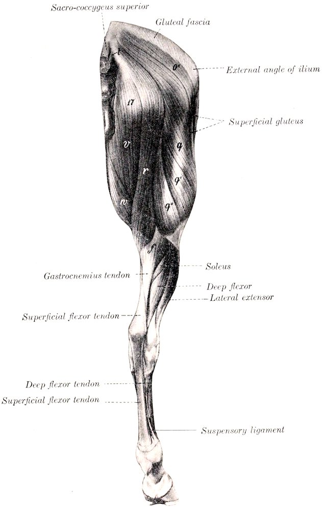
Fig. 199.—Muscles of Pelvic Limb of Horse, Postero-external View. 17, Position of tuber ischii; o′, superficial gluteus; q, q′, q″, biceps femoris; r, semitendinosus; t, sacro-coccygeus lateralis; v, semimembranosus; w, gracilis; f, gastrocnemius. (After Ellenberger-Baum, Anat. f. Künstler.)]
Structure.—The muscle has two heads of origin:[64] The long or vertebral head arises chiefly from the dorsal and lateral sacro-iliac ligaments, the coccygeal fascia, and the intermuscular septum. There is often a large bursa between this head and the trochanter major. The short or ischiatic head arises by a strong tendon from the ventral spine on the tuber ischii. They unite, and a short tendon is detached from the deep face of the muscle, to be inserted into the posterior surface of the femur near the third trochanter (Fig. 455), a bursa being interposed between the tendon and the bone. The muscle then divides into three portions, which terminate on a strong aponeurosis over the junction of the thigh and leg. The anterior branch is directed toward the patella, the middle toward the tibial crest, while the 280posterior one assists in the formation of the posterior contour of the limb. The aponeurosis blends with the deep layer of the fascia cruris, as already described. A synovial bursa occurs under the patellar insertion.
Relations.—Superficially, the skin and fascia; deeply, the sacro-iliac and sacro-sciatic ligaments, the coccygeal fascia, the femur, the obturator, gemellus, quadratus femoris, adductor, semimembranosus, vastus externus, and gastrocnemius muscles, branches of the lateral sacral, gluteal, obturator, femoral and deep femoral vessels, the great sciatic, tibial, peroneal, and posterior gluteal nerves; in front, the superficial and middle glutei; behind and internally, the semitendinosus.
Blood-supply.—Gluteal, obturator, and femoro-popliteal arteries.
Nerve-supply.—Posterior gluteal and great sciatic nerves.
6. Semitendinosus (Biceps rotator tibialis).—This is a long muscle which extends from the first two coccygeal vertebræ to the proximal third of the inner surface of the tibia. It lies at first behind the biceps, then passes downward on the back of the thigh, between that muscle and the semimembranosus (Figs. 178, 179, 199). It has two heads of origin.
Origin.—(1) The transverse processes of the first and second coccygeal vertebræ, the coccygeal fascia, and the intermuscular septum between this muscle and the biceps femoris; (2) the ventral surface of the tuber ischii.
Insertion.—(1) The tibial crest; (2) the crural fascia and the tuber calcis.
Action.—To flex the stifle and rotate the leg inward; also to extend the hip and hock joints, acting with the biceps and semimembranosus in propulsion of the trunk, rearing, etc.
Structure.—The long or vertebral head is small at its origin, but becomes larger by the accession of fibers arising on the intermuscular septum. Below the tuber ischii it is joined by the short head, which arises partly by fleshy fibers, partly by a common tendon with the biceps. The muscle then passes downward on the back of the thigh, and terminates on a wide tendon on the inner surface of the proximal third of the leg. A distinct band passes forward to be inserted on the tibial crest (a bursa lying between the tendon and the tibia), part fuses with the fascia of the leg, while the remainder joins the biceps tendon and concurs in the formation of the tendinous band, which, as before described, terminates on the tuber calcis (Fig. 458). A bursa may occur under the long head where it passes over the tuber ischii.
Relations.—Externally, the skin and fascia, the biceps, and the internal head of the gastrocnemius; internally, the coccygeal fascia, the sacro-sciatic ligament, the semimembranosus; anteriorly, the biceps femoris, branches of the femoral artery, and the great sciatic nerve.
Blood-supply.—Posterior gluteal, obturator, and femoro-popliteal arteries.
Nerve-supply.—Posterior gluteal and great sciatic nerves.
7. Semimembranosus[65] (Figs. 179, 186, 199, 451).—This muscle lies on the inner surface of the preceding muscle and the gastrocnemius, and has two heads of origin.
Origin.—(1) The posterior border of the sacro-sciatic ligament; (2) the ventral surface of the tuber ischii.
Insertion.—The internal epicondyle of the femur, behind the lateral ligament.
Action.—To extend the hip joint and to adduct the limb.
Structure.—The long head, small and pointed above, extends toward the root of the tail, fusing with the sacro-sciatic ligament. Passing downward, it becomes larger and covers in part the posterior aspect of the tuber ischii. A bursa may be found here. Below this it joins the short head, which is much larger. The large belly so formed passes downward and forward, covered in great part by the gracilis, and terminates on a short, flat tendon of insertion at the distal end of the femur.
Relations.—The upper part of the muscle assists in forming the lateral boundary 281of the pelvic outlet. It is related posteriorly and externally to the skin and fascia and the semitendinosus; internally, to the anus and its muscles, the vulva in the female, and the internal pudic artery and nerve (Figs. 452, 453). Below the pelvis the chief relations are: externally, the semitendinosus, biceps, and gastrocnemius, branches of the obturator, femoral, and femoro-popliteal arteries, and the great sciatic nerve and its chief branches; internally, the crus penis and ischio-cavernosus muscle (in the male), and the gracilis; in front, the adductor and the femoral vessels; behind, the skin and fascia.
Blood-supply.—Obturator and femoral arteries.
Nerve-supply.—Great sciatic nerve.
IV. THE INTERNAL MUSCLES OF THE THIGH
The muscles of this group are arranged in three layers.
First Layer
1. Sartorius (Figs. 180, 202, 203).—This long and rather narrow muscle is the most anterior one of the first layer. It extends from the sublumbar region to the lower and inner part of the stifle.
Origin.—The iliac fascia and the tendon of the psoas minor.
Insertion.—The internal straight ligament of the patella and the adjacent part of the tuberosity of the tibia.
Action.—To flex the hip joint and adduct the limb.
Structure.—The muscle is very thin at its origin, but becomes thicker and narrower as it passes downward and a little backward. It terminates near the stifle joint on an aponeurosis which blends with that of the gracilis and with the fascia of the leg.
Relations.—Superficially, the inguinal (Poupart’s) ligament, the abdominal muscles, the skin and fascia, and the saphenous vessels and nerve; deeply, the ilio-psoas, quadriceps femoris, and adductor. It forms the anterior boundary of the femoral canal, in which the femoral artery and vein and the deep inguinal lymph glands are situated (Fig. 451).
Blood-supply.—Femoral artery.
Nerve-supply.—Femoral and saphenous nerves.
2. Gracilis (Figs. 186, 199, 202, 203).—This is a wide, flat, quadrilateral muscle, situated behind the sartorius, which it exceeds greatly in extent.
Origin.—The middle third of the pelvic symphysis, the prepubic tendon and pubo-femoral ligament, and the ventral surface of the pubis behind the prepubic tendon.
Insertion.—The internal straight ligament of the patella, the internal surface of the tibia in front of the lateral ligament of the stifle joint, and the fascia of the leg.
Action.—To adduct the limb. It may also rotate it inward.
Structure.—The muscle arises by a strong tendon, chiefly in common with the opposite muscle. Its direct attachment to the ventral surface of the pelvis is not so extensive as a superficial inspection would suggest. The origin of the muscle presents anteriorly a round perforation for the passage of the external pudic vein. The belly is composed of parallel bundles, and is marked by a superficial furrow which, however, does not indicate a muscular division. It terminates on the inner surface of the stifle on a thin wide tendon which blends in front with that of the sartorius, below with the crural fascia.
Relations.—Superficially, the skin and fascia, the penis or mammary gland, and the saphenous vessels and nerve; deeply, the pectineus, adductor, semimembranosus 282and semitendinosus, and, at the middle of the femur, the femoral vessels; anteriorly, the sartorius. In the upper third of the thigh the sartorius and gracilis are separated by a triangular interval (femoral triangle), in which lie the deep inguinal lymph glands and the femoral vessels.
Blood-supply.—Femoral and deep femoral arteries.
Nerve-supply.—Obturator and saphenous nerves.
Second Layer
1. Pectineus (Figs. 180, 451, 456).—This muscle is fusiform and extends from the anterior border of the pubis to the middle of the inner border of the femur.
Origin.—The prepubic tendon, the pubo-femoral ligament, and the anterior border of the pubis.
Insertion.—The middle of the internal border of the femur, near the nutrient foramen.
Action.—To adduct the limb and flex the hip joint.
Structure.—The belly is cylindrical and contains little fibrous tissue. Its origin is perforated by the pubo-femoral ligament, and is thus divided into two unequal parts. The large upper part arises mainly from the prepubic tendon—only a very small part gaining direct attachment to the pubis. The small lower part does not reach the bone. The insertion is pointed and tendinous.
Relations.—Superficially, the gracilis; deeply, the femur, the vastus internus, the terminal tendon of the psoas major and iliacus, and the deep femoral artery; anteriorly, the sartorius, the femoral vessels, the saphenous nerve, and the deep inguinal lymph glands; posteriorly, the adductor, obturator externus, and quadratus femoris, and the obturator nerve (anterior division).
Blood-supply.—Femoral and deep femoral arteries.
Nerve-supply.—Femoral and obturator nerves.
The femoral canal is exposed in the dissection of the preceding muscles (Figs. 200, 451). It is bounded anteriorly by the sartorius, posteriorly by the pectineus, and externally by the iliacus and vastus internus. Its internal wall is formed by the femoral fascia and the gracilis. Its upper or abdominal opening (Lacuna vasorum) lies behind and a little internal to the internal inguinal ring and is bounded anteriorly by the inguinal ligament, posteriorly by the anterior border of the pubis, and externally by the tendon of the psoas minor. The canal terminates below at the insertion of the pectineus. It contains the deep inguinal lymph glands, the femoral artery and vein, and the saphenous nerve.
2. Adductor[66] (Figs. 179, 186, 451) (Great and small adductors of the thigh).—This fleshy, prismatic muscle lies behind the pectineus and vastus internus. It extends downward and forward from the ventral surface of the pelvis to the internal condyle of the femur.
Origin.—The ventral surface of the pubis and ischium and the tendon of the gracilis.
Insertion.—(1) The posterior surface of the femur from the level of the external trochanter to the groove for the femoral vessels; (2) the internal epicondyle of the femur and the internal lateral ligament of the stifle joint.
Action.—To adduct the limb and assist in extending the hip joint. It may also rotate the femur outward.
Structure.—It is almost entirely fleshy, and is composed of parallel bundles united rather loosely. It is usually possible to separate from the principal mass a small anterior short portion,[67] which is inserted into the femur behind the pectineus. The principal mass[68] is perforated below its middle by the femoral vessels, and is 283thus divided into two branches. The outer branch is inserted into the back of the femur with the short portion, while the inner branch is attached to the internal epicondyle and lateral ligament. There is often a superficial slip which reaches the internal straight patellar ligament. Some fibers pass under the lateral ligament and end on the tendon of the semimembranosus.
Relations.—Internally, the gracilis, and branches of the femoral artery and of the obturator nerve; externally, the femur, the obturator externus, quadratus femoris, biceps femoris, and gastrocnemius, and the femoral, deep femoral, and obturator arteries; anteriorly, the pectineus, vastus internus, and a large branch of the obturator nerve; posteriorly, the semimembranosus and the great sciatic nerve.
Blood-supply.—Femoral, deep femoral, and obturator arteries.
Nerve-supply.—Obturator nerve.
3. Semimembranosus.—Described on p. 280.
Third Layer
1. Quadratus femoris (Ischio-femoralis).—This is a narrow, flat muscle, which lies under cover of the upper part of the adductor (Figs. 455, 456).
Origin.—The ventral surface of the ischium, just in front of the semimembranosus.
Insertion.—An oblique line on the posterior surface of the femur, near the lower part of the internal trochanter.
Action.—To extend the hip joint, and to adduct the thigh and rotate it outward.
Structure.—It is composed of parallel bundles of fibers directed downward, forward, and outward.
Relations.—Internally, the adductor, semimembranosus, and the obturator vessels; externally, the obturator externus and biceps femoris, the deep femoral artery, and the great sciatic nerve.
Blood-supply.—Deep femoral and obturator arteries.
Nerve-supply.—Great sciatic nerve.
2. Obturator Externus (Fig. 456).—This is a pyramidal muscle which extends across the back of the hip joint from the obturator foramen to the trochanteric fossa.
Origin.—The ventral surface of the pubis and ischium, and the margin of the obturator foramen.
Insertion.—The trochanteric fossa.
Action.—To adduct the thigh and to rotate it outward.
Structure.—It is almost entirely fleshy, the muscle-bundles being rather loosely connected. The insertion is pointed, flattened, and partly tendinous. The origin is perforated by the obturator vessels and nerve.
Relations.—Internally, the adductor and quadratus femoris and the deep femoral vessels; externally, the gemellus, the tendon of the obturator internus, the biceps femoris, and the great sciatic nerve; anteriorly, the hip joint, the pectineus, and the external pudic vein.
Blood-supply.—Deep femoral and obturator arteries.
Nerve-supply.—Obturator nerve.
3. Obturator internus (Fig. 451).—This arises by two heads within the pelvic cavity, the tendon emerging through the lesser sciatic foramen.
Origin.—(1) The pelvic surface of the pubis and ischium around the obturator foramen; (2) the pelvic surface of the shaft of the ilium and the wing of the sacrum.
Insertion.—The trochanteric fossa.
Action.—To rotate the femur outward.
284Structure.—The ischio-pubic head lies on the pelvic floor and covers the obturator foramen. It is thin and fan-shaped. The iliac head extends along the lateral wall of the pelvis, and is pennate, with a central tendon throughout. Both terminate on a flat tendon which passes outward through the lesser sciatic foramen to be inserted into the trochanteric fossa. A synovial bursa facilitates the play of the tendon over the external border of the ischium.[69]
Relations.—The pelvic surface is covered by the pelvic fascia and the peritoneum. The obturator vessels and nerve lie between the two heads, and the internal pudic vessels and nerve lie along the dorsal edge of the iliac head.
Blood-supply.—Obturator and internal pudic arteries.
Nerve-supply.—Great sciatic nerve.
4. Gemellus[70] (Fig. 455).—This is a thin, triangular muscle, which extends from the external border of the ischium to the trochanteric fossa.
Origin.—The external border of the ischium near the ischiatic spine.
Insertion.—The trochanteric fossa.
Action.—To rotate the femur outward.
Structure.—Fleshy, some fibers being inserted into the tendon of the obturator internus.
Relations.—Dorsally, the tendon of the obturator internus and the gluteus profundus; ventrally, the obturator externus.