NEUROLOGY (continued 3)
Reading edition
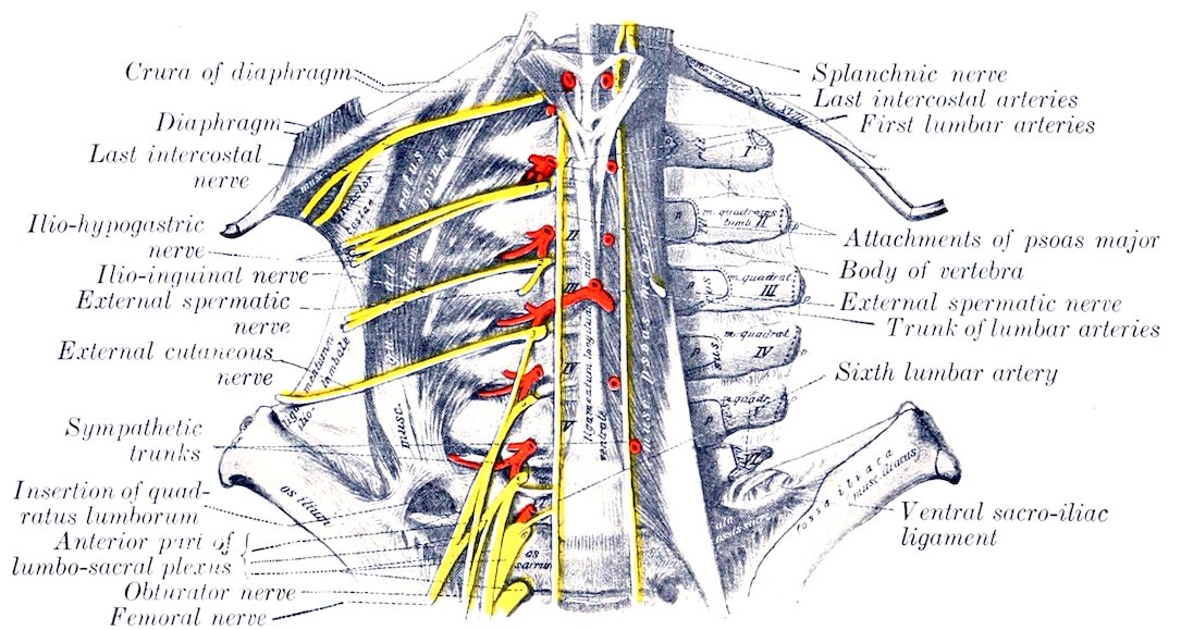
Fig. 524.—Lumbar Nerves of Horse, Ventral View. (After Schmaltz, Atlas d. Anat. d. Pferdes.)
The ventral branch of the second lumbar nerve is connected by an anastomotic branch with that of the third nerve. It gives off a large branch to the psoas muscle and an inguinal branch, and is continued as the ilio-inguinal nerve. The inguinal branch (N. spermaticus externus) runs backward in the substance of the psoas minor, emerges a short distance in front of the circumflex iliac vessels, and runs backward and downward under the peritoneum to the internal inguinal ring. It gives branches to the internal oblique muscle and descends in the inguinal canal along the outer border of the cremaster (to which it detaches filaments) and ends subcutaneously in the scrotum and prepuce in the male, the mammary gland in the female. The ilio-inguinal nerve (N. ilioinguinalis) may be regarded as the continuation of the ventral branch. It runs parallel with the ilio-hypogastric nerve and has a similar arrangement. Its anterior or superficial branch perforates the external oblique muscle a little in front of the point of the hip, runs downward on the front of the thigh and the outer surface of the stifle, and gives off cutaneous branches. The posterior or deep branch runs behind and parallel with that of the 705iliohypogastricus, detaches branches to the internal oblique muscle, and descends the inguinal canal with the inguinal branch, to be distributed to the external genital organs and the surrounding skin in the inguinal region.
The ventral branch of the third lumbar nerve is connected by a small anastomotic branch with the second nerve and furnishes a root of the lumbo-sacral plexus. It gives off a branch to the psoas muscles, an inguinal branch, and is continued as the external cutaneous nerve. The inguinal branch (N. spermaticus externus) passes backward in the substance of the psoas minor, from which it emerges under cover of or near the circumflex iliac vessels. It then runs external to and parallel with the external iliac artery and descends in the inner part of the inguinal canal. It emerges at the external ring with the external pudic artery and ramifies in the external genital organs and the skin of the inguinal region. The external cutaneous nerve of the thigh (N. cutaneus femoris lateralis) runs backward in the substance of the psoas muscles and emerges at the outer border of the psoas minor. It then passes outward and backward on the iliac fascia and accompanies the posterior branch of the circumflex iliac artery. With this vessel it perforates the abdominal wall by passing between the external oblique and the iliacus a short distance below the point of the hip, and descends on the inner face of the tensor fasciæ latæ (near its anterior border) and ramifies subcutaneously in the region of the stifle.
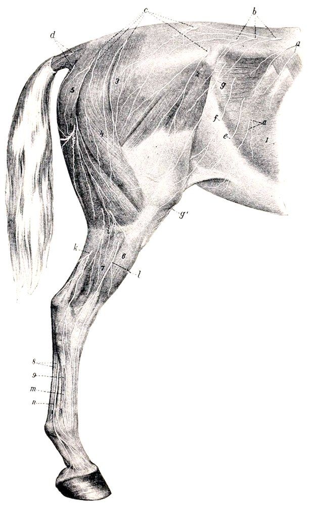
Fig. 525.—Superficial Nerves of Pelvic Limb and Posterior Part of Trunk of Horse. a, Cutaneous branches of sixteenth and seventeenth thoracic nerves; b, cutaneous branches of lumbar nerves; c, cutaneous branches of sacral nerves; d, cutaneous branches of coccygeal nerves; e, f, g, cutaneous branches of last intercostal, ilio-hypogastric, and ilio-inguinal nerves; g′, end of external cutaneous nerve of thigh; h, posterior cutaneous nerve of thigh; i, i, cutaneous branches of great sciatic nerve; k, posterior cutaneous nerve of the leg; l, superficial peroneal nerve; m, terminal part of deep peroneal nerve; n, external plantar nerve; 1, obliquus abdominis externus; 2, tensor fasciæ latæ; 3, gluteus superficialis; 4, biceps femoris; 5, semitendinosus; 6, anterior extensor; 7, lateral extensor; 8, flexor tendons; 9, great metatarsal artery. (After Ellenberger, in Leisering’s Atlas.)
The origin and disposition of some of the foregoing nerves are variable. In some cases the ilio-inguinal nerve ends in the psoas major, and appears then to be absent. The mode of formation of the inguinal nerves is very inconstant. The inner nerve may arise with one of the outer ones from a common trunk, or they may anastomose. The inguinal branch of the ilio-inguinal nerve often receives a twig from the ilio-hypogastric nerve.
The ventral branches of the fourth, fifth, and sixth lumbar nerves concur in the formation of the lumbo-sacral plexus.
Lumbo-sacral Plexus
This plexus (Fig. 451) results substantially from the union of the ventral branches of the last three lumbar and the first two sacral nerves, but it derives a 706small root from the third lumbar nerve also. The anterior part of the plexus lies in front of the internal iliac artery between the lumbar transverse processes and the psoas minor. The posterior part lies partly upon and partly in the texture of the sacro-sciatic ligament. From the plexus are derived the nerves of the pelvic limb, which are now to be described.
Femoral Nerve
The femoral (or anterior crural) nerve (N. femoralis) is derived chiefly from the fourth and fifth lumbar nerves, but commonly receives a fasciculus from the third nerve also (Fig. 524). It is the larger of the two nerves which are given off from the anterior part of the lumbo-sacral plexus. It runs backward at first between the psoas major and minor, then crosses the deep face of the tendon of insertion of the latter, and descends under cover of the sartorius in the furrow between the two heads of the iliacus. It gives off the saphenous nerve, crosses the lower part of the outer head of the iliacus, and divides into several branches which dip into the interstice between the rectus femoris and the vastus internus (Fig. 450). These branches are accompanied by the anterior femoral vessels and innervate the quadriceps femoris. Collateral muscular branches are supplied to the ilio-psoas and sartorius.
The saphenous nerve (N. saphenus) (Figs. 450, 451, 457) is given off as the parent trunk crosses the furrow between the psoas major and the outer head of the iliacus. It descends with the femoral vessels in the femoral canal, and gives branches to the sartorius, gracilis, and pectineus. About the middle of the thigh it divides into several branches which emerge from between the sartorius and gracilis, perforate the deep fascia and ramify on the inner surface and the front of the limb as far downward as the hock. The longer posterior branches accompany the saphenous vessels, while the anterior branches deviate forward toward the stifle and the anterior surface of the leg.
Obturator Nerve
This nerve (N. obturatorius) is derived from the ventral branches of the last three lumbar nerves (Fig. 524). It runs downward and backward, at first above and then upon the external iliac vein, inclines inward across the obturator vein, and passes through the anterior part of the obturator foramen in front of that vessel (Fig. 451). It continues downward through the obturator externus, and divides into several branches which innervate the obturator externus, pectineus, adductor, and gracilis muscles (Fig. 456).
Anterior Gluteal Nerve
This nerve (N. glutæus cranialis) is derived chiefly from the last lumbar and first sacral nerves. It divides into four or five branches which emerge through the great sacro-sciatic foramen with the divisions of the anterior gluteal artery and supply the gluteal and tensor fasciæ latæ muscles. The nerve to the latter and the anterior part of the superficial gluteus passes between the deep part of the gluteus medius and the deep gluteal muscle, and is accompanied on the iliacus by branches of the external circumflex vessels.
Posterior Gluteal Nerve
The posterior gluteal nerve (N. glutæus caudalis) is derived mainly from the sacral roots of the lumbo-sacral plexus (Figs. 451, 526). It divides into two trunks which emerge above the great sciatic nerve. The upper nerve passes backward on the sacro-sciatic ligament and divides into branches which enter the two heads of the biceps femoris; it supplies a branch to the posterior part of the middle gluteus, and a nerve which turns around the posterior border of the latter and enters the 707posterior head of the superficial gluteus. The inferior nerve runs downward and backward on the sacro-sciatic ligament and divides into the posterior cutaneous nerve and muscular branches which supply the semitendinosus. The former (N. cutaneus femoris caudalis) passes through the biceps femoris, emerges between that muscle and the semitendinosus a little below the level of the tuber ischii, and ramifies subcutaneously on the outer and posterior surfaces of the hip and thigh (Fig. 525).
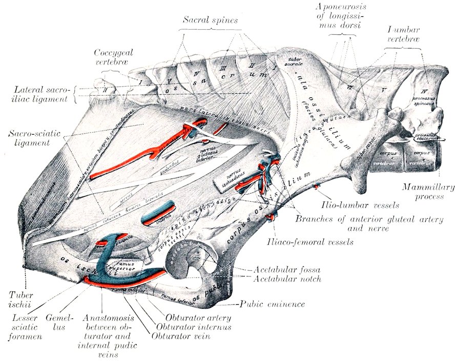
Fig. 526.—Vessels and Nerves of Pelvic Wall of Horse. Nervus ischiadicus = great sciatic nerve; n. glut. inf = anterior gluteal nerve; n. pudendus = internal pudic nerve. (After Schmaltz, Atlas d. Anat. D. Pferdes.)
Great Sciatic Nerve
The great sciatic nerve (N. ischiadicus) (Figs. 451, 455, 526), the largest in the body, is derived chiefly from the last lumbar and the sacral roots of the lumbo-sacral plexus, but may receive a fasciculus from the third sacral nerve also. It emerges through the great sacro-sciatic foramen as a broad flat band—blended at first with the posterior gluteal nerve—which passes downward and backward on the lower part of the sacro-sciatic ligament and on the origin of the deep gluteus muscle. It turns downward in the hollow between the trochanter major and the tuber ischii over the gemellus, the tendon of the obturator internus, and the quadratus femoris. In its descent in the thigh it lies between the biceps femoris externally and the adductor, semimembranosus, and semitendinosus internally, and it is continued between the two heads of the gastrocnemius as the tibial nerve. Its chief branches are as follows:
7081. In the pelvic part of its course the sciatic nerve supplies small branches to the obturator internus, gemellus, and quadratus femoris; the branch to the obturator internus reaches the muscle by passing through the anterior end of the lesser sacro-sciatic foramen.
2. As the sciatic nerve turns down behind the hip joint it gives off a large branch (Ramus muscularis proximalis) which divides to supply the semimembranosus and the short heads of the biceps femoris and semitendinosus.
3. The posterior cutaneous nerve of the leg or external saphenous nerve (N. cutaneus suræ posterior) is detached from the sciatic about the middle of the thigh. It receives a fasciculus from the peroneal nerve and descends with the recurrent tarsal vein on the outer face of the gastrocnemius to the distal third of the leg. Here it perforates the deep fascia and ramifies under the skin on the outer surface of the tarsus and metatarsus (Fig. 525).
4. The peroneal or external popliteal nerve (N. peronæus) (Figs. 455, 460, 527) is a large trunk which arises from the great sciatic nerve very shortly after the latter emerges from the pelvic cavity. It descends with the parent trunk to the origin of the gastrocnemius; here the peroneal nerve deviates outward and forward across the external face of the gastrocnemius under cover of the biceps femoris, and divides at the origin of the lateral extensor muscle into superficial and deep branches. The collateral branches include a twig to the biceps femoris, the reinforcing fasciculus detached about the middle of the thigh to the posterior cutaneous nerve, and, lower down, the external cutaneous nerve of the leg (Ramus cutaneus suræ lateralis). The latter, also known as the peroneal cutaneous nerve, emerges between the middle and posterior divisions of the biceps femoris at the level of the stifle joint and ramifies under the skin. The superficial peroneal or musculo-cutaneous nerve (N. peronæus superficialis) furnishes branches to the lateral extensor, and descends in the furrow between that muscle and the anterior extensor, perforates the deep fascia of the leg, and ramifies under the skin on the front and the outer face of the tarsus and metatarsus. The deep peroneal or anterior tibial nerve (N. peronæus profundus) is the direct continuation in point of size of the peroneal trunk. It dips in between the lateral and anterior extensors of the digit, gives branches to these muscles and the tibialis anterior, and descends in front of the intermuscular septum which separates the former. It continues downward behind the tendon of the anterior extensor and divides on the front of the hock into internal and external branches. The internal branch (Ramus medialis) passes down under the skin on the anterior face of the metatarsus and supplies the cutaneous fibers of this region. The external branch (Ramus lateralis) furnishes a twig to the extensor brevis muscle and descends with the great metatarsal artery. It supplies the skin on the outer face of the metatarsus and the fetlock.
In the greater part of its course in the leg the deep peroneal nerve is separated from the anterior tibial vessels by the tibialis anterior (muscular part of the flexor metatarsi). It should be noted that the outer of the two veins which almost always accompany the artery here is frequently very large, and the layer of muscle which intervenes between it and the nerve is often exceedingly thin. In the distal part of the leg the nerve is in direct contact with the vein, and on the front of the hock it lies behind the vessels.
Tibial Nerve
The tibial nerve (N. tibialis), also termed the posterior tibial, is the direct continuation of the great sciatic nerve (Figs. 451, 455, 458, 459, 527). It passes down between the two heads of the gastrocnemius, and accompanies the recurrent tibial vessels to the distal third of the leg, where it divides into internal and external plantar nerves. In the upper third of the leg it lies along the inner side of the superficial flexor under cover of the internal head of the gastrocnemius; lower down it is covered by the common deep fascia and is situated in the space between the deep flexor and the inner border of the tendo Achillis.
709At the usual point of election for tibial neurectomy, i. e., about a handbreadth above the level of the tuber calcis, the nerve lies in areolar tissue and fat in a fascial compartment formed by the special fascia of the deep flexor in front and by the common fascia and the accessory or tarsal tendon of the semitendinosus and biceps femoris behind and internally.
At its origin the tibial nerve gives off a muscular branch (Ramus muscularis distalis), the divisions of which pass between the two heads of the gastrocnemius and radiate to supply that muscle, the popliteus, the soleus, and the flexors of the digit. Small cutaneous twigs are also detached along the course of the nerve.
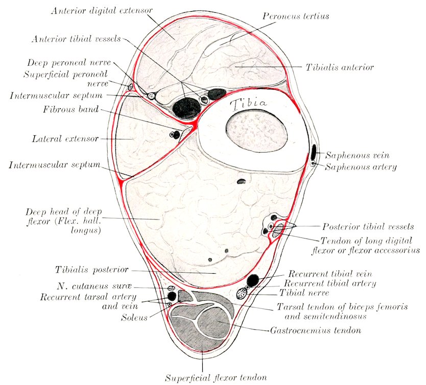
Fig. 527.—Cross-Section of Distal Third of Left Leg of Horse. The deep fascia is shown by red line.
Plantar Nerves
The plantar nerves, internal and external (N. plantaris medialis, lateralis), result from the bifurcation of the tibial nerve in the lower part of the leg (Figs. 458, 459). They continue at first in the same direction and relations as the parent trunk, in direct apposition and inclosed in a common sheath. At the hock they diverge at a very acute angle and descend in the tarsal canal behind the deep flexor tendon in company with the plantar arteries. The internal plantar nerve supplies cutaneous nerves to the inner aspect of the tarsus and metatarsus, descends along the inner border of the flexor tendons behind the superficial plantar metatarsal vessels, and is otherwise arranged like the corresponding volar nerve of the fore limb. The external plantar nerve deviates outward between the two flexor tendons to reach their outer border. It supplies a branch to the suspensory 710ligament, and in its further course resembles the corresponding nerve of the fore limb.
In so-called “tibial” neurectomy the operator really cuts the two plantar nerves which have not yet separated. The anastomotic branch between the two plantar nerves is smaller than that which connects the corresponding nerves of the fore limb, and is absent in 30 per cent. of the cases, according to Rudert.
Sacral Nerves
Five pairs of sacral nerves (Nn. sacrales) are present in the horse.
The small dorsal branches emerge through the dorsal sacral foramina and the space between the sacrum and the first coccygeal vertebra, and ramify in the muscles and skin of the sacral region and the adjacent part of the tail (Fig. 525). The fifth anastomoses with the dorsal branch of the first coccygeal nerve.
The ventral branches leave the vertebral canal through the ventral sacral foramina and the interval between the sacrum and first coccygeal vertebra. They are connected with the sympathetic by rami communicantes, and contribute branches to the pelvic plexus. The first and second are the largest, and unite with each other and with those of the last three lumbar nerves to form the lumbo-sacral plexus. The third and fourth are connected with each other, and the majority of their fibers go to form the pudic and posterior hæmorrhoidal nerves.
The pudic nerve (N. pudendus) (Figs. 451, 455, 526) accompanies the internal pudic artery to the ischial arch, turns around the latter, parting company with the artery, and pursues a flexuous course along the dorsum penis as the nervus dorsalis penis and ramifies in the glans and the penile layer of the prepuce. Within the pelvis it anastomoses with the posterior hæmorrhoidal nerve, and gives branches to the bladder and urethra, the terminal part of the rectum, and the skin and muscles of the anus (Fig. 452). It also supplies the nerve to the ischio-cavernosus muscle and numerous branches to the corpus cavernosum and corpus spongiosum. In the female it terminates in the clitoris and vulva (Fig. 453).
The posterior hæmorrhoidal nerve (N. hæmorrhoidalis caudalis) passes downward and backward above the pudic nerve, with which it anastomoses. It gives twigs to the terminal part of the rectum, the sphincter ani externus, and the surrounding skin (Fig. 452). In the female it supplies twigs to the vulva also (Fig. 453).
The ventral branch of the fifth nerve is small. It gives twigs to the sacro-coccygeus ventralis lateralis and the skin of the root of the tail and joins the first coccygeal nerve.
Coccygeal Nerves
The coccygeal nerves (Nn. coccygei) commonly number five pairs. Their dorsal and ventral branches anastomose to form respectively two trunks on either side, which extend to the tip of the tail and supply its muscular and cutaneous nerves. The dorsal trunk runs with the dorso-lateral artery between the sacro-coccygeus dorsalis and intertransversales muscles (Fig. 454). The ventral trunk accompanies the ventro-lateral artery below the intertransversales.
The Sympathetic Nervous System[210]
The sympathetic nervous system (Systema nervorum sympathicum) is that part of the nervous system which serves (1) to transmit stimuli to the heart-muscle, unstriped muscle, and glands; (2) to conduct impulses from the viscera to the cerebrospinal system.
Many of the fibers are derived from the cerebrospinal system and are rearranged and distributed in the sympathetic system. Numerous sympathetic fibers are contributed to the cerebrospinal 711nerves, through which they are distributed to the unstriped muscular tissue and glands, as vasomotor, pilomotor, and secretory nerves. The sympathetic, like the cerebrospinal system, consists of neurones, each of which comprises the cell-body, an axone, and numerous branched dendrites. The cell-bodies are aggregated into ganglia, some of which are large and more or less constant in position and form, while others are microscopic; and are scattered in an irregular manner through the peripheral part of the system. It is probable that simple visceral reflexes may be mediated by sympathetic neurones alone.
In descriptive anatomy the sympathetic system is regarded as consisting of (1) a chain of ganglia extending along either side of the vertebral column and connected by association fibers to form the sympathetic trunk; (2) central branches to and from the cerebrospinal nerves; (3) peripheral branches, which form plexuses with each other and the cerebrospinal nerves; (4) the peripheral ganglia which are interposed in the plexuses.
The sympathetic trunk (Truncus sympathicus) extends on either side from the base of the cranium to the tail. In it are interposed, at intervals of varying regularity, the ganglia of the sympathetic trunk (Ganglia trunci sympathici). These are connected with the cerebrospinal nerves by central branches, the rami communicantes.
Two kinds of rami communicantes occur. Of these, one type consists largely of medullated fibers derived from the spinal nerves and ganglia; they have therefore a white appearance, and are termed white rami. They contain both efferent and afferent fibers. The efferent splanchnic fibers are derived from the ventral roots of the spinal nerves and terminate in great part about the cells of the nearest sympathetic ganglion; others end in more distant or in peripheral ganglia. The afferent splanchnic fibers are chiefly peripheral processes of the cells of the spinal ganglia, but some are sympathetic fibers which enter the spinal nerve-trunk and terminate about cells of the spinal ganglion. The gray rami consist mainly of non-medullated fibers derived from the sympathetic ganglia directly or through the trunk, which proceed centrally to the spinal nerves and are distributed along the somatic divisions of the latter to unstriped muscle and glands as vasomotor, pilomotor, and secretory fibers. Some go to the membranes of the spinal cord, and a few terminate about cells of the spinal ganglia as sensory sympathetic fibers.
Similar but more complex and irregular communications which exist between the sympathetic system and the cranial nerves—with the exception of the first and second—have been referred to in the accounts of the latter.
It is convenient for descriptive purposes to divide the sympathetic system into cephalic, cervical, thoracic, abdominal, and pelvic parts.
1. The cephalic part (Pars cephalica systematis sympathici) comprises the otic, sphenopalatine, and ciliary ganglia, which may be regarded as homologues of the ganglia of the trunk of other regions. It also includes three plexuses formed by branches derived from the superior cervical ganglion. The ganglion gives off two or three filaments which subdivide to form the internal carotid plexus around the artery of like name. The cavernous plexus surrounds the artery within the cavernous sinus and communicates with that of the opposite side. The external carotid plexus is formed around the homonymous artery, and filaments from it go to the vessel and its branches and to the salivary glands.
2. The cervical part (Pars cervicalis s. sympathici) includes two ganglia and the trunk which connects them.
The superior or anterior cervical ganglion (G. cervicale craniale) lies on the guttural pouch below the occipito-atlantal articulation (Fig. 437). It is reddish-gray in color, fusiform, and about an inch (ca. 2 to 3 cm.) in length. It is connected by rami communicantes with the last four cranial and first cervical nerves, sends branches to form the plexuses mentioned above, and contributes to the pharyngeal plexus.
The cervical trunk of the sympathetic connects the superior and inferior cervical ganglia. On leaving the former it is associated in a common sheath with the vagus along the dorsal face of the common carotid artery. At the root of the neck it separates from the vagus and joins the inferior ganglion.
The inferior or posterior cervical ganglion (G. cervicale caudale) is situated under the first rib and the insertion of the scalenus (Figs. 428, 429). On the right 712side it lies upon the longus colli and the trachea, on the left side upon the same muscle and the œsophagus. It is flattened, very irregular and variable in outline, and is blended more or less with the first thoracic ganglion. It is connected by large gray rami with the ventral branches of the last two cervical nerves, and by small filaments with the recurrent nerve. It receives a nerve—termed by Schmaltz the nervus transversarius—which accompanies the vertebral artery; through it rami come from the second to the sixth cervical nerves inclusive.
A middle cervical ganglion may be present, and occurs oftenest on the right side. It lies on the carotid artery an inch or two in front of the first rib and is intercalated in the vago-sympathetic trunk.
The cardiac nerves (Nn. cardiaci) are formed by branches from the inferior cervical and first thoracic ganglia, together with twigs from the sympathetic trunks and the vagi. They form a plexus (P. cardiacus) on the ventral face of the trachea with each other and with branches of the vagus and recurrent nerves. They are subject to considerable variation, but their general arrangement may be stated as follows:
(a) On the right side there are usually two cardiac nerves. Of these one passes back with the vagus in the angle between the right brachial artery and the common carotid trunk, pierces the pericardium, crosses the aortic arch, and divides into branches which are mingled with those of the left nerves. The second crosses obliquely over the right face of the trachea and joins the vagus, where the latter gives off the right recurrent nerve; a small plexus is formed here, from which two or three cardiac branches are detached. These pass back beneath the trachea and ramify on the atria and ventricles.
(b) On the left side there are commonly three cardiac nerves. One of these is distributed to the great vessels in the anterior mediastinum. The largest passes back at first with the vagus beneath the arch of the left brachial artery, inclines downward, perforates the pericardium, and divides into two branches. One branch passes beneath the bifurcation of the pulmonary artery and is distributed to the left atrium. The larger branch dips in between the aorta and the pulmonary artery, gives twigs to these vessels, and ramifies on the ventricles, especially along the course of the right coronary artery. The third nerve crosses the deep face of the left brachial artery, passes back below the trachea, and unites with filaments of a right cardiac nerve. It passes around the aorta and ramifies chiefly along the course of the left coronary artery on the left face of the ventricles.
3. The thoracic part (Pars thoracalis systematis sympathici) extends below the costo-vertebral joints from the inferior cervical ganglion to the upper border of the diaphragm, and passes between the latter and the psoas minor to be continued by the abdominal part.
The trunk is concealed in the first part of its course by the outer border of the longus colli, but further back it is visible under the pleura.
The ganglia are arranged segmentally at each intercostal space and partly on the heads of the ribs. They are flattened and are small and fusiform, with the exception of the first. This (G. thoracale primum) is extensive, irregularly quadrilateral in outline, and is united with the inferior cervical ganglion. The ganglia are connected with the thoracic nerves by white and gray rami communicantes.
The visceral branches comprise aortic, cardiac, pulmonary, and œsophageal branches, and the splanchnic nerves. The aortic branches ramify on the thoracic artery, forming around that vessel the thoracic aortic plexus (P. aorticus thoracalis). The cardiac branches concur with those of the vagus in forming the cardiac plexus (P. cardiacus). From this branches go to form the coronary plexuses (P. coronarii) along the course of the vessels of like name. The pulmonary branches join with corresponding branches of the vagus and filaments from the cardiac plexus in forming the pulmonary plexus (P. pulmonalis) at the root of the lung. Branches 713of the plexus, on which are minute ganglia, ramify with the bronchi in the substance of the lung.[211] The œsophageal branches join with those of the vagus in the formation of the œsophageal plexus (P. œsophageus).
The great splanchnic nerve (N. splanchnicus major) arises by a series of roots derived from the sixth or seventh to the fifteenth or sixteenth thoracic ganglia inclusive. It extends at first along the inner side of the thoracic trunk, then crosses the latter ventrally, passes back between the crus of the diaphragm and the outer border of the psoas minor, and joins the cœliac ganglion. It is small at its origin, but becomes considerably larger than the sympathetic trunk. Near its termination it bears the small splanchnic ganglion, from which and from the nerve, filaments go to the aorta, the œsophagus, and the vertebræ.
The small splanchnic nerve (N. splanchnicus minor) is formed by roots derived from the last two or three thoracic ganglia. It runs back with the great splanchnic nerve, but ends in the cœliac or in the renal and adrenal plexuses.
This nerve is not always present and is variable in arrangement. The roots communicate by filaments with the great splanchnic.
4. The abdominal part of the sympathetic trunk (Pars abdominalis systematis sympathici) lies along the inner border of the psoas minor, above the aorta on the left side and the posterior vena cava on the right (Fig. 524). The trunk is smaller than the thoracic part, and presents usually six small fusiform lumbar ganglia (G. lumbalia), which are connected by rami communicantes with the ventral divisions of the lumbar nerves. Visceral branches go to the aortic and hypogastric plexuses, and to the cœliac and mesenteric ganglia.
5. The pelvic part of the sympathetic trunk (Pars pelvina systematis sympathici) begins at the last lumbar ganglion and extends along the pelvic surface of the sacrum internal to the emergence of the ventral branches of the sacral nerves (Fig. 451). At the third segment of the sacrum the trunk divides into internal and external branches. The internal branch inclines to the median plane and unites with the opposite branch. At the junction there is often found the small coccygeal ganglion (G. impar), which lies on the coccygeal artery at the joint between the first and second coccygeal vertebræ. A filament from the ganglion accompanies the artery. The external branch communicates with the last two sacral nerves and joins the ventral coccygeal nerves. Ganglia occur near each of the first three sacral foramina, and are connected by gray rami communicantes with the adjacent spinal nerves. The visceral branches are distributed through the hypogastric plexus. They supply motor fibers to the longitudinal and inhibitory fibers to the circular muscular coat of the rectum, motor fibers to the bladder and uterus, and the vasodilator fibers (Nn. erigentes) to the penis.
THE ABDOMINAL AND PELVIC PLEXUSES
The chief plexuses which distribute nerves to the viscera and vessels of the abdominal and pelvic cavities are two in number, the solar and the pelvic. From them fibers proceed to form numerous subsidiary plexuses which are named according to the organs which they supply or the vessels which they enlace.
The Solar Plexus
The solar plexus (Fig. 450) is situated on the dorsal wall of the abdominal cavity in relation to the aorta and the origin of its chief visceral branches. It comprises the cœliaco-mesenteric ganglia and the cœliac and anterior mesenteric plexuses.
714The cœliaco-mesenteric ganglia[212] are two in number, right and left. They are situated on each side of the aorta, in relation to the origin of the cœliac and anterior mesenteric arteries. The right ganglion is concealed by the posterior vena cava; it is irregularly quadrilateral and is about two inches (4 to 6 cm.) in length. The left ganglion is largely covered by the left adrenal; it is narrower than the right one and is three or four inches (ca. 8 to 10 cm.) long. The two ganglia are united by irregular connecting branches in front of and behind the anterior mesenteric artery.[213] Each receives the splanchnic nerve of its own side and branches from the dorsal œsophageal continuation of the vagus nerves. Branches from the ganglia and the cords which connect them pass back to the small stellate posterior mesenteric ganglia, which are situated at the origin of the posterior mesenteric artery.
The cœliac plexus (P. cœliacus) consists of a feltwork of nerve-fibers in which are small ganglia. It surrounds the cœliac artery and from it proceed the following unpaired secondary plexuses which accompany the arterial branches:
1. The gastric plexus (P. gastricus) enlaces the gastric artery and divides like the artery into two parts, forming the anterior and posterior gastric plexuses. These receive branches of the vagus nerves.
2. The hepatic plexus (P. hepaticus) is formed by several nerves of considerable size which accompany the hepatic artery and the portal vein. It receives fibers from the left vagus, ramifies in the liver, and gives off branches which accompany the collateral branches of the hepatic artery and supply the areas in which these vessels are distributed.
3. The splenic plexus (P. lienalis) resembles the preceding in its arrangement. In addition to its terminal branches to the spleen, it gives collateral twigs to the pancreas and the left part of the greater curvature of the stomach.
The anterior mesenteric plexus (P. mesentericus cranialis) is formed mainly by branches from the posterior part of the cœliaco-mesenteric ganglia; it is continuous with the cœliac plexus in front and the posterior mesenteric behind. It surrounds the anterior mesenteric trunk and its branches, and supplies the viscera to which these vessels are distributed.
The posterior mesenteric plexus (P. mesentericus caudalis) accompanies the artery of like name in its distribution.
The secondary plexuses which accompany the branches of the mesenteric arteries give off branches which form two fine peripheral plexuses in the wall of the intestine. One of these, the myenteric plexus (P. myentericus) or plexus of Auerbach, lies between the layers of the muscular coat, and is provided with microscopic ganglia. The other is in the submucous tissue, and is therefore termed the submucous plexus (P. submucosus) or plexus of Meissner.
The following paired plexuses are derived mainly from branches of the cœliac and aortic plexuses:
1. The renal plexuses (P. renales) proceed largely from the cœliaco-mesenteric ganglia, but receive fibers also from the small splanchnic nerves. They enlace the renal arteries and supply the kidneys. Minute renal ganglia occur on the course of the nerves along the renal vessels.
2. The adrenal plexuses (P. suprarenales) are formed by a relatively very large number of fine fibers derived in great part directly from the cœliaco-mesenteric ganglia. Numerous minute ganglia occur in their meshes.
3 a. The spermatic plexuses (P. spermatid) proceed largely from the aortic and renal plexuses. Each accompanies the corresponding (internal) spermatic artery to the testicle.
3 b. The utero-ovarian plexuses (P. ovarici) are the homologues in the female 715of the preceding. They accompany the utero-ovarian arteries to the ovary and the cornua of the uterus.
The hypogastric or pelvic plexus (P. hypogastricus) is the pelvic continuation of the aortic and posterior mesenteric plexuses. The chief nerves which form it enter the pelvic cavity ventral to the large vessels, anastomose with each other and with branches from the third and fourth sacral nerves, and ramify on the pelvic viscera. The peripheral plexuses derived therefrom are named according to the organs which they supply; the chief of these are the hæmorrhoidal, vesical, uterovaginal, prostatic, cavernous (of the penis or clitoris).
THE NERVOUS SYSTEM OF THE OX[214]
The spinal cord resembles that of the horse in conformation and structure. In cattle of medium size its length is about 165 to 170 cm. (ca. 65 to 67 inches) and its weight about 240 to 250 grains (ca. 8 ounces).
In a cow 140 cm. in height Dexler found the weight of the cord (including the intradural nerve-roots) to be 260 g. and the length 162 cm. The lengths of the regions were: cervical 41 cm., thoracic 72 cm., lumbar 32 cm., sacral 7 cm.
The brain has an average weight of about 500 grams (ca. 16 to 17 ounces). Its differences in general form are correlated with those noted in the consideration of the cranial cavity.
The medulla is short, wide, and thick. Its ventral face is strongly convex. The pyramids are narrow, short, and close together in front. Near the decussation there is a well defined oval prominence on either side which indicates the position of the posterior olive. The corpus trapezoideum is large; it has no central part between the pyramids. The external arcuate fibers are distinct. The restiform bodies are short and thick and diverge more strongly than in the horse. The floor of the fourth ventricle is only about two-thirds as long as in the horse. The area acustica is a well-marked oval prominence lateral to the middle part of the limiting sulcus. The tuberculum acusticum at the origin of the auditory nerve is very large. The anterior fovea is distinct. The posterior recess of the fourth ventricle communicates through the foramen of Magendie (Apertura mediana) with the subarachnoid space.
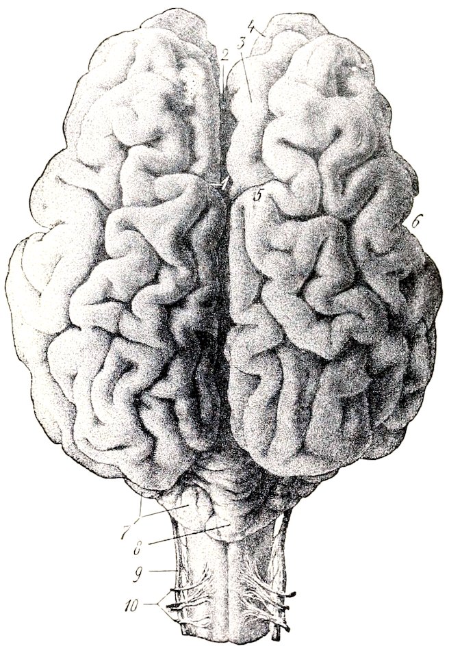
Fig. 528.—Brain of Cow, Dorsal View. 1, Sulcus transversus; 2, longitudinal fissure; 3, antero-internal frontal gyrus; 4, olfactory bulb; 5, marginal or sagittal pole of hemisphere; 6, lateral fissure (of Sylvius); 7, hemisphere of cerebellum; 8, vermis of same; 9, accessory nerve; 10, dorsal root of first cervical nerve. (Ellenberger-Baum, Anat. d. Haustiere.)
716The pons is smaller, both transversely and longitudinally, than that of the horse. It is strongly convex and has a distinct central depression.
The cerebellum is smaller and more angular in form than in the horse. The vermis is large and has a distinct depression on its anterior face for the posterior corpora quadrigemina. The hemispheres are relatively small and are not clearly divided into tabulations. The anterior peduncles are very short.
The cerebral peduncles are short. A small eminence in the locus perforatus posticus is caused by the presence of the interpeduncular ganglion. The internal geniculate body is prominent. The pineal body is long and fusiform, and is often pigmented in spots. The third ventricle forms two considerable recesses in relation to the pineal body: one (R. pinealis) extends up into the body; the other (R. suprapinealis) is a long tubular prolongation in front of it.
The optic tracts cross the cerebral peduncles almost at a right angle.
The pituitary body is situated in a deep fossa and is surrounded by a plexus of vessels.
The cerebral hemispheres are shorter, higher, and relatively wider than in the horse. The frontal poles are small, the occipital large. Viewed from the side, the dorsal surface is strongly convex. The highest point of the dorso-medial border is a little in front of its middle and forms a marked prominence termed the sagittal or marginal pole (Polus sagittalis s. prominentia marginalis dorsalis). Anterior to this the border drops abruptly, being cut into by the deep transverse fissure. The arachnoid on the basal and anterior parts of the hemispheres is usually pigmented. The corpus callosum extends through a little more than a third of the length of the hemisphere. The pattern of the fissures and gyri of the pallium is somewhat simpler than in the horse.
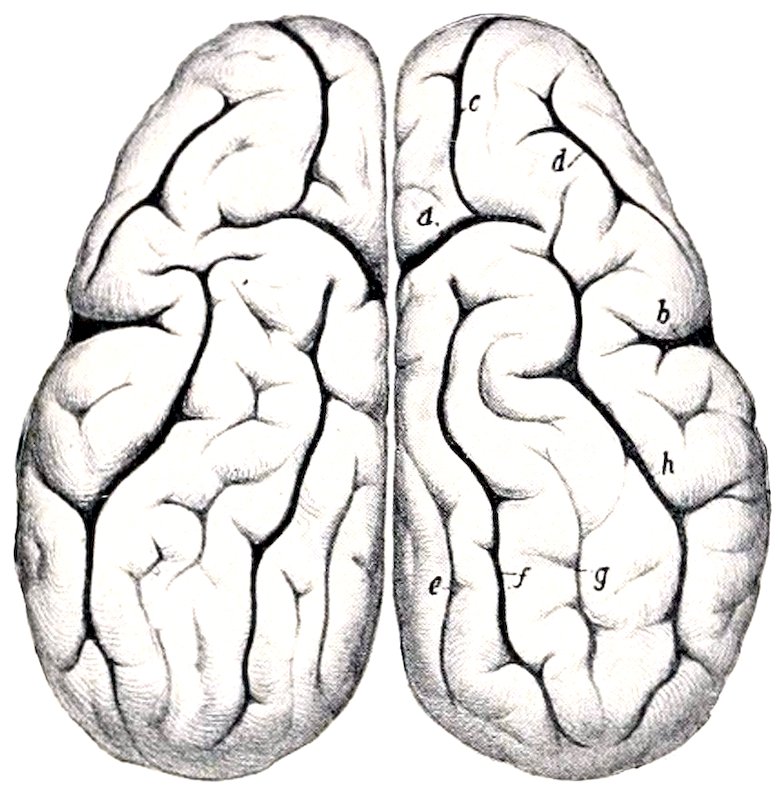
Fig. 529.—Cerebral Hemispheres of Ox, Dorsal View. a, Transverse fissure; b, lateral fissure (of Sylvius); c, coronal fissure; d, diagonal fissure; e, entomarginal fissure; f, marginal fissure; g, ectomarginal fissure; h, suprasylvian fissure. (After Ellenberger, in Leisering’s Atlas.)
1. The lateral fissure (of Sylvius) is very deep. Its middle branch extends almost vertically upward on the middle of the lateral surface of the hemisphere, and is separated by a gyrus of variable width from the suprasylvian fissure. The anterior branch runs forward about parallel with the sulcus rhinalis anterior, from which it is separated by the short gyri of the insula. The latter are covered to a small extent only by the overhanging gyri (operculum). The posterior branch may run back a distance of only about 1.5 cm. and end in T-shaped manner, or it may join the sulcus rhinalis posterior.
2. The suprasylvian fissure is deep and very distinct. It extends in an undulating manner from the lateral surface of the occipital pole to the outer side of the sagittal pole. Here it may be interrupted or may be continued by the coronal fissure, which descends to the frontal pole and divides into two short branches.
3. The diagonal fissure (S. diagonalis) begins in front of the stem of the lateral fissure and runs upward and forward. Its form is very variable.
4. The transverse fissure cuts obliquely into the dorsal border in front of the sagittal pole. It is short and deep and commonly communicates with the suprasylvian and coronal fissures.
5. The marginal fissure is distinct. It extends from the sagittal pole to the occipital pole. It is flexuous and deviates outward somewhat in its posterior two-thirds.
6. The entomarginal fissure lies medial and parallel to the posterior part of the preceding.
7. The ectomarginal fissure is lateral and parallel to the marginal fissure. It does not extend so far in either direction as the latter and is often more or less broken up by annectent gyri.
8. The sulcus rhinalis is more open than in the horse, since it is not overlapped by the gyri above it.
9. The calloso-marginal fissure is flexuous and not so regular as in the horse. It is often 717interrupted at or near the level of the genu of the corpus callosum, in which case the genual fissure continues it downward.
10. The crucial fissure is faint and is confined to the medial surface. It lies about one centimeter in front of the transverse fissure and commonly joins the calloso-marginal fissure below.
11. Short and variable fissures occur in front of and behind the middle branch of the lateral fissure (F. ectosylvia anterior, posterior).
The olfactory bulb is much smaller than in the horse. The external olfactory tract is large, the inner one small and not distinct.
The cranial nerves have in general the same superficial origin as in the horse. The more important differences in other respects are as follows:
The oculomotor nerve is larger than in the horse. It emerges with the fourth, sixth, ophthalmic, and maxillary nerves through the large foramen, which is equivalent to the foramen lacerum orbitale and foramen rotundum of the horse.
The internal branch of the lacrimal nerve supplies twigs to the mucous membrane of the frontal sinus; the large outer branch runs backward and supplies the matrix of the horn. The frontal nerve emerges from the orbit below the supraorbital process. The naso-ciliary nerve is large and sends filaments to the ocular muscles. The ciliary ganglion is somewhat larger than in the horse and is connected with the lower division of the oculomotor nerve by several short filaments. In consequence of the absence of the canine and upper incisor teeth the corresponding branch of the infraorbital nerve is naturally wanting. The mandibular nerve emerges through the foramen ovale. The superficial temporal nerve gives off a large branch which joins the superior buccal division of the facial on the masseter, about half-way between the zygomatic arch and the angle of the jaw. The otic ganglion is larger than in the horse. According to Moussu the buccinator nerve furnishes excito-secretory twigs to the parotid and inferior buccal glands.
The facial nerve divides into its two terminal branches before reaching the border of the jaw. The superior buccal nerve is the larger of the two; it crosses the masseter much lower than in the horse. The relatively small inferior buccal nerve runs beneath the parotid or in the gland substance parallel with the border of the lower jaw, crosses under the insertion of the sterno-cephalicus, and runs forward along the depressor labii inferioris. At the point where it crosses the facial vein and parotid duct it gives off an anastomotic branch to the superior nerve. The auriculo-palpebral nerve is large.
The vagus bears—in addition to the relatively large jugular ganglion—a ganglion nodosum at the point of origin of the superior laryngeal nerve. The trunk is large. The pharyngeal branch is large and anastomoses with the superior and external laryngeal nerves. The latter commonly arises directly from the trunk. The superior œsophageal trunk communicates with the splanchnic nerve, contributes twigs to the hepatic plexus, and ramifies chiefly on the right surface of the rumen and the adjacent surface of the abomasum. The inferior œsophageal trunk supplies branches to the hepatic plexus and to all the divisions of the stomach.
The spinal accessory nerve presents two special features. The part which joins the jugular ganglion bears a small ganglion. The ventral branch supplies both parts of the sterno-cephalicus.
The hypoglossal nerve is large. It is connected with the ventral division of the first cervical nerve by a branch of considerable size, and detaches a long branch which runs backward along the carotid artery.
The spinal nerves resemble those of the horse in origin and general arrangement. The thoracic nerves number thirteen pairs. The more important differences in the limb-plexuses and their branches are as follows:
The brachial plexus is formed by the ventral branches of the last three cervical 718and first thoracic nerves; the second thoracic nerve furnishes no root, but the ventral branch of the sixth cervical goes almost entirely to the plexus after giving off the nerve to the rhomboideus and the cervical part of the serratus magnus.
The differences in the nerves above the elbow are not of sufficient moment to receive notice in this brief account, but below this point there are naturally important special features correlated chiefly with the arrangement of the digits.
The radial nerve is continued below the elbow by a large cutaneous branch (N. cutaneus antibrachii dorsalis) which emerges at the lower border of the external head of the triceps and descends on the dorsal aspect of the limb. It communicates above the carpus with the cutaneous branch of the median nerve and terminates in three dorsal digital nerves; two of these descend along the axial or interdigital side of the dorsal surface of the chief digits, and the third along the inner (abaxial) side of the inner chief digit.
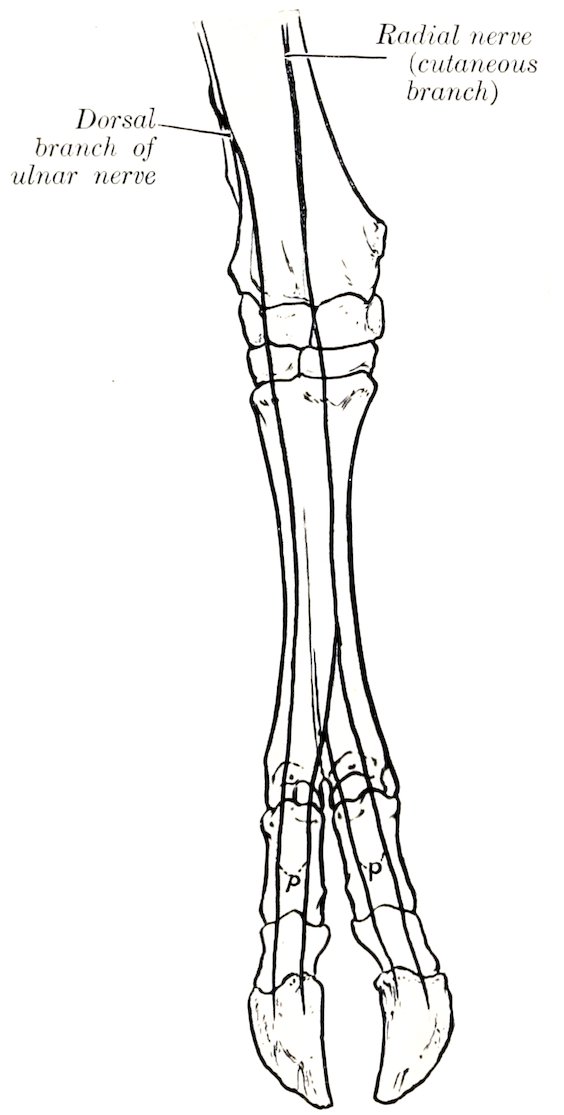
Fig. 530.—Nerves of Distal Part of Right Fore Limb of Ox, Anterior View (Schematic). p, Dorsal digital nerves.
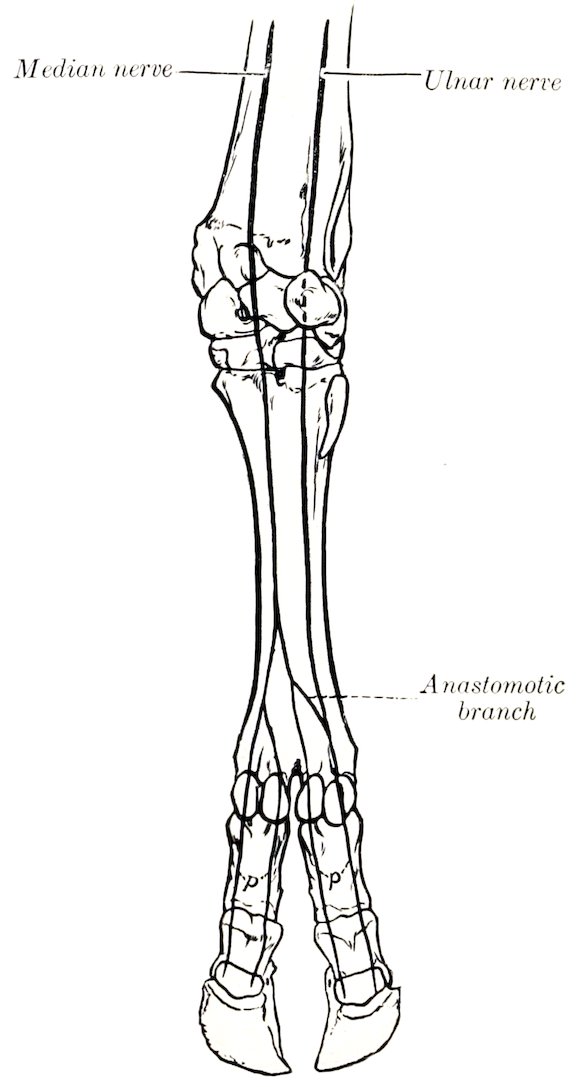
Fig. 531.—Nerves of Distal Part of Right Fore Limb of Ox, Posterior View (Schematic). p, Volar digital nerves.
The ulnar nerve divides at a variable distance down the forearm into two branches. The dorsal or superficial branch emerges between the tendons of the external and middle flexors of the carpus, and is continued as the external dorsal digital nerve on the outer chief digit. The volar or deep branch descends along the superficial digital flexor, gives a branch to the suspensory ligament below the carpus, 719and unites with the external branch of the median nerve to form the external volar digital nerve.
The median nerve is much larger than the ulnar. It passes beneath the pronator teres, descends the forearm as in the horse, and divides in the distal part of the metacarpus into two branches. The internal branch gives twigs to the inner small digit and is continued on the inner side of the volar surface of the inner chief digit as the internal volar digital nerve (N. digitalis medialis digiti III); it also concurs with the outer branch in forming the two digital nerves which descend along the interdigital aspect of the chief digits. The external branch is larger. It bifurcates, and one division unites with the twig from the internal branch to form a common digital trunk. From the latter two digital nerves proceed as mentioned above; these are the external and internal volar digital nerves of the internal and external chief digits respectively (N. digitalis volaris lateralis digiti III, medialis digiti IV). The other division unites with the volar branch of the ulnar nerve to form the external volar digital nerve of the external chief digit (N. digitalis lateralis digiti IV).
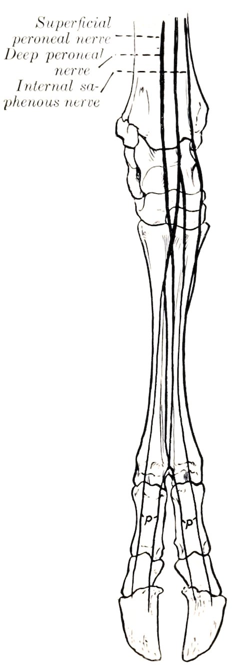
Fig. 532.—Nerves of Distal Part of Right Hind Limb of Ox, Anterior View (Schematic). p, Dorsal digital nerves.
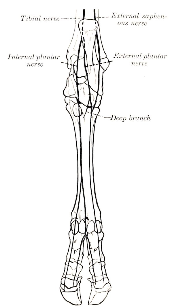
Fig. 533.—Nerves of Distal Part of Right Hind Limb of Ox, Posterior View (Schematic). p, Plantar digital nerves.
The lumbo-sacral plexus and its branches to the pelvis and thigh present no very striking special features.
The superficial peroneal nerve is much larger than in the horse. After crossing 720beneath the peroneus longus it passes down on the front of the tarsus and metatarsus and divides into three branches. The internal and external branches descend as the internal and external dorsal digital nerves on the chief digits. The larger middle branch joins a branch of the deep peroneal nerve in the interdigital space, and from this union proceed the dorsal digital nerves which descend on the opposed surfaces of the chief digits.
The deep peroneal nerve descends in the leg as in the horse and continues down the anterior groove of the metatarsus with the dorsal metatarsal artery. Its terminal branches concur with branches from the superficial peroneal nerve in the formation of the two axial dorsal digital nerves (N. dig. dors. lat. dig. III et dig. dors. med. dig. IV pedis), and with a branch of the internal plantar nerve in the formation of corresponding plantar digital nerves.
The tibial nerve divides at the back of the hock into internal and external plantar nerves. The internal plantar nerve descends between the superficial flexor tendon and the suspensory ligament and divides into two branches; the inner branch descends as the internal plantar digital nerve (N. dig. plant. med. dig. III) along the inner side of the flexor tendons of the inner chief digit; the outer branch turns around the flexor tendons to reach the interdigital space, where it concurs with a branch of the deep peroneal nerve in the formation of two axial plantar digital nerves (N. dig. plant. lat. dig. III et dig. plant. med. dig. IV) which descend on the opposed surfaces of the chief digits. The external plantar nerve descends along the outer border of the flexor tendons, gives a branch to the suspensory ligament and to the external small digit, and continues along the outer face of the external digit as the external plantar digital nerve (N. dig. plant. lat. dig. IV).
The sympathetic system closely resembles that of the horse in its general arrangement, and only a few differential features will receive attention. The superior cervical ganglion is closer to the cranial base and is thicker than in the horse; its branches to the carotid and cavernous plexuses are large, and no connection is formed with the spinal accessory nerve. The cervical trunk is smaller than in the horse, and arises by two or three bundles from the superior cervical ganglion behind its middle. The inferior cervical ganglion is distinctly separable from the first thoracic; the latter is large. There are thirteen pairs of thoracic ganglia. The cœliaco-mesenteric ganglia are plexiform, not compact. There are five pairs of sacral ganglia, and the right and left trunks are connected here by transverse anastomoses.
THE NERVOUS SYSTEM OF THE PIG
The spinal cord weighs about 42 grams (Ellenberger-Baum). It is almost circular in cross-section, except at the enlargements, where it is somewhat flattened dorso-ventrally. The conus medullaris extends to the anterior part of the third sacral segment. The epidural space is occupied by a large quantity of fat.
The brain in adults of medium size weighs about 125 grams (ca. 4 to 4½ ounces). When viewed from above, it is seen to have an elongated oval form. The occipital poles overlie the cerebellum to a small extent only; the frontal poles are rounded and relatively large, but do not conceal the ends of the olfactory bulbs. The medulla is relatively broad. The cuneate tubercle is very large and is limited externally by a groove. The corpus trapezoideum is very wide laterally. The pons is less prominent than in the ox. The cerebellum is very wide and short. Its anterior face is flattened and presents a depression for the corpora quadrigemina. The vermis is large. The hemispheres consist of an inner large and an outer small part. The cerebral peduncles are very short. The posterior corpora quadrigemina 721are wide apart, rounded, and relatively large. The internal geniculate body is prominent. The cerebral hemispheres are somewhat bean-shaped in lateral profile, the convex border being dorsal. The arrangement of the gyri and sulci is simpler than in the horse or ox. The principal fissures are as follows:
1. The lateral fissure (of Sylvius) begins at the sulcus rhinalis and runs upward and somewhat backward on the depressed part of the lateral surface of the hemisphere; it appears unbranched on superficial examination, but when the gyrus which almost completely conceals the insula is raised, an anterior branch is exposed which forms the dorsal boundary of the insula.
2. The suprasylvian fissure pursues a curved course approximately parallel with the dorso-medial border of the hemisphere. Its posterior end is separated by a short interval from the sulcus rhinalis posterior. From its highest point a branch proceeds obliquely upward and forward, crosses the dorso-medial border, and joins the splenial fissure.
3. The diagonal fissure crosses the anterior part of the lateral surface. It is directed obliquely downward and backward.
4. The coronal fissure is constant and deep. It is directed upward and inward over the frontal pole, continues backward near the dorso-medial border, and usually ends by passing obliquely over the border to the inner surface of the hemisphere.
5. The presylvian fissure begins—apparently as a branch of the sulcus rhinalis anterior—about 2 cm. in front of the lateral fissure, and curves over the lateral aspect of the frontal pole.
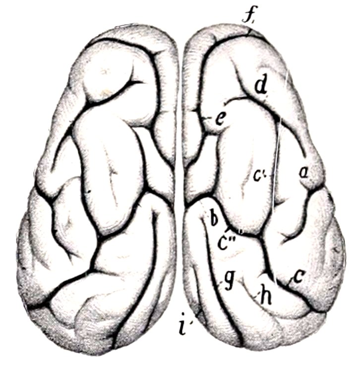
Fig. 534.—Cerebral Hemispheres of Pig, Dorsal View. a, Upper part of lateral fissure (of Sylvius); b, cruciate fissure; c, suprasylvian fissure, with its anterior branch (c′) and upper branch (c″); d, diagonal fissure; e, coronal fissure; f, presylvian fissure; g, marginal fissure; h, ectomarginal fissure; i, entomarginal fissure. (After Ellenberger, in Leisering’s Atlas.)
6. The marginal fissure begins behind the cruciate fissure close to the dorso-medial border and extends in a gentle curve backward almost to the occipital pole.
7. The ectomarginal fissure lies above and nearly parallel to the posterior part of the suprasylvian fissure.
8. The entomarginal fissure lies along the posterior part of the dorso-medial border.
9. The calloso-marginal fissure consists of two separate parts. The more extensive posterior part is termed the splenial fissure. It extends from the tentorial aspect of the hemisphere in a direction parallel with the corpus callosum nearly to the middle of the medial surface; it is continued by the cruciate fissure and is also connected with the suprasylvian and coronal fissures by branches which pass obliquely upward and forward. The anterior part, the genual fissure, lies about midway between the anterior part of the corpus callosum and the dorso-medial border.
10. The cruciate fissure is short and shallow. It runs obliquely upward and forward from the anterior end of the splenial fissure on the inner surface of the hemisphere.
11. The sulcus rhinalis extends in an undulating manner along the entire lower part of the lateral surface of the hemisphere and forms the upper limit of the rhinencephalon.
The olfactory bulbs are very large and the tracts extremely broad and short. The trigonum olfactorium is so prominent as to be termed the tuberculum olfactorium.
722The cranial nerves present the following special features:
The third, fourth, sixth, and the ophthalmic and maxillary divisions of the fifth nerve, emerge together as in the ox.
The lacrimal nerve resembles that of the horse, the frontal that of the ox. The naso-ciliary nerve is relatively large and sends numerous filaments to the ocular muscles. The maxillary nerve has a very short course in the pterygo-palatine fossa. The infraorbital nerve is large in correlation with the development of the snout which receives numerous branches. The mandibular nerve emerges through the foramen lacerum anterius. The superficial temporal nerve is small; according to Moussu it furnishes the excito-secretory fibers to the upper part of the parotid gland, while those going to the lower part are derived from the mylo-hyoid nerve.
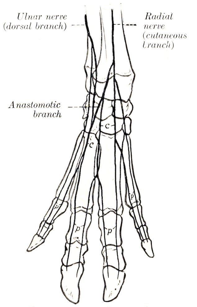
Fig. 535.—Nerves of Distal Part of Right Fore Limb of Pig, Anterior View (Schematic). c, Dorsal common digital nerves; p, dorsal proper digital nerves.
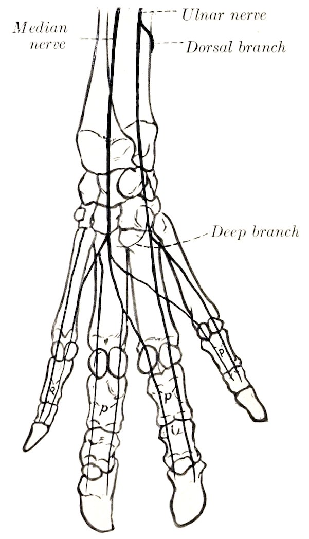
Fig. 536.—Nerves of Distal Part of Right Fore Limb of Pig, Posterior View (Schematic). p, Volar proper digital nerves.
The inferior buccal nerve passes downward and forward under cover of the parotid gland and accompanies the parotid duct, with which it turns around the lower border of the jaw in front of the masseter.
The vagus bears a jugular ganglion and a ganglion nodosum; the latter occurs at the point of origin of the superior laryngeal nerve, and may be as large as a small pea. Proximal to this the vagus is easily divided into two strands, one of which is the accessory component. The œsophageal trunks form a posterior œsophageal plexus, as in man, from which two nerves issue. The inferior nerve is small and ramifies on the parietal surface of the stomach. The superior trunk is much larger; it gives branches to the stomach, crosses the lesser curvature of that organ, and joins the solar plexus.
The hypoglossal nerve may present a small dorsal root, on which there is a minute hypoglossal ganglion (of Froriep).
723The spinal nerves number on each side eight in the cervical region, fourteen (commonly) in the thoracic, seven in the lumbar, and four in the sacral. Some of the special features of the nerves of the limbs are as follows:
The brachial plexus is derived from the same nerves as in the ox, but the root furnished by the sixth cervical is relatively smaller. The plexus consists of two parts, the upper of which emerges above the scalenus, the lower between the two parts of that muscle. The more important differences in the nerves emanating from the plexus from the arrangement in the ox occur in the lower part of the limb. The cutaneous branch of the radial nerve divides at the carpus into branches which concur with the dorsal branch of the ulnar nerve in supplying the dorsal digital nerves, two for each digit. The volar digital nerves, also two for each digit, are formed by the terminal branches of the median nerve and the volar or deep branch of the ulnar nerve. The formation and arrangement of the digital nerves are indicated in the annexed schematic figures.
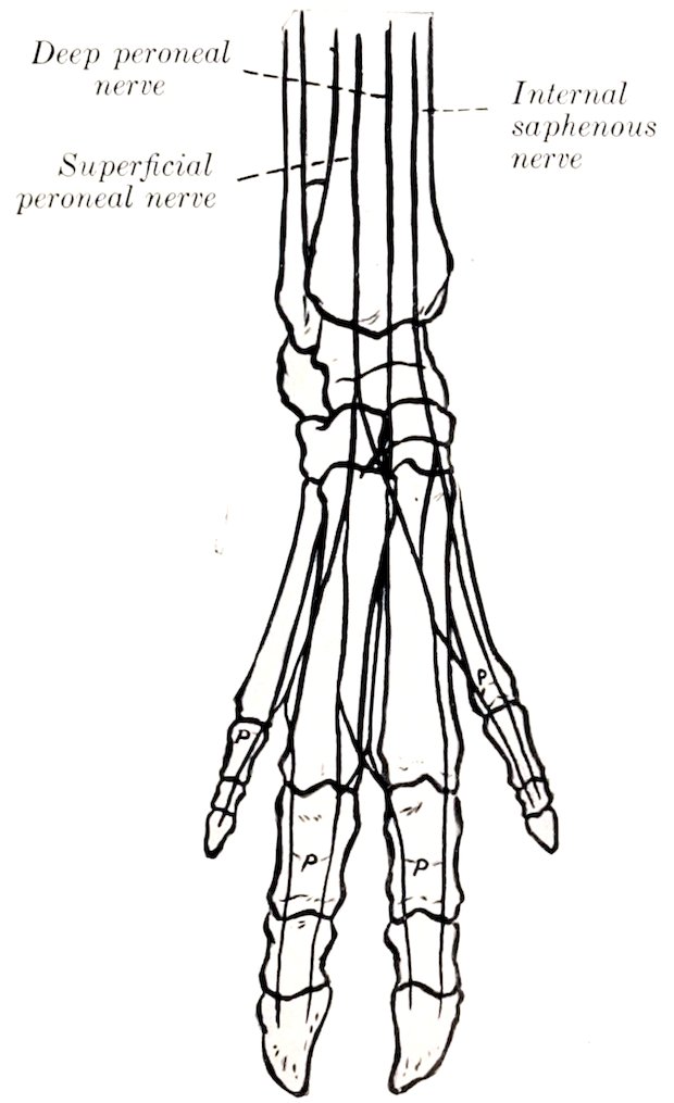
Fig. 537.—Nerves of Distal Part of Right Hind Limb of Pig, Anterior View (Schematic). p, Dorsal proper digital nerves.
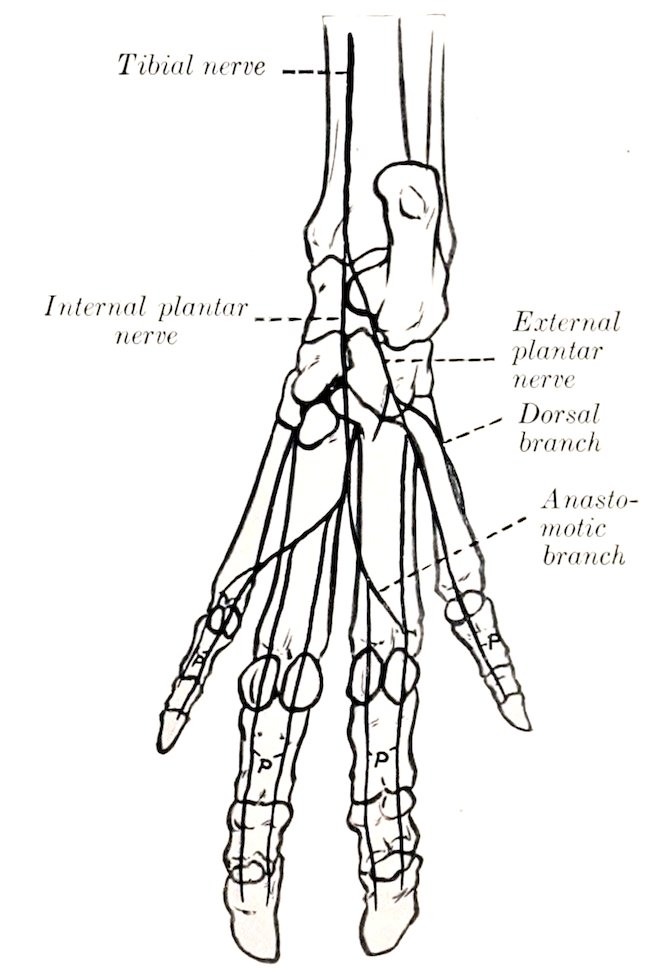
Fig. 538.—Nerves of Distal Part of Right Hind Limb of Pig, Posterior View (Schematic). p, Plantar proper digital nerves.
The lumbo-sacral plexus is derived from the ventral branches of the last three lumbar and first sacral nerves. The distribution of the branches of the plexus differs chiefly in the distal part of the limb. The superficial peroneal nerve is large and terminates by forming the greater part of the dorsal digital nerves. The latter, two for each digit, are also in part formed by the terminal branches of the deep peroneal nerve.
The tibial nerve divides at the tarsus into internal and external plantar nerves. The latter divide to form the plantar digital nerves, two for each digit. In addition, the external plantar nerve supplies a branch to the dorsal aspect of the outer (fifth) digit. The arrangement of the digital nerves is indicated by the annexed schematic figures.
724The sympathetic system of the pig has received very little attention from anatomists. The superior cervical ganglion is long and fusiform. It gives off filaments which join the vagus near the ganglion nodosum. The cervical trunk is short and relatively larger than in the ox; it is inclosed in a common sheath with the vagus in the neck and separates from that nerve to join the middle cervical ganglion at the thoracic inlet.
THE NERVOUS SYSTEM OF THE DOG
The spinal cord is almost circular in cross-section except at the well-marked cervical and lumbar enlargements, where it is compressed dorso-ventrally. The conus medullaris lies over the junction of the sixth and seventh lumbar vertebræ. The length of the cord of a rather large dog was found to be about 38 cm.; of this, the cervical part was 11 cm., the thoracic 17.4 cm., the lumbar about 7 cm., and the sacro-coccygeal about 2.6 cm. (Flatau-Jacobson).
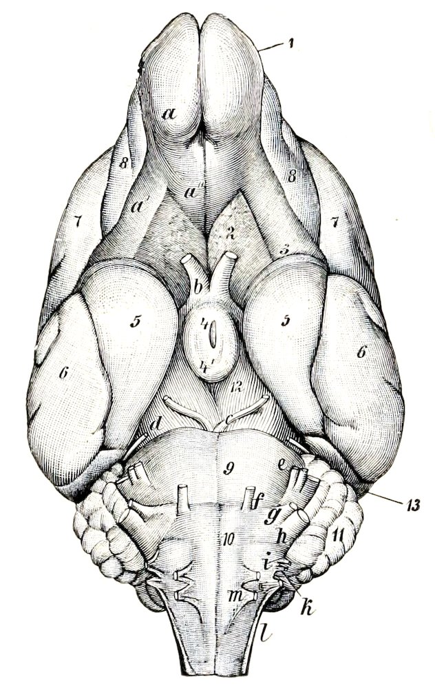
Fig. 539.—Base of Brain of Dog. a, Olfactory bulb; a′, a″, external and internal olfactory tracts; b, optic nerve; c, oculomotor nerve; d, trochlear nerve; e, trigeminus; f, abducens; g, facial; h, auditory; i, glosso-pharyngeal; k, vagus; l, spinal accessory; m, hypoglossal; 2, trigonum olfactorium; 3, anterior perforated substance; 4, tuber cinereum and infundibulum; 4′, mammillary body; 5, pyriform lobe; 6, temporal lobe; 7, parietal lobe; 8, frontal lobe; 9, pons; 10, medulla oblongata; 11, cerebellum; 12, cerebral peduncle; 13, occipital lobe. (Ellenberger-Baum, Anat. d. Hundes.)
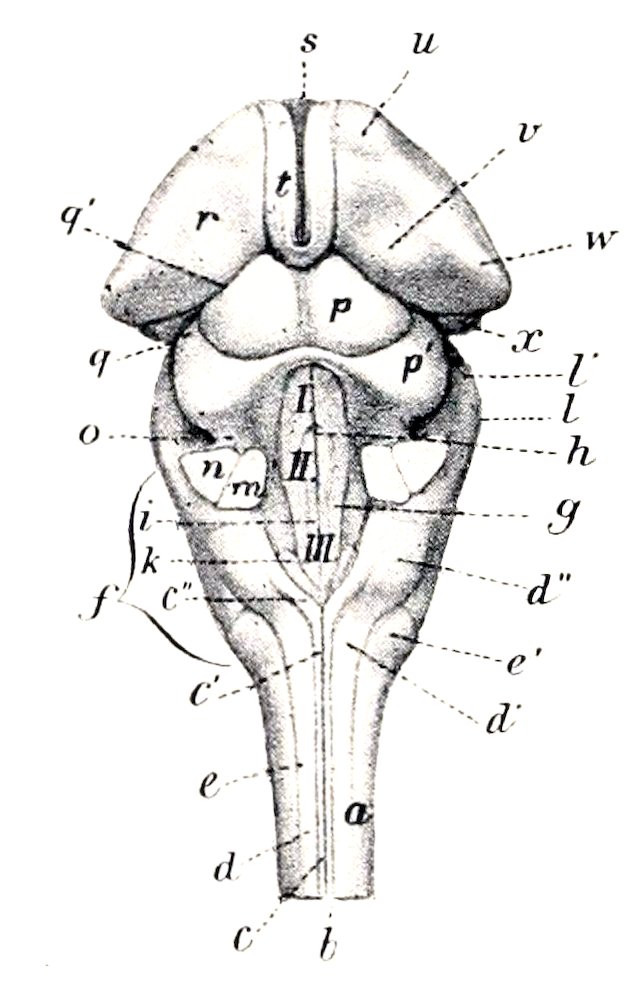
Fig. 540.—Brain Stem of Dog, Dorsal View. a, Spinal cord; b, dorsal median groove; c, c′, funiculus gracilis; c″, clava; d, d′, funiculus cuneatus; d″, tuberculum cuneatum; e, funiculus lateralis; e′, tuberculum Rolandi; f, medulla oblongata; I, II, III, floor of fourth ventricle (fossa rhomboidea); g, limiting groove; h, median fissure; i, eminentia medialis; k, ala cinerea; l, l′, pons; m, posterior peduncle, and n, middle peduncle of cerebellum (cut); o, anterior peduncle of cerebellum; p, p′, corpora quadrigemina, anterior and posterior; q, q′, peduncles (brachia) of corpora quadrigemina; r, thalamus; s, massa intermedia; t, stria medullaris; u, anterior tubercle of thalamus; v, pulvinar; w, corpus geniculatum laterale; x, corpus geniculatum mediale. (After Ellenberger, in Leisering’s Atlas.)
The brain weighs about 60 to 70 g. in dogs of medium size, but there is, of course, a wide range of weight in the different breeds. Thus in small terriers the 725weight is 30 g. or even less, while in very large dogs it may exceed 150 g. It corresponds much more closely with the cranium in size and general form than in the animals previously described; this is specially true of the small breeds, in which the bony crests and frontal sinuses are little developed.
The medulla is broad and thick. Its ventral surface is strongly convex from side to side. The pyramids are large and prominent and are limited by distinct median and lateral grooves. The olivary eminence is a well defined oval elevation situated between the pyramid and the superficial origin of the hypoglossal nerve. The external arcuate fibers form a wide band which crosses the lateral surface obliquely upward and forward, and obscures the groove which limits the restiform body externally. The cuneate tubercle is distinct. The rhomboid fossa is deep and narrow.
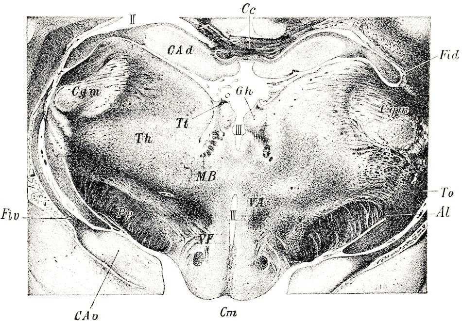
Fig. 541.—Part of Cross-section of Brain of Dog, Passing through Ganglion Habenulæ. Al, Ansa lenticularis of thalamic radiation; C a d, dorsal end of hippocampus; C a v, ventral end of same; C c, corpus callosum; C g m, corpus geniculatum mediale; C m, corpus mammillare; F i d, fimbria; F i v, ventral part of fimbria; G h, ganglion habenulæ; M B, fasciculus retroflexus; P p, pes pedunculi; T h, thalamus; T o, tractus opticus; T t, habenula; V A, fasciculus thalamo-mammillaris; V F, fornix ventralis; II, lateral ventricle; III, third ventricle. (After Probst.)
The pons is relatively small. The corpus trapezoideum is very wide and is divided by the pyramids into two lateral parts. The cerebellum is very broad, but is low and also compressed from before backward. Rather more than half of it is overlapped by the cerebral hemispheres. The anterior surface is accurately adapted to the concave tentorial surfaces of the hemispheres and to the posterior corpora quadrigemina and their commissure. The posterior surface is almost vertical and is convex centrally and flattened laterally. The vermis is prominent and in general well defined, although it is connected in its middle part with the hemispheres. The latter are three-sided and consist of four lobules. The anterior peduncles are very short.
The posterior corpora quadrigemina are large, very wide apart, and prominent, and are connected by a curved commissure. The internal geniculate body is large. 726In the deep interpeduncular fossa are two small bands which indicate the course of the fasciculi retroflexi, tracts which connect the habenular and interpeduncular ganglia. The mammillary body is double. The tuber cinereum is relatively large. The pituitary body is circular and rather small.
The cerebral hemispheres are very broad behind and diminish in width anteriorly; there is a sudden narrowing at the frontal poles, which are flattened laterally. The tentorial surfaces are concave and form a deep cavity which receives the anterior part of the cerebellum. The arrangement of the fissures and gyri is simpler than in the animals previously examined. The chief fissures are as follows:
1. The lateral fissure (of Sylvius) extends upward and backward from the sulcus rhinalis a little behind the middle of the lateral surface of the hemisphere.
2. The ectosylvian fissure has approximately the shape of an inverted u, and curves over the lateral fissure. It is regarded as consisting of three parts—anterior, middle, and posterior (F. ectosylvia anterior, media, posterior).
3. The suprasylvian fissure lies above and approximately concentric with the preceding. It also consists of three parts—anterior, middle, and posterior.
4. The ectomarginal or collateral fissure runs forward on the dorsal aspect of the hemisphere almost parallel with the dorso-medial border. A little in front of the middle of the surface it gives off a short ansate fissure (F. ansata) which runs obliquely inward and forward, and is continued by the coronal fissure.
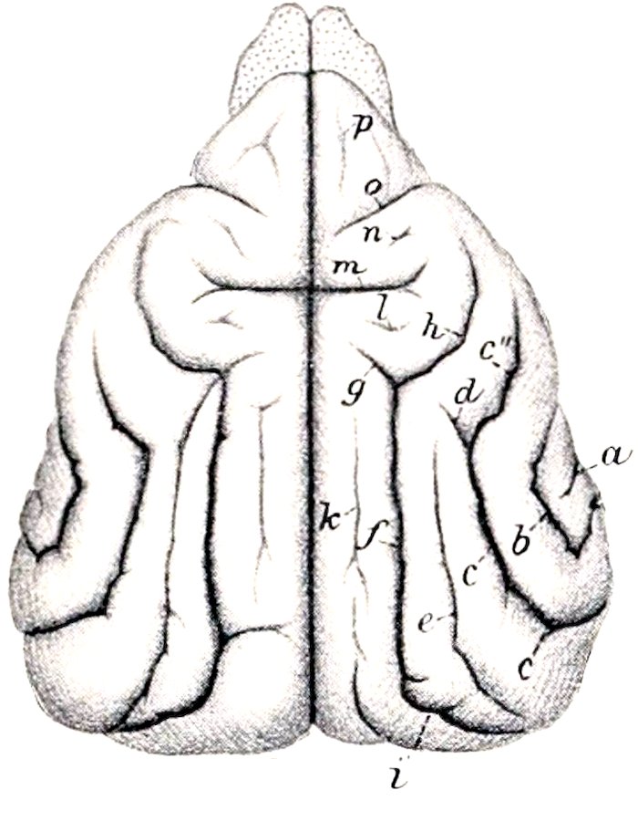
Fig. 542.—Cerebrum of Dog, Dorsal View. a, End of lateral fissure (of Sylvius); b, ectosylvian fissure; c, c′, c″, suprasylvian fissure; d, fissura ansata minor; e, ectolateral fissure; f, ectomarginal or collateral fissure; g, fissura ansata; h, coronal fissure; i, medilateral fissure; k, entomarginal fissure; l, postcruciate fissure; m, cruciate fissure; o, presylvian fissure; p, fissura prorea. (After Ellenberger, in Leisering’s Atlas.)
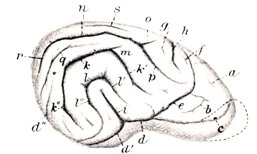
Fig. 543.—Fissures of Lateral Surface of Cerebrum of Dog, Right Side. a, Fissura prorea; b, frontal fissure; c, olfactory fissure; d, d′, sulcus rhinalis; e, presylvian fissure; f, precruciate fissure; g, postcruciate fissure; h, cruciate fissure; i, lateral fissure (of Sylvius); k, k′, k″, suprasylvian fissure; l, l′, l″, ectosylvian fissure; m, fissura ansata minor; n, ectomarginal or collateral fissure; o, fissura ansata; p, coronal fissure; q, ectolateral fissure; r, medilateral fissure; s, entomarginal fissure. (After Ellenberger, in Leisering’s Atlas.)
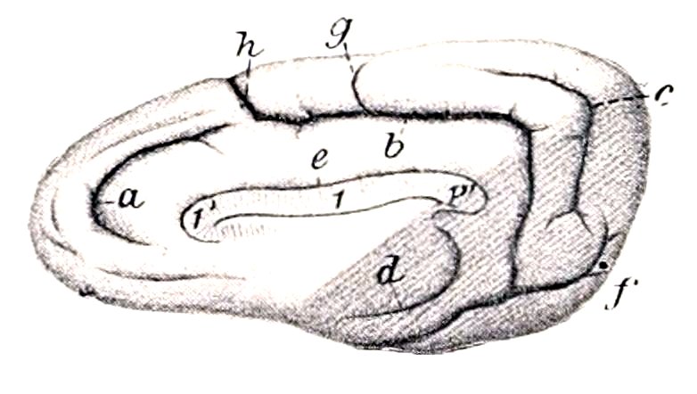
Fig. 544.—Right Cerebral Hemisphere of Dog, Inner Side. 1, Corpus callosum; 1′, genu, 1″, splenium of corpus callosum; a, genual fissure; b, splenial fissure; c, suprasplenial fissure; d, hippocampal fissure; e, sulcus corporis callosi; f, postsplenial fissure; g, lesser cruciate fissure; h, cruciate fissure. (After Ellenberger, in Leisering’s Atlas.)
5. The coronal fissure is the continuation of the ectomarginal. It runs forward and downward, curving around the cruciate fissure, and ends behind the middle of the presylvian fissure.
7276. The ectolateral fissure lies between the posterior part of the ectomarginal fissure and the suprasylvian fissure.
7. The entomarginal fissure lies between the anterior part of the ectomarginal fissure and the dorso-medial border. It is often indistinct and not rarely absent.
8. The presylvian fissure begins at the sulcus rhinalis, a little in front of its middle, and curves over the anterior third of the lateral surface of the hemisphere in front of the coronal fissure.
9. The cruciate fissure is the deepest and most characteristic. It cuts deeply into the anterior third of the dorso-medial border and runs almost straight outward. It is continuous on the inner surface of the hemisphere with the calloso-marginal or splenial fissure.
10. The sulcus rhinalis is continued on the tentorial surface of the hemisphere by the occipito-temporal fissure. Anteriorly it is prolonged as the olfactory fissure, which is concealed by the olfactory bulb and tract.
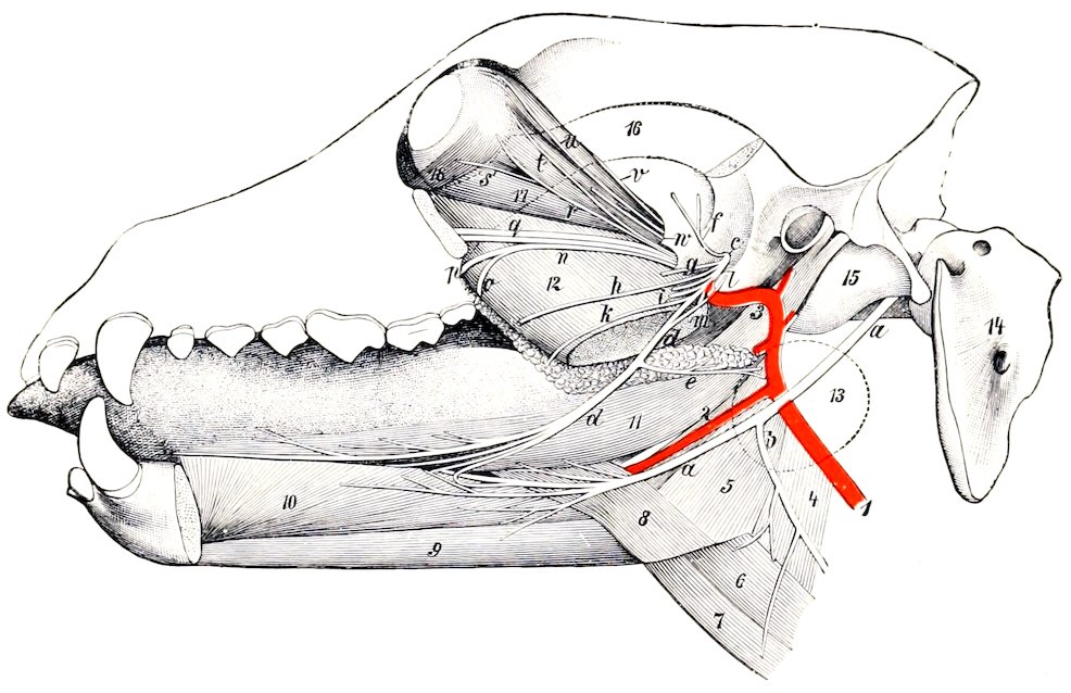
Fig. 545.—Deep Dissection of Head of Dog, showing Especially Trigeminal and Hypoglossal Nerves. a, Hypoglossal nerve; b, cervical branch of a; c, mandibular division of trigeminus; d, lingual nerve; e, nerve to submaxillary gland; f, deep temporal nerve; g, pterygoid nerve; h, buccinator nerve (cut); i, inferior alveolar or dental nerve; k, staphyline branch of lingual nerve; l, chorda tympani; m, mylo-hyoid nerve; n, sphenopalatine nerve; o, lesser palatine nerve; p, great palatine nerve; q, infraorbital nerves; r, subcutaneus malæ; s, branch of oculomotor nerve to inferior oblique muscle; t, lacrimal nerve; u, frontal nerve; v, trochlear nerve; w, abducens; 1, carotid artery; 2, lingual artery; 3, internal maxillary artery; 4, thyro-pharyngeus muscle; 5, hyo-pharyngeus muscle; 6, thyro-hyoideus; 7, sterno-hyoideus; 8, baseo-glossus. 9, genio-hyoideus; 10, genio-glossus; 11, stylo-glossus; 12, pterygoideus internus; 13, outline of submaxillary gland (dotted); 14, atlas; 15, bulba ossea; 16, zygomatic arch (dotted); 17, rectus oculi inferior; 18, obliquus oculi inferior. (Ellenberger-Baum, Anat. d. Hundes.)
11. The calloso-marginal or splenial fissure is parallel with the splenium and middle part of the corpus callosum on the inner surface of the hemisphere. It is continuous in front with the cruciate fissure and runs downward and outward across the tentorial surface to join the occipito-temporal fissure and sulcus rhinalis. It gives off from its highest part the short lesser or accessory cruciate fissure.
12. The suprasplenial fissure runs almost parallel with the posterior part of the calloso-marginal fissure on the inner and tentorial surfaces of the hemisphere.
The olfactory bulb is large and is strongly compressed laterally; its anterior end projects beyond the frontal pole of the hemisphere. The tract is short and is 728also flattened laterally. Both contain a narrow cavity which communicates with the lateral ventricle. The pyriform lobe is large and rounded.
The cranial nerves present the following special features which are worthy of notice:
The ophthalmic nerve gives off lacrimal, frontal, long ciliary, ethmoidal, and infratrochlear branches. The lacrimal nerve arises from the ophthalmic at its origin;[215] its recurrent branch emerges at the orbital ligament and concurs with the zygomatic and frontal nerves in the formation of the anterior auricular plexus. The frontal nerve emerges from the orbit in front of the upper end of the orbital ligament, ramifies in the upper lid and the adjacent skin of the forehead, and sends branches backward to the anterior auricular plexus. The long ciliary nerve accompanies the optic nerve and divides into several branches which pierce the posterior part of the sclera. The ethmoidal nerve gives off internal nasal branches and ends in the muzzle. The infratrochlear nerve runs forward between the internal straight and the superior oblique muscles of the eye and ramifies on the face in the vicinity of the inner canthus.
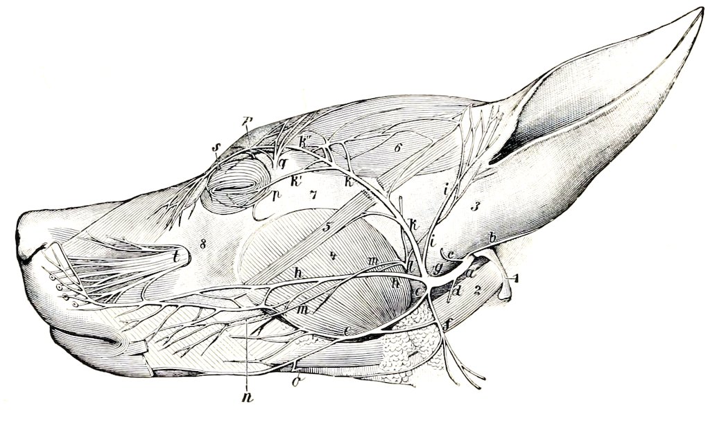
Fig. 546.—Nerves of Face of Dog. Parotid Gland is Removed. a, Facial nerve; b, posterior auricular nerve; c, internal auricular nerve; d, digastric nerve; e, inferior buccal nerve; f, cervical branch of facial; g, auriculo-palpebral; h, superior buccal nerve; i, temporal branch; k, k′, k″, zygomatic branch; l, auriculo-temporal; m, malar branch of l; n, buccinator nerve; o, mylo-hyoid branch; p, subcutaneus malæ; q, lacrimal nerve; r, frontal nerve; s, infratrochlear nerve; t, infraorbital nerve; 1, paramastoid process; 2, digastricus muscle; 3, base of concha; 4, masseter; 5, zygomaticus; 6, scutularis; 7, zygomatic arch; 8, maxilla. (Ellenberger-Baum, Anat. d. Hundes.)
The maxillary nerve, after giving off the zygomatic, divides into infraorbital and sphenopalatine branches. The zygomatic nerve emerges through an opening in the upper part of the orbital ligament and ramifies in the lower lid and on the adjacent surface of the face. The infraorbital nerves, two in number, divide within the infraorbital canal and after their emergence upon the face, thus forming seven or eight external nasal and superior labial branches. The sphenopalatine nerve gives off lesser and greater palatine and posterior nasal nerves.
The mandibular nerve passes out through the foramen ovale. The superficial temporal nerve divides into auricular, temporal, and malar branches; the last-named crosses the masseter and ramifies with the buccal nerves. The mylo-hyoid nerve is given off from the mandibular nerve almost immediately after the latter emerges from the cranium; it innervates the mylo-hyoideus and digastricus and gives off a branch which turns around the lower jaw, joins the inferior buccal nerve, 729and ramifies on the lateral surface of the face. The inferior alveolar nerve arises by a common trunk with the mylo-hyoid; it gives off dental branches and terminates in mental and inferior labial branches. The lingual nerve supplies vasodilator and excito-secretory filaments to the submaxillary and sublingual salivary glands; these fibers are derived from the chorda tympani.
The facial nerve divides near the posterior border of the jaw into four branches. The upper branch is the auriculo-palpebral nerve, which divides after a very short course into anterior auricular and zygomatic branches. The latter curves upward and forward across the zygomatic arch toward the eye, and divides into branches which supply the eyelids and nasal region and concur with the frontal and lacrimal nerves in forming the anterior auricular plexus. The superior buccal nerve accompanies the parotid duct across the masseter. The inferior buccal nerve runs forward along the lower border of the masseter and the mandible. The two nerves ramify on the cheek and anastomose with each other and the infraorbital nerves to form a plexus from which branches go to the muscles of the lips and nostrils. The cervical branch runs downward and backward over the submaxillary gland and ramifies in the cervical panniculus; it communicates with the inferior buccal nerve and sends twigs to the parotido-auricularis muscle and the submaxillary space.
The vagus bears a jugular ganglion and a ganglion nodosum. The latter is situated near the superior cervical ganglion on the rectus capitis anterior major and dorsal to the carotid artery; it is fusiform and may be about half an inch (ca. 1 to 1.5 cm.) long in a large dog. In its course in the neck the nerve is inclosed with the sympathetic trunk in a common sheath and is related ventrally to the common carotid artery. The formation of dorsal and ventral œsophageal trunks by the union of the dorsal and ventral divisions of the two nerves takes place at the hiatus œsophageus. The dorsal trunk concurs with branches of the solar plexus in forming the posterior gastric plexus on the visceral surface of the stomach. The ventral trunk ramifies on the parietal surface of the stomach, forming the anterior gastric plexus, from which a considerable branch passes along the lesser curvature to the pylorus. Some special features of the collateral branches are: Two pharyngeal branches are present. The superior pharyngeal branch arises from the vagus above the ganglion nodosum. It passes under the deep face of the carotid artery and descends on the lateral surface of the pharynx to end in the crico-thyroid muscle. It furnishes a root of the inferior pharyngeal branch, and communicates with the pharyngeal branch of the ninth and with the superior laryngeal and hypoglossal nerves. The inferior pharyngeal branch is formed by the union of roots derived from the superior pharyngeal nerve and the ganglion nodosum, together with filaments from the superior cervical ganglion. It crosses the side of the pharynx behind the superior pharyngeal and ramifies on the posterior constrictor of the pharynx and the origin of the œsophagus. It contributes filaments to the pharyngeal plexus, communicates with the recurrent nerve, and supplies twigs to the thyroid gland. The pharyngeal plexus is formed on the lateral surface of the pharynx by branches of the pharyngeal nerves and the communications above described; branches from it innervate the muscles and mucous membrane of the pharynx. The superior laryngeal nerve arises from the ganglion nodosum and descends over the side of the pharynx, crossing beneath the carotid artery and the superior laryngeal nerve. It passes through the thyroid notch and ramifies in the mucous membrane of the larynx. It communicates with the superior cervical ganglion and the superior pharyngeal branch of the vagus, and gives twigs to the hyo-pharyngeus muscle. Immediately after its entrance into the larynx it gives off a large branch which, instead of uniting with the recurrent as in the other animals, has a peculiar arrangement. It runs back near the dorsal border of the thyroid cartilage, gives a branch to the crico-arytenoideus posterior as it passes over that muscle, and continues 730along the trachea internal to the recurrent nerve. At the thoracic inlet it communicates with the inferior cervical ganglion and continues backward to unite with the vagus at or near the point of origin of the recurrent nerve. Its collateral filaments supply the trachea and concur with the inferior pharyngeal branch in forming a plexus on the cervical part of the œsophagus which innervates that tube.[216] The depressor nerve is a very delicate filament which arises usually from the superior laryngeal nerve and accompanies the vago-sympathetic trunk to the thorax; its fibers reach the heart through the cardiac nerves. The pulmonary and posterior œsophageal plexuses are highly developed.
The hypoglossal nerve gives off a long branch (R. descendens) which runs downward and backward across the pharynx and larynx, communicates with the ventral branch of the first cervical nerve, and supplies the thyro-hyoid, sterno-hyoid, and sterno-thyroid muscles.
The spinal nerves number thirty-six or thirty-seven on either side, and comprise eight cervical, thirteen thoracic, seven lumbar, and five or six coccygeal.
The brachial plexus (Fig. 487) is derived from the ventral branches of the last four cervical and first thoracic nerves; the root supplied by the fifth cervical nerve is very small. The roots unite at the lower border of the scalenus. The more important special features in the arrangement of the nerves which emanate from the plexus are as follows:
The musculo-cutaneous nerve passes between the coraco-brachialis and the brachial artery and descends in the arm in front of the artery. At the shoulder joint it gives off branches to the biceps and coraco-brachialis, and in the lower third of the arm is connected with the median nerve by an oblique branch. It terminates near the elbow by dividing into a branch for the brachialis and a small cutaneous nerve which passes down over the inner face of the elbow and, inclining a little forward, descends over the deep fascia of the forearm to the carpus.
The radial nerve descends behind the ulnar nerve, gives branches to the extensors of the elbow, dips in between the internal head of the triceps and the accessory head of the anconeus, winds around the arm, and divides between the brachialis and the external head of the triceps into two branches. The deep branch (R. profundus) supplies the extensor and supinator muscles on the forearm. The superficial branch (R. superficialis) emerges upon the flexor surface of the elbow and divides into two branches which terminate by supplying two dorsal digital nerves to each digit, except the fifth, which receives its outer dorsal nerve from the ulnar. The inner branch descends along the inner side of the cephalic vein to the carpus, where it divides into dorsal digital nerves for the first digit and the inner side of the second. The outer branch is much larger. It runs downward on the middle of the front of the forearm and supplies the remaining dorsal digital nerves except that to the outer side of the fifth digit.
The ulnar nerve is as large as or larger than the median, with which it is united for some distance. At the distal third of the arm it separates from the median and passes over the internal epicondyle of the humerus. At the proximal part of the forearm it gives off the dorsal branch (R. dorsalis), which supplies cutaneous twigs to the dorso-lateral surface of the lower part of the forearm and carpus and terminates as the external dorsal digital nerve of the fifth digit. Descending under cover of the flexor carpi ulnaris, the ulnar nerve inclines inward under the tendon of insertion of that muscle and divides into superficial and deep branches. The superficial branch (R. superficialis) runs downward along the outer border of the flexor tendons, gives off the external volar digital nerve of the fifth digit (N. dig. vol. lat. dig. V), and a branch (N. met. vol. IV) which descends in the space between the fourth and fifth metacarpal bones and unites with the deep branch. The deep 731branch (R. profundus) descends in the carpal canal and divides under the deep flexor tendon into its terminal branches. The smaller of these supply the volar metacarpal muscles. The larger terminals are the three volar common digital nerves (Nn. dig. vol. comm. II, III, IV), which descend along the second, third, and fourth intermetacarpal spaces, subdivide, and concur with the volar metacarpal branches of the median nerve in forming the volar proper digital nerves (Nn. dig. vol. proprii).
The median nerve descends behind the brachial artery, passes over the internal epicondyle of the humerus, then under the pronator teres, and continues in the forearm under cover of the flexor carpi radialis. It gives branches below the elbow to the flexor and pronator muscles, and lower down a palmar branch to the skin on the inner and posterior aspect of the carpus, and terminates between the superficial and deep flexor tendons by dividing into three volar metacarpal nerves (N. met. vol. I, II, III). These descend in the first, second, and third intermetacarpal spaces and unite with the volar common digital nerves in forming volar proper digital nerves. The arrangement of the digital nerves is indicated in the annexed schematic figures.
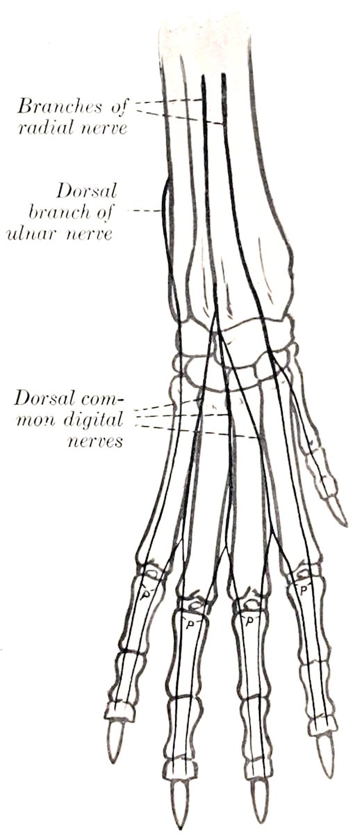
Fig. 547.—Nerves of Distal Part of Right Fore Limb of Dog, Anterior View (Schematic). p, Dorsal proper digital nerves.
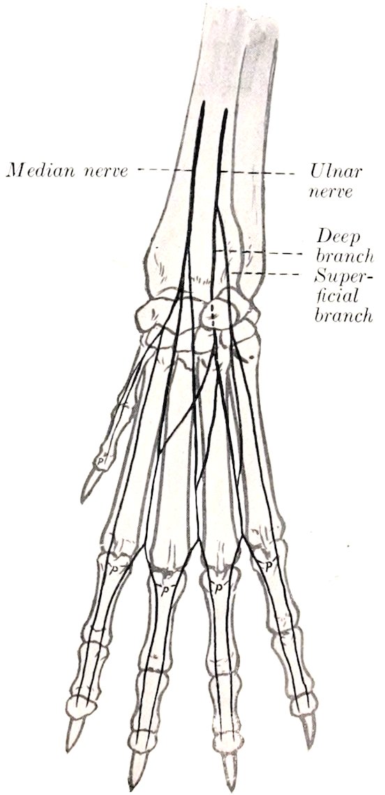
Fig. 548.—Nerves of Distal Part of Right Fore Limb of Dog, Posterior View (Schematic). p, Volar proper digital nerves.
The lumbo-sacral plexus is formed from the ventral branches of the last five lumbar and first sacral nerves. The more important special facts in regard to the nerves of the pelvic limb are as follows:
The saphenous nerve (Fig. 492) is relatively large, and, in fact, might be 732considered as the continuation of the femoral nerve. It descends at first in front of the femoral artery, then passes over the lower part of the inner surface of the thigh with the saphenous artery, continues down the leg with the dorsal branch of that vessel, and anastomoses with branches of the superficial peroneal nerve. It supplies cutaneous twigs from the stifle to the metatarsus.
The peroneal nerve separates from the tibial above the origin of the gastrocnemius, runs downward and forward across the outer head of that muscle, passes between the deep flexor of the digit and the peroneus longus, and divides into superficial and deep branches. The superficial peroneal nerve descends the leg along the peroneus tertius and longus, gives off a dorsal branch to the first digit (N. cutaneus dorsalis medialis), and divides at the proximal part of the metatarsus into three dorsal common digital nerves (Nn. dig. pedis dors. comm. II, III, IV). These descend with the superficial dorsal metatarsal arteries and concur with branches of the deep peroneal nerve in forming dorsal proper digital nerves (Nn. dig. ped. prop. dorsales). The deep peroneal nerve passes down the leg with the anterior tibial vessels. It gives twigs to the hock joint and the extensor brevis muscle, and divides into three dorsal metatarsal nerves (Nn. met. dors. II, III, IV); these descend with the corresponding arteries along the intermetatarsal spaces and unite with the dorsal common digital nerves in supplying the dorsal proper digital nerves.
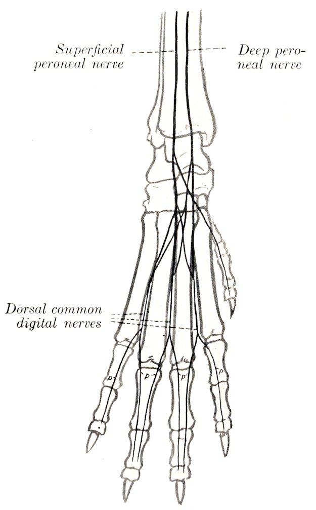
Fig. 549.—Nerves of Distal Part of Right Hind Limb of Dog, Anterior View (Schematic). p, Dorsal proper digital nerves.
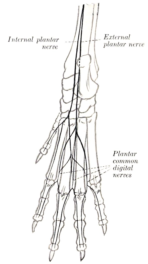
Fig. 550.—Nerves of Distal Part of Right Hind Limb of Dog, Posterior View (Schematic). p, Plantar proper digital nerves.
The tibial nerve divides at the tarsus into two plantar branches. The internal plantar nerve descends along the inner border of the superficial flexor tendon and 733divides near the middle of the metatarsus into two branches. Of these, the internal branch constitutes the first plantar common digital nerve (N. dig. comm. plant. I). The outer branch descends on the superficial flexor tendon and divides into three plantar metatarsal nerves (Nn. met. plant. II, III, IV), which unite with the plantar common digital nerves. The external plantar nerve passes down between the flexor tendons, sends twigs to the muscles on the plantar surface of the metatarsus, and divides into three plantar common digital nerves (Nn. dig. comm, plant. II, III, IV). These descend with the deep plantar metatarsal arteries in the second, third, and fourth intermetatarsal spaces, receive the plantar metatarsal nerves, and divide into plantar proper digital nerves, which pass down the opposed surfaces of the second to the fifth digits.
The sympathetic system presents few special features worthy of mention. The superior cervical ganglion lies above the ganglion nodosum close to the cranial base and internal to the origin of the internal carotid and occipital arteries. It forms connections directly or through the carotid plexus with the last seven cranial and first cervical nerves. The cervical trunk unites with the vagus so intimately as to form a vago-sympathetic trunk. There may be a small middle cervical ganglion in front of the inferior cervical ganglion. The latter is usually distinct from the first thoracic ganglion, which is stellate and lies on the longus colli opposite the second rib and intercostal space.
734