THE DIGESTIVE SYSTEM (continued 3)
Reading edition
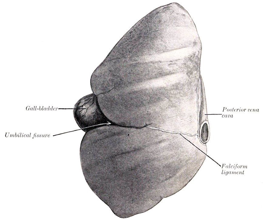
Fig. 306.—Liver of Sheep, Parietal Surface; Hardened in situ.
DIGESTIVE SYSTEM OF THE PIG
THE MOUTH
The rima oris is extensive, the angles of the mouth being situated far back. The upper lip is thick and short, and is blended with the snout; the lower lip is small and pointed. The labial glands are few and small.
The mucous membrane of the cheeks is smooth. The buccal glands are 411compactly arranged in two rows opposite the cheek teeth. The parotid duct opens opposite the fourth or fifth cheek tooth.
The hard palate is long and narrow; it is marked by a median furrow, on each side of which are twenty or more ridges. On its anterior part there is a triangular incisive papilla, at which the naso-palatine ducts open.
The soft palate is very thick. Its direction almost continues that of the hard palate, i. e., it is nearly horizontal. It extends to the middle of the oral surface of the epiglottis.[110] It has a median prolongation termed the uvula. The oral surface presents a median furrow, on either side of which is an oval raised area, marked by numerous crypts; these elevations are the tonsils. Tonsillar tissue also occurs in the lateral walls of the fauces.
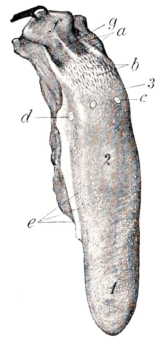
Fig. 307.—Tongue of Pig. 1, Apex; 2, dorsum; 3, root; a, orifices of ducts of lingual glands; b, papillæ of root; c, vallate papilla (not really so distinct as in figure); d, foliate papilla; e, fungiform papillæ; f, epiglottis (pulled back); g, median glosso-epiglottic fold. (Ellenberger-Baum, Anat. d. Haustiere.)
The tongue is long and narrow and the apex is thin. Two or three vallate papillæ are present. The fungiform papillæ are small and are most numerous laterally. The filiform papillæ are soft and very small. On the root there are soft, long, pointed papillæ, directed backward. Foliate papillæ are also present.
There is a well-marked median glosso-epiglottic fold, on either side of which is a depression (Vallecula epiglottica). The frenum linguæ is double.
The dental formula of the pig is:
( 3 1 4 3 )
2 ( I C P M ) = 44
( 3 1 4 3 )
412The formula for the temporary teeth is:
( 3 1 4 )
2 ( Di Dc Dp ) = 32
( 3 1 4 )
The upper incisors are small; they are separated from each other by spaces, and from the canines by a larger interval. The first incisors are the largest, are convergent, and have no distinct neck. The second are narrower and somewhat smaller. The third (corner) incisors are much smaller, are flattened laterally, and have three small eminences on the crown.
The lower incisors are almost horizontal and are convergent and close together. The first and second are about equal in size, prismatic, and deeply implanted in the jaw. The labial surface is slightly convex, the lingual concave and marked near its extremity by a ridge. The third incisor is much smaller and has a short narrow crown and a distinct neck.
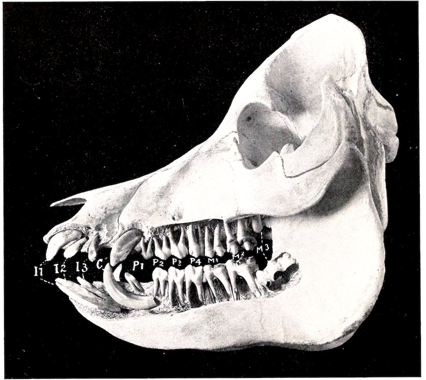
Fig. 308.—Skull of Pig about a Year and a Half Old, Sculptured to Show the Embedded Parts of the Teeth. I1–3, Incisors; C, canines; P1–4, premolars; M1–3, molars.
The canine teeth or tusks of the male are greatly developed and project out of the mouth. The upper canine of the boar may be about three or four inches (ca. 8 to 10 cm.) long. The crown is conical and is curved somewhat backward and outward; the embedded part is curved and has a large pulp cavity.[111] The lower canine may reach a length of eight inches or more (ca. 20 cm.); the crown is prismatic and is curved backward and outward in front of the upper one, so that the friction between the two keeps a sharp edge on the lower tooth. In the sow they are much smaller.
413
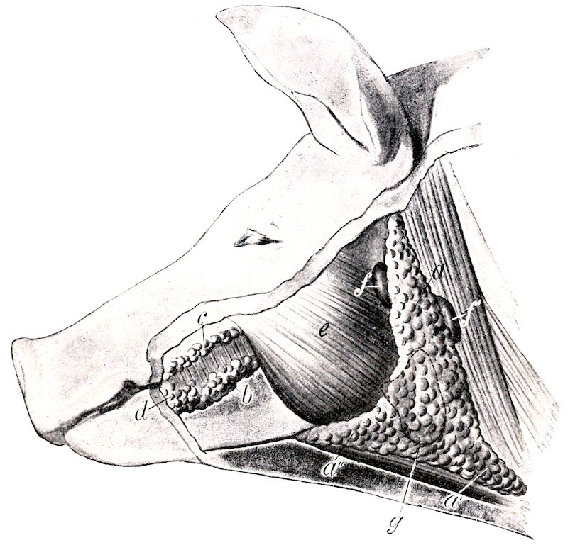
Fig. 309.—Superficial Glands of Head of Pig. a, Parotid gland; a′, a″, cervical and submaxillary angles of a; b, c, ventral and dorsal buccal glands; d, labial glands; e, masseter muscle; f, f′, lymph glands; g, dotted line indicating outline of submaxillary gland, which is concealed. (Ellenberger-Baum, Anat. d. Haustiere.)
The average periods of eruption of the teeth are given in the subjoined table.
Tooth Eruption Change
I1 2 to 4 weeks 12 months
I2 upper 2 to 3 months lower 1½ to 2 month 16 to 20 months
I3 Before birth 8 to 10 months
C Before birth 9 to 10 months
P1 5 months
P2 5 to 7 weeks 12 to 15 months
P3 upper 4 to 8 days lower 2 to 4 weeks.
P4 upper 4 to 8 days lower 2 to 4 weeks
M1 4 to 6 months
M2 8 to 12 months
M3 18 to 20 months
The cheek teeth increase in size from before backward. They have (with certain exceptions noted below) complex tuberculate crowns; the term bunodont is applied to this condition, as distinguished from the selenodont structure in horses and cattle, in which there are prominent ridges of enamel. They are short-crowned teeth with a distinct neck and root. The first tooth in each jaw is small, simple, and appears only once; in the lower jaw it is near the canine, in the upper near the second cheek tooth. The next two teeth are larger, laterally compressed, and sectorial. The fourth tooth below is larger, but otherwise like the preceding ones, while the upper one is much wider and is tuberculate.
414
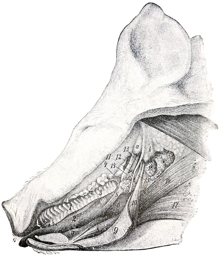
Fig. 310.—Dissection of Mouth and Pharyngeal Region of Pig. 1, Hard palate; 2, tongue showing distinctly fungiform papillæ; 3, foliate papilla; 4, lymph nodules of soft palate; 5, stylo-glossus muscle, out of which a portion has been cut; 6, hyo-glossus; 7, genio-glossus; 8, genio-hyoideus; 9, mylo-hyoideus (reflected); 10, stylo-hyoideus; 11, great cornu of hyoid bone; 12, tensor palati; 13, levator palati; 14, pterygo- et palato-pharyngeus; 15, stylo-pharyngeus; 16, chondro-pharyngeus; 17, sterno- et omo-hyoideus; 18, retropharyngeal lymph glands; a, bulla ossea; b, paramastoid process. (Ellenberger-Baum, Anat. d. Haustiere.)
The parotid gland is large and triradiate. It extends very little on to the masseter muscle and its upper angle does not quite reach the base of the ear. It is pale in color, and is embedded in fat in animals in good condition. On its deep face are several large subparotid lymph glands, some of which are only partially covered by the parotid. The duct arises on the deep face, has a course similar to that of the ox, and perforates the cheek opposite the fourth or fifth upper cheek tooth. Small accessory glands (Glandulæ parotideæ accessoriæ) may be found along the course of the duct.
415The submaxillary gland is small, reddish in color, and oval in outline; it is covered by the parotid. Its superficial face is convex, and is marked by rounded prominences. From its deep face a narrow process extends forward about two or three inches (ca. 5 to 7.5 cm.) beneath the mylo-hyoideus muscle along with the duct. The latter opens near the frenum linguæ, but there is no papilla.
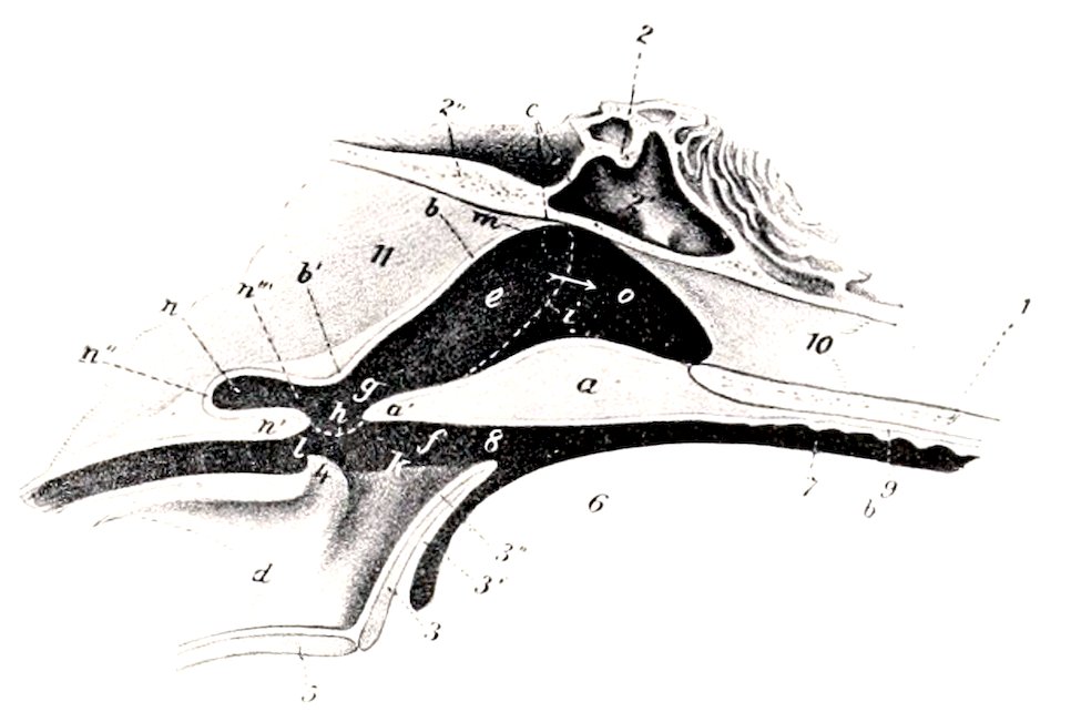
Fig. 311.—Sagittal Section of Pharyngeal Region of Pig, Partly Schematic. 1, Palate bone; 2, sphenoid bone; 2′, sphenoidal sinus; 2″ occipital bone; 3, epiglottis; 4, arytenoid cartilage; 5, thyroid cartilage; 6, root of tongue; 7, mouth cavity; 8, isthmus faucium; 9, hard palate; 10, septum nasi; 11, ventral muscles of head; a, soft palate; a′, free edge of a; b, dorsal wall of pharynx; c, fornix of pharynx; d, cavity of larynx; e, g, naso-pharynx; f, oro-pharynx; h, posterior pillar of soft palate; i, dotted line indicating lateral boundary between nasal cavity and pharynx; k, aditus laryngis; l, entrance to œsophagus; m, Eustachian orifice; n, pharyngeal recess; o, posterior naris. (After Ellenberger, in Leisering’s Atlas.)
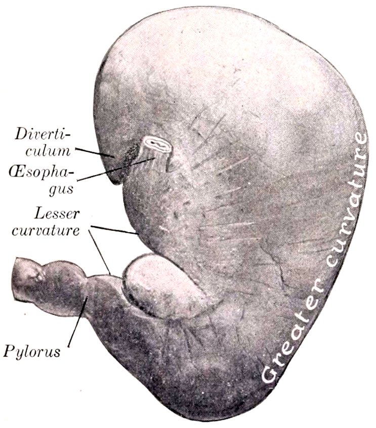
Fig. 312.—Stomach of Pig, Parietal Surface. The organ contained a rather small amount of ingesta and hence is somewhat contracted.
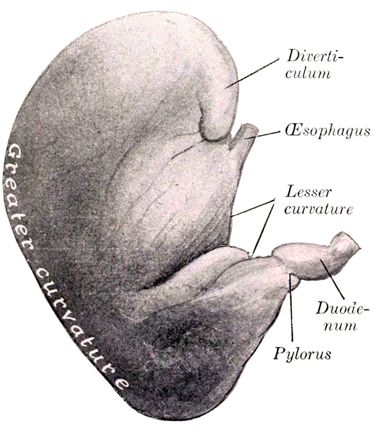
Fig. 313.—Stomach of Pig, Visceral Surface. Organ was fixed in situ and is somewhat contracted.
The sublingual gland has an arrangement similar to that of the ox. The posterior part (Glandula sublingualis grandicanalaris) is reddish-yellow in color, and is about two inches (ca. 5 cm.) long, and half an inch wide; its posterior end is in relation to the submaxillary gland and its duct. The anterior part is much 416larger, being two or three inches (ca. 5 to 7 cm.) long and about twice the width and thickness of the posterior part. All or most of the ducts from the posterior part unite to form the ductus sublingualis major, which opens near the ductus submaxillaris. Eight or ten ductus sublinguales minores convey the secretion from the anterior part through the floor of the mouth.
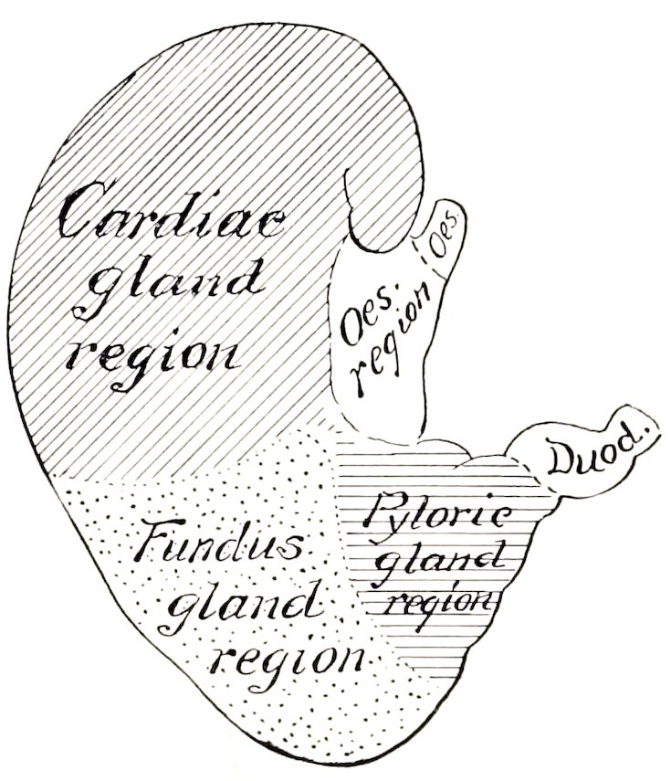
Fig. 314.—Diagram of Zones of Mucous Membrane of Stomach of Pig.
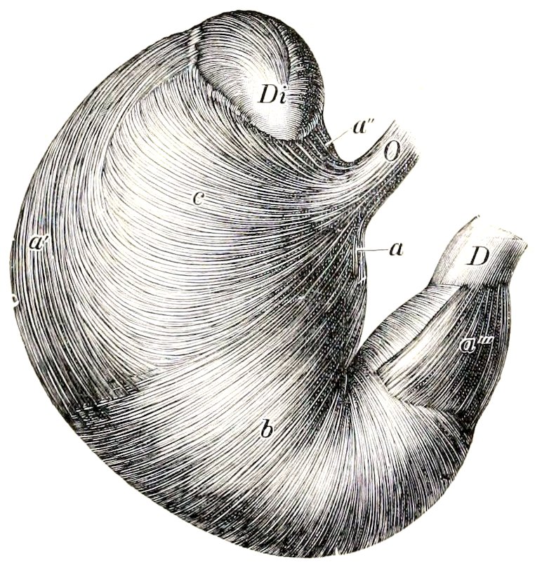
Fig. 315.—Visceral Surface of Stomach of Pig, from which the Serous Coat has been Removed.
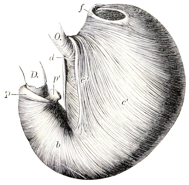
Fig. 316.—Everted Stomach of Pig, from which the Mucous Membrane has been Removed. O, Œsophagus; D, duodenum; Di, diverticulum; a, a′, a″, a‴, longitudinal fibers; b, circular fibers; c, external oblique fibers; c′, internal oblique fibers; c″, cardiac loop; d, fibers which connect branches of cardiac loop; f, fold at entrance to diverticulum; p, pyloric sphincter; p′, pyloric prominence. (Ellenberger-Baum, Anat. d. Haustiere.)
THE PHARYNX
The pharynx presents in its posterior part a median cul-de-sac about an inch and a half (ca. 3 to 4 cm.) long, which is situated between the ventral straight muscles of the head and the origin of the œsophagus. Its lower margin is formed by the junction of the posterior pillars of the soft palate, which contain muscular 417tissue derived from the palatinus and palato-pharyngeus. The vault of the pharynx (Fornix pharyngis) is divided by a median fold of mucous membrane which is a direct continuation of the septum nasi. On either side of this is an infundibulum in which the Eustachian tube opens.
THE ŒSOPHAGUS
The œsophagus is short and nearly straight. It has (according to Rubeli) a potential caliber of nearly 3 inches (ca. 7 cm.) at either end, and about 1⁷⁄₁₀ inches (ca. 4.2 cm.) in its middle part.[112] The muscular coat, except near the cardia, is striated. There are mucous glands in the submucosa to about the middle of the tube.
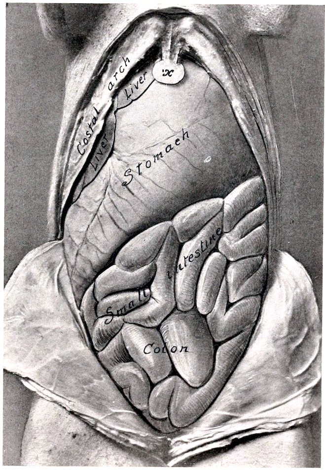
Fig. 317.—Abdominal Viscera of Young Pig, Ventral Aspect. The stomach was very large in this subject.
418
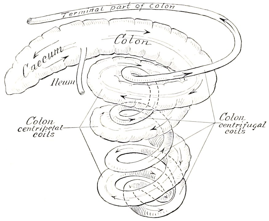
Fig. 318.—Diagram of Cæcum and Colon of Pig. Coils of colon have been pulled apart somewhat.
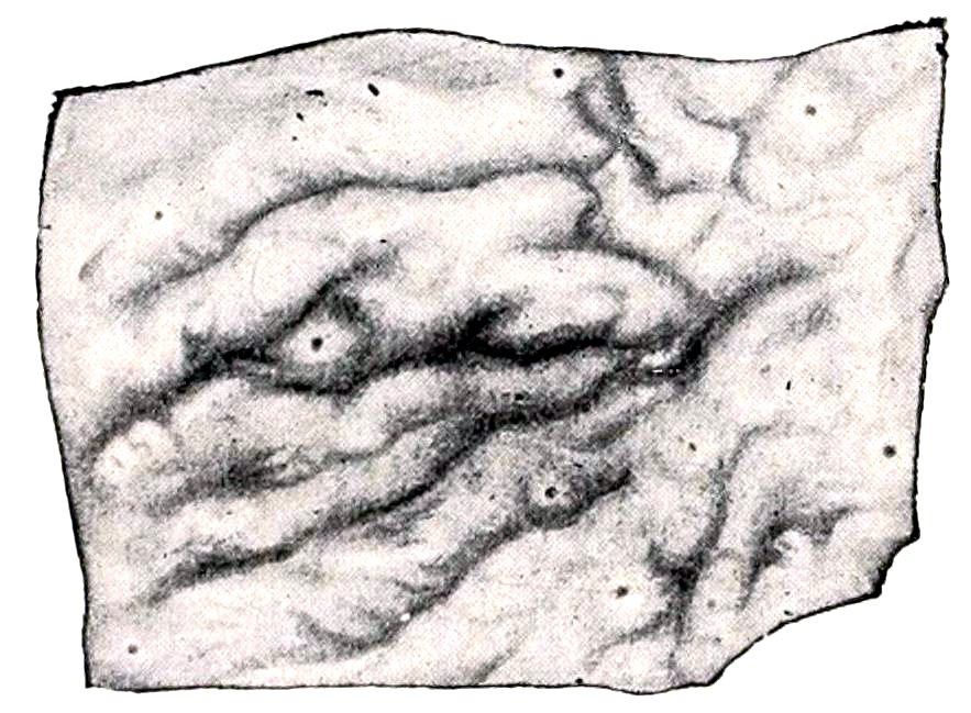
Fig. 319.—Solitary Nodules from Large Intestine of Pig. (Ellenberger-Baum, Anat. d. Haustiere.)
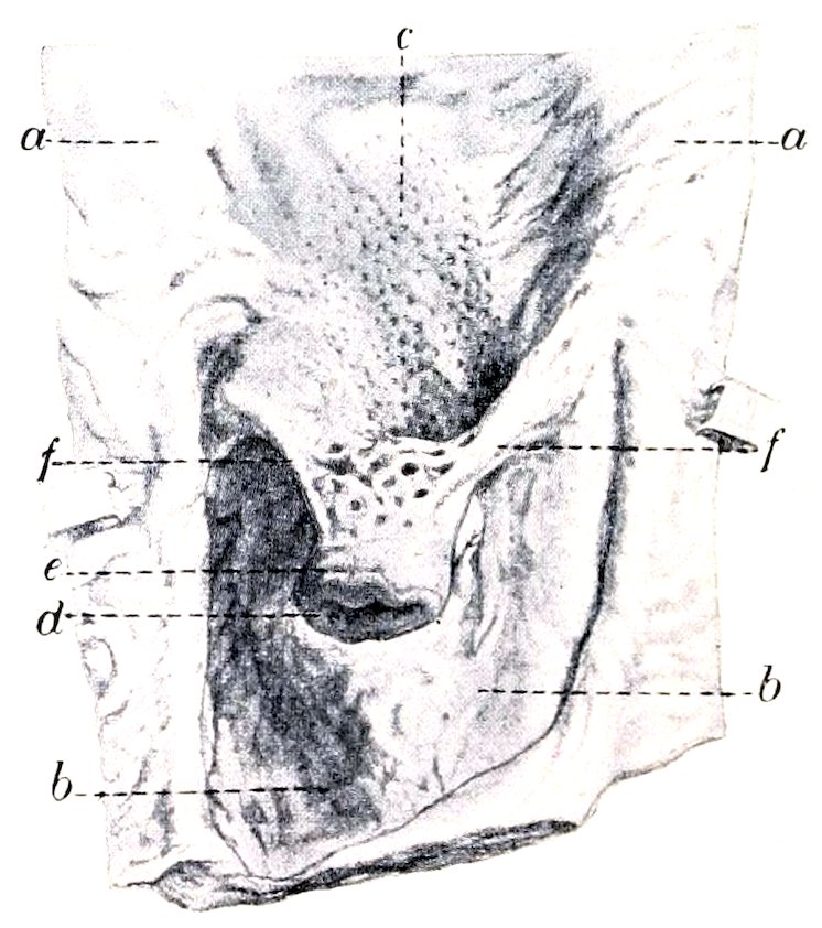
Fig. 320.—Ileo-cæcal Opening and Adjacent Part of Cæcum and Colon of Pig. a, Mucous membrane of colon; b, mucous membrane of cæcum; c, Peyer’s patch; d, ileo-cæcal opening; e, end of ileum; f, folds (frenula) of ileum. (After P. Schumann.)
THE STOMACH
The stomach is large and pyriform. Its average capacity is about 1½ to 2 gallons (ca. 5.7 to 8 liters). When full it extends backward to the last left intercostal space laterally and the umbilicus ventrally. The left portion is large and rounded, while the right portion (Pars pylorica) is small, and bends sharply upward to join the small intestine. The parietal surface is related to the liver, gall-bladder, and diaphragm. The visceral surface is related to the intestine, the mesentery, and the pancreas. The great curvature is related to the diaphragm, spleen, and abdominal floor. The pyloric end lies on the right lateral lobe of the liver. The left extremity presents a flattened conical blind pouch, the diverticulum ventriculi, the apex of which projects backward. The œsophagus joins the stomach very obliquely, almost in the median plane, and about three or four inches (ca. 8 to 10 419cm.) below the twelfth thoracic vertebra. The cardiac opening is slit-like and is bounded above and to the left by a fold which contains a thickening of the internal oblique layer of the muscular coat. The opening into the diverticulum is situated above and a little to the left of the cardia; it is transversely oval, and is bounded (except externally) by a thick fold which contains spirally arranged muscular fibers. The mucous membrane may be divided into four regions. Over a quadrilateral area around the cardia (about one inch on the right and two or three inches on the left side of the orifice) it is œsophageal in character, and presents a number of folds. A sharp line of demarcation separates this from the rest of the mucous membrane, which is soft and glandular. The second or cardiac gland region is pale gray in color and thin (ca. 0.5 to 1 mm.); it extends about to the middle of the stomach. The third or fundus gland region is readily distinguished by its thickness (ca. 3 mm.) and its brownish-red mottled appearance.[113] The fourth or pyloric region is pale, thinner than the preceding, and presents a number of irregular folds. At the pylorus there is a remarkable ridge which projects from the wall of the lesser curvature and diminishes considerably the size of the orifice. It is about an inch and a half (ca. 3 to 4 cm.) long and nearly half an inch (ca. 1 cm.) high. Sometimes it is grooved; in other cases it has the form of a rounded eminence attached by a pedicle to the wall. Fibers from the circular muscular coat extend into it.
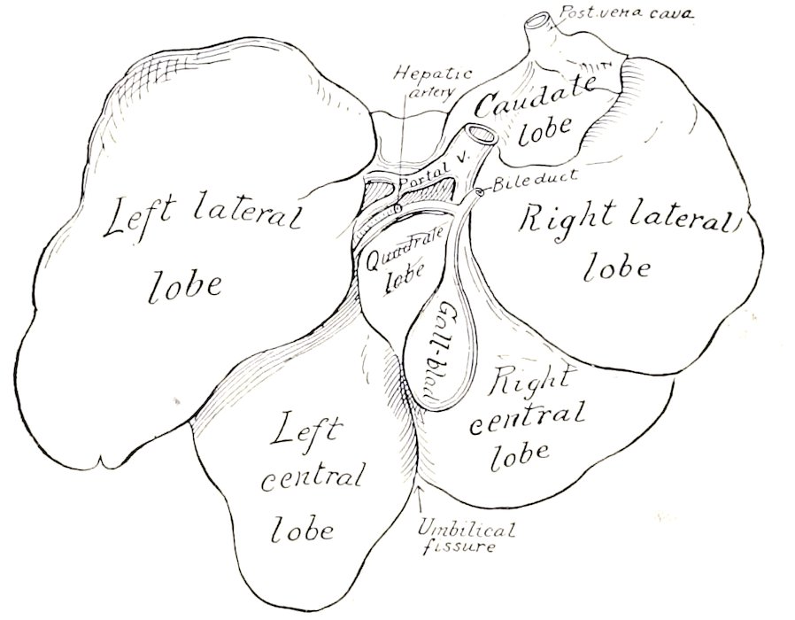
Fig. 321.—Liver of Pig, Soft Specimen Sketched with Lobes Drawn Apart.
THE INTESTINE
The small intestine is 50 to 65 feet (ca. 15 to 20 m.) long. The mesentery of about the first two feet (ca. 60 cm.) is two to two and a half inches (ca. 5 to 6 cm.) long; this part may be termed duodenum. The remainder (Jejuno-ileum) has a mesentery about six to eight inches (ca. 15 to 20 cm.) long, which is thick and contains a quantity of fat, and numerous large lymph glands at its root; the root is attached in the sublumbar region behind the stomach and blends here with the mesentery of the large intestine. The small intestine is arranged in close coils and lies mainly on the left side and floor of the abdomen, from the stomach to the pelvis; some coils, however, lie against the right flank. The opening of the bile-duct is about one or two inches (ca. 2.5 to 5 cm.) from the pylorus, and that of the pancreatic duct about four or five inches (ca. 10 to 15 cm.) beyond it. Peyer’s 420patches and solitary glands are numerous and very distinct; the patches are usually band-like and prominent; the solitary glands form projecting nodules marked by crypts.
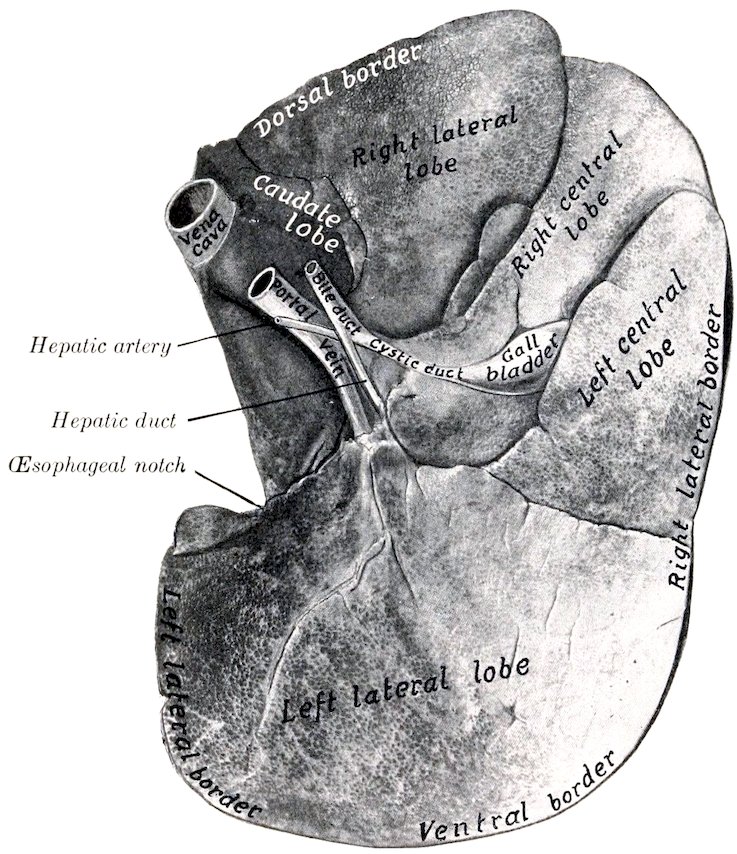
Fig. 322.—Liver of Pig, Visceral Surface. Photograph of specimen hardened in situ.
The large intestine is about 15 feet (ca. 4 to 5 meters) in length, and is for the most part much wider than the small intestine; it is connected by a mesentery with the dorsal abdominal wall between the kidneys. The cæcum is cylindrical, about 8 to 12 inches (ca. 20 to 30 cm.) long, and 3 to 4 inches (ca. 8 to 10 cm.) wide. It lies against the upper part of the right flank, its blind end reaching to within about two inches (ca. 5 cm.) of the pelvic inlet. The ileum joins the cæcum obliquely and projects somewhat into its lumen. The colon has at first about the same caliber as the cæcum, but becomes gradually smaller. Most of it is arranged in three close, double spiral coils in the mesentery, in relation chiefly to the right flank externally, and to the small intestine behind and to the left. On emerging from this spiral labyrinth, it passes forward to the stomach and pancreas and then turns backward; this terminal part is narrow, median in position, and closely attached by a short mesentery to the sublumbar region. The rectum is usually surrounded by a quantity of fat. The cæcum has three longitudinal muscular bands and three rows of sacculations, which are continued a short distance on the colon. The spiral colon has two bands and two series of sacculations, which, however, gradually disappear in the centrifugal part.
421
THE LIVER
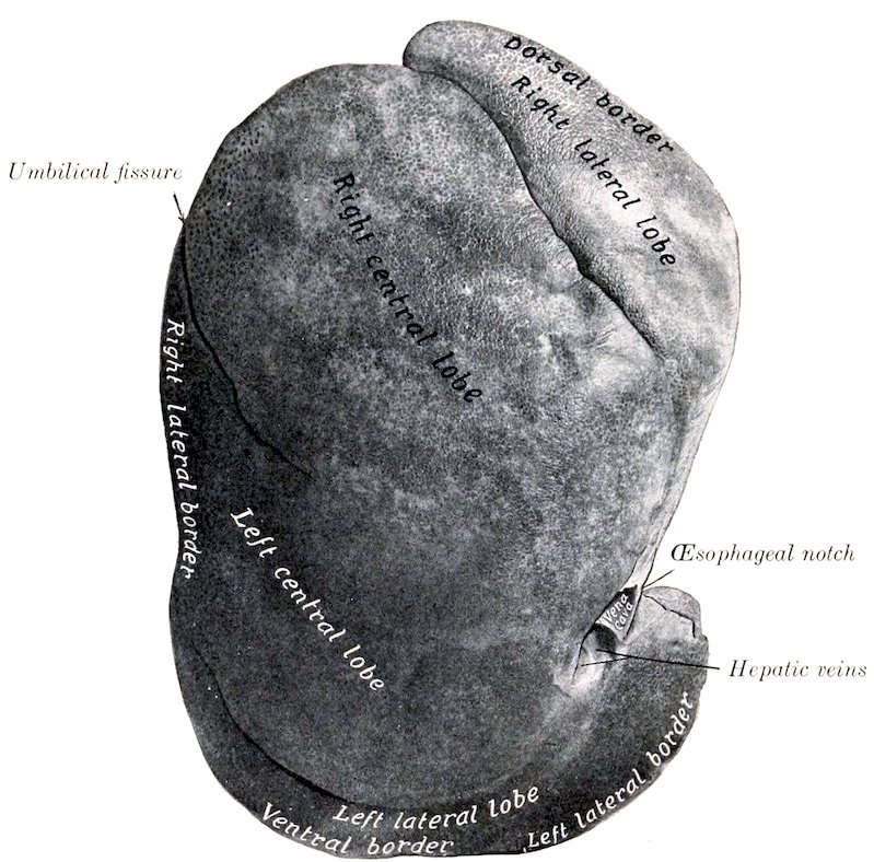
Fig. 323.—Liver of Pig, Parietal Surface. Photograph of specimen hardened in situ.
The liver is relatively large, its average weight being about 4 to 4½ pounds (ca. 2 kg.). It is thick and very strongly curved.[114] It is divided by three deep interlobar incisures into four principal lobes—right lateral, right central, left central, left lateral; the last of these is usually considerably the largest. On the upper part of the right lateral lobe is the caudate lobe and its process. That part of the right central lobe which lies below the portal fissure and to the left of the gall-bladder and cystic duct is homologous with the quadrate lobe of man. The parietal surface is extremely convex, its most anterior part reaching to a plane through the lower part of the sixth or seventh intercostal space. The visceral surface is deeply concave; most of it is related to the stomach, for which there is a correspondingly large and deep gastric impression. There is a duodenal impression on the upper part of the right lateral lobe, but no renal impression, as the right kidney does not touch the liver. The fossa for the gall-bladder is mainly on the right central lobe, but also in part on the adjacent surface of the left central lobe. The posterior vena cava enters the dorsal border of the caudate lobe and soon becomes entirely embedded in the gland substance, emerging only at its passage through the diaphragm. The œsophageal notch is large and is occupied mainly by the large right crus of the diaphragm. The right 422lateral border extends backward to the upper part of the twelfth intercostal space or thirteenth rib. The left lateral border is opposite the eighth rib and intercostal space. The ventral border extends backward about as far as the umbilicus.
There are no lateral ligaments and the falciform ligament disappears early.
The gall-bladder is attached in the fossa vesicæ felleæ on the right central lobe, at a considerable distance from the ventral border. The cystic duct joins the hepatic duct at an acute angle immediately after the emergence of the latter from the portal fissure. The bile-duct (Ductus choledochus) opens at the papilla duodeni about one or two inches (ca. 2.5 to 5 cm.) from the pylorus.
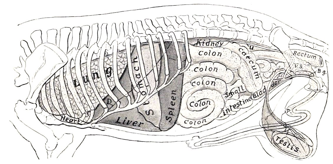
Fig. 324.—Projection of Viscera of Pig on Body-wall, Left Side. D, Costal line of diaphragm; U, ureter; V. S., vesicula seminalis; B. g., bulbo-urethral gland; P, penis.
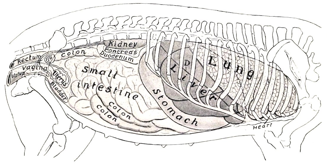
Fig. 325.—Projection of Viscera of Pig on Body-wall, Right Side. D, Costal attachment of diaphragm; O, ovary.
Owing to the large amount of interlobular tissue, the lobules are mapped out sharply; they are polyhedral in form and are 1 to 2.5 mm. in diameter. For the same reason the gland tissue is much less friable than that of the other animals, from which it is easily distinguished.
423
THE PANCREAS
The pancreas is elongated, and is situated transversely on the dorsal abdominal wall, with its right extremity on the ventral surface of the right kidney, and the left end touching the dorsal end of the spleen and the left kidney. The right portion has two lobes; the posterior lobe bends around the great mesenteric artery and the portal vein; the anterior one is pointed, extends downward and forward about in the median plane, and is attached to the portal fissure and the duodenum. The left portion is prismatic and rests mainly on the left sac of the stomach, its left extremity being included between the left kidney and the dorsal end of the spleen. The pancreatic duct passes from the anterior lobe directly through the duodenal wall, opening about five or six inches (ca. 12.5 to 15 cm.) from the pylorus. The interlobular tissue usually contains a good deal of fat.
THE SPLEEN
The spleen is long and narrow. Its long axis is nearly dorso-ventral in direction, and is slightly curved to conform to the left part of the greater curvature of the stomach. The dorsal end lies under the vertebral ends of the last three ribs. The ventral end is smaller, reaches to the ventral abdominal wall, and lies against the left lobe of the liver. The visceral surface has a longitudinal ridge on which the hilus is situated; this divides the surface into nearly equal gastric and intestinal areas. The spleen is attached so loosely to the stomach that it may be regarded as being intercalated in the great omentum. In large subjects it may reach a length of 15 to 18 inches (ca. 38 to 45 cm.) and a width of 3 to 4 inches (ca. 8 to 10 cm.).
DIGESTIVE SYSTEM OF THE DOG
THE MOUTH
The size and form of the mouth vary greatly in different breeds, the cavity being in some long and narrow, in others short and wide. The rima oris is very extensive, so that the labial commissure is opposite the third or fourth cheek tooth. The lips are thin and mobile, and present numerous tactile hairs. The upper lip has a small, central, bare area which forms part of the muzzle, and is marked by a central groove, the philtrum, or (as in the bulldog) a fissure, giving the appearance of harelip. The lateral borders of the lower lip are flaccid and denticulated. The mucous membrane is usually pigmented and forms distinct frena labiorum. The labial glands are small and scanty.
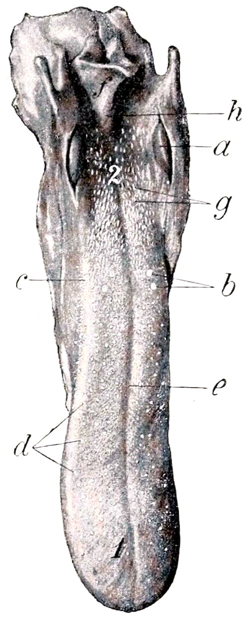
Fig. 326.—Tongue of Dog. a, Tonsil; b, vallate papillæ; c, foliate papilla; d, fungiform papillæ; e, median groove of dorsum; f, epiglottis; g, soft papillæ of root; h, middle glosso-epiglottic fold; 1, tip; 2, root. (Ellenberger-Baum, Anat. d. Haustiere.)
The cheeks are loose and capacious, and their mucous lining is smooth and more or less pigmented. The parotid duct opens usually opposite the third upper cheek tooth. Near the last tooth are the openings of the four or five ducts from the orbital gland.
The hard palate is widest between the fourth pair of cheek teeth. It has 424eight to ten curved ridges on either side of the raphé. Behind the first pair of incisor teeth is the triangular incisive papilla, at which the naso-palatine ducts open. The mucous membrane is usually pigmented.
The soft palate is thick except at its margins. In the resting state of the parts it comes in contact with the oral surface of the epiglottis. Between its anterior and posterior pillars on either side is a marked tonsillar sinus, in which an elongated tonsil is situated; this is reddish in color, about an inch long, and projects somewhat into the fauces between two folds of mucous membrane.
The tongue is wide, thin, and very mobile. It is not pigmented, but has a bright red color. The upper surface is marked by a median groove (Sulcus medianus linguæ), and is thickly beset with short, pointed, filiform papillæ, the free ends of which are directed backward. On the root the papillæ are long and soft; similar papillæ occur on the lateral walls of the isthmus faucium. The fungiform papillæ are small, and are scattered over the dorsum and sides of the tongue, but are absent on the root. There are usually two or three vallate papillæ on either side. Small foliate papillæ are also present. In the inferior part of the tip of the tongue is the lyssa, a fusiform cord, composed of fibrous tissue, muscular tissue, and fat. In large dogs it is about two inches (ca. 4 to 5 cm.) long.
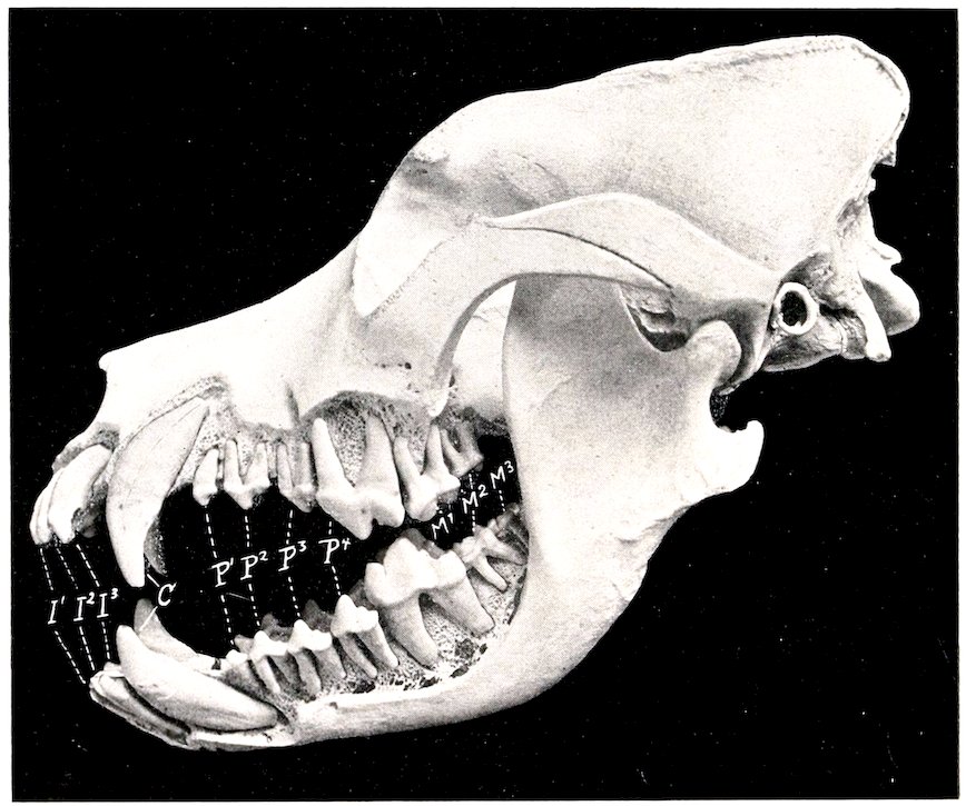
Fig. 327.—Skull of Adult St. Bernard Dog, Sculptured to Show the Embedded Parts of the Teeth. I1–3, Incisors; C, canines; P1–4, premolars; M1–3, molars.
THE TEETH
The formula for the permanent teeth is:
( 3 1 4 2 )
2 ( I C P M ) = 42
( 3 1 4 3 )
425All of the teeth have short crowns and distinct necks; they erupt rapidly. The crowns are white, being destitute of cement.
The incisors are placed almost vertically and close together in the jaw-bones. They do not correspond to an opposing tooth, but rather to parts of two teeth of the other jaw. The crowns are trituberculate, the central projection being the largest. They increase in size from the first to the third. The labial surface is convex, the lingual slightly concave, and marked off from the neck by a V-shaped ridge, the cingulum. The roots are flattened transversely. The lower incisors are smaller than the upper ones. One or two supernumerary teeth may be present.
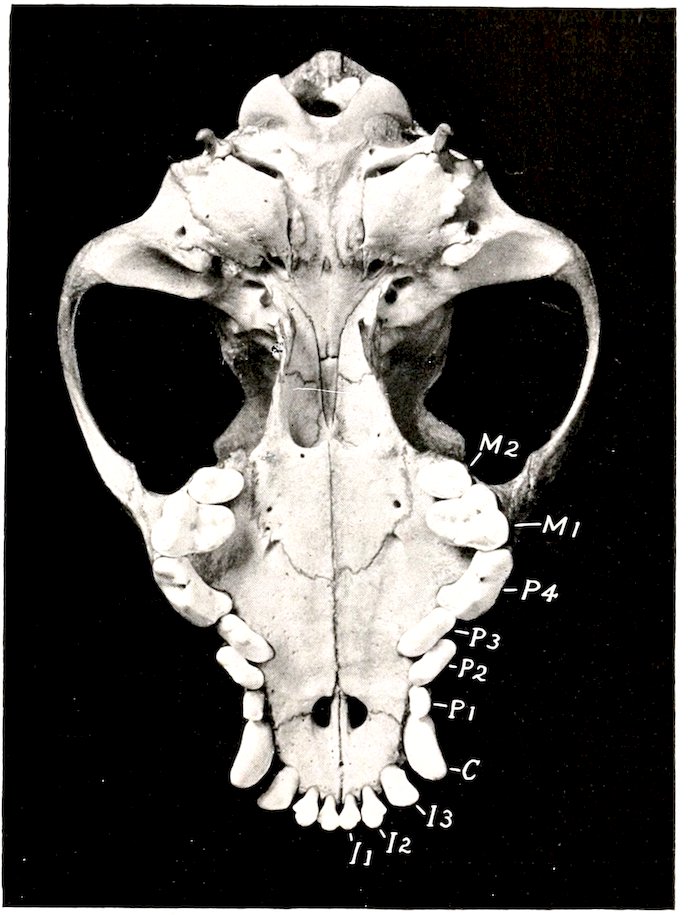
Fig. 328.—Base of Skull of Cocker Spaniel. I1–3, Incisors; C, canine; P1–4, premolars; M1–2, molars. Note the crowding of the canines and premolars on account of the shortness of the jaw.
The canine teeth are large, conical, and curved. The upper canine is separated from the corner incisor by an interval into which the lower canine is received when the jaws are closed.[115] The lower canine is close to the corner incisor. The root is about an inch (ca. 2 to 3 cm.) long and is flattened laterally.
The cheek teeth are typically ⁶⁄₇, but in brachycephalic breeds they are commonly reduced to ⁵⁄₇, and in extreme cases even to ⅘. The reduction occurs at either 426end or at both ends of the series.[116] The first tooth appears only once. The fourth tooth of the upper row and the fifth of the lower row are much larger than the rest, and termed sectorial or carnassial teeth. From these the teeth diminish in size both forward and backward. The upper and lower teeth do not correspond, but rather dovetail. The teeth behind the sectorial ones are tuberculate, i. e., have rounded eminences on the masticatory surface. The others are all sectorial in character, i. e., have sharp-edged, pointed projections, the middle one being the most prominent. The premolars are laterally compressed, and are separated by intervals from the canines and from each other, except in the brachycephalic breeds. The upper molars have wide, somewhat quadrangular crowns, and three roots. The crown of the upper fourth premolar (Dens sectorius) is divided into two pointed lobes and has an antero-internal tubercle; it has three roots. The crown of the first lower molar (Dens sectorius) is compressed laterally and has two pointed, sharp-edged lobes, behind which are one or two tubercles; it has two roots.
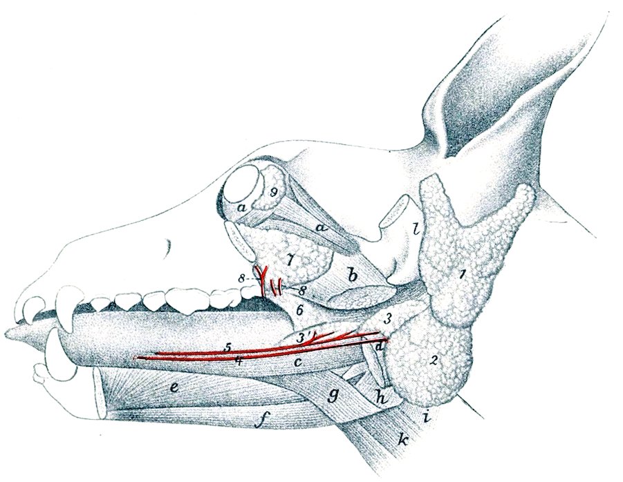
Fig. 329.—Dissection of Head of Dog, Showing Salivary Glands, etc. a, Ocular muscles; b, pterygoideus internus (cut); c, stylo-glossus; d, digastricus (cut); e, genio-glossus; f, genio-hyoideus; g, hyo-glossus; h, thyro-pharyngeus; i, crico-pharyngeus; k, thyro-hyoideus; l, zygomatic process of temporal (sawn off); 1, parotid gland; 2, submaxillary gland; 3, posterior part of sublingual gland; 3′, anterior part of same; 4, submaxillary duct; 5, ductus sublingualis major; 6, palatine glands; 7, orbital or zygomatic gland; 8, ducts of 7; 9, lacrimal gland. (Ducts colored red.) (After Ellenberger, in Leisering’s Atlas.)
The average periods of eruption are given below.
Tooth Eruption Change
I1 4 to 5 weeks 4 to 5 months
I2 4 to 5 weeks
I3 4 weeks
C 3 to 4 weeks 4 to 5 months
P1 4 to 5 months 5 to 6 months
P2 4 to 5 weeks
P3 3 to 4 weeks
P4 3 to 4 weeks
M1 4 months
M2 upper 5 to 6 months lower 4 ½ to 5 months
M3 6 to 7 months
427
THE SALIVARY GLANDS
The parotid gland is small. Its upper part is wide and is divided into two lobes by a deep notch into which the base of the ear is received. The lower end is small and overlaps the submaxillary gland. The duct leaves the gland at its anterior border, crosses the masseter muscle, and opens into the mouth opposite the third upper cheek tooth. Small accessory glands (Glandulæ parotidæ accessoriæ) are sometimes found along the course of the duct.
The submaxillary gland is usually larger than the parotid. In large dogs it is about two inches (ca. 5 cm.) long and an inch or more (ca. 3 cm.) wide. It is rounded in outline and of a pale yellow color. Its upper part is covered by the parotid, but it is otherwise superficial, and is palpable in the angle of junction of the jugular and external maxillary veins. The duct leaves the deep face of the gland, passes along the surface of the digastricus and stylo-glossus, and opens into the mouth near the frenum linguæ on a very indistinct papilla.
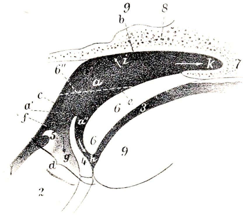
Fig. 330.—Sagittal Section of Pharyngeal and Laryngeal Regions of Dog. 1, Œsophagus; 2, larynx; 3, mouth cavity; 4, epiglottis; 5, arytenoid cartilage; 6, soft palate; 6″, position of soft palate when raised; 7, vomer; 8, base of cranium; 9, root of tongue; a, a′, a″, pharynx; b, fornix of pharynx; c, posterior wall of pharynx; d, floor of pharynx; e, pharyngeal surface of soft palate; f, origin of œsophagus; g, vestibule of larynx; h, isthmus faucium; i, Eustachian opening; k, posterior naris. (After Ellenberger, in Leisering’s Atlas.)
The sublingual gland is divided into two parts. The posterior part (Glandula sublingualis grandicanalaris) lies on the digastricus muscle in intimate relation with the submaxillary gland, but clearly separable from it after removal of the common fibrous capsule. It has a pointed anterior process. The duct (Ductus sublingualis major) accompanies the submaxillary duct, and usually opens beside it, but in some cases joins it. The anterior part (Glandula sublingualis parvicanalaris) is long and narrow; it lies on the stylo-glossus muscle. It has a number (8 to 12) of small ducts (Ductus sublinguales minores), some of which open directly into the mouth, while others join the large duct.
The orbital or zygomatic gland (Glandula zygomatica) represents the superior molar glands of herbivora. It is prismatic and is situated in the orbital region, between the zygomatic arch and the internal pterygoid muscle, in contact with the periorbita. It has four or five ducts (Ductus zygomatici) which open near the last upper cheek tooth; one of them (canal of Nuck) is almost as large as the parotid duct; the others are small.
THE PHARYNX
The fornix is narrow. The orifices of the Eustachian tubes are small and slit-like; the end of the tube causes a rounded projection of the mucous membrane 428(Torus tubarius). The œsophageal opening is relatively small and is encircled by a fold of the mucous membrane. The hyo-pharyngeus muscle is clearly divided into a kerato-pharyngeus and a chondro-pharyngeus.
THE ŒSOPHAGUS
The œsophagus is relatively wide and dilatable except at its origin. The constriction at its origin is termed the isthmus œsophagi. The cardiac end is slightly dilated; it joins the stomach under the tenth or eleventh thoracic vertebra, slightly to the left of the median plane. The muscular tissue is striated and consists mainly of two layers of spiral fibers which cross each other; near the cardia, however, the fibers are longitudinal and circular. There are mucous glands and lymph glands in the submucosa throughout.
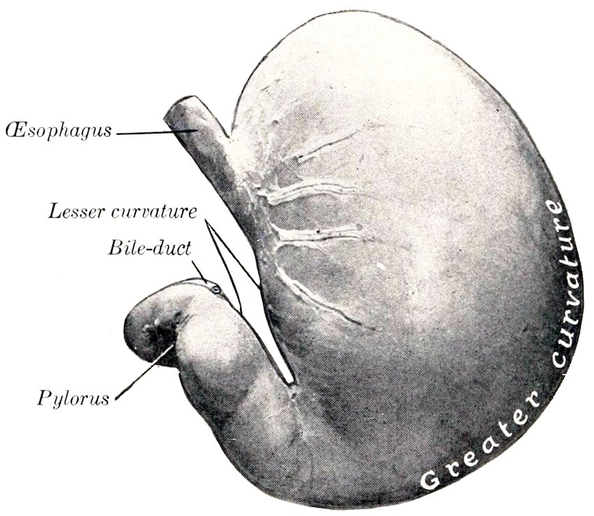
Fig. 331.—Stomach of Dog. Parietal Surface. Organ fixed in situ when well filled.
THE STOMACH
The stomach is relatively large. Its capacity in a dog weighing about 40 pounds is about six to seven pints.
Colin estimates the average capacity at about 3 liters (ca. 6½ pints), with a range between 0.6 and 8 liters (ca. 1⅓ to 17½ pints). Neumayer gives the capacity as 100 to 250 c.c. per kilogram of body-weight (ca. 2.7 ounces per pound). The average capacity of the human stomach is estimated at 35 to 40 ounces—only about one-half of that of a dog of medium size.
When full it is irregularly pyriform. The left or cardiac part (Corpus ventriculi) is large and rounded, while the right or pyloric part (Pars pylorica) is small and cylindrical. When empty, or nearly so, the left sac is strongly contracted; the pyloric part is not materially affected by variations in the amount of ingesta.
The parietal surface of the full stomach is very extensive, strongly convex, and faces partly forward, but largely to the left. It is related to the liver, the left part of the diaphragm, and the left and ventral abdominal wall as far back as a transverse section through the second or third lumbar vertebra.
The visceral surface is much less extensive and is considerably flattened; it faces chiefly to the right, and is related to the intestine, pancreas, and left kidney.
The upper part of the lesser curvature is nearly straight and vertical, but the lower part forms a deep, narrow angle, due to the fact that the pyloric part is directed sharply forward and upward.
429The greater curvature is nearly four times as long as the lesser curvature. In the full stomach it extends considerably behind the left costal arch (having carried the spleen backward); ventrally, it lies on the abdominal wall, almost midway from the xiphoid cartilage to the pubis.
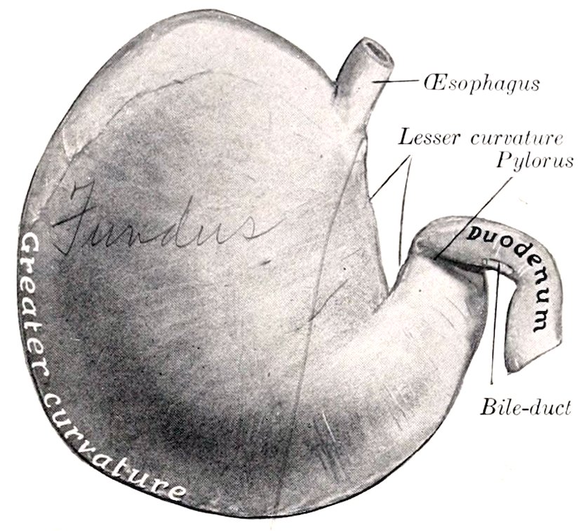
Fig. 332.—Stomach of Dog, Visceral Surface. Organ fixed in situ when well filled.
The left extremity or fundus is large and rounded; it is the most dorsal part of the organ, and lies under the dorsal ends of the eleventh and twelfth ribs.
The pyloric extremity is small and is directed forward and somewhat upward; it lies usually about an inch or two (ca. 3.5 to 5 cm.) below the cardia, a little to the right of the median plane. It is related to the portal fissure of the liver and to the pancreas.
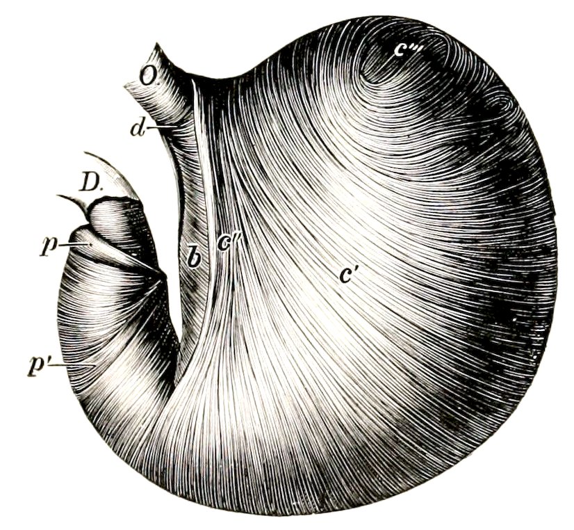
Fig. 333.—Everted Stomach of Dog from which the Mucous Membrane is Removed. O, Œsophagus; D, duodenum; b, circular fibers; c′, internal oblique fibers; c″, cardiac loop; c‴, transition of internal to external oblique fibers; d, fibers uniting branches of cardiac loop; p, pyloric sphincter; p′, antral sphincter. (Ellenberger-Baum, Anat. d. Haustiere.)
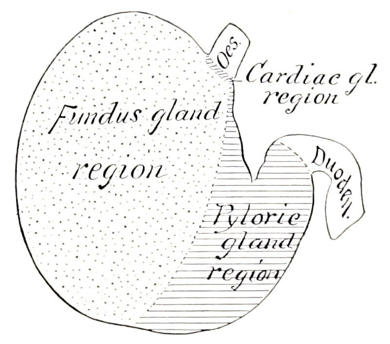
Fig. 334.—Diagram of Zones of Mucous Membrane of Stomach of Dog.
The cardia is situated about two to three inches (ca. 5 to 7 cm.) from the left extremity, and is oval; it lies a little to the left of the median plane, below the tenth or eleventh thoracic vertebra.
When empty, or nearly so, the stomach is separated from the ventral abdominal wall by the liver and small intestine, and the greater curvature extends back to 430the eleventh or twelfth rib. In this state there is not rarely a marked constriction between the right and left portions.
The longitudinal muscular fibers are found on the curvatures and on the pyloric part. The oblique fibers are arranged in two layers: the external layer is a continuation of the longitudinal fibers of the œsophagus onto the body and fundus. The circular layer covers the whole stomach except the fundus, and forms a pyloric sphincter and an antral sphincter. The internal oblique layer is arranged as in the horse, and forms a similar loop-like, cardiac sphincter.
Three regions of the mucous membrane exist. Cardiac glands are found in a very narrow pale zone around the cardiac opening, and also scattered along the lesser curvature. The fundus gland region has a thick, reddish-brown mucous membrane, which lines about two-thirds of the organ. The pyloric mucous membrane is thinner and pale; in the dead subject it is often stained by regurgitated bile.
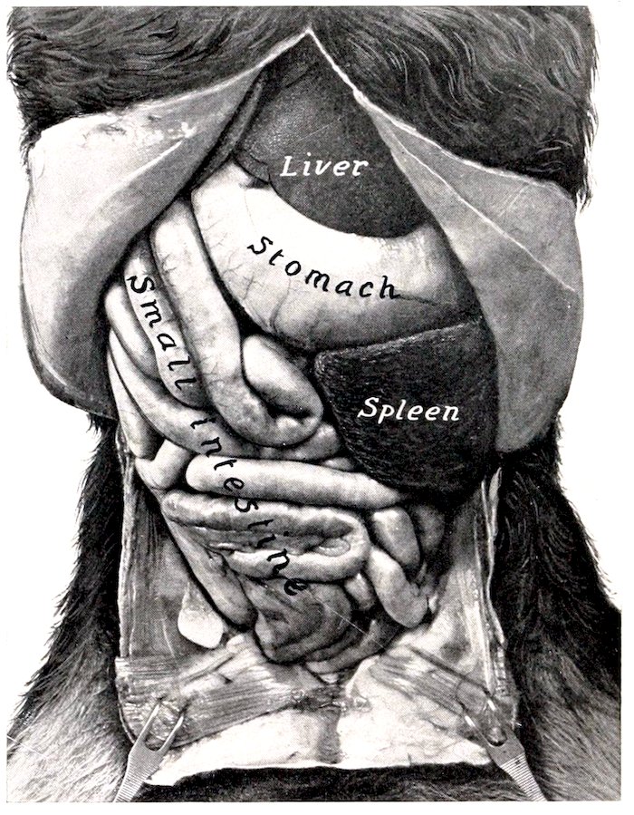
Fig. 335.—Abdominal Viscera of Dog. Superficial ventral view after removal of the great omentum. From photograph of formalin-hardened subject.
THE INTESTINE
The intestine is short—about five or six times the length of the body.
The small intestine has an average length of about thirteen feet (ca. 4 meters). It occupies most of the abdominal cavity behind the liver and stomach. The duodenum begins at the portal fissure and passes backward and upward, at first on the visceral surface of the liver, then in contact with the right flank. Near the pelvis it turns inward and sharply forward, passes along the inner border of the left kidney, and joins the jejunum to the left of the root of the mesentery. The mesoduodenum is given off from the common mesentery; it is a relatively wide 431fold, except at the terminal part. Here there are two short folds, one of which joins the mesocolon; the other passes on to the terminal part of the colon. The first part of the mesoduodenum contains the right branch of the pancreas. Its root is blended with the mesocolon to form a common mesentery. The remainder of the small intestine forms numerous coils, and is attached by a wide mesentery to the sublumbar region. The pancreatic and bile-ducts open into the duodenum about two or three inches (ca. 5 to 8 cm.) from the pylorus; the accessory pancreatic duct opens an inch or two (ca. 2.5 to 5 cm.) further back. The mucous membrane has very long villi. The duodenal (Brunner’s) glands occur only near the pylorus. Peyer’s patches are numerous (about twenty in young subjects), and begin in the duodenum. They are usually elliptical in outline, but the last one is band-like, reaches to the end of the ileum and is four to sixteen inches (ca. 10 to 40 cm.) long in young dogs (Ellenberger). There is an ileo-cæcal valve.[117] The muscular coat is relatively thick.
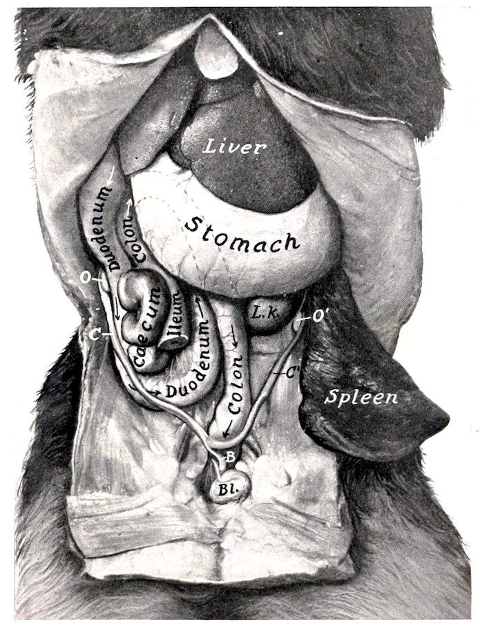
Fig. 336.—Deep Dissection of Abdominal Viscera of Dog (Female). BL, Bladder; B, body; C, C′, cornua of uterus; O, O′, ovaries; L. K., left kidney. The concealed part of the colon is indicated by dotted lines. (From photograph of formalin-hardened subject.)
The large intestine is two to two and a half feet (ca. 60 to 75 cm.) long. Its caliber is about the same as that of the small intestine, and it has neither longitudinal bands nor sacculations.
The cæcum is about three or four inches (ca. 8 to 10 cm.) long, and is twisted in a spiral fashion. The twist is maintained by the peritoneum, which attaches it 432to the ileum. It is situated usually against the middle of the upper part of the right flank, below the duodenum and the right branch of the pancreas.[118]
The colon is attached to the sublumbar region by a mesentery, the mesocolon. It presents three parts, which correspond to the ascending, transverse, and descending colon of man. The first or right part (Colon ascendens) is very short. It begins at the junction of the ileum and cæcum, and passes forward along the inner surface of the duodenum and the right branch of the pancreas till it reaches the pyloric part of the stomach; here it turns inward and crosses the median plane, forming the transverse part (Colon transversum). The third or left part (Colon descendens) passes backward in the sublumbar region along the inner border or ventral surface of the left kidney; then inclines toward the median plane and joins the rectum.[119] The caliber of the colon is about the same throughout. It has no bands nor sacculations.
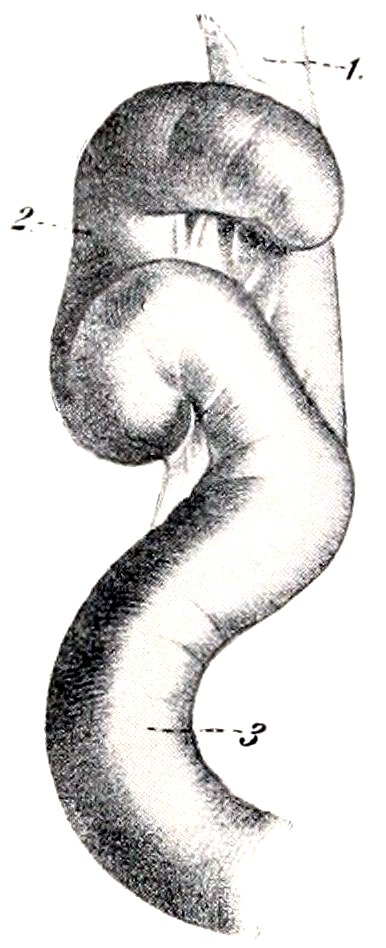
Fig. 337.—Cæcum of Dog. 1, Ileum; 2, cæcum; 3, colon. (From Leisering’s Atlas.)
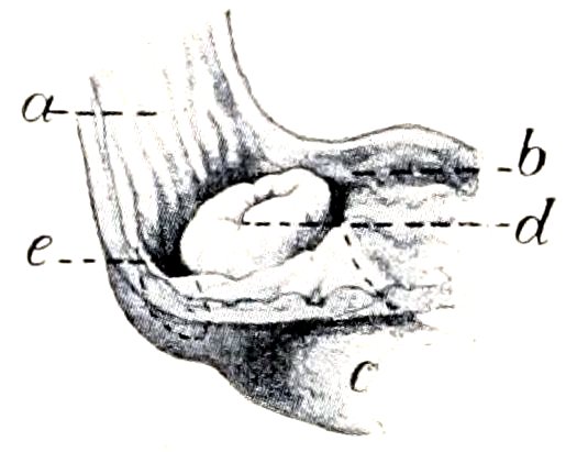
Fig. 338.—Termination of Ileum of Dog. a, Colon; b, cæcum; c, ileum; d, ileo-cæcal opening; e, circular depression around end of ileum. (After P. Schumann.)
The mucous membrane of the cæcum contains numerous solitary glands; they are found also in the first part of the colon.
The rectum is almost completely covered with peritoneum, the line of peritoneal reflection being under the second or third coccygeal vertebra. At the junction of the rectum and anus the mucous membrane has a stratified, squamous epithelium, and contains the anal glands. A small opening on either side leads into two lateral anal sacs (Sinus paranales); these are usually about the size of a hazelnut, and contain a dirty gray, fatty substance, which has a peculiar and very unpleasant odor. The skin which lines these pouches contains coil glands. Further back the skin contains large sebaceous glands and peculiar circumanal glands.
The retractor ani muscle is extensive. It arises from the shaft of the ilium, the pubis, and the symphysis pelvis, and passes upward and backward to end on the first coccygeal vertebræ and the sphincter ani externus. The retractors, together with the coccygei, form a sort of pelvic diaphragm, analogous to that of man.
THE LIVER
The liver is relatively large, weighing usually about 5 per cent. of the body-weight. It is divided into six or seven lobes by fissures which converge at the portal fissure. When the gland is examined in the soft condition, all the lobes are visible on the visceral surface, but usually only four on the parietal surface.
The left lateral lobe is the largest, and is oval in outline. The left central lobe 433is much smaller and is prismatic. The right central lobe is second in size, and presents a somewhat tongue-shaped quadrate lobe, marked off by the deep fissure in which the gall-bladder lies. The right lateral lobe is third in size, and is oval in outline. On its visceral surface is the large caudate lobe; this consists of two parts—on the right, the caudate process; on the left, the papillary process or lobe, both often being subdivided by fissures.
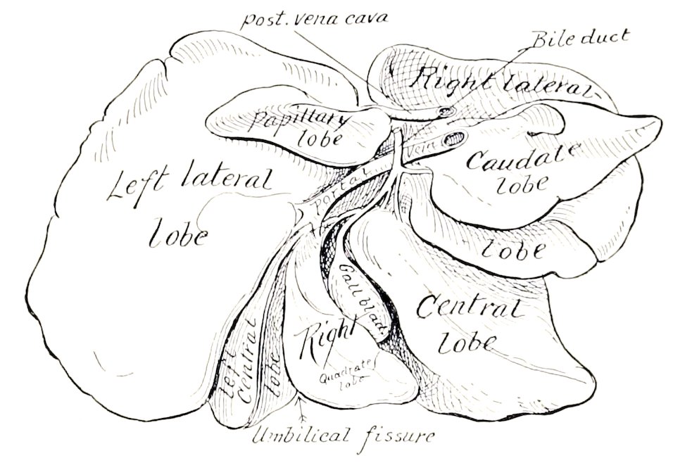
Fig. 339.—Liver of Dog. Soft specimen sketched with lobes drawn apart.
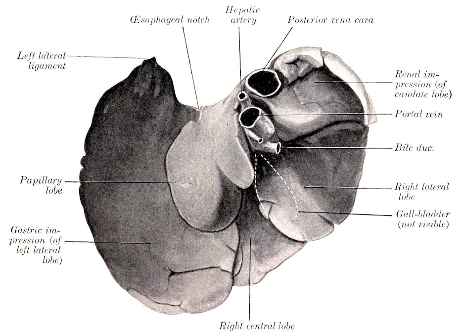
Fig. 340.—Liver of Dog, Visceral Surface; Hardened in situ. Left central lobe, gall-bladder, and great part of right central lobe not visible.
When hardened in situ (with the stomach full), the gland presents the following characters:
The visceral surface is marked by a somewhat oblique ridge (corresponding to 434the lesser curvature of the stomach), which separates two deep cavities. The depression to the left of the ridge is occupied by the body and fundus of the stomach. The depression on the right is smaller, and is occupied by the pyloric part of the stomach, the origin of the duodenum, and the right branch of the pancreas. The gall-bladder is not visible.
When the stomach is empty and contracted, the visceral surface of the liver is strikingly different. There is then a shallow impression for the left part of the stomach on the left lobe, and a large convex area, related to the small intestine and a mass of omentum. The pyloric and duodenal impressions are not much changed. The gall-bladder is visible.
The portal fissure is very deep and might well be termed a fossa. It is bounded on the right (externally) by the caudate process, on the left (internally) by the papillary process or lobe, which is directed ventrally. The hepatic artery enters at the dorsal end and the portal vein centrally, while the bile-duct leaves at the ventral part. The left central lobe is not visible, and the right central one is largely concealed.
The parietal surface is extremely convex, being adapted to the diaphragm and the adjacent part of the ventral wall of the abdomen. On it all the lobes except the papillary are completely or partially visible. The gall-bladder is visible in the fissure between the quadrate and chief portions of the right central lobe.
The dorsal border presents a renal impression on its right portion. The posterior vena cava passes downward and forward at first in a deep groove on the caudate lobe, then largely embedded in the parietal surface of the right lateral lobe; it receives two large hepatic veins just before piercing the diaphragm.
The œsophageal notch is large, and is occupied on the right by the thick margin of the hiatus œsophageus. The left border is opposite to the ninth and tenth ribs; it is thin and is marked by numerous indentations. The caudate process extends usually a little beyond the last right rib, between the right kidney and the right branch of the pancreas. The ventral border lies on the ventral abdominal wall a variable distance behind the xiphoid cartilage.
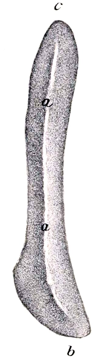
Fig. 341.—Spleen of Dog, Visceral Surface. a, a, Ridge; b, ventral end; c, dorsal end. (After Ellenberger, in Leisering’s Atlas.)
The gall-bladder lies in the fossa vesicæ felleæ, between the two parts of the right central lobe; it does not reach to the ventral border of the liver. The cystic duct joins the hepatic duct at the lower part of the portal fissure, forming with it the bile duct (Ductus choledochus); the latter passes to the right and opens into the duodenum, about two or three inches (ca. 5 to 8 cm.) from the pylorus.
Of the ligaments, the coronary and right lateral are well developed, but the left lateral and falciform are small.
THE PANCREAS
The pancreas is V-shaped, consisting of two long narrow branches, which meet at an acute angle above the pylorus. The right branch extends backward above the first part of the duodenum, below the caudate lobe of the liver and the right kidney, and ends usually a short distance behind the latter. The left branch passes inward and backward on the visceral surface of the stomach, and ends under the left kidney. There are two ducts: the larger one unites with the bile duct, while the smaller one enters the duodenum an inch or two (ca. 3 to 5 cm.) further back.
435
THE SPLEEN
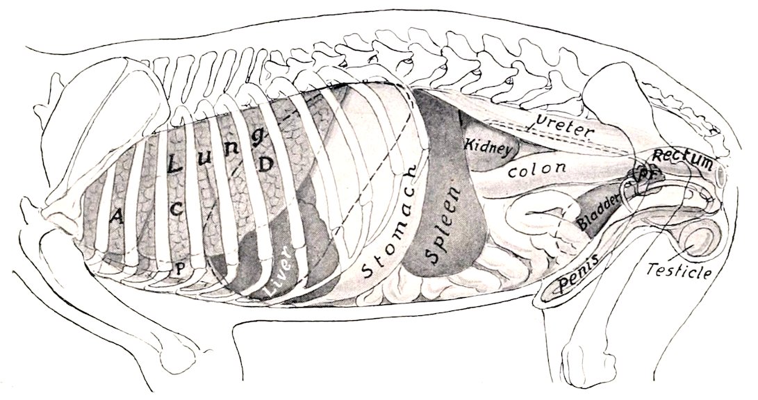
Fig. 342.—Projection of Viscera of Dog, (Male) on Body-Wall, Left Side. A, C, D, Apical, cardiac, and diaphragmatic lobes of lung; P, pericardium; Pr, prostate. Costal attachment and median line of diaphragm are dotted.
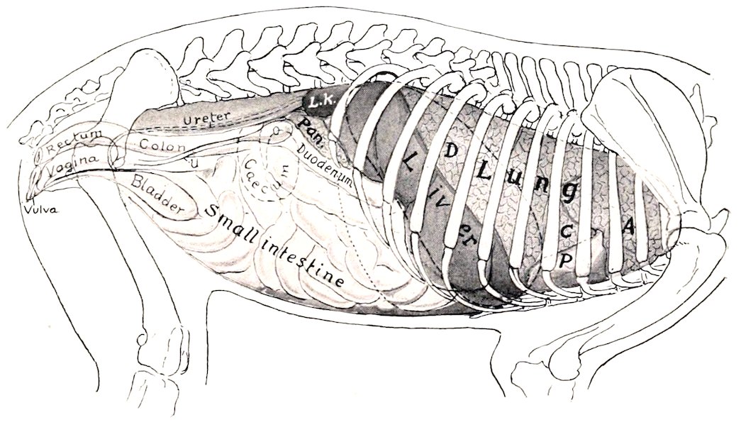
Fig. 343.—Projection of Viscera of Dog (Female) on Body-Wall, Right Side. A, C, D, Apical, cardiac, and diaphragmatic lobes of right lung; P., pericardium; L. K., right kidney; Pan., right branch of pancreas; O, ovary; U, cornu of uterus. Costal attachment and median line of diaphragm are dotted; also posterior contour of stomach.
The spleen is bright red in color in the fresh state. It is somewhat falciform, long, and narrow; the ventral part is the widest. Its position varies in accordance with the fullness of the stomach. When that organ is moderately full the long axis of the spleen corresponds to the direction of the last rib. Its parietal surface is convex and lies largely against the left flank. The visceral surface is concave in its length, and is marked by a longitudinal ridge, on which the vessels and nerves are situated. The dorsal end lies against the left kidney and left crus of the diaphragm. The ventral end is a little further back, and reaches to the ventral wall of the abdomen. It is so loosely attached by the great omentum as to be regarded as an appendage of the latter.
436