ARTHROLOGY (continued 2)
Reading edition
The chief metacarpal bones of the pig, and the second to the fifth of the dog, articulate with each other at their proximal ends, and are connected by interosseous ligaments, which do not, however, unite them closely, as in the horse.
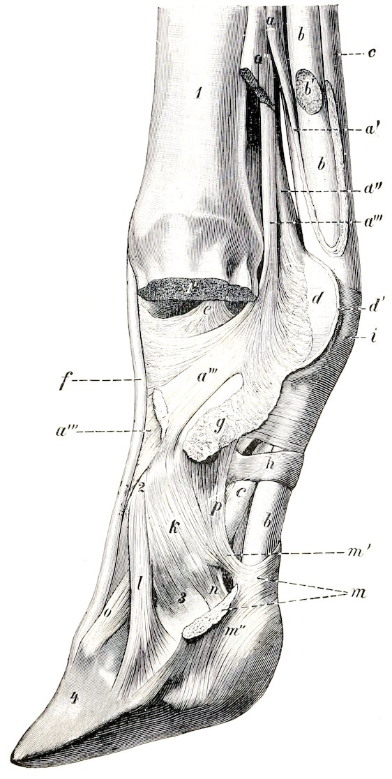
Fig. 168.—Distal Part of Limb of Ox, Showing Ligaments and Tendons. One Digit and Corresponding Articular Part of Metacarpal Bone are Removed. a, Suspensory ligament; a′, branch of a to superficial flexor tendon; a″, a‴, lateral and central branches of a; b, deep flexor tendon; b′, branch of b to digit removed; c, c′, superficial flexor tendon; d, d′, intersesamoid ligament (cut); e, interdigital lateral ligament of fetlock joint; f, tendon of common extensor; g, proximal interdigital ligament; h, digital annular ligament; i, posterior annular ligament of fetlock; k, lateral ligament of pastern joint; l, distal interdigital ligament; m, crucial interdigital ligament (cut); m′, m″, attachments of m to second phalanx and distal sesamoid bone; n, suspensory ligament of distal sesamoid; o, anterior elastic ligament; p, lateral volar ligament of pastern joint; 1, metacarpus, sawn off at 1′; 2, first phalanx; 3, second phalanx; 4, third phalanx. (Ellenberger-Baum, Anat. d. Haustiere.)
METACARPO-PHALANGEAL JOINTS
Ox.—There are two joints, one for each digit. The two capsules communicate posteriorly. The two interdigital lateral ligaments (Ligamenta collateralia interdigitalia) result from the bifurcation of a band which arises in the furrow between the divisions of the distal end of the large metacarpal bone; they spread out and end on the proximal ends of the first phalanges. A strong superior interdigital ligament (Ligamentum interdigitale), consisting of short intercrossing fibers, unites the middles of the interdigital surfaces of the first phalanges.
207Crucial ligaments (Ligamenta phalango-sesamoideæ) connect the sesamoids with the proximal end of the opposite first phalanx.
The intersesamoid ligament connects all four sesamoids, and extends upward much less than in the horse.
The lateral sesamoidean ligaments end almost entirely on the first phalanges.
The superficial inferior sesamoidean ligament is absent. The middle inferior sesamoidean ligaments of each digit are two short strong bands which extend from the distal margins of the sesamoids to the proximal ends of the first phalanges. The deep inferior sesamoidean ligaments are strong and distinctly crucial.
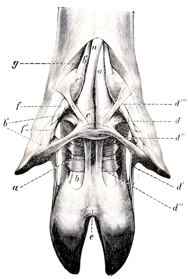
Fig. 169.—Ligaments and Tendons of Digits of Pig, Volar View. a, Superficial flexor tendon; b, deep flexor tendon; b′, branches of b to accessory digits; c, c′, annular ligaments; d-d‴, ligaments of accessory digits; e, cruciate interdigital ligaments; f, f′, spiral band around the flexor tendons of the accessory digits; g, abductor of accessory digit. (Ellenberger-Baum, Anat. d. Haustiere.)
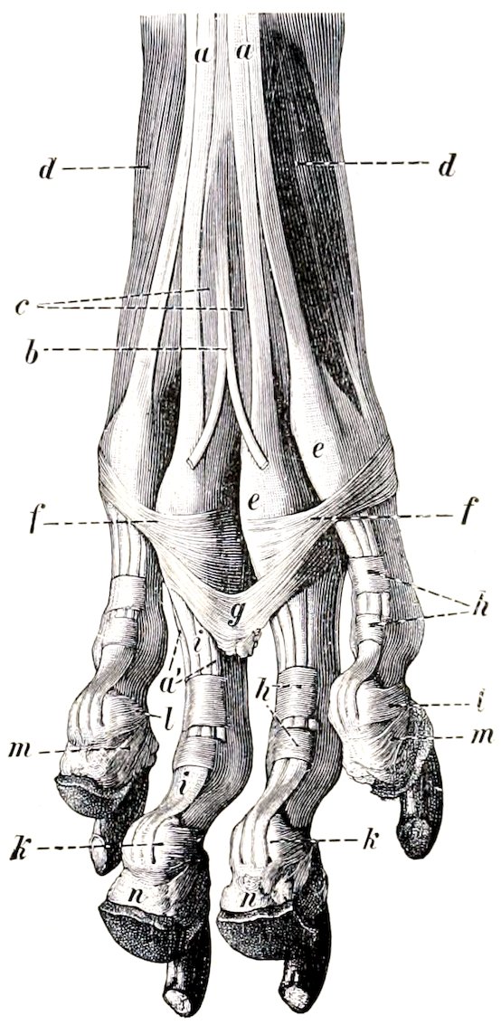
Fig. 170.—Ligaments and Tendons of Paw of Dog, Hind Limb, Volar View. a, a′, Superficial flexor tendon; b, tendon to large pad; c, lumbricales muscles; d, interossei muscles; e, f, annular ligaments at metatarsophalangeal joints; g, suspensory ligament of large pad; h, digital annular ligaments; i, deep flexor tendon; k, distal sesamoid; l, suspensory ligament of k; m, suspensory ligament of digital pad; n, digital pads. (Ellenberger-Baum, Anat. d. Haustiere.)
The suspensory or superior sesamoidean ligament is more distinctly muscular than in the horse—indeed in the young animal it consists almost entirely of muscular tissue. At the distal third of the metacarpus it divides into three branches. These give rise to five subdivisions, either by bifurcation of the lateral branches or trifurcation of the middle branch. The four lateral bands end on the sesamoid bones and the distal end of the large metacarpal bone, and detach slips to the extensor tendons. The middle band passes through the groove between the two divisions of the distal end of the metacarpus, and divides into two branches 208which join the tendons of the proper extensors of the digits; it sends fibers also to the interdigital lateral ligaments and to the central sesamoids. About the middle of the metacarpus the suspensory ligament detaches a band which unites lower down with the superficial flexor tendon, thus inclosing the tendon of the deep flexor of the digit; it also blends with the thick fascia of the region. The latter gives off a band on either side to the accessory digits, and a tendinous band descends from each accessory digit to the third phalanx and sesamoid bone, blending with the tendon of the corresponding proper extensor.
Pig.—There are four metacarpo-phalangeal joints, each of which has a capsule, lateral, intersesamoidean, and crucial sesamoidean ligaments. Since distinct interosseous muscles are present, there are, of course, no suspensory ligaments.
Dog.—There are five metacarpo-phalangeal joints, each having its own capsule and indistinct lateral ligaments. A small sesamoid bone occurs in the anterior part of each capsule, over which the corresponding extensor tendon plays. The intersesamoidean ligaments do not extend above the sesamoids. The cruciate ligaments are present, as well as a fibrous layer which attaches the distal margins of the sesamoids to the posterior surface of the proximal end of the first phalanx.
INTERPHALANGEAL JOINTS
Ox.—The two proximal joints have separate capsules, and broad, but rather indistinct, lateral ligaments. Each joint has also two central and two lateral volar ligaments. The central ligaments are largely fused to form a strong band. The lateral ones extend from the borders of the first phalanx to the proximal end of the second phalanx.
The distal interphalangeal joints have, in addition to the capsules and lateral ligaments, bands which reinforce them on either side. The central or interdigital pair arise in the depressions on the distal ends of the first phalanges, receive fibers from the second phalanges, and end on the interdigital surfaces of the third phalanges at the margin of the articular surface. The lateral pair have a similar course, but are thinner, and end on the corresponding third sesamoid. An elastic band crosses the front of the second phalanx obliquely, from the distal end of the first phalanx to the extensor process of the third phalanx.
The crucial or inferior interdigital ligaments (Ligamenta cruciata interdigitalia) are two strong bands which limit the separation of the digits. They are attached above to the lateral (or abaxial) eminences on the proximal ends of the second phalanges (blending with the lateral ligaments), cross the deep flexor tendon obliquely, and reach the interdigital space, where they intercross and blend. Most of the fibers end on the third sesamoid of the opposite side, but some are attached to the interdigital aspect of the second phalanx and the third sesamoid of the same side. In the sheep there is, instead of the foregoing, a transverse ligament which is attached on either side to the interdigital surfaces of the second and third phalanges and the third sesamoid bone. It is related below to the skin, above to a pad of fat.
Pig.—The interphalangeal joints of the chief digits resemble in general those of the ox. The inferior interdigital ligament resembles, however, that of the sheep, and is intimately adherent to the skin. There is, besides, a remarkable arrangement of ligaments which connect the small digits with each other and with the chief digits.
This apparatus is somewhat complex, but its chief features are as follows: A superior interdigital ligament is attached on either side to the third phalanges of the small digits, while centrally it blends with the annular ligaments of the flexor tendons behind the metacarpo-phalangeal joints of the chief digits. Two bands (central longitudinal interdigital ligaments) arise on the bases of the small digits, cross the flexor tendons obliquely downward and inward, pass through the superior interdigital ligament, and blend below with the inferior interdigital ligament. Two lateral bands (lateral longitudinal interdigital ligaments) are attached in common with the 209superior interdigital ligaments to the third phalanges of the small digits, and blend below with the outer part of the inferior interdigital ligament.
Dog.—Each joint has a capsule and two lateral ligaments. The distal joints have also two elastic dorsal ligaments (Ligamenta dorsalia), which extend from the proximal end of the second phalanx to the ridge at the base of the third phalanx (Fig. 133). They produce dorsal flexion of the joint, and thus raise or retract the claws when the flexor muscles relax. The distal sesamoids are represented by complementary cartilages attached to the volar margins of the articular surfaces of the third phalanges.
Three interdigital ligaments restrict the spreading apart of the digits. Two of these cross the volar surface of the proximal parts of the chief digits, i. e., one for the second and third, the other for the fourth and fifth; they blend with the annular ligaments on either side. The third ligament is attached on either side to the foregoing ligaments and the annular ligaments of the third and fourth digits, and curves downward centrally, ending in the large pad on the paw.
Articulations of the Pelvic Limb
SACRO-ILIAC JOINT
This joint and the pelvic ligaments present no very striking differences in the other animals. The sacro-sciatic ligament in the dog is a narrow but strong band which extends from the posterior part of the lateral margin of the sacrum to the tuber ischii; it is the homologue of the ligamentum sacro-tuberosum of man.
HIP JOINT
Ox.—The shallowness of the acetabulum is compensated by the greater size of the marginal cartilage. The head of the femur has a smaller radius of curvature than that of the horse, and the articular surface extends a considerable distance outward on the upper surface of the neck. The round ligament is entirely intraarticular; it is small, and sometimes absent. The pubo-femoral or accessory ligament is absent.
There are no important differences in the other animals.
STIFLE JOINT
Ox.—There is a considerable communication between the femoro-patellar and inner femoro-tibial joint cavities; this is situated as in the horse, but is wider. A small communication with the external femoro-tibial capsule sometimes occurs. The two femoro-tibial capsules usually communicate. The middle patellar ligament is not sunken, as there is no groove on the tuberosity of the tibia where it is attached. The external patellar ligament fuses completely with the tendon of insertion of the biceps femoris, and a synovial bursa is interposed between them and the external condyle of the femur.
In the other animals there is a single ligamentum patellæ, and the synovial sacs communicate so freely as to constitute a common joint cavity. In the dog the semilunar cartilages are united anteriorly by a transverse ligament, and the posterior part of the capsule contains the two Vesalian sesamoids, which articulate with the condyles of the femur.
TIBIO-FIBULAR JOINTS
Ox.—The proximal end of the fibula fuses with the external condyle of the tibia. The distal end remains separate, and forms an arthrosis with the distal end 210of the tibia; the movement here is imperceptible, as the two bones are closely united by strong peripheral fibers.
Pig.—The superior joint is provided with a capsule which is reinforced in front and behind by fibrous tissue.
The interosseous ligament attaches the shaft of the fibula to the outer border of the tibia.
The inferior joint is included in the capsule of the hock joint, and is strengthened by oblique anterior and posterior ligaments. There is also an interosseus ligament.
Dog.—The arrangement is essentially the same as in the pig, but there is no interosseous ligament in the inferior joint.
HOCK JOINT
Ox.—There is very considerable mobility at the first intertarsal joint, the capsule of which is correspondingly roomy. The short external lateral ligament is attached below on the tibial tarsal only. A strong transverse ligament attaches the external malleolus (distal end of the fibula) to the back of the tibial tarsal bone. The dorsal or oblique ligament is narrow and thin.
Pig.—The arrangement in general resembles that of the ox.
Dog.—The long lateral ligaments are very small, and the short ones double. The plantar ligament is weak, and ends on the fourth metacarpal bone. No distinct dorsal (or oblique) ligament is present. Movement occurs almost exclusively at the tibio-tarsal joint.
The remaining joints resemble those of the thoracic limb.
211