OSTEOLOGY (continued 2)
Reading edition
The temporal part is separated from the orbital part by the deep sphenoidal notch (Incisura sphenoidalis), which is occupied by the orbital wing of the sphenoid. Its external surface forms part of the inner wall of the temporal fossa. The internal surface is largely covered by the orbital wing of the sphenoid in the young subject, but later forms part of the wall of the frontal sinus.
The principal connections of the frontal bone are as follows: (1) The inner border joins its fellow at the frontal suture. (2) The anterior border meets the nasal and lacrimal at the naso-frontal and fronto-lacrimal sutures. (3) Laterally it forms the spheno-frontal suture with the orbital wing of the sphenoid, and also joins the palate bone and maxilla. (4) Posteriorly it meets the parietal at the parieto-frontal (or coronal) suture, and articulates below this with the squamous temporal. (5) The extremity of the supraorbital process unites with the zygomatic process of the temporal bone.
Development.—Each ossifies in membrane from one center which appears in the root of the supraorbital process. In the new-born foal there is a slit between the cranial plate and the orbital and temporal plates which receives the cartilaginous margin of the orbital wing of the sphenoid.
The Temporal Bones
The temporal bone (Os temporale) forms the greater part of the lateral wall of the cranium. It is situated between the occipital behind, the parietal above, the frontal in front, and the sphenoid below. It consists of two distinct parts, squamous and petrous.
1. The squamous temporal (Squama temporalis) is a shell-like plate which has two surfaces and four borders.
The internal surface (Facies cerebralis) is largely overlapped by the surrounding bones, but its central part is free and presents digital impressions and vascular grooves.
56The external surface (Facies temporalis) is convex, and enters into the formation of the temporal fossa. From its lower part there springs the zygomatic process (Processus zygomaticus), which forms the external boundary of the temporal fossa. It is at first directed outward, and is wide and flattened dorso-ventrally. It then turns forward, becomes narrower, and is twisted so that its surfaces are internal and external. Its anterior end is pointed and joins the zygomatic process of the malar bone, with which it forms the zygomatic arch (Arcus zygomaticus). The narrow anterior part has a convex outer surface and a concave inner one. Its upper border has a rough area for articulation with the supraorbital process of the frontal. Its lower border is wide and rough. The wide posterior part presents on its ventral face a surface for articulation with the condyle of the mandible. This surface consists of a transversely elongated condyle (Tuberculum articulare), behind which is the glenoid fossa (Fossa mandibularis). The fossa is limited behind by the postglenoid process, the anterior surface of which is articular. Behind this process is a fossa in which is the external opening of the parieto-temporal canal. The dorsal surface is concave and forms the outer boundary of the temporal fossa. The superior border is sinuous and is continuous behind with the temporal crest.
The posterior process (Processus posterior) springs from the posterior part of the squama. Its external surface is crossed by the temporal crest, which forms here the outer limit of the temporal fossa. The internal surface forms the outer boundary of the parieto-temporal canal, and is elsewhere applied to the petrous portion. It divides into two branches, upper and lower; the upper branch unites with the occipital bone, while the lower one curves downward behind the external auditory process and overlaps the mastoid process.
The superior border of the squamous temporal articulates with the parietal, forming the parieto-temporal suture. The inferior border joins the temporal wing of the sphenoid at the spheno-squamous suture. The anterior border unites with the frontal bone, and the posterior with the parietal.
2. The petrous temporal (Os petrosum) is placed between the occipital behind and the parietal in front, and is largely overlapped externally by the squamous temporal. It has the form of a four-sided pyramid, the base of which is ventral.
The external surface is mainly concealed by the squamous temporal, but two features are visible. A short tube of bone, the external auditory process, protrudes from the lowest part through the notch of the squamous temporal. The process is directed outward, upward, and a little forward. It gives attachment to the annular cartilage of the ear. Its lumen, the external auditory meatus (Meatus acusticus externus), conducts to the cavity of the middle ear (tympanum) in the dry skull, but is separated from it by the tympanic membrane in the natural state. The mastoid process projects ventrally in the interval between the posterior process of the squamous temporal and the root of the paramastoid (or styloid) process of the occipital bone; its outer surface is crossed by a curved groove which leads to the parieto-temporal canal.
The internal surface faces into the cerebellar fossa of the cranium. It is concave and smooth but irregular. In its lower part is the entrance to a short canal, the internal auditory meatus, which transmits the seventh and eighth cranial nerves.
The fundus of the meatus is divided by a crest into a superior and an inferior fossa. In the superior one is the origin of the facial canal, which curves through the bone and opens externally at the stylo-mastoid foramen; it transmits the facial (seventh cranial) nerve. The inferior fossa presents small foramina for the passage of fibers of the auditory (eighth cranial) nerve.
Behind the meatus and near the posterior margin of the surface is the slit-like opening of the aquæductus vestibuli, covered by a scale of bone. Below this is a narrow fissure, the orifice of the aquæductus cochleæ.
57The anterior surface looks upward and forward. The outer part articulates with the parietal bone and the inner part faces into the cerebral fossa of the cranium. A sharp border, the petrosal crest (Crista petrosa), separates this surface from the inner one.
The posterior surface joins the lateral part of the occipital bone.
The base forms the outer boundary of the foramen lacerum basis cranii. It is very irregular and presents a number of important features. The hyoid process is a short rod which projects downward and forward below the base of the external auditory process, inclosed in a bony tube; it is connected by a bar of cartilage with the hyoid bone. The stylo-mastoid foramen is situated between the root of the hyoid process and the mastoid process; it is the external opening of the facial canal, through which the facial nerve emerges. The bulla ossea is a considerable eminence situated centrally; it is thin-walled and incloses a cavity which is part of the tympanum. The muscular process[14] is a sharp spine which projects downward and forward from the anterior part of the base; it gives origin to the tensor and levator palati muscles. External to the root of the preceding is the small petro-tympanic or Glaserian fissure (Fissura petro-tympanica) for the passage of the chorda tympani nerve. The osseous Eustachian tube is a semicanal at the inner side of the root of the muscular process; it leads to the tympanum. At the inner side of the preceding is the slit-like orifice of the petrosal canal, which communicates with the facial canal.
The apex projects upward and backward between the squamous temporal and the occipital bone.
Development.—The petrous temporal may be regarded as consisting of petro-mastoid and tympanic parts. The latter includes the external auditory process, the bulla ossea, and the muscular process; it is developed in membrane. The petro-mastoid is developed in the cartilaginous ear capsule. Its petrous part consists of very dense bone which contains the labyrinth or internal ear and forms the inner wall of the tympanum.
The parieto-temporal canal (Meatus temporalis) is a continuation of the transverse groove which extends outward from the base of the tentorium osseum. It is directed downward, forward, and somewhat outward, and opens externally in front of the root of the auditory process. It is bounded by the squamous temporal externally, the petrous behind, and the parietal in front and internally. Several foramina open from it into the temporal fossa. It contains a large vein (Vena cerebralis dorsalis), the continuation of the transverse sinus of the dura mater.
The foramen lacerum basis cranii (Foramen lacerum et jugulare) is a large irregular opening in the cranial base, bounded internally by the basilar part of the occipital bone, externally by the petrous temporal, and in front by the temporal wing of the sphenoid. It consists of a large anterior part (Foramen lacerum anterius), and a narrow posterior part (Foramen lacerum posterius s. jugulare). It transmits the internal carotid artery, the middle meningeal artery, the mandibular, ninth, tenth, and eleventh cranial nerves, and the inferior cerebral vein.
In the fresh state the foramen is occupied by a dense fibrous membrane which is perforated by apertures for the various structures transmitted. Thus there are three openings in front for the internal carotid artery, the mandibular nerve, and the middle meningeal artery; these are (from within outward) the foramen caroticum, ovale, spinosum.
(B) BONES OF THE FACE
The bones of the face (Ossa faciei) are the Maxilla, Premaxilla, Palatine, Pterygoid, Nasal, Lacrimal, Malar, Superior Turbinal, Inferior Turbinal, Vomer, Mandible, and Hyoid. The last three are single, the others paired.
58
The Maxillae
The maxillæ are the principal bones of the upper jaw and carry the upper cheek teeth. They are situated on the lateral aspect of the face, and articulate with almost all of the facial bones and the frontal and temporal also. For description each may be divided into a body and two processes.
The body (Corpus maxillæ) presents two surfaces, two borders, and two extremities. The external or facial surface (Facies lateralis) is somewhat concave in front and convex behind. On its posterior part is a horizontal ridge, the facial or zygomatic crest (Crista facialis); in a skull of medium size its anterior end is about an inch and a half (3 to 4 cm.) above the third or fourth cheek tooth, and it is continued behind by a corresponding ridge on the malar bone. About two inches (5 cm.) above and a little in front of the anterior end of the crest is the infraorbital foramen (Foramen infraorbitale); this is the external opening of the infraorbital canal.
In the young horse the anterior part of the surface is convex over the embedded parts of the teeth. As the latter are extruded the surface flattens and becomes concave in old subjects.
The internal or nasal surface (Facies nasalis) is concave dorso-ventrally; it forms the greater part of the lateral wall of the nasal cavity. Its upper part is crossed obliquely forward and downward by the shallow lacrimal groove (Sulcus lacrimalis), which contains the naso-lacrimal duct; in the adult the posterior part of the groove is converted into a canal, which is continuous with that on the inner surface of the lacrimal bone. Below the groove is the inferior turbinal crest (Crista conchalis inferior), to which the inferior turbinal bone is attached. Lower down and parallel with the turbinal crest is the palatine process, which projects inward like a shelf. Behind this the surface is rough for articulation with the palate bone; this area is crossed by a groove which concurs with a furrow on the palate bone in the formation of the palatine canal. The posterior part of the bone is excavated to form part of the maxillary sinus.
The superior border is irregular and scaly. Its anterior part is grooved and its posterior part beveled for articulation with the nasal process of the premaxilla and the nasal and lacrimal bones.
The inferior or alveolar border (Processus alveolaris) is thick, and presents six large cavities, the alveoli, for the cheek teeth. The alveoli are separated by transverse interalveolar septa. There is often a small alveolus for the first premolar (“wolf tooth”) close to the first large one. Further forward the border is narrow and forms part of the interdental or interalveolar space (Margo interalveolaris). Behind the last alveolus is a rough area, the alveolar tuberosity.
The anterior extremity is pointed. It joins the premaxilla, and forms with it the alveolus for the canine tooth.
The posterior extremity forms a rounded prominence, the maxillary tuberosity (Tuber maxillare). Internal to the tuberosity is a deep cavity, the maxillary hiatus, in which are three foramina. The upper one, the maxillary foramen, leads into the infraorbital canal. The lower one, the posterior palatine foramen, is the entrance to the palatine canal. The sphenopalatine foramen perforates the inner wall of the hiatus and opens into the nasal cavity.
The zygomatic or temporal process (Processus temporalis) projects backward above and external to the tuberosity; it is overlapped by the corresponding part of the malar and also articulates with the zygomatic process of the temporal. A small curved plate extends inward from it and joins the frontal and palate bones, forming part of the floor of the orbit.
The palatine process (Processus palatinus) is a plate which projects horizontally inward from the lower part of the inner surface of the body. It forms the 59greater part of the basis of the hard palate. Its superior or nasal surface is smooth and concave transversely; on its anterior part, close to the inner border, is a shallow groove in which the organ of Jacobson is situated. The inferior or palatine surface is slightly concave from side to side, and presents along its outer part the palatine groove (Sulcus palatinus). The groove is a continuation of the palatine canal, and contains the palatine artery and nerve. The internal border unites with its fellow to form the median palatine suture; its nasal aspect bears the nasal crest, which forms, with that of the opposite process, a groove for the vomer. The posterior border unites with the horizontal part of the palate bone at the transverse palatine suture.
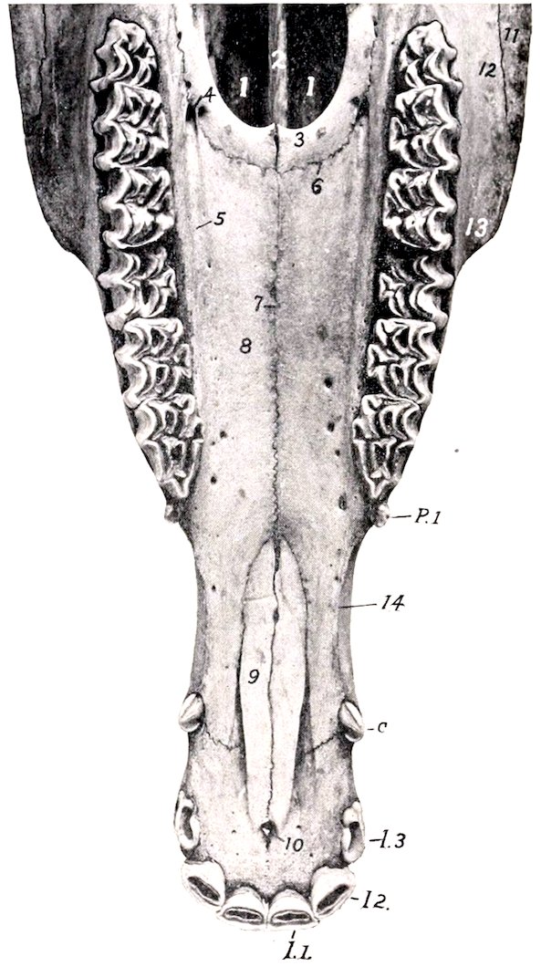
Fig. 31.—Upper Jaw of Horse About Four and a Half Years Old, Ventral View. 1, 1, Posterior nares; 2, vomer; 3, horizontal part of palate bone; 4, anterior palatine foramen; 5, palatine groove; 6, transverse palatine suture; 7, median palatine suture; 8, palate process of maxilla; 9, palate process of premaxilla; 10, foramen incisivum; 11, malar bone; 12, maxilla; 13, anterior end of facial crest; 14, interalveolar space; I. 1–3, incisor teeth; C, canine tooth; P1, first premolar or “wolf” tooth.
The infraorbital or superior dental canal extends almost horizontally from the maxillary foramen to the infraorbital foramen. It is placed at the upper edge of the inner plate of the maxilla, and traverses the maxillary sinus. Near the infraorbital foramen it gives off a small canal (Canalis alveolaris incisivus) which lies above the roots of the premolars and extends also into the premaxilla, carrying vessels and nerves to the teeth there.
Development.—The maxilla ossifies in membrane below and external to the cartilaginous nasal capsule. It has one chief center and a supplementary one in the region of the deciduous canine tooth (Martin).
The Premaxillae
The premaxillæ (Ossa incisiva) form the anterior part of the upper jaw and carry the incisor teeth. Each consists of a body and two processes, nasal and palatine.
The body is the thick anterior part which carries the incisor teeth. Its labial or superior surface is convex and smooth, and is related to the upper lip. The palatine or inferior surface is concave and presents a foramen a little behind its middle.[15] The internal surface is rough, and joins the opposite bone; it is marked by a curved groove, which forms with that on the opposed surface, the incisive 60foramen (Foramen incisivum). The alveolar border (Limbus alveolaris) separates the palatine and labial surfaces; it is curved and thick, and presents three alveoli for the incisor teeth; behind the third alveolus it is rounded and free, forming part of the interalveolar space.
The nasal process (Processus nasalis) projects backward and upward from the body, forming here the lateral wall of the nasal cavity. The two surfaces, facial and nasal, are smooth and rounded. The superior border is free, thick, and smooth. The inferior border is dentated and joins the maxilla; at its anterior end it forms with the latter the alveolus for the permanent canine tooth.[16] The posterior extremity fits into the interval between the nasal bone and the maxilla.
The palatine process (Processus palatinus) is a thin plate which forms the anterior part of the basis of the hard palate. Its nasal or superior surface has a longitudinal ridge which forms with that of the other side a groove for the septal cartilage. The palatine or inferior surface is flat. The internal border is serrated and meets its fellow at the median palatine suture. The external border is separated from the maxilla and the nasal process by the palatine cleft (Fissura palatina). The posterior extremity fits into the interval between the vomer and the palatine process of the maxilla.
Development.—The premaxilla ossifies from a single center. Fusion of the two bones is complete at the end of the third or the beginning of the fourth year.
The Palatine Bones
The palatine bones (Ossa palatina) are situated on either side of the posterior nares, and form the posterior margin of the hard palate. Each is twisted so as to form a horizontal and a perpendicular part.
The horizontal part (Pars horizontalis) is a narrow plate which forms the posterior part of the hard palate. It presents smooth nasal and palatine surfaces. The internal border meets its fellow at the median palatine suture, on the nasal aspect of which is the nasal crest. The anterior border joins the palate process of the maxilla at the transverse palatine suture, and forms with it the anterior palatine foramen. The posterior border is concave and free; it gives attachment to the aponeurosis of the soft palate.
The perpendicular part (Pars perpendicularis) is more extensive and forms the outer boundary of the posterior nares. The nasal or internal surface is in the greater part of its extent concave and smooth; it is marked by a narrow rough area to which the pterygoid bone is attached. Below this the bone curves outward, forming the pterygoid process. The maxillary or external surface (Facies maxillaris) presents three areas for consideration. The largest articulates with the maxilla; it is rough and is crossed by a groove which enters into the formation of the palatine canal. Behind this is a smooth part which assists in forming the pterygo-palatine fossa. The rough area below this is overlapped by the pterygoid process of the sphenoid bone. The superior border is perforated by the sphenopalatine foramen. Behind the foramen the two plates of the bone separate to inclose part of the sphenopalatine sinus. The inner plate curves inward to articulate with the vomer. The outer plate joins the maxilla and frontal and the orbital wing of the sphenoid.
Development.—The palatine bone ossifies in membrane from a single center.
The Pterygoid Bones
The pterygoid bones (Ossa pterygoidea) are narrow, thin, bent plates, situated on either side of the posterior nares. Each has two surfaces and two extremities. The internal surface is smooth, and forms part of the boundary of the posterior nares. The external surface articulates with the palatine, vomer, and sphenoid, 61concurring with the last in the formation of the pterygoid or Vidian canal. The anterior extremity is free, turned slightly outward, and forms the hamulus, a pulley around which the tendon of the tensor palati muscle is reflected.
Development.—The pterygoid ossifies in membrane from a single center.
The Nasal Bones
The nasal bones (Ossa nasalia) are situated in front of the frontal bones and form the greater part of the roof of the nasal cavity. They have an elongated triangular outline, wide behind, pointed in front. Each presents two surfaces, two borders, a base, and an apex.
The external or facial surface is smooth and is convex transversely; the profile contour is usually slightly wavy, with a depression about its middle and a variably prominent area in front.
The internal or nasal surface is smooth and concave from side to side. About in its middle it presents the superior turbinal crest (Crista conchalis dorsalis), which is parallel with the inner border, and has the superior turbinal bone attached to it. Most of this surface faces into the nasal cavity, but its posterior part enters into the formation of the frontal sinus; the latter area is marked off by an oblique ridge.
The internal border is straight, and meets the opposite bone at the nasal suture.
The external border is irregular. Its anterior third is free and concurs with the nasal process in forming the naso-maxillary notch (Incisura naso-maxillaris). Behind this it is scaly for articulation with the end of the nasal process, the maxilla, and the lacrimal, forming the naso-maxillary and naso-lacrimal sutures.
The base or posterior end is beveled and overlaps the frontal bone, forming the naso-frontal suture.
The apex is pointed and thin.
Development.—Each nasal bone ossifies in membrane from a single center. The nasal suture does not close completely even in old age. In some cases the two plates separate to inclose a small air-space in the posterior part.
The Lacrimal Bones
The lacrimal bones (Ossa lacrimalia) are situated at the anterior part of the orbit, and extend forward on the face to the posterior border of the maxilla. Each presents three surfaces and a circumference.
The external aspect is clearly divided into orbital and facial parts by the orbital margin. The orbital surface (Facies orbitalis) is triangular in outline, smooth and concave; it forms part of the inner and front wall of the orbit. Near the orbital margin it presents a funnel-like fossa for the lacrimal sac (Fossa sacci lacrimalis), which is the entrance to the osseous lacrimal canal. Behind this is a depression in which the inferior oblique muscle of the eye takes origin. The facial surface (Facies facialis) is more extensive, and has the form of an irregular pentagon. It is slightly convex and smooth in the foal, flattened in the adult. It usually bears the small lacrimal tubercle, situated nearly an inch (ca. 2 cm.) from the orbital margin.
The nasal or internal surface (Facies nasalis) faces into the frontal and maxillary sinuses. It is concave and very irregular, and is crossed almost horizontally by the osseous lacrimal canal (Canalis lacrimalis osseus).
The orbital margin (Margo orbitalis) is concave, rough above, smooth below.
The circumference articulates above with the frontal and nasal bones, below with the malar and maxilla, in front with the maxilla, and behind with the frontal. The various sutures so formed are designated by combinations of the names of the bones.
Development.—Each ossifies in membrane from a single center.
62
The Malar Bones
The malar or zygomatic bones (Ossa zygomatica) are placed between the lacrimal above and the maxilla below and in front. Each is irregularly triangular in outline and presents three surfaces, three borders, a base, and an apex.
The facial surface is smooth, slightly convex, wide in front, and narrow behind. At its lower part it presents the facial or zygomatic crest, which is continuous in front with the similar ridge on the maxilla and behind with the zygomatic process of the temporal; the crest is rough below, where the masseter muscle is attached to it.
The orbital surface is separated from the facial surface by the concave orbital margin. It is concave and smooth, and forms part of the lower and front wall of the orbit.
The nasal surface is concave and faces into the maxillary sinus. In the young foal a considerable part of it articulates with the maxilla.
The superior border articulates with the lacrimal chiefly, but to a small extent behind with the maxilla also.
The inferior border and the base articulate with the maxilla.
The apex is beveled above and is overlapped by the zygomatic process of the temporal bone.
Development.—Each ossifies in membrane from one or two centers.
The Turbinal Bones
These (Ossa turbinata) are delicate, scroll-like bones, four in number, which are attached to the lateral walls of the nasal cavity. They project into the cavity and greatly diminish its extent. Each is composed of a very thin lamina, cribriform in many places, and covered on both sides with mucous membrane in the fresh state. They are arranged in two pairs, superior and inferior.
The superior or naso-turbinal (Concha dorsalis)[17] is somewhat cylindrical in form, small at its anterior part, and flattened transversely. It is attached to the turbinal crest of the nasal bone and the nasal plate of the frontal bone. The anterior part is rolled like a scroll one and a half times, thus inclosing a cavity which communicates with the middle meatus nasi. The arrangement is best seen on a cross-section (Fig. 37). The posterior part is not rolled, but its lower border is attached to the lateral nasal wall, thus helping to inclose a large space which is part of the frontal sinus. This cavity is separated from that of the scroll-like part by a transverse septum. The internal surface is flattened, and is separated from the septum nasi by a narrow interval, the common meatus (Meatus nasi communis). Another narrow passage, the superior meatus (Meatus nasi superior), separates the upper surface from the roof of the nasal cavity. The space between the lower surface and the inferior turbinal is the middle meatus (Meatus nasi medius). The anterior extremity is prolonged toward the nostril by two small bars of cartilage.
The inferior or maxillo-turbinal bone (Concha ventralis) is shorter and smaller posteriorly than the upper one. It is attached to the inferior turbinal crest, and consists, like the upper one, of an anterior coiled and a posterior uncoiled portion.
To express briefly the mode of coiling of the two bones of the same side we may say that they are rolled toward the septum and each other.
The inferior and posterior borders of the posterior part are attached to the maxilla, thus helping to inclose a cavity which is part of the maxillary sinus. The lower surface is separated from the floor of the nasal cavity by the inferior meatus (Meatus nasi inferior), which is much larger than the other nasal passages. The anterior extremity is prolonged to the nostril by a curved bar of cartilage.
Development.—Each ossifies in cartilage from a single center.
63
The Vomer
The vomer is a median unpaired bone, which assists in forming the lower part of the septum nasi. It is composed of a thin lamina which is bent (except in its posterior part) so as to form a narrow groove (Sulcus septi narium), in which the lower part of the perpendicular plate of the ethmoid bone and the septal cartilage are received. The lateral surfaces, right and left, are highest near the posterior end and diminish gradually to the anterior end; they are slightly convex dorso-ventrally, and are covered by the nasal mucous membrane during life. The inferior border is thin and free in its posterior third, and divides the posterior nares into right and left halves; in the remainder of its extent it is wider and is attached to the nasal crest. The anterior extremity lies above the ends of the palatine processes of the premaxillæ. The posterior extremity consists of two wings (Alæ vomeris) which extend outward below the body of the presphenoid; posteriorly they form a notch (Incisura vomeris), and laterally join the palate and pterygoid bones.
Development.—The vomer is primitively double, and ossifies from a center on either side in the membrane covering the cartilaginous septum nasi; the two laminæ then fuse below and form a groove.
The Mandible
The mandible or inferior maxilla (Mandibula) is the largest and the only movable bone of the face. The two halves of which it consists at birth unite during the second or third month, and it is usually described as a single bone. It carries the lower teeth, and articulates by its condyles with the squamous temporal on either side. It consists of a body and two rami.[18]
The body (Corpus mandibulæ) is the thick anterior part which bears the incisor teeth. It presents two surfaces and a border. The lingual or superior surface (Facies lingualis) is smooth and slightly concave; during life it is covered by mucous membrane, and the tip of the tongue overlies it. The labial or mental surface (Facies mentalis) is convex and is related to the lower lip. It is marked by a median furrow which indicates the position of the primitive symphysis mandibulæ. The curved alveolar border (Limbus alveolaris) presents six alveoli for the incisor teeth, and a little further back two alveoli for the canine teeth in the male; in the mare the latter are usually absent or small.
The rami (Rami mandibulæ) extend backward from the body and diverge to inclose the submaxillary space (Spatium mandibulare). Each ramus is bent so as to consist of a horizontal part (Pars molaris) which bears the lower cheek teeth, and a vertical part (Ramus mandibulæ) which is expanded and furnishes attachment to powerful muscles; the term angle is applied to the most prominent part of the curve. The ramus presents two surfaces, two borders, and two extremities. The external surface is smooth and slightly convex from edge to edge on the horizontal part; at the junction with the body it presents the mental foramen (Foramen mentale), which is the external opening of the mandibular or inferior dental canal. On the vertical part it is somewhat concave and presents a number of rough lines for the attachment of the masseter muscle. The internal surface of the horizontal part is smooth, and presents a shallow longitudinal depression in its middle; above this there is often a faint mylo-hyoid line for the attachment of the muscle of like name. At the lower part of the junction with the body there is a small fossa for the attachment of the genio-hyoid and genio-glossus muscles. On the vertical part the surface is concave, and is marked in its lower and 64posterior part by rough lines for the attachment of the internal pterygoid muscle. In front of its middle is the mandibular or inferior maxillary foramen (Foramen mandibulare), which is the posterior orifice of the mandibular or inferior dental canal (Canalis mandibulæ). The canal curves downward and passes forward below the cheek teeth, opening externally at the mental foramen; it is continued into the body of the bone as a small canal (Canalis alveolaris incisivus), which carries the vessels and nerves to the incisor teeth. The superior or alveolar border forms anteriorly part of the interalveolar space; here it is thin. Behind this it is thick and is excavated by six alveoli for the lower cheek teeth. Behind the last alveolus it curves sharply upward and is narrow and rough. In the young foal there is commonly a small alveolus for the vestige of the first premolar (“wolf tooth”) close to the first large one. The inferior border of the horizontal part is nearly straight; it is thick and rounded in the young horse, becoming narrower and sharp in old subjects. At its posterior part there is a smooth impression (Incisura vasorum) where the facial vessels and parotid duct turn round the bone. Behind this point the border curves sharply upward, forming the angle (Angulus mandibulæ); this part is thick and has two roughened lips, internal and external, separated by a considerable intermediate space; near the condyle it becomes narrower. The anterior extremity joins the body. The superior extremity comprises the coronoid process in front and the condyle behind, the two being separated by the sigmoid notch (Incisura mandibulæ), through which the nerve to the masseter muscle passes. The coronoid process (Processus coronoideus) is thin transversely and curved slightly inward and backward. It projects upward in the temporal fossa, and furnishes insertion to the temporal muscle. The condyle (Capitulum mandibulæ) lies at a much lower level than the end of the coronoid process. It is elongated transversely and articulates with the squamous temporal through the medium of an articular disc. The part below the condyle is usually termed the neck (Collum mandibulæ); on its antero-internal part is a depression (Fovea pterygoidea) for the attachment of the external pterygoid muscle. The middle of the vertical part of the ramus consists to a large extent of a single plate of compact substance which may be so thin in places as to be translucent.
Development.—The mandible develops from two chief centers in the connective tissue which overlies the paired Meckel’s cartilages. At birth it consists of two symmetrical halves which meet at a median symphysis. Fusion usually occurs in the second or third month.
Age changes.—These are associated largely with the growth and later with the reduction of the teeth. In the young horse, in which the teeth are large and are in great part embedded in the bone, the body is thick and strongly curved, and the horizontal part of the ramus is also thick. Later, as the teeth are extruded from the bone, the body becomes flattened and narrower, and the horizontal part of the ramus is thinner, especially in its lower part; the angle and the impression in front of it are more pronounced.
The Hyoid Bone
The hyoid bone (Os hyoideum) is situated chiefly between the vertical parts of the rami of the mandible, but its upper part extends somewhat further back. It is attached to the petrous temporal bones by rods of cartilage, and supports the root of the tongue, the pharynx, and larynx. It consists of a body, a lingual process, and three pairs of cornua.
The body or basihyoid (Basis ossishyoidei) is a short transverse bar, compressed dorso-ventrally. The upper surface is concave and smooth in its middle, and presents at each end a convex facet or tubercle for articulation with the small cornu. The lower surface is slightly roughened for muscular attachment. The anterior border carries medially the lingual process. The posterior border is concave and smooth in its middle, and carries on either side the thyroid cornu. The body, the 65lingual process, and the thyroid processes are fused together, and may be compared to a spur or a fork with a very short handle.
The lingual process (Processus lingualis) projects forward medially from the body, and is embedded in the root of the tongue during life. It is compressed laterally and has a blunt-pointed free end. The lateral surfaces are slightly concave. The upper border is narrow, the lower thick and irregular.
The thyroid cornua or thyrohyoids (Cornua laryngea)[19] extend backward and upward from the lateral parts of the body. They are compressed laterally (except at their junction with the body), and their posterior ends are connected with the anterior cornua of the thyroid cartilage of the larynx.
The small cornua or keratohyoids (Cornua minora) are short rods which are directed upward and forward from either end of the upper surface of the body. Each is somewhat constricted in its middle part and has slightly enlarged ends. The lower end has a small concave facet which articulates with the body. The upper end articulates with the great cornu, or with the middle cornu when present.
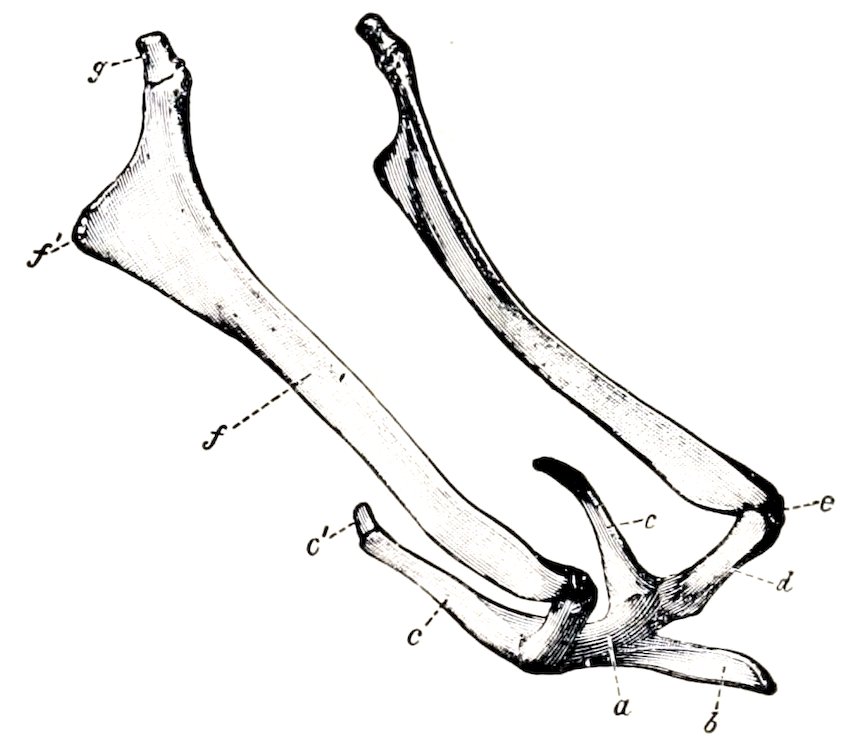
Fig. 32.—Hyoid Bone of Horse, Viewed from the Side and Somewhat from in Front. a, Body; b, lingual process; c, thyroid cornu; c′, cartilage of c; d, small cornu; e, middle cornu; f, great cornu; f′, muscular angle of great cornu; g, cartilage of great cornu. (Ellenberger-Baum, Anat. d. Haustiere.)
The great cornua or stylohyoids are much the largest parts of the bone. They are directed upward and backward, and are connected above with the base of the petrous temporal bones. Each is a thin plate, seven or eight inches (ca. 18 to 20 cm.) long, which is slightly curved in its length, so that the external surface is concave and smooth. The internal surface is convex and smooth. The borders are thin. The upper extremity is large and forms two angles; the upper angle is connected by a rod of cartilage with the hyoid process of the petrous temporal bone; the lower angle is somewhat thickened and rough for muscular attachment. The lower extremity is small, and articulates with the small or the middle cornu.
The middle cornua or epihyoids are small wedge-shaped pieces or nodules interposed between the small and great cornua. They are usually transitory, and unite with the great cornua in the adult.
Development.—The hyoid ossifies in the cartilages of the second and third visceral arches. Each part has a separate center, except the lingual process, which ossifies by extension from the body. The latter has primarily two centers (Martin).
THE SKULL AS A WHOLE
The skull of the horse has the form of a long four-sided pyramid, the base of which is posterior. It is convenient to exclude the mandible and hyoid from present consideration. The division between the cranium (Cranium cerebrale) and the face (Cranium viscerale) may be indicated approximately by a transverse plane through the anterior margins of the orbits.
The superior or frontal surface (Norma frontalis) is formed by the upper part of the occipital, the interparietal, parietal, frontal, and nasal bones. It may be 66divided into parietal, frontal, nasal, and premaxillary regions. The parietal region extends from the occipital crest to the parieto-frontal or coronal suture. It is marked medially by the external sagittal crest, which bifurcates in front, the branches becoming continuous with the frontal crests. The latter curve outward to the root of the supraorbital process. The frontal region is the widest part of the surface, and is smooth and almost flat. It is bounded in front by the naso-frontal suture. On either side of it is the root of the supraorbital process, pierced by the supraorbital foramen. The nasal region is convex from side to side, wide behind, narrow in front. Its profile is in some cases nearly straight; in others it is undulating, with a variably marked depression about its middle and at the anterior end. The premaxillary region presents the osseous nasal aperture (Apertura nasalis ossea) and the foramen incisivum.
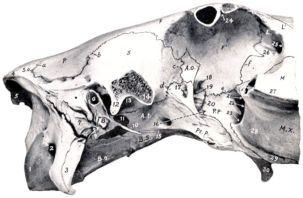
Fig. 33.—Cranial and Orbital Regions of Skull of Horse, Lateral View. The Zygomatic Arch and Supraorbital Processes Have Been Sawn Off. 1, Occipital condyle; 2, condyloid fossa; 3, paramastoid or styloid process; 4, occipital crest; 5, external occipital protuberance; 6, external auditory meatus; 7, mastoid process; 8, hyoid process; 9, stylo-mastoid foramen; 10, muscular process; 11, foramen lacerum anterius; 12, postglenoid process; 13, glenoid cavity; 14, temporal condyle; 15, Vidian groove; 16, alar canal of pterygoid process indicated by arrow; 17, temporal foramen; 18, ethmoidal foramen; 19, optic foramen; 20, foramen lacerum orbitale; 21, maxillary foramen; 22, sphenopalatine foramen; 23, posterior palatine foramen; 24, supraorbital foramen (opened); 25, lacrimal fossa; 26, depression for origin of obliquus oculi inferior; 27, facial crest; 28, maxillary tuberosity; 29, alveolar tuberosity; 30, hamulus of pterygoid bone; S.o., supraoccipital; P, parietal; S, squamous temporal; B.o., basioccipital; B.s., basisphenoid; A.t., temporal wing of sphenoid; A.o., orbital wing of sphenoid; Pt.p., pterygoid process of sphenoid; P.p., perpendicular part of palate bone; F,F′, facial and orbital parts of frontal bone; L,L′, orbital and facial parts of lacrimal bone; M, facial part of malar bone; M.x., maxilla; a, parieto-occipital suture; b, parieto-temporal or squamous suture; c, d, spheno-squamous suture; e, palato-frontal suture; f, fronto-lacrimal suture.
The lateral surface (Norma lateralis) may be divided into cranial, orbital, and maxillary or preorbital regions.
67The cranial region presents the temporal fossa, the zygomatic arch, and the outer part of the petrous temporal bone.
The temporal fossa is bounded internally by the sagittal and frontal crests, externally by the temporal crest and the zygomatic arch, and behind by the occipital crest. Its upper and middle parts are rough for the attachment of the temporal muscle. In its lower posterior part are several foramina which communicate with the parieto-temporal canal. The fossa is continuous in front with the orbital cavity.
The zygomatic arch is formed by the zygomatic processes of the temporal, malar, and maxilla. Its ventral face presents the condyle and glenoid cavity for articulation with the lower jaw, through the medium of the articular disc. Behind the glenoid cavity is the postglenoid process.
The external auditory process projects outward through a deep notch in the lower margin of the squamous temporal below the temporal crest. A little further back is the mastoid process, crossed in its upper part by a groove for the mastoid artery.
The orbital region comprises the orbit and the pterygo-palatine fossa.
The orbit is a cavity which incloses the eyeball, with the muscles, vessels, and nerves associated with it. It is not separated in the skeleton from the temporal fossa. The long axis of the cavity, taken from the optic foramen to the middle of the inlet, is directed forward, outward, and slightly upward. The inner wall (Paries medialis) is complete and extensive. It is concave and smooth, and is formed by the frontal and lacrimal and the orbital wing of the sphenoid. In its extreme anterior part is the fossa for the lacrimal sac. Behind this is a small depression in which the inferior oblique muscle of the eye arises; here the plate which separates the orbit from the maxillary sinus is very thin. The upper wall (Paries superior) is formed by the frontal and to a small extent by the lacrimal bone. It presents the supraorbital foramen, which perforates the root of the supraorbital process. The lower wall (Paries inferior) is very incomplete, and is formed by the malar, the zygomatic process of the temporal, and to a small extent by the maxilla. The external boundary (Paries lateralis) is the supraorbital process. At the extreme posterior part is the orbital group of foramina. Four are situated in front of the pterygoid crest. Of these, the uppermost is the ethmoidal or internal orbital foramen, which transmits the ethmoidal vessels and nerve. The optic foramen is situated a little lower and further back; it transmits the optic nerve. Immediately below the optic is the foramen lacerum orbitale, which transmits the ophthalmic, third, sixth, and sometimes the fourth nerve; commonly there is a very small trochlear or pathetic foramen in the crest for the last-named nerve. The foramen rotundum is below the foramen lacerum, from which it is separated by a thin plate; it transmits the superior maxillary nerve. The alar canal opens in common with the foramen rotundum, and the anterior opening of the pterygoid or Vidian canal is also found here. The temporal foramen (For. alare parvum) is just behind the pterygoid crest and on a level with the foramen lacerum. It is the upper opening of a canal which leads from the alar canal, and through it the anterior deep temporal artery emerges. The inlet of the orbital cavity (Aditus orbitæ) is circumscribed by a complete bony ring, which is nearly circular. Its antero-inferior part (Margo infraorbitalis) is smooth and rounded; the remainder (Margo supraorbitalis) is rough and irregularly notched. During life the cavity is completed by the periorbita or ocular sheath, a conical fibrous membrane, the apex of which is attached around the optic foramen.
Below the orbital cavity is the pterygo-palatine fossa. Its wall is formed by the pterygoid process, the perpendicular part of the palate bone, and the tuber maxillare. Its deep anterior part (maxillary hiatus) contains three foramina. The upper one, the maxillary foramen, is the entrance to the infraorbital canal, which transmits the infraorbital nerve and vessels. The sphenopalatine foramen transmits 68vessels and nerves of like name to the nasal cavity. The lower foramen, the posterior palatine, transmits the palatine artery and nerve to the palatine canal. The upper part of the fossa is smooth, and is crossed by the internal maxillary artery and the maxillary nerve. The lower part is chiefly roughened for the attachment of the internal pterygoid muscle, but is crossed in front by a smooth groove in which the palatine vein lies.
The maxillary or preorbital region is formed chiefly by the maxilla, but also by the premaxilla, and the facial parts of the lacrimal and malar bones. Its contour is approximately triangular, the base being posterior. It offers two principal features. The facial crest extends forward from the lower margin of the orbit, and ends abruptly at a point about an inch and a half (3 to 4 cm.) above the third or fourth cheek tooth;[20] its inferior aspect is rough for the attachment of the masseter muscle. The infraorbital foramen is situated in a transverse plane about an inch (ca. 2 to 3 cm.) in front of the end of the crest and about two inches (5 cm.) above it. The foramen opens forward, and through it the infraorbital artery and nerve emerge. The surface over the premolar teeth varies greatly with age, in conformity with the size of the embedded parts of the teeth. In the young horse the surface here is strongly convex, the outer plate of bone is thin and even defective sometimes in places, and the form of the teeth is indicated by eminences (Juga alveolaria). In the old animal the surface is concave on account of the extrusion of the teeth from the bone. The downward curve of the premaxilla is pronounced in the young subject, very slight in the aged.
The inferior or basal surface (Norma basalis), exclusive of the mandible, consists of cranial, guttural, and palatine regions.
The cranial region (Basis cranii externa) extends forward to the vomer and pterygoid processes (Fig. 28). At its posterior end is the foramen magnum, flanked by the occipital condyles. External to the latter is the condyloid fossa, in which is the hypoglossal foramen, which transmits the hypoglossal nerve and the condyloid artery and vein. Further outward are the paramastoid or styloid processes (Processus jugulares) of the occipital bone. Extending forward centrally is a prismatic bar, formed by the basilar part of the occipital and the body of the sphenoid bone; at the junction of these parts are tubercles for the attachment of the ventral straight muscles of the head. On either side of the basilar part of the occipital is the foramen lacerum basis cranii, bounded externally by the base of the petrous temporal bone. In front of these the region becomes very wide on account of the lateral extension of the zygomatic processes, bearing on the ventral aspect the condyle and glenoid cavity for articulation with the mandible. Beyond this the process turns forward and joins the zygomatic process of the malar, completing the zygomatic arch and the surface for the attachment of the masseter muscle. On either side of the body of the sphenoid is the infratemporal fossa, formed by the temporal wing and the root of the pterygoid process of the sphenoid bone. It is bounded in front by the pterygoid crest, which separates it from the orbit and the pterygo-palatine fossa. In it is the pterygoid or alar foramen, which transmits the internal maxillary artery. A little lower is the entrance to the pterygoid (Vidian) canal.
The guttural region presents the pharyngeal orifice of the nasal cavity. This is elliptical and is divided in its depth medially by the vomer into two posterior nares or choanæ. It is bounded in front and laterally by the palate and pterygoid bones, behind by the vomer. It is flanked by the hamular process of the pterygoid bones. The plane of the opening is nearly horizontal, and the length is about twice the width.
The palatine region comprises a little more than half of the entire length 69of the base of the skull (Fig. 31). The hard palate (Palatum durum) is concave from side to side, and in its length also in the anterior part. It is formed by the palatine processes of the premaxillæ and maxillæ, and the horizontal parts of the palate bones. It is circumscribed in front and laterally by the superior alveolar arch, in which the upper teeth are implanted. The interalveolar space (Margo interalveolaris) is that part of the arch in which alveoli are not present. Behind the last alveolus is the alveolar tuberosity, and internal to this is a groove for the palatine vein. In the middle line is the median palatine suture (Sutura palatina mediana). In the line of the suture, a little behind the central incisors, is the foramen incisivum, through which the palato-labial artery passes. On either side, parallel with the alveolar part of the maxilla, is the palatine groove (Sulcus palatinus), which contains the palatine vessels and nerve. It is continuous at the anterior palatine foramen with the palatine canal, which is situated between the maxilla and the palate bone. The palatine cleft (Fissura palatina) is the narrow interval along the outer margin of the palatine process of the premaxilla; it is closed in the fresh state by cartilage. Scattered along each side of the palate are several vascular foramina. The transverse palatine suture (Sutura palatina transversa) is about half an inch from the posterior border. The latter is in a plane through the last molar teeth, and is concave and free.
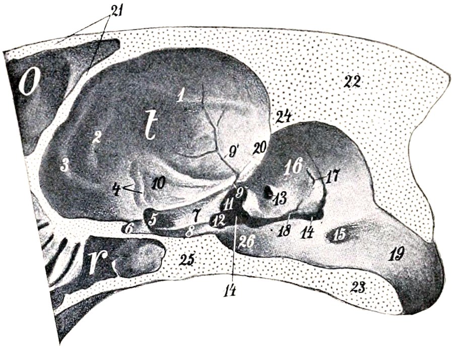
Fig. 34.—Cranial Cavity of Horse as Seen on Sagittal Section of Skull. O, Frontal sinus; r, sphenoidal sinus; t, cerebral compartment of cranium; 1–3, ridges (juga) corresponding to fissures of lateral surface of cerebrum; 4, groove for middle cerebral artery; 5, entrance to for. lacerum orbitale; 6, entrance to optic foramen; 7, 8, grooves on sphenoid bone; 9, incisura spinosa; 9′, groove for middle meningeal artery; 10, fossa for pyriform lobe of cerebrum; 11, incisura ovalis; 12, incisura carotica; 13, internal auditory meatus; 14, foramen lacerum basis cranii; 15, hypoglossal foramen; 16, petrous temporal; 17, orifice of aquæductus vestibuli; 18, orifice of aquæductus cochleæ; 19, foramen magnum; 20, petrosal crest; 21, two plates of frontal bone; 22, supraoccipital; 23, basioccipital; 24, tentorium osseum; 25, body of sphenoid. (After Ellenberger-Baum, Top. Anat. d. Pferdes.)
The posterior or nuchal surface (Norma occipitalis) is formed by the occipital bone. It is trapezoidal in outline, wider below than above, concave dorso-ventrally, convex transversely. It is separated from the superior surface by the occipital crest, and from the lateral surfaces by the superior curved lines (Lineæ nuchæ superiores). Below the crest are two rough areas for the attachment of the complexus muscles. A little lower is a central eminence on the sides of which the ligamentum nuchæ is attached. At the lowest part centrally is the foramen magnum, at which the brain and spinal cord meet; this is bounded laterally by the occipital condyles, which are flanked by the paramastoid or styloid processes (Processus jugulares).
The apex of the skull is formed by the bodies of the premaxillæ and mandible, carrying the incisor teeth.
THE CRANIAL CAVITY
This cavity incloses the brain, with its membranes and vessels. It is relatively small and is ovoid in shape.
70The superior wall or roof (Calvaria) is formed by the supraoccipital, interparietal, parietal, and frontal bones. In the middle line is the internal sagittal crest, which joins the crista galli in front, and furnishes attachment to the falx cerebri. Posteriorly the crest is continued by the sharp anterior margin of the tentorium osseum, which projects downward and forward into the cavity, and gives attachment to the tentorium cerebelli by its sharp lateral edges. Behind this the roof is grooved centrally for the middle lobe or vermis of the cerebellum. Transverse grooves pass from the base of the tentorium osseum to the parieto-temporal canals. The anterior part of the roof is hollowed by the frontal sinus. The occipital part is very thick and strong.
The lateral walls are formed by the occipital, parietal, temporal, and frontal bones, and in part by the orbital wings of the sphenoid. Each is crossed obliquely by the petrosal crest, which concurs with the projecting margin of the parietal bone and the tentorium osseum in dividing the cavity into cerebral and cerebellar compartments. Behind the crest is a depression for the lateral lobe (hemisphere) of the cerebellum. Below this are the internal auditory meatus and the openings of the aquæductus vestibuli and aquæductus cochleæ.
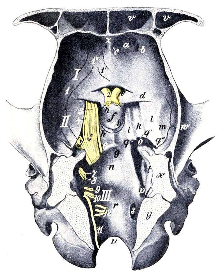
Fig. 35.—Floor of Cranial Cavity of Horse. The roots of the cranial nerves are shown on the left side and are designated by number. I, Anterior cranial fossa; II, middle cranial fossa; III, posterior cranial fossa; a, ethmoidal fossa; b, ethmoidal foramen; c, foramen for nasal branch of ophthalmic artery; d, orbital wing of sphenoid bone; e, optic fossa; f, sella turcica; g, spheno-occipital crest; h, h′, dotted line indicating contour of pituitary body; h″, slight elevation representing dorsum sellæ; i, k, grooves for nerves and cavernous sinus; l, depression for pyriform lobe of cerebrum; m, groove for middle meningeal artery; n, depression for pons; o, foramen lacerum anterius; p, foramen lacerum posterius; q, incisura carotica; q′, incisura ovalis; q″, incisura spinosa; r, depression for medulla oblongata; s, hypoglossal foramen; t, internal auditory meatus; u, foramen magnum; v, frontal sinus; w, zygomatic process of temporal bone; x, section of petrous temporal; y, section of occipital bone; z, crista galli; 1, 1′, 1″, dotted lines indicating position of olfactory tracts and peduncle. (After Ellenberger-Baum, Top. Anat. d. Pferdes.)
The roof and lateral walls are marked by digital impressions and vascular grooves.
The inferior wall or floor (Basis cranii interna) may be regarded as forming three fossæ. The anterior fossa (Fossa cranii anterior) supports the frontal and olfactory parts of the cerebrum. It is formed chiefly by the presphenoid, and lies at a higher level than the middle fossa. In front the fossa is divided medially by the crista galli, lateral to which are the deep ethmoidal or olfactory fossæ for the olfactory lobes. The ethmoidal or internal orbital foramen perforates the cranial wall at the outer side of these fossæ. Further back the central part of the surface is slightly elevated, and is flanked by shallow depressions which support the olfactory peduncles. Posteriorly is a bony shelf which covers the entrance to the optic foramina; the edge of this shelf and the posterior borders of the orbital wings of the sphenoid may be taken as the line of demarcation between the anterior and 71middle fossæ. The middle fossa (Fossa cranii media) is the widest part of the cavity. It extends backward to the internal spheno-occipital and petrosal crests, thus corresponding to the postsphenoid. In its middle is a small fossa, the sella turcica, in which the pituitary body, or hypophysis cerebri, is situated. On either side are two grooves for nerves; the inner one transmits the ophthalmic, third, and sixth nerves to the foramen lacerum orbitale; the outer one leads to the foramen rotundum, and lodges the maxillary nerve. External to the grooves is a depression for the pyriform lobe of the cerebrum. The posterior fossa (Fossa cranii posterior) corresponds to the basilar part of the occipital bone. It contains the medulla, pons, and cerebellum. In front is a median depression (Fossa pontis) for the pons. The surface behind this is concave transversely and slopes gently downward to the foramen magnum; it supports the medulla. On either side are the foramen lacerum basis cranii and the hypoglossal foramen.
The anterior or nasal wall is formed by the cribriform plate of the ethmoid, which separates the cranium from the nasal cavity. It is perforated by numerous foramina for the passage of the olfactory nerve-bundles.
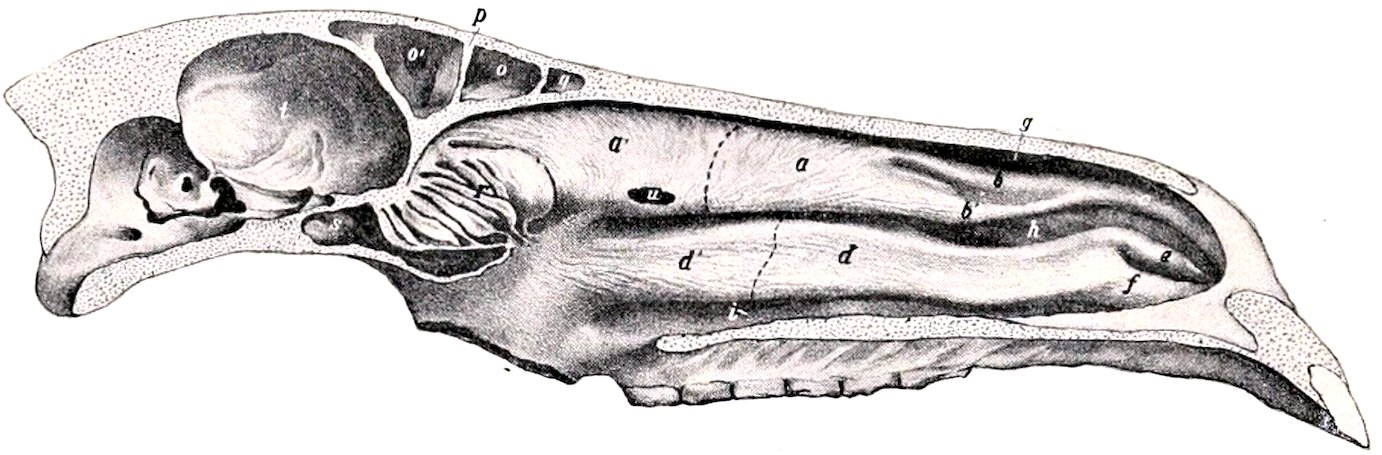
Fig. 36.—Median Section of Skull of Horse Without the Mandible. The septum nasi is removed, but the mucous membrane on the turbinal bones is retained. a, a′, Superior turbinal bone, dotted line indicating limit between anterior coiled part and posterior uncoiled part; b, b′, superior turbinal folds, inclosing bars of cartilage; c, fold of mucous membrane formed by union of b and b′; d, d′, anterior coiled and posterior uncoiled part of inferior turbinal, dotted line indicating septum between them; e, f, inferior turbinal folds, former (alar fold) inclosing bar of cartilage; g, h, i, superior, middle, inferior meatus; o, o′, frontal sinus; partial septum between o and o′; q, nasal part of frontal sinus (nasal sinus); r, lateral mass of ethmoid bone; s, sphenoidal sinus; t, cranial cavity; u, opening made in superior turbinal bone at point where drainage of frontal sinus may be obtained. (After Ellenberger, in Leisering’s Atlas.)
THE NASAL CAVITY
The nasal cavity (Cavum nasi) is a longitudinal passage which extends through the upper part of the face. It is divided into right and left halves by a median septum nasi. The lateral walls are formed by the maxilla, premaxilla, and the perpendicular part of the palate bones. Attached to them are the turbinal bones, which subdivide each nasal fossa into three meatuses (Meatus nasi). This wall is crossed obliquely by the canal and groove for the naso-lacrimal duct, and its posterior part is perforated by the sphenopalatine foramen. The superior wall or roof is formed by the frontal and nasal bones. It is concave from side to side, and nearly straight longitudinally, except in the posterior part, where it curves downward. It presents a median elevation, the nasal crest. The inferior wall or floor is formed by the palatine processes of the premaxillæ and maxillæ, and the horizontal parts of the palate bones. It is wider but considerably shorter than the roof. It is concave transversely, and nearly horizontal from before backward, except in the posterior third, where there is a slight declivity. The anterior part presents a median groove for the cartilaginous septum, and a furrow for the organ of Jacobson on either side. On either side of the palatine processes of the premaxillæ is the palatine cleft.
72The septum nasi is incomplete in the macerated skull. It is formed by the perpendicular plate of the ethmoid behind, and the vomer below. In the fresh state it is completed by a plate of cartilage.
The superior meatus (Meatus nasi superior) is a narrow passage between the roof and the superior turbinal bone. It ends at the cribriform plate of the ethmoid. The middle meatus (Meatus nasi medius) is the space between the two turbinal bones. In its posterior part is the very narrow opening into the maxillary sinus. The inferior meatus (Meatus nasi inferior) is the channel along the floor which is overhung by the inferior turbinal bone. It is much the largest and is the direct path between the anterior and posterior nares.
The external aperture is bounded by the nasal bones and the premaxillæ.
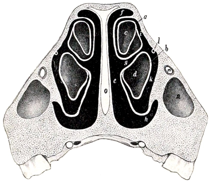
Fig. 37.—Cross-section of Nasal Region of Skull of Horse; the Section Passes Through the Anterior End of the Facial Crest, and Between the Third and Fourth Cheek Teeth. a, Superior, b, inferior turbinal bone; c, d, cavities of a and b; e, common meatus; f, g, h, superior, middle, inferior meatus; i, k, passages to cavities of turbinal bones; l, naso-lacrimal duct; m, infraorbital canal; n, anterior end of maxillary sinus; o, septal cartilage. (After Ellenberger, in Leisering’s Atlas.)
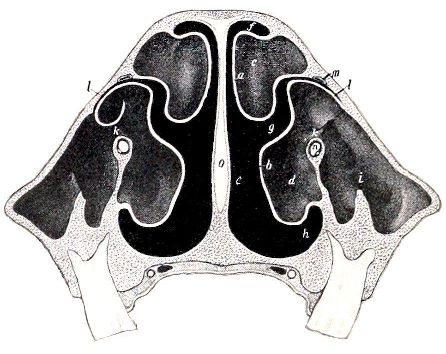
Fig. 38.—Cross-section of Nasal Region of Skull of Horse; the Section is Cut About Halfway between the Orbit and the Anterior End of the Facial Crest, and Passes between the Fifth and Sixth Cheek Teeth. a, Superior, b, inferior turbinal bone; c, d, cavities of a and b; e, common meatus; f, superior, g, middle, h, inferior meatus; i, placed over ridge in maxillary sinus; k, communication between outer and inner (turbinal) part of maxillary sinus; l, naso-maxillary opening; m, naso-lacrimal canal; n, infraorbital canal. (After Ellenberger, in Leisering’s Atlas.)
The posterior extremity or fundus is separated from the cranial cavity by the cribriform plate of the ethmoid, and is largely occupied by the lateral masses of that bone.
THE PARANASAL SINUSES
Connected directly or indirectly with the nasal cavity, of which they are diverticula, are four pairs of air-sinuses (Sinus paranasales), viz., maxillary, frontal, sphenopalatine, and ethmoidal.
The maxillary sinus (Sinus maxillaris) is the largest. Its external wall is formed by the maxilla, the lacrimal, and the malar. It is bounded internally by the maxilla, the inferior turbinal, and the lateral mass of the ethmoid bone. It extends backward to a transverse plane in front of the root of the supraorbital process, and its anterior limit is indicated approximately by a line drawn from the anterior end of the facial crest to the infraorbital foramen. Its upper boundary corresponds to a line drawn backward from the supraorbital foramen parallel to the facial crest. The floor is formed by the alveolar part of the maxilla; it is very irregular and is crossed by bony plates running in various directions. The last 73three cheek teeth project up into the cavity to an extent which varies with age; they are covered by a thin plate of bone. The cavity is divided into anterior and posterior parts by an oblique septum. The outer margin of the septum is commonly about one and a half to two inches (ca. 3.5 to 5 cm.) from the anterior end of the facial crest; from here it is directed inward, backward, and upward. The upper part of the septum (formed by the posterior end of the inferior turbinal bone) is very delicate and usually cribriform.
The position of the septum is quite variable. It is not rare to find it further forward, and in some cases it is further back than is stated above. In the recent state, i. e., when covered by the mucous membrane on both surfaces, it is nearly always complete, but in very exceptional cases there is an opening of variable size in the upper part.
The anterior compartment, often called the inferior maxillary sinus, is partially divided by the infraorbital canal into an external maxillary part and an internal smaller turbinal part. The latter communicates with the middle meatus by a very narrow slit situated at its highest part. The posterior compartment, often called the superior maxillary sinus, is also crossed by the infraorbital canal, internal to which it opens freely into the sphenopalatine sinus. It communicates dorsally with the frontal sinus through the large oval fronto-maxillary opening, situated at the level of the osseous lacrimal canal and the corresponding part of the inner wall of the orbit; the orifice is commonly about one and a half to two inches (ca. 4 to 5 cm.) long and an inch or more (2 to 3 cm.) wide. Just in front of this, and covered by a thin plate, is the narrow naso-maxillary fissure (Aditus naso-maxillaris), by which the sinus opens into the posterior part of the middle meatus.
The foregoing statements refer to the arrangement in the average adult animal. In the foal the cavity (with the exception of its turbinal part) is largely occupied by the developing teeth. In horses five to six years of age the maxillary part of the sinus is still filled up to a large degree by the embedded parts of the teeth. As the teeth are extruded to compensate the wear, more and more of the cavity becomes free, until in old age only the short roots project up in the floor, covered by a layer of bone. Other facts in this connection will be given in the description of the teeth. In exceptional cases the posterior part of the inferior turbinal is smaller than usual and leaves a considerable interval, through which the maxillary sinus communicates with the nasal cavity.
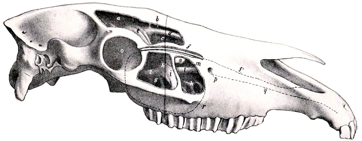
Fig. 39.—Skull of Horse, Lateral View without Mandible. The Sinuses are Opened up. a, Posterior part, b and c, anterior part of frontal sinus; d, roof of superior meatus; e, lateral mass of ethmoid bone; f, f′, naso-lacrimal duct, exposed in its posterior part; g, h, posterior and anterior compartments of maxillary sinus (also designated as superior and inferior maxillary sinuses); i, septum between g and h; k, lower limit of upper thin and partly membranous portion of septum; l, infraorbital canal; m, turbinal part of maxillary sinus; n, bullous prominence of inferior turbinal; o, orbit; p, infraorbital foramen; q, continuation of infraorbital canal to premaxilla; r, limit of maxillary sinus. (After Ellenberger, in Leisering’s Atlas.)
The frontal sinus (Sinus concho-frontalis) consists of frontal and turbinal parts. The frontal part is bounded chiefly by the two plates of the frontal bone, but its floor is formed in part by the lateral mass of the ethmoid. It extends forward to a 74plane through the anterior margins of the orbits, backward to one through the temporal condyles, and outward into the root of the supraorbital process. It is separated from the sinus of the opposite side by a complete septum. It is partially subdivided by a number of bony plates. The turbinal part is situated in the posterior part of the superior turbinal bone, roofed in by the nasal and lacrimal bones. It extends forward to a transverse plane about half-way between the anterior margin of the orbit and the end of the facial crest. Behind it is in free communication with the frontal part over the lateral mass of the ethmoid. It is separated from the nasal cavity by the thin turbinal plate. The frontal and maxillary sinuses communicate through the large opening described above.
The sphenopalatine sinus (Sinus sphenopalatinus) consists of two parts which communicate under the lateral mass of the ethmoid. The sphenoidal (posterior) part is excavated in the body of the presphenoid. The palatine (anterior) part is between the two plates of the perpendicular part of the palate bone, under the lateral mass of the ethmoid; it communicates freely with the maxillary sinus. The septum between the right and left sinuses is not usually median in the sphenoidal part.
In about one-third of the cases (according to Paulli) the sphenoidal and palatine parts are separated by a transverse septum, and the sphenoidal part then communicates only with the lower ethmoidal meatuses.
The term ethmoidal sinus is often applied to the cavity of the largest ethmoturbinal. It communicates with the maxillary sinus.
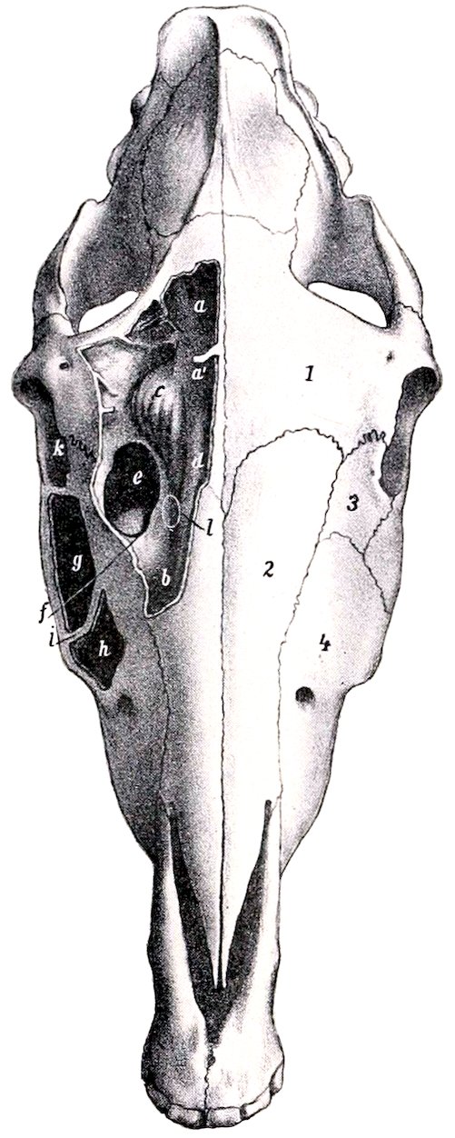
Fig. 40.—Skull of Horse, Dorsal View, with Sinuses Exposed by Removal of the Outer Plate of Bone. 1, Frontal bone; 2, nasal bone; 3, lacrimal bone; 4, maxilla; a, posterior part of frontal sinus; a′, middle part of frontal sinus; b, anterior (turbinal) part of frontal sinus; c, lateral mass of ethmoid bone; d, roof of superior meatus; e, fronto-maxillary opening; f, naso-maxillary opening below plate which forms the anterior margin of e; g, h, posterior and anterior compartments of maxillary sinus—often called the superior and inferior maxillary sinuses; i, septum between g and h; k, orbit; l, point at which superior turbinal bone may be perforated to obtain drainage into nasal cavity. (After Ellenberger, in Leisering’s Atlas.)
The Bones of the Thoracic Limb the scapula
The scapula is a flat bone, situated on the anterior part of the lateral wall of the thorax, and extending obliquely from the vertebral end of the seventh or eighth rib to the sternal end of the first rib. It is curved slightly and slopes outward in adaptation to the form of the thoracic wall. It is triangular in outline, and has two surfaces, three borders, and three angles.
The external surface or dorsum (Facies lateralis s. dorsalis) is divided into two fossæ by the spine (Spina scapulæ), which extends from the vertebral border to the neck of the bone, where it gradually subsides. The free edge of the spine is thick, rough, and in great part subcutaneous. A little above its middle is a variable prominence, 75the tubercle of the spine (Tuber spinæ), to which the trapezius muscle is attached. The supraspinous fossa (Fossa supraspinata) is situated in front of the spine, and the infraspinous fossa (Fossa infraspinata) behind it. The former is much the smaller of the two; it is smooth and is occupied by the supraspinatus muscle. The infraspinous fossa lodges the infraspinatus muscle; it is wide and smooth in its upper part, narrower below, where it is marked by several rough lines for the attachment of the teres minor muscle; near the neck is the nutrient foramen, and a little lower is a vascular groove.
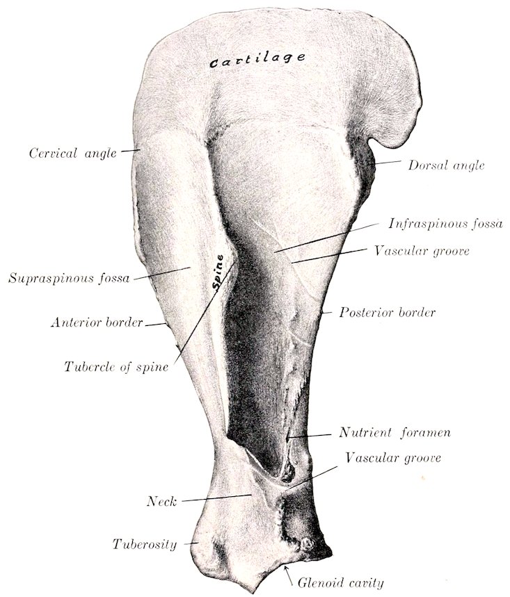
Fig. 41.—Left Scapula of Horse, External View. (After Schmaltz, Atlas d. Anat. d. Pferdes.)
The costal surface or venter (Facies costalis) is hollowed in its length by the subscapular fossa (Fossa subscapularis); this occupies nearly the whole of the lower part of the surface, but is pointed above and separates two rough triangular areas (Facies serrata), to which the serratus magnus is attached. In the lower third there is a vascular furrow with several branches.
The anterior or cervical border (Margo cranialis) is convex and rough above, concave and smooth below.
The posterior or dorsal border (Margo caudalis) is slightly concave. It is thick and rough in its upper third, thin in its middle, and thickens again below.
The superior or vertebral border (Margo dorsalis s. basis) carries the scapular cartilage (Cartilago scapulæ). In the young subject the edge of the bone is thick, 76and is pitted by impressions into which the cartilage fits. The cartilage is the unossified part of the fœtal scapula. Its lower edge fits the depressions and elevations of the bone. It thins out toward the free edge, which is convex and lies alongside of the upper parts of the vertebral spines. In front it continues the line of the scapula, but behind it forms a rounded projection. The lower part of the cartilage undergoes more or less ossification, so that the vertebral border of the bone in old subjects is thin, irregular, and porous.
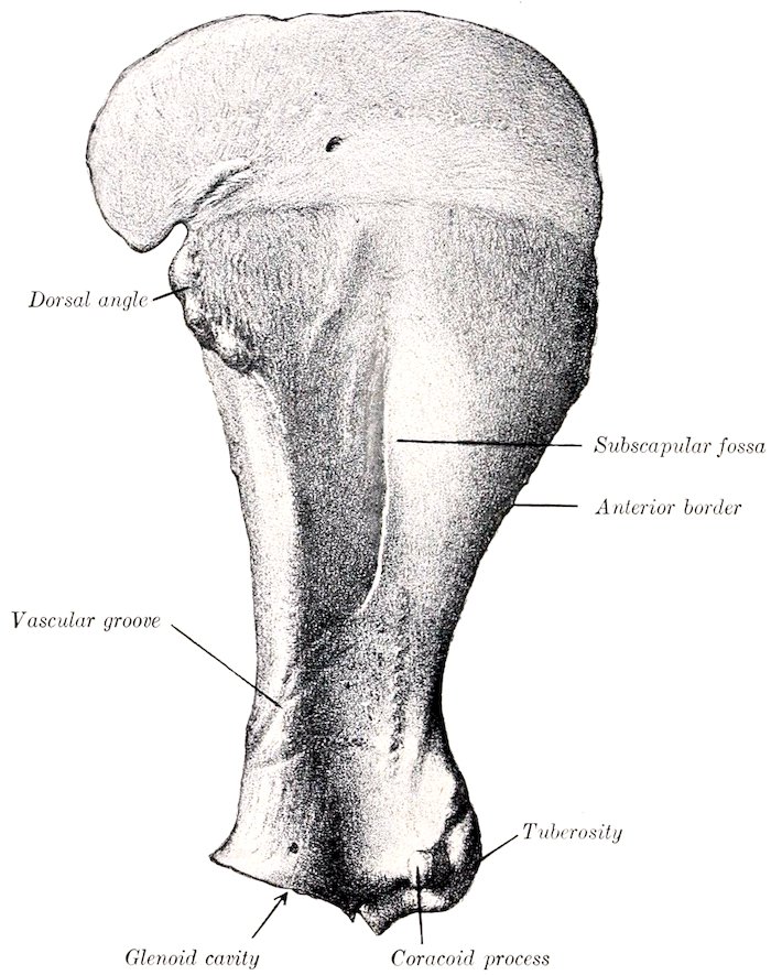
Fig. 42.—Left Scapula of Horse, Costal Surface.—(After Schmaltz, Atlas d. Anat. d. Pferdes.)
The anterior or cervical angle (Angulus cranialis) is at the junction of the anterior and vertebral borders and lies opposite to the second thoracic spine. It is relatively thin and is about a right angle.
The posterior or dorsal angle (Angulus caudalis) is thick and rough; its position can be determined readily in the living animal.
The inferior or articular angle (Angulus glenoidalis) is joined to the body of the bone by the neck of the scapula (Collum scapulæ). It is enlarged, especially in the sagittal direction. It bears the glenoid cavity (Cavitas glenoidalis) for articulation with the head of the humerus. The cavity is oval in outline, and its margin is cut into in front by the glenoid notch (Incisura glenoidalis), and is rounded off 77externally; just above its postero-external part is a tubercle to which a tendon of the teres minor is attached. The bicipital tuberosity or tuber scapulæ is the large rough prominence in front, to which the tendon of origin of the biceps brachii is attached; projecting from its inner side is the small coracoid process (Processus coracoideus), from which the coraco-brachialis muscle arises.
Development.—The scapula has four centers of ossification, viz., one each for the body of the bone, the bicipital and coracoid processes, the anterior part of the glenoid cavity, and the tuber spinæ. The last ossifies after birth and fuses with the spine about the third year. The bicipital tuberosity and coracoid fuse with the body of the bone about the end of the first year.
In old subjects the spongy substance disappears at the middle part of the fossæ, so that the bone consists here of a thin layer of compact substance. Considerable ossification of the cartilage is usual, the borders become much rougher, the muscular lines are more pronounced, and a medullary cavity may appear in the neck. Much variation occurs in dimensions and slope. The average ratio between the length and breadth (scapular index) is about 1 ∶ 0.5, but in many cases the base is relatively wider. The inclination on a horizontal plane varies from 50 to 65 degrees. Exceptionally the coracoid process reaches a length of an inch or more (2½ to 3 cm.), and the chief nutrient foramen may be on the posterior border or in the subscapular fossa.
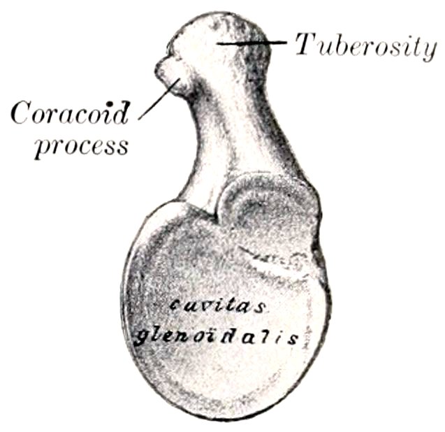
Fig. 43.—Distal End of Left Scapula of Horse, End View. (After Schmaltz, Atlas d. Anat. d. Pferdes.)
THE HUMERUS
The humerus is a long bone which extends from the shoulder above, where it articulates with the scapula, to the elbow below and behind, where it articulates with the radius and ulna. It is directed obliquely downward and backward, forming an angle of about 55 degrees with a horizontal plane. It may be divided into a shaft and two extremities.
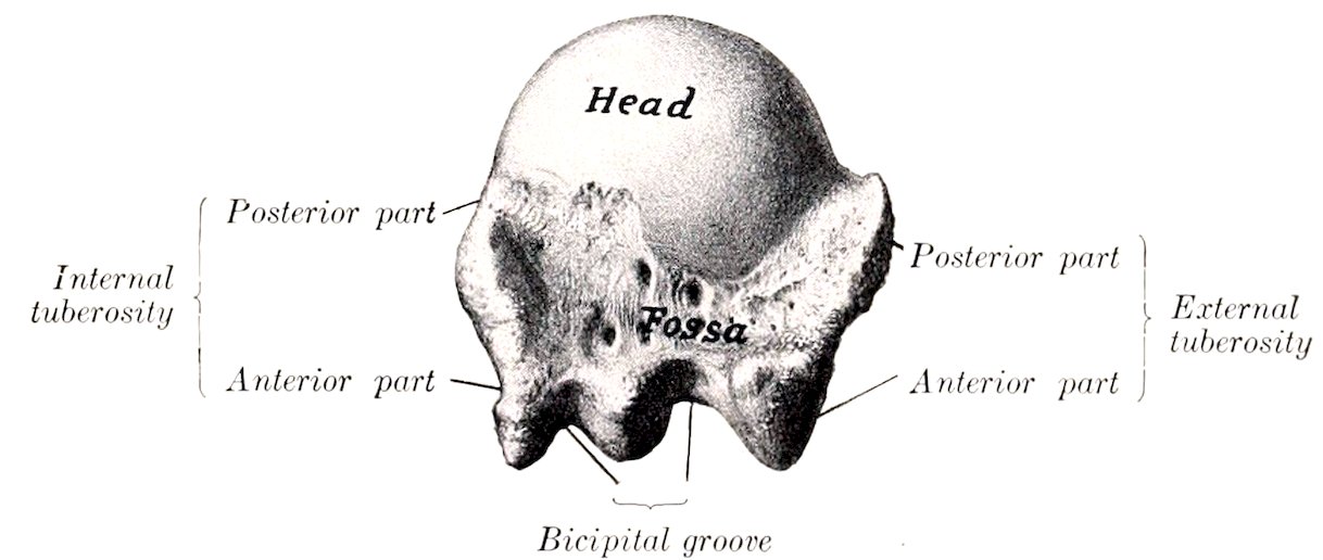
Fig. 44.—Proximal End of Left Humerus of Horse, End View. (After Schmaltz, Atlas d. Anat. d. Pferdes.)
The shaft or body (Corpus humeri) is irregularly cylindrical and has a twisted appearance. It may be regarded as having four surfaces. The external surface is smooth and is spirally curved, forming the musculo-spiral groove (Sulcus musculi brachialis), which contains the brachialis muscle; the groove is continuous with the posterior surface above and winds around toward the front below. The internal surface is nearly straight in its length, rounded from side to side, and blends with the anterior and posterior surfaces. Just above its middle is the internal or teres 78tubercle (Tuberositas teres), to which the tendon of the latissimus dorsi and teres major muscles is attached. The nutrient foramen is in the lower third of this surface. The anterior surface is triangular, wide and smooth above, narrow and roughened below. It is separated from the external surface by a distinct border, which bears on its upper part the deltoid tuberosity (Tuberositas deltoidea). From the latter a rough line curves upward and backward to the outer surface of the neck, and gives origin to the external head of the triceps muscle. Below the tuberosity the border inclines forward, becomes less salient, and ends at the coronoid fossa. The posterior surface is rounded from side to side and smooth.
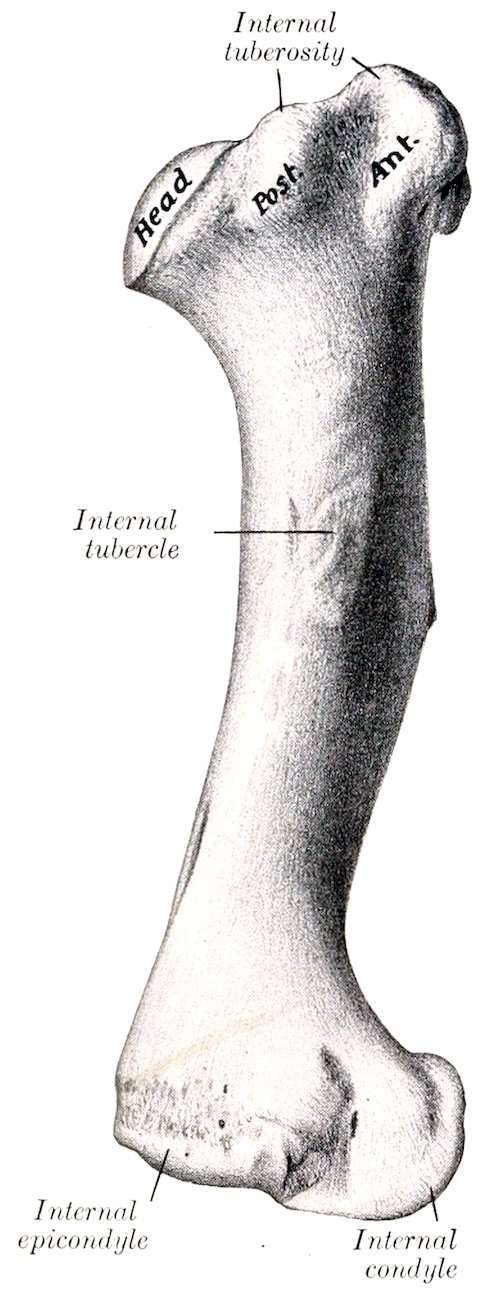
Fig. 45.—Left Humerus of Horse, Internal Surface. (After Schmaltz, Atlas d. Anat. d. Pferdes.)
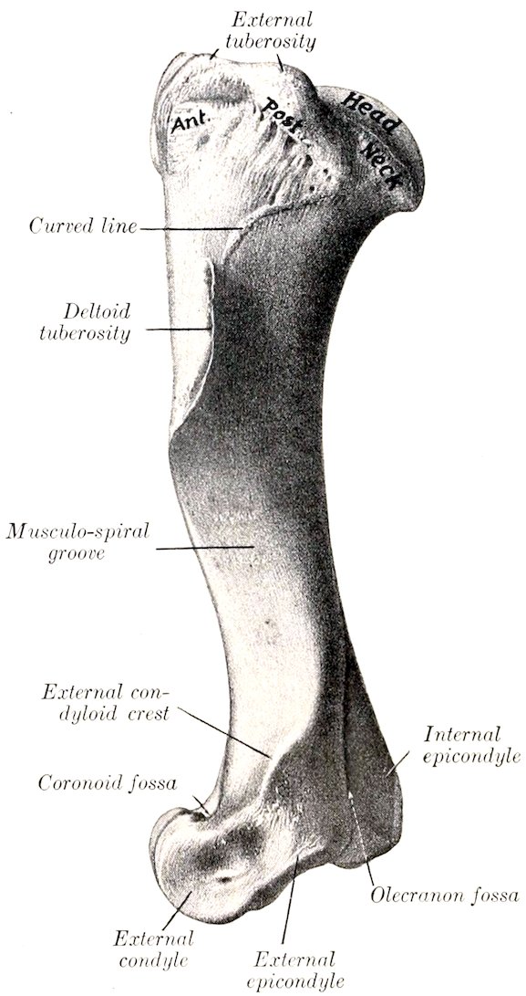
Fig. 46.—Left Humerus of Horse, External Surface. (After Schmaltz, Atlas d. Anat. d. Pferdes.)
The proximal extremity consists of the head, neck, two tuberosities, and the bicipital groove. The head (Caput humeri) presents an almost circular convex articular surface, which is about twice as extensive as the glenoid cavity of the scapula, with which it articulates. In front of the head is a fossa, in which are several foramina. The neck (Collum humeri) is well defined behind, but is practically absent elsewhere. The external tuberosity (Tuberculum majus) is placed 79antero-externally, and consists of two parts; the anterior part forms the outer boundary of the bicipital groove and gives attachment to the external branch of the supraspinatus muscle; the posterior part gives attachment to the short insertion of the infraspinatus, while its outer surface is coated with cartilage, over which the chief tendon of the same muscle passes to be inserted into a triangular facet on the outer aspect of the anterior part. The internal tuberosity (Tuberculum minus) is less salient, and consists of anterior and posterior parts; the anterior part forms the inner boundary of the bicipital groove, and furnishes insertion to the inner branch of the supraspinatus above, and the posterior deep pectoral muscle below; the posterior part gives attachment to the subscapularis muscle. The bicipital or intertubercular groove (Sulcus intertubercularis) is situated in front; it is bounded by the anterior parts of the tuberosities, and is subdivided by an intermediate ridge. The groove is covered in the fresh state by cartilage, and lodges the tendon of origin of the biceps brachii muscle. Just below the intermediate ridge is a small fossa in which several foramina open.
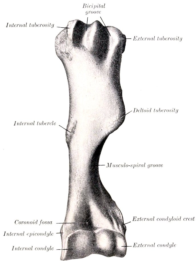
Fig. 47.—Left Humerus of Horse, Anterior View. (After Schmaltz, Atlas d. Anat. d. Pferdes.)
The distal extremity has an oblique surface for articulation with the radius and ulna, which consists of two condyles of very unequal size, separated by a ridge. The internal condyle (Condylus medialis) is much the larger, and is crossed by a sagittal groove, on the anterior part of which there is usually a synovial fossa. Posteriorly 80the groove extends upward considerably above the rest of the articular surface and reaches the olecranon fossa, and this part articulates with the semilunar notch of the ulna. The external condyle (Condylus lateralis) is much smaller and is placed somewhat lower and further back, giving the extremity an oblique appearance; it is marked by a wide shallow groove. The coronoid fossa (Fossa coronoidea) is situated in front above the groove on the internal condyle; it furnishes origin to part of the extensor carpi, and external to it is a rough depression from which the anterior or common extensor of the digit arises. Behind and above the condyles are two thick ridges, the epicondyles. The internal or flexor epicondyle (Epicondylus medialis s. flexorius) is the more salient; it furnishes origin to flexor muscles of the carpus and digit, and presents internally a tubercle for the attachment of the internal lateral ligament of the elbow joint. The external or extensor epicondyle (Epicondylus lateralis s. extensorius) bears externally the external supracondyloid crest (Crista condyloidea lateralis), which forms here the outer boundary of the musculo-spiral groove, and gives origin to the extensor carpi. Below this is a rough excavation in which the external lateral ligament is attached. The lower border of the epicondyle gives attachment to the flexor carpi externus. Between the epicondyles is the deep olecranon fossa (Fossa olecrani).
Development.—The humerus ossifies from six centers, viz., three primary centers for the shaft and epiphyses, and three secondary centers for the external tuberosity, the deltoid tuberosity, and the internal condyle. The proximal end fuses with the shaft at about three and one-half years, the distal at about one and a half years of age.
THE RADIUS
The radius is much the larger of the two bones of the forearm in the horse. It extends in a vertical direction from the elbow, where it articulates with the humerus, to the carpus below. It is gently curved, the convexity being anterior. It consists of a shaft and two extremities.
The shaft (Corpus radii) is curved in its length, somewhat flattened from before backward, and expanded at its ends. It presents for description two surfaces and two borders. The anterior surface (Facies dorsalis) is smooth, slightly convex in its length, and rounded from side to side. The posterior surface (Facies volaris) is correspondingly concave in its length and is flattened in the transverse direction. At its upper part there is a smooth shallow groove, which concurs with the ulna in the formation of the interosseous space of the forearm; the nutrient foramen is in the lower part of this groove. Below this there is in the young subject a narrow, rough, triangular area to which the ulna is attached by an interosseous ligament; in the adult the two bones are fused here. A variable rough elevation below the middle of the surface and close to the internal border gives attachment to the superior check ligament. The internal border (Margo medialis) is slightly concave in its length and is largely subcutaneous; at its proximal end there is a smooth area on which the tendon of insertion of the brachialis muscle lies, and a small rough area just below gives attachment to that muscle and the long internal lateral ligament of the elbow joint. The external border (Margo lateralis) is more strongly curved, but presents no special features.
The proximal extremity or head (Capitulum radii) is flattened from before backward and wide transversely. It presents an articular surface (Fovea capituli) which corresponds to that on the distal end of the humerus; it is crossed by a central sagittal ridge, which has a synovial fossa on its posterior part, and ends in front at a prominent lip, the coronoid process (Processus coronoideus). Just below the posterior border there are two concave facets for articulation with the ulna, and between these and the interosseous space is a quadrilateral rough area at which the two bones are united by an interosseous ligament. At the inner side 81of the anterior surface is the bicipital tuberosity (Tuberositas radii), into which the biceps tendon is inserted. The internal tuberosity is continuous with the preceding eminence, and furnishes attachment to the short part of the internal lateral ligament. The external tuberosity is more salient; it gives attachment to the external lateral ligament and to the anterior and lateral extensor muscles of the digit.
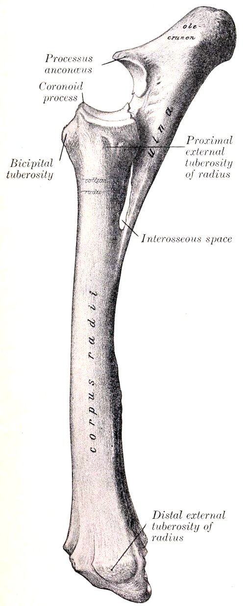
Fig. 48.—Left Radius and Ulna of Horse, External View. (After Schmaltz, Atlas d. Anat. d. Pferdes.)
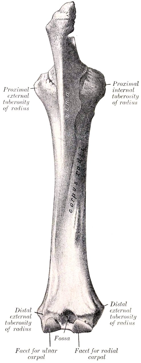
Fig. 49.—Left Radius and Ulna of Horse, Posterior View. (After Schmaltz, Atlas d. Anat. d. Pferdes.)
The distal extremity is also compressed from before backward. It presents 82the carpal articular surface (Facies articularis carpea) which consists of three parts. The inner facet is the largest, is quadrilateral, concavo-convex from before backward, and articulates with the radial carpal bone (or scaphoid); the middle one is somewhat similar in form but smaller, and articulates with the intermediate carpal bone (or semilunar); the outer facet is smaller, is convex, and articulates below with the ulnar carpal (or cuneiform) and behind with the accessory carpal (or pisiform). The anterior surface presents three grooves, separated by ridges. The middle one is vertical and gives passage to the tendon of the extensor carpi radialis; the outer one is similar and contains the tendon of the anterior extensor of the digit; the inner one is small and oblique and lodges the tendon of the extensor carpi obliquus. The posterior aspect is crossed by a rough ridge, below which are three depressions. On either side is a tuberosity (Tuberculum ligamenti) to which the lateral ligament is attached. The outer one is marked by a small vertical groove for the passage of the lateral extensor tendon.
Development.—The radius ossifies from four centers, viz., one each for the shaft, the two extremities, and the outer part of the distal end; the last is morphologically the distal end of the ulna which has fused with the radius, and the line of fusion is often indicated by a distinct groove on the carpal articular surface. The proximal extremity unites with the shaft at about one and a half years, the distal end at about three and a half years.
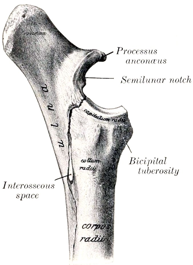
Fig. 50.—Upper Half of Radius and Ulna of Horse, Internal View. (After Schmaltz, Atlas d. Anat. d. Pferdes.)
THE ULNA
The ulna of the horse is a reduced long bone situated behind the radius, with which it is partially fused in the adult.
The shaft (Corpus ulnæ) is three-sided and tapers to a point below. The anterior surface (Facies dorsalis) is applied to the posterior surface of the radius, and below the interosseous space the two bones are fused in the adult. The surface which enters into the formation of the space is smooth and usually presents a small nutrient foramen, directed upward. Above the space it is rough and is attached to the radius by an interosseous ligament which is usually permanent. The internal surface (Facies medialis) is smooth and slightly concave. The external surface (Facies lateralis) is flattened. The internal and external borders are thin and sharp, except at the interosseous space. The posterior border is slightly concave in its length and is rounded. The lower end is pointed and is usually a little below the middle of the radius. It is commonly continued by a fibrous cord to the distal external tuberosity of the radius, but this band may be replaced in part or entirely by bone.
The proximal extremity is the major part of the bone. It projects upward and somewhat backward behind the lower end of the humerus, and forms a lever arm for the extensor muscles of the elbow. The internal surface is concave and smooth. The external surface is convex and is roughened above. The anterior border bears on its middle a pointed projection, the processus anconæus or “beak,” which overhangs the semilunar notch or sigmoid cavity (Incisura semilunaris). The latter is triangular in outline, concave from above downward, and 83articulates with the humerus; in the middle of its lower part is an extensive synovial fossa. Just below the notch are two convex facets which articulate with those on the posterior aspect of the proximal end of the radius. The posterior border is nearly straight, and is thick and rounded. The free end or summit is a rough tuberosity, the olecranon, which gives attachment to the triceps brachii and other muscles.
The primitive distal extremity has, as previously stated, fused with the radius.
Development.—The ulna ossifies from three centers, of which one is for the main part of the bone, one for the olecranon, and one for the distal end. The cartilaginous embryonic ulna extends the entire length of the forearm. The lower part of the shaft is usually reduced to a small fibrous band or may disappear entirely; in some cases a variable remnant of it ossifies. The distal extremity fuses early with the radius. The olecranon unites with the rest of the bone at three to three and a half years. A medullary canal appears to occur constantly in the adult—contrary to the statements of some authors.
THE CARPUS
The carpus of the horse consists of seven or eight bones (Ossa carpi) arranged in two rows, proximal or antibrachial, and distal or metacarpal. The (abbreviated) names and relative positions of the bones of the left carpus as seen from in front are indicated below.
Proximal Row:
Radial Intermediate Ulnar Accessory
Distal Row:
First Second Third Fourth
The Radial Carpal Bone
The radial carpal bone (Os carpi radiale, scaphoid) is the largest bone of the upper row; it is somewhat compressed laterally, and is clearly six-sided. The superior or proximal surface is convex in front, concave behind, and articulates with the inner facet on the distal end of the radius. The inferior or distal surface is also convex in front and concave behind; it articulates with the second and third carpal bones. The external surface bears upper and lower facets on its anterior part for articulation with the intermediate; between and behind these it is excavated and rough. The anterior or dorsal surface is rough and slightly convex. The internal surface and the posterior or volar surface are rough and tuberculate.
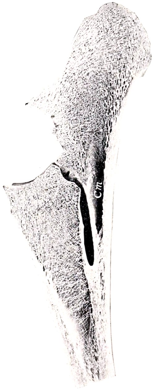
Fig. 51.—Sagittal Section of Upper Part of Radius and Ulna of Horse. Cm, Medullary cavity of ulna.
The Intermediate Carpal Bone
The intermediate carpal bone (Os carpi intermedium, semilunar, lunar) is somewhat wedge-shaped, wider in front than behind. The superior or proximal surface is saddle-shaped, and articulates with the middle facet on the distal end of 84the radius. The inferior or distal surface is smaller, convex in front, concave behind, and articulates with the third and fourth carpal bones. The internal surface has upper and lower facets for articulation with the radial carpal, and between these it is excavated and rough. The external surface is similar to the preceding and articulates with the ulnar carpal. The anterior or dorsal surface is rough and slightly convex. The posterior or volar surface bears a tuberosity on its lower part.
The Ulnar Carpal Bone
The ulnar carpal bone (Os carpi ulnare, cuneiform) is the smallest and most irregular bone of the upper row. The superior or proximal surface is concave, and fits the lower part of the outer facet on the distal end of the radius. The inferior or distal surface is oblique and undulating for articulation with the fourth carpal bone. The internal surface has upper and lower facets for articulation with the intermediate. The anterior or dorsal and external surfaces are continuous, convex, and rough. The posterior or volar surface is oblique, and bears a concave facet for articulation with the accessory carpal bone; below this is a tubercle.
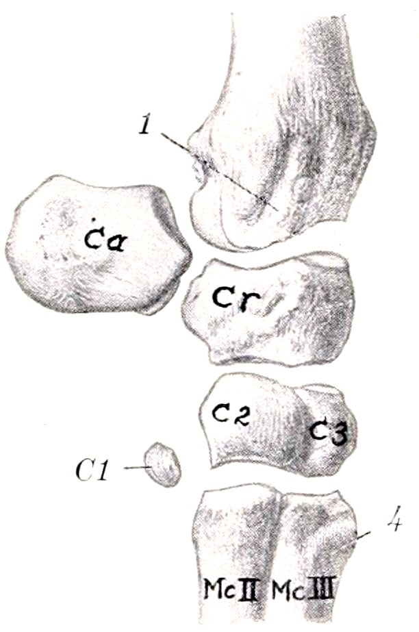
Fig. 52.—Left Carpal Bones of Horse, with Distal End of Radius and Proximal End of Metacarpus; Internal View.
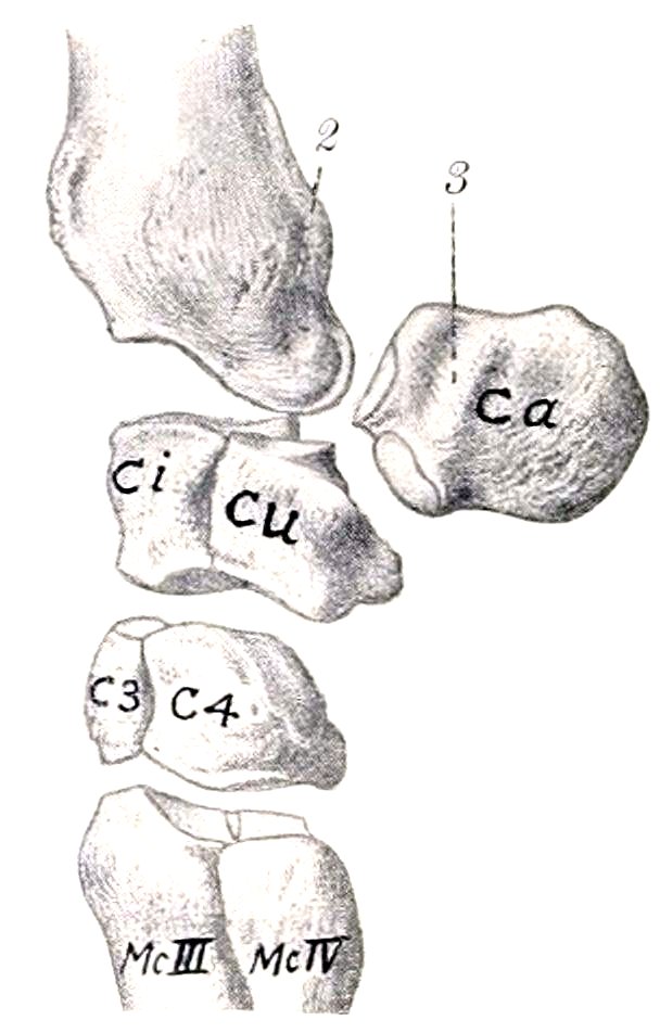
Fig. 53.—Left Carpal Bones of Horse, with Adjacent Ends of Radius and Metacarpus; External View. Ca, Accessory carpal bone; Cr, radial carpal; Ci, intermediate carpal; Cu, ulnar carpal; C1–4, first to fourth carpals; Mc. II, III, IV, metacarpal bones; 1, groove for tendon of extensor carpi obliquus; 2, groove for lateral extensor tendon; 3, groove for tendon of flexor carpi externus; 4, metacarpal tuberosity. (After Schmaltz, Atlas d. Anat. d. Pferdes.)
The Accessory Carpal Bone
The accessory carpal bone (Os carpi accessorium, pisiform) is situated behind the ulnar carpal bone and the outer part of the distal end of the ulna. It is discoid and presents for description two surfaces and a circumference. The internal surface is concave and forms the outer wall of the carpal groove. The external surface is convex and rough; a smooth groove for the outer tendon of the flexor carpi externus crosses its anterior part obliquely downward and slightly forward. The anterior border bears two facets; the upper one is concave and articulates with the back of the outer facet on the distal end of the radius; the lower one is convex and articulates with the ulnar carpal bone. The remainder of the circumference is rounded and rough.
The accessory does not directly bear weight, and may be regarded as a sesamoid bone interposed in the course of the tendons of the middle and external flexors of the carpus, which it enables to act at a mechanical advantage. The posterior border furnishes attachment to the transverse carpal ligament, which completes the carpal canal for the flexors of the digit.
85
The First Carpal Bone
The first carpal bone (Os carpale primum, trapezium) is a small inconstant bone, commonly about the size and shape of a pea, which is situated in the lower part of the internal lateral ligament behind the second carpal bone.
This bone appears to be absent on both sides in about half of the cases; in a good many subjects it is present on one side only. In size it varies from a minute nodule to a discoid mass 10 to 12 mm. in length. In exceptional cases it articulates with both the second carpal and the second (inner) metacarpal bone, in other cases with the former only, but in the majority of specimens no articular facet is present.
The Second Carpal Bone
The second carpal bone (Os carpale secundum, trapezoid) is the smallest constant bone of the lower row, and is irregularly hemispherical in shape. The superior or proximal surface is a convex facet which is continued upon the posterior or volar surface and articulates with the posterior part of the radial carpal. The external surface faces obliquely outward and forward, and bears three facets for articulation with the third carpal bone. The anterior or dorsal and the internal surface bear a tuberosity to which the lateral ligament is attached. The inferior or distal surface is articular and consists of a large flattened facet for the inner (second) metacarpal bone, and a small one for the large (third) metacarpal bone. Some specimens have a small facet on the lower part of the posterior surface which articulates with the first carpal bone.
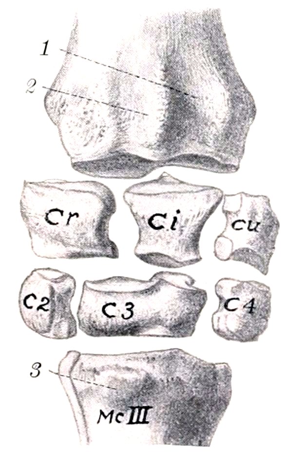
Fig. 54.—Carpal Bones of Horse, with Adjacent Ends of Radius and Metacarpus; Anterior View. The Accessory and First Carpal Bones are Not Shown.
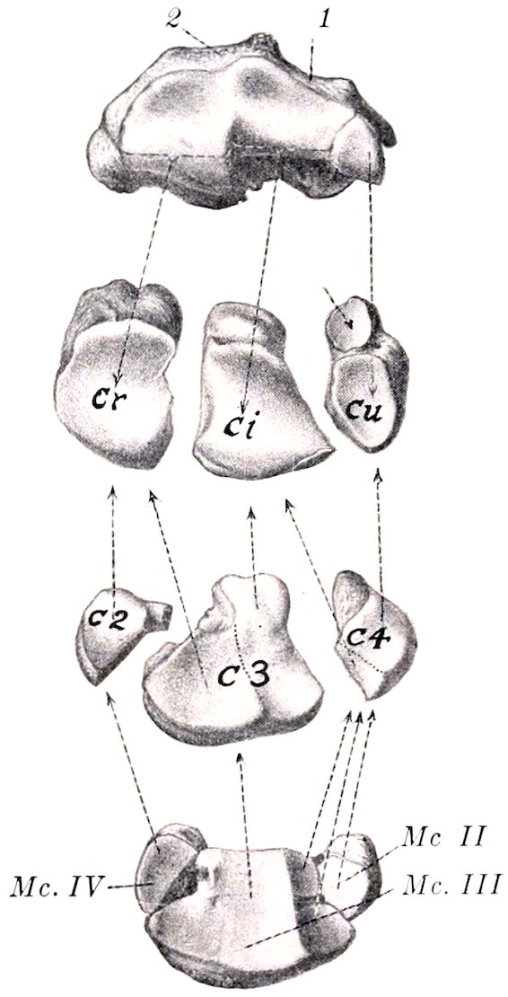
Fig. 55.—Carpal Articular Surface of Radius and Proximal Articular Surfaces of Carpal and Metacarpal Bones, Left Side. The Accessory and First Carpal Bones are not Shown. Cr, Radial carpal; Ci, intermediate carpal; Cu, ulnar carpal; C2, C3, C4, second, third, and fourth carpals; Mc.II, second or inner small metacarpal bone; Mc.III, third or large metacarpal bone; Mc.IV, fourth or outer small metacarpal bone; 1, 2, grooves for tendons of anterior extensor and extensor carpi radialis; 3, metacarpal tuberosity. Arrows indicate relations of facets. Short arrow points to facet on ulnar carpal for articulation with accessory carpal. (After Schmaltz, Atlas. d. Anat. d. Pferdes.)
86
The Third Carpal Bone
The third carpal bone (Os carpale tertium, os magnum) is much the largest bone of the lower row, forming more than two-thirds of the width of the latter. It is flattened from above downward, and is twice as wide in front as behind. The superior or proximal surface consists of two facets separated by an antero-posterior ridge; the inner facet is concave and articulates with the radial carpal; the outer facet—for the intermediate carpal—is concave in front and convex behind, where it encroaches on the posterior surface. The inferior or distal surface is slightly undulating, and articulates almost entirely with the large (third) metacarpal bone, but it usually bears a small oblique facet at its inner side for the inner (second) metacarpal, and there is commonly a non-articular depression externally. The internal surface faces backward and inward, and bears three facets for articulation with the second carpal, between which it is excavated and rough. The external surface has two facets for articulation with the fourth carpal, and is depressed and rough in its middle. The anterior or dorsal surface is convex and is crossed by a rough transverse ridge. The posterior or volar surface is relatively small, and is rounded; its upper part is encroached upon by the superior articular surface, below which it is rough.
The Fourth Carpal Bone
The fourth carpal bone (Os carpale quartum, unciform) is somewhat wedge-shaped, and is readily distinguished from the second by its greater size and its posterior tubercle. The superior or proximal surface articulates with the intermediate and ulnar; it is convex and curves outward, backward, and downward, encroaching on the external and posterior surfaces. The inferior or distal surface bears two inner facets for the large (third) metacarpal and an outer one for the external (fourth) metacarpal bone. The internal surface has two or three facets for articulation with the third carpal, between which it is excavated and rough. The anterior or dorsal surface is convex and rough. The external surface is small, being encroached upon by the superior articular surface. The posterior or volar surface bears a tubercle on its lower part.[21]
The Carpus as a Whole
The bones of the carpus, exclusive of the accessory, form an irregular quadrangular mass, the width of which is about twice the height or the antero-posterior diameter. The anterior or dorsal surface is convex from side to side, depressed along the line of junction of the two rows, and prominent below. The posterior or volar surface is in general slightly convex, but very irregular. It forms with the accessory the carpal groove (Sulcus carpi), which in the recent state is rendered smooth by the posterior ligament; it is converted into the carpal canal (Canalis carpi) for the flexor tendons by the transverse carpal ligament, which stretches across from the accessory bone to the inner side. The proximal surface is widest internally and is elevated in front, concave behind; it is entirely articular and adapted to the carpal articular surface of the radius. The distal surface is also articular and is irregularly faceted in adaptation to the surfaces of the metacarpal bones; each of the lower bones usually articulates with two metacarpal bones, but sometimes the third does not bear on the inner metacarpal bone. The lateral surfaces are both irregular and rough, the internal one being the wider. With the exception of the accessory, ulnar, and second, each bone articulates with two bones of the other row.
Development.—Each ossifies from a single center.
87
THE METACARPUS
Three metacarpal bones (Ossa metacarpalia) are present in the horse. Of these, only one, the third or large metacarpal bone, is fully developed and carries a digit; the other two, the second and fourth, are much reduced, and are commonly called the internal and external small metacarpal or “splint” bones.
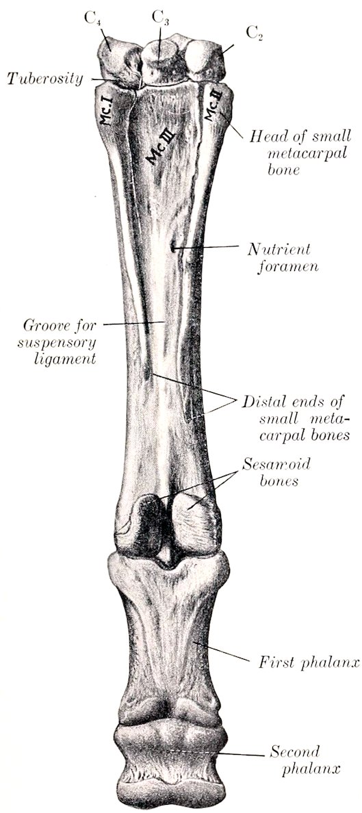
Fig. 56.—Distal Row of Carpal Bones, Metacarpus, First and Second Phalanges, and Proximal Sesamoid Bones of Horse; Posterior View, Left Side. The fourth (external) metacarpal bone is wrongly numbered as I. (After Schmaltz, Atlas. d. Anat. d. Pferdes.)
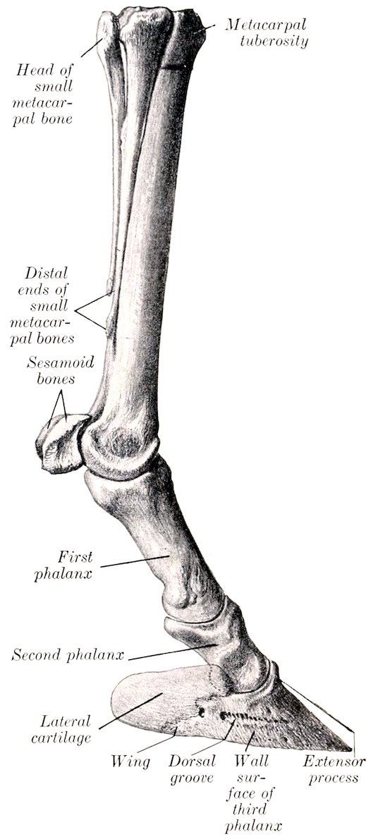
Fig. 57.—Left Metacarpal and Digital Bones of Horse, Internal View. (After Schmaltz, Atlas d. Anat. d. Pferdes.)
The Large Metacarpal Bone
This (Os metacarpale tertium) is a very strong long bone, placed vertically between the carpus above and the first phalanx below. It consists of a shaft and two extremities.
88The shaft (Corpus) is semicylindrical, and presents two surfaces and two borders. The anterior or dorsal surface is smooth, convex from side to side, and nearly straight in its length. The posterior or volar surface is somewhat convex from side to side and, with the small bones, forms a wide groove which lodges the suspensory ligament. On either side of its upper two-thirds it is roughened for the attachment of the small metacarpal bones. The nutrient foramen occurs at the junction of the upper and middle thirds. The lower third is wider and flattened. The borders are rounded.