OSTEOLOGY (continued 4)
Reading edition
127The sphenoidal sinus is almost entirely in the sphenoid bone and does not communicate with the palatine and maxillary sinus. It has one or two openings into the ventral ethmoidal meatuses. There is no cavity in the perpendicular part of the palate bone.
There are several small air-cavities between the lateral mass of the ethmoid and the anterior part of the frontal sinus, which communicate separately with ethmoidal meatuses.
BONES OF THE THORACIC LIMB
The scapula is more regularly triangular than in the horse, relatively wider at the vertebral end and narrower at the lower end. The scapular index is about 1 ∶ 0.6. The spine is more prominent and is placed further forward, so that the supraspinous fossa is narrow and does not extend to the lower part of the bone. The spine is sinuous, bent backward in its middle, forward below. Its free border is somewhat thickened in its middle, but bears no distinct tubercle. Instead of subsiding below as in the horse the spine becomes a little more prominent, and is prolonged by a pointed projection, the acromion, from which part of the deltoid muscle arises. The subscapular fossa is shallow. The areas for the attachment of the serratus muscle are not very distinct. The nutrient foramen is usually in the lower third of the posterior border. The glenoid cavity is almost circular and without any distinct notch. The tuberosity is small and close to the glenoid cavity. The coracoid process is short and rounded. The cartilage resembles that of the horse.
The humerus has a shallow musculo-spiral groove. The deltoid tuberosity is less prominent than in the horse, and the curved line running from it to the neck bears a well-marked tubercle on its upper part. The nutrient foramen is usually in the lower third of the posterior surface. The external tuberosity is very large, and rises an inch or more (ca. 3 cm.) above the level of the head. Its anterior part curves inward over the bicipital groove, and below it externally there is a prominent circular rough area for the insertion of the tendon of the supraspinatus. The anterior part of the internal tuberosity has a small projection which curves over the groove. The groove is undivided. The distal articular surface is decidedly oblique, and the grooves and ridge are very well marked. The coronoid and olecranon fossæ are deep and wide. The external condyloid crest is represented by a rough raised area. The proximal end unites with the shaft at three and one-half to four years, and the distal at about one and one-half years.
The radius is short and relatively broad. It is somewhat oblique, the lower end being nearer the median plane than the upper. The curvature is more pronounced below than above. The shaft is prismatic in its middle part and has anterior, external, and posterior faces. There is a marked increase in width and thickness below. The proximal articular surface presents a synovial fossa which extends inward from the deep groove between the two glenoid cavities. The bicipital tuberosity is represented by a slightly elevated rough area. The posterior facets for the ulna are larger than in the horse. The two bones commonly fuse above the proximal interosseous space and always fuse below it, except near the distal end, where there is a small distal interosseous space. A groove connects the two spaces externally. The distal extremity is large, and is thickest internally. Its articular surface is oblique in two directions, i. e., from within upward and backward. The grooves for the extensor tendons are shallow. The proximal end unites with the shaft at one to one and one-half years, and the distal at three and one-half to four years.
The approximation of the lower ends of the forearms and the carpi gives the “knock-kneed” appearance in cattle. The obliquity of the joint surfaces produces lateral deviation of the lower 128part of the limb in flexion. The facets for the radial and intermediate carpals are narrower than in the horse and run obliquely forward and outward. The surface for the ulnar carpal is extensive and saddle-shaped; its outer part is furnished by the ulna.
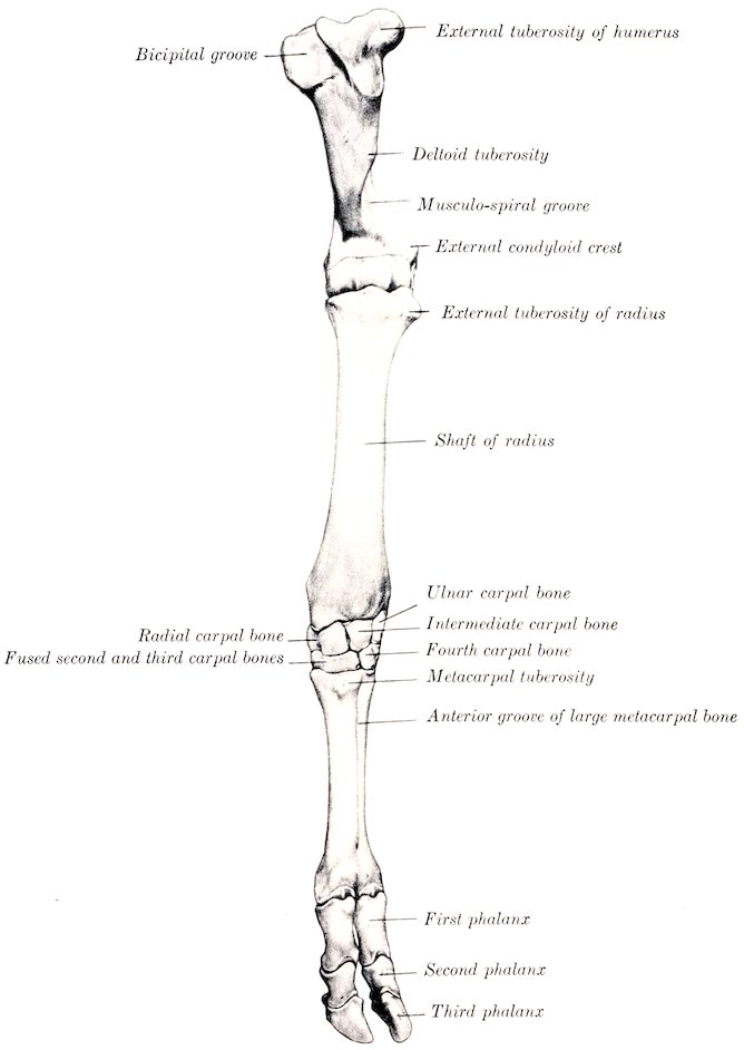
Fig. 99.—Skeleton of Left Fore Limb of Ox, from Shoulder Downward, Anterior View. (After Ellenberger-Baum, Anat. für Künstler.)
The ulna is more fully developed than in the horse. The shaft is complete, three-sided, and strongly curved. It is fused with the radius in the adult, except at the two interosseous spaces mentioned above. Its upper part contains a medullary canal which extends somewhat into the proximal end. The olecranon is large and bears a rounded tuberosity above. The distal end is fused with the 129radius; it projects below the level of the latter, forming the styloid process (Processus styloideus ulnæ), which furnishes part of the facet for the ulnar carpal. The proximal and distal ends unite with the shaft at three and one-half to four years.
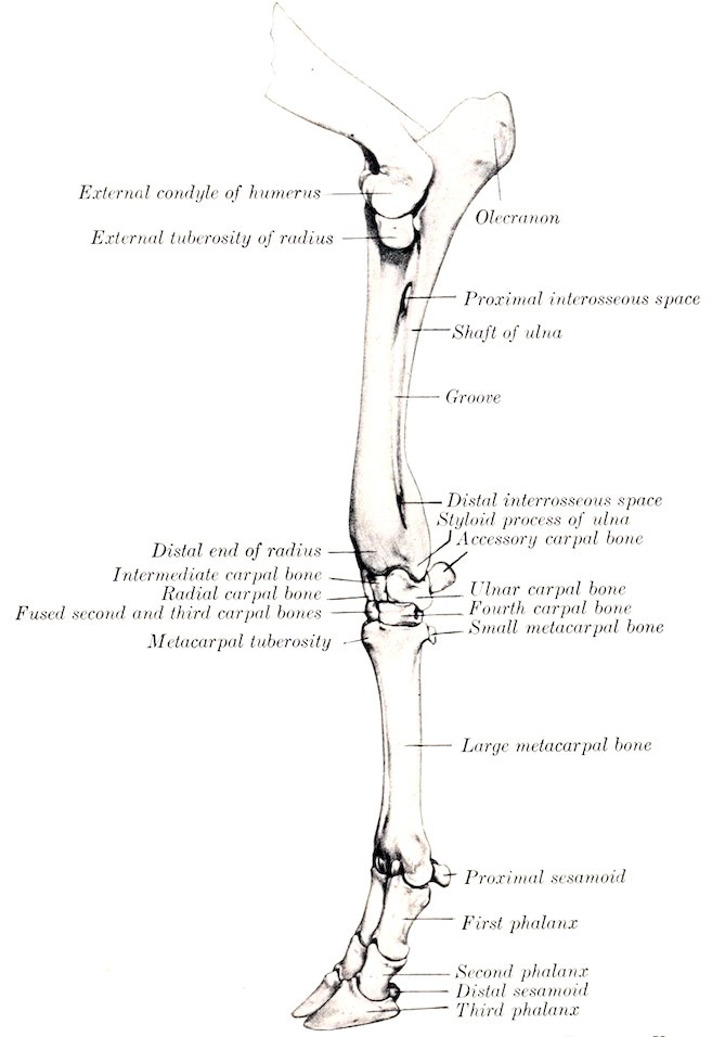
Fig. 100.—Skeleton of Left Fore Limb of Ox, from Elbow Downward, External View. (After Ellenberger-Baum, Anat. für Künstler.)
The carpus consists of six bones, four in the upper row, and two in the lower. The upper row is oblique in conformity with the carpal articular surface of the radius. The radial and intermediate resemble in general those of the horse, but are less regular in shape, and their long axes are directed obliquely backward and inward. The radial is narrower than in the horse and curves upward behind. The intermediate is constricted in its middle, and wider behind than in front. The ulnar is large and very irregular. Its proximal surface is extensive and sinuous and articulates with both radius and ulna. Behind is a large oval facet for articulation with the accessory carpal. The accessory is short, thick, and rounded; it articulates 130with the ulnar carpal only. The first carpal is absent. The second and third carpals are fused to form a large quadrilateral bone. The fourth carpal is a smaller quadrilateral bone.
The metacarpus consists of a large metacarpal and an external small metacarpal bone. The large metacarpal bone (Mc. 3 + 4) results from the fusion of the third and fourth bones of the fœtus, and bears evidences of its double origin even in the adult state. The shaft is shorter than in the horse, and is relatively wider and flatter. The anterior surface is rounded, and is marked by a vertical vascular groove connecting two canals which traverse the ends of the shaft from before backward. The posterior surface is flat and presents a similar but much fainter groove. The borders are rough in the upper third. The proximal end bears two slightly concave facets for articulation with the bones of the lower row of the carpus; the inner area is the larger, and they are separated by a ridge in front and a notch behind. The outer angle has a facet behind for the small metacarpal bone. The inner part of the extremity has anterior and posterior tuberosities. The distal end is divided into two parts by a sagittal notch. Each division bears an articular surface similar to that in the horse, but much smaller. The medullary canal is divided into two parts by a vertical septum which is usually incomplete in the adult.
The small metacarpal bone (Mc. 5) is a rounded rod about an inch and a half (ca. 3.5 to 4 cm.) in length, which lies against the upper part of the outer border of the large bone. Its upper end articulates with the latter, but not with the carpus. The lower end is pointed.
Four cartilaginous metacarpals are present in the early fœtal state, viz., the second, third, fourth, and fifth. The second commonly either disappears or unites with the third; sometimes it develops as a small rod of bone. The third and fourth gradually unite, but can be cut apart at birth. Each has three centers of ossification; the proximal epiphysis fuses with the shaft before birth, the distal at two to two and one-half years.
Four digits are present in the ox. Of these, two—the third and fourth—are fully developed and have three phalanges and three sesamoids each. The second and fifth are vestiges and are placed behind the fetlock; each contains one or two small bones which do not articulate with the rest of the skeleton.
The first phalanges are shorter and narrower than in the horse and are three-sided. The interdigital surface is flattened and its posterior part bears a prominence for the attachment of the interdigital ligaments. The proximal extremity is relatively large, and is somewhat compressed from side to side. The articular surface is concave from before backward and is divided by a sagittal groove into two areas, of which the abaxial one is the larger and higher. Behind these are two facets for articulation with the sesamoid bones. The posterior surface bears two tuberosities separated by a deep depression. The distal extremity is smaller than the proximal, especially in the antero-posterior direction. Its articular surface is divided by a sagittal groove into two convex facets, of which the abaxial one is decidedly the larger. There are depressions on either side for ligamentous attachment. The bone consists at birth of two pieces—the distal end and the fused shaft and upper extremity. Union occurs at one and one-half to two years.
The second phalanges are about two-thirds of the length of the first and are distinctly three-sided. The proximal articular surface is divided by a sagittal ridge into two glenoid cavities, of which the abaxial one is much the larger. There is a central prominence in front and two lateral tubercles behind. The distal extremity is smaller than the proximal. Its articular surface encroaches considerably on the anterior and posterior surfaces, and is divided into two lateral parts by a groove. There is a deep depression for ligamentous attachment on the interdigital side. The bone contains a small medullary canal. The distal end unites with the rest of the bone about the middle of the second year.
The third phalanges resemble in a general way one-half of the bone of the horse. 131Each has four surfaces. The dorsal or wall surface is marked in its lower part by a shallow groove, along which there are several foramina of considerable size; the posterior one of the series is the largest, and conducts to a canal in the interior of the bone. Below the groove the surface is prominent, rough, and porous. Near and on the extensor process are several relatively large foramina. The slope of the surface is very steep posteriorly, but in front it forms an angle of 25 to 30 degrees with the ground plane. The articular surface is narrow from side to side, and slopes downward and backward. It is also oblique transversely, the interdigital side being the lower. It is adapted to the distal surface of the second phalanx, with the exception of a facet behind for the distal sesamoid. The extensor process is very rough. The volar or sole surface is narrow and slightly concave, and presents two or three foramina of considerable size. It is separated from the wall surface by a border which is sharp in front, rounded behind. There is no semilunar crest, since the deep flexor tendon is attached to the thick posterior border of the sole surface. The interdigital surface is smooth and grooved below, rough and porous above. At the upper angle it is perforated by a large foramen, which is equivalent to the volar foramen of the horse and leads to a cavity in the middle of the bone. The surface is separated by a rounded border from the wall surface, and by a sharp edge from the sole surface. The wing or angle is very short and blunt, and there is no lateral cartilage.
Four proximal sesamoids are present, two for each digit. They are much smaller than in the horse. The bones of each pair articulate with the corresponding part of the distal end of the large metacarpal bone by their anterior surfaces, with each other and with the first phalanx by small facets.
The two distal sesamoids are short and their ends are but little narrower than the middle.
BONES OF THE PELVIC LIMB
The ilia are almost parallel to each other and are also less oblique with regard to the horizontal plane than in the horse. They are relatively small. The gluteal line is prominent and is nearly parallel to the external border; it joins the superior ischiatic spine. A rounded ridge separates the two parts of the ventral surface. The surface for articulation with the sacrum is triangular. The internal angle is truncated, does not extend as high as the vertebral spines, and is separated from the opposite angle by a wider interval than in the horse. The external angle is relatively large and prominent; it is not so oblique as in the horse, and is wide in the middle, smaller at either end. The shaft is short and compressed from side to side.
The ischium is large. Its long axis is directed obliquely upward and backward, forming an angle of about 45 to 50 degrees with the horizontal plane. The transverse axis is oblique downward and inward at a similar angle, so that this part of the pelvic floor is deeply concave from side to side. The middle of the ventral surface bears a rough ridge or imprint for muscular attachment. The superior ischiatic spine is high and thin, and bears a series of almost vertical rough lines externally. The tuber ischii is large and three-sided, bearing upper, lower, and external tuberosities. The ischial arch is narrow and deep. The symphysis bears a ventral ridge, which fades out near the ischial arch.
The acetabular branch of the pubis is narrow, and is directed outward and a little forward. The anterior border is marked by a transverse groove which ends below the rough ilio-pectineal eminence. The longitudinal branch is wide and thin.
The acetabulum is smaller than in the horse. The rim is rounded and is usually marked by two notches. One of these is postero-internal and is narrow and deep; it leads to the deep acetabular fossa and is commonly almost converted 132into a foramen by a bar of bone. The other notch is antero-internal, small, and sometimes replaced by a foramen or absent.
The obturator foramen is large and elliptical. Its inner border is thin and sharp.
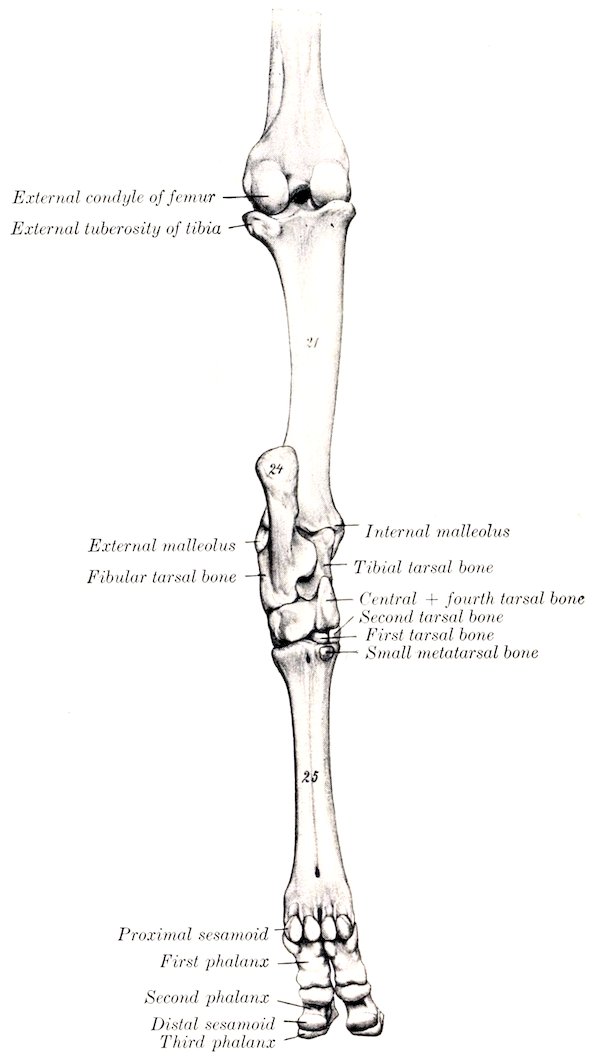
Fig. 101.—Skeleton of Left Hind Limb of Ox, from Middle of Thigh Downward, Posterior View. 21, Tibia; 24, tuber calcis; 25, large metatarsal bone. (After Ellenberger-Baum, Anat. für Künstler.)
The pelvic inlet is elliptical and is more oblique than in the horse. In a cow of medium size the conjugate diameter is about nine and a half inches (ca. 23 to 24 cm.), and the transverse about seven inches (ca. 18 cm.). The anterior end of the symphysis lies in a transverse plane through the junction of the third and fourth 133sacral segments. The roof is concave in both directions. The floor is deeply concave, particularly in the transverse direction. The cavity is narrower and its axis is inclined strongly upward in the posterior part. The distance between the acetabulum and the external angle of the ilium is only a little (ca. 3 to 4 cm.) more than the distance between the former and the tuber ischii.
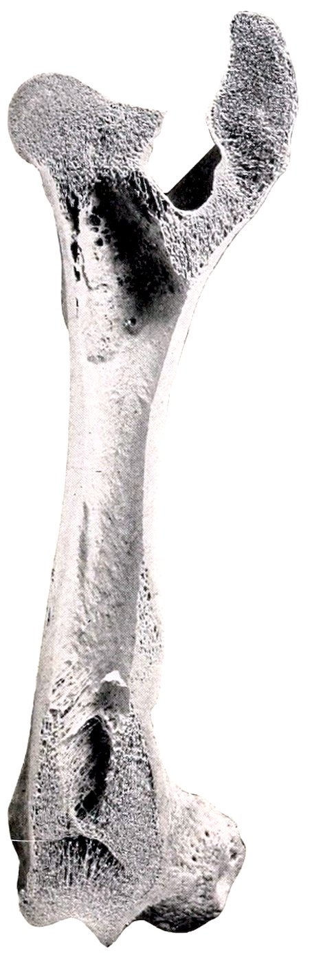
Fig. 102.—Frontal Section of Left Femur of Ox, Front View.
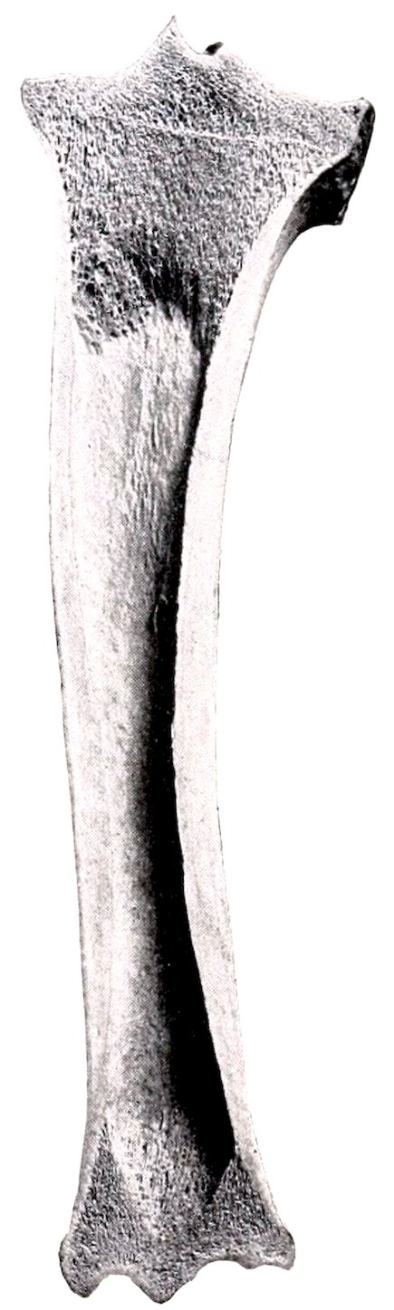
Fig. 103.—Frontal Section of Left Tibia of Ox, Anterior View. These figures show the internal architecture of these bones, and especially the great extent of the medullary cavity as compared with those of the horse (Figs. 67 and 72).
The femur has a relatively small shaft, which is cylindrical in its middle, prismatic below. The trochanter minor has the form of a rough tuberosity, and is situated higher up than in the horse and encroaches on the posterior surface. The trochanteric ridge (Crista intertrochanterica posterior) connects it with the trochanter major. The third trochanter is absent. The supracondyloid (plantar) fossa is shallow. The proximal extremity is very wide. The head is smaller than in the horse and the articular surface extends considerably on the upper surface of the neck. Instead of the notch there is a small depression on the middle of the head for the attachment of the round ligament. The neck is well defined except above. The trochanter major is very massive and is undivided; its external surface is very rough. The trochanteric fossa is deep, but does not extend so far downward as in the horse. The distal end presents no very striking differential features, but the lips of the trochlea are less oblique than in the horse, and converge very slightly 134below. The proximal extremity unites with the shaft at about three and one-half years, the distal at three and one-half to four years.
The tibia resembles that of the horse rather closely, but is somewhat shorter. The shaft is distinctly curved, so that the inner side is convex. The posterior surface is not divided into two areas, and the lineæ musculares are fewer and extend up higher than in the horse. The articular grooves and ridge of the distal end are almost sagittal in direction, and present an extensive but shallow synovial fossa. The external groove is separated by a sharp ridge from an outer area which is for articulation with the external malleolus. The anterior part of the internal malleolus is prolonged downward and has a pointed end. The groove behind it is broad and well defined. Externally there is a deep narrow groove which separates two prominences. The proximal extremity fuses with the shaft at three and one-half to four years, the distal at two to two and one-half years.
The fibula usually consists of the two extremities only. The head is fused with the external condyle of the tibia and bears a small blunt-pointed prolongation below. The distal end remains separate and forms the external malleolus (sometimes called the os malleolare). It is quadrilateral in outline and compressed from side to side. The proximal surface articulates with the distal end of the tibia, and bears a small spine which fits into the groove on that bone. The distal surface rests on the fibular tarsal, and the inner articulates with the external ridge of the tibial tarsal bone. The outer surface is rough and irregular.
The early cartilaginous fibula is complete, but later the shaft is reduced and is usually represented by a fibrous cord which connects the two ends. In some cases, however, the upper part undergoes partial ossification, forming a slender rod which is usually united with the outer border of the tibia and is joined to the head by fibrous tissue.
The patella is long, narrow, and very thick. The anterior surface is strongly convex and very rough and irregular. The articular surface is convex from side to side and nearly straight in the vertical direction. The large prominence on the inner side for the attachment of the accessory cartilage allows prompt determination of the side to which the bone belongs. The apex is more pointed than in the horse.
The tarsus consists of five pieces; the central and fourth, and the second and third tarsal bones are fused.
The tibial tarsal bone is relatively long and narrow, and is somewhat flattened from before backward. It bears a trochlea at either end. The groove and ridges of the proximal trochlea are not spiral, but almost sagittal; the outer ridge is the wider, and articulates with both tibia and fibula. The distal trochlea consists of two condyles divided by a groove, and articulates with the combined central and fourth tarsals. The posterior surface bears a large oval facet for articulation with the fibular tarsal; this occupies most of the surface, and is convex and grooved from above downward. The outer surface presents two facets for articulation with the fibular tarsal, and is excavated and rough elsewhere. The inner surface bears a tuberosity at its upper part, and is flattened below.
The fibular tarsal bone is longer and more slender than in the horse. The distal part of the body is compressed laterally, and bears a projection in front which articulates with the external malleolus. The tuber calcis is marked posteriorly by a wide shallow groove, which is coated with cartilage in the fresh state.
The central and fourth tarsals are fused to form a large bone (Os centrotarsale quartum, scapho-cuboid), which extends across the entire width of the tarsus and articulates with all of the other bones. The greater part of the upper surface is molded on the distal trochlea of the tibial tarsal, and its inner part rises high above the rest posteriorly. Externally there is a narrow undulating surface for articulation with the distal end of the fibular tarsal bone. The posterior surface bears two 135tuberosities, of which the outer one is rounded, the inner more prominent and narrower.
The first tarsal bone is quadrilateral and small. It articulates with the central above, the metatarsus below, and the second tarsal in front.
The second and third tarsals are fused to form a rhomboid piece. The proximal surface is concavo-convex, and articulates with the central component. The distal surface is undulating and rests on the metatarsus. The external surface bears a small facet in front for the fourth tarsal component, and the posterior surface a very small one for the first tarsal bone.
The large metatarsal bone is about one-seventh (ca. 3 cm.) longer than the corresponding metacarpal. Its shaft is compressed laterally and is distinctly four-sided. The groove on the anterior surface is deep and wide. The posterior surface is marked by variable grooves. The upper foramen on this surface does not perforate the shaft, but passes obliquely through the extremity, opening on the posterior part of its proximal surface. The postero-internal angle of the proximal end bears a facet for articulation with the small metatarsal bone.
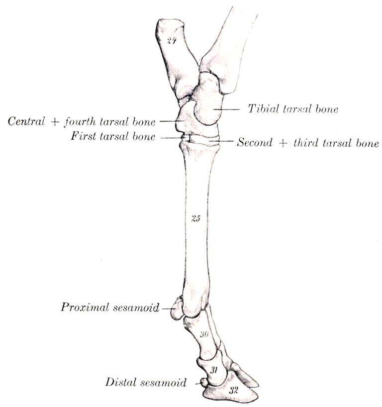
Fig. 104.—Skeleton of Distal Part of Left Hind Limb of Ox, Internal View. 24, Tuber calcis; 25, large metatarsal bone; 30, first phalanx; 31, second phalanx; 32, third phalanx. (After Ellenberger-Baum, Anat. für Künstler.)
The small metatarsal bone is a quadrilateral disc a little less than an inch in width and height. Its anterior face bears a facet for articulation with the large metatarsal bone.
The large metatarsal bone is usually regarded as consisting of the fused third and fourth metatarsal bones. The medullary cavity is subdivided like that of the large metacarpal bone. Some anatomists, however, consider that the ridges at the upper end of each border represent the second and fifth metatarsals (Rosenberg and Retterer). On this basis the small bone would be the first metatarsal.
136The phalanges and sesamoids resemble those of the thoracic limb so closely as to render separate description unnecessary.
SKELETON OF THE PIG
VERTEBRAL COLUMN
The vertebral formula is C7T14–15L6–7S4Cy20–23.
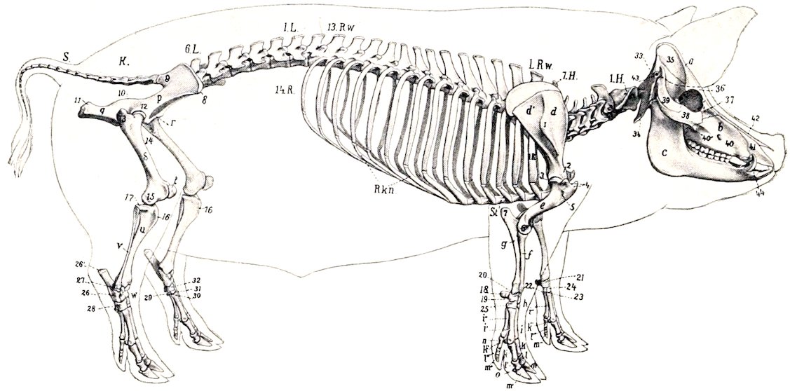
Fig. 105.—Skeleton of Pig, Lateral View. a, Cranium; b, upper jaw; c, lower jaw; 1H.-7H., cervical vertebræ; 1R.w., first thoracic vertebra; 13 R.w., thirteenth thoracic vertebra (next to last); 1L., first lumbar vertebra; 6L., sixth lumbar vertebra (next to last usually); K., sacrum; S., coccygeal vertebra; 1R., first rib; 14R., last rib; R.kn., costal cartilages; St., sternum; d, supraspinous fossa of scapula; d′, infraspinous fossa; 1, spine of scapula; 2, neck of scapula; e, humerus; 3, head of humerus; 4, tuberosities of humerus; 5, deltoid tuberosity; 6, external epicondyle of humerus; f, radius; g, ulna; 7, olecranon; h, carpus; 18–25, carpal bones; i-i⁗, metacarpus; k-k⁗, proximal phalanges; l-l⁗, middle phalanges; m-m⁗, distal phalanges; n, o, sesamoids; p, ilium; 8, external angle of ilium (tuber coxæ); 9, internal angle of ilium (tuber sacrale); 10, superior ischiatic spine; q, ischium; 11, tuber ischii; r, pubis; 12, acetabulum; s, femur; 13, trochanter major; 14, trochanter minor; 15, external epicondyle; t, patella; u, tibia; 16, crest of tibia; 17, external condyle of tibia; v, fibula; w, tarsus; 26–31, tarsal bones; 26′, tuber calcis. (After Ellenberger, in Leisering’s Atlas.)
The cervical vertebræ are short and wide. The bodies are elliptical in cross-section, the long diameter being transverse. The anterior articular surfaces are slightly convex from side to side and concave dorso-ventrally; the posterior ones are slightly concave. A ventral crest is not present. The arches are wide from side to side, but the laminæ are narrow, so that a considerable interval (Spatium interarcuale) separates adjacent arches dorsally. The pedicles are perforated by a foramen on either side in addition to the usual intervertebral foramina. The transverse processes divide into two branches, both of which increase in size from the third to the sixth. The upper branch projects outward and backward; it is short and is thickened at its free end. The lower branch is a quadrilateral plate directed ventrally; each overlaps the succeeding one to a small extent, and the series forms the lateral boundary of a deep and wide groove beneath the bodies. The spines increase in height from the third to the last; the anterior ones are inclined backward, the posterior ones forward. The last cervical is recognized by the great length of its spine (ca. 10 cm. in the adult), the absence of the ventral plate of the transverse process, and the flatness of the body, which bears a pair of 137small facets on its posterior margin for the heads of the first ribs. It has foramina transversaria, and usually two foramina in either side of the arch.
The dorsal arch of the atlas bears a large tuberosity. The ventral tubercle is long, compressed laterally, and projects back under the axis. The wing is flattened and bears a posterior tuberosity. The foramen transversarium passes through the posterior border of the wing to the fossa under the latter, and is not visible dorsally; it is sometimes very small or absent. The sides of the vertebral foramen bear two lateral projections which partially divide it into a ventral narrow part, which receives the odontoid process, and a dorsal larger part for the spinal cord. In the fresh state the division is completed by the transverse ligament, which is attached to the projections.
The axis has a large spinous process, which is directed upward and backward. The odontoid process is a thick cylindrical rod. The transverse process is very small and the foramen transversarium is often incomplete.
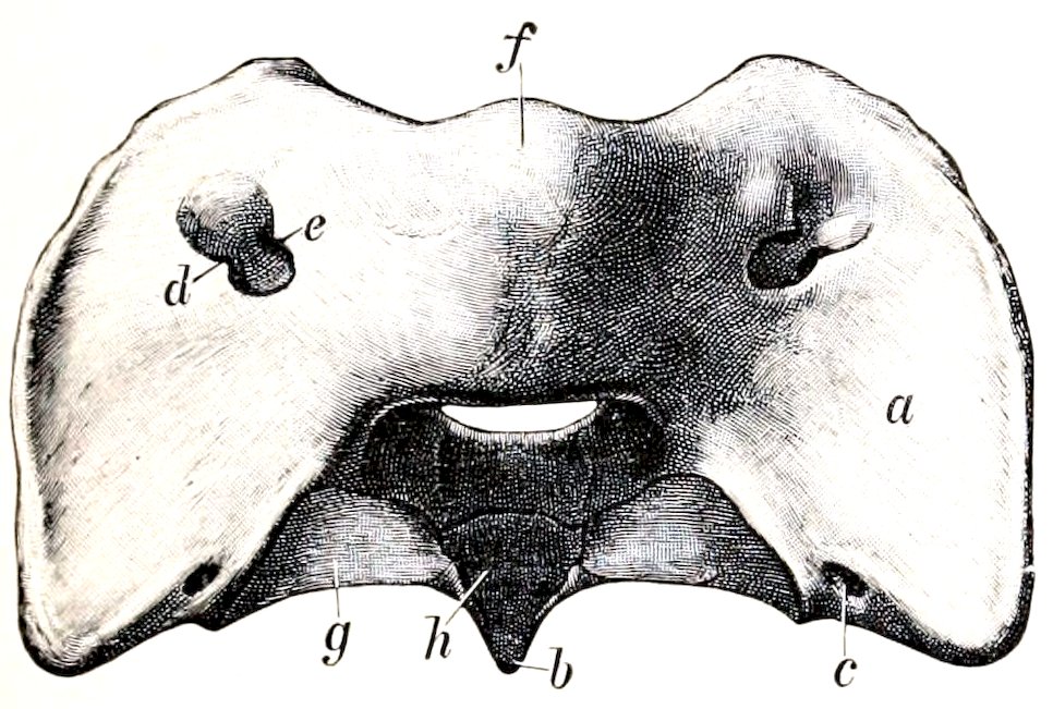
Fig. 106.—Atlas of Pig, Dorsal View. a, Wing; b, ventral tubercle; c, foramen transversarium; d, alar foramen; e, intervertebral foramen; f, dorsal tuberosity; g, articular surface corresponding to that of posterior articular process of typical vertebra; h, facet on ventral arch for odontoid process. (Ellenberger-Baum, Anat. d. Haustiere.)
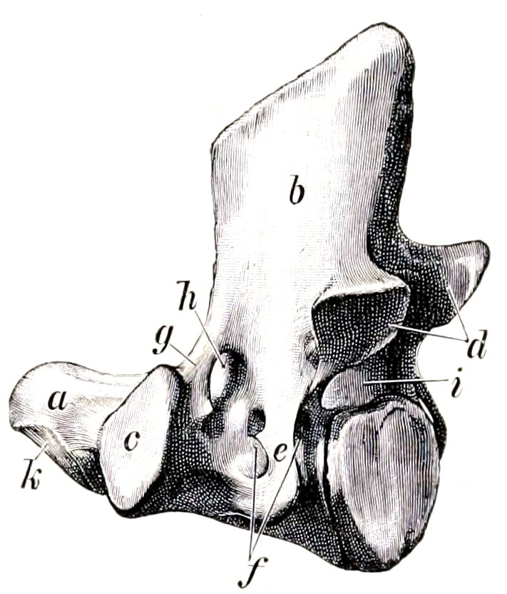
Fig. 107.—Axis of Pig, Left Lateral View. a, Odontoid process (dens); b, spinous process; c, anterior articular process; d, posterior articular processes; e, transverse process; f, foramen transversarium; g, bar of bone which bounds h, intervertebral foramen; i, vertebral foramen. (Ellenberger-Baum, Anat. d. Haustiere.)
The thoracic vertebræ are often fifteen in number. Their bodies are relatively long, constricted in the middle, and without ventral crests. Their extremities are elliptical, depressed in the middle and prominent at the periphery. The arch is perforated by a foramen on either side, and in most of the series there is also a foramen in the posterior part of the root of the transverse process which communicates with the former or with the posterior intervertebral foramen. Sometimes there is a foramen in the anterior part of the process also. There are mammillary processes except on the first two; in the posterior five or six vertebræ they project from the anterior articular processes. The facet for the tubercle of the rib is absent or fused with that for the head in the last five or six. The last transverse process is lumbar in character, plate-like, and about an inch (2 cm.) long. Small accessory processes occur in the posterior part of the region. The first spinous process is broad, very high, and inclined a little forward. The others diminish very gradually in length to the tenth, beyond which they are about equal. The second to the ninth are inclined backward, the tenth is vertical (anticlinal), and the rest incline forward. The width decreases decidedly from the fourth to the tenth, beyond which there is a gradual increase. The summits are slightly enlarged and lie almost in a straight line.
138The occurrence of fifteen thoracic vertebræ appears to be quite common, and some observers have recorded the existence of sixteen and even seventeen; a reduction to thirteen is rare.
The lumbar vertebræ are six or seven in number. The bodies are longer than in the thoracic region and bear a ventral crest. They become wider and flatter in the posterior part of the series. The arches are deeply notched, and are separated by an increasing space dorsally. The mammillary processes project outward and backward. The transverse processes are bent downward and incline a little forward. Their length increases to the fifth and is much diminished in the last. They form no articulation with each other or with the sacrum. The posterior edge of the root of the process is marked by a notch in the anterior part of the series, a foramen in the posterior part. The spines are broad and incline forward, with the exception of the last, which is narrow and vertical.
Lesbre states that six and seven lumbar vertebræ occur with almost equal frequency. The number may be reduced to five, and the number of presacral vertebræ varies from twenty-six to twenty-nine.
The sacrum consists usually of four vertebræ, which fuse later and less completely than in the other domesticated animals. It is less curved than in the ox. The spines are absent, excepting small rudiments on the last two segments. The middle of the dorsal surface is flattened and smooth, and presents openings into the sacral canal between adjacent arches (Spatia interarcualia). On either side are the superior sacral foramina, and tubercles which indicate the fused articular processes. The wings resemble those of the ox. The anterior articular processes are very large. The pelvic surface resembles that of the ox, but is not so strongly curved, and the transverse lines are very distinct.
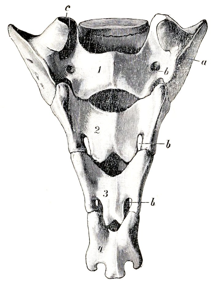
Fig. 108.—Sacrum of Pig, Dorsal View. a, Wing; b, dorsal sacral foramina; c, articular process; 1–4, segments or sacral vertebræ. (Ellenberger-Baum, Anat. d. Haustiere.)
The coccygeal vertebræ are specially characterized by the presence of functional articular processes on the first four or five, beyond which these processes become non-articular and smaller. The arches of the first five or six are complete. The transverse processes are broad and plate-like in the anterior part of the series and diminish very gradually.
The numerical variation here is twenty to twenty-six according to the observation of several anatomists. Lesbre states that he has found twenty-three most frequently.
Curves.—The cervical region is practically straight. The thoracic and lumbar regions form a gentle curve, concave ventrally, the highest point of which is at the junction of the two regions. The sacral promontory is not so pronounced as in the ox, and the sacral curve is flatter.
The Ribs
The ribs number fourteen or fifteen pairs, of which seven are sternal and seven or eight asternal usually. They are in general strongly curved in the improved 139breeds, so that there is a fairly distinct angle, except toward the end of the series. The backward slope of the posterior ribs is slight. The first rib is prismatic, has a large sternal end, and a very short cartilage. The width is greatest in the third to the sixth, and the length in the sixth and seventh usually. The tuberosity fuses with the head on the last five or six. The second to the fifth form diarthrodial joints with their cartilages, which are wide and plate-like.
THE STERNUM
The sternum consists of six segments and resembles that of the ox in general form. The first segment (Manubrium) is long, flattened laterally, and bears a blunt-pointed cartilage on its anterior end; its posterior end forms a diarthrodial joint with the body. The latter is flattened, wide in its middle, narrow at either end. The widest segments are formed of two lateral parts, which are not completely fused in the adult. The last segment has a long narrow part which bears the xiphoid cartilage.
The thorax is long and is more barrel-shaped than in the horse or ox, since the ribs are more strongly curved and differ less in relative length.
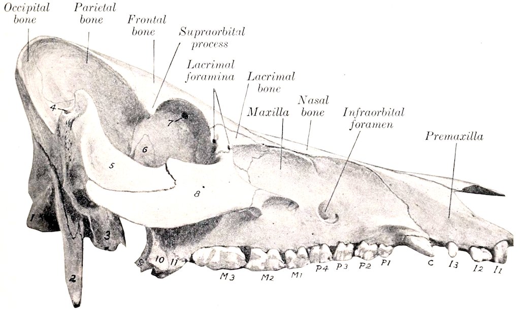
Fig. 109.—Skull of Pig, Lateral View without Mandible. 1, Occipital condyle; 2, paramastoid or styloid process; 3, bulla ossea; 4, external auditory meatus, 5, zygomatic process of temporal bone; 6, sphenoid bone; 7, orbital opening of supraorbital canal; 8, malar bone; 9, pterygoid bone; 10, pterygoid process of sphenoid; 11, pterygoid process of palate bone; I1–3, incisor teeth; C, canine tooth; P1–4, premolars; M1–3, molars.
BONES OF THE SKULL
Cranium
The occipital bone has an extensive squamous part or supraoccipital, which forms a very broad and prominent crest. The latter is concave, and is thick and rough above, where it forms the highest part of the skull; laterally it becomes thinner, turns downward, and is continuous with the temporal crest. Two divergent ridges pass upward from the foramen magnum, and the surface between them is concave and smooth. The greater part of the inner (or anterior) surface of the supraoccipital is united with the parietal bones, but a lower concave area faces 140into the cranial cavity. The foramen magnum is almost triangular, and is narrow above, where it is flanked by two small tuberosities. The paramastoid processes are extremely long and project almost straight downward. The hypoglossal foramen is at the inner side of the root of the process. The basilar part is short and wide; its lower surface bears a thin median ridge and two lateral imprints or tubercles which converge at the junction with the sphenoid bone.
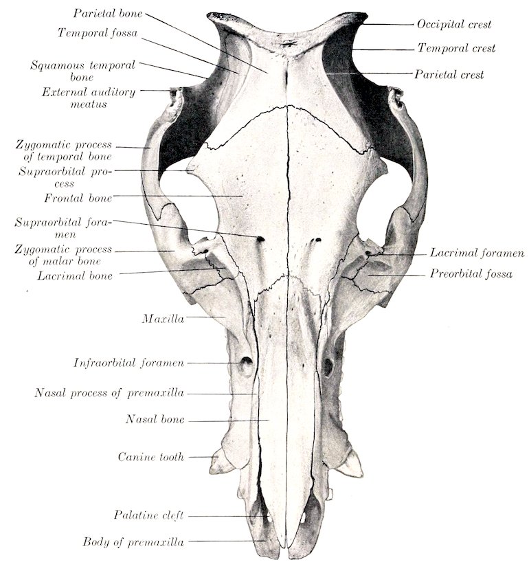
Fig. 110.—Skull of Pig, Dorsal View.
The interparietal fuses before birth with the occipital. The tentorium osseum is absent.
The parietal is overlapped by the occipital bone behind and concurs in the formation of the occipital crest. Its external surface is divided by the parietal crest into two parts. The inner part (Planum parietale) faces upward and forward, and is flattened and smooth. Its inner border is short and straight and unites early with the opposite bone. Its anterior border is concave and joins the frontal bone. The outer part (Planum temporale) faces outward and is more extensive; it is concave, forms a large part of the temporal fossa, and is overlapped below by the squamous temporal. The parietal crest extends in a curve from the occipital crest 141forward and outward to the supraorbital process. The internal surface is concave and is marked by digital impressions. The lower border projects into the cranial cavity and forms a crest which separates the cerebral and cerebellar compartments laterally. The interior forms part of the frontal sinus in the adult. There is no parieto-temporal canal.
The frontal bone is long. The frontal surface slopes downward and forward, the inclination varying in different subjects. The anterior part is concave and is marked by the supraorbital foramen and the groove leading forward from the foramen to the nasal bone. The supraorbital canal opens into the orbit at the upper part of the inner wall of the latter. The supraorbital process is short and blunt-pointed, and is not connected with the zygomatic arch. The gap in the orbital margin is closed by the orbital ligament in the fresh state. The orbital part is extensive and forms the greater part of the inner wall of the orbit. Its upper part is perforated by the orbital orifice of the supraorbital canal, in front of which is the distinct fovea trochlearis. The ethmoidal or internal orbital foramen is situated in the lower part near the junction with the orbital wing of the sphenoid. The temporal part is very narrow and is separated from the orbital plate by a ridge which joins the pterygoid crest below. The interior of the bone is excavated by the frontal sinus in practically its entire extent in the adult. In the young subject the cavity is confined to the anterior part and the rest of the bone is thick.
The temporal bone has a general resemblance to that of the ox. The zygomatic process is short and stout and is bent at a right angle. The upper border of the process is thin; traced from before backward it curves sharply upward and forms a high prominence in front of the external auditory meatus; beyond this it drops rather abruptly and is then continued upward to the occipital crest. The anterior part of the lower border joins the zygomatic process of the malar, which is deeply notched. The condyle is concave in the transverse direction. The postglenoid process is absent, but the articular surface is bounded behind and internally by a crest. There is no parieto-temporal canal. The external auditory canal is very long and is directed upward and outward. The bulla ossea is large, compressed laterally, and bears a pointed muscular process in front. A narrow space intervenes between the bulla and the basilar part of the occipital bone, so that the foramen lacerum resembles that of the horse. The small hyoid process is situated in a deep depression in front of the root of the paramastoid process, and the stylo-mastoid foramen is immediately external to it. The petrous part presents no important differential features. The squamous part (including the root of the zygomatic process) contains an air-cavity, which is continuous with the sphenoidal sinus.
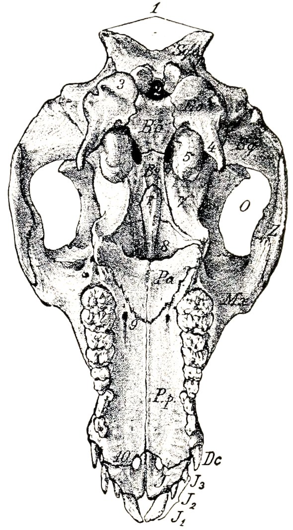
Fig. 111.—Basal Surface of Skull of Young Pig, without the Mandible. Sq.o., Supraoccipital; E.o., exoccipital; B.o., basioccipital; B.s., body of sphenoid; Sq., squamous temporal bone; V, vomer; Mx., maxilla; Pa., horizontal part of palate bone; P.p., palate process of maxilla; Z, malar bone; J, premaxilla; D.c., canine tooth; J1–3, incisor teeth; O, temporal fossa; 1, occipital crest; 2, for. magnum; 3, occipital condyle; 4, paramastoid (styloid) process; 5, bulla ossea; 6, for. lacerum basis cranii; 7, pterygoid process of sphenoid; 8, posterior nares; 9, anterior palatine foramen; 10, palatine cleft. (Struska, Anat. d. Haustiere.)
The sphenoid bone is short and resembles that of the ox in general. The body 142is narrow. The pituitary fossa is very deep, and is limited behind by a prominent dorsum sellæ; the dorsum bears lateral projections, the posterior clinoid processes. The foramen ovale is absent, being included in the foramen lacerum anterius. The other foramina are like those of the ox. The pterygoid process is broad and twisted. Its base is not perforated and its free edge is thin and sharp. It concurs with the pterygoid and palate bones in the formation of the pterygoid fossa, which opens backward and is not present in the horse or ox. The sphenoidal sinus is very large and occupies the body, the temporal wings, and a great part of the pterygoid processes in the adult; it is continued into the temporal bone as mentioned above.
The ethmoid bone has a relatively long perpendicular plate, which is marked by ridges corresponding to the ethmoidal meatuses. The cribriform plate is extensive and very oblique, so that it and the crista galli are almost in line with the basi-cranial axis. A linear series of relatively large foramina is found on either side of the crista. The lateral mass consists of five endoturbinals and eighteen ectoturbinals (Paulli). The lamina papyracea concurs in the formation of the pterygo-palatine fossa.
Face
The maxilla is extensive. Its external surface forms a longitudinal groove, which is continued upon the premaxilla in front and the facial parts of the lacrimal and malar behind. The infraorbital foramen—sometimes double—is large and is situated above the third or fourth cheek tooth. The alveolus for the canine tooth produces a ridge at the anterior end which is very pronounced in the boar. The facial crest extends forward from the root of the zygomatic process and fades out behind the infraorbital foramen; in some specimens it is prominent and thin-edged, in others it is rounded and projects little. The zygomatic process is short but stout and buttress-like; it is overlapped externally by the malar. The maxillary tuberosity forms in the young subject a long bulla, which occupies most of the pterygo-palatine fossa and contains the developing permanent molars; after the eruption of the teeth the tuberosity flattens and joins the vertical part of the palate bone. The palatine process is very long and is marked in its anterior part by transverse ridges corresponding with those of the mucous membrane of the palate. The anterior palatine foramen is near the junction with the palate bone; from it the palatine groove can be traced distinctly along the entire length of the process. The alveolar border presents a large alveolus for the canine tooth at its anterior end; behind this are seven alveoli for the cheek teeth, which increase in size from first to last. The maxillary foramen and infraorbital canal are very large. The maxillary sinus is small.
The body of the premaxilla is narrow and prismatic. It presents three alveoli for the incisor teeth, which are separated by short intervals and diminish in size from before backward. As in the ox, a narrow space separates the right and left bones and takes the place of the foramen incisivum. The palatine process is long and narrow. The nasal process is very extensive and is somewhat rhomboid in outline. Its upper border forms a very long suture with the nasal bone, and the lower articulates to about the same extent with the maxilla. The palatine fissure is relatively wide.
The horizontal part of the palate bone forms a fourth to a fifth of the length of the palate; its palatine surface is triangular, the apex being anterior; its nasal surface is deeply grooved and smooth. A pterygoid process (processus pyramidalis of man) projects backward and downward, and its thick rounded end is received between the pterygoid process of the sphenoid and the pterygoid bone. The perpendicular part is largely overlapped externally by the maxilla and concurs in forming part of the palatine canal. Superiorly the two plates separate and inclose 143an air-cavity which opens into an ethmoidal meatus. The inner plate curves inward and unites with the vomer and ethmoid to form a horizontal plate (Lamina transversalis), which divides the posterior part of the nasal cavity into an upper olfactory part and a lower respiratory part.
The pterygoid bone is nearly vertical in direction, and is narrow in its middle, wide at each end. The external surface is free below and forms the inner wall of the pterygoid fossa. The lower end is notched and forms a distinct hamulus.
The nasal bone is very long and its width is almost uniform, except at the anterior end, which is pointed and reaches almost as far forward as the premaxilla. The facial surface is flattened from side to side. In profile it is nearly straight in some subjects, variably concave in others. The external border is free to a small extent in front only; otherwise it is firmly connected with the premaxilla and maxilla. In the adult the frontal sinus extends into the posterior part of the bone.
The lacrimal bone is very sharply bent. Its facial surface presents a deep depression, surmounted by a ridge or tubercle. On or close to the orbital margin are two lacrimal foramina which lead to the lacrimal canals. The orbital surface presents a fossa in which the inferior oblique muscle of the eyeball arises, and its lower part bears a crest, which is crossed obliquely by a vascular furrow. The upper border articulates with the frontal only. The bone concurs in the formation of the maxillary sinus.
The malar bone is strongly compressed from side to side. Its facial surface is small and presents a fossa which is continuous with the depressions of the maxilla and lacrimal. The orbital surface is still smaller and is smooth and deeply grooved. The zygomatic process is very extensive, especially in the vertical direction. Its external surface is convex and free, and bears a rough eminence in its middle. Its internal surface is concave; it is overlapped in front by the maxilla, and in the remainder of its extent is free and smooth. The upper border is thick and rounded in front, where it forms the lower part of the orbital margin; behind this it forms an extensive notch which receives the zygomatic process of the temporal. (It might be regarded as dividing into frontal and temporal branches.) The lower border is convex and becomes thinner behind.
The turbinal bones resemble those of the ox. The superior turbinal is, however, relatively longer, less fragile, and more firmly attached to the nasal bone. There is no middle turbinal.
The vomer is very long. The anterior extremity reaches to the body of the premaxilla or very close to it. The lower border is received into a groove formed by the nasal crest of the maxillæ and palate bones and in front by the palatine processes of the premaxillæ. The posterior border is concave, thin, and sharp.
The os rostri (or prenasal bone) is situated in the snout between the nostrils. It has the form of a short three-sided prism. The upper surface is convex and is notched at each end. The lateral surfaces are concave, smooth, and converge below, forming a grooved lower border. The posterior surface is triangular, notched centrally, and rough laterally. The anterior surface is deeply pitted and is surrounded by an irregular sharp border.
The mandible is very strong, and differs very much from that of the horse or ox. The body narrows decidedly in front; the lingual surface is deeply concave; the mental surface is strongly convex, slopes downward and backward, and forms a distinct prominence at the point of divergence of the rami. Above this prominence is a pair of foramina. The alveolar border presents six alveoli for the incisor teeth, and a little further back two large cavities for the canine teeth. There are two pairs of mental foramina of considerable size and a variable number of smaller ones. The rami diverge more than in the horse or ox, and the upper part is somewhat incurved. The horizontal part is very thick and strong. Its outer surface is strongly convex from above downward. The inner surface is prominent 144over the roots of the molar teeth and overhangs the concave lower part. The alveolar border is thin in front and widens behind; it does not follow the axis of the ramus, but runs nearly straight and produces the marked overhang noted above. There are seven alveoli for the lower cheek teeth, which increase in size from before backward. The first is small, not always present in the adult, and is separated by short spaces from the second and the canine alveolus. The vertical part is relatively wide above. The condyle is convex in both directions, wide in front, narrow and declivitous behind. The very small and thin-edged coronoid process is not quite so high as the condyle, from which it is separated by a very wide notch. The mandibular foramen is large. The two halves of the bone unite soon after birth in the improved breeds.
The body of the hyoid bone is broad from before backward, short transversely, and bears on its ventral aspect a very short pointed lingual process. The thyroid cornua are wide and curved, concave and grooved dorsally; their ends are attached to the thyroid cartilage of the larynx by rather long bars of cartilage. The small cornua are short, wide, and flattened dorso-ventrally; they are attached to short bars which project from the junction of the body and thyroid cornua. The middle cornu is a little longer than the small cornu, but is relatively slender; it is largely cartilaginous in the young subject and does not ossify at either end. The great cornu is a very slender rod, slightly enlarged at either end; the upper extremity is attached to the hyoid process of the temporal by a rather long and wide bar of cartilage.
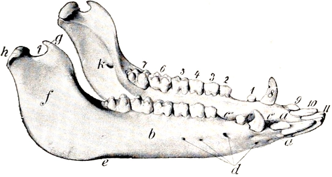
Fig. 112.—Mandible of Pig. a, Body of mandible; b, horizontal part of ramus; f, vertical part of ramus; c, interalveolar border; c′, interval between canine and corner incisor; c″, interval between first and second premolars; d, mental foramina; e, vascular impression; g, coronoid process; h, condyle; i, sigmoid notch; k, mandibular foramen; 1–7, cheek teeth, 8, canine tooth; 9, 10, 11, incisors. (Ellenberger-Baum, Anat d. Haustiere.)
THE SKULL AS A WHOLE
The length and the profile contour vary greatly in different subjects. Primitively the skull is long—especially in its facial part—and the frontal profile is almost straight. The condition is very pronounced in wild or semi-feral pigs, and exists also in the improved breeds during extreme youth. Most of the latter are decidedly brachycephalic when fully developed; the face is “dished” in a pronounced fashion. The frontal region slopes sharply upward, and the nasal region is shortened, and in some specimens even distinctly concave in profile. The supraorbital foramina are about midway between the orbital margin and the frontal suture. The supraorbital grooves extend forward from the foramina to the nasal region and turn outward and downward toward the infraorbital foramina over the ridges which separate the nasal and lateral regions.
The lateral surface is triangular when the mandible is included. The temporal fossa is entirely lateral and its long axis is almost vertical. It is bounded above by the occipital crest, behind by the temporal crest, in front by the parietal crest, and is marked off from the orbital cavity by the supraorbital process and a curved crest which extends from it to the root of the pterygoid process. The zygomatic arch is strong, high, and flattened from side to side. Its root is notched above and bears a projection below. It curves sharply upward behind and forms a pointed recurved projection above and in front of the external auditory meatus. 145The orbit is small. Its margin is deficient behind in the dry skull, thick and rounded in front and below. The cavity is limited below by a ridge on the frontal and lacrimal bones, and is separated by a crest from the temporal fossa. The inner wall is perforated above by the orbital opening of the supraorbital canal, and below by the optic and ethmoidal foramina; on its antero-inferior part is the fossa in which the inferior oblique muscle of the eye takes origin. Two lacrimal foramina are found on or close to the anterior margin. The pterygo-palatine fossa is well defined; its upper part forms a deep groove which leads from the large foramen in the sphenoid (like that of the ox) to the very large maxillary foramen. The preorbital region is deeply grooved in its length and is clearly marked off by a ridge from the nasal and frontal regions. The facial crest is short, usually thin-edged, and lies above the fifth and sixth cheek teeth. A little (ca. 2 cm.) in front of it is the infraorbital foramen. There is a ridged prominence over the canine alveolus. In some skulls the anterior part of the upper jaw is inclined upward.
The most striking features of the basal surface are as follows: The paramastoid processes are extremely long, less flattened than in the horse and ox, and nearly vertical. At the inner side of the root of each is the hypoglossal foramen, and in front of it are the stylo-mastoid foramen and a deep cavity in which the hyoid process is concealed. The bulla ossea is long, compressed laterally, and bears a sharp, short, muscular process. The basioccipital is wide and flattened; it bears a median crest and two lateral tubercles. The posterior nares are small and are wider below than above. On either side of them is the tuberosity of the palate bone, and above this is the pterygoid fossa. The palate is remarkably long and is relatively narrow. It constitutes about two-thirds of the entire length of the skull. It is widest between the canines and premolars and narrow at each end. It is marked by a crest medially and by the palatine foramen and groove laterally. The anterior part bears transverse ridges. It is moderately arched from side to side. In some specimens it is nearly straight or slightly concave in its length; in others it curves upward to a variable degree in front. The posterior end always slopes upward more or less.
The posterior or nuchal surface is remarkable for its height and the breadth of the occipital crest. The central part above the foramen magnum is smooth and concave from side to side, and is bounded laterally by ridges, which converge below and end on two tubercles at the upper margin of the foramen magnum. The surface is separated from the temporal fossæ by the temporal crests, which curve downward and outward and blend with the external auditory canals. The mastoid process has the form of a plate which overlaps the root of the paramastoid process and bears a crest on its anterior part.
The cranial cavity is small, in spite of the great size of the cranium; the discrepancy is due to the enormous development of the frontal sinuses in the adult. It is relatively longer, but much lower than that of the ox. Its width is greatly diminished between the orbits. The olfactory fossæ are extensive and very oblique. The floor resembles that of the ox, but the foramen ovale is absent, the dorsum sellæ is more developed, and the foramen lacerum basis cranii is like that of the horse. Two oblique lateral crests clearly mark the limit between the cerebral and cerebellar compartments. The tentorium osseum and the parieto-temporal canals are absent.
The nasal cavity is very long. Its posterior part is divided by a horizontal plate into olfactory and respiratory parts. The olfactory part or fundus is above, and contains the ethmoturbinals and ethmoidal meatuses. The lower part is continuous with the inferior meatus and leads to the pharyngeal orifice; hence it is sometimes called the naso-pharyngeal meatus. The bony roof is almost complete in front on account of the great length of the nasal bones.
The frontal sinus is a vast excavation in the adult animal. It involves all of 146the roof and almost all of the sides of the cranium, and extends forward into the roof of the nasal cavity a variable distance—sometimes as far as a transverse plane through the infraorbital foramina. The septum between the right and left sinuses is usually deflected in an irregular manner in its middle part, but is practically median at either end. Each sinus is subdivided by numerous septa, some of which are complete. Thus the sinus is divided into compartments, each of which communicates with an ethmoidal meatus.
In the young pig the sinus is small and is confined to the anterior part of the frontal bone. Later it extends backward, outward, and to a less extent forward. In the old subject it penetrates laterally into the supraorbital process and the root of the zygomatic process of the temporal bone, and behind almost down to the foramen magnum and the occipital condyles. It then consists of six to eight compartments usually.
The maxillary sinus is relatively small. Its anterior end is a little less than an inch (ca. 2 cm.) behind the infraorbital foramen, and it extends upward into the lacrimal and backward into the malar bone. The infraorbital canal passes along its floor, and the roots of the molar teeth do not project up into it. It does not communicate with the frontal and sphenoidal sinuses, but with the posterior part of the middle meatus by means of a considerable orifice.
The sphenoidal sinus is very large. It involves the body, pterygoid processes, and temporal wings of the sphenoid bone, and extends into the squamous temporal. It communicates with the ventral ethmoidal meatus.
There is a small sinus in the perpendicular part of the palate bone which communicates with an ethmoidal meatus.
BONES OF THE THORACIC LIMB
The scapula is very wide, the index being about 1 ∶ 0.7. The spine is triangular and is very wide in its middle, which curves backward over the infraspinous fossa and bears a large tuberosity. Its lower part bears a small projection (rudimentary acromion). The anterior border is strongly convex in profile, sinuous when viewed from the front, and thick and rough in its middle. The posterior border is wide, slightly concave, and bears an outer rough lip. The vertebral border is convex, and the cartilage is not so extensive as in the horse and ox. The cervical angle is thin and bent inward a little. The dorsal angle is thick and is about a right angle. The neck is well defined. The rim of the glenoid cavity is rounded and not notched. The tuberosity is just above the antero-internal part of the glenoid cavity and bears no distinct coracoid process.
The humerus has an appearance in profile somewhat like an italic f minus the cross-bar; this is due to the marked backward and forward inclination of the proximal and distal ends respectively. The shaft is decidedly compressed from side to side. The internal surface is extensive and flattened; it is separated from the anterior surface by a distinct border, and bears no teres tubercle. The musculo-spiral groove is shallow. The deltoid tuberosity is small, and there is a larger rounded eminence midway between it and the external tuberosity. The nutrient foramen is on the posterior surface below its middle. The head is more strongly curved and the neck better marked than in the horse or ox. The external tuberosity is very large and extends upon the front of the extremity. It is divided into two high prominences by a wide deep groove. There is a third eminence below and externally for the attachment of the supraspinatus muscle. The bicipital groove is at the front of the inner side; it is undivided and is almost converted into a canal. The outer groove on the distal articular surface is so shallow as to give the appearance of two condyles of similar size. The olecranon fossa is very deep, and the plate of bone which separates it from the coronoid fossa is thin and sometimes 147perforated. The proximal end unites with the shaft at three and a half years, the distal at one year.
The radius is short and narrow, but thick. The shaft increases in size from above downward. The greater part of the posterior surface is in apposition with the ulna; this part is marked by a vascular furrow which runs downward from the upper interosseous space, and has the nutrient foramen at its proximal end. The bicipital tuberosity is represented by a rough area. The distal end is relatively large. Its carpal surface consists of concavo-convex facets for the radial and intermediate carpal bones. There is a wide shallow groove on the middle of the front. The proximal end fuses with the shaft at one year, the distal at three and a half years.
The ulna is massive. It is much longer and considerably heavier than the radius. The shaft is curved. The anterior surface is convex and most of it is rough and attached to the radius by the interosseous ligament. There is a smooth area on the upper third, which concurs with the radius in forming the upper interosseous space, and is marked in its upper part by the nutrient foramen. From this space a vascular furrow passes downward to the lower part of the shaft, where there is often a distal interosseous space for the passage of vessels. The internal surface is extensive, concave, and smooth. The external surface is slightly convex, and its upper part is marked by an oblique rough line or ridge. The proximal extremity is large and is bent inward somewhat; its length is more than one-third of that of the entire bone. The distal extremity is relatively small; it articulates with the ulnar and accessory carpal bones, and is notched in front to accommodate the ridge on the radius. The bone contains a considerable medullary canal. The bone is consolidated at three to three and a half years.
The carpus comprises eight bones, four in each row. The bones of the proximal row resemble those of the ox, with the exception of the accessory, which is more like that of the horse, but has no external groove. The first carpal is small, elongated from before backward, rounded, and articulates in front with the second carpal. The latter is high and narrow, and articulates with the second and third metacarpal bones below. The third carpal articulates with the radial and intermediate above, the third metacarpal bone below. The fourth is the largest bone of the row; it articulates with the intermediate and ulnar above, the fourth and fifth metacarpals below, and bears a tuberosity on its volar aspect.
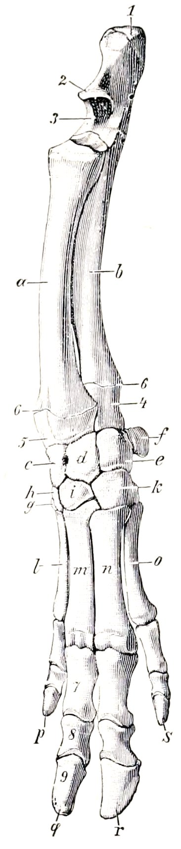
Fig. 113.—Skeleton of Forearm and Manus of Pig, Antero-external View. a, Radius; b, ulna; c, radial carpal; d, intermediate carpal; e, ulnar carpal; f, accessory carpal; g, first carpal; h, second carpal; i, third carpal; k, fourth carpal; l-o, second to fifth metacarpal bones; p-s, second to fifth digits; 1, olecranon; 2, beak of ulna (proc. anconeus); 3, semilunar notch; 4, styloid process of ulna; 5, styloid process of radius; 6, distal epiphyseal lines; 7, first phalanx; 8, second phalanx; 9, third phalanx. (Ellenberger-Baum, Anat. d. Haustiere.)
Four metacarpal bones are present. The first is absent, the third and fourth are large and carry the chief digits, while the second and fifth are much smaller and bear the accessory digits. Their proximal ends articulate with each other and with the carpus as indicated above. The bones are consolidated at about two years of age.
148The third and fourth metacarpals are flattened from before backward, three-sided, and placed close together. The distal end of each bears a trochlea for articulation with the first phalanx and the sesamoids. The third is the wider of the two, and articulates with all of the lower row of the carpus except the first. The fourth articulates with the fourth carpal chiefly, but has a small facet for the third. The second and fifth metacarpals are placed further back than the chief bones. The fifth is considerably the thicker of the two. The proximal ends are small and articulate with the corresponding carpal and metacarpal bones. The distal end is relatively large; its articular surface is condyloid in front, trochlear behind.
Each chief digit comprises three phalanges and three sesamoids. The bones of the chief digits resemble those of the ox in form, but there is no foramen on the interdigital side of the extensor process and the proximal sesamoids are narrow and ridged behind. The phalanges of the accessory digits (which do not reach the ground ordinarily) are similar in form but much smaller.
BONES OF THE PELVIC LIMB
The os coxæ is long and narrow. The ilium and ischium are almost in line with each other and sagittal in direction. The wing of the ilium bends outward much less than in the horse or ox. The gluteal surface is divided into two fossæ by a ridge, which is continuous with the superior ischiatic spine behind. The inner surface presents an extensive rough area behind, which is in apposition with the wing of the sacrum. The smooth iliac area is narrow, and is bounded above by a ridge. The crest or anterior border is convex, and is thick, rough, and prominent in its middle, which forms the highest point of the bone. The internal angle is lower than the crest, is directed backward, and articulates internally with the sacrum. The external angle is lower still and is very little thickened. The ischia in the female are somewhat divergent and flattened behind. The tubera are everted and bear three prominences. There is a crest or tuberosity on the ventral surface. The superior ischiatic spine is like that of the cow, but is slightly incurved and the muscular ridges on its outer face are more pronounced. The symphyseal part of the pubis is thick and the two bones are almost in a horizontal plane. The ilio-pectineal eminence is prominent and the psoas tubercle is well marked.
The acetabulum is placed a little further back than in the ox. The rim is thick and is cut into posteriorly by a narrow fissure, which leads into the deep fossa acetabuli. The three pieces of the os coxæ are fused by the end of the first year, but the crest and the ischial tubera are partially separate till the sixth or seventh year. The symphysis does not usually undergo complete anchylosis. Interischial bones are present.
The inlet of the pelvis is elliptical and very oblique. In a sow of full size the conjugate diameter is about five to six inches (10 to 12 cm.) and the transverse about three and a half to four inches (ca. 8.75 to 10 cm.). In the female the floor is relatively wide and flattened, especially at the outlet, where the tubera are everted; it also has a decided downward inclination behind. The pelvic axis is therefore correspondingly oblique. The ischial arch is wide. In the boar the pubis is much thicker and the ischia are not everted posteriorly. The inlet is smaller. The floor is concave from side to side and slopes decidedly less than in the sow. The superior ischiatic spines are more incurved, and the ischial arch is much narrower and deeper.
The femur has a relatively wide and massive shaft, on which four surfaces might be recognized. The principal nutrient foramen is situated in the proximal third of the anterior surface. The posterior surface is wide, and is limited outwardly by a ridge which extends from the trochanter major to the large external supracondyloid crest. There is no supracondyloid (plantar) fossa. The head is strongly curved, and is marked toward the inner side by a rather large depression for the 149attachment of the round ligament. The neck is distinct. The trochanter major, although massive, does not extend up as high as the head. The trochanteric ridge and fossa resemble those of the ox. The external (third) trochanter is absent. The ridges of the trochlea are similar and almost sagittal.
The shaft of the tibia is slightly curved, convex internally. The tuberosity is grooved in front, and a narrow sulcus separates it from the external condyle. The facet for the fibula is on the posterior border of the latter, and is bounded internally by an eminence. The upper part of the crest is very prominent and curves outward. The distal end resembles in general that of the ox, but is relatively narrower transversely and thicker from before backward.
The fibula extends the entire length of the region, and is separated from the tibia by a wide interosseous space. The shaft is flattened from side to side; the proximal part is wide and deeply grooved externally; the lower part is narrower and thicker. The proximal end is flattened, grooved externally, and articulates internally with the external condyle of the tibia. The distal end forms the external malleolus. It is grooved externally, and articulates with the tibia and tibial tarsal internally, with the fibular tarsal bone distally.
The patella is very much compressed laterally and presents three surfaces.
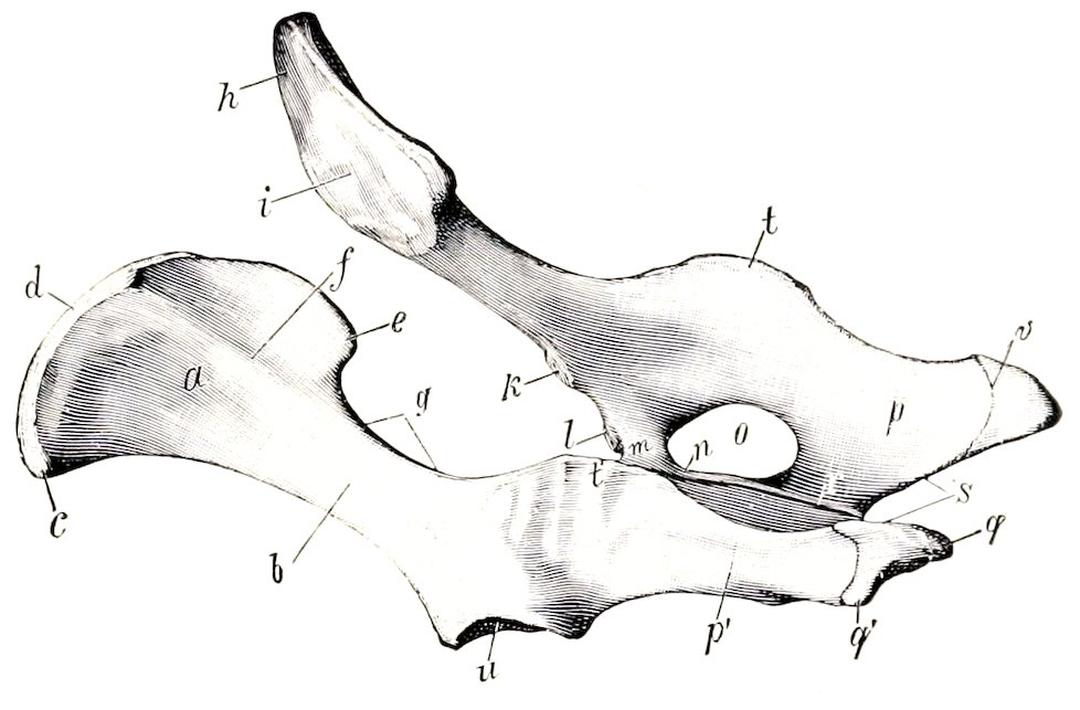
Fig. 114.—Coxal Bones of Pig, Left Posterior View. a, Wing, b, shaft, c, external angle, d, anterior border, e, internal angle, f, gluteal line of ilium; g, great sciatic notch; h, smooth, i, rough part of ventral surface of ilium; k, psoas tubercle; l, ilio-pectineal eminence; m, acetabular branch, n, symphyseal branch of pubis; o, obturator foramen; p, p′, ischium; q, q′, tuber ischii; r, symphysis; s, ischial arch; t, t′, superior ischiatic spine; u, acetabulum; v, epiphyseal line. (Ellenberger-Baum, Anat. d. Haustiere.)
The tarsus comprises seven bones. The tibial and the fibular tarsal resemble in general those of the ox. The axis of the tibial is, however, slightly oblique downward and inward, and its distal end bears a double trochlea for articulation with the central and fourth tarsals. The tuber calcis is deeply grooved posteriorly. The central tarsal is narrow transversely and thick. Its proximal surface is deeply concave, and the posterior bears a large tubercle. The first tarsal is high and narrow; it articulates with the central and second tarsals and the second metatarsal bone. The second tarsal is small and somewhat prismatic; it articulates with the central above, the third in front, the first behind, and the second and third metatarsals below. The third tarsal is much larger, and is compressed from above downward, wide in front, narrow behind. It articulates with the central tarsal above, the third metatarsal below, the second tarsal internally, and the fourth tarsal externally. The fourth tarsal is large. Its external face is crossed by an oblique groove for the tendon of the peroneus longus. The internal surface articulates with the central and third tarsals. The proximal surface supports the tibial and fibular tarsal bones, and the distal surface rests on the fourth and fifth metatarsals. It ossifies from two centers.
The four metatarsal bones resemble the corresponding bones of the fore limb, but are somewhat longer. The proximal ends of the third and fourth each have a considerable projection behind; the process on the third has a facet for articulation 150with a discoid sesamoid bone. The second and fifth are placed more on the posterior aspect of the large bones than is the case in the fore limb.
The first and second phalanges are a little longer and narrower than those of the fore limb.
SKELETON OF THE DOG
VERTEBRAL COLUMN
The vertebral formula is C7T13L7(6)S3Cy20–23.
The cervical vertebræ are relatively longer than in the ox and the pig. The bodies of the typical vertebræ diminish in length from first to last and are compressed dorso-ventrally. The anterior extremity is moderately convex and the posterior slightly concave; both are oblique. The median ridge and lateral grooves on the upper surface of the body are very well marked. The second, third, and fourth have distinct ventral spines. The spinous process of the third has the form of a long low crest; in the remainder it is higher, blunt-pointed, and inclined forward. The transverse processes of the third, fourth, and fifth project downward and backward, and divide into two branches; of these, the anterior one is thin, and the posterior is thick and tuberculate at its free end. The process of the sixth has two parts; one of these is an extensive quadrilateral plate directed downward and outward and ridged on its inner surface; the other part is short and blunt, and is directed outward and a little backward and upward. The seventh is readily distinguished by its shortness, the length of its spine, and the single transverse process. The posterior articular processes bear tubercles which are large on the third, fourth, and fifth.
The ventral arch of the atlas is narrow from before backward, and bears a small tubercle posteriorly. The upper surface of the dorsal arch is strongly convex and rough centrally. The wings are wide, flattened, and almost horizontal. The upper surface is rough. There is an alar notch (Incisura alaris) on the anterior border instead of the anterior foramen. The foramen transversarium is present.
The body of the axis is flattened dorso-ventrally, especially in front. The odontoid process is rounded and relatively long, reaching almost to the occipital bone; it is inclined upward a little. The articular surfaces which flank it are condyloid in form and very oblique. The inferior surface is wide, and is divided by a median crest into two fossæ. The transverse processes are single, pointed, directed backward and outward, and perforated by relatively large foramina transversaria. The spinous process is thin and of moderate height, but very long; it is prolonged forward so as to overhang the dorsal arch of the atlas, and is terminated behind by a tuberosity which is connected by two crests with the posterior articular processes. The anterior notches are large and are never converted into foramina.
The bodies of the thoracic vertebræ are wide and compressed dorso-ventrally, especially at each end of the region. Their convex anterior surfaces are depressed in the middle. The posterior facets for the heads of the ribs are absent on the last three or four. The transverse processes resemble those of the horse. They bear mammillary processes except at the anterior end of the region. The facets for the tubercles of the ribs are large and concave in the anterior part of the series, and become smaller and slightly convex further back. The last three have accessory processes also. The first three or four spinous processes are about equal in length. Behind this they become gradually shorter to the tenth, and then remain equal. The backward slope is most marked in the ninth and tenth. The eleventh is practically vertical (anticlinal vertebra) and the last two incline slightly forward.
151The bodies of the lumbar vertebræ are decidedly flattened dorso-ventrally, and increase in width from first to last. The length increases to the sixth. The transverse processes are plate-like and are directed forward and downward. Their length increases to the fifth and sixth. They form no joints with each other or with the sacrum. Their extremities are enlarged, with the exception of the last. The accessory processes project backward over the posterior notches of the first five. The anterior articular processes are large, compressed laterally, and bear mammillary processes. The spinous processes are broad below, narrower above, and with the exception of the last, incline a little forward. Their height diminishes behind the fourth.
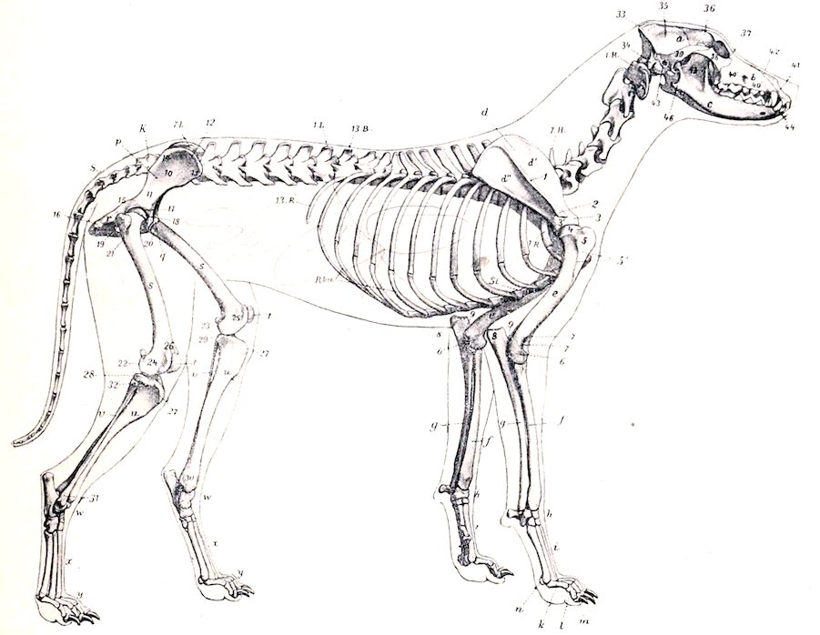
Fig. 115.—Skeleton of Dog, Lateral View. a, Cranium; b, face; c, mandible; 1H-7H, cervical vertebræ; 13B, last thoracic vertebra; 1L-7L, lumbar vertebræ; K, sacrum; S, coccygeal vertebræ; 1R-13R, ribs; R.Kn., costal cartilages; St., sternum; d, scapula; d′, supraspinous fossa; d″, infraspinous fossa; 1, spine of scapula; 2, acromion; 3, tuberosity of scapula; 3′, articular end of scapula; e, humerus; 4, head of humerus; 5, external tuberosity of humerus; 5′, deltoid ridge; 6, 6′, epicondyles of humerus; 7, external condyloid crest; 7′, coronoid fossa; f, radius; g, ulna; 8, olecranon; 9, “beak” of ulna; h, carpus; i, metacarpus; k, proximal phalanges; l, middle phalanges; m, distal phalanges; n, sesamoid; p, ilium; 10, wing of ilium; 11, shaft of ilium; 12, crest of ilium; 13, external angle of ilium (tuber coxæ); 14, internal angle of ilium (tuber sacrale); 15, superior ischiatic spine; q, pubis; r, ischium; 16, tuber ischii; 17, acetabulum; s, femur; 18, head of femur; 19, trochanter major; 20, trochanter minor; 21, trochanter tertius; 22, 23, condyles; 24, 25, epicondyles; 26, trochlea; t, patella; u, tibia; 27, tuberosity of tibia; 28, 29, condyles of tibia; 30, internal malleolus; v, fibula; 31, external malleolus; 32, head of fibula; w, tarsus; x, metatarsus; y, phalanges; 33, occipital bone; 34, paramastoid (styloid) process; 35, parietal bone; 36, frontal bone; 37, lacrimal bone; 38, malar bone; 39, squamous temporal; 40, maxilla; 40′, infraorbital foramen; 41, premaxilla; 42, nasal bone; 43, external auditory meatus; 44, canine tooth; 45, masseteric fossa; 46, angular process of mandible. (After Ellenberger, in Leisering’s Atlas.)
The sacrum results from the early fusion of three vertebræ. It is short, wide, and quadrangular. The spines are fused to form a median crest, which is notched, however, between the summits of the spines. On either side are two tubercles, vestiges of the fused articular processes. The pelvic surface is deeply 152concave and presents two pairs of foramina. The wings are prismatic and very high. Their lateral surfaces are extensive, face almost directly outward, and bear an auricular surface on the lower part. The anterior surface of the body of the first vertebra is extensive, depressed in its middle, and bears a prominent lip below. The anterior articular processes are large and have extensive, slightly concave facets which face upward and inward. The posterior articular processes are small. The transverse processes of the last vertebra project backward and may articulate or fuse with those of the first coccygeal. The sacral canal is strongly compressed dorso-ventrally.
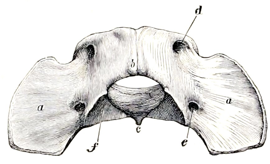
Fig. 116.—Atlas of Dog, Viewed from Above and Behind. a, Wing; b, dorsal arch and tubercle; c, ventral tubercle; d, intervertebral foramen; e, foramen transversarium; f, articular surface for axis. (Ellenberger-Baum, Anat. d. Hundes.)
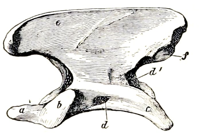
Fig. 117.—Axis of Dog, Left Lateral View. a, Odontoid process; b, articular surface for atlas; c, transverse process; d; foramen transversarium; d′, posterior opening of d; e, spine; f, posterior articular process. (Ellenberger-Baum, Anat. d. Hundes.)
The coccygeal vertebræ are fully developed in the anterior part of the region. The arch is complete in the first six usually. The first three or four have well developed articular processes at each end. Behind this the posterior pair quickly disappears, and the anterior ones become non-articular and gradually reduced in size. The transverse processes of the first five or six are relatively large; behind this they quickly disappear. Hæmal arches (or chevron bones) in the form of a V or Y occur ventrally at the intercentral junctions of the third, fourth, and fifth usually. They transmit the middle coccygeal artery, which passes between pairs of ventral tubercles further back.
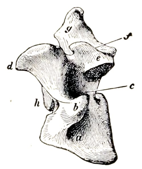
Fig. 118.—Sixth Cervical Vertebra of Dog, Left View. a, Ventral plate, b, lateral part of transverse process; c, foramen transversarium; d, anterior articular process; f, accessory process; g, spinous process; h, articular head of body. (Ellenberger-Baum, Anat. d. Hundes.)
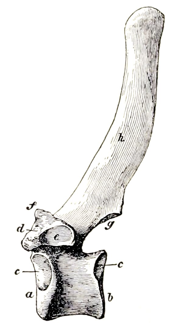
Fig. 119.—Fourth Thoracic Vertebra of Dog, Left View. a, Head; b, glenoid cavity; c, facet for head of rib; d, transverse process; e, facet for tubercle of rib; f, mammillary process; g, posterior articular process; h, spinous process. (Ellenberger-Baum, Anat. d. Hundes.)
153Curves.—A gentle curve, convex ventrally, is formed by the cervical and the anterior part of the thoracic region. The posterior thoracic and the lumbar vertebra form a second curve, concave ventrally. The sacral promontory is well marked. The sacrum and the anterior part of the coccygeal region constitute a third and more pronounced curve, concave ventrally. In long-tailed dogs the sacro-coccygeal region is somewhat S-shaped.
Variations.—Numerical variations are not common except in the coccygeal region. The number of thoracic vertebræ may be twelve or fourteen, with or without compensatory change in the lumbar region. Girard recorded a case with eight lumbar and the usual number of thoracic vertebra. Six lumbar with fourteen thoracic vertebræ have been met with. The first coccygeal sometimes unites with the sacrum.
THE RIBS
Thirteen pairs of ribs are present, of which nine are sternal and four asternal. They are strongly curved, narrow, and thick. Those in the middle of the series are the longest. The first eight or nine increase in width in their lower part. The last rib is usually floating. The costal cartilages are long and curve forward; the length and curvature of the first pair is a striking special feature.
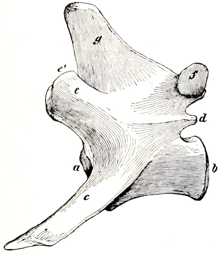
Fig. 120.—Fourth Lumbar Vertebra of Dog, Left View. a, b, Articular surfaces of body; c, transverse process; d, accessory process; e, anterior articular process; e′, mammillary process; f, posterior articular process; g, spinous process. (Ellenberger-Baum, Anat. d. Hundes.)
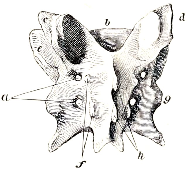
Fig. 121.—Sacrum of Dog, Dorsal View. a, Dorsal sacral foramina; b, articular surface of body of first segment; c, d, anterior articular processes; e, wing; f, rudiments of articular processes; g, lateral part; h, spinous processes. (Ellenberger-Baum, Anat. d. Hundes.)
THE STERNUM
This is long, laterally compressed, and consists of eight sternebræ, which fuse only in exceptional cases and in extreme old age. The first segment is the longest; its anterior end is blunt-pointed and bears a short conical cartilage. It widens at the point of articulation of the first pair of cartilages. The last segment is also long, thinner than its predecessors, wide in front, and narrow behind, where it bears a narrow xiphoid cartilage.
The thorax is distinctly barrel-like and is not decidedly compressed anteriorly like that of the horse and ox. The inlet is oval and is relatively wide on account of the marked curvature of the first pair of ribs and cartilages.
BONES OF THE SKULL
In the following descriptions of the separate bones an intermediate type—e. g., a fox terrier—is selected, and the most striking differences in the brachycephalic and dolichocephalic breeds will be considered in the section on the skull as a whole.
154
Cranium
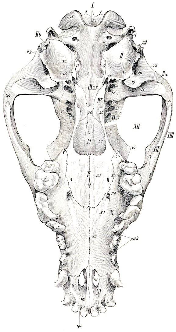
Fig. 122.—Base of Skull of Dog, without Mandible. I, Occipital; II, tympanic part of temporal; IIa, squamous part of temporal; IIb, mastoid part of temporal; III, sphenoid; IV, pterygoid; V, palate bone; VI, vomer; VII, malar; VIII, zygomatic arch; IX, inner wall of orbit; X, palatine process of maxilla; XI, premaxilla; XII, orbital cavity; 1, 1, tubercles above foramen magnum; 2, foramen magnum; 3, occipital condyle; 4, notch between occipital condyles; 5, condyloid fossa; 6, hypoglossal foramen; 7, paramastoid (styloid, jugular) process; 8, for. lacerum and posterior opening of carotid canal; 9, petro-occipital synchondrosis; 10, petro-tympanic fissure; 11, tubercle; 12, bulla ossea; 13, muscular process of petrous; 14, carotid foramen; 15, osseous Eustachian tube; 16, postglenoid process; 17, glenoid cavity; 18, for. ovale; 19, posterior opening of alar canal; 20, external opening of parieto-temporal canal; 21, stylo-mastoid foramen; 22, external auditory meatus; 23, temporal crest; 24, zygomatic process of temporal bone; 25, body of postsphenoid; 26, body of presphenoid; 27, anterior opening of alar canal; 28, for. lacerum orbitale; 29, optic foramen; 30, hamulus of pterygoid; 31, horizontal part of palate bone; 32, perpendicular part of palate bone; 33, median palatine suture; 34, palato-maxillary suture; 35, posterior nasal spine; 36, anterior palatine foramen; 37, palatine groove; 38, alveolar border of maxilla; 39, palatine process of maxilla; 40, pterygoid process of maxilla; 41, palatine fissure; 42, body of premaxilla; 43, palatine process of premaxilla; 44, alveolar border of premaxilla; 45, supraorbital process of frontal bone. (Ellenberger-Baum, Anat. d. Hundes.)
The occipital bone is similar in position to that of the horse. The crest, is prominent, angular, and directed backward. Just below the crest are two rough imprints or tubercles for muscular attachment. The surface below these is convex from side to side and concave from above downward. On either side, at the junction 155with the squamous, there is a foramen which communicates with the parieto-temporal canal. The condyles are somewhat flattened and are widely separated above; at the inner side of each is a short condyloid canal, which opens into the parieto-temporal canal. The paramastoid processes are very short. The basilar part is wide and joins the bulla ossea on either side; its lower surface is flattened and the tubercles are at the junction with the bulla. The hypoglossal foramen is small and is close to the foramen lacerum posterius; the latter is bounded in front by the bulla ossea, behind and internally by the occipital bone.
The interparietal bone fuses with the occipital before birth. It forms the high posterior part of the sagittal crest, and is wedged in between the two parietal bones. The tentorium osseum is thin and curved, concave ventrally. Its base concurs with the occipital and parietal bones in the formation of a transverse canal which is continuous with the parieto-temporal canals.
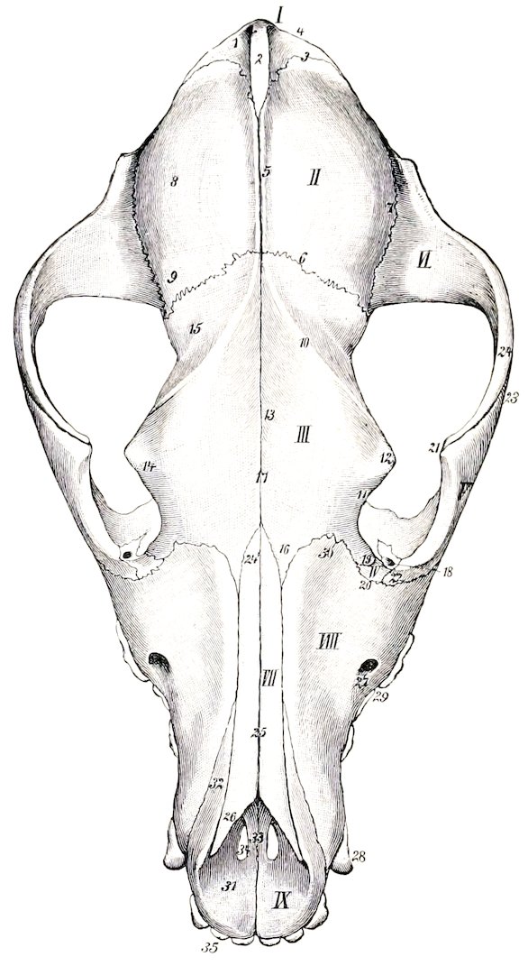
Fig. 123.—Skull of Dog, Dorsal View. I, Occipital; II, parietal; III, frontal; IV, lacrimal; V, malar; VI, squamous temporal; VII, nasal; VIII, maxilla; IX, premaxilla; 1, supraoccipital; 2, interparietal; 3, parieto-occipital suture; 4, occipital crest; 5, sagittal crest; 6, parieto-frontal suture; 7, squamous suture; 8, parietal eminence; 9, antero-external angle of parietal bone; 10, frontal crest; 11, 14, orbital margin; 12, supraorbital process; 13, frontal fossa; 15, temporal part of frontal bone; 16, nasal process of frontal bone; 17, frontal suture; 18, lacrimal foramen; 19, maxillary process of frontal bone; 20, lacrimo-maxillary suture; 21, frontal process of malar bone; 22, lacrimal process of malar; 23, zygomatic process of malar; 24, zygomatic process of squamous temporal; 24′, posterior end of nasal bone; 25, nasal suture; 26, anterior end of nasal bone; 27, infraorbital foramen; 28, canine tooth; 29, cheek tooth; 30, frontal process of maxilla; 31, body, 32, nasal process, 33, palatine process of premaxilla; 34, palatine fissure; 35, incisor teeth. (Ellenberger-Baum, Anat. d. Hundes.)
The parietal bone is rhomboid in outline and is strongly curved. It is extensive and forms the greater part of the roof of the cranial cavity. At the junction of the right and left bones there is a prominent sagittal crest which is continued upon the frontal bones. The lower border articulates with the temporal wing of the sphenoid by its anterior part and with the squamous temporal in the remainder of its extent. The external surface enters into the formation of the temporal fossa. The internal surface is marked by digital impressions, and by grooves for the middle meningeal artery and its branches.
The external surface of the frontal bone is crossed by a frontal crest, which extends in a curve from the sagittal crest to the supraorbital process, and separates the frontal and temporal parts. The frontal parts of the two bones form a central depression and slope downward and forward. The supraorbital process is very short, so that the orbital margin is incomplete as in the pig. The supraorbital foramen is absent. In front there 156is a narrow pointed nasal part which fits in between the nasal bone and the maxilla. The orbital and temporal parts are relatively extensive. Two ethmoidal foramina are commonly present. The frontal sinus is confined to the frontal bone.
The parts of the temporal bone fuse early. The zygomatic process curves widely outward and forward. Its anterior part is beveled below and articulates extensively with the corresponding process of the malar. The articular surface for the condyle of the mandible consists of a transverse groove which is continued upon the front of the large postglenoid process. Behind the latter is the lower opening of the parieto-temporal canal. There is no condyle. The mastoid part is small, but bears a distinct mastoid process. The external auditory meatus is large and the canal very short, so that one can see into the tympanum in the dry skull. The India ossea is very large and is rounded and smooth; the inner side is united to the basioccipital. Above this junction and roofed in by the union of the petrous part and the basioccipital is the petro-basilar canal (Canalis petrobasilaris); this transmits a vein from the floor of the cranium to the foramen lacerum posterius. The latter is in reality a depression and is situated behind the bulla ossea. In its posterior part is a foramen which transmits the ninth, tenth, and eleventh cranial nerves. The carotid canal branches off from the petro-basilar, passes forward external to it through the inner part of the bulla ossea, and opens in front at the carotid foramen; it transmits the internal carotid artery. The Eustachian opening is immediately external to the carotid foramen. The muscular and hyoid processes are extremely rudimentary. The petrous part projects into the cranial cavity and forms a sharp prominent petrosal crest. The inner surface presents a deep floccular fossa above the internal auditory meatus. The anterior surface is also free. The anterior angle is perforated by a canal for the fifth cranial nerve (Canalis nervi trigemini).
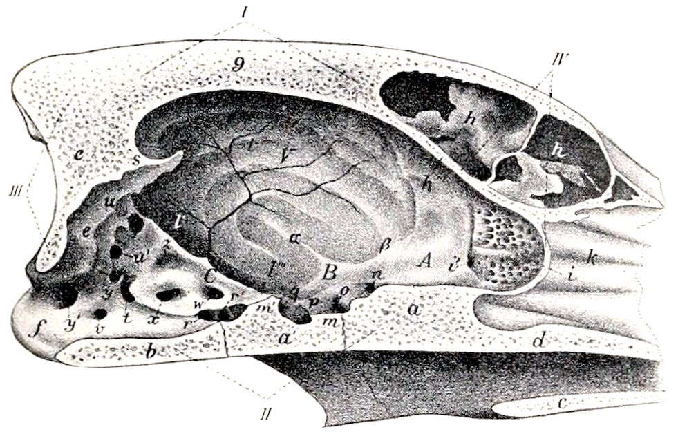
Fig. 124.—Cranial Cavity of Dog, as Seen on Sagittal Section of Skull. I, Roof of cranium; II, base of cranium; III, posterior wall of cranium; IV, anterior wall of cranium; A, anterior cranial fossa; B, middle cranial fossa; C, posterior cranial fossa; a, body of presphenoid; a′, body of postsphenoid; c, palate bone; d, vomer; e, occipital; f, occipital condyle; g, sagittal crest; h, frontal sinus; h′, cranial plate of frontal bone; i, cribriform plate of ethmoid bone; i′, ethmoidal foramen; k, ethmoturbinals; l, parietal bone; l′, l″, squamous temporal bone; l‴, temporal wing of sphenoid bone; m, sella turcica; m′, dorsum sellæ; n, optic foramen; o, foramen lacerum orbitale; p, foramen rotundum; q, foramen ovale; r, r′, carotid foramina; s, tentorium osseum; t, foramen lacerum; u, u′, orifices of parieto-temporal canal; v, condyloid canal; w, canal for trigeminal nerve; x, internal auditory meatus; y, y′, orifices of canal for inferior occipital sinus; z, floccular fossa. (After Ellenberger, in Leisering’s Atlas.)
The body of the sphenoid bone is flattened dorso-ventrally. The sella turcica is shallow, but the dorsum sellæ is well developed and bears posterior clinoid processes. A pair of anterior clinoid processes project back from the roots of the orbital wings. The latter are relatively small and are crossed externally by a crest, which is continued forward upon the palate bone. The temporal wings are extensive and articulate above with the parietals. Perforating the roots of the wings are the following foramina, named from before backward: The optic passes through the orbital wing. The foramen lacerum orbitale is a little lower and is at the junction of the wings. The foramen rotundum opens into the alar canal, which passes through the root of the short but wide pterygoid process. The 157foramen ovale is near the posterior border of the temporal wing. There is no sphenoidal sinus.
The ethmoid bone is highly developed. The cribriform plate is extensive, and the olfactory fossæ are very deep. The crista galli is little developed, and often incomplete. The perpendicular plate is long. The lateral masses are greatly developed and bulge upward into the frontal sinus. There are four large endoturbinals and six ectoturbinals. The lamina papyracea is extensive and forms the inner wall of the maxillary sinus. Its lower border joins the palatine process of the maxilla and the horizontal part of the palate bone. A shelf-like plate extends inward from its lower part and concurs with the similarly incurved part of the palate bone in forming the transverse lamina (Lamina transversalis), which divides the olfactory fundus of the nasal cavity from the naso-pharyngeal meatus.
Face
The maxilla is short, but very high posteriorly. The facial crest is absent. The infraorbital foramen is over the alveolus for the third premolar. The frontal process fits into a deep notch between the nasal and orbital parts of the frontal bone, and the middle part of the posterior border lies along the orbital margin. There are more or less pronounced ridges, juga alveolaria, over the canine and molar teeth. The zygomatic process is short and thin; it is completely overlapped outwardly by the malar, and is perforated by a number of foramina (Foramina alveolaria). A maxillary tuberosity is not present in the adult, but there is a pointed projection, the pterygoid process, behind the last alveolus. The internal surface bears a short turbinal crest on its anterior part, behind which it is deeply concave and forms the outer wall of the maxillary sinus. The palatine process is short, wide behind, and moderately arched from side to side. The anterior palatine foramen is situated at or close to the transverse palatine suture about midway between the median suture and the alveolar border. The palatine groove is distinct. The large alveolus for the canine tooth is completed by the premaxilla. The small alveolus for the first premolar is separated from the preceding one by a small interval. The next two consist of anterior and posterior parts for the roots of the teeth. The fourth and fifth are much larger and are divided into three parts. The last is small and consists of three divisions. The infraorbital canal is short.
The body of the premaxilla is compressed dorso-ventrally, and contains three alveoli for the incisor teeth, which increase in size from first to third; it also completes the inner wall of the large alveolus for the canine tooth. The foramen incisivum is very small except in large skulls. The interalveolar border is wide and very short. The nasal process is wide at its origin and tapers to a sharp point behind; the anterior part curves upward, backward, and a little inward, and forms the lateral margin of the osseous nasal aperture; the posterior part extends backward a long distance between the nasal bone and the maxilla. The palatine process turns upward and outward, forming with its fellow a wide groove for the septal cartilage; the posterior end is pointed and fits into a notch between the palatine processes of the maxillæ, and supports the end of the vomer. The palatine fissure is short but wide.
The horizontal part of the palate bones is extensive, forming about one-third of the hard palate. It presents a variable number of lesser palatine foramina. There is usually a pointed posterior nasal spine at the end of the median suture. The palatine canal is sometimes formed entirely in this bone. The perpendicular part is even more extensive. Its external surface is chiefly free and forms most of the inner wall of the large pterygo-palatine fossa. The maxillary foramen is situated in a deep recess between this bone and the zygomatic process of the maxilla. Just above it there is commonly another foramen which opens into the nasal 158cavity. The posterior palatine and sphenopalatine foramina are situated further back and a little lower; the former is immediately below the latter. A horizontal plate extends from the inner surface, meets that of the opposite bone and completes the lamina transversalis spoken of in the description of the ethmoid bone. There is no palatine sinus.
The pterygoid bones are very wide and short. They form a considerable part of the lateral boundaries of the posterior nares. The lower and posterior holders are free and at their angle of junction there is a variable hamulus.
The nasal bones are (in most breeds) long and wider in front than behind. The external surface is variably concave in its length and is inclined toward the median suture so as to form a central groove. The inner borders turn downward and form an internal nasal crest which becomes very prominent behind. The posterior parts fit into a notch formed by the frontal bones. The anterior ends form an almost semicircular nasal notch.
The lacrimal bone is very small. The facial part extends very little or not at all beyond the orbital margin. The orbital surface is small and triangular, and presents the entrance to the lacrimal canal.
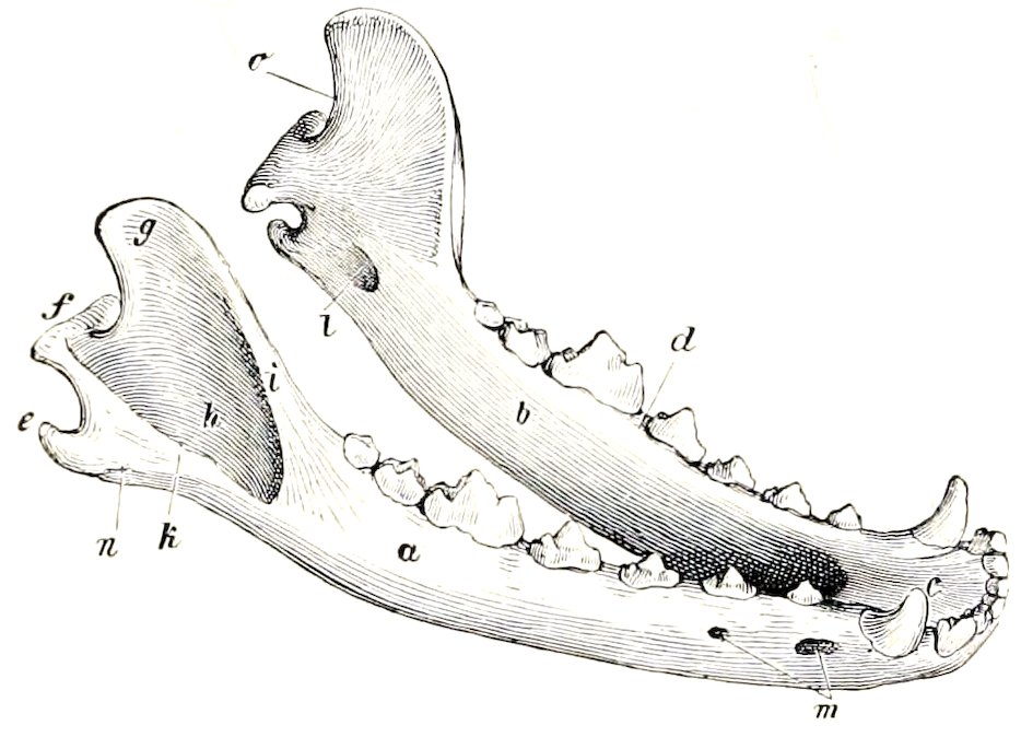
Fig. 125.—Mandible of Dog, Right-Anterior View. a, Right ramus; b, left ramus; c, body; d, alveolar border; e, processus angularis; f, condyle; g, coronoid process; h, masseteric fossa; i, k, crests which form the upper and lower boundaries of fossa; l, mandibular foramen; m, mental foramina; n, masseteric line; o, sigmoid notch. (Ellenberger-Baum, Anat. d. Hundes.)
The large zygomatic process constitutes the bulk of the malar bone. It is very long and is strongly curved. The upper border is convex, free in front, where it forms part of the orbital margin, beveled behind for articulation with the similar process of the temporal bone. Between these it bears an eminence, the processus frontalis, to which the orbital ligament is attached. The body of the bone may be considered to consist of a lacrimal process directed upward and fitting in between the lacrimal and maxilla, and a maxillary process directed downward. The facial surface is convex.
The superior turbinal bone is in its anterior part a simple plate, attached by one edge to the nasal bone; it curves downward and inward, and its free border is thickened and everted. The posterior part is wider and resembles the ethmoturbinals, with which it is connected.
The inferior turbinal bone is short and very complex. It is attached to the nasal surface of the maxilla by a basal lamina, which divides into two secondary lamellæ. The latter detach numerous tertiary lamellæ, which are coiled and have thick free edges (Fig. 373).
The vomer is not in contact with the posterior part of the floor of the nasal cavity, and does not divide the posterior nares. The posterior end is narrow and deeply notched. Near the posterior nares the two plates curve outward and join the palate bones and assist in forming the lamina transversalis.