ANGIOLOGY: THE VASCULAR SYSTEM (continued 3)
Reading edition
THE PORTAL VEIN
The portal vein (V. portæ) is a large trunk which returns the blood carried to the viscera by the gastric, splenic, and mesenteric arteries. Its peripheral tributaries correspond closely with the branches of the arteries, but the terminal trunks do not. The vein is formed behind the pancreas and below the posterior vena cava by the confluence of the anterior and posterior mesenteric and splenic veins. It passes forward, traverses the posterior part of the pancreas very obliquely, inclines 597ventrally and a little to the right, and reaches the portal fissure of the liver. Here it divides into three branches which enter the liver and ramify in the substance of the gland like an artery, terminating in the lobular capillaries. From the lobules the blood passes into the hepatic veins and through these to the posterior vena cava. Thus the blood which is distributed to the stomach, nearly the entire intestinal tract, the pancreas, and the spleen, passes through two sets of capillaries prior to its return to the heart, viz., the capillaries of these viscera and of the liver.
1. The anterior mesenteric vein (V. mesenterica cranialis) is the largest of the portal radicles. It is situated to the right of the artery of like name, and its tributaries correspond in general to the branches of the artery. Usually a single colic vein corresponds to the two arteries of the right portions of the colon.
2. The posterior mesenteric vein (V. mesenterica caudalis) is the smallest of the radicles of the portal. It accompanies the artery in the colic mesentery and its rectal branches anastomose with those of the internal pudic vein.
3. The splenic vein (V. lienalis) is the very large satellite of the splenic artery. It is formed by the union of two radicles at the base of the spleen. On leaving the hilus of the spleen it passes inward between the anterior pole of the left kidney and the saccus cæcus of the stomach and above the left end of the pancreas, receives commonly the posterior gastric vein (V. gastrica caudalis), and unites with the anterior mesenteric at the posterior border of the pancreas.
The collateral tributaries of the portal veins are as follows:
(1) Pancreatic veins (Rami pancreatici).
(2) The gastro-duodenal vein (V. gastroduodenalis) corresponds mainly to the extrahepatic branches of the hepatic artery.
(3) The anterior gastric vein (V. gastrica cranialis) joins the portal at the portal fissure.
THE COMMON ILIAC VEINS (Fig. 456)
These (V. iliacæ communes) are two very large but short trunks which result from the union of the internal and external iliac veins of each side at the sacro-iliac articulation. The left one is the longer and crosses obliquely over the terminal part of the aorta. The chief tributaries of each are as follows:
1. The last lumbar vein.
2. The circumflex iliac veins (Vv. circumflexæ ilium profundæ) are the two satellites of each corresponding artery, on either side of which they are placed. They may open directly into the posterior vena cava or into the external iliac vein.
3. The ilio-lumbar vein (V. iliolumbalis) may open into the common iliac, the external iliac, or the internal iliac vein.
THE INTERNAL ILIAC VEINS
The internal iliac or hypogastric veins (Vv. hypogastricæ), right and left, are usually formed by the confluence of lateral sacral, gluteal, and internal pudic veins. The obturator vein may open into them also. They are short trunks and are smaller than the external iliac veins. The tributaries correspond in general to the branches of the three arteries of like names. The internal pudic veins receive affluents from the venous plexuses of the prepuce and penis in the male; of the mammary gland, vulva, vagina, and vestibular bulb in the female.
THE VEINS OF THE PELVIC LIMB
The external iliac vein (V. iliaca externa) (Figs. 450, 451) lies behind the corresponding artery at the brim of the pelvis. It is the upward continuation of the femoral vein, and unites at the sacro-iliac joint with the internal iliac to form the common iliac vein. Its tributaries are as follows:
5981. The obturator vein (V. obturatoria) is a satellite of the artery and usually opens into the external iliac at the insertion of the psoas minor. Its radicles anastomose with those of the internal and external pudic veins (Figs. 451, 455).
2. The iliaco-femoral or external circumflex veins (Vv. circumflexæ femoris laterales) are the two satellites of the homonymous artery. They open a little higher than the obturator.
The femoral vein (Figs. 450, 451) lies behind the artery in the upper part of the thigh, external to it lower down. Its chief tributaries are:
1. A very large but short trunk formed by the union of the deep femoral and the external pudic vein. The deep femoral vein (V. profunda femoris) corresponds otherwise to the artery. The external pudic vein (V. pudenda externa) arises chiefly from a rich plexus of large veins situated above and along the sides of the penis and prepuce in the male, the mammary glands in the female. It passes through a foramen in the anterior part of the tendon of origin of the gracilis and runs outward in the subpubic groove behind the pectineus to unite with the deep femoral vein. The right and left veins are connected by a large transverse anastomosis and each has a large connection with the obturator vein. Each receives the posterior abdominal vein (V. epigastrica caudalis) which accompanies the artery of like name. The subcutaneous abdominal vein (V. abdominalis subcutanea) arises in the skin and panniculus of the ventral abdominal wall, anastomoses with the internal and external thoracic and deep abdominal veins, and joins the external pudic or posterior abdominal vein.
A small vein accompanies the external pudic artery in the inguinal canal.
2. The anterior femoral vein (V. femoris cranialis) accompanies the artery.
3. The saphenous vein (V. saphena) (Figs. 451, 458, 460) arises at the inner side of the flexion surface of the tarsus as the upward continuation of the internal metatarsal vein. Its course is distinctly visible. It ascends on the subcutaneous surface of the tibia and the popliteus muscle, inclosed between layers of the deep fascia, inclines a little backward to the proximal part of the leg, then deviates slightly forward, runs upward on the gracilis, passes between that muscle and the sartorius, and joins the femoral or the external pudic vein.[173] On the upper part of the capsule of the hock joint it forms an arch with the anterior tibial vein. The vein has numerous valves. The satellite artery is relatively small and lies in front of the vein as far as the junction with the recurrent tibial vein, which it accompanies on the leg. It receives the recurrent tibial vein (V. recurrens tibialis)[174] at the proximal fourth of the leg. This vessel arises at the inner surface of the tarsus and forms an arch with the posterior tibial vein at the level of the tuber calcis. It ascends in the furrow in front of the gastrocnemius tendon, inclines forward at the proximal third of the leg, and joins the saphenous vein at an acute angle. It has numerous valves. A smaller vein from the anterior face of the metatarsus joins the saphenous at the hock.
4. Muscular branches which correspond to the arteries.
5. The posterior femoral or femoro-popliteal vein (V. femoris caudalis) is a satellite of the artery. It receives the recurrent tarsal or external saphenous vein (V. tarsea recurrens), which arises at the outer side of the hock, ascends on the deep fascia of the external surface of the leg in front of the tendo Achillis, passes between the biceps femoris and semitendinosus, and joins the posterior femoral vein. It is connected with the recurrent tibial vein by a large anastomotic branch which crosses in front of the tuber calcis. Usually a branch from it ascends along the great sciatic nerve and anastomoses with the obturator vein.
599The popliteal vein (V. poplitea) lies along the inner side of the artery (Fig. 459). It is formed by the confluence of anterior and posterior tibial veins.
Two anterior tibial veins (Vv. tibiales anteriores) usually accompany the artery of like name; the outer vein is much the larger. In other cases there is a single large vein in the proximal part of the leg, two lower down. They arise from a number of anastomosing radicles on the front of the capsule of the hock joint, chiefly as the continuation of the perforating tarsal vein. The origin of the chief vein is connected with the saphenous by a large anastomotic branch.
The posterior tibial vein (V. tibialis posterior) is commonly double (Fig. 458). It arises at the level of the tuber calcis, where it has a communication with the recurrent tibial vein. It is a satellite of the artery.
The internal or great metatarsal vein (V. metatarsea dorsalis medialis) (Figs. 458, 460) arises from the venous arch above the sesamoids at the fetlock, but is practically the upward continuation of the internal digital vein. It ascends along the inner border of the deep flexor tendon, then in the groove on the inner aspect of the proximal part of the large metatarsal bone to the capsule of the hock joint, and is continued by the saphenous vein.
The external metatarsal vein (V. metatarsea plantaris lateralis) arises from the venous arch above the fetlock, but appears to be the upward continuation of the external digital vein. It ascends along the outer border of the deep flexor tendon in front of the plantar nerve, and is connected with the deep metatarsal vein at the proximal part of the metatarsus by a transverse branch. It then passes upward along the deep flexor tendon in relation to the plantar nerves and the inner tarsal artery and is continued by the recurrent tibial vein.
The deep metatarsal vein (V. metatarsea plantaris medialis) arises from the plantar venous arch, passes forward between the branches of the suspensory ligament, and ascends on the posterior face of the large metatarsal bone. At the proximal end of the metatarsus it is connected with the external metatarsal vein by a transverse branch. It then passes (as the perforating tarsal vein) through the vascular canal of the tarsus and forms the chief radicle of the anterior tibial vein.
The plantar venous arch (Arcus venosus plantaris) and the digital veins are arranged like those of the thoracic limb.
The Lymphatic System
The lymphatic system (Systema lymphaticum) is subsidiary to the venous part of the circulatory system, from which it arises in the embryo. It consists of the lymph vessels and glands.
The lymph vessels (Vasa lymphatica) contain a colorless fluid, the lymph, which contains numerous lymphocytes.[175] They resemble the veins in structure but have thinner walls and are provided with more numerous valves. The vessels are sacculated opposite the segments of the valves and have a characteristic beaded appearance when distended. The collecting lymph vessels do not usually form rich plexuses, as veins often do, their branching is more limited and less tree-like than that of the blood-vessels, and their caliber therefore increases less from the periphery toward their termination. All of the lymph is ultimately carried into the venous system by two trunks, the thoracic duct and the right lymphatic duct. Almost all of the lymph passes through at least one group of lymph glands before entering the blood-vascular system.
The lymph glands or nodes (Lymphoglandulæ) are intercalated in the course of the lymph vessels. They vary widely in size, some being microscopic, others 600several inches in length. In form they may be globular, ovoid and flattened, elongated, or irregular. In certain situations they are aggregated into groups, and a knowledge of the position of these and the territory drained into them is important. It is convenient, when possible, to indicate their position with regard to arteries on the course of which they are placed. In color they are usually gray or yellowish-brown in the dead subject, pink or reddish-brown during life, but this varies according to their position and functional state. The bronchial glands are often blackened by infiltration with carbon. The mesenteric glands are creamy or white while the chyle is passing through, but pink at other times. Vessels which carry lymph to a gland are called afferent; the efferent vessels which convey it away are larger and fewer. Each gland has a depression, the hilus, at which the blood-vessels enter and the efferent lymph vessels emerge.
Lymph nodules or follicles (Noduli lymphatici) are minute masses of lymphoid tissue which occur in certain mucous membranes. They may be solitary, as in the solitary glands of the intestine, or aggregated into masses or patches, as in the tonsils and the so-called Peyer’s patches.
The lymph nodule or follicle is the unit of structure of the lymph gland. It consists essentially of an artery surrounded by a reticulum of connective tissue, the meshes of which contain numerous lymphocytes. Surrounding this is a rich plexus of lymph vessels, forming the so-called sinus, inclosed in some cases by a fibrous capsule. The gland consists of a mass of follicles, inclosed in a fibrous capsule, from which trabeculæ pass in and unite the follicles. Beneath the capsule is the peripheral sinus, which consists of a very rich plexus of lymph vessels; to this the afferent vessels pass at various points of the surface. In the cortical substance the cells are in rounded masses, the cortical nodules, while in the medullary substance they lie around the arteries, forming the so-called medullary cords. The medulla is redder than the cortex, since it is more vascular; it contains the central lymph sinuses, which have a similar structure to the peripheral sinus.
The hæmolymph glands differ from the lymph glands in color and structure. They are of a deep red color, which is due to the high vascularity of the cortical substance. The peripheral sinuses especially are greatly developed and contain numerous red blood-cells. There is no clear division into cortical and medullary substance, and the trabeculæ contain smooth muscle-cells. Some have afferent and efferent lymph vessels and others do not. They resemble the spleen in some respects, but their significance is not yet clear. They are numerous in the ox and sheep, much fewer in the dog, and apparently are absent in the horse. They occur along the course of the aorta, in the perineal fat, at the portal fissure, and with the gastric and mesenteric lymph glands. In the ox they are also found under the trapezius muscle, under the skin of the upper part of the flank, and in other places less constantly.
The tissue or lymph-spaces are interstices of varying size between cells or in the meshes of connective tissue. They contain a fluid derived from the blood-plasma, which is usually called lymph. They are drained by the veins and lymph vessels. The large serous sacs are often included in this category.
The exact relationship between the lymphatic capillaries and the tissue spaces is still a matter of controversy. It is held by some that the lymph vessels are in direct communication with the tissue spaces, while others maintain that the lymphatics are complete closed tubes. Communication between the spaces and vessels is in general very free. Mall has shown that granules injected into the hepatic artery are returned by the lymphatics as well as by the veins, and intramuscular injections will enter the lymph vessels of the tendon in spite of the absence of lymphatics in muscle.
Lymphatic System of the Horse
THE THORACIC DUCT (Figs. 428, 429)
The thoracic duct (Ductus thoracicus) is the chief collecting trunk of the lymphatic system. It begins as an elongated irregular dilatation, the cisterna or receptaculum chyli, which is situated between the right side of the aorta and the right crus 601of the diaphragm at the first and second lumbar vertebræ. The duct enters the thorax through the hiatus aorticus and runs forward on the right of the median plane between the vena azygos and the aorta, covered by the pleura. At the sixth or seventh thoracic vertebra it inclines somewhat ventrally, crosses obliquely over the left face of the œsophagus, and passes forward on the left side of the trachea to the inlet of the thorax. The extrathoracic terminal part passes downward and forward a variable distance (3 to 4 cm.) on the deep face of the left scalenus muscle, bends inward and backward under the bicarotid trunk, and opens into the upper part of the origin of the anterior vena cava just behind the angle of junction of the jugular veins. The terminal bend is ampullate and sometimes divides into two very short branches which open close together.
Since the duct develops from a plexus of ducts in the embryo, considerable variation from the more usual course occurs. There is often a left duct which arises at the cisterna or at a variable point from the right duct, runs across the left intercostal arteries parallel to the latter, and unites with it over the base of the heart or further forward. The two are connected by cross-branches. In some cases the left duct is the larger, and there may indeed be none on the right side. Other variations are common.
The chief tributaries of the thoracic duct are as follows:
1. The two lumbar trunks (Trunci lumbales) are formed by the confluence of the efferent ducts of the lumbar glands, and commonly unite with each other and with the posterior intestinal trunk before opening into the cistern.
2. The intestinal trunks (Trunci intestinales), two or three in number, receive the efferents of the lymph glands of the intestine, stomach, liver, and spleen.[176]
In its course through the thorax the thoracic duct receives efferents from the intercostal, mediastinal, and bronchial glands. At the thoracic inlet it is joined by ducts from the prepectoral and right axillary glands, and by the left tracheal duct. The duct is provided with several pairs of valves; the best developed are at its termination.
THE RIGHT LYMPHATIC DUCT
This vessel (Ductus lymphaticus dexter)—when present—collects the lymph from the right side of the head, neck, and thorax, and from the right thoracic limb. It is most frequently absent, being represented by a number of short ducts which terminate in the thoracic duct, the right jugulo-brachial junction, or the origin of the anterior vena cava. When present in its typical form, it results from the confluence of efferent ducts from the right axillary and prepectoral lymph glands with the right tracheal duct. It lies on the deep face of the scalenus muscle above the terminal part of the right jugular vein. It is more or less ampullate and usually opens into the anterior vena cava to the right of the thoracic duct. It may be connected with the latter by considerable anastomoses and may join it.
The duct is very variable in form and in regard to its afferents. Often it is a very short, irregular, and bulbous trunk; in some cases it is about an inch and a half (ca. 3 to 4 cm.) in length and receives the tracheal duct at its terminal bend. The lymphatico-venous connections here need further study.
THE LYMPH GLANDS AND VESSELS OF THE HEAD AND NECK
1. The submaxillary lymph glands (Lg. submaxillares) (Figs. 437, 462) are arranged in two elongated groups in the submaxillary space along each side of the omo-hyoid muscles. The two groups are in apposition in front of the insertion of these muscles and diverge posteriorly in the form of a V, extending backward about four or five inches (ca. 10 to 12 cm.). They are covered by the skin and a 602thin layer of fascia and panniculus, and are therefore palpable. Anteriorly they are firmly attached to the mylo-hyoidei, but otherwise they are rather movable in the normal state. Each group is related externally to the external maxillary artery and the anterior belly of the digastricus, below to the external maxillary vein, and above to the lingual and sublingual veins.
They receive afferent vessels from the lips, nostrils, nasal region, cheeks, the anterior part of the tongue, the jaws, the floor of the mouth, and the greater part of the hard palate and nasal cavity. The efferent vessels pass to the anterior cervical and pharyngeal glands.
The superficial lymph vessels of the face converge to twelve to fifteen trunks which turn around the lower border of the jaw with the facial vessels. Those of the lips form plexuses at the commissures. The nasal mucous membrane is richly supplied with lymph vessels which accompany the veins; posteriorly they communicate with the subdural and subarachnoid spaces and send efferents to the pharyngeal and anterior cervical glands.
2. The pharyngeal lymph glands (Figs. 436, 437, 569) may consist of two groups. One lies on the lateral surface of the pharynx along the course of the external carotid artery. These glands are related externally to the stylo-maxillaris and digastricus and often to the submaxillary gland also, above to the guttural pouch. Other glands (Lg. retropharyngeales) are commonly found on the guttural pouch along the course of the internal carotid artery. They lie below the artery and are covered by the aponeurosis of the mastoido-humeralis and the cervical end of the submaxillary gland.
They receive afferent vessels from the cranium, the posterior part of the tongue, the soft palate, pharynx, guttural pouch, larynx, posterior part of the nasal cavity, and efferents from the submaxillary glands.
3. The anterior cervical lymph glands (Lg. cervicales craniales) are situated chiefly along the course of the common carotid artery in the vicinity of the thyroid gland, under cover of the cervical angle of the parotid gland. Some occur between the thyroid and the submaxillary salivary gland, others above and partly upon the thyroid. They are related deeply to the posterior part of the larynx, the trachea, the thyroid gland, and the œsophagus; below to the external maxillary vein and the outer border of the omo-hyoideus.[177]
These glands are variable. Often there are none in front of the thyroid and the group may extend back a considerable distance along the course of the carotid artery.
Their afferents are deep lymph vessels from the head, the pharynx, larynx, guttural pouch, and thyroid gland, and efferents from the submaxillary and pharyngeal glands. Their efferent vessels go to the middle and posterior cervical glands.
4. The middle cervical lymph glands (Lg. cervicales mediæ) form an inconstant group situated a little in front of the middle of the neck on the trachea below the carotid artery. The group is usually small and in some cases is absent, being replaced by a number of glands occurring at intervals along the course of the carotid artery. In other subjects the group consists of several glands of considerable size. They are intercalated in the course of the tracheal lymph ducts.
5. The posterior cervical or prepectoral lymph glands (Lg. cervicales caudales) form a large group below the trachea at the entrance to the thorax (Fig. 466). They occupy the interstices between the vessels and muscles and extend forward a variable distance on the ventral aspect of the trachea. They are covered by the panniculus and sterno-cephalicus. Their afferent vessels come from the head, neck, thorax, and thoracic limb. They receive efferent ducts of the anterior and middle cervical, prescapular, and axillary glands. Their efferents go to the thoracic duct on the left, to the right lymphatic duct on the right, or open directly into the vena cava.
6. The prescapular or superficial cervical lymph glands (Lg. cervicales 603superficiales) lie on the anterior border of the anterior deep pectoral muscle, in relation to the omo-hyoideus internally and the mastoido-humeralis externally (Figs. 431, 441, 466). They are on the course of the ascending branch of the inferior cervical artery. They receive afferents from the neck, breast, shoulder, and arm. Their efferents pass to the prepectoral glands.
The tracheal ducts, right and left (Ductus trachealis dexter, sinister), are collecting trunks for the lymph of the head and neck. They lie on the trachea in relation to the carotid arteries. The right one goes to the prepectoral glands or to the right lymphatic duct, the left one to the terminal part of the thoracic duct.
THE LYMPH GLANDS AND VESSELS OF THE THORAX
1. The intercostal lymph glands (Lg. intercostales) are small and are situated at the sides of the bodies of the thoracic vertebræ, in series corresponding to the intercostal spaces. They receive afferents from the vertebral canal, the spinal muscles, the diaphragm, intercostal muscles, and pleura. The efferent vessels go to the thoracic duct.
In the young subject these glands are more numerous than in the adult, and there are also glands along the dorsal face of the thoracic aorta which seem to disappear later.
2. The anterior mediastinal lymph glands (Lg. mediastinales craniales) (Fig. 428) are numerous. Some are situated on the course of the brachial arteries and their branches; on the right side they are related deeply to the trachea, on the left to the œsophagus also. They are variable in size and disposition and are continuous in front with the prepectoral glands. Other glands lie along the ventral face of the trachea on the anterior vena cava and the right atrium of the heart; these are continuous behind with the bronchial glands. A few glands usually occur along the dorsal surface of the trachea, and there is often one at the angle of divergence of the brachiocephalic trunk. The afferent vessels come chiefly from the pleura, the pericardium, the heart, the thymus or its remains, the trachea, and the œsophagus. Their efferent vessels pass to the prepectoral glands and the thoracic duct.
The glands along the ventral face of the trachea are frequently enlarged and usually pigmented in dissecting-room subjects. The left recurrent nerve lies above them on the ventral face of the trachea.
3. The bronchial lymph glands (Lg. bronchiales) are grouped around the terminal part of the trachea and the bronchi. One group lies on the upper surface of the bifurcation of the trachea and is continued a short distance backward under the œsophagus (Fig. 428). Others lie below the trachea and bronchi and also occupy the angle between the aortic arch and the pulmonary artery, concealing the left recurrent nerve and often covering the vagus in this part of its course. Small glands occur along the chief bronchi in the substance of the lungs (Lg. pulmonales). They receive the deep and most of the superficial lymph vessels of the lungs, and the efferents from the posterior mediastinal glands. Their efferent vessels go to the thoracic duct and the anterior mediastinal glands. The deep lymph vessels of the lung arise in plexuses which surround the terminal bronchi and accompany the bronchi to the root of the lung. The superficial vessels form a rich network under the pleura; most of them pass to the bronchial glands.
The bronchial glands are commonly pigmented except in young subjects and are often enlarged and indurated.
4. The posterior mediastinal lymph glands (Lg. mediastinales caudales) are usually small and are scattered along the posterior mediastinum above the œsophagus. They receive afferent vessels from the œsophagus, mediastinum, diaphragm, and liver. The efferents go to the bronchial and anterior mediastinal lymph glands, partly to the thoracic duct directly.
604Sometimes one or two small glands are situated in the acute angle between the posterior vena cava and the diaphragm.
THE LYMPH GLANDS AND VESSELS OF THE ABDOMEN AND PELVIS
The lymph glands of the abdomen consist of two main groups, parietal and visceral. The parietal glands lie in the subperitoneal or subcutaneous tissue; they receive the lymph vessels from the abdominal and pelvic walls, from parts of the viscera, and from the proximal lymph glands of the pelvic limbs. The visceral glands lie on the walls of the viscera or in the peritoneal folds which connect the organs with the wall or with adjacent viscera. They receive all or most of the lymph vessels from the organs with which they are connected.
The parietal glands comprise the following:
1. The lumbar lymph glands (Lg. lumbales) lie along the course of the abdominal aorta and posterior vena cava (Fig. 450). Some are placed along the lower surface and sides of the vessels, others above. A few small glands may be found above the sublumbar muscles. The small nodes which are situated at the hilus of the kidneys are often termed the renal lymph glands. They receive afferent vessels from the lumbar wall of the abdomen and the paired viscera (kidneys, adrenals, genital organs), also the inguinal vessels and the efferents of the iliac glands. Their efferents go to the thoracic duct, constituting the lumbar trunks of origin of that vessel.
2. The internal iliac lymph glands (Lg. iliacæ internæ) are grouped about the terminal part of the aorta and the origins of the iliac arteries (Fig. 450). Their afferent vessels come chiefly from the pelvis, pelvic viscera, and tail, and they receive efferent vessels of the external iliac and deep inguinal glands.
3. The external iliac lymph glands (Lg. iliacæ externæ) form a group on either side on the iliac fascia at the bifurcation of the circumflex iliac artery (Fig. 450). Their afferent vessels come from the flank and abdominal floor, the outer surface of the thigh, and the precrural glands. The efferent vessels go to the lumbar and internal iliac glands.
4. The sacral lymph glands (Lg. sacrales) are small nodes situated along the borders of the sacrum and on its pelvic surface. They receive afferents from the roof of the pelvis and from the tail, and their efferent vessels pass to the internal iliac glands.
5. The superficial inguinal lymph glands (Lg. inguinales superficiales) lie on the abdominal tunic in front of the external inguinal ring (Fig. 457). They form an elongated group along the course of the subcutaneous abdominal artery, on either side of the penis in the male, above the mammary glands in the female; in the latter they are often termed mammary. Their afferents come from the inner surface of the thigh, the abdominal floor, the sheath and scrotum in the male, and the mammary glands in the female. The efferent vessels ascend through the inguinal canal and go to the deep inguinal and lumbar glands.
6. The ischiatic lymph gland (Lg. ischiadica) is a small node which may be found at the lesser sciatic notch. It receives lymph from the adjacent parts and from the popliteal glands, and sends efferents to the sacral and internal iliac glands.
The visceral glands include the following:
1. The gastric lymph glands (Lg. gastricæ) are situated along the course of the gastric arteries. Several occur along the attachment of the gastro-phrenic ligament. A group lies at the lesser curvature a short distance below the cardia. There is another small group on the visceral surface where the posterior gastric artery divides into its primary branches. Two or three small nodes are usually found on the ventral aspect of the pylorus. Other small glands are scattered 605along the course of the gastro-epiploic and short gastric arteries in the great and gastro-splenic omenta. The efferent vessels pass largely to the cœliac radicle of the thoracic duct, but along the left part of the great curvature they go to the splenic glands.
2. The hepatic lymph glands (Lg. hepaticæ) lie along the portal vein and hepatic artery and in the lesser omentum. Their efferent vessels go to the cœliac radicle of the cisterna chyli.
Many of the lymph vessels from the parietal surface of the liver pass in the falciform and lateral ligaments to the diaphragm and anastomose with its lymphatics. Some pass through the diaphragm with the vena cava and go to the mediastinal glands.
The pancreatic lymph vessels follow the course of the blood-vessels which supply the gland; most of them go to the splenic and hepatic glands.
3. The splenic lymph glands (Lg. lienales) lie along the course of the splenic blood-vessels. Their afferent vessels come from the subscapular network of the spleen, from the greater curvature of the stomach, and from the left part of the pancreas. The efferents pass to the cœliac radicle of the thoracic duct.
4. The mesenteric lymph glands (Lg. mesentericæ) are situated in the great mesentery near its root. They are numerous and hence lie close together. They receive a very large number of afferent vessels (400 to 500) from the small intestine. They have several considerable efferents which concur in the formation of the intestinal radicles of the cisterna chyli.
The lymph vessels of the intestine form three sets of capillary plexuses, viz., in the subserosa, submucosa, and mucosa. The lymph follicles, solitary and aggregate, lie in the zone of the plexus of the mucosa. The collecting vessels arise from the subserous plexus.
5. The lymph glands of the cæcum are numerous and are distributed along the course of the cæcal blood-vessels. Their efferents enter into the formation of an intestinal radicle of the cisterna chyli.
6. The lymph glands of the great colon are extremely numerous and are placed close together along the colic blood-vessels. Their efferent vessels are large and numerous. They converge to two large trunks which concur with those of the cæcum and small intestine to form an intestinal radicle of the cisterna chyli.
The intestinal radicles of the cisterna chyli are formed by the confluence of efferents from the intestinal lymph glands. The anterior trunk lies on the left side of the anterior mesenteric artery, passes between that vessel and the cœliac artery, turns sharply backward across the right renal vessels and opens into the cisterna. It is formed by the union of the cœliac trunk with efferents from the glands of the small intestine, cæcum and colon. It is about four inches (ca. 10 cm.) in length and is ampullate. The posterior trunk receives vessels from the small intestine and small colon. It usually opens into a trunk formed by the union of the right and left lumbar ducts. It is usually ampullate at its termination (Franck). The arrangement of these collecting trunks is, however, very variable.
7. The lymph glands of the small colon are situated in part on the wall of the bowel along the attachment of the mesentery, in part between the layers of the latter along the course of the blood-vessels. The efferent vessels go to the lumbar glands and to the posterior intestinal radicle of the thoracic duct.
The lymph vessels of the rectum pass chiefly to the lumbar and internal iliac glands.
8. The anal lymph glands (Lg. anales) form a small group on either side of the sphincter ani externus (Figs. 451, 453). They receive afferents from the anus, perineum, and tail. Their efferents go to the internal iliac glands.
THE LYMPH GLANDS AND VESSELS OF THE THORACIC LIMB
1. The axillary lymph glands (Lg. axillares), some ten to twelve in number, are grouped on the inner face of the distal part of the teres major and the tendon of the latissimus dorsi at the angle of junction of the external thoracic and subscapular 606veins with the brachial (Fig. 466). Their efferents include most of the lymph vessels of the limb, which come directly or as efferents from the cubital glands. They receive also lymph vessels from the thoracic wall. The efferents accompany the brachial blood-vessels and end in the prepectoral glands and the thoracic and right lymphatic ducts.
2. The cubital lymph glands (Lg. cubitales), usually eight to ten in number, form a discoid oval group at the inner side of the distal part of the shaft of the humerus (Figs. 441, 446). They lie behind the biceps muscle on the brachial vessels and median nerve and are covered by the deep fascia and the posterior superficial pectoral muscle. They receive as afferents most of the vessels from the limb below this point. Their efferents pass chiefly to the axillary glands, but in part to the prescapular glands also.
A number of superficial lymph vessels ascend with or near the subcutaneous veins (cephalic and accessory cephalic) and join the prescapular and prepectoral glands. Superficial vessels from the chest-wall and shoulder run across the latter to the prescapular glands. The superficial lymphatics of the pectoral region form a plexus which drains into the prepectoral and prescapular glands by a number of vessels which accompany the cephalic vein. The deep lymph vessels of the pectoral region run with the external thoracic vein to the axillary glands.
THE LYMPH GLANDS AND VESSELS OF THE PELVIC LIMB
1. The precrural or subiliac lymph glands (Lg. subiliacæ) are situated in the fold of the groin on the anterior border of the tensor fasciæ latæ, about midway between the point of the hip and the patella (Figs. 450, 451, 457). They lie on the course of the posterior branch of the circumflex iliac artery, and number usually about a dozen. They receive superficial lymph vessels from the hip, thigh, and flank. Their efferent vessels ascend with the posterior circumflex iliac vein, enter the abdomen near the external angle of the ilium, and join the external iliac lymph glands.
2. The deep inguinal lymph glands (Lg. inguinales profundæ) form a large group situated in the upper part of the femoral canal between the pectineus and sartorius muscles (Figs. 451, 457). They cover the femoral vessels and are related superficially to the inguinal ligament. They receive nearly all of the lymph vessels of the limb below them. Their efferent vessels ascend to the internal iliac glands.
3. The popliteal lymph glands (Lg. popliteæ), usually four to six in number, lie behind the origin of the gastrocnemius and between the biceps femoris and semitendinosus at the division of the posterior femoral artery into its primary branches (Fig. 455). They receive the deep lymph vessels of the distal part of the limb. Their efferent vessels chiefly follow the course of the femoral vessels to the deep inguinal glands, but one or two ascend in company with a vein along the great sciatic nerve and may enter a gland at the lesser sciatic notch. From this a vessel accompanies the internal pudic vein and joins the internal iliac glands.
Several superficial lymph vessels ascend with or near the internal metatarsal and saphenous veins, enter the femoral canal, and end in the deep inguinal glands.
The Fœtal Circulation
The blood of the fœtus is oxygenated, receives nutrient matter, and gives off waste matter by close contiguity with the maternal blood in the placenta. The chief differences in the blood-vascular system as compared with that which obtains after birth are correlated with this interchange.
The umbilical arteries, right and left, are large vessels which arise from the 607internal iliac arteries and pass downward and forward in the umbilical folds of peritoneum on either side of the bladder to the umbilicus. Here they are incorporated with the umbilical vein and the urachus in the umbilical cord, ramify in the allantois, and end as the capillaries of the fœtal placenta. They conduct the impure blood to the placenta. After birth these vessels retract with the bladder to the pelvic cavity; their lumen becomes greatly reduced and the wall thickened so that they are cord-like and are termed the round ligaments of the bladder.
The umbilical vein receives the oxygenated blood from the placenta. Its radicles converge to form in the horse a single large trunk which separates from the other constituents of the umbilical cord on entering the abdomen and passes forward along the abdominal floor in the free border of the falciform ligament of the liver. It enters the latter at the umbilical fissure and joins the portal vein, so that the blood conveyed by it passes through the capillaries of the liver before entering the posterior vena cava.
In the ox and dog some of the blood in the umbilical vein is conveyed directly to the vena cava by the ductus venosus (Arantii). This vessel is given off within the liver from a venous sinus formed by the confluence of the portal and umbilical veins and passes directly to the posterior vena cava.
The foramen ovale is an opening in the septum between the atria, by which the latter communicate with each other. It is guarded by a valve (Valvula foraminis ovalis) which prevents the blood from passing from the left atrium to the right. After birth the foramen soon closes, but this part of the septum remains membranous, and there is a deep fossa ovalis in the right atrium which indicates the position of the former opening. In some cases the foramen persists to a variable extent in the adult without apparent disturbance of the circulation.
The pulmonary circulation is very limited in the fœtus, and most of the blood which enters the pulmonary artery passes through the ductus arteriosus to the aorta. This vessel is larger than the divisions of the pulmonary which go to the lungs and joins the left side of the aortic arch. After birth the pulmonary circulation undergoes promptly an enormous increase and the ductus is rapidly transformed into a fibrous cord—the ligamentum arteriosum.
The only arterial blood in the fœtus is that carried by the umbilical vein. This blood is mixed in the liver with the venous blood of the portal vein, and after passing through the capillaries of the liver is carried by the hepatic veins to the posterior vena cava. The latter receives also the venous blood from the posterior part of the trunk and the pelvic limbs. It is generally believed that the blood carried into the right atrium by the posterior vena cava passes largely, if not entirely, through the foramen ovale into the left atrium, while the blood flowing into it through the anterior vena cava passes into the right ventricle. On this basis the blood received by the left atrium consists chiefly of mixed blood from the posterior vena cava, since the small amount of blood conveyed by the pulmonary veins is venous. This mixed blood passes into the left ventricle and is forced into the systemic arteries. The venous blood from the anterior part of the body and the thoracic limbs is conveyed by the anterior vena cava to the right atrium, passes into the right ventricle, and is forced into the pulmonary artery. A small amount is carried to the lungs, but the bulk of it passes by the ductus arteriosus into the aorta behind the point of origin of the brachiocephalic trunk (anterior aorta), and is carried to the posterior part of the body, a large part passing by the umbilical arteries to the placenta.
608
THE BLOOD-VASCULAR SYSTEM OF THE OX
The Pericardium and Heart
The pericardium is attached by two fibrous bands (Ligamenta sternopericardiaca) to the sternum opposite the facets for the sixth costal cartilages; these ligaments, right and left, are embedded in the mass of fat which separates the apex of the pericardium from the floor of the thorax.
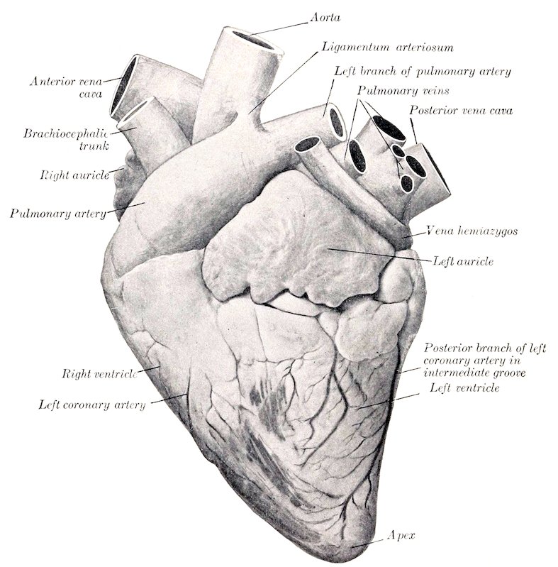
Fig. 467.—Heart of Ox, Left View.
The heart of the adult ox has an average weight of about 5½ to 6 pounds (ca. 2.5 to 2.7 kg.), or about 0.4 to 0.5 per cent. of the body-weight. Its length from base to apex is relatively longer than that of the horse and the base is smaller in both its diameters.
A shallow intermediate groove (Sulcus intermedius) extends from the coronary groove down the left side of the posterior border, but does not reach the apex. The amount of fat in and near the grooves is much greater than in the horse.
The heart is situated more to the left of the median plane than in the horse and is opposite to the third, fourth, and fifth ribs when it is contracted. It has extensive 609contact with the lateral wall of the thorax on the left side, but none on the right side, where a considerable thickness of lung covers the pericardium.[178]
Two bones, the ossa cordis, develop in the aortic fibrous ring. The right one is in apposition with the atrio-ventricular rings and is irregularly triangular in form. Its left face is concave and gives attachment to the right posterior cusp of the aortic valve. The right surface is convex from before backward. The base is superior. The posterior border bears two projections separated by a notch. It is usually a little more than an inch (ca. 4 cm.) in length. The left bone is smaller and is inconstant. Its concave right border gives attachment to the left posterior cusp of the aortic valve. There is a large fleshy moderator band in the right ventricle.
The Arteries[179]
The great arterial trunks in the thorax resemble those of the horse in general disposition.
The left coronary artery is much larger than the right one; it gives off a branch which descends in the intermediate groove, and terminates by running downward in the right longitudinal groove. The right artery, after emerging from the interval between the right auricle and the pulmonary artery, divides into branches which are distributed to the wall of the right ventricle.
The brachiocephalic trunk (anterior aorta) is usually four or five inches (ca. 10 to 12 cm.) in length.
The brachial arteries give off in the thorax the following branches:
1. A common trunk for the subcostal, dorsal, superior or deep cervical, and vertebral arteries.
(1) The subcostal artery commonly arises separately, but may be given off as in the horse. It supplies the first three intercostal arteries.
(2) The dorsal artery is relatively small. It usually ascends in front of the first costo-vertebral joint and is distributed as in the horse.
(3) The superior or deep cervical artery may arise from a common stem (Truncus vertebro-cervicalis) with the vertebral, or may constitute a branch of that artery. It passes up between the first thoracic and last cervical vertebræ or between the sixth and seventh cervical and is distributed as in the horse.
(4) The vertebral artery passes along the neck as in the horse to the intervertebral foramen between the second and third cervical vertebræ, gives off a muscular branch, and enters the vertebral canal (Fig. 469). It runs forward on the floor of the canal—connected with its fellow by two or three transverse anastomoses—and divides in the atlas into two branches. The smaller internal division (cerebrospinal artery) passes forward to the floor of the cranium and concurs with the condyloid artery and branches of the internal maxillary in the formation of a large rete mirabile. The large external branch emerges through the intervertebral foramen of the atlas and ramifies in the muscles of the neck in that region, compensating for the smallness of the branches of the occipital artery. It also sends a branch to the rete mirabile. The collateral branches detached to the cervical muscles are large and compensate for the small size of the deep cervical artery. The collateral spinal branches of the vertebrals pass through the intervertebral foramina, divide into anterior and posterior branches, and form two longitudinal trunks which are connected by cross-branches so as to form irregular polygonal figures.
2. The internal thoracic artery.
6103. The inferior cervical artery corresponds usually to the ascending branch of that vessel in the horse.
4. The external thoracic artery is large and usually gives off a branch which is equivalent to the descending branch of the inferior cervical artery of the horse.
THE COMMON CAROTID ARTERY
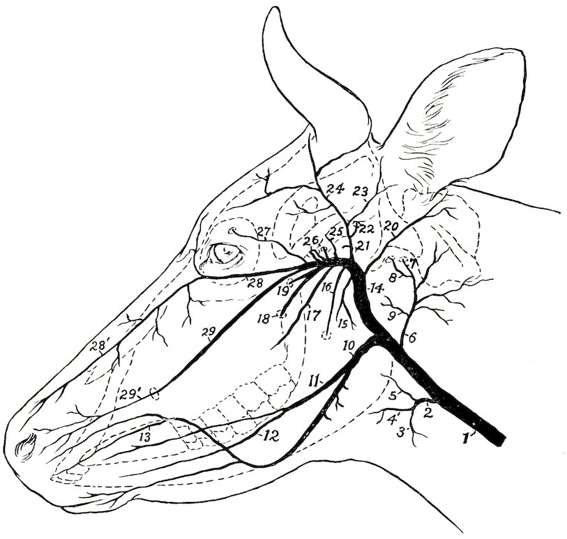
Fig. 468.—Schema of Chief Arteries of Head of Cow. 1, Common carotid artery; 2, thyro-laryngeal; 3, thyroid; 4, laryngeal; 5, pharyngeal; 6, occipital; 7, condyloid; 8, middle meningeal; 9, pharyngeal; 10, external maxillary; 11, lingual; 12, sublingual; 13, superior labial; 14, internal maxillary; 15, masseteric; 16, inferior alveolar; 17, buccinator; 18, great palatine; 19, sphenopalatine; 20, posterior auricular; 21, superficial temporal; 22, posterior meningeal; 23, anterior auricular; 24, artery to matrix of horn; 25, deep temporal; 26, arteries to rete mirabile; 27, frontal; 28, malar; 28′, dorsal nasal continuation of malar; 29, infraorbital; 29′, lateral nasal continuation of 29.
The carotid arteries usually arise from a common trunk about two inches (ca. 5 cm.) in length, but in exceptional cases are given off separately from the brachiocephalic. Each pursues a course similar to that of the horse and is accompanied by the small internal jugular vein, but is separated from the external jugular vein by the omo-hyoid and sterno-mastoid muscles. It divides at the digastricus into occipital, external maxillary, and external carotid arteries. In addition to tracheal, œsophageal, and muscular branches, it gives off the thyroid and laryngeal arteries. The thyroid artery (A. thyreoidea cranialis) bends around the anterior end of the thyroid gland, in which it ramifies. The accessory thyroid artery is usually absent. The laryngeal artery may arise with the thyroid.
6111. The occipital artery is relatively small. It gives off:
(1) The pharyngeal artery, which, however, may arise from the external carotid.
(2) Several branches to the muscles (chiefly the flexors) and to the atlanto-occipital joint.
(3) The condyloid artery passes into the cranium through the anterior foramen in the condyloid fossa, and joins the vertebral in the formation of the rete mirabile about the pituitary gland. Before entering the cranium it gives off a branch to the pharyngeal lymph glands, and the middle meningeal artery; the latter passes through the foramen lacerum. Another branch enters the temporal canal and gives twigs to the temporalis muscle and the mucous membrane of the frontal sinus. A muscular branch emerges from the intervertebral foramen of the atlas. A diploic branch goes into the occipital condyle and squama, and emits twigs to the occipital muscles.
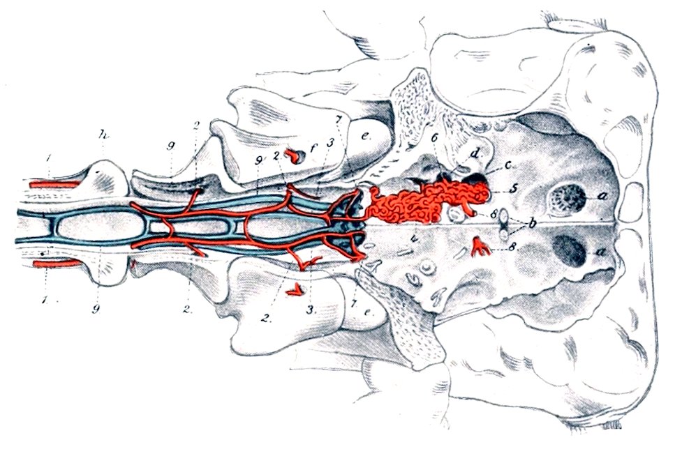
Fig. 469.—Floor of Cranium and Anterior Part of Vertebral Canal of Ox. 1, Vertebral artery; 2, muscular branches of 1; 3, branches of 1 to the rete mirabile, 4; 5, branches of internal maxillary artery to rete; 6, branch of internal maxillary artery entering cranium through foramen ovale; 7, condyloid artery; 8, emergent artery from rete, distributed like internal carotid artery of horse; 9, 9′, longitudinal vertebral sinuses; a, cribriform plate; b, optic foramina; c, for. lacerum orbitale + rotundum; d, foramen ovale; e, occipital condyle; f, g, h, first, second, and third cervical vertebræ. (After Leisering’s Atlas.)
2. The external maxillary artery is smaller than that of the horse, but pursues a similar course. The lingual artery is large, and often arises separately from the common carotid; it gives off a branch to the submaxillary gland, and the sublingual artery. After turning around the jaw the facial gives off the two labial arteries. The superior labial is large; it usually gives off a branch which runs forward almost parallel with the lateral nasal. The angular artery is absent or rudimentary, and the lateral and dorsal nasal arteries spring from branches of the internal maxillary.
3. The external carotid artery passes upward between the stylo-hyoideus and the great cornu of the hyoid bone, turns forward across the external face of the latter, and divides into superficial temporal and internal maxillary arteries. It gives off the following collateral branches:
(1) Branches to the parotid and submaxillary glands.
(2) The pharyngeal artery, which, however, often arises from the occipital.
(3) The posterior auricular, which resembles that of the horse and sends a stylo-mastoid branch into the tympanum. It may arise from the superficial temporal.
612(4) The masseteric artery resembles that of the horse, but is smaller.
The superficial temporal artery is large and presents the following special features: (1) Its transverse facial branch passes into the central part of the masseter. (2) It gives off a branch which corresponds to the posterior meningeal artery of the horse, enters the temporal canal, and ramifies in the dura mater, giving off twigs to the external ear, the temporal muscle, and the frontal sinus. (3) It usually gives off the anterior auricular artery. (4) Branches are supplied to the frontalis muscle and the eyelids, (5) A large branch passes around the outer side of the base of the horn-core, supplies the matrix of the horn, and anastomoses across the back of the frontal eminence with the artery of the opposite side.
The internal maxillary artery is less curved than in the horse and is entirely extraosseous, since the alar canal is absent. The principal differential features in its branching are as follows:
(1) The ophthalmic artery forms a rete mirabile within the periorbita. Its frontal branch enters the supraorbital canal and ramifies chiefly in the frontal sinus.
(2) Several branches take the place of the internal carotid artery. One of these enters the cranial cavity through the foramen ovale and several small ones pass through the foramen which represents the foramen rotundum and foramen lacerum orbitale of the horse. They concur with the vertebral and condyloid arteries in the formation of an extensive rete mirabile on the cranial floor around the sella turcica. From each side of the rete an artery arises which is distributed in general like the internal carotid of the horse.
(3) The malar artery is large; it arises by a common trunk with the infraorbital and gives off the dorsal nasal and the angular artery of the eye.
(4) The infraorbital artery is large and emerges from the infraorbital foramen to form the lateral nasal artery.
(5) The palatine artery is smaller than in the horse and usually arises by a common trunk with the sphenopalatine. It passes through the palatine canal and along the palatine groove, enters the nasal cavity through the incisive fissure, and does not go to the upper lip. It forms a rete mirabile about the naso-palatine canal and terminates in the mucous membrane of the anterior part of the nasal cavity.
ARTERIES OF THE THORACIC LIMB
The brachial artery pursues the same course in the arm as that of the horse. At the elbow it becomes the median.[180] The chief differential features in its branches are as follows:
1. The subscapular artery is almost as large as the continuation of the brachial. The posterior circumflex artery sends branches backward and downward into the triceps, taking the place in part of the deep brachial artery. The thoracico-dorsal artery supplies branches to the pectoral muscles and the triceps as well as the teres major and latissimus dorsi; it may arise directly from the brachial.
2. The deep brachial artery is small.
3. The superior collateral ulnar artery is often double, and does not extend to the carpus.
The median artery (posterior radial artery) descends along the inner part of the posterior surface of the radius and divides near the middle of the forearm into the radial and ulnar arteries. It gives off at the upper third of the forearm the common interosseous artery, a large vessel which anastomoses with the deep brachial, passes through the proximal interosseous space, and descends (as the dorsal interosseous) in the groove between the radius and ulna, and concurs in the formation of the rete carpi dorsale. At the distal end of the forearm it sends a branch 613through the distal interosseous space, which passes downward, assists in forming the rete carpi volare, and is continued in the metacarpus as the external deep volar metacarpal artery. This is a small vessel which passes down under the outer border of the suspensory ligament and assists in forming the deep volar arch near the fetlock.
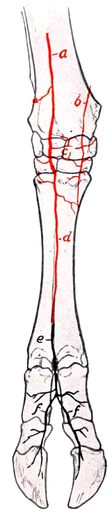
Fig. 470.—Arteries of Distal Part of Right Fore Limb of Ox, Anterior View. a, Interosseous artery; b, dorsal branch of radial artery; c, rete carpi dorsale; d, dorsal metacarpal artery; e, dorsal common digital artery; f, f, dorsal proper digital arteries.
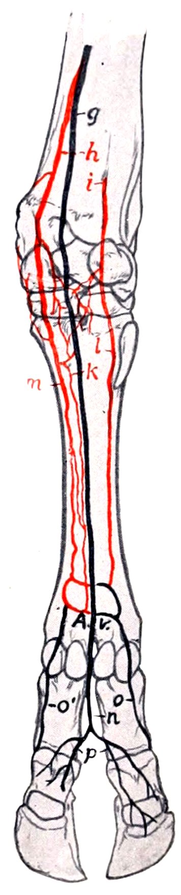
Fig. 471.—Arteries of Distal Part of Right Fore Limb of Ox, Posterior View. g, Ulnar artery; h, radial artery; i, volar branch of common interosseous artery; k, l, m, deep volar metacarpal arteries; A.v., volar arches; n, volar common digital artery; o, o′, p, volar proper digital arteries.
The radial artery is smaller than the ulnar. It descends on the flexor carpi internus, passes over the postero-internal surface of the carpus, and is continued as the internal deep volar metacarpal. At the distal end of the forearm and at the carpus it furnishes branches to the retia carpi. Another branch (A. met. perforans proximalis) runs outward between the suspensory ligament and the large metacarpal bone, passes through the proximal foramen of the bone, and unites with the interosseous artery; before passing through the foramen it detaches the middle deep volar metacarpal artery (A. met. volaris prof. III.), which descends on the posterior face of the metacarpal bone, receives an anastomotic branch from the internal deep artery, and concurs in the formation of the deep volar arch above the fetlock.
The ulnar artery, the larger of the two divisions of the median, descends under 614cover of the flexor carpi internus without giving off any large branches in the forearm. It passes through the carpal canal and continues along the inner side of the deep flexor tendon as the volar common digital artery. At the distal third of the metacarpus this vessel is joined by a branch from the internal deep volar metacarpal artery, forming the superficial volar arch. Near this another branch of the common digital (or of the internal digital) passes around the outer border of the flexor tendons to the posterior face of the metacarpal bone and concurs with the deep volar metacarpal arteries in the formation of the deep volar arch. A branch from the arch passes forward through the inferior foramen of the metacarpal bone and joins the dorsal metacarpal artery which descends from the rete carpi dorsale in the anterior metacarpal groove.
The volar common digital artery (A. metacarpea volaris superficialis III.) passes into the interdigital space and divides into two volar proper digital arteries, which descend along the interdigital surfaces of the chief digits and pass through the foramina at the upper part of the interdigital surfaces of the third phalanges, enter the cavities in these bones, and ramify in a manner similar to the corresponding vessels in the horse. The volar common digital detaches a branch (A. interdigitalis perforans) which passes forward through the upper part of the interdigital space and anastomoses with the dorsal metacarpal artery.
The volar proper digital arteries (or the common digital) give off, in addition to other collaterals, branches which correspond to the arteries of the plantar cushion of the horse. These pass to the bulbs of the claws and anastomose with each other and with the inner and outer digital arteries, forming an arch from which numerous branches are distributed to the matrix of the hoofs.
The dorsal metacarpal artery (A. met. dorsalis III.) is a small vessel which arises from the rete carpi dorsale, descends in the groove on the anterior face of the metacarpal bone, and is joined by the inferior perforating metacarpal artery from the deep volar arch to constitute the dorsal common digital artery. This vessel (A. digitalis communis dorsalis III.) divides into two dorsal proper digital arteries.
The internal digital artery (A. digiti III. medialis) is the continuation of the internal deep volar metacarpal artery. It descends on the inner side of the inner digit and terminates at the bulb of the claw by anastomosing with the corresponding volar proper digital artery. It gives off a branch to the rudimentary digit and forms a transverse anastomosis behind the first phalanx with the volar common digital or its inner division.
The external digital artery (A. digiti IV. lateralis) arises from the deep volar arch, passes down on the outer side of the external digit and is distributed like the inner one.
BRANCHES OF THE THORACIC AORTA
The bronchial and œsophageal arteries often arise separately.
Ten pairs of intercostal arteries usually arise from the aorta. The other three come from the subcostal artery.
The two phrenic arteries are very variable in origin. They may come from the aorta, the cœliac, left ruminal, or an intercostal or lumbar artery.
BRANCHES OF THE ABDOMINAL AORTA
The cœliac artery is about four to five inches (ca. 10 to 12 cm.) in length. It passes downward and curves forward between the rumen and pancreas on the left and the right crus of the diaphragm and the posterior vena cava on the right. It gives off five chief branches.
1. The hepatic artery arises from the convex side of the curve of the cœliac artery as it crosses the posterior vena cava. It passes downward, forward, and to 615the right above the portal vein to the portal fissure, and gives off the following branches:
(1) Pancreatic branches.
(2) Dorsal and ventral branches to the liver. The ventral branch is the larger; it gives off the right gastric artery, which runs in the lesser omentum to supply the origin of the duodenum and the pylorus, anastomosing with the dorsal branch of the omaso-abomasal artery.
(3) The cystic artery supplies the gall-bladder.
(4) The gastro-duodenal artery divides into right gastro-epiploic and pancreatico-duodenal branches. The right gastro-epiploic artery anastomoses with the left gastro-epiploic. The pancreatico-duodenal artery anastomoses with the first intestinal branch of the anterior mesenteric artery.
2. The right ruminal artery (A. ruminalis dextra) is the largest branch, and usually arises by a short common trunk with the splenic. It runs downward and backward on the right face of the dorsal sac of the rumen to the posterior transverse fissure, in which it turns around to the left and anastomoses with branches of the left ruminal artery. It gives off a pancreatic branch, dorsal and ventral coronary arteries, branches to the great omentum, and ramifies on both surfaces of the rumen.
3. The left ruminal artery (A. ruminalis sinistra) runs downward on the anterior part of the right face of the rumen, enters the anterior furrow, in which it runs from right to left, and continues backward in the left longitudinal groove, anastomosing with branches of the right artery. It supplies chiefly the left face of the rumen, but not its posterior part. It usually gives off near its origin the reticular artery (A. reticularis); this rather small vessel passes forward on the dorsal curvature of the rumen and turns downward in the rumino-reticular groove, in the bottom of which it runs around ventrally to the right side. It gives off a branch which passes to the left of the cardia and along the lesser curvature of the reticulum to the neck of the omasum. The reticular branches anastomose with the omaso-abomasal and left ruminal arteries.
4. The omaso-abomasal artery (A. gastrica sinistra) appears as the continuation of the cœliac. It passes forward and downward to the greater curvature of the omasum and divides after a course of four or five inches into two branches. The dorsal branch curves sharply backward on the dorsal surface of the omasum, continues along the lesser curvature of the abomasum, and anastomoses with the hepatic artery. It supplies branches to the omasum and to the lesser curvature and pyloric part of the abomasum. The ventral branch (A. gastroepiploica sinistra) runs forward and downward over the anterior extremity of the omasum, passes backward in the great omentum close to the ventral curvature of the abomasum, and anastomoses with the right gastro-epiploic. A considerable branch from it curves around in front of the neck of the omasum to communicate with the reticular artery. Another branch runs back across the left side of the omaso-abomasal junction, and a third goes to the lesser (ventral) curvature of the omasum.
5. The splenic artery usually arises by a common trunk with the right ruminal artery. It passes forward and to the left across the dorsal curvature of the rumen and enters the hilus of the spleen.
The anterior mesenteric artery arises from the aorta just behind the cœliac, and has about the same caliber as the latter. It passes downward and a little to the right, and crosses the colon as it emerges from the spiral to run backward. After detaching twigs to the pancreas it gives off in succession the following branches:
1. A branch (A. colica media) passes to the colon as it emerges from the spiral arrangement. This artery is comparable to the middle colic or artery of the small colon of the horse; it runs backward along the terminal part of the colon, which it supplies.
616
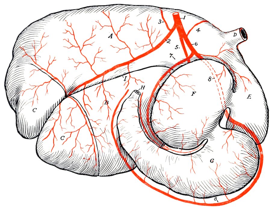
Fig. 472.—Gastric Arteries of Ox, Right View (Partly Schematic). 1, Cœliac artery; 2, right ruminal; 3, splenic; 4, reticular; 5, left ruminal; 6, omaso-abomasal; 7, dorsal branch of 6; 8, ventral branch of 6 (= left gastro-epiploic); A, dorsal sac of rumen; B, ventral sac of rumen; C, C, posterior blind sacs; D, œsophagus; E, reticulum; F, omasum; G, abomasum; H, duodenum; I, right longitudinal furrow of rumen. By an oversight the reticular artery is shown as arising from the omaso-abomasal.
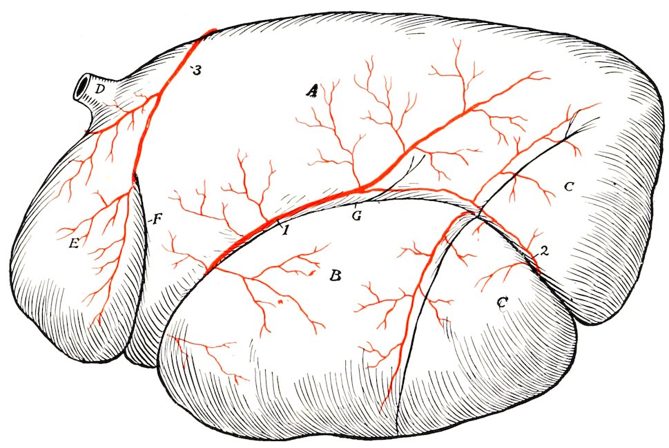
Fig. 473.—Gastric Arteries of Ox, Left View (Partly Schematic). 1, Left ruminal artery; 2, continuation of right ruminal artery; 3, reticular artery, which disappears into rumino-reticular groove; A, dorsal sac of rumen; B, ventral sac of rumen; C, C, posterior blind sacs; D, œsophagus; E, reticulum; F, rumino-reticular groove; G, left longitudinal furrow of rumen.
6172. The ileo-cæco-colic artery ramifies on the right face of the spiral part of the colon. It gives off the ileo-cæcal artery, which divides into ileal and cæcal arteries.
3. An artery (Ramus collateralis) runs in the mesentery in a curve along the ventral border of the coils of the colon. (This vessel is absent in the sheep.)
4. An artery which constitutes the direct continuation of the anterior mesenteric pursues a course in the mesentery corresponding to the series of mesenteric lymph glands.
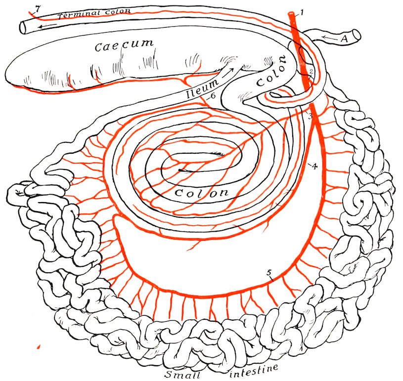
Fig. 474.—Plan of Branches of Anterior Mesenteric Artery of Ox. 1, Anterior mesenteric artery; 2, middle colic artery; 3, ileo-cæco-colic artery; 4, ramus collateralis; 5, continuation of anterior mesenteric artery, giving off branches to small intestine; 6, cæcal artery; 7, colic branch of posterior mesenteric artery; A, termination of duodenum.
The two preceding vessels are essentially the arteries of the small intestine, which they supply with the exception of its initial and terminal parts. The first gives off no considerable branches in its course along the ventral border of the coils of the colon, but on curving upward along the latter it anastomoses with the second artery and detaches numerous branches to the small intestine which form series of superposed anastomotic arches. It supplies, roughly speaking, about one-third of the small intestine and terminates by joining the ileal artery. The second artery gives off numerous branches which also form arches and supply about the first two-thirds of the small intestine, exclusive of the small part supplied by branches of the cœliac artery. Both arteries give branches to the lymph glands.[181]
618The posterior mesenteric artery arises from the aorta near its termination. It is small and supplies branches to the terminal part of the colon and to the rectum.
The renal arteries arise from the aorta close together. The right one passes outward and forward across the dorsal face of the posterior vena cava to the hilus of the kidney. The left one runs backward, but necessarily varies in direction in conformity with the position of the kidney (q. v.).
The spermatic arteries resemble those of the horse.
The utero-ovarian arteries are small.
The five pairs of lumbar arteries derived from the aorta are distributed much as in the horse. The sixth usually comes from the internal iliac artery.
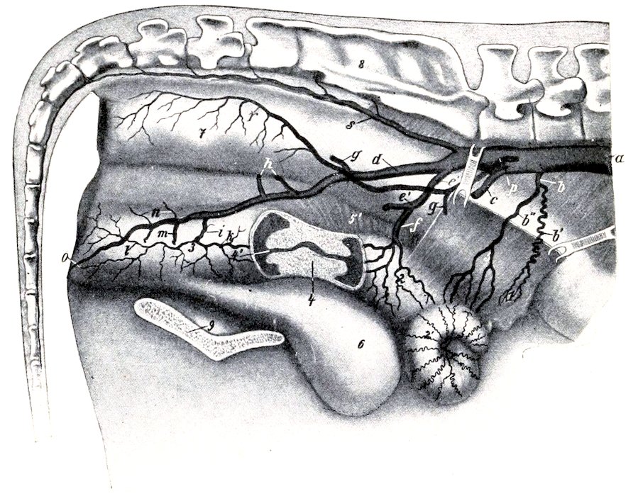
Fig. 475.—Pelvic Arteries of Cow. Part of the right wall of the uterus and vagina is removed and the cervix uteri is shown in sagittal section. a, Aorta; b, utero-ovarian artery; b′, ovarian, and b″, uterine branch of b; c, external iliac artery; d, right internal iliac artery; e, common trunk of umbilical artery (e′) and middle uterine artery (f); g, ilio-lumbar artery; h, anterior gluteal arteries; i, middle hæmorrhoidal artery; k, posterior uterine artery; l, perineal artery; m, posterior gluteal artery; n, obturator arteries; o, artery of clitoris; p, posterior mesenteric artery; r, anterior hæmorrhoidal artery; s, middle sacral artery; 1, ovary; 2, apparent body of uterus, really apposed horns; 2′, cornu; 3, vagina; 4, cervix uteri; 4′, vaginal part of uterus (os uteri); 5, 5′, broad ligaments, large part of right one removed; 6, urinary bladder; 7, rectum; 8, sacrum; 9, symphysis pelvis. (After Zieger.)
The middle sacral artery is a vessel about 5 mm. in diameter which continues the aorta. It arises from the dorsal face of the aorta at the angle of divergence of the internal iliacs, runs backward on the pelvic surface of the sacrum a little to the left of the median line, and is continued as the middle coccygeal artery. It gives off small collateral branches to the spinal cord and the muscles of the tail and the lateral coccygeal arteries. The latter may have a common trunk of origin, and each divides into dorsal and ventral branches. The middle coccygeal artery runs through the ventral (hæmal) arches of the coccygeal vertebræ. The coccygeal arteries are connected at pretty regular intervals by segmental anastomoses.
The internal iliac arteries are much longer than in the horse. Each passes backward on the sacro-sciatic ligament and divides about the middle of the pelvic wall into posterior gluteal and internal pudic branches. The chief differences in its distribution are: (1) A large trunk gives origin to the umbilical and middle 619uterine arteries. The umbilical artery is usually largely obliterated and its terminal branches receive their blood through anastomoses with the internal pudic. It gives off near its origin two small vessels, the ureteral artery (A. ureterica) and the deferential artery (A. deferentialis), which accompany the ureter and the vas deferens respectively. The middle uterine artery (A. uterina media) is very large. It is distributed chiefly to the cornu of the uterus, and compensates for the small size of the utero-ovarian artery. (2) The ilio-lumbar artery is relatively small and is distributed chiefly to the sublumbar muscles. It is sometimes replaced by branches of the circumflex iliac and gluteal arteries. (3) The anterior gluteal artery is commonly represented by several vessels. (4) The obturator artery is represented by several small branches which supply the obturator and adductor muscles. (5) The iliaco-femoral and lateral sacral arteries are absent. The absence of the latter is compensated by the middle sacral and gluteal arteries. (6) The posterior gluteal (or ischiatic) artery is large. It emerges through the lesser sciatic notch and ramifies in the biceps femoris and adjacent muscles. (7) The internal pudic artery (A. urethro-genitalis) is the direct continuation of the internal iliac. It gives off branches to the rectum, bladder, urethra, and genital organs. In the male it supplies the accessory genital glands and divides into dorsal and deep arteries of the penis; the a. dorsalis penis runs along the dorsum penis to the glans and gives twigs to the prepuce; the a. profunda penis gives off a perineal branch and enters the corpus cavernosum penis. In the female it gives off a large posterior uterine artery, which supplies the posterior part of the uterus and gives branches to the vagina and bladder. It ends as the a. clitoridis, which supplies the clitoris and adjacent parts.
ARTERIES OF THE PELVIC LIMB
The external iliac artery has the same course as in the horse. The circumflex iliac artery is large. A branch from it emerges between the abdominal and lumbar muscles near the external angle of the ilium and ramifies like the terminals of the ilio-lumbar artery of the horse.
The femoral and popliteal arteries pursue a similar course to those of the horse. The chief differences in their branches are as follows:
1. The external pudic artery is distributed chiefly to the scrotum in the male. In the cow it is usually termed the mammary and is very large, especially during lactation. Each divides at the base of the mammary gland into two branches which are distributed to the anterior and posterior parts (“quarters”) of the gland. A small branch accompanies the subcutaneous abdominal vein to the xiphoid region.
2. The deep femoral artery gives off an obturator branch which passes up through the obturator foramen to supply the obturator internus and compensates otherwise for the absence of the obturator artery.
3. The anterior femoral artery is large. It often gives off the external circumflex artery of the thigh, which perforates the proximal end of the quadriceps, gives branches to that muscle, the iliacus, glutei, and tensor fasciæ latæ.
4. The saphenous artery is large. It descends in front of the homonymous vein to the postero-internal surface of the hock, where it divides into two plantar branches. The internal plantar artery (A. plantaris medialis) is the direct continuation of the saphenous. It descends at first along the inner border of the superficial flexor tendon and is continued as the internal superficial plantar metatarsal artery along the inner side of the deep flexor tendon with the internal plantar nerve. It anastomoses at the proximal end of the metatarsus with the perforating metatarsal artery, assisting in the formation of the proximal plantar arch. Near the fetlock it concurs with the perforating branch of the dorsal metatarsal artery in the formation of the distal plantar arch. Below this it is continued as the internal 620digital artery. The external plantar artery (A. plantaris lateralis) is small. It descends along the outer border of the deep flexor tendon with the external plantar nerve, concurs with the perforating tarsal and the internal plantar arteries in the formation of the proximal plantar arch, and gives branches to the rete tarsi dorsale. Continuing downward along the deep flexor tendon as the external superficial plantar metatarsal artery, it assists in forming the distal plantar arch and becomes the external digital artery.

Fig. 476.—Arteries of Distal Part of Right Hind Limb of Ox, Anterior View. a, Anterior tibial artery; b, proximal perforating metatarsal artery; c, dorsal metatarsal artery; d, dorsal common digital artery; e, e′, dorsal proper digital arteries.
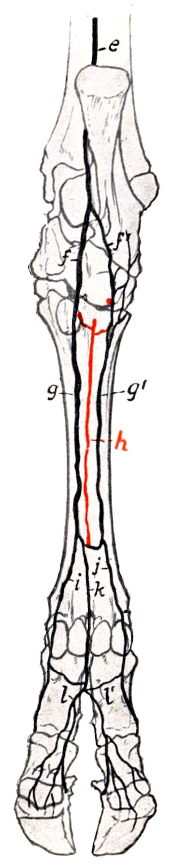
Fig. 477.—Arteries of Distal Part of Right Hind Limb of Ox, Posterior View. e, Saphenous artery; f, f′, internal and external plantar arteries; g, g′, internal and external superficial plantar metatarsal arteries; h, deep plantar metatarsal artery; i, j, internal and external plantar digital arteries; k, plantar common digital artery; l, l′, internal and external plantar digital arteries.
The posterior tibial artery is relatively small and is distributed chiefly to the muscles on the posterior surface of the tibia. Lower down it is replaced by the saphenous artery as described above.
The anterior tibial artery has the same course as in the horse. It is continued down the groove on the front of the metatarsal bone as the dorsal metatarsal artery. This is the chief artery of the region and is accompanied by two veins. It detaches the proximal perforating metatarsal artery, which passes through the proximal foramen of the metatarsal bone and concurs with the superficial plantar 621arteries in forming the proximal plantar arch. Near the distal end of the metatarsal bone it gives off the distal perforating metatarsal artery which passes back through the distal metatarsal foramen and assists in forming the distal plantar arch. The two arches are connected by the small deep plantar metatarsal artery which lies in the groove on the posterior face of the large metatarsal bone.
The dorsal common digital artery is the direct continuation of the dorsal metatarsal. It divides into two branches which unite in the interdigital space with the corresponding branches of the plantar common digital to form the proper digital arteries.
The plantar common digital artery descends from the distal plantar arch, anastomoses in the interdigital space with the internal and external digitals, and divides into two branches which join those of the dorsal common digital artery as before mentioned.
The foregoing is a brief statement of the more common arrangement of the vessels in the distal part of the limb, but minor variations are very common.
The Veins[182]
The vena hemiazygos usually takes the place of the vena azygos. It lies along the left side of the aorta and the bodies of the thoracic vertebræ, turns down across the left face of the aorta and left pulmonary artery, runs back over the left auricle and opens into the great coronary vein or the right atrium. It receives the intercostal veins.
Two jugular veins occur on either side. The internal jugular vein (V. jugularis interna) is a relatively small vessel which accompanies the carotid artery. It arises by occipital, laryngeal, and thyroid radicles, receives tracheal, œsophageal, and muscular branches, and joins the external jugular near its termination. It is sometimes absent, but in some cases it appears, on the other hand, to be large enough to interfere with venesection practised on the external jugular. The external jugular vein (V. jugularis externa) is very large and corresponds to the single jugular of the horse. It is separated from the carotid artery in the greater part of its course by the sterno-cephalicus and omo-hyoideus muscles.
The inferior cerebral vein usually does not unite with the occipital; the latter is continued by the internal jugular vein.
The orbital veins form a network between the periorbita and the muscles of the eyeball. This plexus communicates with the cavernous sinus and with the superior cerebral vein. It is also drained by the frontal vein which runs in the supraorbital canal and groove and joins the angular vein of the eye.
The dorsal nasal vein is usually double.
The superior labial vein usually joins the infraorbital.
The vena reflexa is absent and the radicles which are received by it in the horse go to the internal maxillary vein.
The sublingual vein is very large.
The veins of the thoracic limb differ chiefly in the distal part; the special features are as follows:
The dorsal digital veins ascend on the front of the digits and are connected with the other digital veins by transverse branches. They unite near the fetlock to form the dorsal metacarpal vein. This runs upward on the anterior face of the metacarpus and carpus, inclines to the inner surface of the radius, and joins the accessory cephalic or the cephalic vein.
The volar digital veins are larger than the dorsal. They lie on the interdigital 622surfaces of the digits and unite in the interdigital space to form a trunk which is a satellite of the volar common digital artery.
The internal and external digital veins lie in front of the corresponding arteries. They are connected with the volar digital vein by a large branch which passes between the flexor tendons and the first phalanx. At the distal end of the metacarpus each inclines forward and anastomoses with the volar common digital vein to form the volar venous arch. The inner vein is continued as the internal volar metacarpal vein along the inner border of the suspensory ligament, and becomes a satellite of the radial artery in the forearm, while the outer one is continued on the posterior face of the metacarpal bone by two irregular veins, the external and middle volar metacarpals. The latter anastomose freely with each other and with the inner vein. They unite below the carpus or join the volar common digital vein.
The accessory cephalic vein is the upward continuation of the dorsal metacarpal vein and is much larger than in the horse.
The posterior vena cava is partially embedded in the medial border of the liver. Its abdominal part has a thicker wall than in the horse. Its affluents correspond to the arteries of which they are satellites. The renal veins are large and thick-walled; they run obliquely forward and join the vena cava at an acute angle. The left one is much the longer.
Two middle sacral veins usually accompany the artery.
The veins of the mammary glands deserve special notice. They converge to a venous circle at the base of the udder, which is drained chiefly by two pairs of veins. The subcutaneous abdominal vein (anterior mammary or “milk” vein) is very large in animals of the dairy breeds and its course along the ventral wall of the abdomen is easily followed. It is usually flexuous. It emerges at the anterior border of the udder about two or three inches (ca. 5 to 8 cm.) from the linea alba, runs forward (deviating a little outward), dips under the panniculus, passes through a foramen in the abdominal wall about a handbreadth from the median plane, and joins the internal thoracic vein. The external pudic vein (middle mammary vein) is also of considerable size. It ascends in the inguinal canal as a satellite of the artery and joins the external iliac vein. The right and left veins are connected at the posterior border of the base of the udder by a large transverse branch. From the latter arises the perineal vein (posterior mammary vein), which runs medially upward and backward to the perineum, turns around the ischial arch, and joins the internal pudic vein. In the male these veins are relatively small.
The deep veins of the thigh and leg resemble those of the horse, but there is no recurrent tibial vein.
The saphenous vein is much smaller than in the horse.
The recurrent tarsal vein (external saphenous) is large. It is the upward continuation of the external plantar metatarsal vein and anastomoses with the anterior tibial and saphenous veins. It arises on the outer face of the hock, ascends at first in front of the tendo Achillis, then crosses the latter externally, passes up between the biceps femoris and semitendinosus, and joins the posterior femoral vein.
There are three chief metatarsal veins. The great dorsal metatarsal vein arises at the distal part of the metatarsus by the union of the dorsal digital vein and a large branch from the venous arch above the sesamoids. It ascends superficially between the long and lateral extensor tendons and becomes the chief radicle of the anterior tibial vein. The internal plantar metatarsal vein arises from the venous arch above the fetlock, ascends along the inner border of the suspensory ligament, passes through the vascular canal of the metatarsus and centro-tarsal (as the perforating tarsal) and joins the anterior tibial vein. The external plantar 623metatarsal vein is larger than the preceding. It passes superficially over the outer face of the hock and is continued by the recurrent tarsal vein.
The digital veins differ from those of the fore limb chiefly in that the dorsal vein is large and the plantar absent or small.
The portal vein is formed usually by the confluence of two radicles, gastric and mesenteric. It receives the right gastro-epiploic vein and veins of the pancreas. The gastric vein is the largest affluent. It is formed by the junction of three veins, the left of which receives the splenic vein. The anterior mesenteric vein is also formed by the confluence of three radicles; it usually receives the small posterior mesenteric vein. The portal tributaries are in general satellites of the corresponding arteries.
The Lymph Vessels and Glands
The thoracic duct arises from a small cisterna chyli and is very variable. It is exceptional to find a single trunk throughout, as is often the case in the horse. The duct is largely covered by fat and small lymph glands. There are often two ducts, one on either side of the aorta, which may join at a variable point or terminate close together at the junctions of the jugular and brachial veins. In other cases the duct is single for a variable distance, then bifurcates or divides into three or four branches, which are often connected by plexiform anastomoses.
The efferent vessels from the intestine converge to a large trunk which accompanies the anterior mesenteric artery and vein. It passes below the pancreas and receives usually a large duct which is formed by efferent vessels from the stomach, liver, and spleen; this duct sometimes opens directly into the cisterna. The gastro-intestinal trunk receives the collecting duct (truncus lumbalis) of the lumbar lymph vessels, forms a bend around the right renal vessels, and joins the cisterna.
The lymph glands in the ox are in general less numerous but larger than those of the horse, and in some situations a single large gland occurs instead of a group of smaller ones, as found in the latter animal.
The submaxillary lymph glands are usually two in number, right and left; each is situated under the external maxillary vein between the submaxillary salivary gland and the sterno-cephalicus muscle. Usually one or two hæmolymph glands are near it. There is generally a small gland on the deep face of the anterior part of the submaxillary salivary gland, and small nodes may be found further forward in the submaxillary space.
A large subparotid lymph gland lies on the postero-superior part of the masseter muscle, partly under cover of the upper end of the parotid gland. Several hæmolymph glands lie on its deep face.
The pharyngeal lymph glands number two or three on each side. Of these, two large suprapharyngeal glands (Lg. retropharyngeales) are situated about an inch apart between the dorsal wall of the pharynx and the ventral straight muscles (Figs. 281, 365). These are two to three inches long. Enlargement of them is likely to cause difficulty in swallowing and in respiration. Behind these are several hæmolymph glands. An atlantal gland is situated below the wing of the atlas on the spinal accessory nerve. It is discoid, oval in outline, and may be an inch and a half or more in length. It is partly covered by the upper end of the submaxillary salivary gland.
A smaller lymph gland (parapharyngeal) is usually found along the lower border of the carotid artery (i. e., ventral to the atlantal gland) on the lateral wall of the pharynx and under cover of the submaxillary salivary gland or at its posterior border.
Two or three small anterior cervical lymph glands commonly lie along the carotid artery a little further back.
624The middle cervical lymph glands comprise a series of small nodes along the dorsal face of the trachea.
In the sheep and goat there is a lymph gland at the middle of the neck, in the angle between the spinalis and complexus, covered by the splenius. It receives vessels from the atlantal gland and sends efferents to the prescapular gland.
The prescapular or superficial cervical lymph gland is situated at the anterior border of the supraspinatus under cover of the mastoido-humeralis and omo-transversarius. It is elongated and may be an inch or more in width and four or five inches long. Two occur in exceptional cases.
A chain of hæmolymph glands lies along the front of the shoulder, covered above by the trapezius. Others are frequently found (in the calf especially) on the surface of the omo-transversarius at the point where it passes under the mastoido-humeralis (Forgeot).
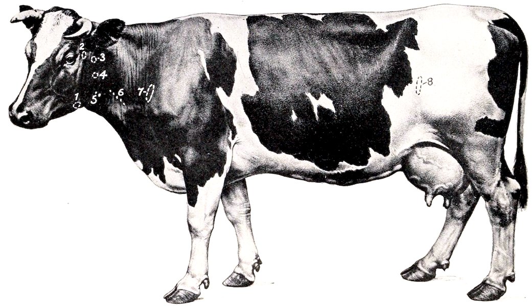
Fig. 478.—Superficial Lymph Glands of Cow Projected on Surface of Body. 1, Submaxillary; 2, subparotid; 3, atlantal; 4, parapharyngeal; 5, anterior cervical; 6, middle cervical; 7, prescapular; 8, precrural. (With use of fig. in Ellenberger-Baum, Anat. f. Künstler.)
The posterior cervical or prepectoral lymph glands number commonly three or four on each side. One or two are placed on the brachial vessels, one below the brachial vein at the first rib, and one at the junction of the common trunk of the external and subcutaneous thoracic veins with the brachial.
The intercostal lymph glands are situated, as in the horse, at the upper parts of the intercostal spaces. Associated with them are hæmolymph glands.
The sternal lymph glands are situated along the course of the internal thoracic artery at the lower part of the intercostal spaces. The largest is close to the thoracic inlet. Several glands occur in the fat about the apex of the pericardium.
4. Several anterior mediastinal lymph glands are placed along the œsophagus and the trachea.[183] Hæmolymph glands occur here also.
Usually three large bronchial lymph glands are present (Fig. 289). One is situated at the origin of each chief bronchus and the third is under the trachea at the origin of the special bronchus of the apical lobe of the right lung. Another 625may lie between the aorta and the left branch of the pulmonary artery. These glands are often pigmented. Other small nodes are situated on the bronchi within the lungs.
The posterior mediastinal lymph glands comprise two or three of large size placed along the dorsal wall of the œsophagus. There may be a single gland about eight inches (ca. 20 cm.) long, the anterior half of which lies on the œsophagus (Fig. 289).[184] A small gland is situated in the acute angle formed by the posterior vena cava and the diaphragm.
A single axillary lymph gland is situated on the distal part of the teres major on the course of the vein from the latissimus dorsi.
The lumbar lymph glands form an irregular series scattered along the abdominal aorta and posterior vena. A bean-shaped renal lymph gland occurs at the hilus of each kidney. Hæmolymph glands occur along the course of the aorta.
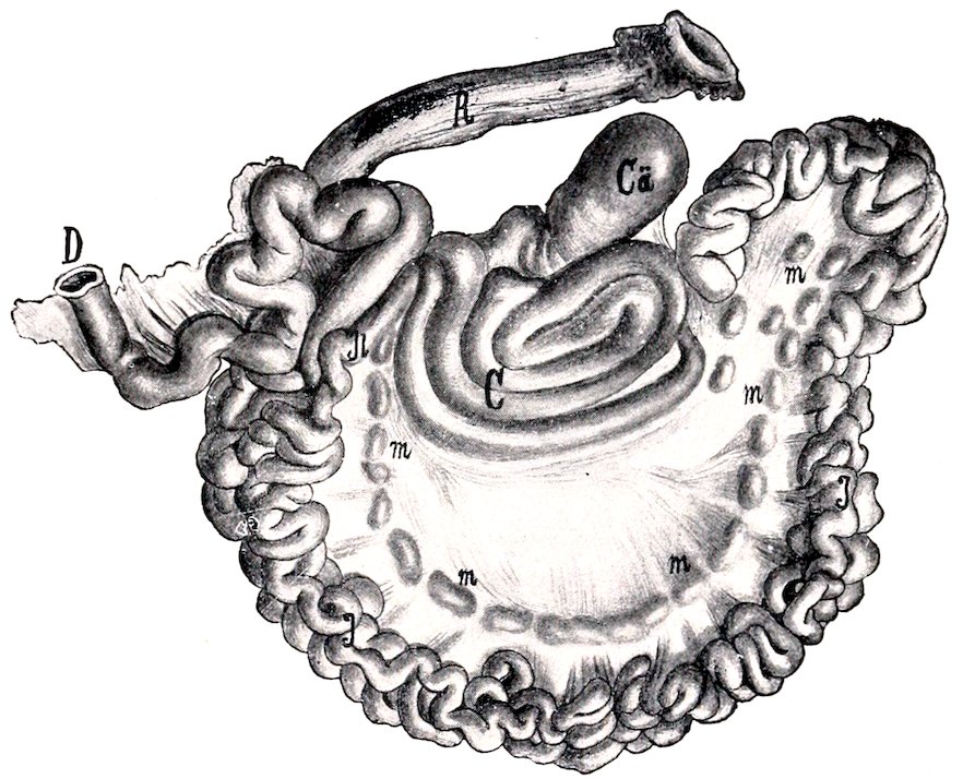
Fig. 479.—Intestine of Ox, Spread Out. Ca, Cæcum; C, colon; D, duodenum; H, ileum; J, jejunum; R, rectum; m, mesenteric lymph glands. (After Edelmann.)
The internal iliac lymph glands comprise a group of five or six at the termination of the aorta and the origin of the vena cava. A discoid gland, two inches or more in diameter, occurs near the side of the pelvic inlet at the angle of divergence of the circumflex iliac artery from the external iliac. A small gland is sometimes found at the origin of the prepubic artery.
The external iliac lymph glands number commonly two on either side, and are situated near the point of the hip in front of the anterior branch of the circumflex iliac artery.
The superficial inguinal lymph glands are situated centrally below the prepubic tendon and in the narrow space between the origins of the graciles. In the male there are usually two or three on either side of the penis behind the sigmoid flexure. In the cow they are supramammary, i. e., are situated above the posterior margin 626of the base of the udder; two large glands which are in apposition with each other medially are constant, and above these there are often two smaller glands.
An ischiatic lymph gland, which is discoid and usually about an inch in diameter, is situated on the lower part of the sacro-sciatic ligament near the lesser sciatic notch under cover of the biceps femoris.
An anal lymph gland is situated on the retractor ani on either side.
The gastric lymph glands are numerous and comprise: (1) a chain along the course of the right ruminal artery and two or three in the left groove of the rumen; (2) several on the reticulum above and below the junction with the omasum; (3) a series along the course of the superior omasal vessels and the lesser curvature of the abomasum; (4) an extensive chain along the ventral face of the omasum and the great curvature of the abomasum at the attachment of the great omentum.
Two or three large hepatic or portal lymph glands are found at the upper part of the portal fissure of the liver.
No lymph glands occur at the hilus of the spleen. The efferent vessels from the spleen appear to go to glands along the course of the cœliac artery.
A large discoid cœliac lymph gland lies on the pancreas and the cœliac artery, covering the latter as it gives off its primary branches.
The mesenteric lymph glands are large and numerous. They form a chain in the lower part of the mesentery along the course of the great mesenteric artery. They are chiefly elongated and narrow, and some have a length of six to eight inches (15 to 20 cm.) or more. Sometimes several unite and form a narrow band about two feet in length. A large gland occurs at the origin of the spiral part of the colon, a group is found near the anterior mesenteric trunk, and others are scattered along the coils of the colon, which are best seen on the right side. A chain of small lymph glands extends along the terminal part of the colon and the dorsal face of the rectum.
The precrural lymph gland is situated on the aponeurosis of the external oblique at the anterior border of the tensor fasciæ latæ a short distance above the stifle. It is elongated like the prescapular gland and may be six inches (ca. 15 cm.) long. Several subcutaneous hæmolymph glands occur in this vicinity; others are situated further forward on the flank not far from the last rib. There is usually a round, flattened lymph gland about an inch in diameter on the upper part of the quadriceps femoris under cover of the tensor fasciæ latæ, and one or more small ones occur on the surface of the latter muscle.
No deep inguinal lymph glands are present.
A single popliteal lymph gland an inch or more in length is situated somewhat lower down on the gastrocnemius than in the horse, behind the tibial and peroneal nerves.
CIRCULATORY SYSTEM OF THE PIG
THE HEART
The pericardium is attached to the sternum from a point opposite to the third rib as far as the xiphoid cartilage, and also to the sternal part of the diaphragm.
The heart is small in proportion to the body-weight, especially in fat animals. It is relatively short and wide. The apex is blunt and is marked by a notch (Incisura apicis). Its long axis is more oblique, but it is less asymmetrical with regard to the median plane, than in the horse or ox. The lower border of the left auricle (appendix) is marked by several notches and is situated at a lower level than the 627right one. The right longitudinal groove is placed far back and there is no posterior sulcus.
The Arteries
The pulmonary vessels present no remarkable features.
The aorta resembles that of the horse and ox in its course and relations, but the arch is more strongly curved. There is no common brachiocephalic trunk (anterior aorta).
The brachiocephalic artery arises first from the aortic arch and passes forward below the trachea to the first rib. Here it gives off the common carotid arteries, and is continued around the first rib as the right brachial.
The superior cervical, vertebral, and dorsal arteries usually arise by a common trunk.
The inferior cervical artery is large; its ascending branch gives off the posterior thyroid artery and parotid branches.
The internal and external thoracic arteries give branches to the pectoral mammary glands; the external may be absent.
The left brachial artery arises from the aortic arch just above the brachiocephalic. It curves forward and downward and turns around the anterior border of the first rib. Its superior cervical, vertebral, and dorsal branches usually arise separately.
The carotid arteries arise from the brachiocephalic close together or by a very short common trunk. They terminate in occipital and internal and external carotid divisions.
The occipital artery resembles that of the horse.
The internal carotid artery usually arises by a common trunk with the occipital. After giving off a large meningeal branch it passes through the foramen lacerum and forms with the opposite artery a rete mirabile which resembles that of the ox, but is smaller and is not connected with the vertebral and condyloid arteries.
The external carotid artery has the same course and termination as in the horse. Its branches present the following special features: (1) The lingual artery is relatively large and supplies the pharyngeal artery, the sublingual, muscular branches, and branches to the soft palate, submaxillary gland, and larynx. (2) The external maxillary artery is small and is distributed chiefly to the submaxillary gland, the pharyngeal lymph glands, the masseter and the panniculus. It does not extend upon the lateral surface of the face. (3) The posterior auricular artery is long and relatively large; it gives off the posterior meningeal.