ANGIOLOGY: THE VASCULAR SYSTEM (continued 2)
Reading edition
7. The nutrient artery of the humerus (A. nutritia humeri) is a short vessel which enters the nutrient foramen of the humerus. It often arises from the ulnar.
8. The anterior radial artery (A. collateralis radialis inferior) passes downward and a little outward on the anterior face of the humerus under cover of the biceps and brachialis to the front of the elbow joint, where it is joined by the radial nerve. It then descends on the anterior surface of the radius under cover of the anterior extensor of the digit to the carpus, where it concurs in the formation of the rete carpi dorsale, anastomosing with the posterior radial and interosseous arteries. It supplies branches to the elbow joint, the biceps, brachialis, and the extensors of the carpus and digit. A cutaneous branch emerges between the distal end of the biceps and the brachialis.
THE POSTERIOR RADIAL ARTERY
The posterior radial or median artery (A. mediana) is the direct continuation of the brachial. It passes downward and slightly backward, at first on the inner surface of the humerus, and then over the capsule and internal lateral ligament of the elbow joint, under cover of the posterior superficial pectoral muscle.[163] Below the elbow it dips under the flexor carpi internus and passes down the inner part of the posterior surface of the radius. In the distal part of the forearm it inclines backward and is separated from the radius by the reinforcing band (Caput tendineum) of the superficial flexor of the digit and is continued by the large metacarpal artery.
560It is accompanied by the median nerve, which lies in front of the artery at its origin, then crosses over it obliquely at the elbow joint and becomes posterior. In the arm the brachial vein lies behind and partly upon the artery; lower down there are usually two satellite veins, anterior and posterior. The chief collateral branches are as follows:
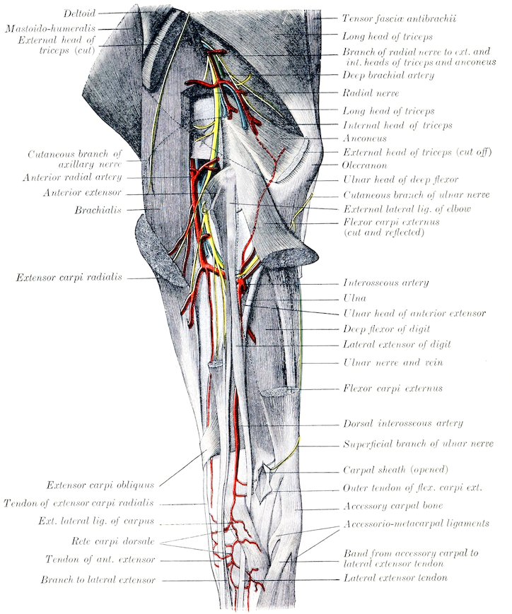
Fig. 443.—Dissection of Left Forearm of Horse, External Surface. (After Schmaltz, Atlas d. Anat. d. Pferdes.)
1. Articular branches for the elbow joint.
5612. Muscular branches to the flexors of the carpus and digit. The largest of these arise at the proximal third of the forearm.
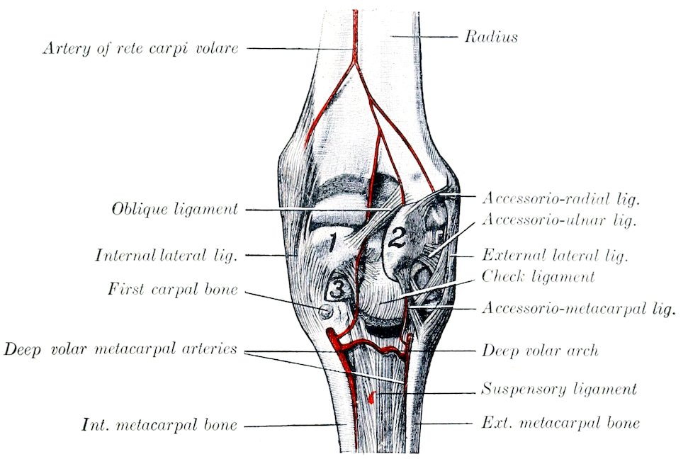
Fig. 444.—Deep Dissection of Right Carpus of Horse, Posterior View. 1, Radial carpal bone; 2, accessory carpal bone; 3, second carpal bone. (After Schmaltz, Atlas d. Anat. d. Pferdes.)
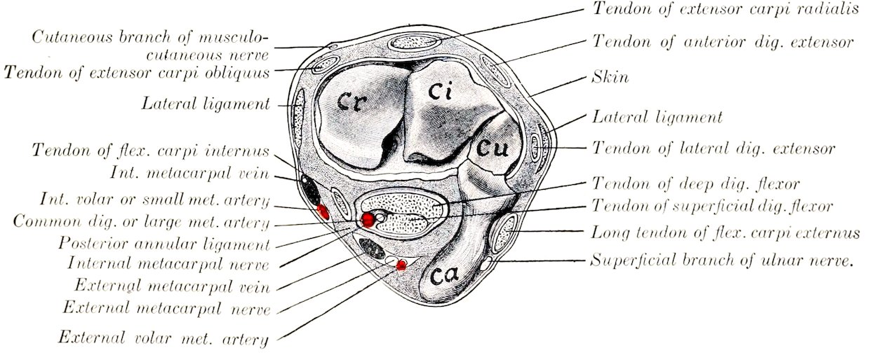
Fig. 445.—Cross-section of Right Carpus of Horse. The soft structures are cut at the level of the radio-carpal articulation, exposing the proximal row of carpal bones. Cr, Radial carpal bone; Ci, intermediate carpal bone; Cu, ulnar carpal bone; Ca, accessory carpal bone. (After Schmaltz, Atlas d. Anat. d. Pferdes.)
3. The common interosseous artery (A. interossea communis) is a vessel of considerable size which arises at the level of the interosseous space, through which it passes. Before entering the space it gives off a small branch, the volar interosseous (A. interossea volaris), which descends to the radial head of the perforans. In the space it supplies the nutrient arteries of the radius and ulna. Emerging from the space it gives off branches to the flexor carpi externus, a small recurrent branch (A. interossea recurrens) which passes upward on the outer surface of the ulna and anastomoses with the deep brachial and ulnar arteries, and is continued 562as the dorsal interosseous artery. This vessel (A. interossea dorsalis) passes down between the anterior and lateral extensors of the digit and concurs with the anterior radial in forming a network on the anterior surface of the carpus, the rete carpi dorsale. From the latter arise two small vessels, the internal and external dorsal metacarpal arteries (A. metacarpea dorsalis medialis, lateralis), which run distally in the grooves between the large and small metacarpal bones and anastomose with the volar metacarpal arteries.
4. The artery of the rete carpi volare (A. retis carpi volaris) is a small vessel which arises at the distal third of the forearm and passes downward to the posterior surface of the carpus, where it concurs with branches of the volar metacarpal arteries in forming the rete carpi volare.
5. The external volar metacarpal artery (A. metacarpea volaris lateralis) is a small vessel which arises just above the carpus under cover of the flexor carpi medius and anastomoses with the ulnar artery, forming the supracarpal arch. From the latter a branch descends with the outer branch of the median nerve, inclines outward toward the posterior border of the accessory carpal bone, and arrives at the head of the external metacarpal bone. Here it is connected with the internal volar metacarpal artery, usually by two transverse branches, thus forming the deep volar or subcarpal arch (Arcus volaris profundus). One of these branches lies between the subcarpal check ligament and the suspensory ligament; the other (not always present) lies beneath the latter on the large metacarpal bone. A small branch descends to the fetlock with the external metacarpal nerve. Below the arch the artery pursues a flexuous course downward on the posterior face of the large metacarpal bone alongside of the external small metacarpal and under cover of the suspensory ligament. At the distal third of the metacarpus it commonly unites with the corresponding vessel of the inner side to form a short trunk which passes backward through the angle of divergence of the branches of the suspensory ligament and joins the external digital or the common digital artery.
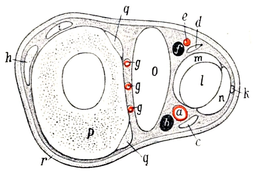
Fig. 446.—Cross-section of Middle of Right Metacarpus of Horse. a, Common digital (or large metacarpal) artery; b, internal metacarpal vein; c, internal metacarpal nerve; d, external metacarpal nerve; e, branch of external volar metacarpal artery; f, external metacarpal vein; g, g, g, deep volar or interosseous metacarpal arteries; h, tendon of anterior extensor; i, tendon of lateral extensor; k, anastomotic branch connecting metacarpal nerves; l, deep flexor tendon; m, check ligament; n, superficial flexor tendon; o, suspensory ligament; p, large metacarpal bone; q, q, small metacarpal bones; r, skin (After Ellenberger, in Leisering’s Atlas.)
6. The internal volar or small metacarpal artery (A. metacarpea volaris medialis) is given off from the posterior radial at an acute angle, usually a little above the external one or by a common trunk with it. It passes down the inner side of the carpus behind the tendon of the flexor carpi internus and embedded in the posterior annular ligament. Arriving at the proximal end of the inner metacarpal bone it becomes more deeply placed and is connected with the external volar artery by one or two transverse branches as stated above. It then pursues a flexuous course downward alongside of the inner small metacarpal bone, like the corresponding external artery, with which it commonly unites as described above. It is larger than the external artery and supplies the nutrient artery to the large metacarpal bone.
The foregoing account describes the most common arrangement of the dorsal and volar metacarpal arteries. Variations in their origin and connections are common, but have no great surgical importance. Collateral branches are omitted for the same reason. In some cases the 563internal volar metacarpal is connected with the common digital or large metacarpal artery a little below the carpus by a branch passing obliquely across the inner border of the deep flexor tendon; this forms a superficial volar arch.
THE COMMON DIGITAL ARTERY
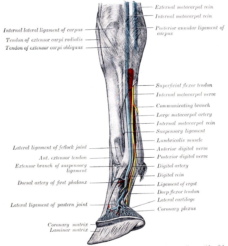
Fig. 447.—Dissection of Right Carpus, Metacarpus, and Digit of Horse, Inner View. (After Schmaltz, Atlas d. Anat. d. Pferdes.)
The common digital or large metacarpal artery[164] is the direct continuation of the posterior radial. It descends in the carpal canal along the inner side of the flexor tendons in company with the inner branch of the median nerve and a satellite vein. Continuing down the limb it preserves this relation to the tendons to the distal third of the metacarpus, where it inclines toward the middle line of the limb behind the suspensory ligament, and divides into the internal and external digital arteries. In the metacarpus the artery is related to the vein in front and the artery behind, and is covered by the fascia and skin. It furnishes collateral branches to the suspensory ligament, the flexor tendons, and the skin.
564
THE DIGITAL ARTERIES
The internal and external digital arteries (A. digitalis medialis, lateralis) are formed by the bifurcation of the common digital at the distal third of the metacarpus. They diverge, pass down over the outer surface of the corresponding sesamoid at the fetlock, and descend parallel with the borders of the deep flexor tendon to the volar grooves and foramina of the third phalanx. Entering the latter the two arteries unite in the semilunar canal and form the terminal arch (Arcus terminalis), from which numerous branches pass through the bone to the wall surface and ramify in the matrix of the wall and sole of the hoof. A number of branches emerge through the foramina at the lower border, where they anastomose with each other in arciform fashion. These branches were named by Spooner the inferior communicating arteries, and the anastomotic arch formed by them is termed the circumflex artery of the third phalanx (Chauveau) or the artery of the lower border of the third phalanx (Leisering).
Each artery is accompanied by a vein and by the digital nerves. Above the fetlock the artery is most deeply placed and is covered by the vein; the nerve is behind the vein. At the fetlock the artery has become superficial and is related to the vein in front and the posterior branch of the nerve behind. The anterior branch of the nerve crosses over the artery obliquely to the lateral aspect of the first phalanx. The vessels and nerves are crossed obliquely by a small tendinous band, the ligament of the ergot, which begins in the fibrous basis of the ergot at the back of the fetlock and extends downward and forward to end in the fascia at the side of the pastern joint.
In addition to branches to the joints, tendons and synovial sheath, ergot, and skin, the digital arteries give off:
1. The artery of the first phalanx or perpendicular artery (A. phalangis primæ), a short trunk which arises at a right angle about the middle of the first phalanx, and divides into dorsal and volar branches (Rami dorsales et volares). The dorsal (anterior) branch passes between the first phalanx and the extensor tendon and ramifies on the front of the digit, anastomosing with its fellow. The volar (posterior) branch dips in between the flexor tendons and the inferior sesamoidean ligaments and anastomoses with the opposite artery.
2. The artery of the plantar cushion (A. toricæ phalangis tertiæ) arises at the proximal border of the lateral cartilage and passes backward and downward to ramify in the plantar cushion and the matrix of the heels and frog.
3. The dorsal artery of the second phalanx (Ramus dorsalis phalangis secundæ) arises a little above the navicular bone, and passes forward under cover of the lateral cartilage and the extensor tendon to the front of the second phalanx, where it anastomoses with the opposite vessel. It gives branches to the skin, the tendon, the coffin joint, and the coronary matrix of the hoof.
4. The volar artery of the second phalanx (Ramus volaris phalangis secundæ) is smaller than the preceding, opposite to which it arises. It passes above the proximal border of the third sesamoid and unites with the opposite artery.
The arteries of the second phalanx form what is termed by Chauveau the coronary circle. The dorsal (anterior) part of the circle gives off commonly an artery (A. coronalis phalangis tertiæ) near either side of the extensor tendon, which divides into two branches. The central branch unites with that of the opposite side, while the other joins a branch of the artery of the plantar cushion. In this way is formed the circumflex artery of the coronary cushion, an anastomotic arch which lies on the extensor tendon at the coronet. In some cases descending branches of the arteries of the first phalanx concur in the formation of the arch.
5. The dorsal artery of the third phalanx or preplantar artery (A. dorsalis phalangis tertiæ) arises at the deep face of the wing of the third phalanx, passes outward through the notch or foramen there, and runs forward in the groove on the wall surface. It gives off ascending and descending branches, which ramify in the matrix 565of the wall of the hoof, anastomosing above with the circumflex artery of the coronary cushion and below with the circumflex artery of the third phalanx. Before passing through the wing it detaches a retrograde branch to the plantar cushion, and after emerging one which ramifies on the outer aspect of the lateral cartilage.
The terminal part of the digital artery after giving off the preceding vessel is sometimes termed the plantar.
BRANCHES OF THE THORACIC AORTA
In addition to the coronary arteries and the common brachiocephalic trunk (which have been described), the thoracic part of the aorta gives off branches to the thoracic walls and viscera and to the spinal cord and its membranes. The visceral branches (Rami viscerales) are the bronchial and œsophageal, which arise by a broncho-œsophageal trunk. The parietal branches (Rami parietales) are the intercostal and phrenic arteries.
1. The broncho-œsophageal (Truncus broncho-œsophageus) is a short, usually bulbous, trunk which arises at the sixth thoracic vertebra from the aorta or in common with the first aortic intercostal arteries. It passes (under cover of the vena azygos) down the right face of the aorta toward the bifurcation of the trachea and divides into bronchial and œsophageal branches.
(a) The bronchial artery (A. bronchialis) crosses the left face of the œsophagus to the bifurcation of the trachea, where it divides into right and left branches. Each enters the hilus of the corresponding lung above the bronchus, which it accompanies in its ramification. It supplies the lung tissue and also detaches twigs to the bronchial lymph glands and the mediastinum.
(b) The œsophageal artery (A. œsophagea) (Fig. 429) is a small vessel which passes backward above the œsophagus in the posterior mediastinum and anastomoses with the œsophageal branch of the gastric artery. It detaches twigs to the œsophagus and the mediastinal lymph glands and pleura, and also gives off two branches which pass between the layers of the ligaments of the lungs and ramify in the subpleural tissue. Very commonly there is another artery which runs backward ventral to the œsophagus.
In some cases there is no broncho-œsophageal trunk, the bronchial and œsophageal arising separately. In other cases the second aortic intercostal arises in common with them also.
2. The intercostal arteries (Aa. intercostales) (Figs. 428, 429) number eighteen pairs. The first arises from the deep cervical artery, the next three from the subcostal branch of the dorsal artery, and the remainder from the aorta. The aortic intercostals arise from the dorsal face of the aorta in pairs close together; the fifth and sixth usually spring from a common stem. Each passes across the body of a vertebra to the corresponding intercostal space, detaches twigs to the vertebræ and the pleura, and divides into dorsal and ventral branches. The dorsal branch (Ramus dorsalis) gives off a spinal branch (Ramus spinalis) which passes through the intervertebral foramen, gives twigs to the membranes of the spinal cord, perforates the dura, and reinforces the ventral spinal artery. A muscular branch passes to the muscles and skin of the back. The ventral branch (Ramus ventralis) is much the larger. It passes downward, at first almost in the middle of the intercostal space between the intercostal muscles, then gains the posterior border of the rib and is subpleural. Each is accompanied by a vein and nerve, the artery being in the middle and the vein in front. At the lower part of the space it unites with a ventral intercostal branch of the internal thoracic or the asternal artery. It supplies the intercostal muscles, the ribs and the pleura, and gives off perforating branches which pass out to the serratus magnus, the abdominal muscles, and the skin.
3. The phrenic arteries (Aa. phrenicæ) are two or three small vessels which arise at the hiatus aorticus from the ventral aspect of the aorta, often by a common 566trunk. They supply the crura of the diaphragm. In some cases they arise in common with an intercostal artery.
BRANCHES OF THE ABDOMINAL AORTA
The collateral branches of the abdominal part of the aorta are distributed chiefly to the walls and contents of the abdominal cavity, but some branches are supplied to the spinal cord and its membranes, and others extend into the pelvis and to the scrotum. The visceral branches are the cœliac, anterior mesenteric, renal, posterior mesenteric, and the spermatic or utero-ovarian. The parietal branches are the lumbar arteries.
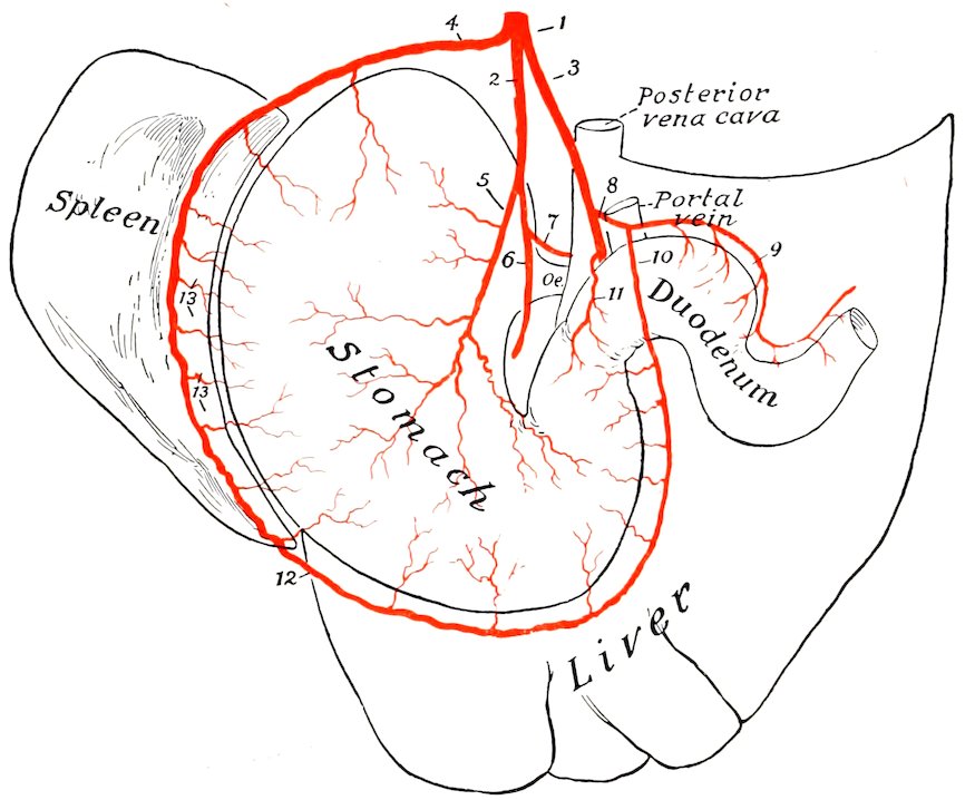
Fig. 448.—Plan of Branches of Cœliac Artery of Horse. 1, Cœliac artery; 2, gastric artery; 3, hepatic artery; 4, splenic artery; 5, posterior gastric artery; 6, anterior gastric artery; 7, œsophageal branch; 8, gastro-duodenal artery; 9, pancreatico-duodenal artery; 10, right gastro-epiploic artery; 11, pyloric artery; 12, left gastro-epiploic artery; 13, short gastric branches of splenic.
I. The cœliac artery or axis (A. cœliaca) is an unpaired vessel, usually half an inch or less (ca. 1 cm.) in length, which arises from the ventral aspect of the aorta at its emergence from the hiatus aorticus. It divides on the dorsal surface of the pancreas into three branches—the gastric, hepatic, and splenic.
1. The gastric artery (A. gastrica sinistra) passes downward and forward in the gastro-phrenic ligament, gives off œsophageal and pancreatic branches, and divides above and behind the cardia into anterior and posterior branches.
(a) The anterior branch (Ramus cranialis) crosses the lesser curvature just to the right of the cardia and ramifies on the parietal surface of the stomach. The branches pursue a flexuous course toward the greater curvature and anastomose with the short gastric arteries and the gastric branch of the hepatic artery.
(b) The posterior branch (Ramus caudalis) is distributed in a similar fashion on the visceral surface.
567(c) The œsophageal branch (Ramus œsophageus) passes through the hiatus œsophageus into the thoracic cavity above the œsophagus and anastomoses with the œsophageal branch of the broncho-œsophageal artery.
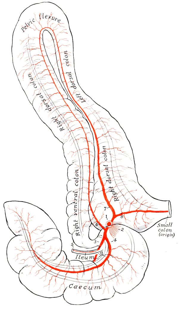
Fig. 449.—Plan of Chief Branches of Anterior Mesenteric Artery of Horse. 1, Stump of anterior mesenteric artery; 2, stumps of arteries of small intestine; 3, external cæcal artery, 4, internal cæcal artery; 5, ileal artery; 6, ventral colic artery; 7, dorsal colic artery; 8, middle colic or first artery of small colon.
The gastric artery often arises by a common trunk with the splenic. The two terminal branches may arise separately, or the anterior from the splenic and the posterior from the hepatic. The œsophageal branch often arises from the splenic or the posterior gastric.
568
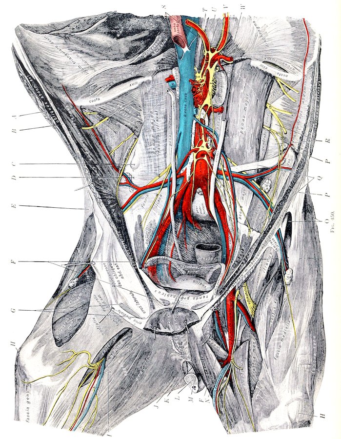
Fig. 450.—Dissection of Sublumbar Region, Pelvic Inlet, and Inner Surface of Thigh of Horse. Blood-vessels: 1, Aorta; 2, posterior vena cava; 3, hepatic; 4, gastric; 5, splenic; 6, 7, renal; 8, anterior mesenteric; 9, arteries of small intestine; 10, ileo-cæco-colic; 11, dorsal colic; 12, artery of small colon; 13, posterior mesenteric; 14, external iliac; 15, internal iliac; 16, circumflex iliac; 17, 19, anterior and posterior branches of 16; 18, anastomosis with intercostals; 20, spermatic; 21, cremasteric; 22, lateral sacral; 23, internal pudic; 24, umbilical; 25, femoral; 26, deep femoral; 27, anterior femoral; 28, branch to gracilis; 29, saphenous. Lymph vessels and glands: a, Lymph vessels; b, lumbar glands; c, internal iliac glands; d, external iliac glands; e, precrural glands; f, deep inguinal glands (position indicated by dotted line). Nerves and ganglia: 1, 2, Left and right cœliaco-mesenteric ganglia; 3, posterior mesenteric ganglion; 4, connection between 1 and 2; 5, connection between hepatic and splenic nerves; 6, 7, connections between anterior and posterior mesenteric ganglia; 8, splanchnic nerve; 9, lumbar trunk of sympathetic; 10, pelvic branches of posterior mesenteric ganglion; 11, internal spermatic nerve; 12, last thoracic nerve; 13, ilio-hypogastric nerve; 14, anastomosis between 13 and 15, ilio-inguinal nerve; 16, 17, cutaneous and muscular branches of 15; 18, 19, muscular and scrotal branches of external spermatic nerve; 20, external cutaneous nerve of thigh; 21, femoral nerve; 22, saphenous nerve; 23, branch of 22 to sartorius. A, Cut surface of obliquus abdominis externus; B, cut surface of obliquus abdominis internus; C, depression due to disappearance of fat in interval between outer border of psoas major and iliacus; D, thick part of iliac fascia which extends from tendon of psoas minor to external angle of ilium; E, superficial fascia of flank; F, internal inguinal ring; G, external inguinal ring; H, patella; I, femoral canal; J, semimembranosus; K, suspensory ligaments of penis; L, cross-section of penis; M, stump of spermatic cord; N, ilio-psoas; O, tensor fasciæ latæ; P, P, extremities of external angle of ilium; Q, remains of iliac fascia; R, ilio-lumbar ligament or deep layer of lumbo-dorsal fascia; S, line of attachment of gastro-pancreatic fold; T, tendon of right crus of diaphragm; U, right crus; V, aorta at hiatus aorticus; W, left crus of diaphragm. The position of the kidneys is indicated by dotted lines. (After Schmaltz, Atlas d. Anat. d. Pferdes)
2. The hepatic artery (A. hepatica) is larger than the gastric. It passes forward to the right and ventrally on the dorsal surface of the pancreas, covered by 569the gastro-pancreatic fold, crossing obliquely under the vena cava, and reaches the inner border of the portal vein. It divides into three or four branches which enter the portal fissure of the liver and ramify within the gland with the portal vein and the hepatic duct. It gives off the following collateral branches:
(a) Pancreatic branches (Aa. pancreaticæ) are given off as the artery crosses the pancreas, in which it is partly embedded.
(b) The pyloric artery (A. gastrica dextra) arises above the first curve of the duodenum. It descends to the pylorus, sending branches to the first part of the duodenum and the pylorus, and anastomoses with the gastric and right gastro-epiploic arteries. It may arise from the gastro-duodenal.
(c) The gastro-duodenal artery (A. gastroduodenalis) passes to the second curve of the duodenum and divides into the right gastro-epiploic and the pancreatico-duodenal. The right gastro-epiploic artery (A. gastroepiploica dextra) crosses over the posterior surface of the duodenum and enters the great omentum, in which it runs to the left, parallel with the greater curvature of the stomach. It gives branches to the latter and to the omentum and forms an anastomotic arch with the left gastro-epiploic artery. The pancreatico-duodenal artery (A. pancreaticoduodenalis) divides into pancreatic and duodenal branches. The former (Ramus pancreaticus) supplies the middle part of the pancreas and is often replaced by a number of variable twigs. The duodenal branch (Ramus duodenalis) passes to the right along the lesser curvature of the duodenum and anastomoses with the first branch of the anterior mesenteric artery.
3. The splenic artery (A. lienalis) is the largest branch of the cœliac. It passes to the left (with the large satellite vein) on the left extremity of the pancreas and across the saccus cæcus of the stomach. Entering the suspensory ligament of the spleen, it runs in the hilus of the spleen to the apex, beyond which it is continued as the left gastro-epiploic. It gives off the following branches:
(a) Pancreatic branches (Rami pancreatici) to the left extremity of the pancreas.
(b) Splenic branches (Rami lienales), which plunge into the substance of the spleen.
(c) Gastric branches (Aa. gastricæ breves), which pass in the gastro-splenic omentum to the greater curvature of the stomach, where they bifurcate and anastomose with the gastric arteries.
570(d) The left gastro-epiploic artery (A. gastroepiploica sinistra) is the continuation of the splenic artery. It passes to the right in the great omentum, parallel with the greater curvature of the stomach, and anastomoses with the right gastro-epiploic. It gives off branches to the greater curvature of the stomach, and twigs to the omentum.
II. The anterior or great mesenteric artery (A. mesenterica cranialis) arises from the ventral face of the aorta at the first lumbar vertebra. It is a large unpaired trunk, about an inch (ca. 2 to 3 cm.) in length, which passes ventrally between the vena cava and the left adrenal into the root of the great mesentery, when it divides into three branches—left, right, and anterior.[165]
1. The left branch gives off at once about fifteen to twenty arteries of the small intestine (Aa. intestinales).[166] These come off close together and pass in divergent fashion between the layers of the great mesentery, each dividing into two branches which anastomose with adjacent branches to form a series of arches. In the anterior part of the series secondary arches are formed by the union of branches given off from the primary set of arches. From the convex side of these arches terminal branches pass to the wall of the small intestine, in which they ramify and form a vascular network. They are accompanied by satellite veins and by nerves and lymph vessels. The first artery anastomoses with the pancreatico-duodenal, and the last with the ileal branch of the great mesenteric artery. Branches are supplied to the mesenteric lymph glands.
2. The right branch (A. ileo-cæco-colica) might be regarded as the continuation of the trunk. It runs downward and a little forward and to the right and gives off the ileal, the two cæcal, and the ventral colic arteries.
(1) The ileal or ileo-cæcal artery (A. ilea) passes in retrograde fashion along the terminal part of the ileum and unites with the last branch of the left division.
(2) The external or inferior cæcal artery (Ramus cæcalis lateralis) passes between the cæcum and the origin of the colon and runs on the external muscular band of the cæcum to the apex, where it anastomoses with the internal artery. Besides numerous collaterals to the cæcum, it gives off the artery of the arch, which passes along the lesser curvature of the base of the cæcum and runs on the outer face of the origin of the great colon.
(3) The internal or superior cæcal artery (Ramus cæcalis medialis) passes along the inner band to the apex of the cæcum, where it anastomoses with the external cæcal artery.
(4) The ventral, right, or direct colic artery (A. colica ventralis) runs along the opposed surfaces of the ventral (first and second) parts of the great colon to the pelvic flexure, where it unites with the dorsal colic artery. It supplies the ventral parts of the great colon and sends a branch to the base of the cæcum.
3. The anterior branch divides after a very short course into the dorsal and middle colic arteries.
(1) The dorsal, left, or retrograde colic artery (A. colica dorsalis) is a large vessel which passes along the dorsal (fourth and third) parts of the great colon to the pelvic flexure, where it joins the ventral colic artery.
(2) The middle colic artery or first artery of the small colon (A. colica media) is a much smaller vessel which passes to the origin of the small colon, enters the colic mesentery, and forms an arch by joining the first branch of the posterior mesenteric artery close to the lesser curvature of the bowel. It sends an anastomotic branch to the dorsal colic artery.
571III. The renal arteries (Aa. renales), right and left, are relatively large vessels which arise from the aorta near the anterior mesenteric. The right artery is the longer of the two. It crosses over the dorsal surface of the vena cava to the right and somewhat forward. At the hilus it divides into several (five to eight) branches; some of these enter the gland at the hilus, while others pass to the ventral surface and enter there. The left artery is short and usually arises a little further back; it passes directly outward to the kidney and is then disposed like the right one. Small collateral branches are supplied to the ureters and the adrenals. The latter also receive small vessels directly from the aorta. The distribution within the kidney has been described.
Variations in the renal arteries are frequent. Two or more arteries may occur on one or both sides. Accessory arteries are more common on the left side and usually enter the posterior pole of the gland. They may arise from the aorta, the external iliac, or the circumflex iliac artery.
IV. The posterior or small mesenteric artery (A. mesenterica caudalis) is an unpaired vessel which arises from the ventral face of the aorta at the fourth lumbar vertebra, i. e., about five or six inches (ca. 12 to 15 cm.) behind the origin of the anterior mesenteric artery. It is much smaller than the latter and supplies the greater part of the small colon and rectum. It descends in the colic mesentery and divides into two branches. The anterior branch (A. colica sinistra) gives off three or four arteries which divide and form anastomotic arches close to the bowel. The first arch is formed by union with the middle colic branch of the anterior mesenteric. The posterior branch or anterior hæmorrhoidal artery (A. hæmorrhoidalis cranialis) passes backward in the upper part of the mesentery and the mesorectum and terminates near the anus by anastomosing with the internal pudic artery. Three or four of its anterior collateral branches form arches.
V. The spermatic arteries (Aa. spermaticæ internæ), right and left, are long slender arteries which arise from the aorta near the posterior mesenteric and supply the testicle and epididymis. Each passes backward in a narrow fold of peritoneum (Plica vasculosa) to the internal inguinal ring and descends through the inguinal canal to the scrotum. In the canal and scrotum it pursues a very flexuous course in the anterior border of the spermatic cord, surrounded by the pampiniform plexus of veins, and associated closely with the spermatic nerves and lymphatics and unstriped muscle-fibers. It passes between the epididymis and testicle, runs in flexuous fashion along the attached border of the latter, turns around the posterior pole, and runs forward on the free border to the anterior pole. The largest branches arise from its ventral part, pass tortuously up either side of the gland embedded in the tunica albuginea, and give off fine branches to the gland substance. Small collateral branches are detached to the ureter and the spermatic cord.
The utero-ovarian arteries in the female correspond to the preceding vessels, but are much shorter. Each is placed in the anterior part of the broad ligament of the uterus and divides into ovarian and uterine branches. The ovarian branch (A. ovarica) pursues a flexuous course to the ovary, which it supplies. The uterine branch (A. uterina cranialis) passes to the concave border of the cornu of the uterus, which it supplies, anastomosing with the uterine artery.
VI. The lumbar arteries (Aa. lumbales) are in series with the intercostal arteries and have a similar origin and distribution. There are usually six pairs of lumbar arteries, of which five arise from the aorta and the sixth from the internal iliac or the lateral sacral at the junction of the last lumbar vertebra and the sacrum. Each passes across the body of a lumbar vertebra to the intertransverse space, gives branches to the sublumbar muscles, and divides into dorsal and ventral branches. The dorsal branch (Ramus dorsalis), the larger of the two, passes upward to ramify in the extensor muscles of the spine and the skin of the loins; it gives off a spinal branch (Ramus spinalis) which comports itself like the corresponding branch of an aortic intercostal artery. The ventral branch (Ramus 572ventralis) runs outward in the intertransverse space, passes between the transversus and obliquus internus abdominis, gives branches to these muscles, and ends in the obliquus externus, the panniculus, and the skin of the flank.
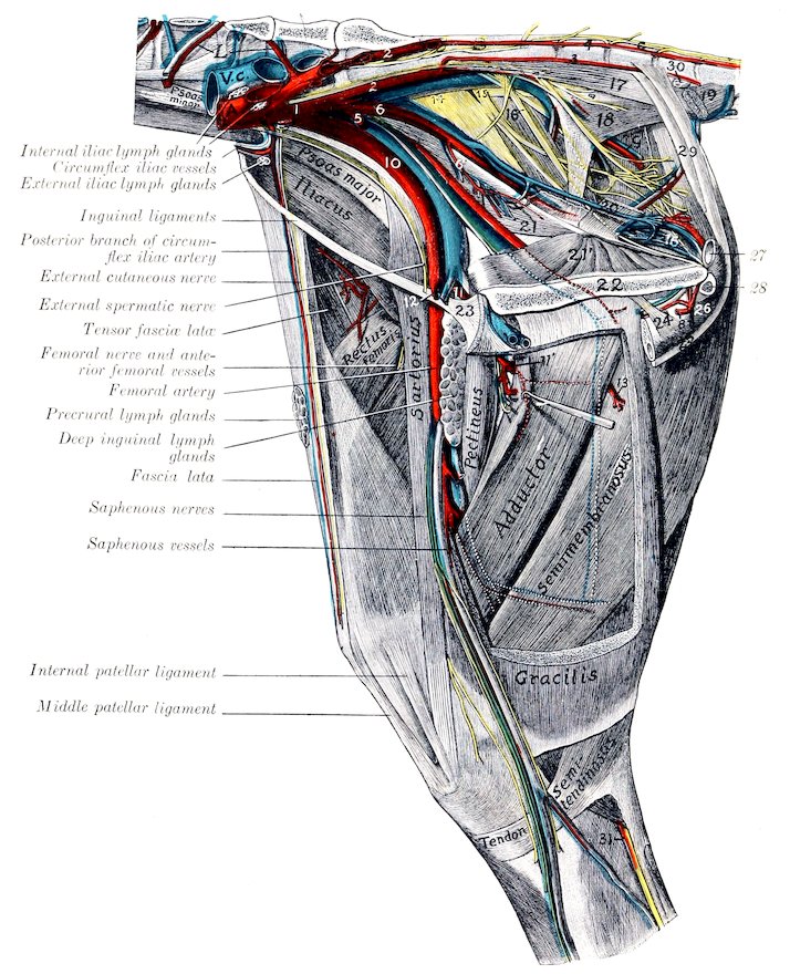
Fig. 451.—Dissection of Pelvis, Thigh, and Upper Part of Leg of Horse, Internal View. L, Lumbar vessels; V.c., posterior vena cava; A, aorta (termination); C, sympathetic trunk; 1, internal iliac artery, 2, lateral sacral artery; 3, middle coccygeal artery; 4, lateral coccygeal artery; 5, umbilical artery (cut off); 6, internal pudic artery; 6′, vesico-prostatic artery; 7, obturator artery; 8, a. profunda penis (from left obturator); 9, posterior gluteal (or ischiatic) vessels; 10, external iliac artery; 11, 11′, deep femoral artery; 12, prepubic artery; 13, branch of deep femoral artery; 14, great sciatic nerve; 15, posterior gluteal nerve; 16 (above), internal pudic nerve; 16 (below near pelvic outlet), suburethral venous plexus; 17, sacro-coccygeus inferior; 18, coccygeus; 19, recto-coccygeus; 20, retractor ani; 21, 21′, two heads of obturator internus; 22, symphysis pelvis; 23, prepubic tendon; 24, suspensory ligament of penis; 25, retractor penis muscle; 26, bulbo-cavernosus muscle; 27, urethra (cross-section); 28, crus penis (cross-section); 29, suspensory ligaments of anus (= upper part of retractor penis muscle); 30, anal lymph glands; 31, tibial nerve. (After Schmaltz, Atlas d. Anat. d. Pferdes.)
573
THE INTERNAL ILIAC ARTERY
The internal iliac or hypogastric arteries (Aa. hypogastricæ) result from the bifurcation of the aorta under the fifth or sixth lumbar vertebra. They diverge at an angle of about 60 degrees, and each passes backward under the wing of the sacrum, then inclines downward on the pelvic surface of the shaft of the ilium, and divides a little above the psoas tubercle into iliaco-femoral and obturator arteries. The chief branches are as follows:
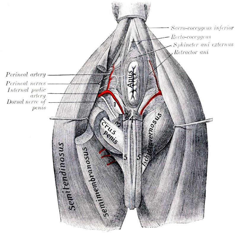
Fig. 452.—Dissection of Perineum of Horse (Male). 1, Bulbo-urethral (Cowper’s) gland; 2, transversus perinei muscle (?); 3, ischio-urethral muscle; 4, retractor penis muscle; 5, bulbo-cavernosus muscle. (After Schmaltz, Atlas d. Anat. d. Pferdes.)
1. The last pair of lumbar arteries pass up through the foramina at the junction of the last lumbar vertebra and the sacrum.
2. The internal pudic artery (A. pudenda interna) arises near the origin of the internal iliac. It passes backward and somewhat downward, at first along the iliac head of the obturator internus, then above the superior ischiatic spine on the inner surface of the sacro-sciatic ligament, perforates the latter and runs for a variable distance in its substance or on its external face. It then re-enters the pelvic cavity, passes backward on the retractor ani to the ischial arch, and divides 574into the perineal and the artery of the bulb in the male, perineal and artery of the clitoris in the female. It is accompanied by the internal pudic nerve. Its chief branches are as follows:
(1) The umbilical artery is given off from the internal pudic about an inch (ca. 2 to 3 cm.) from the origin of that vessel. It is a very large artery in the fœtus, in which it curves downward and forward at the side of the bladder in the edge of the umbilical fold of peritoneum, passes through the umbilical opening, becomes a component of the umbilical cord and ramifies in the fœtal placenta. After birth it extends only to the vertex of the bladder and is much reduced. Its lumen is almost obliterated and its wall is very thick, giving the vessel a cord-like character, hence it is commonly termed the round ligament of the bladder.[167] It gives off small vesical branches (Aa. vesicates craniales) to the bladder, and twigs to the prostate and vasa deferens in the male. In the mare a small branch runs forward along the ureter into the broad ligament of the uterus.
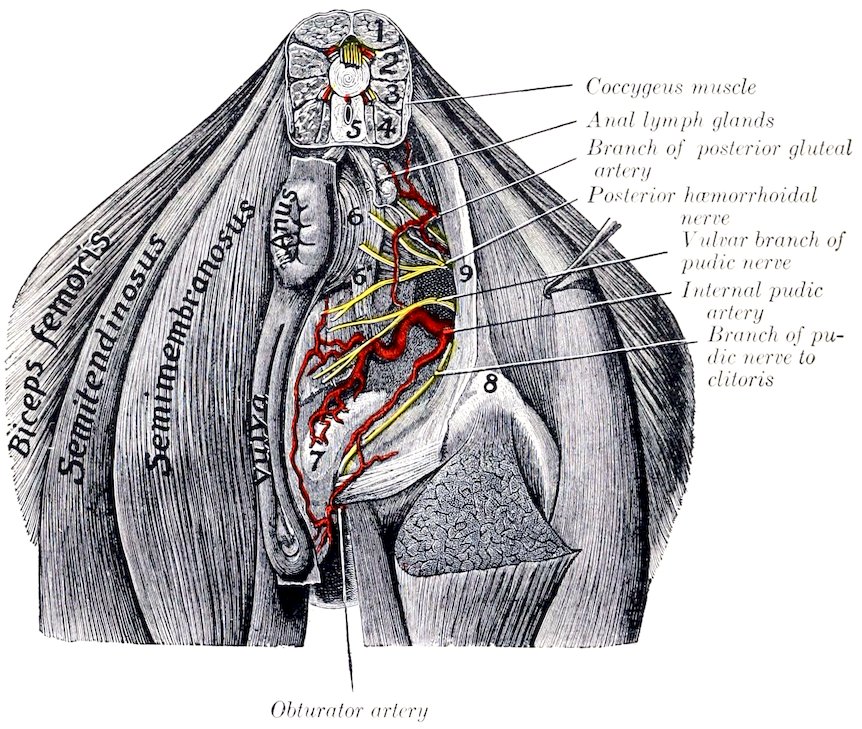
Fig. 453.—Dissection of Perineum of Mare. 1, Sacro-coccygeus superior; 2, sacro-coccygeus lateralis; 3, intertransversales; 4, sacro-coccygeus inferior; 5, recto-coceygeus; 6, sphincter ani externus; 6′, branch connecting sphincter ani with constrictor vulvæ; 7, vestibular bulb; 8, tuber ischii; 9, sacro-sciatic ligament. (After Schmaltz, Atlas d. Anat. d. Pferdes.)
(2) The middle hæmorrhoidal or vesico-prostatic artery (A. hæmorrhoidalis media) in the male arises usually near the prostate and runs backward lateral to the rectum. It supplies branches to the rectum, bladder, urethra, and accessory genital glands. In the female the homologous vessel is much larger and gives off the posterior uterine artery (A. uterina caudalis). This runs forward on the side of the vagina, to which it gives branches, and ramifies on the body of the uterus, anastomosing with the anterior and middle uterine arteries. (In some cases this artery arises from the internal iliac or the umbilical.)
(3) The perineal artery (A. perinei) is relatively small in the male. It passes 575upward at the side of the anus, which it supplies, and gives twigs to the bulbo-cavernosus muscle and the skin of the perineum. In the female it is large and is distributed to the anus and vulva, and gives a large branch to the vestibular bulb.
(4) The artery of the bulb (A. bulbi urethræ) may be regarded as the direct continuation of the internal pudic in the male. It lies at the side of the urethra above the ischial arch, dips under the bulbo-cavernosus muscle, and ramifies in the corpus spongiosum. Before doing so it gives off a small branch which turns around the ischial arch to reach the dorsum penis, and anastomoses with the deep branch of the obturator.
(4a) The artery of the clitoris (A. clitoridis) is the homologue in the female of the preceding vessel, but is much smaller. It passes to the ventral surface of the vulva with the internal pudic nerve, supplies the clitoris, and gives twigs to the vulva.
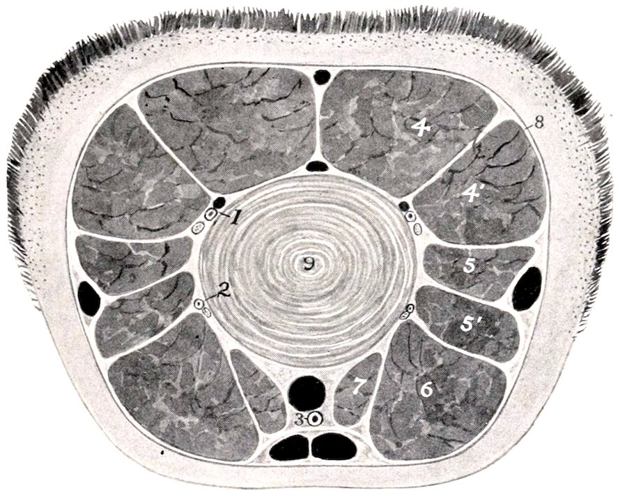
Fig. 454.-Cross-section of Tail of Horse. 1, Dorso-lateral coccygeal vessels and nerve; 2, ventro-lateral coccygeal artery and nerve; 3, middle coccygeal artery; 4, sacro-coccygeus superior; 4′, sacro-coccygeus lateralis; 5, 5′, intertransversales; 6, sacro-coccygeus inferior; 7, recto-coccygeus; 8, coccygeal fascia; 9, fibro-cartilage between fourth and fifth coccygeal vertebra. The veins are black.
3. The lateral sacral artery (A. sacralis lateralis) arises at the lumbo-sacral articulation (Fig. 451). It passes backward under the wing of the sacrum, then along the pelvic surface of the bone below the inferior sacral foramina and the nerves emerging from them, and is continued by the lateral coccygeal artery. The branches are as follows:
(1) Spinal branches (Rami spinales) enter the vertebral canal through the foramen between the last lumbar vertebra and the sacrum and through the ventral sacral foramina. They give off branches to the spinal cord and its membranes which reinforce the ventral spinal artery, and others which emerge through the dorsal sacral foramina and supply the muscles and skin of the croup.
(2) The middle coccygeal artery (A. coccygea) is an unpaired vessel which arises from the right or left lateral sacral or from a lateral coccygeal artery. It passes backward on the pelvic surface of the sacrum to the median line and continues in that position along the tail between the depressor muscles, supplying these and the skin.
576
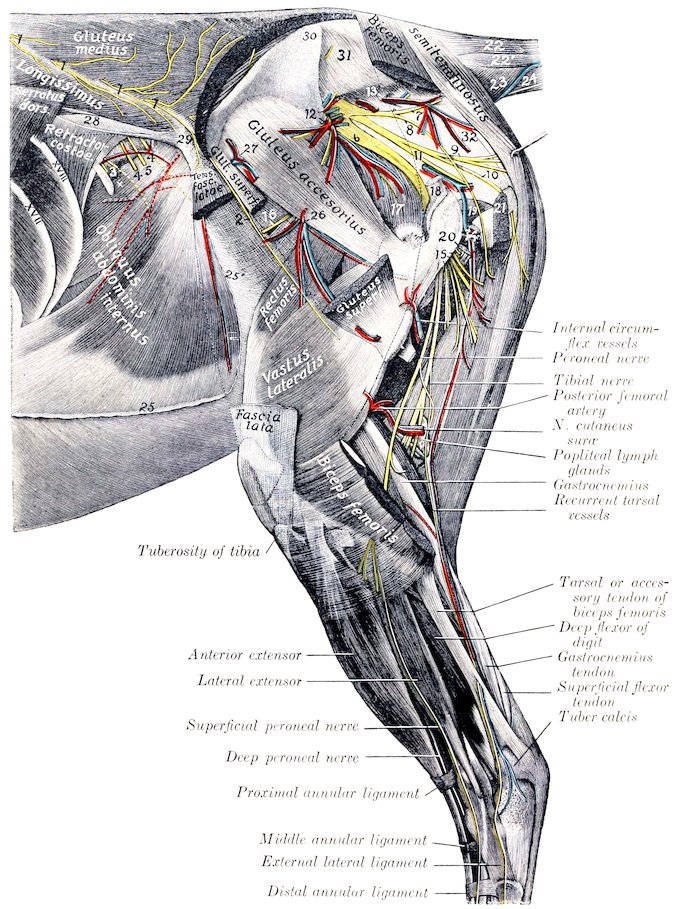
Fig. 455.—Dissection of Pelvis, Thigh, and Leg of Horse, External View. 1, Dorsal branches of last intercostal and first three lumbar nerves; 2, cutaneous branch of fourth lumbar nerve; 3, ventral branches of last intercostal nerve: 4, branches of ilio-hypogastric nerve; 5, superficial branch of ilio-inguinal nerve; 6, great sciatic nerve; 7, 8, nerves to biceps femoris (from posterior gluteal nerve); 9, nerve to semitendinosus (from same); 10, posterior cutaneous nerve of thigh; 11, internal pudic nerve; 12, anterior gluteal vessels and nerves; 13, posterior gluteal (or ischiatic) vessels; 14, branches of obturator vessels; 15, proximal muscular branches of great sciatic nerve to biceps femoris, semitendinosus, and semimembranosus; 16, nerve to tensor fasciæ latæ (from anterior gluteal nerve); 17, gluteus profundus; 18, stump of gluteus medius; 19, gemellus; 20, trochanter major; 21, stump of biceps femoris; 22, sacro-coccygeus superior; 22′, sacro-coccygeus lateralis; 23, coccygeus; 24, sacro-coccygeus inferior; 25, line of fusion of aponeuroses of internal and external oblique muscles of abdomen; 25′, aponeurosis of external oblique muscle (lamina iliaca); 26, branches of iliaco-femoral vessels; 27, ilio-lumbar vessels; 28, ilio-lumbar ligament or deep layer of lumbo-dorsal fascia; 29, external angle of ilium; 30, internal angle of ilium; 31, lateral sacro-iliac ligament; 32, sacro-sciatic ligament. (After Schmaltz, Atlas d. Anat. d. Pferdes.)
577(3) The posterior gluteal or ischiatic artery (A. glutea caudalis) emerges through the upper part of the sacro-sciatic ligament and runs backward and downward on the latter under cover of the biceps femoris (Fig. 455). It gives branches to that muscle and to the semitendinosus and semimembranosus, and anastomoses with the obturator, deep femoral, and posterior femoral arteries.
(4) The lateral coccygeal artery (A. caudalis lateralis ventralis) continues the direction of the lateral sacral, but is much smaller than the preceding vessel. It passes back between the depressor and intertransversales muscles of the tail and divides into two branches which supply twigs to the muscles and skin.
4. The ilio-lumbar artery (A. iliolumbalis) arises at a right angle from the internal iliac and runs outward behind the sacro-iliac joint, crossing the ventral surface of the ilium under cover of the iliacus muscle. It gives branches to the ilio-psoas and longissimus, and terminates by turning around the external border of the ilium a little behind the external angle, giving branches to the gluteus medius and tensor fasciæ latæ (Fig. 455).
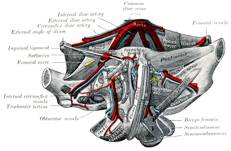
Fig. 456.—Deep Dissection of Ventral Wall of Pelvis of Horse (Male). 1, External pudic artery and small satellite vein; 2, anastomosis between external pudic and internal circumflex veins; 3, accessory ligament; 4, round ligament; 5, transverse ligament; 6, head of femur; 7, obturator externus (stumps); 8, inner margin of obturator foramen; 9, origin of gracilis; 10, origin of adductor; 11, bulbo-cavernosus; 12, retractor penis; 13, suspensory ligament of penis. (After Schmaltz, Atlas d. Anat. d. Pferdes.)
5. The anterior gluteal artery (A. glutea cranialis) is the largest branch of the internal iliac. It arises usually opposite to the preceding vessel and passes outward through the great sacro-sciatic foramen, dividing into several branches as it emerges; these enter the gluteal muscles (Fig. 455).
6. The iliaco-femoral or external circumflex artery (A. cireumflexa femoris lateralis) passes downward and outward across and beneath the shaft of the ilium, under cover of the iliacus muscle, and dips in between the rectus femoris and vastus externus. It is accompanied by two satellite veins. It gives collateral branches to the ilio-psoas, the glutei, and the tensor fasciæ latæ, supplies the nutrient artery of the ilium, and terminates in the quadriceps femoris (Fig. 455).
7. The obturator artery (A. obturatoria), the inner terminal branch of the internal iliac, passes downward and backward on the pelvic surface of the shaft of 578the ilium along the ventral border of the iliac head of the obturator internus, accompanied by the satellite vein and nerve, which lie in front of the artery (Fig. 451). On reaching the anterior border of the obturator foramen it dips under the obturator internus and passes obliquely through the foramen. In this part of its course it gives off a vesical branch and twigs to the obturator internus and the hip joint. After its emergence from the pelvic cavity it runs backward on the ventral face of the ischium, passes through the semimembranosus and, in the male, enters the crus penis, forming the arteria profunda penis. It anastomoses with the internal pudic, and usually with the external pudic by a branch which runs forward on the dorsum penis. Large collateral branches are detached to the adductors of the thigh, the biceps femoris and semitendinosus, forming anastomoses with the deep femoral and posterior femoral arteries. In the female the terminal part is small and enters the root of the clitoris.
ARTERIES OF THE PELVIC LIMB
The main arterial trunk of each pelvic limb descends as far as the lower border of the popliteus muscle, where it divides into the anterior and posterior tibial arteries. The different parts of the trunk receive special names which correspond to the several regions through which it passes. In the abdomen it is termed the external iliac artery, in the proximal two-thirds of the thigh it is called the femoral artery, while below this it is termed the popliteal artery.
The External Iliac Artery (Figs. 450, 451)
The external iliac artery (A. iliaca externa) arises from the aorta under the fifth lumbar vertebra, usually just in front of the origin of the internal iliac. It descends at the side of the pelvic inlet along the tendon of the psoas minor, crosses the insertion of that muscle, and reaches the level of the anterior border of the pubis, where it becomes the femoral artery. It is covered by the peritoneum and fascia, and is related behind to the corresponding vein. Its branches are as follows:
1. The circumflex iliac artery (A. circumflexa ilium profunda) arises from the external iliac at its origin or from the aorta directly. It passes across the iliac fascia toward the external angle of the ilium, near which it divides into two branches. The artery lies between the fascia and the peritoneum and is accompanied by two veins and the external cutaneous nerve of the thigh. The anterior branch gives twigs to the iliacus and psoas muscles, and passes downward and forward in the flank between the obliquus internus and transversus abdominis, in which it ramifies. The posterior branch perforates the abdominal wall and runs downward on the inner face of the tensor fasciæ latæ to the fold of the flank, supplying branches to that muscle, the panniculus, the precrural lymph glands, and the skin.
2. The cremasteric artery (A. spermatica externa) is a very small vessel which arises in a variable manner. It springs most often from the external iliac near its origin, but may come from the circumflex iliac, the aorta between the external and internal iliac, or the latter vessel. It accompanies the cremaster muscle to the inguinal canal, supplies twigs to that muscle, the tunica vaginalis, and other constituents of the spermatic cord (Fig. 450).
2a. The middle uterine artery (A. uterina media) of the female is regarded as the homologue of the preceding vessel. It has a similar origin, but is a much larger artery, which passes between the layers of the broad ligament and reaches the horn of the uterus close to its junction with the body. It is distributed to both, and anastomoses with the anterior and posterior uterine arteries.
579
The Femoral Artery (Figs. 450, 451)
The femoral artery is the main arterial trunk of the thigh. It begins at the level of the anterior border of the pubis, from which it is separated by the femoral vein. It descends almost vertically in the femoral canal behind the sartorius muscle, covered at first by the internal femoral fascia and lower down by the gracilis. After passing over the insertion of the pectineus, it perforates the adductor muscle, crosses in the vascular groove of the posterior surface of the femur, and is continued between the two heads of the gastrocnemius as the popliteal artery. It is related at its origin to the sartorius in front, the femoral vein behind (which separates it from the pectineus), and the iliacus externally. Lower down it is related superficially to the deep inguinal lymph glands, and deeply to the vastus internus, while the saphenous nerve is in front of it, and the vein passes to its external face. The chief branches are as follows:
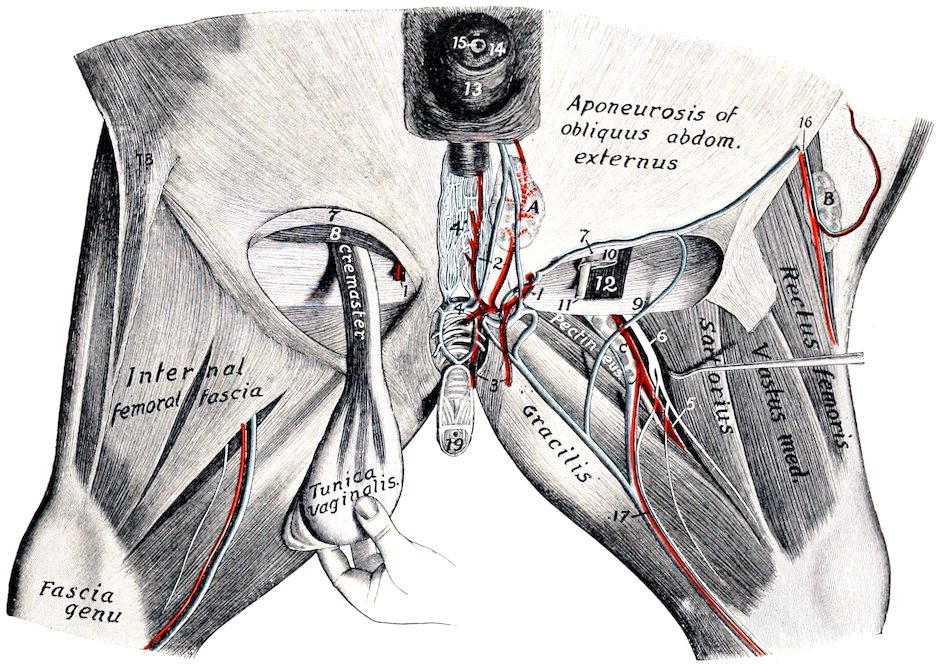
Fig. 457.—Dissection of Inguinal Region and Inner Surface of Thigh of Horse. 1, External pudic artery; 2, anterior branches of 1; 3, posterior branches of 1; 4, transverse anastomosis between external pudic veins; 4′, venous plexus of dorsum penis; 5, femoral artery; 6, saphenous nerve; 7, anterior border of external inguinal ring; 8, obliquus abdominis internus; 9, posterior border of external inguinal ring; 10, tunica vaginalis; 11, vas deferens; 12, cremaster muscle; 13, prepuce; 14, glans penis; 15, external urethral orifice; 16, posterior branches of circumflex iliac vessels; 17, saphenous vessels; 18, fold of flank; 19, penis (cut); A, superficial inguinal lymph glands; B, precrural lymph glands; C, deep inguinal lymph glands. (After Schmaltz, Atlas d. Anat. d. Pferdes.)
1. The prepubic artery (Truncus pudendo-epigastricus) arises from the femoral artery at its origin, usually by a common trunk with the deep femoral. It passes forward and a little inward and downward across the edge of the inguinal ligament, and then runs on the abdominal surface of the ligament to the inner part of the internal inguinal ring, where it divides over the upper border of the internal oblique muscle into the posterior abdominal and external pudic arteries.
(1) The posterior abdominal artery (A. epigastrica caudalis) passes along the 580external border of the rectus abdominis and anastomoses in the umbilical region with the anterior abdominal artery (Figs. 272). It supplies branches to the rectus and obliquus internus muscles.
(2) The external pudic artery (A. pudenda externa) descends on the inguinal ligament through the inner part of the inguinal canal and emerges at the inner angle of the external ring. In the male it divides into the subcutaneous abdominal artery and the dorsal artery of the penis. The former runs forward a short distance from the linea alba, and gives branches to the superficial inguinal lymph glands, the sheath, and the scrotum. The latter passes to the dorsum penis and ends at the glans as the arteria glandis. It gives off collateral branches (Rami profundi penis) to the corpus cavernosum, one of which usually passes backward and anastomoses with a branch of the obturator artery. Branches are also supplied to the superficial inguinal lymph glands, the prepuce, and the scrotum. In the female the mammary artery takes the place of the dorsal artery of the penis; it plunges into the base of the mammary gland.
2. The deep femoral artery (A. profunda femoris) arises either by a common trunk with the prepubic or a little distal to it (Fig. 451). It passes backward and downward across the inner face of the femoral vein, then below the pubis in the space between the hip joint and the pectineus muscle. On reaching the obturator externus it inclines more ventrally and outward, passes out between the internal border of the femur and the quadratus femoris, and ramifies in the biceps femoris and semitendinosus. It supplies large collateral branches to the adductor muscles and twigs to the deep inguinal lymph glands and the hip joint.
3. The anterior femoral artery (A. femoralis cranialis) arises a little distal to the preceding vessel from the opposite side of the femoral trunk. It passes forward, outward, and a little downward across the deep face of the sartorius, and dips in between the rectus femoris and vastus internus (Fig. 451). It is related externally to the ilio-psoas and the femoral nerve.
In some cases this artery is replaced by a large branch of the external circumflex, which passes between the ilio-psoas and rectus femoris and enters the interstice between the latter muscle and the vastus internus.
4. Innominate muscular branches (Rami musculares) of variable size and arrangement are given off to the muscles of this vicinity.
5. The saphenous artery (A. saphena) is a small vessel which arises from the femoral about its middle or from a muscular branch, and emerges between the sartorius and gracilis or through the latter to the inner surface of the thigh (Fig. 457). It is joined by the large saphenous vein and the saphenous nerve and passes down superficially on the fore part of the gracilis, continues on the deep fascia of the leg, and divides above the hock into two branches, which accompany the radicles of the vein. It gives off cutaneous twigs and anastomoses with the recurrent tibial artery.
In some cases this anastomosis does not occur. The artery may be larger and directly continuous with the internal tarsal artery.
6. The articular branch (A. genu suprema) is a slender artery which arises from the femoral just before it passes through the adductor. It descends along the posterior border of the vastus internus to the inner surface of the stifle joint, where it ramifies.
7. The nutrient artery of the femur (A. nutritia femoris) is given off at the middle of the femur and enters the nutrient foramen.
8. The posterior femoral or femoro-popliteal artery (A. femoris caudalis) is a large vessel which arises from the posterior face of the femoral just before the trunk passes between the two heads of the gastrocnemius (Fig. 455). It is very short and divides into two branches. The ascending branch passes upward and outward 581between the adductor (in front) and the semimembranosus (behind), and enters the biceps femoris, in which it ramifies. The descending branch passes downward and backward on the external head of the gastrocnemius, then curves upward between the biceps femoris and semitendinosus (crossed externally by the tibial nerve) and divides into branches to these muscles. A branch is detached from the convexity of the curve which passes down between the heads of the gastrocnemius, gives branches to that muscle and the flexor perforans, and is continued by a slender artery which accompanies the tibial nerve and unites with the recurrent tibial artery. A branch ascends alongside of the sciatic nerve between the biceps and semitendinosus and anastomoses with a descending branch of the obturator.
The Popliteal Artery
This artery (A. poplitea) is the direct continuation of the femoral. It lies between the two heads of the gastrocnemius, at first on the posterior face of the femur, then on the femoro-tibial joint capsule. It then passes down through the popliteal notch under cover of the popliteus, inclines outward, and divides near the upper part of the interosseous space into anterior and posterior tibial arteries. The satellite vein lies along its inner side. Collateral branches are supplied to the stifle joint and the gastrocnemius and popliteus.
POSTERIOR TIBIAL ARTERY
The posterior tibial artery (A. tibialis posterior) is much the smaller of the two terminals of the popliteal. It lies at first between the tibia and the popliteus, then between that muscle and the deep and inner heads of the deep flexor. Lower down it descends along the tendon of the inner head, becomes superficial in the distal third of the leg, and passes behind the tendon. At the level of the tuber calcis it forms an ᔕ-shaped curve, runs downward along the deep flexor tendon accompanied by the plantar nerves, and divides at the lower part of the hock into internal and external plantar arteries. The collateral branches are as follows:
1. The nutrient artery of the tibia (A. nutritia tibiæ).
2. Muscular branches (Rami musculares) to the popliteus and deep flexor.
3. The external tarsal artery (A. tarsea lateralis) arises at the distal end of the leg and is distributed to the outer surface of the hock. It gives off a small recurrent branch which ascends along the external border of the gastrocnemius tendon and anastomoses with a branch of the posterior femoral artery.
4. The recurrent tibial artery (A. tibialis recurrens) is given off from the second part of the curve, ascends in relation to the tibial nerve, and anastomoses with the descending branch of the posterior femoral and with the saphenous artery.
5. Small branches (internal calcaneal) to the inner aspect of the hock.
6. The plantar arteries, internal and external (A. plantaris medialis, lateralis), are the small terminals of the posterior tibial. They descend along the sides of the tarsal sheath of the deep flexor with the plantar nerves to the proximal part of the metatarsus, where they unite with the perforating tarsal artery to form the plantar arch (Arcus plantaris). Four plantar metatarsal arteries proceed from this arch. The two slender superficial plantar metatarsal arteries (A. metatarsea plantaris superficialis medialis, lateralis) descend on either side of the deep flexor tendon with the plantar nerves and unite with the great metatarsal or with the corresponding digital artery. The two deep or interosseous plantar metatarsal arteries (A. metatarsea plantaris profundus medialis, lateralis) descend between the suspensory ligament and the corresponding small metatarsal bone and unite near the fetlock with the great metatarsal. The inner artery is the larger of the 582two and usually appears to be the continuation of the perforating tarsal. It supplies the nutrient artery of the large metatarsal bone. In other cases it is the direct continuation of the internal plantar artery, the external artery only uniting with the perforating tarsal.
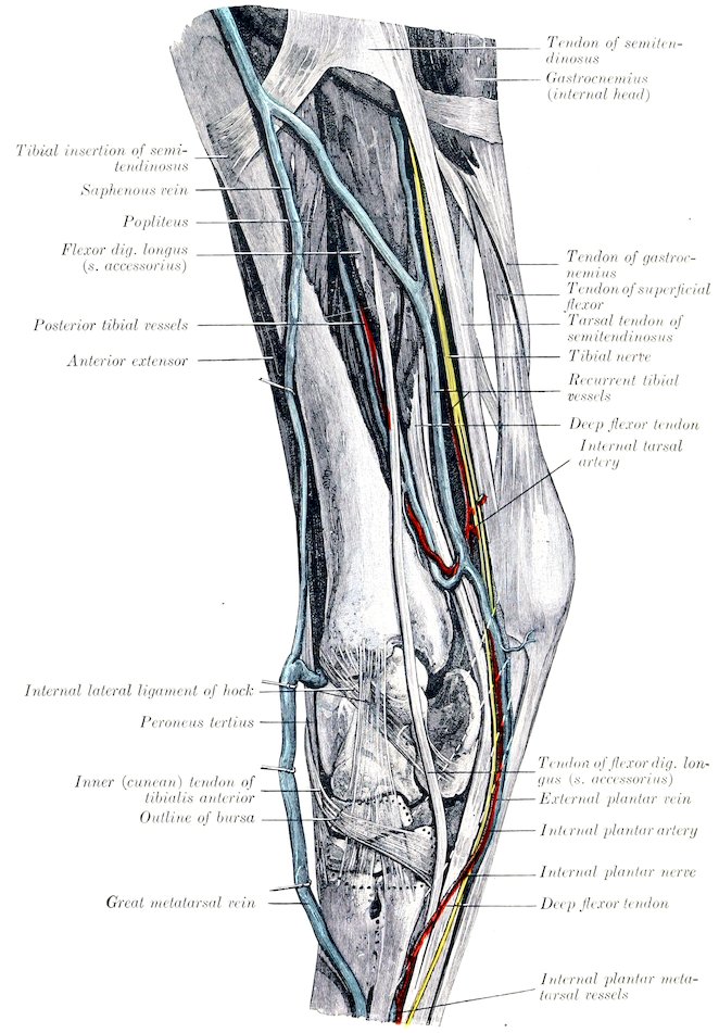
Fig. 458.—Dissection of Leg and Hock of Horse, Inner View. (After Schmaltz, Atlas d. Anat. d. Pferdes.)
583
ANTERIOR TIBIAL ARTERY
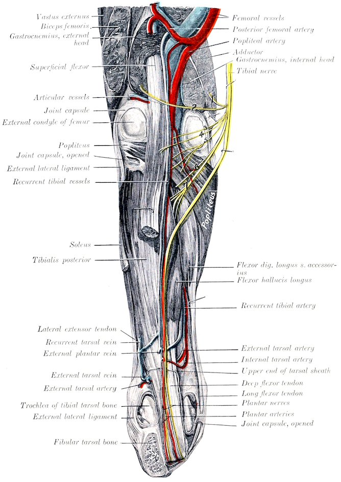
Fig. 459.—Deep Dissection of Right Stifle, Leg, and Hock of Horse, Posterior View. The hock is flexed at a right angle, and the tuber calcis is sawn off. The tibial nerve is drawn inward to show its muscular branches. Branches of tibial nerve: 1, 2, to gastrocnemius; 3, 4, to superficial flexor; 5, to popliteus; 6, to flexor longus or accessorius; 7, to deep flexor. (After Schmaltz, Atlas d. Anat. d. Pferdes.)
584
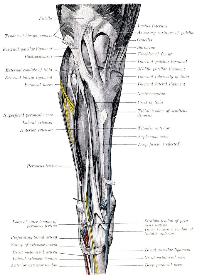
Fig. 460.—Superficial Dissection of Right Stifle, Leg, and Hock of Horse, Front View. (After Schmaltz, Atlas d. Anat. d. Pferdes.)
The anterior tibial artery (A. tibialis anterior) is much the larger of the two terminal branches of the popliteal. It passes forward through the upper part of the interosseous space and descends with two satellite veins on the outer part, of the front of the tibia, under cover of the tibialis anterior. At the lower part of the leg it deviates to the outer border of the tendon of this muscle, passes on to the capsule of the hock joint, gives off the perforating tarsal artery, and is continued as585 the great metatarsal artery. It gives off muscular branches to the dorso-lateral group of muscles of the leg and articular branches to the hock. The peroneal branch (A. peronea) is a variable vessel which descends along the fibula under the lateral extensor; it gives off muscular branches and one which perforates the fascia and divides into ascending and descending cutaneous twigs.
The perforating tarsal artery (A. tarsea perforans) is given off under cover of the outer tendon of the peroneus tertius. It passes backward through the vascular canal of the tarsus with a satellite vein and unites on the upper part of the suspensory ligament with the plantar arteries (or only with the external plantar) to form the plantar arch.
In well-injected specimens it is seen that there is a fine arterial network (Rete tarsi dorsale) on the flexor surface of the hock, which is formed by twigs from the anterior tibial and lateral tarsal arteries. From it proceed two very slender dorsal metatarsal arteries. The inner one descends in the furrow between the inner small and large metatarsal bones, and anastomoses usually in the proximal part of the metatarsus with the internal superficial plantar metatarsal, uniting sometimes with the deep plantar metatarsal. The external vessel passes down under the periosteum on the anterior face of the large metatarsal bone and becomes lost or joins the large metatarsal artery.
Sometimes the anterior tibial artery passes undivided through the tarsus, gives off the internal deep plantar metatarsal, and is continued as a very large internal superficial plantar metatarsal along the deep flexor tendon, thus resembling the arrangement in the fore limb.
In a few cases the perforating tarsal is a large vessel, directly continuing the anterior tibial, and is continued by a large internal deep plantar metatarsal. The great metatarsal is then small. Other variations are common.
THE GREAT METATARSAL ARTERY
This artery (A. metatarsea dorsalis lateralis) is the direct continuation of the anterior tibial. It passes downward and outward under the extensor brevis and the tendon of the lateral extensor, at first on the joint capsule and then in the oblique vascular groove on the upper part of the large metatarsal bone. It then descends in the groove formed by the apposition of the large and external small metatarsals, inclines inward between the two, and divides on the lower part of the posterior face of the large metatarsal bone into the internal and external digital arteries. It is not usually accompanied by a vein. Beyond this the arterial arrangement is the same as in the thoracic limb.
THE VEINS[168]
PULMONARY VEINS
The terminal pulmonary veins (Vv. pulmonales), usually seven or eight in number, return the aërated blood from the lungs and open into the left atrium of the heart. They are destitute of valves. Their tributaries arise in the capillary plexuses in the lobules of the lungs, and unite to form larger and larger trunks which accompany the branches of the bronchi and pulmonary arteries. A very large vein is formed by the union at an acute angle of a trunk from each lung, where the latter are adherent to each other.
SYSTEMIC VEINS
The Veins of the Heart (Figs. 426, 427)
The coronary sinus (Sinus coronarius) is a very short bulbous trunk which receives most of the blood from the wall of the heart. It is situated just 586below the termination of the posterior vena cava and is covered in part by a thin layer of ventricular muscle-fibers. It opens into the right atrium just below the posterior vena cava. It is formed by the union of two tributaries. The great cardiac or left coronary vein (V. cordis magna) begins at the left side of the apex of the heart, ascends in the left longitudinal groove and turns backward in the coronary groove, in which it winds around the posterior border of the heart to the right side and joins the coronary sinus. The middle cardiac or right coronary vein (V. cordis media) begins on the right side of the apex, ascends in the right longitudinal groove and joins the coronary sinus, or opens separately into the atrium just in front of the orifice of the left vein, so that a common trunk (coronary sinus) does not then exist.
The small cardiac veins (Vv. cordis minores), three to five in number, are small vessels which return some blood from the right ventricle and atrium; they open into the latter near the coronary groove in spaces between the musculi pectinati.
THE ANTERIOR VENA CAVA (Fig. 429)
The anterior vena cava (V. cava cranialis) returns to the heart the blood from the head, neck, thoracic limbs, and the greater part of the thoracic wall. It is formed at the ventral part of the thoracic inlet by the confluence of the two jugular and two brachial veins. Its origin is attached to the first pair of ribs. It passes backward in the anterior mediastinum, at first median and ventral to the common carotid trunk, then deviates to the right of the anterior aorta, and opens into the right atrium opposite to the third rib. The demarcation between vein and atrium is not very distinct. It contains no valves except at the mouths of its radicles. Its length is about five to six inches (ca. 12 to 15 cm.) and its caliber about two inches (ca. 5 cm.) in a subject of medium size. Its right face is crossed by the right phrenic nerve, and on the left it is related to the anterior aorta and brachiocephalic artery. It receives, in addition to small pericardial and mediastinal veins, the following tributaries:
1. The internal thoracic vein (V. thoracica interna) is a satellite of the artery of that name. It opens into the anterior vena cava at the first rib.
2. The vertebral vein (V. vertebralis) corresponds to the homonymous artery. On the right side it terminates either in front of the deep cervical vein or by a short common trunk with it. On the left side it almost always unites with the deep cervical and vertebral vein to form a common trunk.
3. The deep or superior cervical vein (V. cervicalis profunda) corresponds to the artery. On the right side it leaves the artery at the first intercostal space, crosses the right face of the trachea and opens into the vena cava; it may form a common trunk with the dorsal or vertebral. On the left side there is nearly always a common trunk for all three.
4. The dorsal vein (V. costo-cervicalis) corresponds to the artery. On the right side it leaves the artery on entering the thorax, crosses the right face of the trachea, and opens into the vena cava in front of the deep cervical or by a common trunk with it. On the left side it almost always joins the deep cervical and vertebral to form a short common trunk which crosses the left face of the intrathoracic part of the brachial artery opposite the second rib and opens into the anterior vena cava.
The Vena Azygos
The vena azygos (Fig. 429) is an unpaired vessel which arises at the level of the first lumbar vertebra by radicles coming from the psoas and the crura of the diaphragm; it is connected with the first lumbar vein. It passes forward along the right side of the bodies of the thoracic vertebræ, in contact usually with the 587thoracic duct, which separates the vein from the aorta. At the seventh vertebra it leaves the spine, curves downward and forward over the right side of the thoracic duct, trachea, and œsophagus, and opens into the right atrium opposite the third intercostal space. Its tributaries are:
1. The last fourteen intercostal veins (Vv. intercostales) of the right side. On the left side the last four to seven intercostal veins usually empty into the vena hemiazygos. This vessel runs on the left side of the aorta from the fourteenth to the eleventh thoracic vertebra, passes between the aorta and the spine, and joins the vena azygos. In its absence its tributaries join the vena azygos.
2. The œsophageal vein (V. œsophagea), satellite of the œsophageal artery, joins the vena azygos as it inclines downward.
3. The bronchial vein (Vena bronchialis) unites with the preceding to form a short common trunk (Ellenberger-Baum), or empties into the great coronary vein (Chauveau).[169]
VEINS OF THE HEAD AND NECK
Jugular Veins
The jugular veins (Vv. jugulares), right and left (Fig. 431), arise behind the posterior border of the lower jaw about two and a half inches (ca. 6 to 7 cm.) below the temporo-maxillary articulation by the union of the superficial temporal and internal maxillary veins. Each passes downward and backward, at first embedded more or less in the parotid gland, and continues in the jugular furrow to the thoracic inlet, where it unites with its fellow and the two brachial veins to form the anterior vena cava. In the neck it is covered by the skin, fascia, and panniculus, and is superficial to the carotid artery, from which it is separated in the anterior two-thirds of the region by the omo-hyoideus muscle.[170] It contains valves at the mouths of its tributaries and has several pairs of semilunar valves variably disposed along its course. Its tributaries are as follows:
1. The internal maxillary vein (V. maxillaris interna) is larger than the external maxillary. It may be considered to begin as the continuation backward of the buccinator vein where the vessel crosses the alveolar border of the mandible (about two inches (ca. 5 cm.) behind the last molar tooth). It runs backward on the inner surface of the ramus below the external pterygoid and covered by the internal pterygoid muscle for a distance of about three inches (ca. 7 to 8 cm.), then inclines a little downward and runs ventral to the artery for about an inch (ca. 2 to 3 cm.). It crosses the external face of the artery at the posterior border of the jaw, and is joined by the superficial temporal vein to form the jugular. Its principal radicles are:
(1) The dorsal lingual vein (V. dorsalis linguæ), which is a satellite of the lingual nerve.
(2) The inferior alveolar or dental vein (V. alveolaris mandibulæ), a satellite of the corresponding artery. It often unites with the preceding.
(3) Pterygoid veins (Rami pterygoidei).
(4) The deep temporal vein (V. temporalis profunda) is a large vessel which receives tributaries from the temporalis muscle and emissaries from the parieto-temporal canal. It is connected with the anterior cerebral vein and usually with 588the meningeal veins by its frontal branch. The latter drains chiefly the lacrimal gland and passes behind the supraorbital process.
2. The superficial temporal vein (V. temporalis superficialis) is a satellite of the corresponding artery. It is formed by the confluence of the anterior auricular and transverse facial veins. The former receives the superior cerebral vein (V. cerebralis dorsalis), which is the emissary of the transverse sinus of the dura mater; it emerges from the parieto-temporal canal behind the postglenoid process. The transverse facial vein (V. transversa faciei) runs at first above the artery of like name, then plunges deeply into the masseter and unites in front with the facial vein. It is connected with the vena reflexa.
3. The inferior masseteric or maxillo-muscular vein (V. masseterica) joins the jugular at the upper border of the sterno-cephalicus tendon. It is a short trunk formed by the confluence at the posterior border of the jaw of masseteric and pterygoid veins. The former is usually connected by a large branch with the buccinator vein.
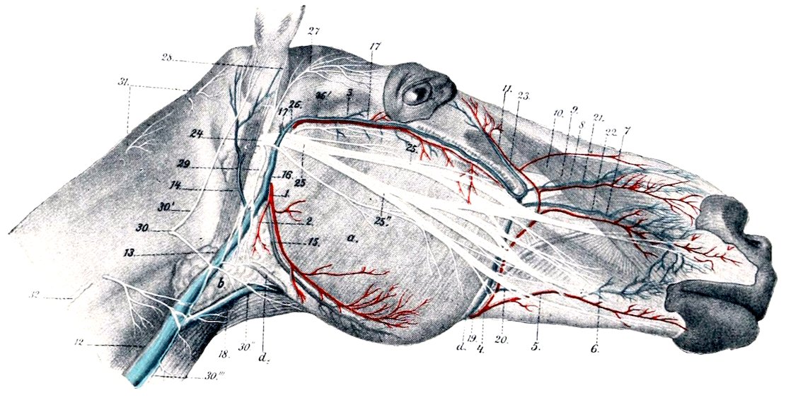
Fig. 461.—Superficial Vessels and Nerves of Head and Anterior Part of Neck of Horse. a, Masseter; b, parotid gland; c, parotido-auricularis; 1, masseteric artery; 2, parotid branch; 3, transverse facial artery; 4, facial artery; 5, 6, inferior labial artery; 7, superior labial artery; 8, lateral nasal artery; 9, infraorbital artery; 10, dorsal nasal artery; 11, angular artery of eye; 12, 13, jugular vein; 14, great auricular vein; 15, masseteric vein; 16, superficial temporal vein; 17, transverse facial vein; 17′, deep temporal vein; 18, external maxillary vein; 19, facial vein; 20, labial vein; 21, dorsal nasal vein; 22, lateral nasal vein; 23, angular vein of eye; 24, 25, facial nerve; 25′, 25″, superior and inferior buccal nerves; 26, transverse facial nerve; 27, auriculo-palpebral nerve; 28, anterior auricular nerve; 29, cervical branch of facial nerve; 30, ventral branch of second cervical nerve; 30′, 30″, 30‴, auricular and cutaneous branches of 30; 31, branches of dorsal division of first and second cervical nerves; 32, dorsal branch of spinal accessory nerve. (After Leisering’s Atlas.)
4. The great auricular vein (V. auricularis magna) is a satellite of the posterior auricular artery above, but joins the jugular a variable distance below and behind the point of origin of the artery.
5. The inferior cerebral vein (V. cerebralis ventralis) is an emissary of the cavernous sinus of the dura. It is a satellite of the internal carotid artery, receives the condyloid vein, and joins the jugular near the occipital vein or by a common trunk with it.
6. The occipital vein (V. occipitalis) arises in the fossa atlantis by the union of muscular and cerebrospinal branches. The former comes from the muscles of the poll and passes through the foramen transversarium. The latter is connected with the occipital sinus of the dura mater and emerges from the spinal canal by the intervertebral foramen.
7. The external maxillary or facial vein (V. maxillaris externa) arises by radicles which correspond in general to the branches of the artery of like name. It passes 589down over the cheek along the anterior border of the masseter muscle behind the artery, crossing over the parotid duct, which lies behind the vein lower down. Thus on the ramus and as they turn around its lower border the artery is in front, the vein in the middle, and the duct posterior. In the submaxillary space the vein is ventral to the artery for some distance, then parts company with the artery, runs straight backward along the lower border of the parotid gland, and opens into the jugular vein at the posterior angle of the gland.[171] The chief differences in the tributaries of the vein as compared with the branches of the corresponding artery are as follows:
The labial veins (Vv. labiales) form a plexus in the submucous tissue of the cheek from which two veins emerge. The upper one passes back and joins the buccinator vein. The lower one (V. labialis communis) joins the external maxillary vein.
Three veins connect with the external maxillary at the anterior border of the masseter.
The upper one is the transverse facial, which unites close to the end of the facial crest.
A little lower is the large valveless vena reflexa or alveolar vein. This passes back under the upper part of the masseter on the maxilla, turns around the tuber maxillare, perforates the periorbita, and joins the ophthalmic vein. It is relatively small at each end, but presents one or two large fusiform dilatations. It receives the following tributaries: (a) The palatine vein (V. palatina major), which separates from the palatine artery at the anterior palatine foramen and passes in the groove between the tuber maxillare and the palate bone. The palatine veins form a very rich plexus of valveless vessels in the submucosa of the hard palate, which consists of several layers anteriorly. (b) The sphenopalatine vein (V. sphenopalatina) forms a rich plexus of valveless vessels on the turbinal bones and the septum nasi. It is usually joined by the infraorbital vein (V. infraorbitalis) to form a short common trunk.
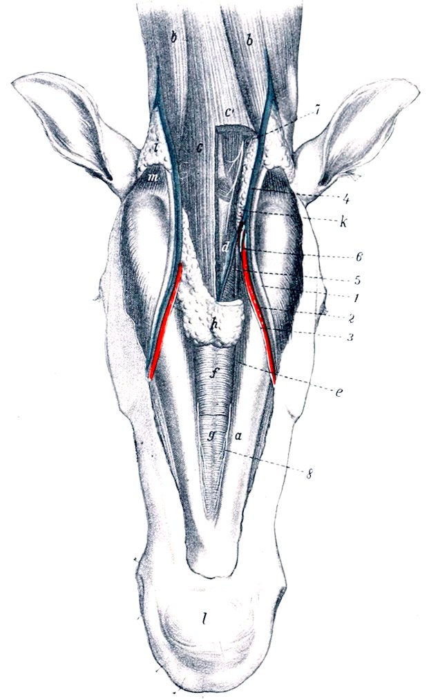
Fig. 462.—Dissection of Submaxillary Space and Adjacent Part of Neck of Horse. a, Ramus of mandible; b, sterno-cephalicus muscle; c, c′, omo-hyoidei and sterno-hyoidei (portion removed on right side); d, hyoid bone; e, anterior belly of digastricus; f, g, mylo-hyoideus; h, submaxillary lymph glands (portion removed on right side); i, parotid gland; k, submaxillary gland; l, chin; m, stylo-maxillaris; 1, parotid duct; 2, facial vein; 3, facial artery; 4, external maxillary vein; 5, sublingual vein; 6, sublingual artery; 7, ventral branch of first cervical nerve; 8, mylo-hyoid nerve. (After Ellenberger, in Leisering’s Atlas.)
The venous plexuses are remarkably developed in certain parts of the nasal mucosa. On the septum a little below its middle and on the turbinals the veins are in several layers. The olfactory region does not share in this arrangement and the veins here are small and join the ethmoidal vein.
590(c) The ophthalmic vein (V. ophthalmica) is a short trunk, connected in front with the vena reflexa and behind with the cavernous sinus through the foramen lacerum orbitale. It receives veins which correspond to the arterial branches.
The buccinator vein (V. buccinatoria) extends backward from the external maxillary along the lower border of the depressor labii inferioris and buccinator under cover of the masseter, passes between the ramus of the mandible and the tuber maxillare and is continued as the internal maxillary vein. It has a large fusiform dilatation and is valveless. It receives a large common labial vein from the labial plexus, and is usually connected with the masseteric vein.
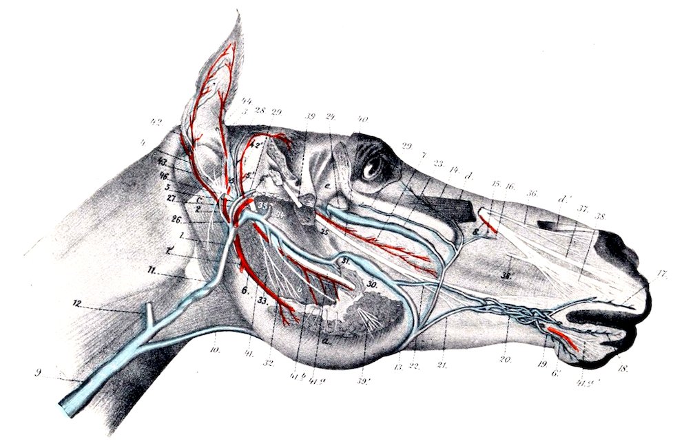
Fig. 463.—Deeper Vessels and Nerves of Head of Horse. The parotid gland, most of the masseter muscle, and a portion of the ramus of the mandible are removed. a, Remnant of masseter muscle; b, internal pterygoid muscle; c, stylo-mandibularis; d, d′, levator labii superioris proprius (portion removed); e, periorbita; 1, masseteric artery; 1′, parotid branch; 2, trunk for anterior (3) and posterior (4) auricular arteries; 5, 5′, superficial temporal artery; 5″, transverse facial artery; 6, inferior alveolar (or dental) artery; 6′, mental continuation of 6; 7, buccinator artery; 8, infraorbital artery; 9, 11, jugular vein; 10, external maxillary vein; 12, inferior cerebral vein; 13, facial vein; 14, angular vein of eye; 15, dorsal nasal vein; 16, lateral nasal vein; 17, superior labial vein; 18, 19, inferior labial veins; 20, labial plexus; 21, 22, emergent veins of plexus; 23, vena reflexa; 24, trunk of sphenopalatine and infraorbital veins; 25, palatine vein; 26, great auricular vein; 27, 28, superficial temporal vein; 29, transverse facial vein; 30, buccinator vein; 31, dorsal lingual vein; 32, inferior alveolar or dental vein; 33, pterygoid vein; 34, deep temporal vein; 35, superior cerebral vein; 36, external nasal nerve; 37, anterior nasal nerve; 38, superior labial nerve; 39, masseteric nerve; 39′, end branches of 39; 40, buccinator nerve; 41, pterygoid nerve; 41a, inferior alveolar or dental nerve; 41b, mylo-hyoid nerve; 42, posterior auricular nerve; 43, auricular branch of vagus; 44, internal auricular nerve; 45, auriculo-palpebral nerve (cut); 46, digastric nerve. (After Leisering’s Atlas.)
The lingual vein (V. lingualis) is not a satellite of the artery. It is formed at the side of the lingual process of the hyoid bone by the confluence of several veins which come from the substance of the tongue. One or two considerable vessels run partly in the substance of the hyo-glossus, and another in the genio-hyoideus. The vein is at first covered by the mylo-hyoideus, perforates that muscle, runs back along the omo-hyoideus in relation to the submaxillary lymph glands, and joins the external maxillary near the posterior border of the jaw. Near its termination it receives the sublingual vein, or the latter may open into the external maxillary directly.
8. The thyroid vein (V. thyreoidea) is a large vessel which joins the jugular near the external maxillary vein. It receives anterior thyroid, laryngeal, and pharyngeal radicles, and sometimes a posterior thyroid vein.
5919. Muscular, tracheal, and œsophageal veins.
10. The cephalic vein (Vena cephalica) enters the jugular near its termination. It will be described with the veins of the thoracic limb.
11. The inferior cervical vein (V. cervicalis ascendens) accompanies the artery. It may open into the brachial vein.
The Sinuses of the Dura Mater
These (Sinus duræ matris) are blood-spaces between the meningeal and periosteal layers of the dura mater and are lined with endothelium. In many places the lumen is crossed by fibrous strands. They receive the veins of the brain, communicate with the meningeal and diploic veins, and with veins outside of the cranium; their connections with the latter are by means of small emissary veins (Emissaria). They convey the blood directly or indirectly to the jugular veins. Some are paired, others unpaired. They may be divided into dorsal and basilar systems. The dorsal system comprises the following:
The superior longitudinal or sagittal sinus (S. sagittalis superior) is situated in the upper border of the falx cerebri along the internal sagittal crest. It begins at the crista galli and ends at the tentorium osseum by dividing into two transverse sinuses. It receives the superior cerebral veins. Along each side are small pouches (Lacunæ laterales) into which the veins open. The lumen of the sinus is traversed by fibrous bands and is partially divided by a longitudinal septum.
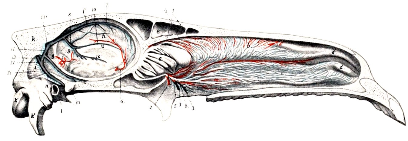
Fig. 464.—Median Section of Head of Horse, Upper Part with Septum Nasi Removed. a, Lateral mass of ethmoid bone; b, superior turbinal; c, inferior turbinal; d, d′, turbinal folds; e, frontal sinus; f, falx cerebri; g, tentorium cerebelli; h, medial surface of hemisphere; i, cerebellum; k, occipital bone; k′, occipital condyle; k″, paramastoid process; l, external auditory meatus; m, temporal condyle; n, parieto-temporal canal; 1, branches of ethmoidal artery; 2, 2′, branches of sphenopalatine artery; 3, 3′, branches of sphenopalatine vein; 4, branches of ethmoidal nerve; 5, 5′, branches of sphenopalatine nerve; 6, artery of corpus callosum; 7, superior sagittal sinus; 8, straight sinus; 9, vena magna cerebri; 10, inferior sagittal sinus; 11, 11′, transverse sinuses; 12, superior petrosal sinus; 13, superior occipital sinus; 14, superior cerebral vein; 15, corpus callosum; 16, fornix. (After Ellenberger, in Leisering’s Atlas.)
The transverse sinuses (S. transversi), right and left, pass outward in the transverse grooves of the parietal bones, enter the parieto-temporal canals, and are continued by the superior cerebral veins to the superficial temporal veins. The two sinuses are connected by the sinus communicans.
The inferior longitudinal or sagittal sinus (S. sagittalis inferior) runs backward on the upper surface of the corpus callosum along the concave edge of the falx cerebri and joins the great cerebral vein (of Galen) to form the straight sinus.
The straight sinus (S. rectus) passes upward and backward between the cerebral hemispheres and in the tentorium cerebelli and joins the superior sagittal sinus. The point of meeting is the confluence of the sinuses (Confluens sinuum).
The occipital sinuses (S. occipitales) lie on either side of the vermis cerebelli. They empty anteriorly into the sinus communicans and communicate behind with the spinal veins.
The superior petrosal sinuses (S. petrosi superiores) pass in the tentorium cerebelli to end in the transverse sinuses.
592The basilar system consists of the following:
The cavernous sinuses (S. cavernosi) lie in the inner grooves of the root of the temporal wings of the sphenoid bone at either side of the sella turcica. The two are connected by a wide traverse branch (Sinus intercavernosus) behind and below the posterior part of the pituitary body. Each is continuous in front with the ophthalmic vein and below with the inferior petrosal sinus. The third, sixth, and the ophthalmic and maxillary divisions of the fifth nerve lie along the outer wall of the sinus. The internal carotid artery traverses the sinus and is connected with its fellow by a transverse branch which lies in the corresponding part of the sinus. An oval opening in the floor communicates with the inferior petrosal sinus and transmits the internal carotid artery.
The sinus is not subdivided by strands of fibrous tissue as in man, but a few delicate bands attach the artery to its wall.
The inferior petrosal sinuses (S. petrosi inferiores)[172] lie along the borders of the basilar part of the occipital bone, inclosed in the thick dura which closes the foramen lacerum. The anterior part extends about half an inch (ca. 12 mm.) under the temporal wing of the sphenoid. Here communications exist with veins in the pterygo-palatine fossa. The posterior end is bulbous and lies in the condyloid fossa; it communicates with the condyloid vein issuing from the hypoglossal foramen and is drained by the inferior cerebral vein; it also receives an emissary vein from the parieto-temporal canal. The roof of the sinus is perforated by an oval opening at the carotid notch which opens into the cavernous sinus and transmits the internal carotid artery; the latter forms the first bend of its S-shaped curve in the petrosal and the second in the cavernous sinus.
The basilar plexus (Plexus basilaris) is a venous plexus situated on the upper surface of the basilar part of the occipital bone. It is connected through the hypoglossal foramen with the inferior petrosal sinus and communicates behind with the spinal veins.
The Veins of the Cranium
The veins of the brain (Venæ cerebri) do not in general accompany the cerebral arteries. They have very thin walls, no muscular coat, and no valves. They are arranged in two sets, superficial or cortical and deep or central. The superficial veins are more numerous and larger than the arteries. They lie on the surface of the brain in the pia mater and the subarachnoid space.
The superior cerebral veins drain the upper and outer part of the cerebral cortex. They receive veins from the medial surfaces of the hemispheres and open into the superior sagittal sinus. Their terminal parts are bulbous and their openings are directed obliquely forward, i. e., contrary to the direction of the bloodstream in the sinus.
The inferior cerebral veins lie on the inferior and external aspect of the hemispheres. They open into the inferior system of sinuses. The middle cerebral vein runs in the lateral fissure (of Sylvius) and opens into the cavernous sinus.
The deep cerebral veins issue from the central or ganglionic parts of the brain at the transverse fissure. They converge to form the great cerebral vein (of Galen) (V. magna cerebri). This passes upward and backward behind the splenium of the corpus callosum and is continued as the straight sinus to join the sagittal sinus.
The superior cerebellar veins ramify on the upper surface of the cerebellum. They open into the superior system of sinuses and the great cerebral vein. The inferior cerebellar veins are larger and go chiefly to the basilar plexus. The veins of the medulla and pons end in the inferior system of sinuses.
The meningeal veins (Venæ meningeæ) arise in capillary plexuses in the superficial and deep faces of the dura mater. Some end in the sinuses of the dura, others accompany the meningeal arteries.
593The diploic veins (Venæ diploicæ) are anastomosing channels in the spongy substance of the cranial bones. Their walls are thin, consisting in many places only of the endothelium, and they have no valves. Some open inward into venous sinuses, others into extracranial veins.
Spinal Veins
Two longitudinal spinal veins or sinuses (Sinus columnæ vertebralis) extend along the floor of the vertebral canal, one on either side of the superior common ligament. They are continuous in front with the basilar plexus. They lie in the grooves on the bodies of the vertebræ and are connected by a series of transverse anastomoses which pass between the central part of the bodies of the vertebræ and the superior common ligament or in channels in the bone. They receive veins from the spinal cord, the dura mater, and the bodies of the vertebræ (Venæ basis vertebræ). Through the intervertebral foramina efferent vessels connect with the vertebral, intercostal, lumbar, and lateral sacral veins.
VEINS OF THE THORACIC LIMB
The brachial vein (V. brachialis) is a satellite of the extrathoracic part of the brachial artery. It arises at the inner side of the distal end of the shaft of the humerus and passes upward in the arm behind the artery under cover of a layer of fascia and the posterior superficial pectoral muscle. At the shoulder it is ventral to the artery, crosses the anterior border of the first rib, and concurs with its fellow and the two jugulars in the formation of the anterior vena cava. The roots of the vein are somewhat variable, but most often four radial veins in addition to a large oblique branch from the cephalic unite in its formation. Its tributaries correspond in general to the branches of the artery, but a few differences are worthy of notice. The thoracico-dorsal vein joins the brachial directly or opens into the deep brachial. The external thoracic or “spur” vein (V. thoracica externa) is a large vessel which arises in the ventral wall of the abdomen, passes forward (embedded more or less in the panniculus) along the outer border of the posterior deep pectoral muscle, and joins the brachial vein near the first rib. It often communicates with the subscapular vein.
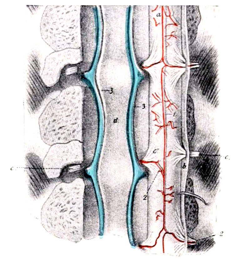
Fig. 465.—Spinal Vessels of Horse. The vertebral canal has been opened by sawing off the arches. The nerve-roots are cut on one side and the spinal cord turned over to right. 1, Ventral or middle spinal artery; 2, reinforcing branches from vertebral, intercostal, or lumbar arteries (according to region); 3, longitudinal vertebral sinuses; a, ventral surface of spinal cord; b, dura mater (cut); c, nerve-roots; d, superior common ligament. (After Ellenberger, in Leisering’s Atlas.)
The cephalic vein (V. cephalica) arises at the inner side of the carpus as the continuation of the internal superficial metacarpal vein. It runs upward on the deep fascia of the forearm at first in the furrow between the flexor carpi internus and the radius. Toward the middle of the forearm it inclines gradually forward on the inner surface of the radius, accompanied by a cutaneous branch of the median nerve, and arrives at the insertion of the biceps. Here it detaches a large branch (Ramus communicans) which passes upward and backward over the inner insertion of the biceps, the posterior radial artery, and the median nerve, and joins the brachial vein. The vein to this point is often termed the internal subcutaneous vein of the forearm (V. cephalica antebrachii). It is continued (as the V. cephalica humeri) in the furrow between the mastoido-humeralis and the anterior superficial pectoral with a branch of the inferior cervical artery, crosses the deep face of the 594cervical panniculus, and opens into the terminal part of the jugular or the brachial vein. It receives an accessory cephalic vein (V. cephalica accessoria), which arises from the carpal network, runs upward along the inner border of the extensor carpi, turns along the lower border of the brachialis, passes under the superficial tendon of the biceps and joins the cephalic.
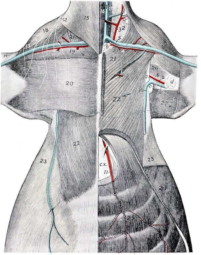
Fig. 466.—Dissection of Pectoral Region and Anterior Part of Abdominal Wall of Horse. 1, Jugular vein; 2, loose connective tissue of axillary space; 3, ascending branch of inferior cervical artery; 4, descending branch of same; 5, cephalic vein; 6, median nerve; 7, ulnar nerve; 8, brachial artery; 9, brachial vein; 10, external thoracic or “spur” vein; 11, anterior abdominal artery and vein; 12, branches of sixth cervical nerve; 13, cutaneous branch of axillary nerve; 14, cutaneous branch of musculo-cutaneous nerve; 15, cervical panniculus; 16, sterno-cephalicus; 17, scalenus; 18, mastoido-humeralis; 19, anterior superficial pectoral; 20, posterior superficial pectoral; 21, anterior deep pectoral; 22, posterior deep pectoral; 23, abdominal panniculus; 24, obliquus abdominis externus; 25, rectus abdominis; 26, coraco-brachialis; 27, tensor fasciæ antibrachii; a, prescapular lymph glands; b, prepectoral lymph glands; c, axillary lymph glands; d, cubital lymph glands; c.x., xiphoid cartilage. (After Schmaltz, Atlas d. Anat. d. Pferdes.)
595The deep veins of the forearm are variable. Commonly two posterior radial veins (Venæ medianoradiales) accompany the artery of like name, one in front and one behind. A third vein arises by radicles emerging from the proximal part of the deep flexor; it joins the posterior satellite of the posterior radial artery or forms one of the roots of the brachial vein. The common interosseous vein joins the posterior satellite. The anterior radial vein (V. collateralis radialis) is a satellite of the artery. The ulnar vein is usually double at its proximal part, and communicates with the deep brachial vein.
There are three chief metacarpal veins. The internal metacarpal vein (V. metacarpea volaris superficialis medialis) arises from the volar venous arch above the fetlock. It is the largest vein of the region and lies in front of the large metacarpal artery. It separates from the artery at the proximal end of the metacarpus, passes upward on the inner part of the posterior surface of the carpus, under cover of the superficial layer of the posterior annular ligament, and is continued as the cephalic vein. It communicates at its proximal part with the origin of the posterior radial veins by a short but relatively large branch. The external metacarpal vein (V. metacarpea volaris superficialis lateralis) arises from the venous arch above the fetlock and passes upward behind the external border of the suspensory ligament in front of the external branch of the median nerve and accompanied by a small artery. At the proximal end of the metacarpus it is connected with the deep metacarpal vein by two transverse anastomoses which pass across the suspensory ligament. It then passes upward as a satellite of the external volar metacarpal artery and concurs in the origin of the ulnar and posterior radial veins. The deep metacarpal vein (V. metacarpea volaris profunda medialis) arises from the venous arch, passes forward between the two branches of the suspensory ligament, and ascends on the posterior surface of the large metacarpal bone. At the proximal end of the latter it communicates with the other metacarpal veins, ascends with the small metacarpal artery, and concurs in forming the radial veins.
The venous arch (Arcus venosus volaris), from which the metacarpal veins arise, is situated above the sesamoids of the fetlock between the suspensory ligament and the deep flexor tendon. It is formed by the junction of the two digital veins.
The digital veins, internal and external (V. digitalis medialis, lateralis), drain the venous plexuses of the foot. They arise at the upper edge of the lateral cartilages and ascend in front of the corresponding arteries.
It is convenient to recognize the following venous plexuses of the foot, which, however, communicate very freely:
1. The coronary plexus encircles the upper part of the foot. It is attached to the terminal part of the extensor tendon, the lateral cartilages, and the bulbs of the plantar cushion.
2. The dorsal (or laminal) plexus covers the dorsal or wall surface of the third phalanx in the deep layer of the matrix of the wall of the hoof. It forms the circumflex vein of the third phalanx or vein of the distal border of the third phalanx, which corresponds to the artery of like name.
3. The volar plexus is in the deep layer of the matrix of the sole of the hoof and on the deep surface of the lateral cartilages. It communicates around the inferior border of the third phalanx with the dorsal plexus and through the lateral cartilages with the coronary plexus.
The deep vein of the third phalanx accompanies the terminal part of the digital artery. It drains the intraosseous plexus.
THE POSTERIOR VENA CAVA (Figs. 270, 450)
The posterior vena cava (V. cava caudalis) returns almost all of the blood from the abdomen, pelvis, and pelvic limbs. It is formed by the confluence of the right 596and left common iliac veins at the fifth lumbar vertebra, above the terminal part of the aorta and chiefly to the right of the median plane.
The mode of origin is variable. In some cases there is a common trunk formed by the union of the two internal iliac veins so that the arrangement resembles the termination of the aorta. In other cases the internal iliac vein does not exist.
It passes forward on the ventral face of the psoas minor to the right of the abdominal aorta. At the last thoracic vertebra it separates from the aorta and runs forward between the right crus of the diaphragm and the pancreas till it reaches the liver. Here it inclines downward along the inner border of the right lobe and the parietal surface of the liver, largely embedded in the gland substance, and passes through the foramen venæ cavæ of the diaphragm. It then runs forward and somewhat downward between the mediastinal lobe and the main mass of the right lung at the upper margin of a special fold of the right pleura, accompanied by the right phrenic nerve, and opens into the posterior part of the right atrium. It receives the following tributaries:
1. The lumbar veins (Vv. lumbales) correspond to the arteries. Five pairs usually empty into the vena cava. Sometimes the corresponding veins of opposite sides unite to form a common trunk. The first communicate with the vena azygos.
2. The spermatic veins (Vv. spermaticæ internæ) (Fig. 450) accompany the arteries of like name. In the spermatic cord they form the pampiniform plexus about the artery and nerves. Their termination is variable. The right one commonly joins the vena cava near the renal vein, often by a common trunk with the left one. Frequently the left vein joins the left renal, and sometimes the right one ends similarly.
2a. The utero-ovarian veins are much larger than the preceding and are satellites of the arteries in the broad ligaments. The ovarian branch is plexiform near the ovary. The uterine branches form a rich plexus in the wall of the uterus. The trunk is very short.
3. The renal veins (Vv. renales), satellites of the arteries, are of large caliber and thin-walled. The right vein passes inward and backward on the ventral face of the kidney between the artery in front and the ureter behind. It joins the vena cava above the right adrenal. The left vein is somewhat longer. It passes inward at first like the right one, then bends around the posterior end of the adrenal, crosses the origin of the renal artery and opens into the vena cava a little further back than the right one. They receive veins from the adrenals, but some adrenal veins open directly into the vena cava.
4. The hepatic veins (Vv. hepaticæ) return the blood from the liver, and open into the vena cava as it lies in the groove in the liver. Three or four large vessels open into the vena cava just before it leaves the liver, and numerous small ones discharge into its embedded part.
5. The phrenic veins (Vv. phrenicæ), two or three in number, return the blood from the diaphragm. They are very large in comparison with the arteries, and join the vena cava as it lies in the caval opening.
In some cases there is a small middle sacral vein (V. sacralis media), a satellite of the artery. It opens into the angle of junction of the common iliac veins or into one of the latter.