THE DIGESTIVE SYSTEM (continued 2)
Reading edition
The ventral portions of the colon have four longitudinal muscular bands (Tæniæ) which produce four rows of sacculations (Haustra). On the first part the external and internal bands are free; the dorsal band is covered by the adhesion to the overlying dorsal division, while the ventral band is largely covered by the adhesion to the cæcum. On the left ventral part the external and ventral bands are free; of the two dorsal bands, the inner one is concealed by the mesocolon, the outer one is chiefly free. The pelvic flexure has a band on its lesser curvature, which is continued on to the third part, concealed by the peritoneal attachment; these parts are practically non-sacculated. Near the diaphragmatic flexure two 369other bands appear, so that the flexure has three bands; two of these are dorsal and free, the third is ventral and concealed. The last part has three bands, of which the inner and outer ones are free, the ventral one covered.
The Small Colon
The small colon (Colon tenue) begins at the termination of the great colon, behind the saccus cæcus of the stomach and below the left kidney, and is continued by the rectum at the pelvic inlet. Its length is about ten to twelve feet (ca. 3.5 m.), and its diameter about three to four inches (ca. 7.5 to 10 cm.).
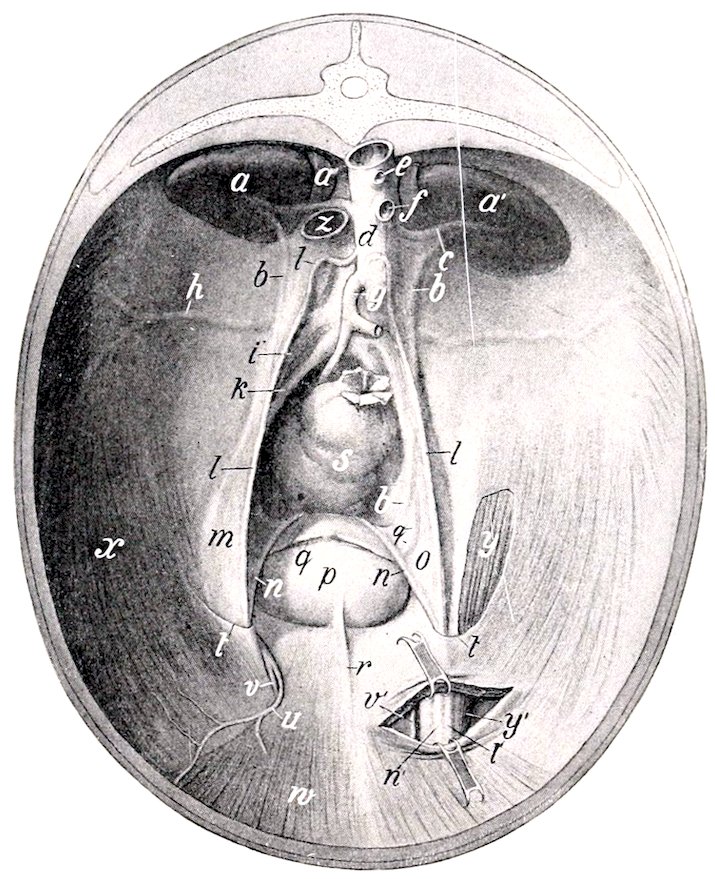
Fig. 272.—Pelvic Inlet and Posterior Part of Abdominal Wall of Horse, Viewed from the Front. The left inguinal canal is partially opened. The peritoneum is retained except over a part of the left cremaster muscle. The sublumbar region is greatly foreshortened in this view. a, a′, Kidneys; a″, adrenal; b., ureter; c, renal artery; d, aorta; e, cœliac artery (cut); f, anterior mesenteric artery (cut); g, posterior mesenteric artery; h, circumflex iliac artery; i, external iliac artery; k, internal iliac artery; l, spermatic artery in peritoneal fold (plica vasculosa), m; n, vas deferens, inclosed in urogenital fold, o; p, urinary bladder; q, round ligament of bladder; r, middle ligament of bladder; s, rectum; t, margin of vaginal ring; u, posterior abdominal artery; v, external pudic artery; w, rectus abdominis muscle; x, transversus and obliquus internus abdominis; y, y′, cremaster externus; z, posterior vena cava (cut). (After Ellenberger-Baum, Top. Anat. d. Pferdes.)
Its coils lie in the space between the stomach and the pelvic inlet, dorsal to the left portions of the great colon. They are mingled with those of the small intestine, from which they are easily distinguished by the tæniæ and sacculation.
It is attached to the sublumbar region by the colic mesentery, and to the termination of the duodenum by a short peritoneal fold (Lig. duodeno-colicum). The great omentum is also attached to the origin of the bowel. The colic mesentery is narrow at its origin, but soon reaches a width of about three feet (ca. 37080 to 90 cm.). Its parietal border is attached along a line extending from the ventral surface of the left kidney to the sacral promontory; it is continuous in front with the root of the great mesentery, and behind with the mesorectum.
There are two tæniæ and two rows of sacculations. Of the tæniæ, one is free, the other concealed by the mesentery. When the bowel is hardened in situ its lumen between the pouches is reduced to a narrow slit.
The Rectum
The rectum (Intestinum rectum) is the terminal part of the bowel; it extends from the pelvic inlet to the anus.[95] Its length is about one foot (ca. 30 cm.). Its direction may be straight or oblique. The first or peritoneal part of the rectum is like the small colon, and is attached by a continuation of the colic mesentery termed the mesorectum. The second or retroperitoneal part forms a flask-shaped dilatation termed the ampulla recti; it is attached to the surrounding structures by connective tissue and muscular bands.
The first part of the rectum is related to the small colon and the pelvic flexure of the great colon. It is frequently deflected to the left by the latter. The second part of the rectum is related dorsally and laterally to the pelvic wall. Ventrally the relations differ in the two sexes. In the male they are the bladder, the terminal parts of the vasa deferentia, the vesiculæ seminales, the prostate, the bulbo-urethral (Cowper’s) glands, and the urethra. In the female they are the uterus, vagina, and vulva.[96]
Structure of the Large Intestine.—The serous coat covers the different parts in varying degrees. It does not cover (a) the opposed surfaces of the cæcum and colon which are between the layers of the cæco-colic fold and mesocolon; (b) the areas of attachment of the cæcum and colon to the pancreas, right kidney, and sublumbar region; (c) the second part of the rectum.
The muscular coat consists of longitudinal and circular fibers. The bulk of the former is in the bands already described. Some of the circular fibers pass from one part of the colon to another, where they are attached to each other, forming the fibræ transversæ coli. The muscular coat of the second part of the rectum presents special features. The longitudinal layer of fibers is very thick and consists of large bundles, loosely united. A large band, the recto-coccygeus, is detached from it on either side, and passes upward and backward to be inserted into the fourth and fifth coccygeal vertebræ.
The submucous tissue is abundant in the wall of the rectum, so that the mucous membrane is loosely attached to the muscular coat, and forms numerous folds when the bowel is empty.
The mucous membrane of the large intestine is thicker and darker in color than that of the small intestine. It forms large crescentic or semilunar folds corresponding to the external constrictions. It has no villi, Brunner’s glands, or Peyer’s patches. The intestinal glands (of Lieberkühn) are large and numerous. Solitary glands are also numerous, especially at the apex of the cæcum and in the left dorsal part of the colon.
Blood-supply.—Greater and lesser mesenteric and internal pudic arteries. The veins go to the portal vein.
Nerve-supply.—Mesenteric plexus of the sympathetic nerves.
371The anus is the terminal orifice of the alimentary canal. It is situated below the root of the tail, where it forms a round projection, with a central depression when contracted. It is covered externally by an integument which is thin, hairless, and provided with numerous sebaceous and sweat glands. The mucous lining is pale, glandless, and covered with a thick, squamous, stratified epithelium.
There are three muscles of the anus.
1. The sphincter ani internus is merely a terminal thickening of the circular coat of the bowel.
2. The sphincter ani externus is a broad band of striped muscle-fibers outside the internal sphincter. Some fibers are attached to the coccygeal fascia above, others to the perineal fascia below. Its action is to close the anus.
3. The retractor ani (M. levator ani) is a flat muscle which lies between the rectum and the sacro-sciatic ligament. It arises from the superior ischiatic spine and the sacro-sciatic ligament, and ends under the external sphincter. Its action is to reduce the partial prolapse which the anus undergoes during defecation.
The suspensory ligament of the anus is a band of unstriped muscle which arises from the first coccygeal vertebra, passes downward over the retractor, and unites with its fellow below the anus. In the male it is largely continued by the retractor penis muscle; in the female it blends with the constrictor vulvæ. It may act as an accessory sphincter of the anus.
Blood-supply.—Internal pudic artery.
Nerve-supply.—Hæmorrhoidal and perineal nerves (for the sphincter ani externus and retractor ani).
THE PANCREAS
The pancreas is situated transversely on the dorsal wall of the abdomen, the greater part being to the right of the median plane. Its central part lies under the sixteenth and seventeenth thoracic vertebræ.
When fresh it has a reddish cream color, but if left in the unpreserved cadaver it rapidly decomposes and becomes dark. It resembles the salivary glands in appearance, but is softer, and its lobules are more loosely united. Its average weight is about twelve ounces (ca. 350 g.).
When hardened in situ its shape is very irregular. It is triangular in outline, and presents for description two surfaces, three borders, and three angles.[97]
The dorsal surface faces upward and forward. It is partially covered by peritoneum. It is related chiefly to the ventral surface of the right kidney and adrenal, the posterior vena cava, the portal vein, the cœliac artery and its divisions, the gastro-phrenic ligament and the saccus cæcus of the stomach, the right and caudate lobes of the liver, and the gastro-pancreatic fold. There are grooves for the divisions of the cœliac artery, and a large one for the splenic vein.
The ventral surface looks downward and backward; it is in general concave. It presents two impressions, separated by an oblique ridge. The smaller of these (Impressio cæcalis) lies to the right, and is caused by the pressure of the base of the cæcum; the larger one (Impressio colica) indicates the area of contact with the terminal part of the great colon and its junction with the small colon. It has usually no peritoneal covering except over a small area at the anterior angle.
The right border is nearly straight; it is related to the second part of the duodenum and the caudate lobe of the liver.
The left border is slightly concave, and is related to the first part of the duodenum, the left sac of the stomach, and the splenic vessels.
372The posterior border presents a deep notch to the right of the median plane for the portal vein, which passes through the gland very obliquely. There is a thin bridge of gland tissue dorsal to the vein, thus forming the portal ring (Annulus portarum). The root of the great mesentery is in contact with the border just to the left of the median plane.
The anterior or duodenal angle (Caput pancreatis) is attached to the concavity of the second curve of the duodenum, and the adjacent part of the right lobe of the liver. The ducts leave at this extremity.
The left or splenic angle (Cauda pancreatis) corresponds to the tail of the pancreas in man. It is in contact with the base of the spleen, the left kidney and adrenal body, and the saccus cæcus of the stomach.
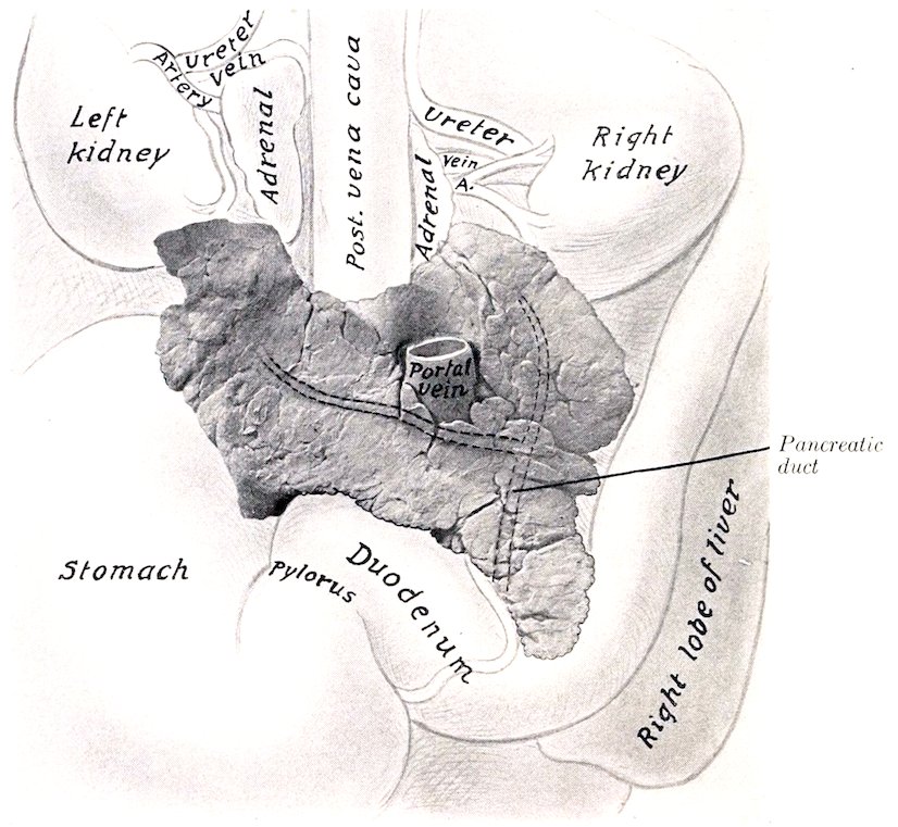
Fig. 273.—Pancreas of Horse with Chief Relations, Ventral View. The pancreatic duct and its two chief radicles are indicated by dotted lines, since they are in the substance of the gland.
The right angle is rounded, and lies on the ventral surface of the right kidney and adrenal body.
The pancreas is attached dorsally by connective tissue to the kidneys and adrenal bodies, the gastro-phrenic ligament and the suspensory ligament of the spleen, the posterior vena cava, the portal fissure, and the gastro-pancreatic fold. The ventral surface is mainly attached by areolar tissue to the base of the cæcum and the terminal part of the great colon.
There are almost invariably two ducts. The large one is termed the pancreatic duct (Ductus pancreaticus [Wirsungi]). It is formed by the union of two radicles which come from the right and left extremities, and passes through the duodenal angle to end at the duodenal diverticulum alongside of the bile-duct. The 373duct is nearly half an inch (ca. 1 cm.) wide, and is very thin-walled. It is situated in the substance of the gland near its dorsal surface; none of it is free. The accessory pancreatic duct (Ductus pancreaticus accessorius [Santorini]) arises either from the chief duct or its left radicle, and ends on a papilla in the duodenum opposite the chief duct.
Structure.—The pancreas belongs to the class of tubulo-alveolar glands, the alveoli being long, like those of the duodenal glands; in other respects it resembles the serous salivary glands very closely. It has no proper capsule and the lobules are rather loosely united.
Vessels and Nerves.—The arteries of the pancreas come from the branches of the cœliac and anterior mesenteric arteries. The nerves are derived from the cœliac and mesenteric plexuses of the sympathetic.
THE LIVER
The liver (Hepar) is the largest gland in the body. It is situated obliquely on the abdominal surface of the diaphragm. Its highest point is at the level of the right kidney, its lowest on the left side, usually about three or four inches (ca. 8 to 10 cm.) from the abdominal floor, opposite the lower end of the seventh or eighth rib. The greater part of it lies to the right of the median plane.
It is red-brown in color and is rather friable. Its average weight is about ten to twelve pounds (ca. 5 kg.). When in the body, or if hardened in situ, it is strongly curved and accurately adapted to the abdominal surface of the diaphragm. When removed in the soft state, it flattens out into a cake-like form quite different from its natural configuration. It presents for description two surfaces and a circumference, which may be divided into four borders.
The parietal surface (Facies diaphragmatica) is strongly convex, and lies against the diaphragm. It faces chiefly upward and forward. It presents, just to the right of the median plane, a sagittal groove for the posterior vena cava (Fossa venæ cavæ). The vein is partially embedded in the substance of the gland, and receives the hepatic veins.
The visceral surface (Facies visceralis) faces in general downward and backward; it is concave and irregular, being moulded on the organs which lie against it. It presents, a little to the right of the median plane, the portal fissure (Porta hepatis). Through this the portal vein, hepatic artery, and hepatic plexus of nerves enter, and the hepatic duct and lymph vessels leave the liver. The portal or hepatic lymph glands are also found here. The pancreas is attached at and to the right of the fissure, and the gastro-hepatic omentum to the left of it. Above the fissure is a ridge which represents the caudate lobe (Lobus caudatus Spigelii), and is continued to the right by the pointed caudate process (Processus caudatus). Further to the left there is a large depression (Impressio gastrica) for the stomach. To the right of this may be seen a groove passing to the right and dorsally; this is the duodenal impression (Impressio duodenalis). Ventral to these is a large depression for the great colon (Impressio colica). Dorsal to this is a smaller depression for the blind end of the base of the cæcum.[98] Coils of the small intestine may also lie on this surface, and the apex of the spleen may reach to it when the stomach is empty.
The dorsal border (Margo obtusus) is thick for the most part. It presents from right to left: (1) a depression for the right kidney (Impressio renalis); (2) a notch, which is the dorsal end of the fossa venæ cavæ; (3) a deep notch (Impressio 374œsophagea) which is mainly occupied by the thick margin of the œsophageal opening of the diaphragm.
The ventral border is thin, and is marked by two deep interlobar fissures or incisures (Incisuræ interlobares), which partially divide the organ into three principal lobes—right, middle, and left. The right lobe is the largest, except in old subjects, in which it is frequently much atrophied. The middle lobe is the smallest. It is marked by several small fissures, and by the umbilical fissure (Incisura umbilicalis); the latter contains the umbilical vein in the fœtus, which is transformed into the round ligament after birth.
The right border is thin and long. It is nearly vertical, and extends backward to about the middle of the sixteenth rib.
The left border is thin and short. It extends backward to a point opposite the lower part of the ninth or tenth rib. The ventral and lateral borders together constitute the margo acutus.
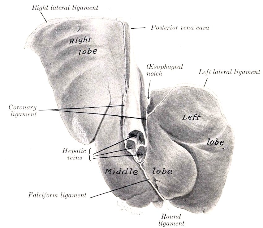
Fig. 274.—Liver of Young Horse, Hardened in situ, Parietal Surface.
The liver is held in position largely by the pressure of the other viscera and by its close application to the diaphragm. It has six ligaments.
1. The coronary ligament (Lig. coronarium hepatis) attaches it closely to the diaphragm. It consists of two laminæ. The right one is attached to the right of the fossa venæ cavæ; the left one begins to the left of the vena cava and passes upward and outward, becoming continuous with the left lateral ligament at the left margin of the œsophageal notch; it detaches a middle fold which extends to the notch and is continuous with the small omentum. The two laminæ unite below the vena cava to form the next ligament.
2. The falciform ligament (Lig. falciforme hepatis) is a crescentic fold which attaches the middle lobe to the sternal part of the diaphragm and to the abdominal floor for a variable distance. In its concave free edge is found.
3753. The round ligament (Lig. teres hepatis), a fibrous cord which extends from the umbilical fissure to the umbilicus; it is the vestige of the umbilical vein, which in the fœtus carries the blood from the placenta to the liver.
4. The right lateral ligament (Lig. triangulare dextrum) is a wide fold which attaches the dorsal border of the right lobe to the costal part of the diaphragm.
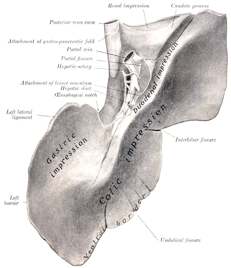
Fig. 275.—Liver of Horse, Visceral Surface. Specimen from middle-aged subject, hardened in situ.
5. The left lateral ligament (Lig. triangulare sinistrum) attaches the dorsal edge of the left lobe to the tendinous center of the diaphragm.
6. The hepato-renal or caudate ligament (Lig. hepatorenale) attaches the caudate process to the right kidney and the base of the cæcum. The gastro-hepatic omentum and the mesoduodenum have been described.
As stated above, the liver is divided by fissures into three principal lobes—right, middle, and left. The right lobe is the largest in the young subject and is 376irregularly quadrilateral in form. On its dorsal part is the caudate lobe, which ends in a pointed process directed outward, and assists in forming the cavity for the right kidney. The middle lobe is normally much the smallest. The left lobe is oval in outline and thickest centrally. In old or middle-aged subjects it often exceeds the right one in size. In some cases the atrophy of the right lobe is so extreme that the middle lobe may exceed it in size.[99]
The hepatic duct (Ductus hepaticus) is formed at the ventral part of the portal fissure by the union of right and left chief lobar ducts. It is two or three inches (ca. 5 to 8 cm.) long and about half an inch (ca. 1 to 1.5 cm.) wide. It passes between the two layers of the lesser omentum, and pierces the wall of the duodenum about five or six inches (ca. 12 to 15 cm.) from the pylorus, alongside of the pancreatic duct. The ducts pass obliquely through the wall of the duodenum for about half an inch (ca. 1 cm.) before opening into the diverticulum duodeni. The arrangement forms an effective valve, which prevents regurgitation from the intestine. There is no gall-bladder.
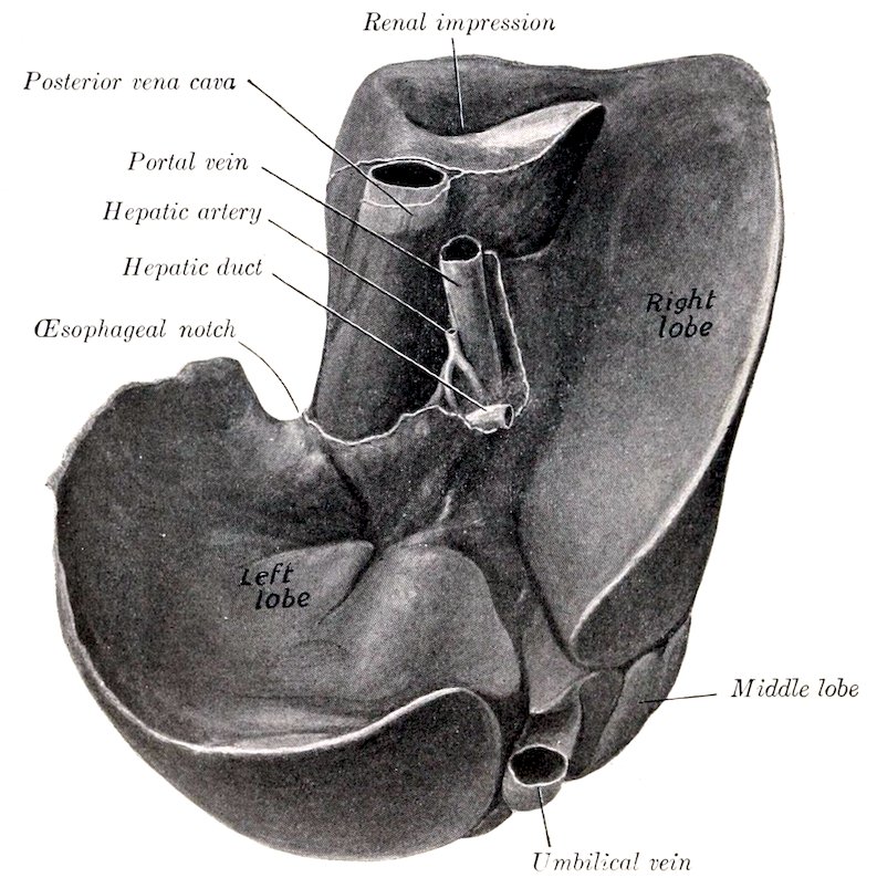
Fig. 276.—Liver of New-born Foal, Hardened in situ. Visceral Surface. The differences, when compared with the organ in the adult, are very striking.
Structure.—The liver is covered by an outer serous, and an inner fibrous coat. The serous coat covers the gland except at the attachment of the pancreas and at the portal fossa; it is reflected from it to form the ligaments and the lesser omentum. The fibrous capsule is in general thin; it sends laminæ into the ligaments, and also 377delicate strands into the gland substance. At the portal fissure it is abundant and surrounds the vessels and ducts, which it accompanies in the portal canals of the gland substance.
The gland substance is composed of the parenchyma and the interstitial tissue. The parenchyma is made up of lobules, 1 to 2 mm. in diameter, which are held together by a small amount of interlobular connective tissue. On account of the very small amount of the latter, the lobulation of the horse’s liver is not usually at all distinct to the naked eye; for the same reason the organ is also quite friable.[100]
Vessels and Nerves.—The portal vein enters at the portal fissure. It conveys blood from the digestive tract and the spleen, which contains various products of digestion and numerous white blood-cells. The hepatic artery also enters at the portal fissure; it may be termed the nutrient vessel. All the blood is returned from the liver to the posterior vena cava by the hepatic veins. The portal vein and the hepatic artery both divide into interlobular branches, which run together in the portal canals of the interlobular tissue. The branches of the portal vein (Venæ interlobulares) give off intralobular branches which form plexuses of capillaries in the lobules and give rise to a central vein (Vena centralis). The branches of the hepatic artery (Rami arteriosi interlobulares) are of relatively small size. They supply mainly (if not exclusively) the interlobular tissue, the capsule, and the walls of the vessels and ducts. The hepatic veins[101] (Venæ hepaticæ) empty into the vena cava as it lies in the fossa of the gland. Their ultimate radicles are the central lobular veins, which emerge from the bases of the lobules and join the sublobular veins (Venæ sublobulares); the latter unite to form the hepatic veins. The largest hepatic veins, three or four in number, join the posterior vena cava just before it leaves the liver to pass through the diaphragm.
The nerve-supply comes from the hepatic plexus, composed of branches from the vagus and sympathetic nerves.
THE SPLEEN
The spleen (Lien) is the largest of the ductless glands.[102] It is situated chiefly in the left parachondrium, in close relation to the left part of the great curvature of the stomach, to which its long axis corresponds. Its size and weight vary greatly in different subjects, and also in the same subject under different conditions, depending chiefly on the great variability of the amount of blood contained in it. The average weight is about 35 ounces (ca. 1 kg.), its length about 20 inches (ca. 50 cm.), and its greatest width about 8 to 10 inches (ca. 20 to 25 cm.). It is usually bluish-red or somewhat purple in color. In the natural state it is soft and yielding, but not friable.
The weight appears to vary ordinarily from about one to eight pounds, although in large horses the latter figure even may be exceeded without any apparent evidence of disease. There does not seem to be any constant relation to the body-weight. For example, the spleen of a colt about ten months old weighed three and a half pounds, while it often weighs less than two 378pounds in horses weighing 1000 to 1200 pounds. The chief variation in outline consists of increase of width, especially of the dorsal part.
It extends obliquely in a curved direction from the left crus of the diaphragm and the saccus cæcus of the stomach to the ventral third of the ninth or tenth rib. It presents for description two surfaces, two borders, and two extremities.
The parietal or external surface (Facies parietalis) is convex, and lies chiefly against the diaphragm, but is in direct contact with the upper parts of the last two ribs and to a small extent with the flank at the lumbo-costal angle.
The visceral or internal surface (Facies visceralis) is in general concave. It is divided into two unequal portions by a longitudinal ridge; on this is a groove, the hilus, in which the vessels and nerves are situated. The area in front of the ridge (Facies gastrica) is moulded on the great curvature of the stomach; it is about two inches (ca. 5 cm.) wide. The area behind the ridge (Facies intestinalis) is much more extensive; it is related chiefly to the small colon, the small intestine, and the great omentum. It may be marked by one or two fissures.
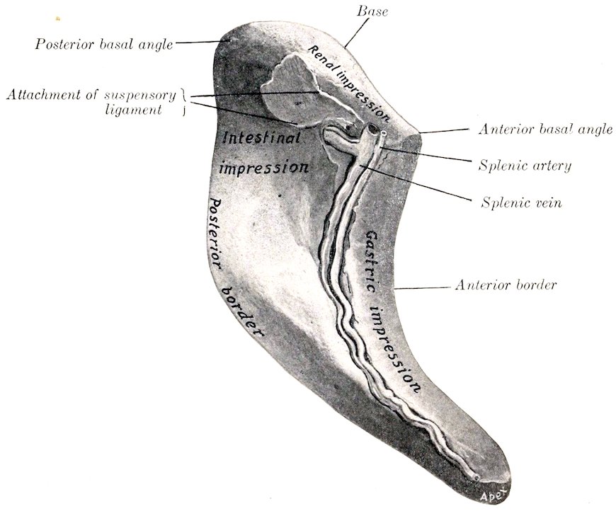
Fig. 277.—Spleen of Horse, Hardened in situ. Visceral Surface. The area marked intestinal impression is related to the first coil of the small colon.
The anterior border (Margo anterior) is concave and thin.
The posterior border (Margo posterior) is convex and thin.
The base or dorsal extremity is beveled, and fits into the interval between the left kidney and the left crus of the diaphragm and the psoas major. When hardened in situ, it shows an impression (Facies renalis) where it lies against the kidney. The left extremity of the pancreas touches it also. The anterior basal angle fits in between the saccus cæcus of the stomach and the left kidney; the posterior basal angle usually lies against the flank just behind the last rib.
The apex or ventral extremity is small; it lies—when the stomach is not full—between the left lobe of the liver and the left dorsal portion of the colon; when 379the stomach is full, the spleen is pushed further back and loses contact with the liver.
The spleen is attached by two peritoneal folds, the suspensory ligament and the gastro-splenic omentum. The suspensory ligament (Lig. suspensorium lienis) attaches the base to the left crus of the diaphragm and the left kidney; it contains a quantity of elastic tissue. The part which passes to the diaphragm is the ligamentum phrenico-lienale, and blends with the gastro-phrenic ligament; the part which goes to the kidney is termed the ligamentum renolienale. The gastro-splenic omentum (Lig. gastrolienale) passes from the hilus to the left part of the great curvature of the stomach. It is narrow above, where it joins the suspensory ligament; below it becomes much wider and is continuous with the great omentum.
Small globular or lenticular masses of splenic tissue may be found in the gastro-splenic omentum. They are termed accessory spleens (Lienes accessoriæ).
Structure.—The spleen has an almost complete serous coat. Subjacent to this and intimately united with it is a capsule of fibrous tissue (Tunica albuginea), which contains many elastic fibers and some unstriped muscular tissue. Numerous trabeculæ (Trabeculæ lienis) are given off from the deep face of the capsule and ramify in the substance of the organ to form a supporting network. In the interstices of this framework is the spleen pulp (Pulpa lienis), a dark red, soft, grumous material. This is supported by a delicate adenoid reticulum, and contains numerous leukocytes, the large splenic cells, red blood-corpuscles, and pigment. The pulp is richly supplied with blood. The branches of the splenic artery enter at the hilus and pass along the trabeculæ. The arteries which enter the pulp have a sheath of lymphoid tissue, which collects on the vessel wall at certain points, forming small lymph nodules, the so-called Malpighian corpuscles (Noduli lymphatici lienales). These are visible to the naked eye as white spots, about as large as the head of a pin. The blood passes into cavernous spaces lined by endothelium which is continuous with the cells of the reticulum of the pulp. From these the veins arise. The splenic vein runs in the hilus in company with the artery and nerves, and joins the posterior gastric vein to form a large radicle of the portal vein.
Blood-supply.—Splenic artery.
Nerve-supply.—Splenic plexus.
THE PERITONEUM
The general disposition of the peritoneum has been described, and other facts in regard to it were mentioned in the description of the viscera. It is now desirable to study it as a continuous whole.[103] (Figs. 256, 257, 278, 279.)
We may consider the peritoneum as consisting of two sacs—a greater and a lesser. The greater sac lines the greater part of the abdominal cavity, and covers most of the viscera which have a peritoneal investment. The lesser sac is an introversion or recess of the greater sac, formed during the development of the viscera. The two sacs communicate by a relatively narrow passage, termed the epiploic foramen of Winslow (Foramen epiploicum). This opening is situated on the visceral surface of the liver above the portal fissure. It can be entered by passing the finger along the caudate lobe of the liver toward its root. Its dorsal (or anterior) wall is formed by the caudate lobe and the posterior vena cava. Its ventral (or posterior) wall consists of the pancreas, the gastro-pancreatic fold, and the portal vein. The walls are normally in contact, and the passage merely a potential one. It is usually about four inches (ca. 10 cm.) in length. It is narrowest at the right 380extremity, where it is about an inch (ca. 2.5 to 3 cm.) wide.[104] If the finger is passed into the foramen from right to left, it enters the cavity of the lesser sac. If now an opening is made in the great omentum and the other hand introduced through it, the fingers of the two hands touch each other over the lesser curvature of the stomach. The formation and boundaries of the lesser sac should now be examined by spreading out the great omentum. It will be found that the latter now incloses a considerable cavity behind the stomach; this is termed the omental cavity (Bursa omentalis). Passing forward over the lesser curvature of the stomach, we enter another space, the vestibule of the omental cavity (Vestibulum bursæ omentalis). This space is closed on the left by the gastro-phrenic ligament, below and on the right by the lesser omentum, and dorsally by the gastro-pancreatic fold, which is attached to the dorsal border of the liver and to the posterior vena cava. Above the œsophageal notch the fingers can be passed around the border of the liver and the vena cava till the coronary ligament is encountered. Thus the vestibule is closed except (1) on the right, where it communicates with the cavity of the greater sac by the epiploic foramen; and (2) behind, where it communicates with the cavity of the omentum.
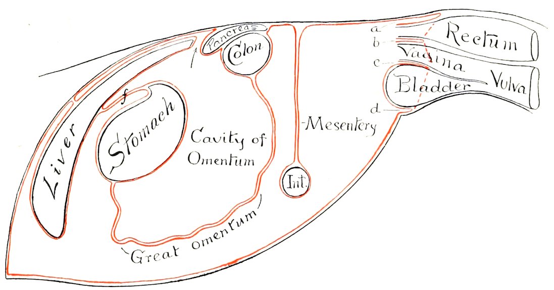
Fig. 278.—Diagram of General Arrangement of Peritoneum (of Mare) in Sagittal Tracing. a, Pouch between rectum and roof of pelvis, continuous with b, recto-genital pouch; c, vesico-genital pouch; d, pouch below bladder and its lateral ligaments; f, lesser omentum; Int., small intestine. The arrow points to the epiploic foramen (of Winslow).
The general arrangement of the great omentum has already been indicated. We may now trace its line of attachment, which would correspond to the mouth of the sac. Beginning at the ventral part of the great curvature of the stomach, the line passes to the ventral face of the pylorus, then crosses obliquely the first part of the duodenum to the point where the pancreas is adherent to it. Here it passes to the anterior face of the terminal transverse part of the great colon, runs along this transversely (from right to left), and continues for some ten or twelve inches (ca. 25 to 30 cm.) on the small colon. It then forms an acute angle, passes inward and forward along the small colon to the dorsal part of the hilus of the spleen, where it blends with the suspensory ligament of the latter, and forms a recess (Recessus lienalis) behind the saccus cæcus of the stomach. It now passes along the hilus of the spleen, and is continued to the great curvature of the stomach by the gastro-splenic omentum. It is convenient to regard the spleen as being intercalated 381in the left part of the great omentum; on this basis the gastro-splenic omentum would be that part of the great omentum which connects the hilus of the spleen with the great curvature of the stomach. The great omentum is relatively small in the horse, and is usually not visible when the abdomen is opened. It is generally folded up in the space between the visceral surface of the stomach and the intestine.[105]
The lesser sac furnishes the peritoneal covering for: (1) the visceral surface of the stomach and a small area of the first curve of the duodenum; (2) a large part of the dorsal surface of the pancreas and portal vein; (3) a small part of the visceral surface of the liver above the attachment of the lesser omentum and the portal fossa; (4) the posterior vena cava, from the level of the epiploic foramen [of Winslow] to its passage through the diaphragm (in so far as it is not embedded); (5) the part of the parietal surface of the liver between the right and middle divisions of the coronary ligament; (6) the corresponding part of the diaphragm, and the right part of the right crus of the same; (7) part of the anterior surface of the terminal part of the great colon, and the origin of the small colon; (8) the left extremity of the pancreas (inconstant); (9) the spleen.
We may now trace the peritoneum in a longitudinal direction, beginning in front. It is reflected from the ventral abdominal wall and the diaphragm upon the liver, forming the ligaments and serous coat of the gland. It leaves the visceral surface of the liver as lesser omentum, and the crura of the diaphragm as the gastro-phrenic ligament, reaches the saccus cæcus and lesser curvature of the stomach and the first curve of the duodenum, covers these organs, and is continued by the great omentum.
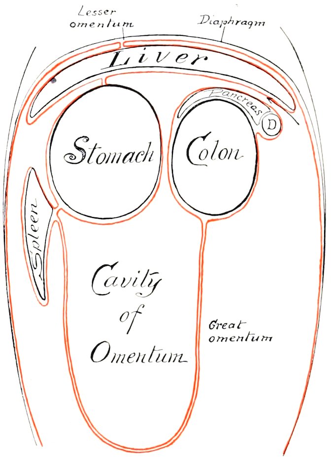
Fig. 279.—Diagram of Abdominal Peritoneum in Frontal (Horizontal) Tracing. D, Duodenum. The arrow indicates the epiploic foramen (of Winslow). By an oversight the leader line to the great omentum is omitted, and the coronary ligament of the liver is erroneously marked lesser omentum; the latter extends from liver to stomach, but is not marked.
On the left it passes from the left crus of the diaphragm and the left kidney to form the suspensory ligament of the spleen, clothes that organ, and leaves it to be continued by the great omentum.
On the right it passes from the right crus of the diaphragm and the dorsal border of the liver to the concave border of the duodenum, forming the gastro-pancreatic fold (second part of the mesoduodenum), and covering part of the dorsal surface of the pancreas. From the margin of the pancreas, the right kidney, and a small area of the sublumbar region behind the latter, it passes on to the base of the cæcum and the terminal part of the great colon. From these it passes on the right to the duodenum, forming the third part of the mesoduodenum. On the left it clothes part of the ventral surface and the outer border of the left kidney, from which it passes to the base of the spleen, forming the ventral layer of the suspensory ligament of the latter. Behind the terminal part of the great colon it is reflected around the great mesenteric artery to form the great mesentery. Behind this it is reflected almost transversely from the roof of the cavity and from the origin of the small colon on to the duodenum, forming the terminal part of the mesoduodenum. 382The line of origin of the colic mesentery begins on the inner part of the ventral surface of the left kidney, and extends to the sacral promontory, where the mesorectum begins. At the termination of the latter the peritoneum is reflected from the rectum on the dorsal and lateral walls of the pelvic cavity. Below the rectum it forms the urogenital fold, and passes on to the dorsal surface of the bladder, covers its anterior part, and is reflected on to the body-wall laterally and ventrally, forming the lateral and middle ligaments of the bladder. In the female the broad ligaments of the uterus replace the urogenital fold, with which they are homologous.
In the new-born foal certain folds are specially large. The falciform ligament of the liver extends to the umbilical opening, and contains in its free edge the large umbilical vein. The bladder—at this time an abdominal organ—has a ventral median fold, which connects it and the urachus with the abdominal floor. This is flanked on either side by a fold which also extends to the umbilicus, and contains the large umbilical artery.
DIGESTIVE SYSTEM OF THE OX
THE MOUTH
The cavity of the mouth is shorter and wider than that of the horse, and the vestibule is more capacious.
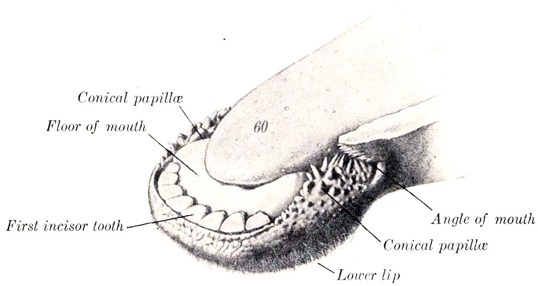
Fig. 280.—Anterior Part of Lower Jaw of Ox. 60, Tip of tongue. (After Ellenberger-Baum, Anat. für Künstler.)
The lips are thick, wide, and comparatively immobile. The middle part of the upper lip and the surface between the nostrils is bare, and is termed the muzzle (Planum nasolabiale). It is smooth, and (in health) is kept cool and moist by a clear fluid secreted by a layer of subcutaneous glands (Glandulæ nasolabiales) about half an inch (ca. 1.5 cm.) thick. It shows irregular lines, mapping out small polygonal areas on which the orifices of the gland-ducts are visible. A narrow bare strip also exists along the edge of the lower lip. The remainder of the integument is provided with ordinary and tactile hairs. The free edge and the lining membrane present verrucose, horny papillæ. The labial glands form compact masses near the angles of the mouth.
The cheeks are more capacious than in the horse. The mucous membrane presents large conical pointed papillæ, which are directed toward the fauces and are covered with a horny epithelium. The largest of these have a length of about 383half an inch (ca. 1 to 1.5 cm.) and are situated around the angle of the mouth and parallel with the cheek teeth. The orifice of the parotid duct is opposite the fifth upper cheek tooth. (In the sheep and goat it is opposite the fourth tooth.) The buccal or molar glands are very well developed, and are arranged in three parts. The dorsal row extends from the maxillary tuberosity to the angle of the mouth. Its lobules are of a light yellow color. The ventral part consists of a compact brownish mass which reaches from the angle of the mouth a short distance under the masseter muscle. The middle part consists of loosely arranged yellow lobules.
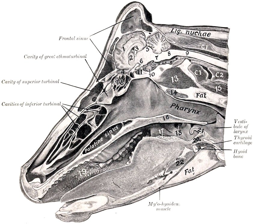
Fig. 281.—Sagittal Section of Head of Cow, Cut a little to the Right of the Median Plane. 1, Cerebral hemisphere; 2, corpus striatum; 3, hippocampus; 4, olfactory bulb; 5, corpora quadrigemina; 6, optic nerve; 7, pons; 8, medulla oblongata; 9, spinal cord; 10, pituitary body; 11, sphenoid sinus; 12, lateral mass of ethmoid; 13, ventral straight muscles; 14, pharyngeal (retropharyngeal) lymph gland; 15, longus colli; 16, soft palate; 17, vallate papillæ; 18, tonsillar sinus; 19, conical papillæ of cheek; 20, hyo-epiglotticus muscle; 21, epiglottis; 22, hyo-glossus muscle; C1, C2, atlas, axis. Subject was hardened with mouth open.
A linear series of large papillæ exists on the floor of the mouth on each side of the frenum linguæ. Near these are found the openings of the small ducts of the sublingual gland. The papilla on which the submaxillary duct opens is wide, hard, and has a serrated edge.
The hard palate is wide, and is usually more or less pigmented. The body of the premaxilla is covered with a thick layer of dense connective tissue, which has a 384thick, horny epithelial covering—forming the so-called dental plate or pad. The palatine ridges extend from this backward about two-thirds of the length of the hard palate. They are nearly straight, and, for the most part, are serrated on the free edge. A median furrow extends between the ridges. The posterior third of the palate is smooth. Between the dental plate and the first ridge is the triangular papilla incisiva; on either side of this is a deep furrow, in which is the oral opening of the naso-palatine canal. This canal is two inches or more (ca. 5 to 6 cm.) in length and opens on the floor of the nasal cavity; it also communicates by a slit-like opening with the organ of Jacobson.
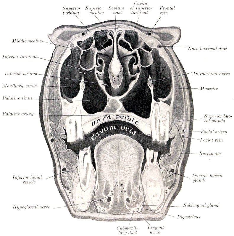
Fig. 282.—Cross-section of Head of Ox. The section passes through the internal canthi. L.a., Lingual arteries. The arrow indicates the communication between the maxillary and palatine sinuses.
The soft palate is somewhat shorter than that of the horse, but is long enough to close the isthmus of the fauces. The posterior pillars do not extend to the entrance of the œsophagus. The azygos muscle is much better 385developed than in the horse. The fibrous aponeurosis is for the most part replaced by muscular tissue.
The isthmus faucium is wide and dilatable. On either side, behind the anterior pillar of the soft palate, is a deep depression, the sinus tonsillaris; external to this is the compact bean-shaped tonsil, which is about one to one and a half inches (ca. 3 to 4 cm.) in length. The tonsil does not project into the fauces, but outward instead: hence it does not occupy the tonsillar sinus, and is not visible internally, as is the case in most animals.
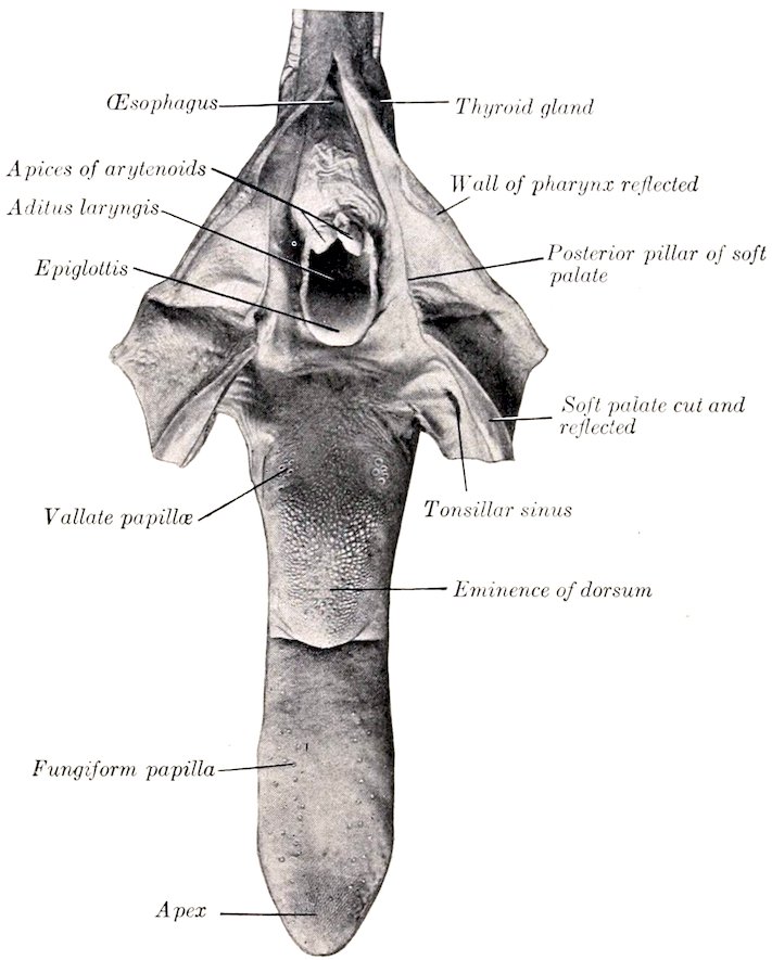
Fig. 283.—Tongue of Ox, Dorsal Aspect. Pharynx, soft palate, and origin of œsophagus are cut dorsally and reflected.
THE TONGUE
The tongue of the ox is often variably pigmented. The root and body are wider than that of the horse, but the free part is more pointed. The posterior part of the dorsum forms a remarkable elliptical prominence, which is sharply defined in front. The filiform papillæ in front of this prominence are large and horny, with sharp points directed backward. They impart to the tongue its rasp-like roughness. The papillæ on the prominence are large, broad and horny; some have a blunt conical form, others are rounded or flattened. Behind the prominence, the papillæ are long and soft, i. e., not horny. The fungiform papillæ are numerous and distinct; they are scattered more generally over the dorsum and edges of the free 386part. The vallate papillæ are about twenty or thirty in number; they are smaller than those of the horse, and are irregularly distributed on either side of the posterior part of the prominence of the dorsum. The foliate papillæ and the lingual fibrous cord are absent. The muscles are well developed; the hyo-glossus arises by additional portions from the great and middle cornua of the hyoid bone. The tongue is highly protractile and is the chief organ of prehension.
THE TEETH
The dental formula of the ox is:
( 0 0 3 3 )
2 ( I C P M ) = 32
( 4 0 3 3 )
The incisors are absent from the upper jaw. There are eight incisors in the lower jaw, arranged in a somewhat fan-like manner. They are simple teeth, without infundibulum. The crown is white, short, and shovel-shaped. The root is rounded, and is embedded in the jaw in such a manner as to allow a considerable degree of movement. There is a distinct neck. In addition to the simple numerical designation, the following terms are commonly applied to the individual teeth: pinchers or central, first intermediate, second intermediate, and corner incisors. It is probable that the latter are much modified canines. The incisors of the ox do not advance out of the alveoli, as is the case in the horse; in old age, however, the gum retracts so that the roots are partly exposed and may come into wear.[106]
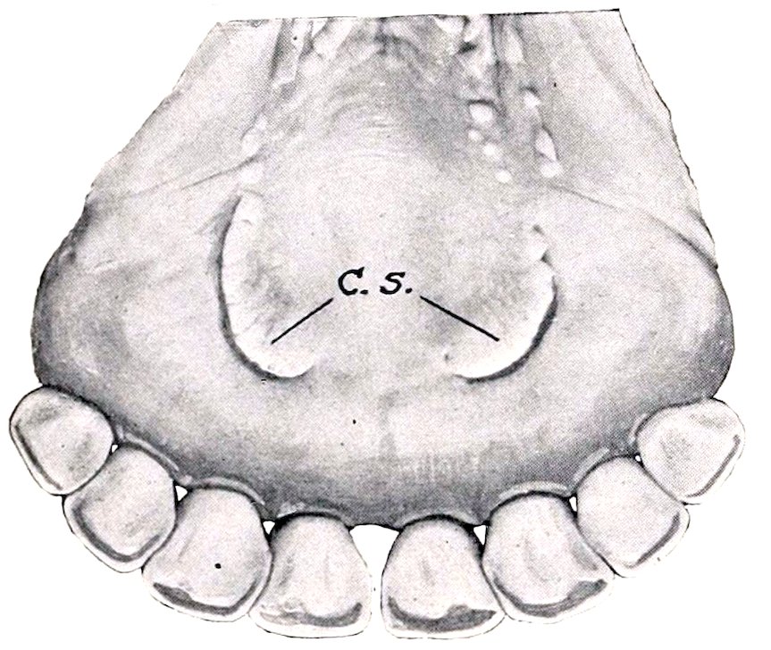
Fig. 284.—Incisor Teeth of Ox, Lingual Aspect. C.s., Caruncula sublingualis.
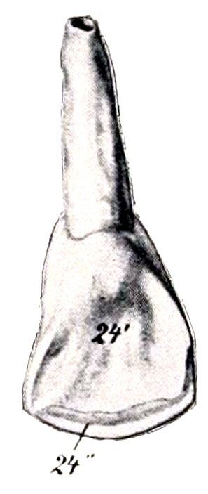
Fig. 285.—Incisor Tooth of Ox. 24′, Lingual surface of crown; 24″, masticatory surface. The crown is clearly marked off from the rounded root by a neck. (After Ellenberger-Baum, Anat. für Künstler.)
The deciduous incisors differ from the permanent set chiefly in being much smaller. The crowns are narrower and diverge more.
The canines are absent (unless the fourth incisors be considered to represent them).
387
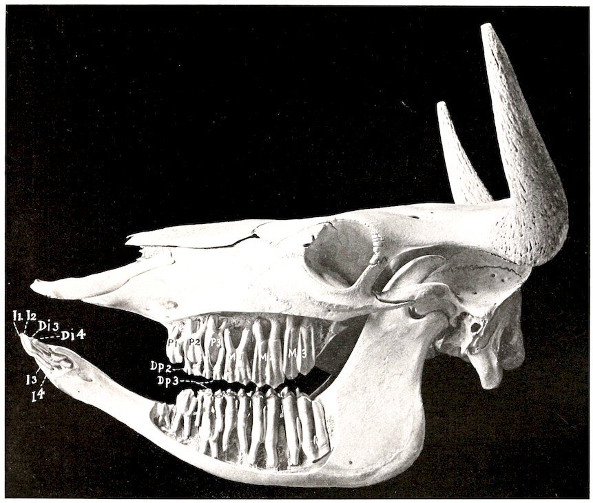
Fig. 286.—Skull of Ox Two Years of Age, Sculptured to Show the Embedded Parts of the Teeth. 11–4, Permanent incisor teeth, the third and fourth not erupted; Di.3, 4, third and fourth deciduous incisors; P1–3, upper permanent premolars, only first erupted; Dp.2, 3, deciduous premolars; M1–3, molars.
The cheek teeth (Fig. 286) resemble those of the horse in number and general arrangement. They are, however, smaller, and also differ in the fact that they 388progressively increase in size from before backward. This feature is so marked that the first tooth is quite small, and the space occupied by the first three (i. e., the premolars) is only about one-half of that required for the posterior three (i. e., the true molars). The enamel folds stand out even more prominently in relief on the masticatory surface than in the horse. The occurrence of wolf-teeth is rare.
The formula of the deciduous teeth is:
( 0 0 3 )
2 ( Di Dc Dp ) = 20
( 4 0 3 )
TABLE OF AVERAGE PERIODS OF ERUPTION OF THE TEETH IN THE OX
Teeth Eruption
A. Temporary:
First incisor (Di 1) Birth to 3 weeks.
Second incisor (Di 2)
Third incisor (Di 3)
Fourth incisor (Di 4)
First cheek tooth (Dp 1) Birth to 3 weeks.
Second cheek tooth (Dp 2) Birth to few days.
Third cheek tooth (Dp 3)
B. Permanent:
First incisor (I1) 1½ to 2 years.
Second incisor (I2) 2 to 2½ years.
Third incisor (I3) 3 years.
Fourth incisor (I4) 3½ to 4 years.
First cheek tooth (P1) 2 to 2½ years.
Second cheek tooth (P2) 1½ to 2½ years.
Third cheek tooth (P3) 2½ to 3 years.
Fourth cheek tooth (M1) 5 to 6 months.
Fifth cheek tooth (M2) 1 to 1½ years.
Sixth cheek tooth (M3). 2 to 2½ years.
The eruption of the permanent teeth is subject to great variation. The above figures are the average of observations of improved breeds under favorable conditions.
THE SALIVARY GLANDS
The parotid gland is smaller than that of the horse, and is light red-brown in color. It has somewhat the form of a very narrow long triangle, and lies chiefly on the posterior part of the masseter muscle. The thick wide upper end partly covers a large subparotid lymph gland. The small lower end is bent forward and fits into the angle of union of the jugular and external maxillary veins; it lies on the submaxillary gland. The parotid duct leaves the lower part of the deep face; in its course it resembles that of the horse, but it pierces the cheek opposite the fifth upper cheek tooth.
The submaxillary gland is very large, and is pale yellow in color. It is covered to a small extent by the parotid. Its general form resembles that of the horse, but its lower (or anterior) end is large and rounded, and is separated by a small interval only from the gland of the other side. This part can be distinctly felt in the living animal, and is related externally to a large submaxillary lymph gland. The duct leaves the middle of the superficial face of the gland, crosses the stylo-hyoideus and the intermediate tendon of the digastricus, and is then disposed as in the horse.
The sublingual gland consists of two parts. The dorsal part (Glandula sublingualis parvicanalaris) is long, thin, and pale yellow in color. It extends from the anterior pillar of the soft palate about to the symphysis of the jaw. It has numerous small tortuous ducts (Ductus sublinguales minores), which open between the papillæ under the side of the tongue. The ventral part is shorter and thicker, and 389is salmon pink in color. It has a single duct (Ductus sublingualis major), which either opens alongside of or joins the submaxillary duct.
THE PHARYNX
The pharynx is short and wide. The vault (Fornix pharyngis) is divided into two cul-de-sacs by a median fold of mucous membrane (Septum nasi membranaceum), which is a continuation of that of the septum nasi; on the outer wall of each is the relatively small opening of the Eustachian tube, which is covered by a simple fold of mucous membrane. The posterior nares are small. The entrance to the œsophagus is large.
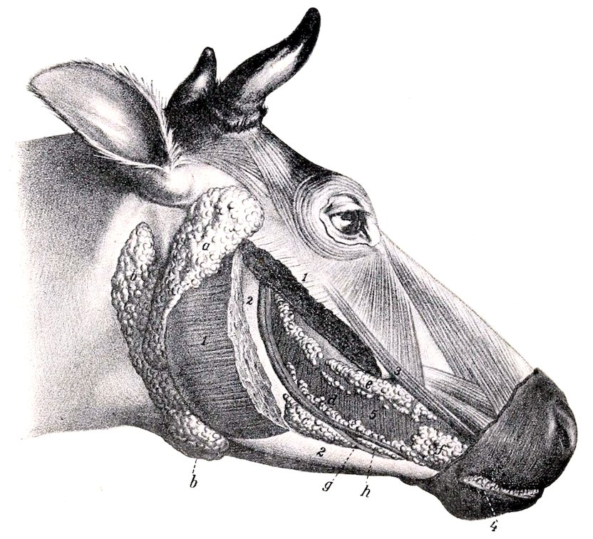
Fig. 287.—Salivary Glands of Ox. a, Parotid gland; b, submaxillary gland; c, inferior, d, middle, and e, superior buccal glands; f, labial glands; g, buccinator nerve; h, buccinator vein; 1, masseter (cut); 2, ramus of mandible; 3, zygomaticus muscle; 4, conical papillæ of lip; 5, buccinator muscle. (After Ellenberger, in Leisering’s Atlas.)
On the dorsal wall of the pharynx are two large pharyngeal lymph glands, which, when enlarged, cause difficulty in swallowing and breathing (Fig. 281).
THE ŒSOPHAGUS
This is much shorter, wider, and more dilatable than that of the horse. Its average diameter (when moderately inflated) is about two inches (ca. 5 cm.), and its length in a large animal is about three to three and a half feet (ca. 90 to 100 cm.). The wall is relatively thin, and the muscular tissue is striped throughout. The latter consists of two strata of spiral fibers, except near the stomach, where they are longitudinal and circular. Fibers are continued into the wall of the stomach 390for some distance. There are submucous glands in the first part of the tube. There is no terminal dilatation, and no part in the abdominal cavity.
A very large mediastinal lymph gland lies above the posterior part of the œsophagus and may, if enlarged, obstruct it (Fig. 289).
THE ABDOMINAL CAVITY
The abdominal cavity of the ox is very capacious, both absolutely and relatively, as compared with that of the horse. This is due to several factors. The lumbar portion of the spine is about one-fourth longer than that of the horse. The transverse diameter between the last ribs is greater. The costal attachment of the diaphragm is almost vertical in direction from the lower part of the ninth rib to the extreme upper part of the thirteenth; in fact, the attachment to the last rib is not constant. Thus the abdomen is increased at the expense of the thorax, and the last three or four ribs enter more largely into the formation of the abdominal wall than in the horse. The flank is also much more extensive. The ilia, on the other hand, do not extend forward beyond a transverse plane through the middle of the last lumbar vertebra. The epigastric and mesogastric regions would be separated by a plane through the lower end of the tenth pair of ribs, or through the last thoracic vertebra. The chief differential features in the arrangement of the peritoneum will be described with the viscera.
THE PELVIC CAVITY
The pelvic cavity is relatively long and narrow. The inlet is more oblique than that of the horse; it is elliptical in outline, and the transverse diameter is smaller than that of the horse. The anterior (pubic) part of the floor is about horizontal, but the posterior (ischial) part slopes upward and backward to a marked degree; this part is also deeply concave transversely. The roof is concave in both directions. The peritoneum extends backward as far as the first coccygeal vertebra, so that the retroperitoneal part of the cavity is short.
THE STOMACH
General Arrangement.—The stomach of the ox is very large, and occupies nearly three-fourths of the abdominal cavity. It fills all of the left half of the cavity (with the exception of the small space occupied by the spleen) and extends considerably over the median plane into the right half.
It is compound, being composed of four divisions, viz., rumen, reticulum, omasum, and abomasum.[107] The division is clearly indicated externally by furrows or constrictions. The first three divisions may be regarded as proventriculi or œsophageal sacculations, the fourth being the stomach proper (in the narrower sense of the term). The œsophagus opens into the stomach on a sort of dome formed by the rumen and reticulum, and is continued through the latter by the œsophageal groove. From the ventral end of the latter a groove traverses the ventral wall of the omasum, thus giving a direct path to the abomasum for finely divided or fluid food. The abomasum joins the small intestine.
Capacity.—The capacity of the stomach varies greatly, depending on the age and size of the animal. In cattle of medium size it holds 30 to 40 gallons, in large animals 40 to 60, in small 25 to 35. The relative sizes of the four parts vary with age. In the new-born calf the rumen and reticulum together are about half as 391large as the abomasum; in ten or twelve weeks this ratio is reversed. During this period the omasum appears to be contracted and functionless. At four months the rumen and reticulum together are about four times as large as the omasum and abomasum together. At about one and one-half years the omasum equals (or nearly so) the abomasum in capacity. The four divisions have now reached their definitive relative capacities, the rumen constituting about 80 per cent., the reticulum 5 per cent., the omasum 7 or 8 per cent., and the abomasum 8 or 7 per cent. of the total amount.
Exterior and Relations
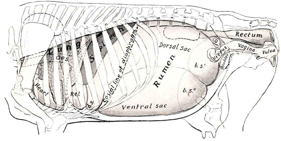
Fig. 288.—Projection of Viscera of Cow on Body-wall, Left Side. Oes., Œsophagus; Ret., reticulum; b.s., anterior blind sac; b.s.′, b.s.″, posterior blind sacs of rumen; O, ovary. The left kidney, concealed by the dorsal sac of the rumen, is indicated by dotted line. The median line of the diaphragm is dotted.
The rumen occupies almost all of the left half of the abdominal cavity, and extends considerably over the median plane ventrally and in its middle. It is somewhat compressed laterally, and may be described as having two surfaces, two curvatures or borders, and two extremities. The parietal (or left) surface (Facies parietalis) is convex and is related to the diaphragm, spleen, and the left wall of the abdomen. It extends from the lower part of the seventh intercostal space almost to the pelvis. The visceral (or right) surface (Facies visceralis) is somewhat irregular, and is related chiefly to the omasum and abomasum, the intestine, the liver, pancreas, kidneys, the uterus in the female, and the posterior aorta and vena cava. The dorsal curvature (Curvatura dorsalis) is convex, following the curve formed by the diaphragm and sublumbar muscles. It is firmly attached to the left portion of the crura of the diaphragm and the sublumbar muscles by peritoneum and connective tissue. The ventral curvature is also convex and lies on the floor of the abdomen. The surfaces are marked by the right and left longitudinal furrows (Sulci longitudinales), which indicate externally the division of the rumen into dorsal and ventral sacs.[108] The reticular (or anterior) extremity (Extremitas reticularis) is divided ventrally by a transverse groove (Sulcus ruminis cranialis) into two sacs. The dorsal sac is the longer of the two, and curves ventrally over the round, blind end of the ventral sac. The former only is continuous 392with the reticulum, a ventral groove (Sulcus rumino-reticularis) being the only external line of demarcation. Dorsally no natural separation exists, the rumen and reticulum together forming a dome-like vestibule (Atrium ventriculi) on which the œsophagus terminates. The pelvic (or posterior) extremity (Extremitas pelvina) extends nearly to the pubis, and is related to the intestine and bladder, and the uterus in the cow. It is divided into dorsal and ventral blind sacs (Saccus cæcus caudalis dorsalis, ventralis) by a deep transverse groove (Sulcus ruminis caudalis) in which the longitudinal furrows terminate. The blind sacs are marked off from the remainder of the rumen by the dorsal and ventral coronary grooves (Sulcus coronarius dorsalis, ventralis).
The left longitudinal groove (Sulcus longitudinalis sinister) begins at the anterior transverse groove, passes somewhat dorsally and backward and divides into two grooves. The ventral one is the chief groove and passes backward to terminate in the angle of union of the posterior blind sacs. The dorsal (accessory) groove curves dorsally and then backward to terminate in the dorsal coronary sulcus. On the right surface there are two longitudinal grooves. The dorsal one is the chief sulcus (Sulcus longitudinalis dexter). It extends in a curved direction (the convexity being dorsal) and is continuous with the left longitudinal groove by means of the grooves between the dorsal and ventral blind sacs at each end. The ventral groove (Sulcus accessorius) curves somewhat ventrally and backward to rejoin the chief sulcus between the posterior blind sacs.
The reticulum is the smallest of the four divisions in the ox. It is also the most anterior, and lies on the concave surface of the diaphragm, so that the median plane divides it into two nearly equal portions. It is somewhat pyriform, but much compressed from before backward. The parietal or phrenic surface (Facies diaphragmatica) is convex and lies against the diaphragm and liver.[109] The ruminal (or posterior) surface (Facies ruminalis) is flattened by the pressure of the other three compartments; it ends dorsally by joining the wall of the rumen, the concave line of junction forming, in the interior of the stomach, the lower margin of the large rumino-reticular orifice. The omasal (or right) surface is narrow and concave, and is connected with the omasum. The dorsal curvature by its left portion joins the rumen to form the atrium, and by its right portion touches the liver. The ventral curvature lies on the sternal portion of the diaphragm, opposite the sixth and seventh ribs. The right extremity forms a rounded cul-de-sac, which is in contact with the liver, omasum, and abomasum.
The omasum (Fig. 294) is ellipsoidal in form and somewhat compressed laterally. It is very clearly marked off from the other divisions. It is situated almost entirely to the right of the median plane, opposite the seventh to the eleventh ribs. The parietal (right) surface (Facies dextra) lies against the diaphragm, liver, and lateral wall of the abdomen. The contact with the latter extends over a small area only (ventral part of seventh to ninth intercostal spaces). The visceral (left) surface (Facies sinistra) is in contact with the rumen and reticulum. The dorsal (greater) curvature is convex, and is related to the diaphragm, liver, and vena cava. The ventral (lesser) curvature is concave, and rests on the abomasum chiefly, the junction with the latter appearing as a constriction near the middle of the curvature; at its anterior part there is a neck (Collum omasi) which is the connection with the reticulum.
The abomasum (Fig. 294) is an elongated pyriform or flask-shaped sac, which lies for the most part on the abdominal floor, to the right of the ventral sac of the rumen. Its narrow posterior part is strongly curved, the concavity being dorsal. The parietal (right) surface lies against the abdominal wall from the seventh to the twelfth intercostal space. The visceral (left) surface is related chiefly to the ventral sac of the rumen. The dorsal (lesser) curvature is concave, and is in contact with the omasum; the junction of the two sacs is indicated by a constriction about four or 393five inches (ca. 10 to 12 cm.) from the anterior extremity of the abomasum. The ventral (greater) curvature is convex, and rests on the abdominal wall from the xiphoid cartilage to the ventral part of the last (right) intercostal space. The anterior extremity forms a rounded blind sac which lies against the reticulum. The posterior (or pyloric) extremity is much smaller and bends upward and forward to join the duodenum. About six to eight inches (ca. 15 to 20 cm.) from the pylorus is a constriction which marks off the pyloric portion from the fundus.
The rumen is attached by peritoneum and connective tissue to the crura of the diaphragm and left psoas muscles, from the hiatus œsophageus backward to about the third or fourth lumbar vertebra.
The lesser omentum attaches the right face of the omasum and the pyloric portion of the abomasum to the visceral surface of the liver.
A small area of the anterior part of the right face of the rumen is adherent to the adjacent surface of the abomasum. The greater part of the ventral curvature of the omasum is attached by connective tissue to the dorsal face of the abomasum.
(Adhesion of the reticulum to the diaphragm is frequently present, but is pathological; the same is true in regard to adhesion to the liver.)
Interior
The cavity of the rumen is partially divided into dorsal and ventral sacs by the pillars; these are folds of the wall, strengthened by additional muscular fibers, and correspond with the grooves on the outside. The two most extensive ones project like shelves into the anterior and posterior ends of the cavity forming the blind sacs at either extremity; they are termed the chief pillars. The anterior chief pillar (Pila cranialis) projects obliquely backward and upward from the ventral wall, and has a thick concave free edge which is opposite to the tenth and eleventh ribs. Its width from the middle of the free edge to its anterior border is about eight to ten inches (ca. 20 to 25 cm.). It is continued on either side by the relatively narrow longitudinal pillars, which connect it with the posterior pillar. The posterior pillar (Pila caudalis) is more nearly horizontal than the anterior one, and separates the large dorsal and ventral posterior blind sacs. Its concave free border is about a hand’s breadth in front of a transverse plane through the external angles of the ilia. From it are detached three accessory pillars on either side; of these, two pass around the blind sacs to meet the corresponding pillars of the opposite side. They thus mark off the posterior blind sacs from the general cavity, and are termed the coronary pillars. It will be noticed that the ventral coronary pillar is complete, while the dorsal one is not. The other accessory pillars join the right and left longitudinal pillars. The right longitudinal pillar is in part double; its ventral division fades out about the middle of the surface, while the dorsal one joins the posterior chief pillar. The distance between the middles of the anterior and posterior pillars is only about sixteen to eighteen inches (ca. 40 to 45 cm.) in a cow of medium size. In this space the dorsal and ventral sacs communicate freely.
The anterior dorsal blind sac of the rumen is separated from the reticulum by an almost vertical fold formed by the apposition of the walls of the two compartments. This rumino-reticular fold (Pila rumino-reticularis) is opposite to the seventh rib and intercostal space. Its free upper edge is concave and forms the lower and outer margin of the large, oval rumino-reticular orifice (Ostium ruminoreticulare). The inner margin of the fold if continued would end about at the cardia, but there is in this region no demarcation between rumen and reticulum; hence it is termed the atrium ventriculi or vestibule.
If the rumino-reticular fold and an imaginary completion of it be taken as the boundary between the rumen and reticulum, it will be seen that the cardia belongs to the latter. The outer border of the rumino-reticular fold ends above and behind the cardia, the inner one just 394behind the œsophageal groove and about four to five inches lower down. Undoubtedly food or water swallowed with a moderate degree of force passes first into the rumen. Foreign bodies (which are commonly swallowed by cattle) are, however, found in the reticulum.
The cardiac orifice is opposite the seventh rib, a little to the left of the median plane and about four inches (ca. 10 cm.) below the spine; it is not funnel-shaped.
The mucous membrane of the rumen is brown in color, except on the margins of the pillars, where it is pale. It is for the most part thickly studded with large papillæ, many of which are nearly half an inch (ca. 1 cm.) long. The edges of the chief pillars and a large part of the wall of the middle of the dorsal sac are, however, not papillated. The papillary arrangement is most marked in the blind sacs. The papillæ vary much in size and form; the largest are foliate, many are narrow or filiform, and others are conical or club-shaped. The mucous membrane on the inner wall of the atrium is finely wrinkled and non-papillated, while above and externally it is papillated. The papillary arrangement also extends over the edge of the rumino-reticular fold an inch or two (ca. 2 to 5 cm.).
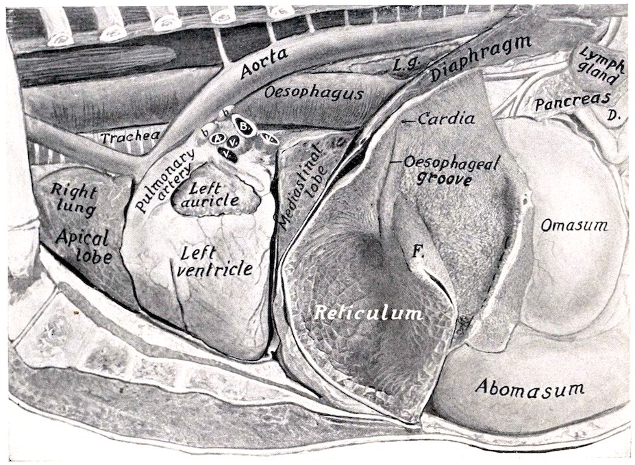
Fig. 289.—Thoracic and Anterior Abdominal Viscera of Ox, Deep Dissection. Most of the rumen has been removed and the left wall of the reticulum cut away. A., Left pulmonary artery; B., left bronchus; V. V. V., pulmonary veins; b. b., bronchial lymph glands; L.g., posterior mediastinal lymph gland; D., termination of duodenum; F., rumino-reticular fold.
The œsophageal groove (Sulcus œsophageus) is a semicanal which begins at the cardia and passes downward (ventrally) on the right wall of the reticulum to end at the reticulo-omasal orifice. It is about six or seven inches (ca. 15 to 18 cm.) in length. Its axial direction is practically vertical, but it is twisted in a spiral fashion; thus its thickened edges or lips project first backward, then to the left, and finally forward. The twist involves chiefly the left lip, and the relative positions of the lips is reversed at the ventral end.
The mucous membrane on the lips of the œsophageal groove is brown and wrinkled, like that of the atrium; in the bottom of the groove it is pale, like that of the œsophagus, marked by longitudinal folds, and presents pointed horny papillæ on the lower part.
The reticulo-omasal orifice (Ostium reticulo-omasicum) is situated in the right 395wall of the reticulum about five or six inches above the bottom of the latter. It is rounded, and is limited below and laterally by the junction of the lips of the œsophageal groove.
In the reticulum the mucous membrane is raised into folds about half an inch high, which inclose four-, five-, or six-sided spaces (Cellulæ reticuli); this peculiar arrangement suggested the scientific name, and also the popular term “honeycomb.” These cells are subdivided by smaller folds, and the bottoms are studded with pointed horny papillæ. The cells grow smaller and gradually disappear near the œsophageal groove and the edge of the rumino-reticular fold; an inch or two from the latter the mucous membrane has the papillary arrangement of the rumen. At the reticulo-omasal orifice there are peculiar horny papillæ, which are curved and resemble the claws of a small bird.
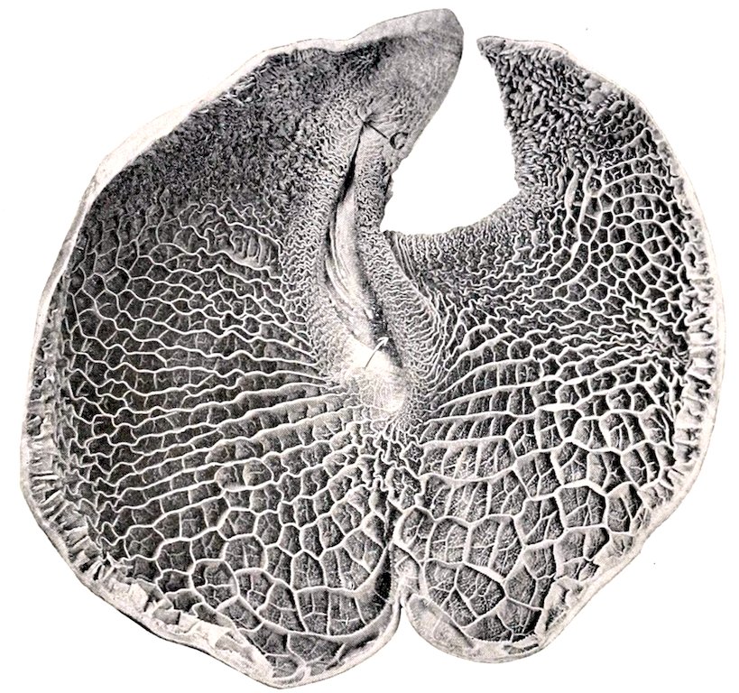
Fig. 290.—Reticulum of Ox, from Photograph. The specimen was cut along the greater curvature and laid open by reflecting the anterior wall. C, is the cardia. The arrow points to the reticulo-omasal orifice. The spiral twist of the œsophageal groove was of necessity partly undone by reflecting the wall; its lips are drawn apart, showing some of the peculiar birdclaw-like papillæ in the lower part.
The cavity of the omasum is occupied to a considerable extent by about a hundred longitudinal folds, the laminæ (Laminæ omasi), which spring from the dorsal and lateral walls. The largest of these—a dozen or more in number—have a convex attached edge, and a ventral, free, slightly concave edge which reaches to within a short distance of the ventral wall of the sac. If these are drawn apart or a cross-section is made, it will be seen that there is a second order of shorter laminæ, and a third order still shorter; finally, there is a series of very low folds or lines. The food is pressed into thin layers in the narrow spaces between the laminæ (Recessus interlaminares), and reduced to a fine state of division by being ground down by the numerous rounded horny papillæ which stud the surfaces of the folds. The ventral wall of the omasum forms a groove (Sulcus omasi), which 396connects the reticulo-omasal opening with the omaso-abomasal opening; it is about three or four inches (ca. 8 to 10 cm.) long, and is directed backward and a little downward and to the right. It is free from leaves, but marked usually by slight folds and small papillæ; it forms a direct path from the reticulum to the abomasum for fluid and finely divided food. In the neck of the omasum there are thick folds, and a number of the peculiar papillæ already mentioned as occurring in the lower part of the œsophageal groove. The omaso-abomasal orifice (Ostium omaso-abomasicum) is oval, and is about four inches (ca. 10 cm.) long. It is bounded in front by a thick muscular pillar which fades out above on the lateral walls of the omasum. Laterally there are two folds (Valvulæ terminales), which probably prevent regurgitation of the contents of the abomasum.
The cavity of the abomasum is divided by a constriction into two areas. The first of these (fundus gland region) is lined with a soft glandular mucous membrane, which forms about a dozen large, slightly spiral folds (Plicæ spirales). The second part (pyloric region) is much narrower and resembles in appearance the corresponding region of the horse’s stomach. A small cardiac gland zone surrounds the omaso-abomasal orifice. The pyloric orifice is small and round.
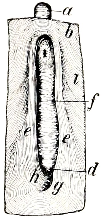
Fig. 291.—Musculature of Œsophageal Groove and Adjacent Part of Stomach of Ox, seen from within after Removal of Mucous Membrane. a, Œsophagus; b, inner muscular layer of atrium; d, reticulo-omasal orifice; e, e′, muscle of lips of groove; g, lower end of muscle of larger lip, which curves around the reticulo-omasal orifice in part and spreads out in the inner muscular layer of the reticulum in part; h, lower end of muscle of smaller lip which in part curves around the reticulo-omasal orifice and is in part continuous with the inner muscular layer of the omasal groove and the omasum; i, inner muscular layer of reticulum. (Ellenberger-Baum, Anat. d. Haustiere.)
Structure.—The serous coat invests all of the free surface of the stomach. The surface of the rumen which is attached to the dorsal abdominal wall is of course uncovered, as well as the adjacent area to which the spleen is attached. The furrows are bridged over by the peritoneum and superficial muscle-fibers, and contain fat and (in most cases) branches of the gastric arteries. The lesser omentum passes from the visceral surface of the liver to the right (parietal) face of the omasum and the pyloric portion of the abomasum. There is a fold between the ventral curvature of the omasum and the dorsal curvature of the abomasum. The great omentum is formed by the peritoneum leaving the stomach along the left longitudinal and posterior transverse furrows and the right surface of the rumen below the longitudinal furrow, also the ventral curvature of the abomasum. Its superficial (ventral) part is attached on the right to the duodenum, while the deep (dorsal) part blends with the mesentery. It conceals the greater part of the intestine on the right side, with the exception of the duodenum, and covers the ventral sac of the rumen almost entirely. It is not lace-like, as in the horse, and contains a large amount of fat in animals in good condition. The epiploic foramen (of Winslow) is almost sagittal in direction.
The muscular coat of the rumen consists of an external longitudinal, and a thicker internal circular layer. The latter forms the bulk of the chief pillars, where it is about one-half to one inch (ca. 1 to 2 cm.) thick. Scattered bundles of striped muscle-fibers radiate from the cardia in the wall of the atrium.
The muscular coat of the reticulum consists of two chief layers which begin and end at the œsophageal groove; they pass in a circular or oblique fashion around the sac, the fibers of the two layers crossing each other at an acute angle. The walls of the cells contain a central muscular layer. The lips of the œsophageal groove contain a thick layer of longitudinal fibers, which are largely continuous at or cross each other below the reticulo-omasal orifice. The bottom of the groove 397has two layers of oblique, unstriped muscle-fibers, with a variable outer layer of striped muscle continuous with that of the œsophagus.
The muscular coat of the omasum consists of a thin external longitudinal layer and a thick internal circular layer. At the omasal groove there is an additional inner layer of longitudinal fibers. The laminæ contain three muscular strata—a central layer of vertical fibers, flanked on either side by a layer of longitudinal fibers, which are continuous at the attached border with the muscularis mucosæ.
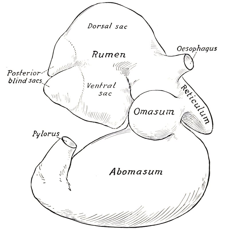
Fig. 292.—Stomach of New-born Calf, Right View. The rumen is raised.
The muscular coat of the abomasum consists of longitudinal and circular layers; the latter forms a well-developed pyloric sphincter.
The mucous membrane of the first three divisions is destitute of glands, and is covered with a thick, stratified, squamous epithelium; the superficial part of the latter is horny, and is shed in large patches in the rumen and omasum. The corium (Tunica propria) is papillated. The mucous membrane of the abomasum is glandular, and corresponds to that of the right sac of the stomach of the horse. The short fundus glands occur in that part which presents the large folds, while the long pyloric glands are found in the remainder, except about the omaso-abomasal orifice, where cardiac glands occur. The mucosa of the fundus gland region is very thin as compared with that of the horse. There is a round prominence on the pyloric valve.
THE INTESTINE
The intestine of the ox lies almost entirely to the right of the median plane, chiefly in contact with the right face of the rumen. It is attached to the sublumbar region by a common mesentery.
398The small intestine has an average length of about 130 feet (ca. 40 m.) and a diameter of about two inches (ca. 5 to 6 cm.).
The duodenum is about three or four feet (ca. 1 m.) in length. It passes forward to the visceral surface of the liver; here it forms, ventral to the right kidney, an S-shaped curve. Thence it runs backward almost to the external angle of the ilium, where it turns on itself, passes forward alongside of the terminal part of the colon, and joins the mesenteric part (jejunum) under the right kidney. It is attached to the liver by the lesser omentum, to the abomasum by the great omentum, and to the terminal part of the colon. The bile-duct opens in the ventral part of the S-shaped curve, about two feet (ca. 60 cm.) from the pylorus. The pancreatic duct opens about a foot (ca. 30 cm.) further back.
The remainder of the small intestine is arranged in numerous very close coils, in contact chiefly with the right face of the ventral sac of the rumen, forming a sort of festoon at the edge of the mesentery. It is not subject to much variation in position, but a few coils may find their way behind the blind sacs of the rumen to the left side.
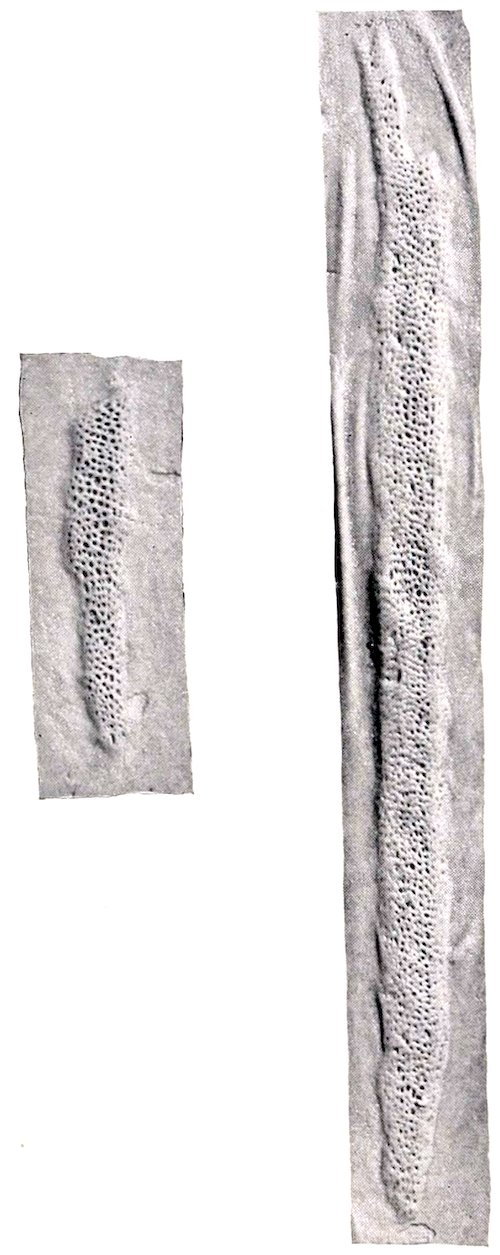
Fig. 293.—Peyer’s Patches of Small Intestine of Ox (about ⅓ natural size).
The orifices of the pancreatic duct and the bile-duct are on papillæ, no diverticulum being present. There are permanent transverse folds of the mucous membrane (Plicæ circulares). Duodenal glands (of Brunner) occur in the first fifteen feet or more (4 to 5 m.), intestinal glands throughout. Peyer’s patches are larger and more distinct than in the horse, and vary greatly in size and number; in adult cattle there are eighteen to forty; in calves, twenty to fifty-eight. They usually have the form of narrow bands. Close to the ileo-cæcal valve there is always a patch, and in most cases there is also a patch on the cæcal side of the valve.
The large intestine is much smaller in caliber than that of the horse, has no bands, and is not sacculated. Most of it is situated between the layers of the common mesentery.
The cæcum is about 20 to 30 inches (ca. 50 to 60 cm.) long, 4 to 5 inches (ca. 10 to 12 cm.) wide, and has a capacity of about 2½ gallons (ca. 9 to 10 liters). It begins opposite the lower part of the eleventh rib, and extends upward and backward against the right flank; its rounded blind end is free, and lies near or in the pelvic inlet.
399
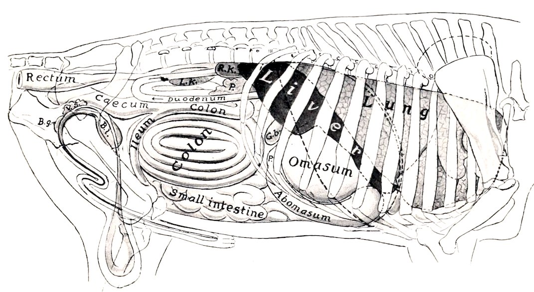
Fig. 294.—Projection of Viscera of Bull on Body-wall, Right Side. P., Pylorus; G.b., gall-bladder; R.K., right kidney; L.K., left kidney; P., (above duodenum) pancreas; Bl., urinary bladder; V.s., vesicula seminalis; B.g., bulbo-urethral (Cowper’s) gland. Costal attachment and median line of diaphragm are indicated by dotted lines.
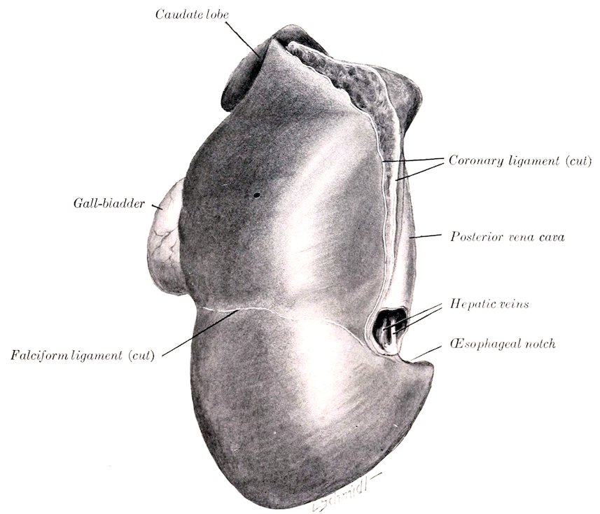
Fig. 295.—Liver of Ox, Parietal Surface; Hardened in situ.
The colon is about 35 feet (ca. 10 m.) long; its diameter is at first about the same as that of the cæcum, but diminishes to about two inches (ca. 5 cm.). Its capacity is about 6 to 8 gallons (ca. 25 to 30 liters). With the exception of its first and terminal parts, it is arranged in double elliptical coils between the layers of the mesentery; the adjacent parts are therefore alternately centripetal and centrifugal (Gyri centripetales et centrifugales). The first part (Ansa proximalis) is marked off from the cæcum only by the ileo-cæcal opening; it forms an S-shaped 400curve and is continued by the spiral part (Ansa spiralis). The coils of this are attached to each other by connective tissue, and are best seen from the inner side. The bowel gradually diminishes in caliber, and the terminal part (Ansa distalis) leaves the spiral mass, passes forward to the great mesenteric artery and turns backward between the duodenum and the initial sigmoid part. From the ventral surface of the right kidney it passes backward, forms an S-shaped curve near the pelvic inlet, and joins the rectum; this part is attached to the sublumbar muscles by a short mesentery, and is also attached to the dorsal part of the duodenum.
The rectum is somewhat shorter than that of the horse, and is usually covered with peritoneum as far back as the first coccygeal vertebra. The retroperitoneal part is surrounded by a quantity of fat. The anus is not prominent.
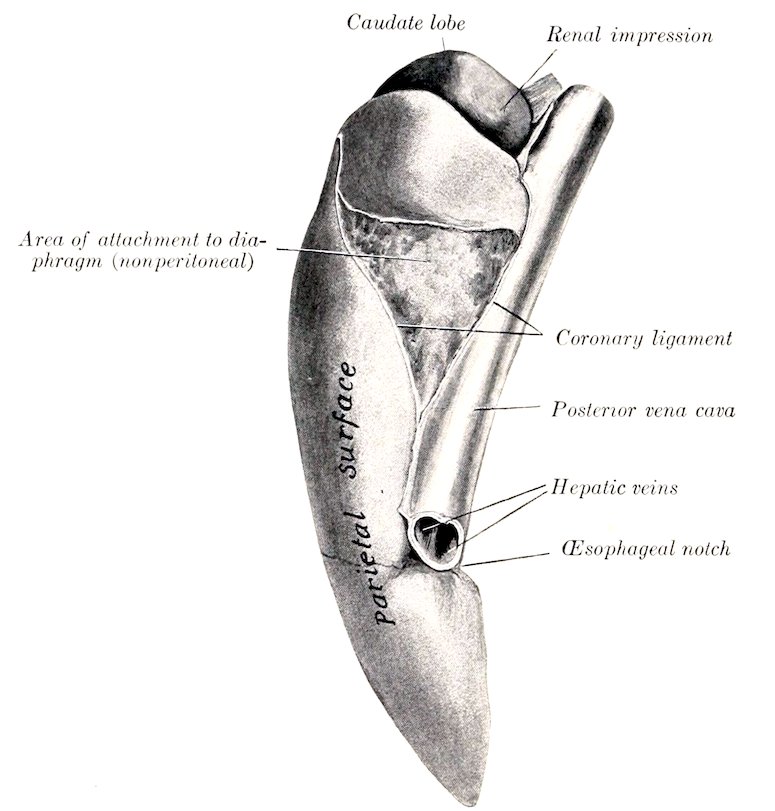
Fig. 296.—Liver of Ox, Left Marginal View; Hardened in situ.
The serous coat is of course absent on the adherent surfaces of the spiral part of the colon. There is a large amount of fat in the mesentery. The longitudinal muscular coat is evenly distributed, consequently there is no sacculation of the bowel. There is a valvular mucous fold at the ileo-cæcal orifice. A Peyer’s patch occurs in the beginning of the cæcum and one in the first part of the colon.
THE LIVER
The liver lies almost entirely to the right of the median plane. Its long axis is directed obliquely downward and forward, about parallel with the median plane, and corresponds to the curvature of the right portion of the diaphragm. It is less extensive, but thicker than that of the horse. Its average weight is about 10 to 12 pounds (ca. 4.5 to 5.5 kg.).
401In small subjects the weight (according to Schmaltz) varies from 6½ to 10 pounds (ca. 3 to 4.5 kg.), in large subjects from 11 to 13 pounds (ca. 5 to 6 kg.). According to Schneider, the average weight is about 13 pounds (ca. 6 kg.)—a little over 1 per cent. of the live weight and about 2 per cent. of the dressed carcass.
When hardened in situ, its configuration adapts it accurately to the structures with which it is in contact.
The parietal surface is convex and is for the most part applied to the right part of the diaphragm, but a small part of it is in direct contact with the last two ribs and with the flank at the lumbo-costal angle. It faces upward, forward, and to the right.
The curvature of the parietal surface is not regular. It is marked, except in its ventral part, by a blunt oblique ridge which corresponds to the basal border of the right lung. This prominence divides the surface into two areas. Of these, the external one is directed outward, is only slightly convex, and often shows impressions of the last three ribs. The inner area presents a depression produced by the right crus of the diaphragm, and otherwise is regularly convex and adapted to the tendinous center and sternal portion of the diaphragm. The falciform ligament is attached to the surface from the œsophageal notch to the umbilical fissure.
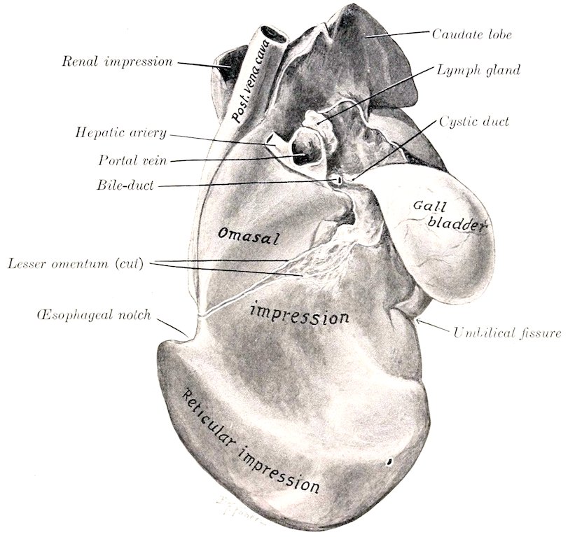
Fig. 297.—Liver of Young Ox, Visceral Surface; Hardened in situ.
The visceral surface is concave and very irregular; it presents impressions of the chief organs which are in contact with it—the omasum and reticulum. It is also related to the pancreas and duodenum.
The following markings are quite distinct on the visceral surface of well hardened specimens: (1) The omasal impression (Impressio omasica) is a deep central cavity below the portal fissure. (2) The reticular impression (Impressio reticularis) is a smaller marginal depression below the œsophageal notch and the inner part of the preceding, from which it is separated by a rounded ridge. (3) The abomasal impression (Impressio abomasica), present only in the calf, is produced by the anterior part of the abomasum. It lies along the ventral part of the surface, and is separated by ridges from the preceding impressions. It disappears as the omasum and reticulum 402increase in size and displace the abomasum from contact with the liver. (4) Shallow grooves for the S-shaped portion of the duodenum (Impressio duodenalis) may be found above and external to the portal fissure. (5) The fossa of the gall-bladder (Fossa vesicæ felleæ) is distinct when that organ is full. In the calf the omasal impression is small, and the abomasal one large in correspondence with the relative sizes of these two sacs.
The portal fissure is a well defined rounded depression, situated dorsal to the omasal impression. It contains, besides the vessels and duct, several large hepatic lymph glands. Above and external to it a part of the pancreas is attached.
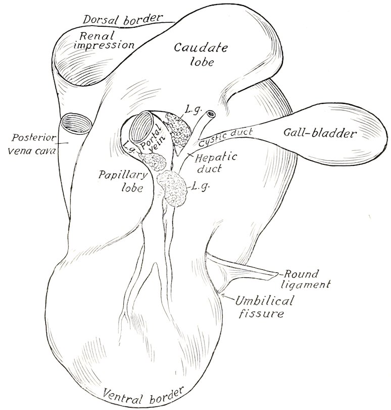
Fig. 298.—Line Drawing of Liver of Ox, Visceral Surface of Soft Specimen. L.g., Hepatic lymph glands at portal fissure.
The dorsal border is short and thick; it extends backward a short distance beyond the upper part of the last rib. It presents the large, thick, quadrilateral caudate lobe, and a deep depression for the right kidney and adrenal.
The ventral border is short and thin and has no interlobar incisures.
The right (or external) border is marked by a small umbilical fissure, in which the ligamentum teres is attached in the young subject.
The left border presents the œsophageal notch below its middle. Above this it is practically median in position, and lodges the posterior vena cava, which is partially embedded in the gland. Below the notch the border extends an inch or two (ca. 2.5 to 5 cm.) to the left of the median plane opposite the ventral third of the sixth rib.
There is no left lateral ligament. The falciform ligament is usually present, 403but the ligamentum teres is found only in young subjects. The lesser omentum leaves the liver along a line extending from the œsophageal notch to the portal fissure. The only distinct lobe in the adult is the caudate (Processus caudatus).
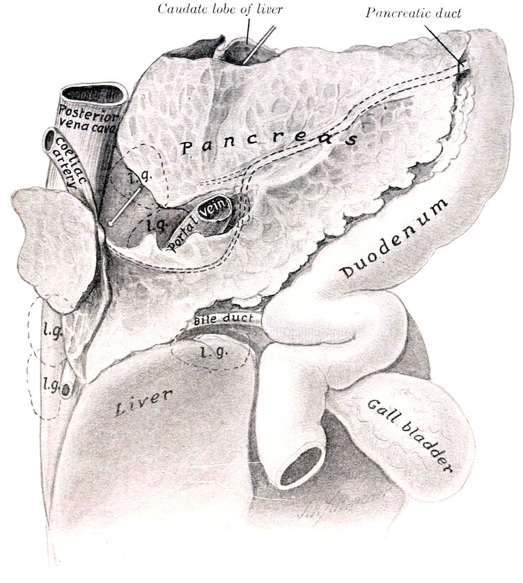
Fig. 299.—Pancreas of Ox, Gastro-intestinal Surface, with Related Organs. The position of some of the larger lymph glands (l.g.) is indicated by dotted line; also the intraglandular course of the pancreatic duct. Probe is passed through epiploic foramen (of Winslow).
A gall-bladder (Vesica fellea) is present. This is a pear-shaped sac, four to six inches (ca. 10 to 15 cm.) long, which lies partly on the visceral surface of the liver (to which it is attached), but largely on the abdominal wall at the lower part of the eleventh or twelfth intercostal space. It may be regarded as a diverticulum of the bile-duct, enlarged to form a reservoir for the bile. Its neck is continued by the cystic duct (Ductus cysticus), which joins the hepatic duct at an acute angle just outside of the portal fissure, to form with it the bile-duct (Ductus choledochus). The latter is short and enters the second bend of the S-shaped curve of the duodenum, i. e., about two feet (ca. 60 cm.) from the pylorus. Several small ducts (Ductus hepato-cystici) open directly into the gall-bladder.
In the new-born calf the liver is relatively much larger than in the adult. The visceral surface presents, below the portal fissure, a large rounded eminence, which is caused by the presence in the underlying gland substance of a large venous sinus into which the umbilical and portal veins empty. A large vessel, the ductus venosus, leads from this sinus directly to the posterior vena cava. Just external to this elevation is a small but distinct lobe about two inches long (Processus papillaris?). The umbilical fissure is deep and partially divides the gland into two chief lobes.
404
THE PANCREAS
The pancreas of the ox is irregularly quadrilateral in form, and lies almost entirely to the right of the median plane. Its weight is about the same as that of the horse. Its dorsal surface is related to the crura of the diaphragm, the cœliac and anterior mesenteric arteries, and the liver. It is attached to the liver at and external to the portal fissure, and to the crura of the diaphragm. Between these adhesions it is free and forms the ventral wall of the epiploic foramen (of Winslow). On the right side it extends backward beyond the caudate lobe of the liver between the layers of the mesoduodenum; here it is in contact with the upper part of the flank at the lumbo-costal angle, and is related above to the right kidney and by its ventro-lateral border to the ventral part of the duodenum. The gastro-intestinal or ventral surface is in contact with the dorsal curvature of the rumen and the intestine. The portal vein passes through the gland. The left extremity is small; the right or posterior is wide and thin and is often divided into two branches. The duct leaves the posterior part of the ventro-lateral (or right) border and enters the duodenum about twelve inches (ca. 30 cm.) further back than the bile-duct.
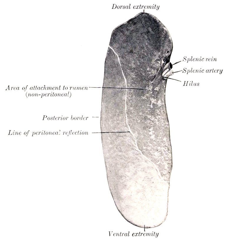
Fig. 300.—Spleen of Ox; Visceral Surface.
THE SPLEEN
The spleen has an elongated, elliptical outline, both extremities being thin, rounded, and similar in size.
Its average weight is about 30 ounces (ca. 800 to 900 gm.), or about ⅙ per cent. of the body-weight. Its length is about 16 to 20 inches (ca. 40 to 50 cm.), 405its width about 5 to 6 inches (ca. 12 to 15 cm.), and in the middle its thickness is about an inch (ca. 2 to 3 cm.).
The dorsal extremity lies under the upper ends of the last two ribs; the ventral extremity is usually opposite the seventh or eighth rib, about a handbreadth above its junction with the cartilage. The parietal surface is convex, and is related to the diaphragm. The visceral surface is concave, and is related chiefly to the left face of the rumen, but also usually to a narrow adjacent area of the reticulum. The dorsal part is attached to the left crus of the diaphragm and the left surface of the rumen by peritoneum and connective tissue; the ventral part is free. The hilus is situated on the dorsal third of the visceral surface, near the anterior border.
About one-half of the visceral surface of the spleen is attached directly to the rumen and is not covered by peritoneum; the line of reflection of the latter crosses the surface obliquely, from the upper part of the posterior border to the anterior border a little below the middle. Similarly there is a narrow uncovered area on the upper part of the parietal surface along the anterior border. The hilus is not a groove, but a simple depression. When hardened in situ, the organ is seen to be somewhat twisted, so that the upper part of the parietal surface faces upward and forward, while below it is directed outward.
DIGESTIVE SYSTEM OF THE SHEEP
The lips are thin and mobile; the upper one is marked by a very distinct philtrum, and otherwise is not bare.
The anterior part of the hard palate is prominent and smooth; on the posterior part of this area there are two narrow depressions in the form of a V, at the deep anterior ends of which the naso-palatine ducts open. The ridges of the palate are irregular and their edges are smooth. The posterior third or rather more of the palate is not ridged and presents numerous orifices of ducts of the palatine glands. The mucous membrane is often more or less pigmented.
The mucous membrane of the cheeks is covered with large papillæ, many of which are long and sharp-pointed, while others are short and blunt. There is also a series of conical papillæ on the floor of the mouth under the lateral part of the tongue.
The tongue resembles that of the ox in form, but the tip is comparatively smooth; the papillæ here are very numerous, but short and blunt. This difference is in conformity with the dissimilarity in the mode of prehension. The prominence of the dorsum is commonly not so pronounced nor so sharply marked off in front as in the ox. The root is smooth. The mucous membrane of the dorsum is often pigmented in spots.
The dental formula is the same as that of the ox. The incisor teeth form a narrow and strongly curved arch. The crowns are long and narrow; their labial surfaces are strongly convex and end at a sharp edge which is used in cropping the grass. The roots are more firmly embedded than in the ox.
The cheek teeth resemble those of the ox. There is a thinner layer of cement, which is usually black.
The average periods of eruption of the teeth are indicated in the subjoined table.
Teeth Temporary Permanent
I1. At birth or first week 1 to 1½ years
I2 First or second week 1½ to 2 years
I3 Second or third week 2½ to 3 years
I4 Third or fourth week 3½ to 4 years
P1 2 to 6 weeks. 1½ to 2 years
P2
P3
M1 3 months (lower), 5 months (upper)
M2 9 to 12 months
M3 1½ to 2 years
406
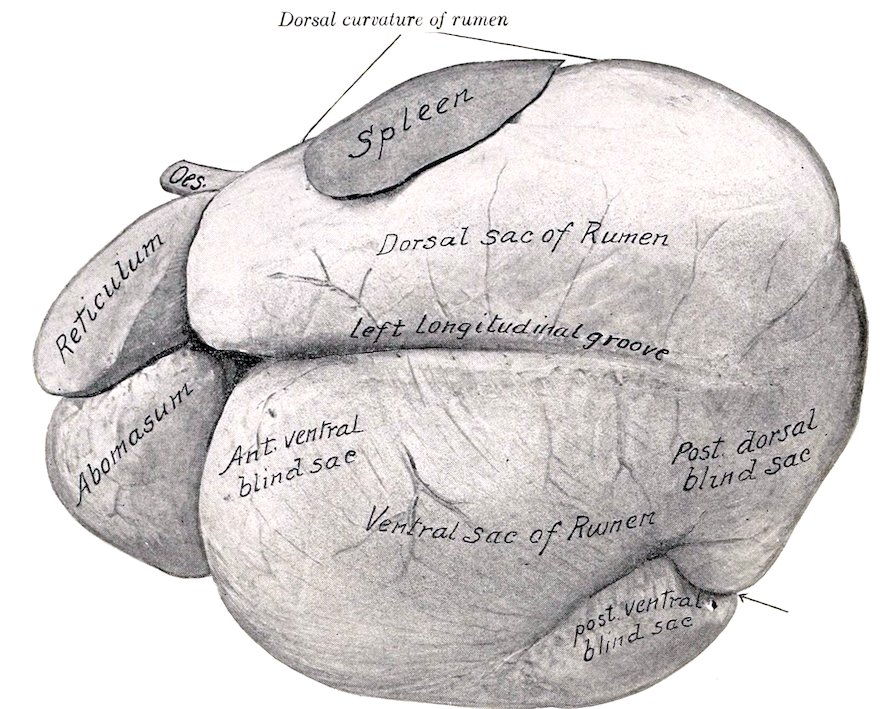
Fig. 301.—Stomach of Sheep, Left View. From photograph of specimen hardened in situ. The reticulum is somewhat contracted. Arrow points to posterior furrow between blind sacs.
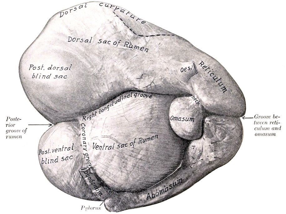
Fig. 302.—Stomach of Sheep, Right View. From photograph of specimen hardened in situ. Dotted line indicates position of spleen.
407The salivary glands resemble those of the ox in general, but the parotid duct crosses the surface of the masseter muscle, and opens opposite the third or fourth cheek tooth.
The tonsil is bean-shaped and about half an inch (ca. 12 mm.) in length. It does not project into the fauces. The mucous membrane of the latter presents two deep and very narrow tonsillar sinuses on either side.
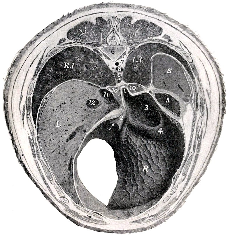
Fig. 303.—Cross-section of Sheep, through Ninth Thoracic Vertebra, Anterior View. 1, Cardiac orifice; 2, œsophageal groove; 3, rumino-reticular orifice; 4, rumino-reticular fold; 5, dorsal sac of rumen; 6, body of ninth thoracic vertebra; 7, vena hemiazygos; 8, aorta; 9, posterior mediastinal lymph gland; 10, 10, crura of diaphragm; 11, posterior vena cava; 12, portal vein; R.l., right lung; L.l., left lung; S, spleen (cut very obliquely); L, liver; R, reticulum (posterior wall). Arrow points to reticulo-omasal orifice.
The fornix of the pharynx presents a median plicated fold which is a continuation of the septum nasi. The pharyngeal orifice of the Eustachian tube has the form of a crescentic slit, placed about on a level with the inferior nasal meatus. There are two large lymph glands and a number of hæmolymph glands above the pharynx.
The œsophagus has a lumen of about an inch when moderately distended; otherwise it resembles that of the ox.
408
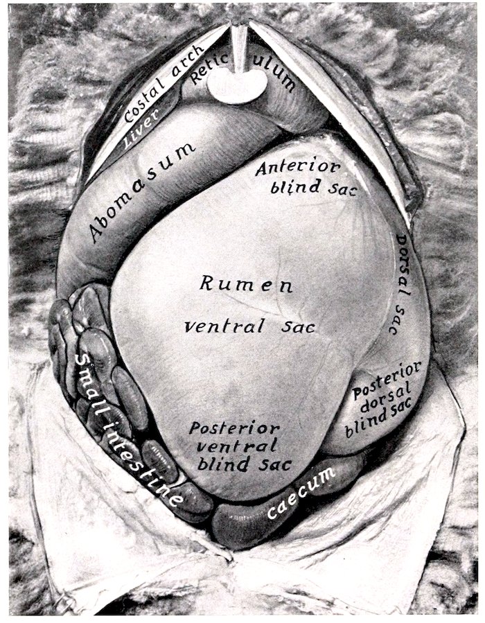
Fig. 304.—AbdomiNal Viscera of Sheep, Superficial Ventral View.
The stomach is like that of the ox in its general arrangement. Its average capacity is about four gallons (ca. 15 liters). The cardiac opening is opposite the middle of the eighth intercostal space; it is just to the left of the median plane and about two to three inches (ca. 5 to 7 cm.) below the vertebral column. The ventral sac of the rumen is relatively larger and extends further to the right of the median plane than in the ox. There are no coronary pillars in the dorsal sac, but its upper part is papillated. The papillæ of the rumen are relatively large and somewhat tongue-like; the largest are about a fourth of an inch (ca. 6 mm.) in length. The reticulum is relatively larger than in the ox. Its ventral part curves more backward and less to the right than in the ox. The parietal surface extends forward as far as the sixth rib or intercostal space, and is related to the diaphragm and liver. The fundus lies on the sternal part of the diaphragm and in contact with the abomasum. The œsophageal groove is disposed much like that of the ox, and is about four to five inches (ca. 8 to 10 cm.) long. The omasum is much smaller than the reticulum, its capacity being only about one pint. It is oval and compressed laterally. It is situated almost entirely to the right of the median plane, opposite to the ninth and tenth ribs, higher than in the ox, and has no contact with the abdominal wall. It is related on the right to the liver and gall-bladder, on the left to the rumen, and below to the abomasum. The laminæ are less numerous than in the ox; in the neck which connects the omasum with the reticulum they have 409the form of low thick ridges, and bear long, pointed, horny papillæ. The abomasum is relatively larger and longer than in the ox. Its capacity is about twice that of the reticulum and averages about two quarts (ca. 1.75 to 2 liters). The blind anterior end lies almost centrally in the xiphoid region, in relation to the liver and reticulum. Its body extends backward a little below and almost parallel with the right costal arch. The pylorus is opposite the ventral end of the eleventh or twelfth intercostal space. The cells of the reticulum are much shallower than in the ox. At the reticulo-omasal opening and on the adjacent part of the laminæ of the omasum are large, pointed, horny papillæ. The number of laminæ is less than in the ox.
The general arrangement of the intestine is like that of the ox. The small intestine is about 80 feet (ca. 24 to 25 m.) long; its average diameter is about an inch (ca. 2 to 3 cm.), the caliber increasing in its terminal part, where a very extensive Peyer’s patch is found. The cæcum is about 10 to 12 inches (ca. 25 to 30 cm.) long, 2 inches (ca. 5 cm.) wide, and has a capacity of about a quart (ca. 1 liter). The colon is about 15 feet (ca. 4 to 5 m.) long. Its caliber is at first about the same as that of the cæcum, but diminishes to about the width of the small intestine.
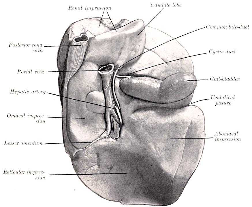
Fig. 305.—Liver of Sheep, Visceral Surface; Hardened in situ.
The liver weighs about 20 to 25 ounces (ca. 550 to 700 gm.). It lies entirely to the right of the median plane. The parietal surface is related almost exclusively to the right part of the diaphragm. The visceral surface presents extensive reticular and abomasal impressions, and a small omasal impression internal to the portal fissure. The umbilical fissure is deep and partially divides the gland into 410two chief lobes. The caudate lobe is prismatic and blunt-pointed. The neck of the gall-bladder is long. The bile-duct joins the pancreatic duct to form a common duct (Ductus choledochus communis) which opens into the duodenum about one foot (ca. 30 cm.) from the pylorus.
The pancreas is arranged as in the ox. Its duct unites with the bile-duct.
The spleen (Fig. 301) is somewhat triangular, with the angles rounded off. It weighs about three or four ounces (ca. 100 grams). Its length is about five inches (ca. 12 to 13 cm.) and its greatest width about four inches (ca. 10 cm.). The parietal surface is convex and is related to the diaphragm, to which rather more than the anterior third is adherent. The visceral surface is concave and its anterior half is attached to the dorsal curvature of the rumen. The dorsal end or base is attached to the left crus of the diaphragm under the last two or three ribs. The hilus is at the posterior basal angle. The ventral end is narrower and thinner than the base; it is situated opposite the ninth and tenth ribs above their middles.