NEUROLOGY
Reading edition
THE NERVOUS SYSTEM
The nervous system is a complex mechanism by which the organism is brought into functional relation with its environment, and its various parts are coördinated. For purposes of gross description it is divided primarily into two parts, central and peripheral.
The central nervous system (Systema nervorum centrale) comprises (a) the spinal cord (Medulla spinalis), and (b) the brain (Encephalon).
The peripheral nervous system (Systema nervorum periphericum) includes (a) the cerebral and spinal nerves with their ganglia, and (b) the sympathetic nervous system.
The division into central and peripheral parts is quite arbitrary and is employed purely as a matter of convenience of description. The fibers of which the nerves are composed either arise or end within the central system, and therefore constitute an integral part of the latter. The structural and functional unit of the nervous system is the neurone, which consists of the cell-body, usually termed the nerve-cell, and all its processes. The processes arise as outgrowths from the cell-body and conduct impulses to or from the cell. They vary greatly in length, some being less than a millimeter long, while others extend from a cell in the spinal cord to the distal end of a limb. A nerve is composed of such processes, usually inclosed in a protecting and insulating sheath, and united into bundles by connective tissue. The neurones are embedded in a peculiar supporting tissue termed neuroglia. This consists of a very intricate feltwork of glia-fibers, many of which are connected with the small glia-cells or astrocytes. In addition the nervous tissue is invested closely by a vascular layer of connective tissue, the pia mater, from which ingrowths extend into the nervous substance proper.[186]
To the naked eye the central nervous system appears to be composed chiefly of two kinds of substance, white and gray. The white matter (Substantia alba) is dead white in color and is soft in the natural state. It consists largely of medullated nerve-fibers, packed closely together and arranged more or less clearly in large or small bundles or tracts (Funiculi, fasciculi). The gray matter (Substantia grisea) is usually brownish-gray in color, often with a tinge of pink. It is softer than the white substance and much more vascular. It is composed chiefly of cell-bodies and non-medullated processes. In some situations it is modified to form the gelatinous substance (Substantia gelatinosa), which is pale yellowish-gray and jelly-like.
Ganglia are gray masses found on the dorsal roots of the spinal nerves and on the course of many nerves. They are commonly ovoid in form and are inclosed in a fibrous capsule. They are composed largely of the cell-bodies of neurones, but have connected with and passing through them nerve-fibers (processes) which extend peripherally and centrally. In origin and function the spinal ganglia belong properly to the central system, but it is customary to include them with the peripheral part in gross anatomical descriptions.
Nerves are conducting trunks composed of bundles of parallel nerve-fibers. They are enveloped in a fibro-elastic sheath, the epineurium, which contains the blood and lymph vessels. They are classified according to their central connections as cranial or cerebral, spinal, and sympathetic. The groups of cells of which the nerve-fibers are processes are termed the nuclei of origin or termination of the nerves, according as the latter conduct impulses in a peripheral or central direction. On the same basis the nerve-fibers are designated efferent and afferent 645respectively. A bundle of fibers which passes from one nerve-trunk to another is called an anastomotic branch (Ramus anastomoticus). In some situations the exchange of branches between adjacent nerves is so free as to constitute a nerve-plexus (Plexus nervorum). The term ramus communicans is properly restricted to branches which connect the ventral divisions of the spinal nerves with adjacent ganglia or nerves of the sympathetic system. The terminal twigs of the nerves are designated according to their distribution as muscular branches (Rami musculares), cutaneous nerves (Nervi cutanei), and articular nerves (Nervi articulares). The muscular branches are motor in function, the cutaneous and articular sensory, but all contain vasomotor fibers which control the caliber of the blood-vessels.
THE MENINGES
The central organs of the nervous system are inclosed in three meninges or membranes. From without inward these are: (1) the dura mater, (2) the arachnoidea, and (3) the pia mater.
THE DURA MATER
The dura mater is a dense resistant membrane of white fibrous tissue; in the fresh state it is bluish-white in color. On account of the difference in its arrangement within the cranium from that in the spinal canal it is customary to describe it as consisting of two parts, cerebral and spinal; these portions are continuous with each other at the foramen magnum.
The cerebral dura mater (Dura mater encephali) is adherent to the interior of the cranium, and may be regarded as forming an internal periosteum for the bones here as well as being an envelope of the brain. Its outer surface is connected with the bony wall of the cranial cavity by numerous fine fibrous strands and by blood-vessels; hence it appears rough in many places when separated from the wall. The degree of adhesion varies greatly at different points. It is most firmly attached at the various projections, e. g., the internal sagittal crest, the tentorium osseum, the petrosal crest; also at the base and the foramen magnum. Before the sutures are closed the dura is connected with the sutural ligaments and through them is continuous with the external periosteum. The cranial nerves receive sheaths from the dura, which is thus continuous without the cranium with the epineurium and periosteum. Along the roof and sides (except as noted above) the adhesion is relatively slight, and lymph-spaces are said to exist between the dura and the bone. The internal surface of the dura is smooth and glistening, since it is lined by endothelium and is moistened by a fluid resembling lymph; it forms the outer boundary of the subdural space. In accordance with its double function the dura is composed of two layers, which are, however, intimately adherent to each other in most places in the adult. The venous sinuses are channels between the two layers and are lined by endothelium. They have been described with the other vessels.
Several folds or septa are given off from the inner surface of the dura; these project into the cavity between the gross subdivisions of the brain. These septa are: (1) the falx cerebri; (2) the tentorium cerebelli; and (3) the diaphragma sellæ.
The falx cerebri is a sickle-shaped median partition which projects downward in the great longitudinal fissure between the cerebral hemispheres. It is attached above to the internal sagittal crest, in front to the crista galli, and behind to the tentorium osseum. Its superior border is convex and separates into two layers which inclose the superior longitudinal sinus. Its lower border is concave and lies over the corpus callosum. The falx is thick above, but much thinner below, and in some places cribriform.
646In the horse the posterior part of the falx cerebri does not extend to the corpus callosum, and hence the hemispheres are here in contact and adherent to each other over a small area. In the ox and pig the falx does not descend so far as in the horse, while in the dog it completely separates the hemispheres.
The tentorium cerebelli is a crescentic transverse fold which separates the cerebellum from the posterior poles of the cerebral hemispheres. It is attached above to the tentorium osseum and laterally to the petrosal crests. Its lower border is thin, concave, and free; it forms the upper and lateral boundaries of an opening (Incisura tentorii) which incloses the mid-brain.
The diaphragma sellæ is a small circular fold which roofs over the sella turcica and the pituitary body. It is perforated centrally by an opening (Foramen diaphragmatis) for the infundibulum.
The falx cerebelli, a sickle-shaped fold which projects into the median notch between the cerebellar hemispheres in man, is not present in the domesticated animals. There is instead merely a slight thickening of the dura.
The spinal dura mater (Dura mater spinalis) forms a tube around the spinal cord from the foramen magnum to the second or third segment of the sacrum. It is separated from the periosteum of the spinal canal by a considerable epidural space (Cavum epidurale) which is occupied by fatty connective tissue and veins. It is held in position chiefly by the sheaths which it furnishes to the roots of the spinal nerves, and in its anterior part by two ligaments; the latter connect it with the inferior atlanto-occipital membrane and with the odontoid process. It is large in proportion to its contents, but its diameter is not uniform. It is largest in the atlas, small in the thoracic region, and becomes very small in its terminal part, where it incloses the delicate filum terminale of the spinal cord.
The subdural space (Cavum subdurale) is the cavity between the inner surface of the dura mater and the arachnoidea. It is a mere capillary space which contains just sufficient fluid to moisten its surfaces; this fluid is usually regarded as lymph, which is replenished by filtration through the walls of the blood-vessels. The space is in communication with the lymph-paths of the nerve-sheaths.
THE ARACHNOIDEA
The arachnoidea is a very delicate and transparent membrane situated between the dura and pia mater. Its outer surface forms the inner wall of the subdural space and is covered by a layer of endothelium continuous with that of the opposed surface of the dura mater. Between it and the pia mater is the subarachnoid space (Cavum subarachnoideale) which contains the cerebrospinal fluid. An inner surface can scarcely be said to exist, since deeply the membrane becomes a reticulum of fine fibers which traverse the subarachnoid space and are attached to the pia mater. The arachnoidea furnishes sheaths to the cerebral nerves from their superficial origins to a variable but usually short distance beyond the emergence from the dursal sac. In the case of the optic nerve this sheath extends to the eyeball.
The arachnoidea of the brain (Arachnoidea encephali), except in the case of the great longitudinal and transverse fissures, does not dip into the sulci on the surface of the brain. On the summits of the gyri it is so closely attached to the pia mater that the two form practically a single membrane. Its outer part bridges over the sulci, and here the subarachnoid space is partially divided up by the loose arachnoid tissue into intercommunicating cavities. In certain situations the arachnoidea is separated from the pia by spaces of considerable depth and extent. These enlargements of the subarachnoid space are termed subarachnoid cisterns (Cisternæ subarachnoidales). In them the subarachnoid tissue does not form a close network, but consists of a relatively small number of long, thread-like strands which traverse the cavity.
647The chief cisternæ are: (1) the cisterna magna, which is at the angle formed between the posterior face of the cerebellum and the upper surface of the medulla oblongata. It communicates with the fourth ventricle through the foramen of Magendie (in man); and behind with the wide subarachnoid space of the spinal cord; (2) the cisterna pontis on the ventral surface of the pons; (3) the cisterna basalis, which lies at the base of the cerebrum and is divided by the optic chiasma into two parts (cisterna chiasmatis, cisterna interpeduncularis); (4) the cisterna fossæ lateralis, situated at the lower part of the lateral fissure (of Sylvius), and continuous with the cisterna basalis.
Along the upper border of the falx cerebri the arachnoidea bears bulbous excrescences, the arachnoid granulations or Pacchionian bodies. These are inclosed in thin evaginations of the dura mater and project into the sagittal sinus or the parasinoidal sinuses along either side of it. In some cases they are sufficiently large to exert pressure on the bone and produce in it depressions of variable depth.
The spinal arachnoidea (Arachnoidea spinalis) is directly continuous with that of the brain at the foramen magnum. It forms a relatively wide tube around the spinal cord, so that the latter (inclosed in the pia) is surrounded by a very considerable quantity of cerebrospinal fluid. The spinal subarachnoid space is traversed by fewer arachnoid trabeculæ than is the case in the cranium. It is partially subdivided by three imperfect septa. One of these, the septum dorsale, is median and dorsal. The other two, the ligamenta denticulata, are lateral and will be described with the pia mater.
PIA MATER
The pia mater is a delicate and very vascular membrane which invests closely the surface of the brain and spinal cord and sends processes into their substance. It also furnishes sheaths to the nerves, which blend outside of the dural sac with the epineurium.
The pia mater of the brain (Pia mater encephali) follows accurately all the inequalities of the surface, dipping into all the fissures and sulci of the cerebrum, and into the larger fissures of the cerebellum. Its external surface, which forms the inner boundary of the subarachnoid space, is covered by a layer of endothelium. From its deep face numerous trabeculæ are given off which penetrate into the substance of the brain and cord, forming a path for the blood-vessels, and concurring with the neuroglia in forming the supporting tissue of the nervous substance proper. The larger blood-vessels of the brain lie within the subarachnoid space, but the smaller vessels ramify in the pia, forming rich plexuses. The twigs which penetrate into the gray matter are inclosed in pial sheaths. They are end-arteries, i. e., constitute the entire supply of the district which they enter and do not anastomose with adjacent vessels.
Two important folds of the pia extend into the two great transverse fissures of the brain; these processes overlie the third and fourth ventricles and are known as the telæ chorioideæ of these cavities. They constitute paths for the deeper vessels and their edges contain vascular convolutions which are known as chorioid plexuses. They will receive further consideration more appropriately later.
The spinal pia mater (Pia mater spinalis) is thicker and denser than that of the brain. It has a strong outer layer of fibrous tissue, most of the fibers of which are longitudinal. The inner layer is vascular and adheres closely to the surface of the cord because it sends numerous processes into it. It sends a fold into the median ventral fissure and also helps to form the median dorsal septum of the cord. Along the median ventral line it forms a band-like thickening, the linea splendens, along which the ventral spinal artery runs. On each side the pia mater gives off a strong longitudinal band, the ligamentum denticulatum, which is connected externally with the dura mater. The inner or pial border extends in a line between the dorsal and ventral roots of the nerves. The outer or dural border is denticulated 648and to a large extent free. The denticulations are attached to the dura between the nerve-roots.
NERVOUS SYSTEM OF THE HORSE
The Spinal Cord
The spinal cord (Medulla spinalis) is the part of the central nervous system which is situated in the vertebral canal. It extends from the foramen magnum to about the middle of the sacrum.
In the horse its average length is about 76 to 78 inches (ca. 190 to 195 cm.), and its weight about 8½ to 9 ounces (ca. 240 to 255 grams).
It is approximately cylindrical, but more or less flattened dorso-ventrally. There is no line of demarcation between it and the medulla oblongata, but for practical purposes the division is usually assumed to be at the posterior border of the foramen magnum. Its posterior part tapers rapidly to a point, forming the conus medullaris. This is prolonged for a short distance by the slender filum terminale.
Forty-two pairs of spinal nerves are connected with the sides of the spinal cord. They are classified as eight cervical, eighteen thoracic, six lumbar, five sacral, and five coccygeal. According to the attachments of these series of nerves the spinal cord is divided into cervical, thoracic, lumbar, and sacral parts.
In a horse about 16½ hands high these parts measured 65 cm. (ca. 26 in.), 86 cm. (ca. 34.4 in.), 27 cm. (ca. 10.8 in.), and 15 cm. (ca. 6 in.) respectively (Dexler).
In the embryo these divisions correspond primitively to the regions of the vertebral column, but later, through unequal growth of the cord and spine, the correspondence between the two is not at all exact in the anterior regions and is lost in the last two. The lumbar part of the cord in the horse ends at the junction of the fifth and sixth lumbar vertebræ, so that the roots of the last lumbar nerve must run backward the length of the last lumbar vertebra to reach the intervertebral foramen through which it emerges. The conus medullaris reaches only to the anterior part of the sacral canal, so that the roots of the sacral and coccygeal nerves extend backward in the spinal canal for a considerable distance, forming a leash of bundles, in the center of which lie the conus medullaris and the filum terminale. This arrangement is expressively designated the cauda equina.
Each pair of spinal nerves is attached by its root-fibers to a certain length of the cord, and the latter is, therefore, regarded as consisting of as many segments as there are nerves. It is to be noted, however, that there is no line of demarcation between the segments other than the intervals between the root-fibers of adjacent nerves.
The segments are of different lengths; the longest are the third to the sixth cervical, which measure 11, 10, 10, and 8.5 cm. respectively. The spinal nerves are in general designated according to the vertebræ behind which they emerge from the vertebral canal. In the neck, however, there are eight pairs of nerves and only seven vertebræ; here the first nerve emerges through the intervertebral foramen of the atlas and the eighth between the last cervical and the first thoracic vertebra.
In the greater part of the thoracic region the spinal cord is fairly uniform in size, but there are two conspicuous wide enlargements which involve the segments with which the nerves of the limbs are connected. The cervical enlargement (Intumescentia cervicalis) begins gradually in the fifth cervical vertebra and subsides in the second thoracic. Its maximum transverse diameter is about an inch (ca. 25 mm.) and its dorso-ventral nearly half an inch (ca. 12 mm.). The lumbar enlargement (Intumescentia lumbalis) is situated in the fourth and fifth lumbar vertebræ. It is a little narrower than the cervical enlargement, and its dorso-ventral 649diameter is also slightly smaller. Behind this the cord tapers rapidly to form the conus medullaris. The tip of the latter is continued by a delicate glistening strand, the filum terminale, which is composed largely of fibrous tissue continued from the pia mater, covered by arachnoid.
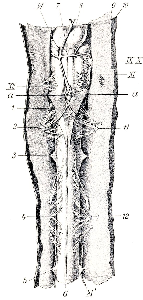
Fig. 495.—Ventral View of Medulla Oblongata and First and Second Segments of Spinal Cord of Horse; the Membranes are Cut and Reflected. 1, Lig. suspensorium arachnoideale; 2, right cerebrospinal artery; 3, 5, digitations of lig. denticulatum; 4, free border of lig. denticulatum; 6, middle spinal artery; 7, basilar artery; 8, pons; 9, arachnoidea; 10, dura mater; 11, 12, ventral root-bundles of first and second segments of spinal cord; VI., N. abducens; IX, X, glossopharyngeus and vagus; XI, accessory, medullary part; XI′, accessory, spinal part; a, line between medulla oblongata and spinal cord. (Dexler, in Ellenberger-Baum, Anat. d. Haustiere.)
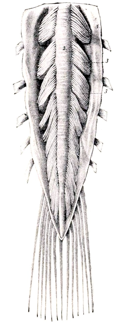
Fig. 496.—Cauda Equina. 1, Dura and arachnoidea divided and reflected; 2, spinal cord; 3, nerve-roots. (From Leisering’s Atlas, reduced.)
The surface of the spinal cord is divided into two similar halves by a dorsal median groove (Sulcus medianus dorsalis) and a ventral median fissure (Fissura mediana ventralis). On either side of the former is the dorso-lateral groove (Sulcus dorsalis lateralis) at which the fibers of the dorsal nerve-roots enter the cord; it is faint except at the enlargements, and is represented by two grooves in the first cervical segment. The ventral root-fibers as they emerge from the cord do not form a continuous series, but arise from a zone (Area radicularis ventralis) 3 to 5 mm. in width a little lateral to the ventral median fissure, and no groove is found here. In the greater part of the cervical region and the anterior part of the thoracic region there is a shallow dorsal paramedian groove (Sulcus intermedius dorsalis) a short distance lateral to the median 650groove. These grooves indicate the division of the white matter of the cord into columns to be described later.
Examination of cross-sections of the spinal cord shows that it is a bilaterally symmetrical structure, incompletely divided into right and left halves by a ventral fissure and a dorsal septum. The ventral median fissure (Fissura mediana ventralis) is narrow and penetrates nearly to the middle of the dorso-ventral diameter of the cord. It is occupied by a fold of pia mater. The dorsal median septum (Septum medianum dorsale) is a partition which descends from the posterior median groove to about the middle of the cord. It apparently consists of condensed neuroglia with an admixture of pial tissue. The two halves of the cord are connected by commissures of gray and white matter. The gray commissure (Commissura grisea) is a transverse band of gray matter at the ventral end of the dorsal septum. It is divided into dorsal and ventral parts by the central canal of the cord. The white commissure (Commissura alba) is a bridge of white matter which connects the ventral columns of the cord over the dorsal end of the ventral median fissure, and constitutes a conducting path from one side to the other.
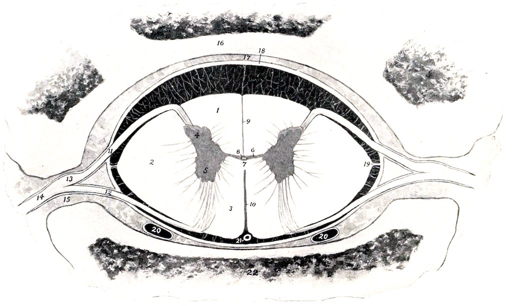
Fig. 497.—Cross-section of Spinal Cord in situ, in Part Schematic. 1, Dorsal column; 2, lateral column; 3, ventral column; 4, dorsal horn; 5, ventral horn; 6, gray commissure; 7, white commissure; 8, central canal; 9, dorsal septum; 10, ventral fissure; 11, dorsal nerve-root; 12, ventral nerve-root; 13, spinal ganglion; 14, nerve; 15, intervertebral foramen; 16, arch of vertebra; 17, epidural space; 18, dura mater (represented a little too thick); 19, ligamentum denticulatum; 20, 20, longitudinal venous sinuses; 21, middle spinal artery. The subdural and subarachnoid spaces (black) are traversed by delicate trabeculæ. The outer part of the arachnoid and the pia mater are not shown.
The central canal of the cord (Canalis centralis), the spinal vestige of the lumen of the embryonal neural tube, is a minute passage which tunnels the gray commissure. It opens at its anterior end into the posterior part of the fourth ventricle of the brain, and its terminal part in the conus medullaris forms a slight dilatation, the ventriculus terminalis. It is lined by epithelium and is surrounded by a layer of modified neuroglia (Substantia grisea centralis).
The gray matter of the spinal cord as seen in cross-sections resembles roughly a capital H the cross-bar of the letter being formed by the gray commissure. Each lateral part is considered as consisting of dorsal and ventral gray columns or horns (Columna grisea dorsalis, ventralis). In the greater part of the cord the 651dorsal column or horn is elongated and narrow and tapers to a point which extends almost to the surface of the cord at the attachment of the dorsal root-fibers of the spinal nerves. Its apex or tip consists of gray matter which is lighter in color and less opaque than that of the rest of the horn and is termed the substantia gelatinosa (Rolandi). The ventral column or horn is short, thick and rounded, and is separated from the surface of the cord by a thick layer of white matter, through which the fibers of the ventral roots of the spinal nerves pass. From the middle of the cervical region to the lumbar region there is a medial projection of gray matter on the lower part of the dorsal column; this is the nucleus dorsalis or Clarke’s column. In the anterior part of the cord there is an outward projection of the gray matter at the base of the ventral horn; this is termed the lateral column or horn. The demarcation between the gray and white matter is in many places indistinct; this is especially the case laterally, where processes of gray matter extend into the white substance, producing what is known as the formatio reticularis.
Cross-sections of the spinal cord present the following gross regional characters: (1) The cervical cord near the medulla is compressed dorso-ventrally, its width is about 18 mm. and its greatest thickness about 8 mm. It has dorsally a deep median sulcus and a distinct dorso-lateral sulcus. Lateral grooves are also present. The dorsal cornua are strongly everted. Each has an expanded head, which comes very close to the surface of the cord, and has an extensive cap of substantia gelatinosa. The neck is distinct. The ventral cornua are short and blunt and diverge very little. The gray commissure is about in the middle of the section and 2.5 mm. in length. According to Dexler the column between the median and lateral grooves dorsally is the funiculus cuneatus, the funiculus gracilis being very small and not showing on the surface in this region. In the middle of the cervical region the diameters are about 16 mm. and 10 mm. respectively. The ventral surface is somewhat flattened. The dorsal cornua have pointed ends and turn decidedly outward. The ventral cornua are short and thick and are directed very slightly outward; their ends are about 4 mm. from the ventral surface. The gray commissure is just above the middle of the section and is about 2 mm. long. The cervical enlargement measures about 25 mm. transversely and 12 mm. vertically. The dorsal cornua are smaller than the ventral and have a large cap of substantia gelatinosa. The ventral cornua are short and thick, curve strongly outward, and are about 4 mm. from the ventral surface. Each bears a prominence on its inner side near the base. The gray commissure is considerably above the middle of the section and is about 4 mm. long. (2) In the middle of the thoracic region the cross-section is biconvex, the ventral surface being the more strongly curved. The transverse diameter is about 15 mm. and the dorso-ventral about 10 mm. The gray columns are close together, the gray commissure being only about 1 mm. in length, and lying considerably above the middle of the section. The dorsal cornua are short and have slightly enlarged ends. The ventral cornua have a uniform diameter, turn very little outward, and end about 3 mm. from the ventral surface. (3) The lumbar enlargement is much flattened, especially dorsally. The transverse diameter is about 22 mm. and the dorso-ventral 9 to 10 mm. The cornua are very large. The ventral cornua are thick and rounded and turn sharply outward; they end about 2 mm. from the ventral surface. The dorsal cornua are smaller and shorter and do not diverge so strongly. The gray commissure is about in the middle of the section and is about 3 mm. long. In the third lumbar vertebra the cord is about 3 mm. narrower and thicker, and both surfaces are about equally convex. The dorsal cornua are smaller, considerably everted, and constricted in the middle. The ventral cornua are very short and do not turn outward. (4) In the first sacral vertebra the cord is almost round and is 5 to 6 mm. in diameter; the cornua are relatively very large and the commissure has the form of a high intermediate mass.
The ventral horn contains large cells, the axones of which emerge as the fibers of the ventral nerve-roots (Fila radicularia). The axones of many cells cross to the opposite side in the white commissure and pass out in a ventral root of that side, or enter the white matter and pass forward and backward, associating various segments of the cord. Some pass to the ventral horn of the opposite side at the same or at different levels. Others pass to the periphery of the cord, join the cerebellospinal fasciculus, and extend to the cerebellum. Scattered through the gray matter are many smaller cells with axones which pursue a short course and serve to connect different parts of the gray matter.
The white matter of the spinal cord is divided into three pairs of columns. The dorsal columns (Funiculi dorsales) lie on either side of the dorsal median septum and extend outward to the dorso-lateral groove and the dorsal gray column. The ventral columns (Funiculi ventrales) are situated on either side between the median fissure and the ventral gray columns. They are connected above the fissure by the white commissure. The lateral columns (Funiculi laterales) lie external to the gray columns on either side; their limits are indicated superficially by the dorso-lateral groove and the emergence of the ventral root-fibers. The paramedian 652groove (where present) indicates a subdivision of the dorsal column into two fasciculi or tracts; the inner of these is the fasciculus gracilis (Goll’s column); the outer, the fasciculus cuneatus (Burdach’s column).
The amounts of gray and white matter vary greatly in different parts of the cord both absolutely and relatively. In cross-section the absolute areas of both are greatest in the enlargements. The relative area of gray matter is smallest in the thoracic region (except at its anterior end), and increases from the lumbar enlargement backward.
Investigations have shown that in man the columns of white matter are subdivided into fasciculi or tracts, which constitute definite conducting paths of greater or less length. Our knowledge of the tracts in the domesticated animals is very limited, and it is quite unsafe to make inferences from the arrangement in man. As evidence of this it may be noted that the ventral cerebrospinal or direct pyramidal tract of man cannot be recognized as such.
The dorsal white columns consist essentially of two sets of axones. The afferent or sensory axones which come from the cells of the spinal ganglia enter as the dorsal roots of the spinal nerves and divide into two branches in the vicinity of the dorsal gray column. The anterior (ascending) branches form the direct sensory path to the brain and extend in the fasciculus cuneatus and fasciculus gracilis or corresponding tracts to nuclei in the medulla oblongata. The posterior (descending) branches extend backward for varying distances and give off numerous collaterals to cells of the gray column, thus forming part of the mechanism for the mediation of reflex action. Some collaterals cross in the white commissure to the opposite side. Many of these fibers are collected in the comma-shaped tract between the fasciculus gracilis and cuneatus. The second set of axones arises from the smaller cells of the gray column. They enter the white matter, divide into anterior and posterior branches, forming the fasciculi proprii or ground bundles of the cord. Some branches cross to the opposite side. The function of this set of axones is chiefly to associate various levels of the cord.
The lateral columns contain some axones of the dorsal nerve-roots, which (in man) are grouped in the marginal tract of Lissauer, situated just dorsal to the apex of the dorsal horn. The cerebellospinal fasciculus (direct cerebellar tract of Flechsig) extends along the periphery of the lateral column. It contains the axones of the cells of the nucleus dorsalis (Clarke’s column), which proceed to the medulla oblongata and enter the cerebellum by the restiform body. The rubrospinal tract of Monakow appears to take the place of the lateral cerebrospinal fasciculus or crossed pyramidal tract of man. It lies at the inner side of the cerebellospinal fasciculus. Its fibers arise in the nucleus ruber of the mid-brain, cross the median plane (decussation of Forel), and pass backward in the tegmentum and medulla oblongata to the lateral column of the cord. It is a path for motor impulses coming from the cerebral cortex and the cerebellum. The lateral fasciculus proprius or ground-bundle is deeply situated at the side of the gray columns. The bulk of its fibers are axones of cells of the posterior cornu which divide into anterior and posterior branches. They are intersegmental paths which associate different levels of the gray matter of the cord. The significance of the remaining fibers is not yet known.
The ventral white columns do not contain a ventral cerebrospinal or direct pyramidal tract, as in man. There is a small tract (Fasciculus intracommissuralis ventralis) above the white commissure, which separates it from the rest of the ventral column. It extends to the middle of the thoracic region. It consists of intersegmental fibers, and contains in the anterior part of the cervical cord in the sheep and goat both crossed and direct pyramidal fibers. The descending cerebellospinal fasciculus extends from the cerebellum to the lumbar region. In the cervical region it occupies a semilunar area which reaches almost to the surface ventro-laterally. Scattered fibers belonging to it lie also in the medial part of the ventral column. Posteriorly it diminishes in size and comes to occupy a position next to the ventral median fissure, corresponding to the sulco-marginal fasciculus of man (Dexler).
The Brain
The brain or encephalon is the part of the central nervous system that is situated in the cranial cavity. It is the enlarged and highly modified cephalic part of the primitive neural tube. It conforms rather closely in size and shape to the cavity in which it lies.
Its average weight without the dura mater is about 23 ounces (ca. 650 gm.), and forms about ⅐ of 1 per cent. of the body-weight.
It is desirable to examine the general external configuration of the brain before studying its various parts in detail.[187]
When divested of its membranes and vessels (Figs. 499, 500), its ventral 653surface or base presents the median brain stem, which is continuous with the spinal cord without any natural line of demarcation; it divides in front into two branches, the cerebral peduncles, each of which disappears into the mass of the corresponding cerebral hemisphere. The brain stem consists of three parts. The medulla oblongata is the posterior part which extends forward as the direct continuation of the spinal cord. The pons (Varolii) is a transversely elongated mass which appears to turn up on either side into the cerebellum. The cerebral peduncles extend forward from the pons and diverge to plunge into the ventral part of the cerebral hemispheres. The area between them is the interpeduncular space. It is largely covered by the pituitary body or hypophysis, a yellowish-brown, discoid structure, which is connected with the base of the cerebrum by a delicate tube called the infundibulum.[188] On drawing the pituitary body gently aside, the infundibulum is seen to be attached to a slight gray eminence, the tuber cinereum. Behind this is the mammillary body, a well-marked round prominence. The posterior part of the space is perforated by numerous openings for the passage of small arteries, and hence is termed the locus perforatus posticus. Two large bands of white matter, the optic tracts, cross the anterior ends of the cerebral peduncles and unite at the optic chiasm or commissure, forming the anterior boundary of the interpeduncular space. Above and in front of the chiasm the hemispheres are separated by the median longitudinal fissure. In contact with the anterior extremity of each hemisphere is the olfactory bulb, an oval enlargement adapted to the ethmoidal fossa of the cranium. This appears as a gray swelling on a wide flat band, the olfactory peduncle, which is continued behind by two divergent bands, the olfactory tracts or striæ. The internal tract disappears after a very short course on to the inner surface of the hemisphere. The external tract is larger and longer; it runs backward, inclines at first outward and then curves inward and disappears on the concealed or tentorial surface of the hemispheres. It is separated from the lateral cerebral gyri by a distinct groove (Sulcus rhinalis). Along the inner side of the stria are two eminences. The anterior of these is the trigonum olfactorium, a gray elevation situated in the angle of divergence of the inner and outer striæ. Behind this is a depression, the fossa transversa,[189] which is continued across the external stria and sharply limits the second and much larger eminence, the pyriform lobe.
The superficial origins of most of the cranial nerves are visible on the base of the brain.
The olfactory nerve-fibers join the convex surface of the olfactory lobe and give it a shaggy appearance in specimens which have been removed intact—a difficult proceeding.
The second or optic nerves converge to the optic chiasm.
The third or oculomotor nerve arises from the inner part of the cerebral peduncle.
The fourth or trochlear nerve may be seen emerging between the pons and the hemisphere, but its connection with the brain is not visible.
The fifth or trigeminal nerve is connected with the lateral part of the pons.
The sixth or abducent nerve arises just behind the pons and lateral to the pyramid of the medulla.
The seventh or facial and the eighth or auditory nerves arise close together just behind the pons proper on the extremity of the corpus trapezoideum.
The ninth or glosso-pharyngeal, the tenth or vagus, and the eleventh or spinal 654accessory nerves are connected by a linear series of roots with the lateral aspect of the ventral surface of the medulla. The spinal part of the accessory nerve comes forward along the edge of the medulla to join its medullary root.
The twelfth or hypoglossal nerve arises from the posterior part of the medulla along the lateral edge of the pyramid.
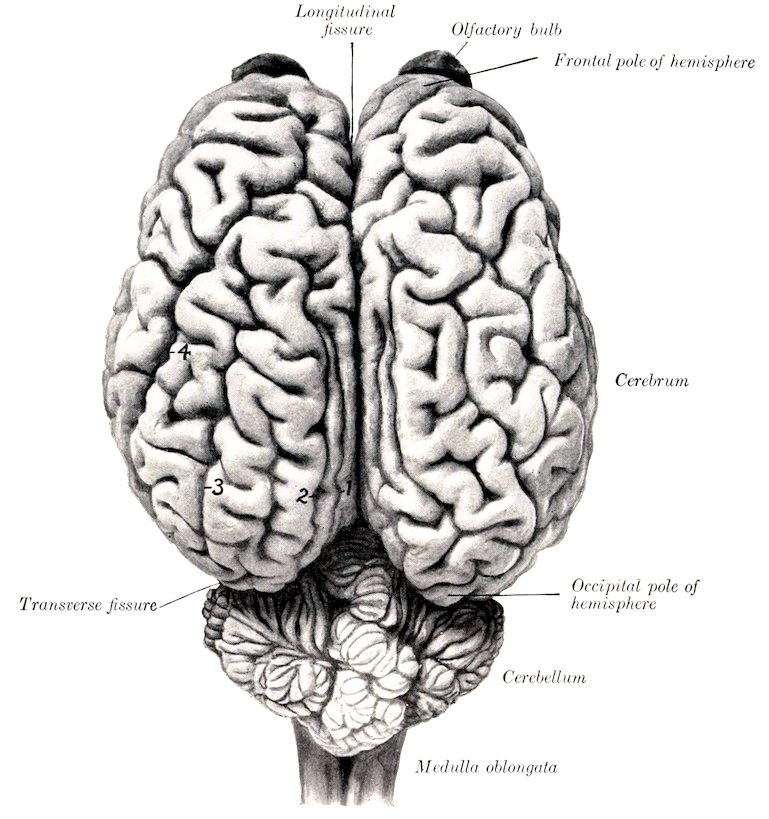
Fig. 498.—Brain of Horse, Dorsal View, about ⁵⁄₇ Natural Size. 1, Entomarginal fissure; 2, marginal fissure; 3, ectomarginal fissure; 4, suprasylvian fissure.
The parts that are visible when the brain is viewed from above are the cerebral hemispheres, the cerebellum, and part of the medulla oblongata. The cerebral hemispheres form an ovoid mass, and are separated from each other by the median longitudinal fissure, in which the falx cerebri is situated. Their surfaces are marked by thick folds, the gyri cerebri, separated by sulci. The upturned ends of the olfactory bulbs are seen in front of the frontal poles of the hemispheres. The occipital poles of the hemispheres overlie the anterior part of the cerebellum, from which they are separated by the transverse fissure and the tentorium cerebelli contained in it. The cerebellum is a much smaller rounded mass which conceals the greater part of the medulla oblongata. Its surface is divided into a middle 655lobe, the vermis, and two lateral hemispheres. It is marked by numerous gyri and narrow sulci which have in general a transverse direction. The posterior third of the medulla oblongata is not covered by the cerebellum.
The brain is developed from the expanded cephalic part of the neural tube of the embryo. The process comprises a series of thickenings, flexures, and unequal growth and expansion of various parts of the tube. In the higher animals the result is that the tubular character of the brain is not very evident, since the lumen comes to consist of four irregular cavities, the ventricles, which are connected by narrow passages. The tube is first subdivided by two constrictions into three brain vesicles, termed respectively the hind-brain or rhombencephalon, the mid-brain or mesencephalon, and the fore-brain or prosencephalon. The hind-brain gives rise to three secondary segments and the fore-brain to two. The annexed table indicates the origin of the principal structures of the fully developed brain from the primitive vesicles. It has become customary to describe the brain with reference to its embryological relations.
TABLE INDICATING THE DERIVATION OF THE PRINCIPAL PARTS OF THE BRAIN
Primary Segments. Secondary Segments. Derivatives. Cavities.
Rhombencephalon (posterior vesicle) Myelencephalon Medulla oblongata Fourth ventricle
Metencephalon Pons
Cerebellum
Isthmus rhombencephali Anterior cerebellar peduncles
Anterior medullary velum
Mesencephalon (middle vesicle) Mesencephalon Corpora quadrigemina Cerebral aqueduct
Cerebral peduncles
Prosencephalon (anterior vesicle) Diencephalon {Optic thalami } Posterior part of third ventricle
{Subthalamic tegmenta
{Pineal body
Telencephalon {Pituitary body } Anterior part of third ventricle. Lateral ventricles and olfactory continuations.
{Optic nerves and retinæ
{Cerebral hemispheres
{Olfactory tracts and bulb
THE RHOMBENCEPHALON
The Medulla Oblongata
The medulla oblongata lies on the basilar part of the occipital bone. It is quadrilateral in outline, but much wider in front than behind, and compressed dorso-ventrally. Its length, measured from the root of the first cervical nerve to the pons, is about two inches (ca. 5 cm.).
Its ventral surface is convex in the transverse direction. It is marked by a median fissure (Fissura mediana ventralis) which is continuous behind with the similar fissure of the spinal cord. The posterior part of the fissure is faintly marked, but in front it becomes deeper and ends in a small depression (Foramen cæcum) behind the central part of a transverse band, the corpus trapezoideum. On either side of the fissure is a rounded tract, the pyramid (Pyramis), which is bounded externally by a faint lateral groove (Sulcus intermedius ventralis). The pyramids join the pons in front; behind they become narrower and disappear into the substance of the medulla, in which their fibers intercross, forming the decussation of the pyramids (Decussatio pyramidum).[190]
The superficial origin of the sixth cranial nerve (N. oculomotorius) is just lateral to the anterior end of the pyramid. The corpus trapezoideum is a transverse band which extends across the surface immediately behind the pons. It is crossed by the pyramids, which cut off a small central part. The lateral part extends out to the roots of the seventh and eighth nerves on either side. Behind the outer part of the corpus trapezoideum there is a variably developed rounded eminence, the tuberculum faciale. The root-fibers of the twelfth or hypoglossal nerve form an oblique linear series lateral to the posterior part of the pyramid.
656
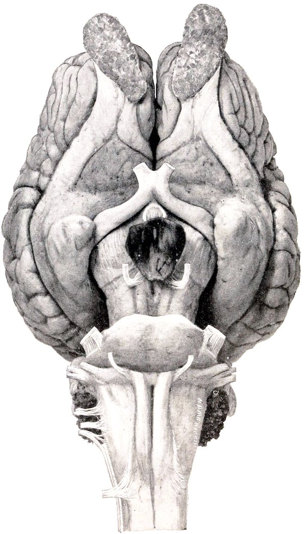
Fig. 499.—Base of Brain of Horse, Natural Size.
The dorsal surface is largely concealed by the cerebellum and forms the greater part of the floor of the fourth ventricle. The dorsal median fissure (Fissura mediana dorsalis), the direct continuation of the corresponding groove of the spinal cord, extends forward to about the middle of the surface. Here the restiform bodies, which constitute the lips of the fissure, diverge to form the lateral boundaries of a triangular depression; this is the posterior part of the rhomboid fossa or floor of the fourth ventricle of the brain. The dorso-lateral fissure winds outward and forward to the lateral aspect of the medulla, where it presents the roots of the ninth, tenth, and eleventh cranial nerves. External to it is a distinct oval eminence on the anterior part of the lateral column, termed the tuberculum cinereum. The central canal of the cord is continued in the posterior part of the medulla, 657inclines dorsally, and opens in the posterior angle of the fourth ventricle. Hence it is customary to distinguish a closed and an open part of the medulla. The dorsal aspect of the latter, which is concealed at present, will be considered later in the description of the fourth ventricle.
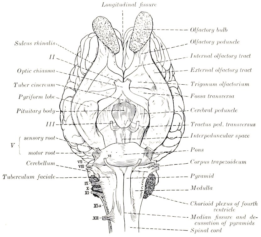
Fig. 500.—Line Drawing of Base of Brain of Horse. (Key to Fig. 499.)
The lateral surface is narrow behind, wider and rounded in front. From it the root-fibers of the ninth, tenth, and eleventh cranial nerves arise in a linear series, and alongside of it the spinal part of the eleventh nerve passes forward to join the medullary root. Close inspection reveals the presence of striæ which curve obliquely downward and backward from the surface of the restiform body toward the hypoglossal root-fibers; these are the external arcuate fibers (Fibræ arcuatæ externæ). The recess between the lateral aspect of the medulla and the cerebellum is occupied by an irregular mass of villous projections of the pia mater, containing tufts of vessels; this is the chorioid plexus of the fourth ventricle, and is the lateral edge of the tela chorioidea of the ventricle. On raising the chorioid plexus it is seen that the tela chorioidea is attached to the dorsal aspect of the medulla, and reinforces here the wall of the fourth ventricle; also that the restiform body terminates in front by entering the base of the cerebellum, forming its posterior peduncle.
In the medulla the fiber tracts of the spinal cord either terminate in the nuclei of the gray matter or undergo changes in their relative position, and new tracts appear. The gray matter is highly modified and forms masses which have no homologues in the cord. The central canal of the closed part of the medulla is surrounded by a thicker layer of gray matter than is the case in the spinal cord. As the medulla opens out this gray matter is naturally spread in the floor of 658the fourth ventricle. The dorsal horns of gray matter become wide, spread apart, and are broken up to a great extent in the formatio reticularis. Two elongated masses of cells appear above the central gray matter; these are the nucleus gracilis and nucleus cuneatus, and in them the fasciculi of like name gradually end. In front of these are the terminal nuclei of the afferent or sensory cranial nerves and of the sensory portions of the mixed nerves. The ventral horns are succeeded by the nuclei of origin of the efferent or motor cranial nerves and the motor root-fibers of the mixed nerves. Of the twelve pairs of cranial nerves, the last eight are connected with nuclei in the medulla and pons. The posterior olivary nucleus (Nucleus olivaris caudalis) is a conspicuous gray mass which lies above the pyramid on each side. On cross-sections it appears as a wavy layer of gray matter which is folded on itself so as to inclose a mass of white matter. It is about 2 cm. long in the horse, but is smaller in circumference than in man and hence does not cause any very distinct external enlargement (olivary eminence), as in the latter. It is connected with the opposite side of the cerebellum by the cerebello-olivary fibers. At its inner side are two small accessory olivary nuclei. The pyramidal tracts, which are small in ungulates, send most of their fibers across to the opposite side in the posterior part of the medulla, forming the pyramidal decussation. Some fibers, however, continue in the ventral column of the same side of the cord, and others are connected with the nuclei of the motor nerve-roots. From the nucleus gracilis and nucleus cuneatus fibers arise which are traceable forward to the thalamus. These are the internal arcuate fibers (Fibræ arcuatæ internæ), which curve across the median plane below the central gray matter and form with those of the opposite side the decussation of the fillet (Decussatio lemniscorum). Beyond the decussation the fibers form an important longitudinal tract which extends forward in the mid-brain. This is the fillet or lemniscus, the chief continuation of the sensory conducting path from the dorsal roots of the spinal nerves. The external arcuate fibers, some of which were seen on the lateral aspect of the medulla, have a similar origin. Part of them (Fibræ dorsales) pass directly to the dorsal aspect of the restiform body of the same side; others (Fibræ ventrales) cross to the opposite side, descend close to the ventral fissure, and then curve upward and forward to the restiform body. The decussation of the arcuate fibers forms the distinct median raphé seen on cross-sections of the medulla anterior to the pyramidal decussation. The restiform body, situated dorso-laterally, contains, in addition to the arcuate fibers, the cerebello-olivary fasciculus before mentioned, and the cerebellospinal fasciculus or direct cerebellar tract. The dorsal longitudinal fasciculus corresponds to the ventral ground-bundle of the spinal cord, displaced dorsally by the decussation of the pyramids and fillet. In the posterior part of the medulla it is not marked off from the fillet, along the dorsal edge of which it lies. From the level of the hypoglossal nucleus forward it is distinct and can be traced as a conspicuous tract in the ventral margin of the gray matter of the floor of the fourth ventricle and of the central gray matter of the mid-brain. Below the restiform body and related externally to the external arcuate fibers, there is a considerable bundle of longitudinal fibers, the spinal root (Tractus spinalis) of the fifth nerve; internal to it is the terminal nucleus of the sensory root of the nerve.
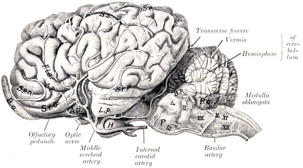
Fig. 501.—Brain of Horse, Left Lateral View. The larger arteries are shown. The nerve-roots are indicated by Roman numerals. P.c., Chorioid plexus of fourth ventricle; B.p., middle peduncle of cerebellum; P, pons; H, pituitary body; L.p., pyriform lobe; A.p., trigonum olfactorium; B.o., olfactory bulb; S.r.a., sulcus rhinalis anterior; S.r.p., sulcus rhinalis posterior; F.l., lateral fissure (of Sylvius); F.l.a., anterior branch of lateral fissure; S.p., presylvian fissure; S.p.n., anterior presylvian fissure; S.s., suprasylvian fissure; S.e., ectomarginal fissure.
659
THE PONS
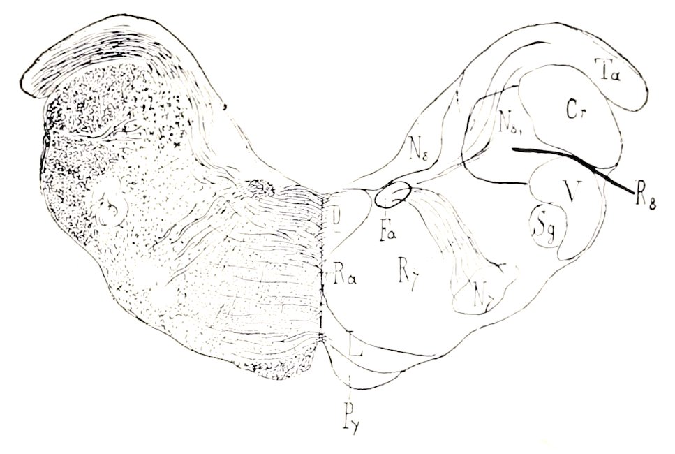
Fig. 502.—Cross-section of Medulla Oblongata of Horse, Passing through Facial Nucleus. Cr, Corpus restiforme; D, dorsal longitudinal fasciculus; Fa, ascending part of facial nerve; L, fillet; N7, nucleus of facial nerve; N8, triangular nucleus of vestibular root of eighth nerve; N8′, spinal root of eighth nerve; Py, pyramid; Ra, raphé; R7, radicular part of facial nerve; R8, vestibular root of eighth nerve; Sg, substantia gelatinosa; Ta, posterior end of tuberculum acusticum; V, spinal root of trigeminus. (Ellenberger-Baum, Anat. d. Haustiere.)
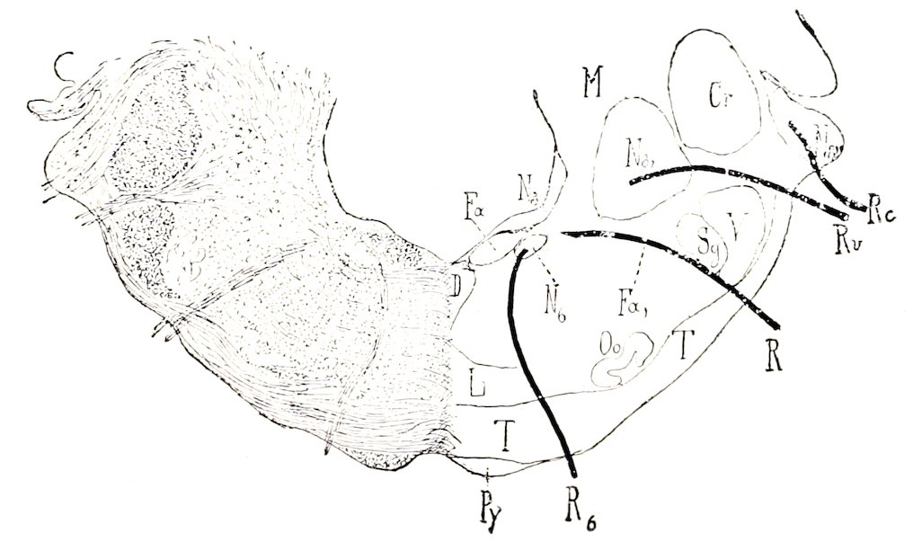
Fig. 503.—Cross-section of Medulla Oblongata of Horse; Section Passes through Middle of Corpus Trapezoideum. Cr, Corpus restiforme; D, dorsal longitudinal bundle; Fa, ascending part of facial nerve; Fa′, emergent or descending part of facial nerve; R7, root of facial nerve; L, fillet; M, central white matter of cerebellum; N6, abducens nucleus; N8, triangular nucleus; N8′, nucleus of Deiters; N8″, tuberculum acusticum; Oo, anterior olive; Py, pyramid; R6, root of abducens nerve; R7, root of facial nerve; Rc, cochlear nerve; Rv, vestibular nerve; Sg, substantia gelatinosa; T, corpus trapezoideum; V, spinal root of trigeminus. (Ellenberger-Baum, Anat. d. Haustiere.)
The pons (Varolii) is that part of the brain stem which lies between the medulla and the cerebral peduncles; it is marked off from these ventrally by anterior and posterior grooves. Viewed from below it is elongated transversely, convex in both directions, and presents a wide shallow median groove (Sulcus basilaris), which lodges the basilar artery. Laterally a large part of its mass curves upward and backward into the base of the cerebellum, forming the middle cerebellar peduncle (Brachium pontis). The superficial origin of the trigeminal (fifth) nerve is at the 660lateral limit of the ventral surface. Transverse striations indicate the course of its superficial (ventral) fibers which connect the two sides of the cerebellum. The dorsal surface is blended on either side with the overlying anterior peduncles of the cerebellum; the central free portion forms the anterior part of the floor of the fourth ventricle, and will be considered in the account of that cavity.
On cross-section the pons is seen to be composed of dorsal and ventral parts. The dorsal part (Pars dorsalis pontis) consists superficially of a layer of gray matter covered by the ependyma of the fourth ventricle. Beneath this the median raphé of the medulla is continued into the pons, dividing it into similar halves. In the anterior part of the pons the fillet divides into an inner and outer part, the medial and lateral fillets (Lemniscus medialis, lateralis); the latter arches outward to reach the outer side of the anterior cerebellar peduncle. The dorsal longitudinal fasciculus becomes sharply defined into a round bundle which lies close to the raphé under the gray matter of the floor of the fourth ventricle. In cross-section the formatio reticularis forms a large area below the superficial gray matter and the longitudinal bundles. Dorso-laterally is the large rounded section of the anterior cerebellar peduncle. Lower down is a large bundle, the sensory root of the fifth nerve. In front of this is the motor nucleus of the same nerve, external to which is its motor root. The ventral part of the pons (Pars basilaris pontis) is composed of transverse and longitudinal fibers, and a large amount of gray matter which is broken up into small masses (Nuclei pontis) by the intersection of the fibers. The transverse fibers are gathered laterally into a compact mass which turns upward and backward and enters the central white matter of the cerebellum, forming the middle cerebellar peduncle. Centrally the fibers are arranged in bundles which intercross. The transverse fibers are chiefly of two kinds. Some arise from the Purkinje cells of the cerebellar cortex and pass either to the opposite side of the cerebellum or turn at the raphé and run forward and backward in the brain stem. Others are axones of cells of the nuclei pontis, and pass to the hemispheres of the cerebellum. The corpus trapezoideum is mainly the central continuation of the cochlear division of the auditory nerve. Above it is the small anterior olivary nucleus (Nucleus olivaris nasalis). The longitudinal fibers of the ventral part of the pons consist chiefly of the cerebrospinal or pyramidal fasciculi. These come from the ventral part (pes) of the cerebral peduncles and are situated laterally, interspersed among the deep transverse fibers in the anterior part of the pons. Toward the posterior part the bundles incline toward the median plane and become collected into a compact mass which appears superficially at the posterior border. Many fibers come from the cerebral cortex and terminate in the nuclei of the gray matter of the pons; they may be designated corticopontile fibers.
THE CEREBELLUM
The cerebellum is situated in the posterior fossa of the cranium, and is separated from the cerebral hemispheres by the transverse fissure and the tentorium cerebelli which occupies it. It overlies the pons and the greater part of the medulla, from which it is separated by the fourth ventricle. Its average weight is about two ounces (ca. 60 gm.) or about 9 per cent. of the weight of the entire brain. Its shape is approximately globular but very irregular. It is somewhat compressed dorso-ventrally and its transverse diameter is the greatest.
The anterior surface faces upward and forward and is covered partially by the tentorium cerebelli. The posterior surface is almost vertical. The ventral surface or base lies over the fourth ventricle, and is connected by three pairs of peduncles with the medulla, pons, and mid-brain.
It is customary to recognize three gross divisions of the cerebellum, viz., the median vermis and two lateral hemispheres. The vermis (cerebelli) is curved in a circular manner so that its two extremities are close together or even in contact on the ventral surface. The anterior extremity is termed the lingula; it lies between the cerebellar peduncles and gives attachment to the anterior medullary velum, a thin lamina which forms the anterior part of the roof of the fourth ventricle. The posterior extremity, the nodulus, gives attachment to the posterior medullary velum which covers the posterior recess of the fourth ventricle. The hemispheres (Hemisphæria cerebelli) are clearly separated from the vermis by two deep paramedian fissures. They lie in the lateral depressions of the cerebellar compartment of the cranium.
In tracing the fissures from behind forward it will be noticed that they are nearly sagittal as far as the anterior surface, where they diverge widely, so that the vermis forms all of the fore part of the cerebellum.
661The surface of the cerebellum is further cut up into numerous gyri or folia by narrow and relatively deep sulci, many of which approach a transverse direction. Certain of the sulci are more pronounced than the others, and by means of them it is possible to define groups of gyri. Such groups are termed lobes, and have received specific names, derived chiefly from the systematic descriptions of the human cerebellum.
The lobes of the vermis are readily distinguished on a median section. Enumerated from the anterior to the posterior extremity they are: (1) lingula, (2) lobus centralis, (3) lobus ascendens, (4) lobus culminis, (5) lobus clivi, (6) tuber vermis, (7) pyramis, (8) uvula, (9) nodulus. Each hemisphere is cut into laterally by two sulci which mark off two sagittal discoid masses, termed by Ziehen tabulations. The external tabulation consists of four or five lobules, the lowest of which is regarded as the flocculus. The inner part of the hemisphere is divided into three or four lobes. In the absence of a satisfactory morphological basis it seems undesirable to deal with the lobation of the cerebellum in further detail.
The cerebellar peduncles, three on each side, join the central white matter of the cerebellum at the base. The posterior peduncle is the restiform body of the medulla, a large rounded tract derived from the lateral and ventral columns of the cord. Near the middle of the medulla it inclines outward, forms the lateral wall of the fourth ventricle, and ends by entering the central white matter of the cerebellum. The middle peduncle is formed, as previously seen, by the brachium pontis. The anterior peduncles (Brachia conjunctiva) pass forward on either side on the dorsal surface of the pons, forming the lateral boundary of the fore part of the fourth ventricle. They disappear under the corpora quadrigemina into the substance of the mid-brain. At the point of disappearance the trochlear (fourth) nerve emerges from the mid-brain. In some cases two or three bundles of fibers (Fila lateralia pontis) arise in the angle between the middle and anterior peduncle, curve obliquely forward and downward over the outer aspect of the latter, and spread out on the ventral face of the cerebral peduncle just in front of the pons.
On sagittal section the cerebellum is seen to consist of a layer of cortical gray matter (Substantia corticalis) and the medullary white matter. The white matter consists of a large basal mass (Corpus medullare), which is joined by the peduncles, and gives off primary laminæ to the lobules; from these secondary and tertiary laminæ arise, the latter entering the gyri. The arrangement on sagittal section is tree-like, hence the classical term “arbor vitæ” which is applied to it. The central gray matter consists of groups of cells which form small nuclei embedded in the central white substance.
As noted above the central gray matter does not form a large nucleus, the corpus dentatum, which is so conspicuous an object on sagittal sections of the cerebellar hemisphere in man.
The principal connections established by the peduncular fibers of the cerebellum are as follows: The posterior peduncle (Corpus restiforme) is composed of afferent and efferent fibers which connect the cerebellum with the medulla and spinal cord. The cerebellospinal fasciculus or direct cerebellar tract, which arises from the cells of the nucleus dorsalis (Clarke’s column) of the cord, ends in the cortex of the vermis; many of its fibers cross to the opposite side. Numerous arcuate fibers from the nucleus gracilis and nucleus cuneatus of the same and opposite sides establish connections with cells of the cerebellar cortex. Olivocerebellar fibers (chiefly afferent) connect with the olivary nucleus of the same and of the opposite side of the medulla oblongata. The nucleo-cerebellar fasciculus comprises fibers derived from the nuclei of the fifth, eighth, and tenth cranial nerves (Edinger). The descending cerebellospinal fasciculus consists of fibers which terminate in relation with cells of the ventral horns of the spinal cord. The chief facts concerning the middle peduncle have been mentioned in the description of the pons. The anterior peduncle is essentially an efferent tract, the fibers of which pass forward to the tegmentum of the cerebral peduncle, the subthalamic region, and the thalamus. After the peduncles disappear under the corpora quadrigemina, they converge and many of their fibers intercross, forming the decussation of the superior peduncle. A considerable number of fibers end in the nucleus ruber. Thence impulses are transmitted in two directions: first, by thalamo-cortical fibers to the cerebral cortex; second, by the rubrospinal tract through the brain stem and lateral columns of the cord to the ventral horn cells. The ventro-lateral cerebellospinal fasciculus (Gowers’ tract) is an ill-defined tract which connects the spinal cord with the cerebellum. Its fibers appear to be axones of cells of the posterior horns of the cord; they pass in the lateral column of the cord, become scattered in passing through the reticular formation of the medulla and pons, and enter the cerebellum by way of the anterior medullary velum.
662
The Fourth Ventricle
The fourth ventricle (Ventriculus quartus) is the cavity of the rhombencephalon; it communicates with the central canal of the spinal cord behind, and through the aqueduct with the third ventricle in front. It is somewhat rhomboid in outline, elongated from before backward and narrowest behind. It is lined completely by an epithelium (Ependyma) and contains a small amount of fluid.
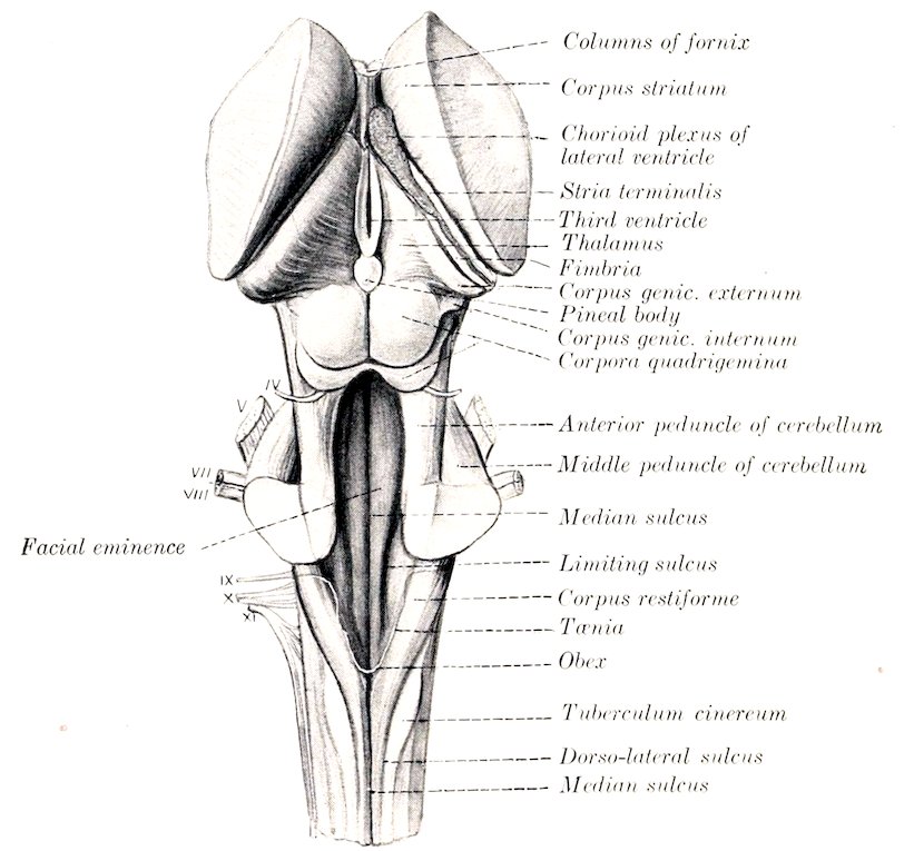
Fig. 504.—Brain Stem and Basal Ganglia of Horse, Dorsal View. The cranial nerve-roots are designated by Roman numerals.
Its floor (Fossa rhomboidea) is formed by the medulla and pons and is marked by three longitudinal furrows which converge behind. It is widest and deepest a little in front of its middle. The posterior part narrows to a point at the opening of the central canal, and on account of its appearance in man it has been termed the calamus scriptorius. The median sulcus (Sulcus medianus) extends the entire length of the floor and is deepest toward the ends. The limiting sulci (Sulci limitantes) begin on either side of the opening of the central canal and extend forward as the lateral limits of the rhomboid fossa. Just beyond the middle of the fossa they expand into a shallow depression, the anterior fovea (Fovea nasalis). On either side of the median sulcus and margined by the limiting sulcus is a slightly rounded column, the eminentia medialis. Opposite the fovea this presents an elongated prominence, the colliculus facialis, so named because it overlies the bend formed by the fibers of origin of the facial nerve. External to the limiting sulcus is a long fusiform elevation, the area acustica, from which a band of fibers (Striæ acusticæ) winds over the anterior end of the restiform body to the superficial origin of the cochlear nerve.
663The lateral wall is formed by the restiform body and the anterior peduncle of the cerebellum.
The roof is formed in its middle part by the vermis of the cerebellum, covered by the epithelium before mentioned. There is commonly a dorsal recess (Recessus tecti s. fastigium) between the extremities of the vermis. The anterior part of the roof is formed by a thin lamina of white substance, the anterior medullary velum (or valve of Vieussens), which extends backward from the corpora quadrigemina, and is attached on either side to the anterior peduncles of the cerebellum. Its anterior part is relatively thick and contains the decussation of the fibers of the trochlear nerves. Posteriorly it blends with the white matter of the cerebellum. A thin lamina of white matter, the posterior medullary velum, backed by pia mater, completes the roof posteriorly. After removal of the cerebellum the line of attachment (Tænia ventriculi quarti) to the medulla is seen; it begins centrally over the opening of the central canal, runs forward on the inner face of the restiform body, and turns outward behind the brachium pontis. The thick part which stretches over the posterior angle of the ventricle is termed the obex. The posterior part of the ventricle forms three recesses, of which two are lateral and the third median and posterior. The lateral recesses communicate with the subarachnoid space by distinct openings (Aperturæ laterales). The layer of pia which strengthens the roof here is named the tela chorioidea of the fourth ventricle. It is triangular in outline and closely adherent to the velum. It forms three fringed masses which contain vascular convolutions and are designated the median and lateral chorioid plexuses of the fourth ventricle. They appear to lie within the ventricle, but are really excluded from the cavity by the epithelial lining, which they invaginate.
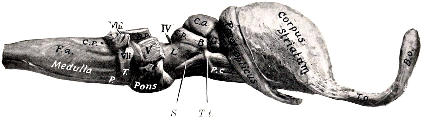
Fig. 505.—Brain Stem and Basal Ganglia of Horse, Right View. F.a., External arcuate fibers; C.r., corpus restiforme; P, pyramid; T, corpus trapezoideum; B.p., middle peduncle of cerebellum; P.c., cerebral peduncle; S, sulcus lateralis; T.t., tractus transversus; L., trigonum lemnisci; C.a., corpus quad, ant.; C.p., corpus quad, post.; B, commissure of C.p.; G, corpus geniculatum internum; T.o., olfactory peduncle; B.o., olfactory bulb.
THE MESENCEPHALON
The mesencephalon or mid-brain connects the rhombencephalon with the fore-brain. In the undissected brain it is covered dorsally by the cerebral hemispheres. It consists of a dorsal part, the corpora quadrigemina, and a larger ventral part, the cerebral peduncles, which are visible on the base of the brain. It is traversed longitudinally by a narrow canal, the cerebral aqueduct, which connects the fourth ventricle with the third.
The corpora quadrigemina[191] are four rounded eminences which lie under the posterior part of the cerebral hemispheres. They consist of two pairs, separated by a transverse groove. The anterior pair (Colliculi nasales) are larger and much 664higher than the posterior pair. They are gray in color, almost hemispherical, and are separated by a narrow furrow which leads forward to the subpineal fovea. A wide groove intervenes between them and the optic thalami. The posterior pair (Colliculi caudales) are relatively small and are paler than the anterior pair. They are marked by a wide median depression, and are limited below by a transverse furrow (Sulcus postquadrigeminus), at either side of which the trochlear (fourth) nerve emerges. Laterally each is prolonged to the inner geniculate body by a band of white matter termed the inferior brachium.[192]
The cerebral peduncles or crura cerebri (Pedunculi cerebri) appear on the base of the brain as two large rope-like stalks which emerge from the pons close together and diverge as they extend forward to enter the cerebrum. At the point of disappearance the optic tract winds obliquely across the peduncle. About half an inch further back a small tract (Tractus peduncularis transversus) curves across the peduncles, and behind this near the median line is the superficial origin of the oculomotor (third) nerve. The triangular depression between the diverging peduncles is the interpeduncular space (Fossa interpeduncularis). It is covered to a large extent by the pituitary body, a discoid brown mass which is connected with the base of the brain by a hollow stalk, the infundibulum. The posterior part of the space is pierced by numerous minute openings which transmit blood-vessels, and is therefore termed the locus perforatus posticus. The objects in the space belong to the diencephalon, and will be described later. The lateral aspect of the peduncle is marked by a groove (Sulcus lateralis mesencephali) which indicates the division into a dorsal part, the tegmentum, and a ventral part the basis pedunculi; these are separated by a layer of dark gray matter, the substantia nigra. The triangular area (Trigonum lemnisci) above the lateral groove is faintly marked by fibers passing obliquely upward and backward to the anterior cerebellar peduncle; these belong to the fillet or lemniscus, an important tract that connects the thalamus and corpora quadrigemina with the sensory reception nuclei of the opposite side of the medulla.
The aqueduct of the cerebrum or of Sylvius (Aquæductus cerebri) is the canal which extends through the mid-brain from the fourth to the third ventricle. It is surrounded by a layer of gray matter (Stratum griseum centrale), in the ventral part of which are the nuclei of origin of the oculomotor and trochlear nerves, and laterally nuclei of the mesencephalic roots of the trigeminal nerves.
THE DIENCEPHALON
The diencephalon or inter-brain comprises the thalamus and a number of other structures grouped about the third ventricle, the cavity of this division of the brain.[193] To expose its dorsal aspect, the greater part of the cerebral hemispheres, the corpus callosum, the fornix, the hippocampus, and the tela chorioidea of the third ventricle must be removed.
The thalamus (or optic thalamus) is the principal body in this part of the brain. It is a large ovoid gray mass placed obliquely across the dorsal face of each cerebral peduncle, so that the long axes of the two thalami would meet in front about at a right angle. Medially they are fused to a large extent, and around the area of adhesion they are separated by a sagittal circular space, the third ventricle. The dorsal surface is convex in both directions, and is separated from the overlying hippocampus by the tela chorioidea (or velum interpositum). Laterally it is separated 665from the nucleus caudatus by an oblique groove in which there is a band of white matter termed the stria terminalis or tænia semicircularis. Internally it is bounded by a narrow band, the stria medullaris, on which is a delicate ridge of ependyma termed the tænia thalami. The striæ unite posteriorly and blend with the stalk of the pituitary body. Near this point they present a small enlargement caused by the nucleus habenulæ. Anteriorly there is a small eminence, the anterior tubercle. The posterior part of the thalamus has the form of a rounded ridge which is continuous laterally with the optic tract.[194] Behind the point of origin of the tract, in the angle between the thalamus and the cerebral peduncle, is the internal geniculate body (Corpus geniculatum mediale), a well defined oval prominence.
The outer surface is separated from the lenticular nucleus by the internal capsule, an important mass of white matter composed of fibers passing to and from the cerebral cortex. These fibers go to form a large part of the ventral portion (basis) of the cerebral peduncle. From the entire external surface of the thalamus fibers pass into the internal capsule and radiate to reach the cerebral cortex; similarly fibers coming from the cortex converge in the internal capsule to enter the thalamus. This arrangement is termed the thalamic radiation. Ventral to the thalamus proper is the subthalamic tegmental region. This is the continuation of the tegmental part of the cerebral peduncle into the diencephalon. It contains the red nucleus (Nucleus ruber) an important ganglion on the course of the motor tracts. It receives numerous fibers from the cerebral cortex and the corpus striatum. From it fibers proceed to the thalamus and to the spinal cord; the fibers to the cord (Tractus rubro-spinalis of Monakow) cross to the opposite side and extend back in the tegmentum to the lateral columns of the cord. Lateral to the red nucleus a conspicuous lenticular area of dark gray matter is visible on cross-sections of the subthalamic region; this is the subthalamic nucleus (Nucleus hypothalamicus s. corpus Luysi), which consists of pigmented nerve-cells scattered through a dense network of fine medullated fibers, and is richly supplied with capillary blood-vessels. The two nuclei are connected by a transverse commissure (Commissura hypothalamica), which crosses the floor of the third ventricle above the mammillary body.
The pineal body or epiphysis is a small ovoid or fusiform red-brown mass situated in a deep central depression between the thalami and corpora quadrigemina. It is variable in size, but is commonly about 10 to 12 mm. long and 6 mm. wide. It is attached at the postero-superior quadrant of the third ventricle by a short stalk, in which is a small recess of that cavity. Its base blends in front with the junction of the striæ medullares of the thalamus. Immediately under the posterior part of the stalk is a short transverse band of white matter, the posterior commissure.
The pineal body is inclosed in a fibrous capsule from which numerous trabeculæ pass inward, dividing the organ into spaces occupied by round epithelial cells of the same origin as the ependyma of the ventricle.
The mammillary body (Corpus mammillare) is a white round elevation a little larger than a pea which projects ventrally at the anterior end of the median furrow of the interpeduncular space. While it is a single body in external form in the horse, sections show that it is double in structure and contains a nucleus of gray matter on either side.
Three sets of fibers are connected with the mammillary body. The anterior pillar of the fornix curves down in the lateral wall of the third ventricle to the body and many of the fornix fibers end in it. A bundle (Fasciculus thalamo-mammillaris) passes upward and backward from it into the anterior part of the thalamus, and a tract (Fasciculus pedunculo-mammillaris) extends back in the floor of the third ventricle to the tegmentum of the mid-brain.
The pituitary body or hypophysis was mentioned as covering part of the interpeduncular space. It is oval in outline, flattened dorso-ventrally, and nearly an inch (ca. 2 cm.) in width. It is attached by a delicate tubular stalk, the infundibulum, to the tuber cinereum, a small gray prominence situated between the optic chiasm in front and the mammillary body behind.
The pituitary body consists of two parts which can be distinguished readily on sections by their color. The glandular (or anterior) lobe is brown in color and is inclosed in a fibrous capsule. 666It is glandular in character and there is good ground for the view that it is an organ of internal secretion. Besides the chief cells which stain lightly, it contains large deeply staining chromophile cells. It arises as an outgrowth from the primitive mouth cavity. The cerebral (or posterior) lobe is pale and is connected with the infundibulum so as to form a rather flask-shaped arrangement. It is almost entirely inclosed by the glandular part. It arises as an outgrowth from the primitive diencephalon, but loses most of its earlier nervous character.
The optic chiasm and tracts form the anterior boundary of the interpeduncular space. The optic chiasm or commissure (Chiasma opticum) is formed by the convergence of the optic nerves and the crossing of the major part of the fibers of the nerve of one side to the tract of the opposite side. From the chiasm each optic tract (Tractus opticus) curves over the cerebral peduncle outward, backward, and upward to the posterior part of the thalamus and the internal geniculate body; some fibers reach the anterior quadrigeminal body.
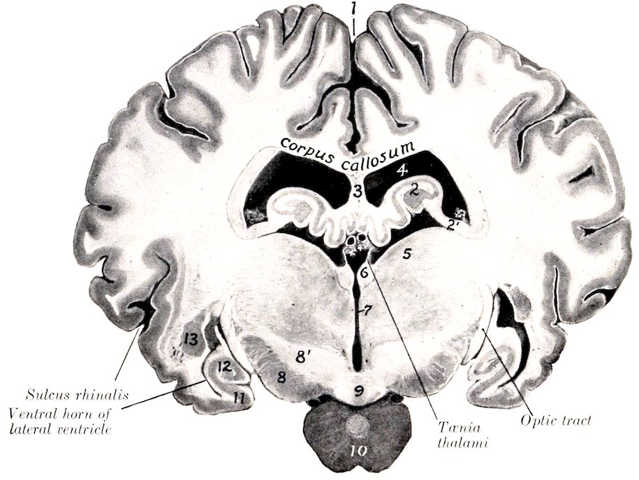
Fig. 506.—Cross-section of Brain of Horse, Natural Size. Section passes through posterior part of third ventricle and is viewed from behind. 1, Longitudinal fissure; 2, hippocampus; 2′, fimbria; 3, septum pellucidum; 4, lateral ventricle; 5, thalamus; 6, habenula; 7, third ventricle; 8, cerebral peduncle; 8′, hypothalamus; 9, mammillary body; 10, pituitary body; 11, pyriform lobe; 12, ventral end of hippocampus; 13, amygdaloid nucleus. Between the upper parts of the tæniæ thalami is the chorioid plexus of the third ventricle, and above this are the internal cerebral veins.
All the fibers in the chiasm are not derived from the optic nerves. The posterior part contains fibers which pass from one tract to the other and are connected with the internal geniculate bodies; this bundle is called Gudden’s commissure (Commissura inferior). Above it is Meynert’s commissure (Commissura superior), the fibers of which enter the subthalamic body.
The third ventricle (Ventriculus tertius) is the narrow annular space between the thalami. It communicates by means of the aqueduct with the fourth ventricle behind, and in front it is continuous with the lateral ventricle on each side through the interventricular foramen. Its floor is formed by the structures of the interpeduncular space and to a small extent by the tegmentum of the cerebral peduncles. The roof is formed in the strict sense only by the ependyma, above which is a fold of pia mater, termed the tela chorioidea of the third ventricle or velum interpositum. The roof is invaginated by two delicate chorioid plexuses 667which appear to lie within the ventricle, although they are excluded from the cavity by the epithelium. When the tela is removed, the delicate ependyma of the roof is torn away with it, leaving the line of attachment to the stria medullaris to constitute the tænia thalami. The anterior wall is formed by the lamina terminalis (s. cinerea), a thin layer of gray matter which extends upward from the optic chiasm to the corpus callosum. A distinct rounded band of white matter extends across its posterior face, bulging into the ventricle. This is the anterior commissure (Commissura nasalis) of the cerebrum; its fibers extend to the olfactory bulb and to the pyriform lobe. A similar but more slender posterior commissure (Commissura caudalis) crosses the posterior wall above the entrance to the aqueduct; the connections of its fibers are not yet clearly known. The interventricular foramen (of Monroe) is situated on either side of the anterior part of the ventricle and leads outward and slightly upward between the anterior pillar of the fornix and the anterior tubercle of the thalamus. The cavity presents three recesses or diverticula, of which two are ventral and the third is supero-posterior. The optic recess (Recessus opticus) lies above the optic chiasm. Just behind it is the infundibular recess (Recessus infundibuli) which extends through the infundibulum to the pituitary body. The pineal recess (Recessus pinealis) is in the stalk of the pineal body.
THE TELENCEPHALON
The telencephalon or end-brain comprises two principal parts, the cerebral hemispheres and the optic part of the hypothalamus. The latter has been considered as a matter of convenience in the description of the diencephalon.
The Cerebral Hemispheres
The cerebral hemispheres (Hemisphæria) form the greater part of the fully developed brain. Viewed from above (Fig. 498) they form an ovoid mass, of which the broader end is posterior, and the greatest transverse diameter is a little behind the middle. The two hemispheres are separated by a deep median cleft, the longitudinal fissure of the cerebrum, which is occupied by a sickle-shaped fold of dura mater, the falx cerebri. In front the separation is complete, and it appears to be behind also, but here the two hemispheres are attached to each other over a small area by the pia mater. When the hemispheres are gently drawn apart, it is seen that the fissure is interrupted in its middle part at a depth of a little more than an inch (ca. 3 cm.) by a white commissural mass, the corpus callosum; this connects the hemispheres for about half of their length. The transverse fissure separates the hemispheres from the cerebellum, and contains the tentorium cerebelli.
The convex or dorso-lateral surface[195] conforms closely to the cranial wall. The medial or internal surface is flat and sagittal and bounds the longitudinal fissure; to a large extent it is in contact with the falx cerebri, but behind the great cerebral vein the two hemispheres are in contact and are attached to each other over a small area as noted above. In well hardened specimens there is usually an impression for the vein in front of the area of adhesion. The base or ventral surface (Fig. 499) is irregular. Its anterior two-thirds is adapted to the cerebral fossa of the cranial floor. Crossing this area in front of the optic tract is a depression, the transverse fossa (vallecula or fossa of Sylvius), which leads outward to the lateral fissure (fissure of Sylvius), and lodges the middle cerebral artery. In front of the fossa there is a considerable rounded elevation known as the trigonum olfactorium. The trigonum and the inner part of the fossa are pierced 668by numerous openings for the passage of small blood-vessels and are equivalent to the locus perforatus anticus of man. Behind the outer part of the fossa is the rounded anterior end of the pyriform lobe. Traced backward the lobe curves upward and inward over the optic tract and the thalamus to the tentorial aspect of the hemisphere; its continuation, the hippocampus, forms part of the floor of the lateral ventricle, and will be examined later.
The posterior part or tentorial area is flattened, faces inward and backward as well as downward, and rests largely on the tentorium cerebelli; on its anterior part there is a shallow depression adapted to the corpora quadrigemina and the pineal body.
The frontal pole or anterior extremity (exclusive of the olfactory bulb) is compressed laterally, and the occipital pole or posterior extremity forms a blunt point.
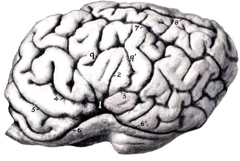
Fig. 507.—Left Cerebral Hemisphere of Horse, Lateral View. The Olfactory Bulb is Cut Off. 1, Lateral fissure (of Sylvius); 2, 3, 4, middle, posterior, and anterior branches of 1; 5, presylvian fissure; 6, 6′, sulcus rhinalis, anterior et posterior; 7, suprasylvian fissure; 8, ectomarginal fissure; 9, 9′, ectosylvian fissure.
The hemisphere comprises: (1) the pallium or mantle, which consists of an outer layer of gray matter, the cortex (Substantia corticalis), covering a large mass of white matter (Centrum semiovale); (2) the rhinencephalon or olfactory portion of the brain; (3) the corpus callosum and fornix, the great commissural white masses; (4) the lateral ventricle and certain important structures associated therewith.
The pallium is thrown into numerous folds, the gyri cerebri, which are separated by sulci or fissures of varying depth. The general pattern of the gyri and sulci is similar in normal brains of the same species, but the details are very variable and are never alike on the two hemispheres of the same brain. In the horse the arrangement is complicated by the existence of numerous short accessory fissures which cut into the gyri at right angles and tend to confuse the observer. The principal fissures and sulci of the convex surface (Figs. 497, 507) are as follows:
1. The lateral fissure (Fissura lateralis Sylvii) ascends on the lateral surface of the hemisphere as the continuation of the fossa transversa in front of the pyriform lobe. After crossing the external olfactory tract it divides into three branches; of these one passes upward, one runs obliquely forward and upward, and the third is directed upward and backward.
2. The suprasylvian fissure (F. suprasylvia)[196] is long and divides a large part 669of the convex surface of the hemisphere into dorsal and lateral portions. It begins on the dorso-medial border near its anterior end and, inclining gradually downward, passes back to end on reaching the tentorial surface. It is continuous internally with the transverse fissure usually and in front with the presylvian fissure.
3. The presylvian fissure (F. præsylvia) is the continuation forward of the preceding. It passes forward, outward, and downward almost to the frontal pole, and then inclines backward to end at the groove which marks the upper limit of the rhinencephalon (Sulcus rhinalis).
4. The marginal fissure (F. marginalis) extends along the dorso-medial border. It begins a little in front of the middle of the border and turns around the occipital pole to end on its tentorial aspect.
5. The entomarginal fissure (F. entomarginalis) lies internal to the dorso-medial border. It does not extend quite as far forward as the marginal fissure, from which it is separated by a narrow gyrus.
6. The ectomarginal fissure (F. ectomarginalis) lies about midway between the marginal fissure and the posterior part of the suprasylvian fissure.
7. The sulcus rhinalis is a very distinct furrow on the lower part of the lateral surface which marks off the olfactory part of the brain (rhinencephalon) from the rest of the hemisphere. It is undulating and is highest where it is crossed by the lateral fissure (of Sylvius).[197]
On the medial surface (Fig. 508) the main fissures and sulci are as follows:
1. The calloso-marginal fissure (F. calloso-marginalis) is extensive and well defined. It is approximately parallel to the dorso-medial border of the hemisphere, from which it is about half an inch distant. It begins in front a short distance below and in front of the anterior bend (genu) of the corpus callosum and forms a C-shaped curve, its posterior part extending on the tentorial surface to a point behind the depression for the corpora quadrigemina. It separates the marginal gyri above from the gyrus fornicatus, which extends down to the corpus callosum.
2. The transverse fissure (F. transversa) begins a little behind the middle of the calloso-marginal fissure, passes obliquely upward and forward to the dorso-medial border—into which it cuts deeply—and usually joins the suprasylvian fissure.[198]
3. The sublimbic fissure (F. sublimbica) curves over the gyrus fornicatus a short distance above the corpus callosum. Its middle part is commonly indistinct, and it is often divided into anterior and posterior parts.
4. The callosal sulcus (Sulcus corporis callosi) separates the corpus callosum from the gyrus fornicatus.
The hippocampus (or cornu Ammonis) is a gyrus which curves from the deep face of the pyriform lobe around the thalamus and forms the posterior part of the floor of the lateral ventricle. It can be displayed by cutting away the brain stem up to the optic tract and the interventricular foramen. Viewed from below the hippocampal gyrus is seen to form a semicircular curve from the apex of the pyriform lobe to the angle of divergence of the posterior pillars of the fornix, i. e., to a point under the central part of the corpus callosum. It is separated deeply by the hippocampal fissure from the gyrus dentatus. Along the concave margin of the latter is a band of white matter, termed the fimbria, which is the prolongation of the greater part of the posterior pillar of the fornix into this region. The ventricular surface of the hippocampus (Fig. 511) is covered with a thin layer of white matter, the alveus, which is also derived from the posterior pillar of the fornix, and 670is therefore continuous with the fimbria. The two hippocampi are connected at their highest parts by transverse fibers which constitute the hippocampal commissure.
The interval between the hippocampus and fimbria on the one hand and the brain stem on the other is a lateral continuation of the great transverse fissure of the brain, and is termed the chorioid fissure. It is occupied by a fold of pia mater, the tela chorioidea of the third ventricle, or velum interpositum. This fold is triangular in outline and its apex reaches to the interventricular foramen. Its base is continuous at the transverse fissure with the pia which covers the surface of the brain. Its middle part lies over the epithelial roof of the third ventricle, as has been seen (Fig. 506). The lateral borders will be seen on the floor of the lateral ventricles, where they form thick rounded bands containing convolutions of blood-vessels, known as the chorioid plexuses of the lateral ventricles (Fig. 511).
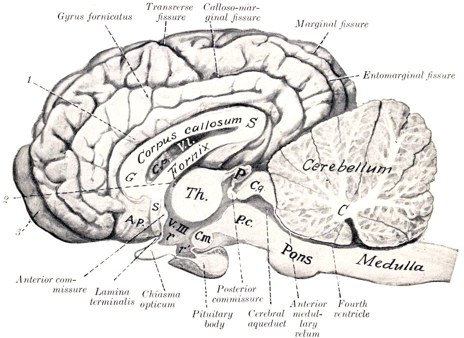
Fig. 508.—Median Section of Brain of Horse. The membranes and vessels are removed. C, Central white matter (corpus medullare) of cerebellum; P.c., cerebral peduncle; C.q., corpora quadrigemina; P, pineal body; Th., thalamus; V.III., third ventricle; r, optic recess; r′, infundibular recess; C.m., mammillary body; s, subcallosal gyrus; A.p., area parolfactoria; G, genu of corpus callosum; S, splenium of same; 1, callosal sulcus; 2, interventricular foramen; 3, olfactory bulb. The cerebral lobe of the pituitary body is distinguished by its lighter color.
When the tela is pulled out one may easily get the impression that the lateral ventricle communicates with the exterior by means of the chorioid fissure. Such is not the case, since the chorioid plexus is covered by the epithelial lining of the ventricle, which has been torn away.
The rhinencephalon or olfactory part of the brain comprises the olfactory bulb, peduncle and tracts or roots, the trigonum olfactorium, the area parolfactoria, and the pyriform lobe.[199]
671The olfactory bulb (Bulbus olfactorius) is an oval enlargement which curves upward in front of the frontal pole of the hemisphere. Its convex superficial face fits into the ethmoidal fossa and receives numerous olfactory nerve-fibers through the cribriform plate; hence it is very difficult to remove the bulb intact. It contains a considerable cavity (Ventriculus bulbi olfactorii) which is connected with the lateral ventricle by a small canal in the middle olfactory tract. The deep face is largely in contact with the frontal pole of the hemisphere and is connected with the olfactory peduncle.
The gray matter of the bulb is external and is thickest on the convex anterior surface. The posterior surface consists to a large extent of fibers which are the axones of the mitral cells of the deep layer of the gray substance and go to form the peduncle and striæ.
The olfactory peduncle (Tractus olfactorius) is a very short but wide band of white substance which arises in the olfactory bulb and extends back to be continued by the olfactory tracts.
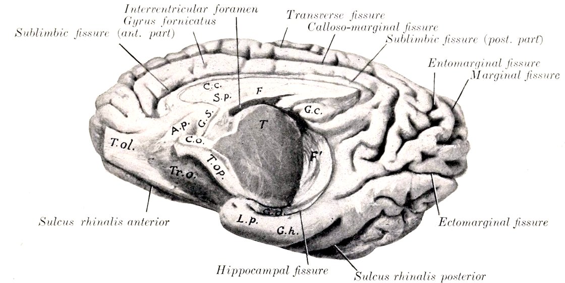
Fig. 509.—Medio-ventral Aspect of Right Cerebral Hemisphere of Horse. The olfactory bulb is cut off. T.ol., Olfactory peduncle; A.p., area parolfactoria; Tr.O., trigonum olfactorium; T.op., optic tract; C.o., chiasma opticum; G.s., subcallosal gyrus; C.c., corpus callosum; S.p., septum pellucidum; F, fornix; G.c., callosal gyrus; T, cut surface of thalamus; F′, fimbria; G.d., gyrus dentatus; L.p., pyriform lobe; G.h., hippocampal gyrus.
The olfactory tracts or striæ (Striæ olfactorii) are three in number. The external tract (Stria lateralis) is much the largest and most distinct. It passes backward, upward, and outward, widens out and joins the pyriform lobe. It is clearly defined dorsally by the sulcus rhinalis and is marked off from the trigonum olfactorium by the sulcus arcuatus. The intermediate tract (Stria intermedia) is short, ill defined, and flat; it contains the canal which opens into the anterior horn of the lateral ventricle. The internal tract (Stria medialis) is small, short, and not well defined; it bends over to the parolfactory area (of Broca) on the inner face of the hemisphere below the genu of the corpus callosum.
The trigonum olfactorium is the prominent gray area situated in the angle of divergence of the internal and external olfactory striæ. It is bounded externally by the external olfactory stria, from which it is defined by the arcuate sulcus. It is continuous with the area parolfactoria on the medial surface; behind the latter a band descends from the rostrum of the corpus callosum and is continuous below with the anterior perforated space; it is termed the subcallosal gyrus or peduncle of the corpus callosum.
672The pyriform lobe (Lobus piriformis) is the well-marked prominence on the base lateral to the optic tract and cerebral peduncle, from which it is separated by a deep fissure. Its nipple-like apex lies behind the fossa transversa and covers the optic tract. The external surface is marked by one or two sulci (lobi piriformis). The lobe contains a cavity, the ventral horn of the lateral ventricle.
The fibers of the olfactory tracts go to the pyriform lobe and hippocampus, the trigonum olfactorium, the area parolfactoria, the subcallosal gyrus, and part of the gyrus fornicatus. The central connections of the olfactory apparatus are complex and are not yet fully understood. The anterior cerebral commissure contains fibers which pass from the olfactory bulb of one side by way of the inner tracts to the bulb of the opposite side; also fibers which cross in it from the inner tract of one side to the pyriform lobe of the opposite side. Many fibers pass by way of the septum pellucidum, fornix, and fimbria to the hippocampus. Other fibers pass in the anterior pillar of the fornix to the mammillary body and thence to the thalamus by the thalamo-mammillary bundle.
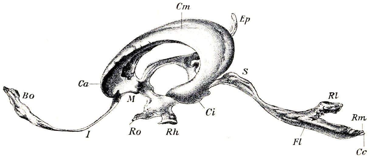
Fig. 510.—Lateral View of Cast of Cavities of Brain of Horse. Bo, Cavity of olfactory, which communicates through the canal I with a lateral ventricle; Ca, anterior horn, Cm, body, Ci, ventral horn of lateral ventricle; M, isthmus which connects lateral and third ventricles Ep, suprapineal recess, below which is the small infrapineal recess; Ro, optic recess; Rh, infundibular and pituitary recess; S, aqueduct; Fl, ridge corresponding to sulcus limitans; Rl, lateral recess, Rm, posterior recess, of fourth ventricle; Cc, beginning of central canal of spinal cord. (Dexler.)
The corpus callosum is the great transverse commissure which connects the two cerebral hemispheres through about half of their length. On median section (Fig. 508) it is seen to be arched from before backward, white in color, and composed substantially of transverse fibers. The middle part or truncus (Truncus corporis callosi) slopes downward and forward and is thinner than the ends. The anterior thickened end, the genu, bends downward and backward and thins out to form the rostrum; the latter is continuous with the lamina terminalis. The posterior end, the splenium, also thick, lies at a considerably higher level than the genu. The upper surface is convex in its length, concave transversely. Its central part forms the floor of the longitudinal fissure. It is covered by a thin layer of gray matter (Induseum griseum), in which are strands of longitudinal fibers (Striæ longitudinales); the latter are arranged in median and lateral bundles (Stria medialis, striæ laterales).[200] The lower surface has the reverse configuration, and forms the roof of the lateral ventricles. Along the median line the septum pellucidum is attached to it. The fibers of the corpus callosum (with the exception of the longitudinal striæ) run transversely and spread out laterally in all directions in the central white matter of the hemispheres to the cortex, forming the radiation of the corpus callosum (Radiatio corporis callosi).
673The fornix is a bilateral structure composed of white fibers which arch chiefly over the thalamus and the third ventricle. It is described as consisting of a body and two pairs of pillars. The body (Corpus fornicis) is formed by the fusion of the two arches of which the fornix is composed. It is triangular and overlies the anterior parts of the thalami and the third ventricle. The upper surface gives attachment to the septum pellucidum and on either side forms part of the floor of the lateral ventricle. The lateral border is related to the chorioid plexus of the lateral ventricle and forms the inner boundary of the interventricular foramen. The anterior pillars or columns (Columnæ fornicis) (Fig. 512) are two slender round bundles which emerge from the body in front of the interventricular foramen and diverge slightly as they curve downward and backward to the mammillary body.
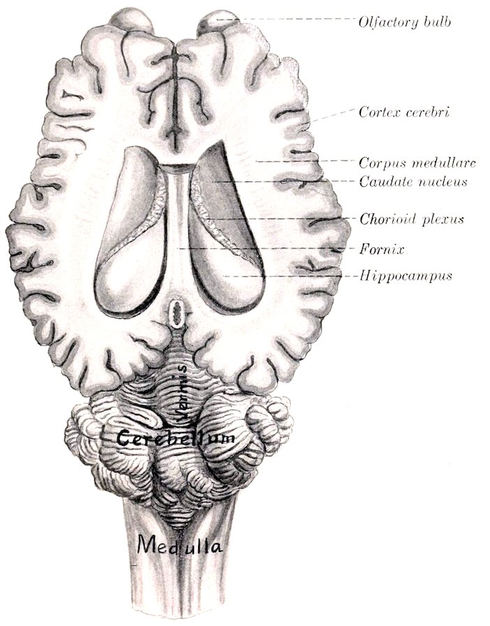
Fig. 511.—Brain of Horse, with Lateral Ventricles Opened by Removal of Upper Part of Cerebral Hemispheres.
From the mammillary body the greater part of these fornix fibers are continued to the thalamus by the thalamo-mammillary fasciculus (or bundle of Vicq d’Azyr). Others pass to the cerebral peduncle. A portion of the fibers cross to the opposite thalamus and cerebral peduncle.
The posterior pillars (Crura fornicis) are much larger bands which diverge widely from the posterior angles of the body. Each curves outward and backward over the thalamus (from which it is separated by the tela chorioidea) and is chiefly continued as the fimbria along the concave border of the hippocampus. The pillars 674give off fibers to form the alveus or white matter which covers the ventricular face of the hippocampi, and between them are transverse fibers which constitute the hippocampal commissure.
The septum pellucidum is the median partition between the two lateral ventricles. Its convex upper border blends with the corpus callosum and its concave lower border joins the fornix. Its anterior part is received into the genu of the corpus callosum. Traced backward it diminishes in height and the two edges meet at an acute angle at the splenium.
The septum consists of two layers (Laminæ septi pellucidi) which are in direct contact with each other. They consist of nerve-fibers and gray matter. The latter exists in considerable amount in the thicker part of the septum adjacent to the anterior pillars of the fornix. Many of the fibers of the septum pass up through the corpus callosum to the gyrus fornicatus. Others are connected with the subcallosal gyrus and the parolfactory area.
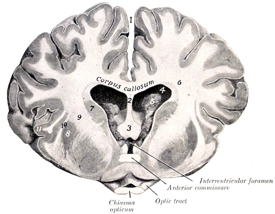
Fig. 512.—Cross-section of Brain of Horse, about Natural Size. Section passes through chiasma opticum, and is viewed from in front. 1, Longitudinal fissure; 2, septum pellucidum; 3, columns (anterior pillars) of fornix; 4, lateral ventricle; 5, chorioid plexus; 6, corpus medullare (central white matter) of hemisphere; 7, caudate nucleus; 8, lenticular nucleus; 9, internal capsule; 10, external capsule; 11, insula.
The lateral ventricle (Ventriculus lateralis) is the irregular cavity in the interior of each cerebral hemisphere.[201] Each communicates with the third ventricle through the interventricular foramen (of Monro), and by a small canal with the cavity of the olfactory bulb. It is usual to describe the ventricle as consisting of three parts, viz., the central part or body, and anterior and inferior horns. The anterior horn (Cornu nasale) is the part in front of the interventricular foramen; it communicates ventrally with the olfactory cavity. The central part (Pars centralis) extends back to the splenium of the corpus callosum. It opens into the third ventricle through the interventricular foramen, which is situated between the fornix and the anterior part of the thalamus. The inferior horn (Cornu ventrale) 675curves downward and forward into the pyriform lobe. The roof of the ventricle is formed by the corpus callosum and the inner wall is the septum pellucidum. After removal of the roof the floor is seen to be formed chiefly by two bodies. The anterior one is the caudate nucleus (Nucleus caudatus), a somewhat pear-shaped gray eminence, the long axis of which is directed obliquely upward, backward, and outward. Its anterior large end is termed the head, and the posterior long tapering end the tail. The posterior body, the hippocampus, is white on its ventricular surface, which is strongly convex. It curves outward and backward and then turns downward and forward to join the pyriform lobe. The two bodies are separated by an oblique groove which is occupied by the chorioid plexus of the lateral ventricle. This is the thickened edge of a fold of pia mater, the tela chorioidea (or velum interpositum), which lies between the hippocampus and the thalamus. It contains convolutions of small blood-vessels and in old subjects there may be calcareous concretions in it. The plexuses of the two sides are continuous through the interventricular foramen. On drawing the chorioid plexus backward a narrow white band, the stria terminalis (or tænia semicircularis), is seen along the margin of the caudate nucleus, where it bounds the intermediate groove. The plexus partially covers a wider white band which is blended with the white substance of the hippocampus; this is the posterior pillar of the fornix and its continuation, the fimbria.