THE MUSCULAR SYSTEM
Reading edition
MYOLOGY
The muscles (Musculi) are the active organs of motion. They are characterized by their property of contracting when stimulated. Muscular tissue is of three kinds: (a) Striated or striped; (b) non-striated, unstriped, or smooth; and (c) cardiac. Only the first of these varieties will be considered in this section. The striped muscles, being for the most part directly or indirectly connected with the skeleton, are often termed skeletal or somatic, while unstriped muscle may be spoken of as visceral or splanchnic. The former cover the greater part of the skeleton, and thus in a large measure determine the form of the animal. They are red in color, the shade varying in different muscles and under various conditions.
Muscles vary greatly in form, and may be classified as—(a) Long; (b) short; (c) flat; (d) ring-like or orbicular. Long muscles are found chiefly in the limbs, while the flat or broad muscles occur principally in the trunk, where they assist in forming the walls of the body cavities. The ring-like or orbicular muscles circumscribe orifices which they close, and are hence termed sphincters.
Attachments.—The muscles are attached to bones, cartilages, ligaments, fasciæ, or the skin. In all cases the attachment is by means of fibrous tissue, the muscle-fibers not coming into direct relation with the bone or cartilage. The perimysium of the muscle may fuse directly with the periosteum or perichondrium (fleshy attachment), or the union may be by means of intermediary fibrous structures called tendons or aponeuroses (tendinous attachment). Tendons may be funicular, ribbon-like, or in the form of membranous sheets; to the latter the term aponeurosis is commonly applied.
In certain positions, especially where tendons play over joints or are subjected to great pressure, sesamoid bones develop in the original tendon tissue. Some of these are large and constant, as the patella and the great sesamoids of the fetlock.
The accessory structures connected with the muscles are the synovial membranes and the fasciæ.
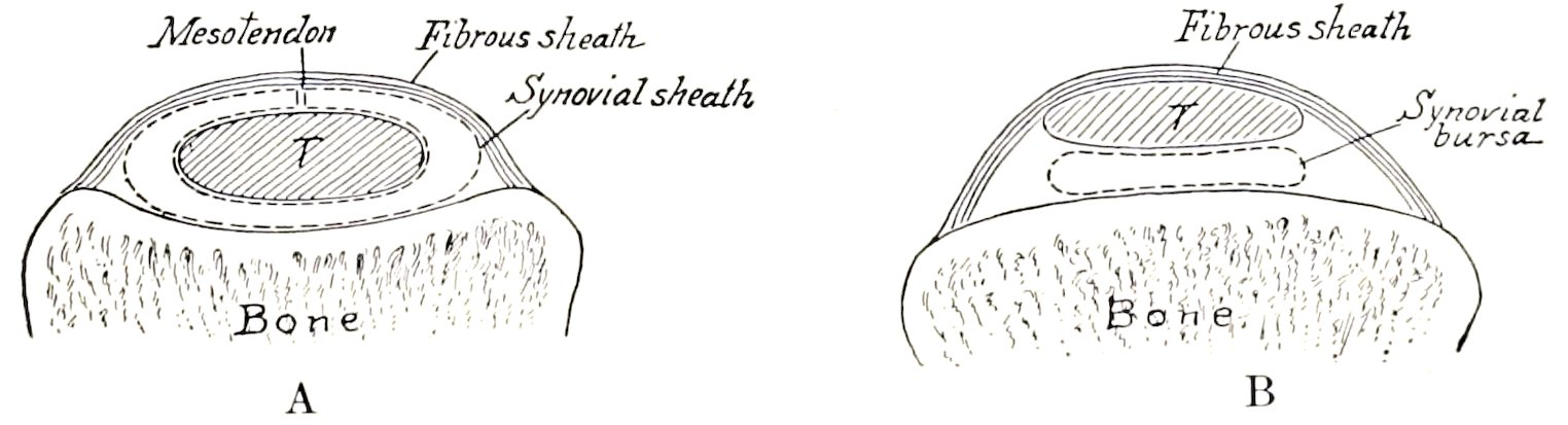
Fig. 171.—Diagrams of Cross-sections of Tendon Sheath (A) and Bursa (B); T, Tendon.
The synovial membranes are arranged in two principal forms: (a) Bursal; (b) vaginal. A bursa (Bursa mucosa) is a simple sac interposed between the tendon or muscle and some deeper seated structure—most commonly a bony prominence. A vagina tendinis or tendon sheath differs from a bursa in the fact that the synovial sac is folded around the tendon more or less completely, so that two layers can be distinguished; of these, the inner one adheres closely to the tendon, while the outer lines the groove or canal in which the tendon lies. The two layers are continuous 212along a fold termed the mesotendon. The arrangement is shown in Fig. 171.[41] The articular synovial membranes in some places form extra-articular pouches, which facilitate the play of tendons.
The fasciæ are sheets of connective tissue, mainly of the white fibrous variety, with a greater or less admixture of elastic fibers in certain cases. Two layers may usually be recognized. Of these, the superficial fascia (Fascia subcutanea) is composed of loose connective tissue which may contain more or less fat and is subcutaneous. The deep fascia is composed of one or more layers of dense fibrous tissue spread over the surface of the muscles chiefly. Its deep face may be very loosely attached to the underlying structures or may fuse with the epimysium, tendons, bones, or ligaments. In some parts, especially the limbs, septal plates pass between the muscles and are attached to the bones or ligaments; these are termed intermuscular septa. In this way many muscles are inclosed in fibrous sheaths which hold them in position. Not uncommonly special bands stretch across the grooves in which tendons play, converting these into canals. Such bands are termed vaginal or annular ligaments. The deep fascia is often so distinctly tendinous in structure, furnishing attachment to special tensor muscles, as to render the distinction between fascia and aponeurosis quite arbitrary. Bursæ occur in certain situations between the fascia and underlying structures (subfascial bursæ), or between the fascia and the skin (subcutaneous bursæ).
It is convenient to divide the description of a muscle into seven heads, viz.: (1) Name, followed by important synonyms; (2) position and form; (3) attachments; (4) action; (5) structure; (6) relations; (7) blood and nerve supply.
1. The name is determined by various factors, viz.: (a) The action, e. g., extensor, adductor, etc.; (b) the shape, e. g., quadratus, triangularis; (c) the direction, e. g., rectus, obliquus; (d) the position, e. g., the subscapularis, iliacus; (e) the division (into heads, etc.), e. g., biceps, triceps, etc.; (f) the size, e. g., major, minor, etc.; (g) the attachments, e. g., sterno-cephalicus, mastoido-humeralis; (h) the structure, e. g., semitendinosus. In most cases two or more of these factors have combined to produce the name, e. g., adductor magnus, longus colli, obliquus externus abdominis.
2. The shape is, in many cases, sufficiently definite to allow the use of such terms as triangular, quadrilateral, fan-shaped, long, flat, fusiform, ring-like, etc.
3. The attachments are in most cases to bone, but many muscles are attached to cartilage, ligaments, fascia, the skin, etc. It is usual to apply the term origin to the attachment which always or more commonly remains fixed when the muscle contracts. The term insertion designates the movable attachment. Such a distinction cannot always be made, as the action may be reversible, or both attachments may be freely movable.
4. The action belongs rather to physiological study, but is briefly indicated in anatomical descriptions.
5. The structure includes the direction of the muscle-fibers, the arrangement of the tendons, the synovial membranes, and any other accessory structures, e. g., annular ligaments and reinforcing sheaths and bands. The relation of the muscle-fibers to the tendon varies, and this fact has given rise to special terms. Thus a muscle in which the fibers converge to either side of the tendon is termed bipennate; while one in which this arrangement exists only on one side of the tendon is called unipennate. The terms fleshy and tendinous are used to indicate the relative amounts of muscular and tendinous tissue. The muscular tissue is often spoken of as the belly (Venter) of the muscle. In the case of the long muscles, the origin is 213often termed the head (Caput). Muscles having two or more heads are called biceps, triceps, etc. Digastric muscles are those which have two bellies joined by an intermediate tendon. Ring-like muscles which circumscribe openings are termed sphincters, on account of their action.
6. The relations are, of course, important on surgical grounds.
7. The nerve-supply is of clinical interest, and is important for the determination of homologies.
FASCIÆ AND MUSCLES OF THE HORSE
PANNICULUS CARNOSUS
The panniculus carnosus (Musculus cutaneus) is a thin muscular layer developed in the superficial fascia. It is intimately adherent in great part to the skin, but has very little attachment to the skeleton. It does not cover the entire body, and may be conveniently divided into facial, cervical, thoracic, and abdominal portions, each of which will be described with the muscles of the corresponding region.
The Fasciæ and Muscles of the Head
The muscles of the head may be divided into three groups, viz.: (1) Superficial muscles, including the panniculus and those of the lips, cheeks, nostrils, eyelids, and external ear; (2) the orbital muscles; (3) the muscles of mastication.
The superficial fascia forms an almost continuous layer, but is very scanty around the natural orifices. It contains a number of the thin superficial muscles, so that care must be exercised in removing the skin. Over the frontal and nasal bones the fascia blends with the periosteum.
The deep fascia is of special interest in three regions. The temporal fascia covers the temporalis muscle, and is attached to the parietal and frontal crests internally, and to the zygomatic arch externally. The buccal fascia covers the buccinator muscle and the free part of the outer surface of the ramus of the jaw. Superiorly it is attached to the facial crest, and posteriorly it forms a band (Ligamentum pterygomandibulare) which stretches from the hamulus of the pterygoid bone to the mandible behind the last molar tooth. It is directly continuous with the pharyngeal fascia, which is attached to the great and thyroid cornua of the hyoid bone, covers the lateral walls of the pharynx, and blends dorsally with the median raphé of the constrictor muscles of the latter.
SUPERFICIAL MUSCLES
1. Panniculus carnosus.—The facial panniculus (M. cutaneus faciei) consists of a thin and usually incomplete muscular stratum, which covers the submaxillary space and the masseter muscle. A branch from it passes forward to the angle of the mouth and blends with the orbicularis oris; this part (M. cutaneus labiorum) retracts the angle of the mouth. (A number of the superficial muscles of the face may be considered modified parts of the panniculus, e. g., the corrugator supercilii, malaris, zygomaticus, etc.)
MUSCLES OF THE LIPS AND CHEEKS
1. Orbicularis oris.—This is the sphincter muscle of the mouth; it is continuous with the other muscles which converge to the lips. It lies between the skin and the mucous membrane of the lips, and is intimately adherent to the 214former. Most of the fibers run parallel to the free edges of the lips and have no direct attachment to the skeleton.
Action.—It closes the lips.
Blood-supply.—Palato-labial, facial, and mental arteries.
Nerve-supply.—Facial nerve.
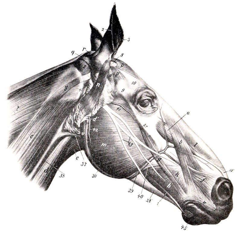
Fig. 172.—Muscles of Head of Horse, Lateral View. The Panniculus is Removed. a, Levator labii superioris proprius; b, levator nasolabialis; c, mastoido-humeralis; d, sterno-cephalicus; d′, tendon of d; e, omo-hyoideus; f, dilatator naris lateralis; g, zygomaticus; h, buccinator; i, depressor labii inferioris; k, orbicularis oris; l, dilatator naris superior; m, masseter; n, parotido-auricularis; o, zygomatico-auricularis; p, interscutularis; p′, fronto-scutularis, pars temporalis; q, cervico-auricularis profundus major; r, cervico-auricularis superficialis; s, obliquus capitis anterior; t, splenius; v, stylo-maxillaris; y, mastoid tendon of mastoido-humeralis; 2, posterior, 3, anterior, border of external ear; 8, scutiform cartilage; 9, zygomatic arch; 10, depression behind supraorbital process; 18, temporo-mandibular articulation; 27, facial crest; 30′, angle of jaw; 37, external maxillary vein; 38, jugular vein; 39, facial vein; 40, parotid duct; 41, transverse facial vein; 42, masseteric vein; 43, facial nerve; 44, parotid gland; 45, chin; x, wing of atlas. (After Ellenberger-Baum, Anat. für Künstler.)
2. Levator nasolabialis (Levator labii superioris alæque nasi).—This thin muscle lies directly under the skin, and chiefly on the lateral surface of the nasal region.
Origin.—The frontal and nasal bones.
Insertion.—(1) The upper lip and the outer wing of the nostril; (2) the commissure of the lips.
Action.—(1) To elevate the upper lip and the commissure; (2) to dilate the nostril.
215Structure.—The muscle arises by a thin aponeurosis. The belly is also thin, and divides into two branches, between which the lateral dilator of the nostril passes. The dorsal branch reaches the nostril and upper lip, blending with the lateral dilator; the ventral one is much smaller, and blends at the labial commissure with the orbicularis and buccinator.
Relations.—Superficially, the skin, fascia, and lateral dilator (in part); deeply, the levator labii superioris proprius, lateral dilator (in part), buccinator, branches of the facial vessels and nerve, and the infraorbital artery and nerve.
Blood-supply.—Facial and palato-labial arteries.
Nerve-supply.—Facial nerve.
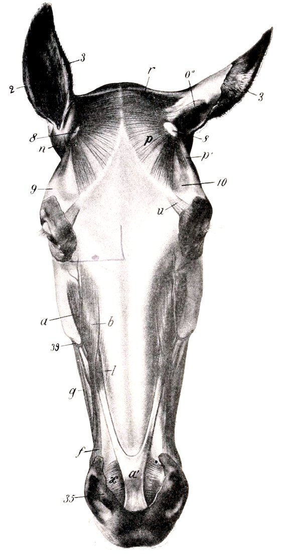
Fig. 173.—Muscles of Head of Horse, Dorsal View. The Panniculus is Removed. a, Levator labii superioris proprius; a′, common tendon of a with opposite muscle; b, levator nasolabialis; f, dilatator naris lateralis; g, zygomaticus; l, dilatator naris superior; n, parotido-auricularis; o″, scutulo-auricularis superficialis superior; p, interscutularis; p′, fronto-scutularis, pars temporalis; r, cervico-auricularis superficialis; u, corrugator supercilii; x, transversus nasi; 2, posterior, 3, anterior, border of external ear; 8, scutiform cartilage; 9, zygomatic arch; 10, supraorbital depression; 35, inner wing of nostril, containing lamina of alar cartilage; 39, facial vein. (After Ellenberger-Baum, Anat. für Künstler.)
3. Levator labii superioris proprius.—This lies on the dorso-lateral aspect of the face, partly covered by the preceding muscle.
Origin.—The lacrimal, malar, and maxillary bones at their junction.
Insertion.—The upper lip, by a common tendon, with its fellow.
Action.—Acting with its fellow, to elevate the upper lip. This action, if carried to the fullest extent, results in eversion. In unilateral action the lip is drawn upward and to the side of the muscle acting.
Structure.—The muscle has a short, thin tendon of origin. The belly is at first flattened, but becomes narrower and thicker, then tapers over the false nostril, to terminate in a tendon. The tendons of the two muscles unite over the alar cartilages of the nostrils, forming an expansion which spreads out in the substance of the upper lip.
Relations.—Superficially, the skin, the levator nasolabialis, and the angular vessels of the eye; deeply, the superior and transverse dilators of the nostril and the infraorbital artery and nerve.
Blood-supply.—Facial artery.
Nerve-supply.—Facial nerve.
4. Zygomaticus.—This very thin muscle lies immediately under the skin of the cheek.
Origin.—The fascia covering the masseter muscle below the facial crest.
Insertion.—The commissure of the lips, blending with the buccinator.
Action.—To retract and raise the angle of the mouth.
216Structure.—Fleshy, with a thin aponeurotic origin.
Relations.—Superficially, the skin; deeply, the buccinator.
Blood-supply.—Facial artery.
Nerve-supply.—Facial nerve.
5. Incisivus superior (Depressor labii superioris).—This lies under the mucous membrane of the upper lip.
Origin.—The alveolar border of the premaxilla from the second incisor to the first cheek tooth.
Insertion.—The upper lip.
Action.—To depress the upper lip.
6. Incisivus inferior.—This is arranged in the lower lip like the preceding muscle in the upper one.
Origin.—The alveolar border of the mandible from the second incisor to a point near the first cheek tooth.
Insertion.—The skin of the lower lip and the prominence of the chin.
Action.—To raise the lower lip.
7. Mentalis (Levator menti).—This is situated in the prominence of the chin. Its fibers arise from each side of the body of the mandible and are inserted into the skin of the chin. It is mingled with fat and strands of connective tissue, in which the roots of the tactile hairs are embedded. It raises and corrugates the skin to which it is attached.
8. Depressor labii inferioris.—This muscle lies on the outer surface of the ramus of the mandible, along the ventral border of the buccinator.
Origin.—The alveolar border of the mandible near the coronoid process and the maxillary tuberosity, in common with the buccinator.
Insertion.—The lower lip.
Action.—To depress and retract the lower lip.
Structure.—The tendon of origin and the belly are fused with the buccinator as far forward as the first cheek tooth. From this point forward the belly is distinct and rounded, terminating in a tendon which spreads out in the lower lip, blending with the orbicularis and the muscle of the opposite side.
Relations.—Superficially, the skin, masseter, facial vessels, and parotid duct; deeply, the mandible and inferior labial artery.
Blood-supply.—Facial artery.
Nerve-supply.—Facial nerve.
9. Buccinator.—This muscle lies in the lateral wall of the mouth, extending from the angle of the mouth to the maxillary tuberosity.
Origin.—The external surface of the maxilla above the interdental space and the molar teeth; the alveolar border of the mandible at the interdental space and also posteriorly where it turns upward to the coronoid process; the pterygo-mandibular ligament.
Insertion.—The angle of the mouth, blending with the orbicularis oris.
Action.—To flatten the cheeks, thus pressing the food between the teeth; also to retract the angle of the mouth.
Structure.—Two layers may be recognized. The superficial layer (Pars buccalis) extends from the angle of the mouth to the masseter. It is incompletely pennate, having a longitudinal raphé on which most of the muscle-fibers converge. The upper fibers are directed chiefly downward and backward, the lower ones upward and backward. The deep layer (Pars molaris) consists mainly of longitudinal fibers. It blends in part with the superficial layer of the orbicularis; it has a small tendinous attachment to the coronoid process behind, and is united below with the depressor labii inferioris.
Relations.—Superficially, the skin and fascia, the zygomaticus, levator nasolabialis, lateral dilator of the nostril, the superior buccal glands, the parotid duct, 217the facial vessels, and branches of the facial nerve; deeply, the mucous membrane of the mouth and the inferior buccal glands.
Blood-supply.—Facial and buccinator arteries.
Nerve-supply.—Facial nerve.
MUSCLES OF THE NOSTRILS
1. Levator nasolabialis.—This has been described (p. 214).
2. Dilatator naris lateralis (M. caninus).—This thin, triangular muscle lies on the lateral nasal region, and passes between the two branches of the levator nasolabialis.
Origin.—The maxilla, close to the anterior extremity of the facial crest.
Insertion.—The outer wing of the nostril.
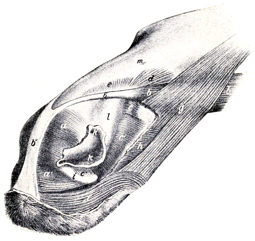
Fig. 174.—Nasal and Superior Labial Muscles of Horse. a, a′, Dilatator naris transversus; b, levator labii superioris proprius; b′, tendon of b; b″, common tendon of two levatores labii superioris proprii; c, c′, dilatator naris inferior; d, e, dilatator naris superior; f, orbicularis oris; g, levator nasolabialis, a portion of which is removed; h, dilatator naris lateralis (cut); i, cornu of alar cartilage; k, nostril; k′, false nostril; l, nasal diverticulum; m, nasal bone. (After Ellenberger-Baum, Top. Anat. d. Pferdes.)
Action.—To dilate the nostril.
Structure.—The muscle has a flat tendon of origin, passes between the two branches of the levator nasolabialis, and spreads out in the external wing of the nostril. The lower fibers blend with the orbicularis oris.
Relations.—Superficially, the skin, fascia, and the labial branch of the levator nasolabialis; deeply, the maxilla and the nasal branch of the levator nasolabialis.
Blood-supply.—Facial artery.
Nerve-supply.—Facial nerve.
3. Dilatator naris transversus (M. transversus nasi).—This is an unpaired, quadrilateral muscle, which lies between the nostrils. It consists of two layers.
Attachments.—Superficial layer, the superficial faces of the laminæ of the alar cartilages; deep layer, the convex edges of the cornua of the same.
Action.—To dilate the nostrils.
218Structure.—It is composed of transverse fleshy fibers, which blend below with the orbicularis.
Relations.—Superficially, the skin, fascia, and tendinous expansion of the levator labii superioris proprius; deeply, the alar cartilages, the extremity of the septum nasi, and the palato-labial artery.
Blood-supply.—Palato-labial artery.
Nerve-supply.—Facial nerve.
4. Dilatator naris superior (Pars dorsalis m. lateralis nasi).—This very thin muscle occupies the angle between the nasal process of the premaxilla and the nasal bone.
Origin.—The lateral border of the nasal bone.
Insertion.—The inner wall of the false nostril and the parietal lamina of the septal cartilage.
Action.—To dilate the vestibule of the nasal cavity.
Structure.—Fleshy.
Relations.—Superficially, the skin, fascia, and levator labii superioris proprius; deeply, the parietal cartilage and false nostril.
Blood-supply.—Facial artery.
Nerve-supply.—Facial nerve.
5. Dilatator naris inferior (Pars ventralis m. lateralis nasi).—This is a similar but thicker muscle, which lies on the nasal process of the premaxilla.
Origin.—The maxilla and the nasal process of the premaxilla.
Insertion.—The cartilaginous prolongations of the turbinal bones and the inner wall of the false nostril.
Action.—To rotate the turbinal outward and dilate the nostril and the vestibule of the nasal cavity.
Structure.—Fleshy. A division into two or more parts may be recognized. A small part posteriorly is inserted into the cartilage of the dorsal turbinal bone, while the bulk of the muscle is inserted into the cartilage of the ventral turbinal bone. A few fibers also pass between the cornu of the alar cartilage and the outer wing of the nostril.
Relations.—Superficially, the levator labii superioris proprius, the levator nasolabialis, and the lateral nasal artery; deeply, the maxilla, the premaxilla, and the anterior nasal branch of the infraorbital nerve.
Blood-supply.—Facial artery.
Nerve-supply.—Facial nerve.
MUSCLES OF THE EYELIDS
1. Orbicularis oculi.—This is a flat, elliptical, sphincter muscle, situated in and around the eyelids, the portion in the upper lid being much broader than that in the lower. The chief attachment is to the skin of the lids, but some bundles are attached to the palpebral ligament at the inner canthus and to the lacrimal bone. Its action is to close the lids.
2. Corrugator supercilii.—This is a very thin, small muscle, which arises over the root of the supraorbital process and spreads out in the upper eyelid, blending with the orbicularis. Its action is to assist in raising the upper lid or, especially in pathological conditions, to wrinkle the skin.
3. Malaris.—This is a very thin muscle, which varies much in different subjects. It extends from the fascia in front of the orbit to the lower lid. Its action is to depress the lower lid.
The foregoing muscles receive their blood-supply from the facial, transverse facial, supraorbital, and infraorbital arteries; the nerve-supply is derived from the facial nerve.
2194. Levator palpebræ superioris.—This slender, flat muscle is almost entirely within the orbital cavity. It arises on the pterygoid crest, passes forward above the rectus oculi superior and below the lacrimal gland, and terminates in a thin tendon in the upper lid.
Action.—To elevate the upper lid.
Blood-supply.—Ophthalmic artery.
Nerve-supply.—Oculomotor nerve.
MUSCLES OF MASTICATION
The muscles of this group are six in number in the horse. They arise chiefly from the upper jaw and the base of the cranium, and are all inserted into the mandible.
1. Masseter.—This muscle extends from the zygomatic arch and facial crest over the broad part of the mandibular ramus. It is semi-elliptical in outline.
Origin.—By a strong tendon from the zygomatic arch and the facial crest.
Insertion.—The outer surface of the broad part of the ramus of the mandible.
Action.—Its action is to bring the jaws together. Acting singly, it also carries the lower jaw toward the side of the contracting muscle.
Structure.—The superficial face of the muscle in its upper part is covered by a strong, glistening aponeurosis, and several tendinous intersections partially divide the muscle into layers. The fibers of the superficial layer take origin from the malar and maxilla only, and diverge somewhat to their insertion close to the thick ventral border of the lower jaw. The fibers of the deep layer arise from the entire area of origin, and pass straight to the border of the mandible; it will be noted that a small part, near the temporo-maxillary joint, is not covered by the superficial layer. The two layers are separable only above and behind; elsewhere they are fused.
Relations.—Superficially, the skin and panniculus, the parotid gland, the transverse facial and masseteric vessels, and the facial nerve; deeply, the ramus of the mandible, the buccinator, depressor labii inferioris, and mylo-hyoideus muscles, the superior buccal glands, the buccinator nerve, and two large varicose veins which join the facial vein at the anterior edge of the muscle. The facial vessels and parotid duct run along the anterior edge of the muscle; the duct, however, bends forward about the middle of the border and leaves the muscle.
Blood-supply.—Transverse facial and masseteric arteries.
Nerve-supply.—Mandibular nerve.
2. Temporalis.—This muscle occupies the temporal fossa.
Origin.—The rough part of the temporal fossa and the crests which surround it.
Insertion.—The coronoid process of the mandible, which it envelops.
Action.—Chiefly to raise the lower jaw, acting with the masseter and internal pterygoid muscles.
Structure.—The surface of the muscle is covered with a glistening aponeurosis, and strong tendinous intersections are found in its substance. The inner edge of the muscle is quite thin, but as the fibers converge toward the much smaller area of insertion, the muscle becomes nearly an inch thick. It fuses somewhat with the masseter.
Relations.—Superficially, the scutiform cartilage and anterior muscles of the external ear and the orbital fat; deeply, the temporal fossa and the deep temporal vessels and nerves.
Blood-supply.—Superficial and deep temporal, and mastoid arteries.
Nerve-supply.—Mandibular nerve.
3. Pterygoideus internus (s. medialis).—This muscle occupies a position on the inner surface of the ramus of the mandible similar to that of the masseter on the outside.
220Origin.—The crest formed by the pterygoid process of the sphenoid and the palate bone.
Insertion.—The concave inner surface of the broad portion of the ramus of the mandible, and the inner lip of the lower border.
Action.—Acting together, to raise the lower jaw; acting singly, to produce also lateral movement of the jaw.
Structure.—The muscle is capable of division into two parts. The principal part is superficial (internal), and its fibers are, for the most part, vertical in direction. It contains much tendinous tissue (septa). The smaller portion is external to the foregoing, and its fibers are directed downward and backward.
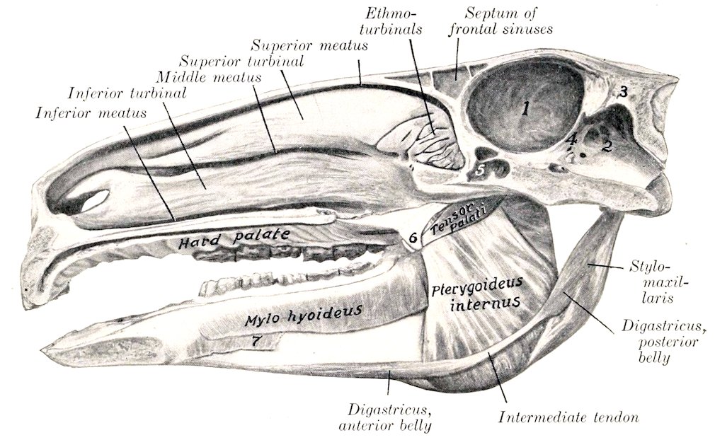
Fig. 175.—Sagittal Section of Head of Horse, Showing Deep Pterygo-maxillary Region, and Nasal and Cranial Cavities. 1, Cerebral compartment of cranial cavity; 2, cerebellar compartment of same; 3, tentorium osseum; 4, tentorium cerebelli; 5, sphenoidal sinus; 6, hamulus of pterygoid bone-tendon of tensor palati cut off short at anterior border of hamulus; 7, mylo-glossus. The olfactory mucous membrane is shaded.
Relations.—Externally, the ramus of the mandible, the external pterygoid muscle, the inferior alveolar vessels and nerve, and the lingual and mylo-hyoid nerves; internally, the great cornu of the hyoid bone, the tensor palati, pterygo-pharyngeus, palato-pharyngeus, mylo-hyoideus, digastricus, and stylo-hyoideus muscles, the guttural pouch, the external maxillary vessels, the ninth and twelfth nerves, the submaxillary salivary gland, the submaxillary and parotid ducts, and the submaxillary and pharyngeal lymph glands.
Blood-supply.—Internal maxillary, masseteric, and inferior alveolar arteries.
Nerve-supply.—Mandibular nerve.
4. Pterygoideus externus (s. lateralis).—This muscle is considerably smaller than the preceding one, and is situated external to its upper part.
Origin.—The external surface of the pterygoid process of the sphenoid bone.
Insertion.—The inner surface of the neck and the inner part of the anterior border of the condyle of the mandible.
Action.—Acting together, to draw the lower jaw forward; acting singly, to move the jaw laterally, i. e., toward the side opposite to the muscle acting. The 221latter action is due to the fact that the origin is nearer to the median plane than the insertion.
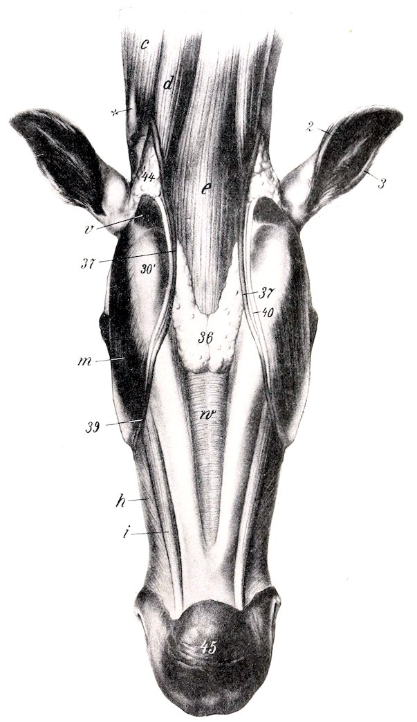
Fig. 176.—Submaxillary and Laryngeal Regions of Horse, after Removal of Skin and Panniculus. c, Mastoido-humeralis; d, sterno-cephalicus; e, omo-hyoideus and sterno-hyoideus; h, buccinator; i, depressor labii inferioris; m, masseter; v, stylo-maxillaris; w, mylo-hyoideus; 2, posterior, 3, anterior, border of external ear; 30′, angle of jaw; 36, submaxillary lymph glands; 37, external maxillary vein; 39, facial continuation of 37; 40, parotid duct; 44, parotid gland; 45, prominence of chin; x, wing of atlas. (After Ellenberger-Baum, Anat. für Künstler.)
Structure.—The muscle is almost entirely fleshy, and the fibers are almost longitudinal in direction. Some of them are inserted into the edge of the interarticular meniscus.
Relations.—Externally, the temporo-maxillary articulation and the temporalis muscle; internally, the internal pterygoid and tensor palati muscles. The internal 222maxillary artery crosses the lower face of the muscle and dips in between it and the tensor palati. The mandibular nerve lies on the ventral surface, and the buccinator nerve perforates the origin of the muscle.
Blood-supply.—Internal maxillary and inferior alveolar arteries.
Nerve-supply.—Mandibular nerve.
5. Stylo-maxillaris (s.-mandibularis) (M. jugulomandibularis).—This is a short muscle extending from the paramastoid or styloid process of the occipital bone to the posterior border of the lower jaw; it is covered by the parotid gland.
Origin.—The paramastoid or styloid process of the occipital bone, in common with the posterior belly of the digastricus.
Insertion.—The posterior border of the ramus of the mandible.
Action.—To depress the lower jaw and open the mouth.
Structure.—The muscle contains a good deal of tendinous tissue. It blends with the posterior belly of the digastricus.
Relations.—Superficially, the parotid gland, the tendon of the sterno-cephalicus, and the fibrous expansion which connects it with the tendon of the mastoido-humeralis; deeply, the guttural pouch, the external carotid artery, the ninth and twelfth nerves, the pharynx, and the submaxillary gland.
Blood-supply.—External carotid artery.
Nerve-supply.—Facial nerve.
6. Digastricus.—This muscle is composed of two fusiform, flattened bellies, united by a round tendon.
Origin.—The paramastoid or styloid process of the occipital bone, in common with the preceding muscle.
Insertion.—The inner surface of the lower border of the mandible behind the symphysis.
Action.—It assists in depressing the lower jaw and opening the mouth. If the mandible be fixed and both bellies contract, the hyoid bone and the base of the tongue are raised, as in the first phase of deglutition.
Structure.—The posterior belly has the appearance of a branch detached from the inner surface of the stylo-maxillaris. It passes downward and forward, and is succeeded by a small rounded tendon. The latter perforates the tendon of insertion of the stylo-hyoideus, and is provided with a synovial sheath. The anterior belly is larger and terminates by thin tendinous bundles.
Relations.—The posterior belly has practically the same relations as the stylo-maxillaris. The intermediate tendon is in contact externally with the internal pterygoid muscle, the submaxillary gland and duct, and the external maxillary artery. The anterior belly lies in the submaxillary space between the ramus of the jaw and the mylo-hyoideus muscle; the sublingual vessels run along its upper border.
Blood-supply.—External carotid and sublingual arteries.
Nerve-supply.—Facial and mandibular nerves.
THE HYOID MUSCLES
This group consists of eight muscles, one of which, the hyoideus transversus, is unpaired.
1. Mylo-hyoideus.—This muscle, together with its fellow, forms a sort of sling between the two rami of the lower jaw, in which the tongue is supported.
Origin.—The inner surface of the alveolar border of the mandible.
Insertion.—(1) A median fibrous raphé extending from the symphysis to the hyoid bone; (2) the lingual process and body of the hyoid bone.
Action.—It raises the floor of the mouth, the tongue, and the hyoid bone.
Structure.—Each muscle consists of a thin curved sheet, the fibers passing downward from their origin and then curving toward the median raphé. It is 223chiefly fleshy, and is thickest behind. The anterior superficial part of the muscle is termed the mylo-glossus.
Relations.—On the superficial surface of the muscles are the ramus, the internal pterygoid and digastricus muscles, and the submaxillary lymph glands. The deep surface is in contact with the mucous membrane of the mouth, the stylo-glossus, hyo-glossus, and genio-hyoideus muscles, the sublingual gland and vessels, the submaxillary duct, and the lingual and hypoglossal nerves.
Blood-supply.—Sublingual artery.
Nerve-supply.—Mylo-hyoid branch of the mandibular nerve.
2. Stylo-hyoideus.—This is a slender, fusiform muscle, having a direction nearly parallel to that of the great cornu of the hyoid bone (Fig. 436).
Origin.—The heel-like prominence on the proximal extremity of the great cornu of the hyoid bone.
Insertion.—The anterior part of the thyroid cornu of the hyoid bone.
Action.—It draws the base of the tongue and the larynx upward and backward.
Structure.—It arises by a thin, short tendon, and has a fusiform belly. The tendon of insertion is perforated for the passage of the intermediate tendon of the digastricus, and at this point there is a small synovial sheath.
Relations.—Superficially, the internal pterygoid muscle and the parotid gland; deeply, the guttural pouch, the pharynx, the external carotid and maxillary arteries, and the hypoglossal nerve.
Blood-supply.—External carotid artery.
Nerve-supply.—Facial nerve (stylo-hyoid branch).
3. Occipito-hyoideus (M. jugulo-hyoideus; occipito-styloideus).—This is a small triangular muscle, which lies in the space between the paramastoid process and the great cornu of the hyoid bone.
Origin.—The paramastoid (styloid) process of the occipital bone.
Insertion.—The proximal extremity and ventral edge of the great cornu of the hyoid bone.
Action.—It carries the distal extremity of the great cornu backward and upward. Acting with the genio-hyoideus and digastricus, it raises the hyoid bone and the larynx.
Structure.—The muscle is somewhat triangular, its fibers being longer as the ventral border is approached. It blends with the posterior belly of the digastricus.
Relations.—Superficially, the parotid gland; deeply, the guttural pouch.
Blood-supply.—Occipital artery.
Nerve-supply.—Facial nerve.
4. Genio-hyoideus.—This is a long, spindle-shaped muscle, which lies under the tongue in contact with its fellow of the opposite side (Fig. 243).
Origin.—The angle of union of the rami of the mandible.
Insertion.—The extremity of the lingual process of the hyoid bone.
Action.—It draws the hyoid bone and tongue forward.
Structure.—The muscle arises by a short tendon, which is succeeded by the belly, composed of long bundles of parallel fibers.
Relations.—Below, the mylo-hyoideus; above, the hyo-glossus, stylo-glossus, genio-glossus, the sublingual gland, submaxillary duct, and the lingual nerve.
Blood-supply.—Sublingual artery.
Nerve-supply.—Hypoglossal nerve.
5. Kerato-hyoideus.—This small triangular muscle lies in the space between the thyroid and small cornu, under cover of the hyo-glossus (Fig. 243).
Origin.—The posterior edge of the small cornu and the adjacent part of the ventral border of the great cornu.
Insertion.—The dorsal edge of the thyroid cornu.
Action.—It raises the thyroid cornu and the larynx.
224Relations.—The muscle is crossed outwardly by the lingual artery.
Blood-supply.—Lingual artery.
Nerve-supply.—Glosso-pharyngeal nerve.
6. Hyoideus Transversus.—This is a thin, unpaired muscle, which extends transversely between the two small cornua of the hyoid bone.
Attachments.—The small cornua close to the junction with the great cornua.
Action.—When relaxed, its upper surface is concave; when it contracts, it elevates the root of the tongue.
Structure.—Fleshy, composed of parallel transverse bundles.
Blood-supply.—Lingual artery.
Nerve-supply.—Glosso-pharyngeal nerve.
7. Sterno-thyro-hyoideus, and
8. Omo-hyoideus.—These are described with the muscles on the ventral surface of the neck.
The Fasciæ and Muscles of the Neck
It is convenient to divide the muscles of the neck into ventral and lateral groups, the two lateral groups being separated from each other by the ligamentum nuchæ.
THE FASCIÆ OF THE NECK
The superficial fascia is in part two-layered, and contains the cervical portion of the panniculus. The fasciæ of the right and left sides are attached along the dorsal line of the neck to the ligamentum nuchæ, while along the ventral line they meet in a fibrous raphé. A deep layer is detached which passes underneath the panniculus, bridges over the jugular furrow, and crosses over the deep face of the mastoido-humeralis and omo-hyoideus to join the superficial layer. It again separates to pass under the cervical trapezius, and become attached to the ligamentum nuchæ. Along the ventral line a septum is detached which separates the sterno-cephalici. Two other layers in front of the shoulder inclose the prescapular lymph glands.
The deep fascia also forms two layers. The superficial layer is attached to the wing of the atlas and the lower edge of the trachelo-mastoideus and scalenus. Passing downward, it incloses the trachea, and, together with the deep layer, furnishes sheaths for the vagus and sympathetic nerves and the carotid artery. Passing upward it detaches septa between the extensor muscles of the spine. Anteriorly it covers the thyroid gland, the guttural pouch, the adjacent vessels and nerves and the larynx, and is attached to the mastoid process of the temporal bone and the thyroid cornu of the hyoid bone. Posteriorly, it is attached to the first rib and the cariniform cartilage of the sternum. The deep layer (prevertebral fascia) covers the ventral surface of the longus colli, and incloses the trachea and œsophagus. Anteriorly, it forms, with the corresponding layer of the opposite side, a septum between the guttural pouches; posteriorly, it becomes continuous with the endothoracic fascia. A fascia propria forms a tubular sheath around the trachea, inclosing also the recurrent nerves.
VENTRAL MUSCLES
This group consists of eleven pairs of muscles which lie almost entirely ventral to the vertebræ.
1. Panniculus carnosus.—The cervical panniculus (platysma myoides of man) has a fleshy origin on the cariniform cartilage (manubrium) of the sternum. 225it passes forward, outward, and upward, crossing over the sterno-cephalicus and jugular furrow obliquely. On reaching the surface of the mastoido-humeralis it adheres closely to this muscle, and soon thins out, to be continued over the splenius and trapezius by an aponeurosis which is difficult to remove from the latter muscle. Scattered bundles may be traced on the ventral surface of the neck to the facial portion. The right and left muscles meet at a ventral median raphé in pennate fashion.
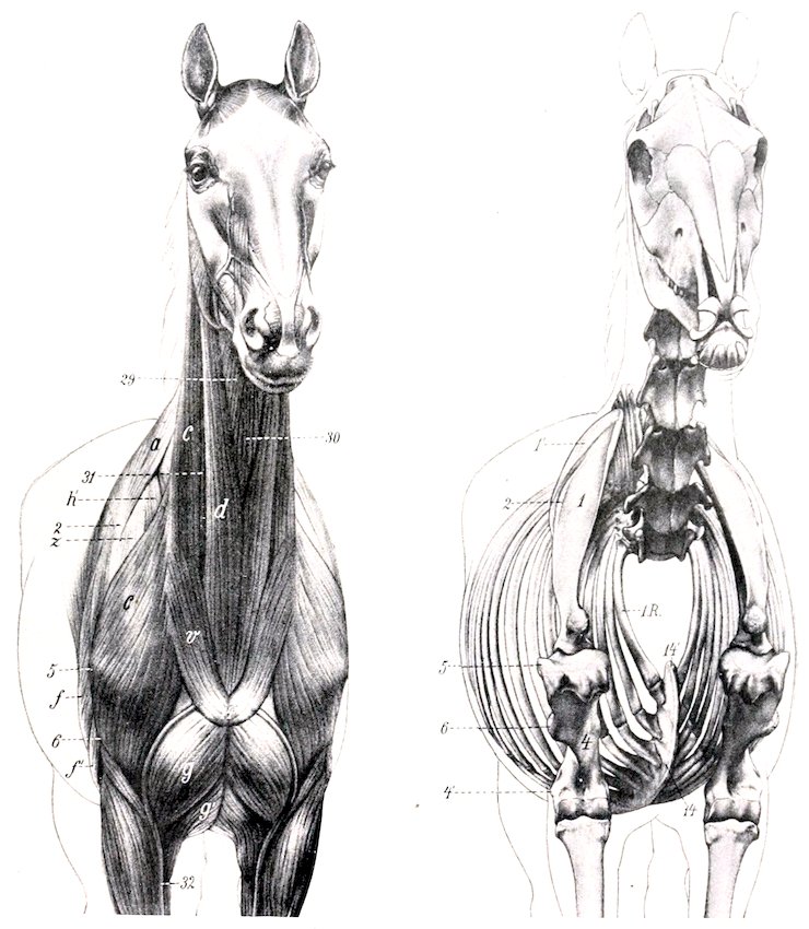
Fig. 177.—Antero-lateral View of Muscles and Skeleton of Horse. a, Trapezius; c, mastoido-humeralis; d, sterno-cephalicus; f, long head of triceps; f′, external head of triceps; g, anterior superficial pectoral muscle; g′, posterior superficial pectoral; h′, anterior deep pectoral; v, cervical panniculus; z, supraspinatus; 29, omo-hyoideus; 30, sterno-thyro-hyoideus; 31, jugular vein; 32, cephalic vein; 1, scapula; 1′, cartilage of scapula; 2, spine of scapula; 4, shaft of humerus; 4′, external epicondyle; 5, external tuberosity of humerus; 6, deltoid tuberosity; 14, ventral border (“keel”) of sternum; 14′, cariniform cartilage; l.R., first rib. (After Ellenberger-Baum, Anat. für Künstler.)
2. Mastoido-humeralis.—This is described on p. 252.
3. Sterno-cephalicus (Sterno-maxillaris s.-mandibularis).[42]—This is a long, narrow 226muscle, extending along the ventral and lateral aspects of the trachea from the sternum to the angle of the jaw. It forms the ventral boundary of the jugular furrow.
Origin.—The cariniform cartilage of the sternum.
Insertion.—The posterior border of the lower jaw.
Action.—Acting together, to flex the head and neck; acting singly, to incline the head and neck to the side of the muscle contracting.
Structure.—The two muscles are fused at their origin, which is fleshy. Near the middle of the neck they separate, and, becoming thinner, each muscle passes under the parotid gland and terminates in a flat tendon.
Relations.—Superficially, the cervical panniculus; deeply, the sterno-thyro-hyoideus and omo-hyoideus muscles. The upper edge of the muscle is related to the jugular vein, which lies in the jugular furrow. The carotid artery, the vagus, sympathetic, and recurrent nerves also lie along the upper edge at the root of the neck. The tendon passes under the submaxillary vein and the parotid gland, having the submaxillary gland and stylo-maxillaris muscle on its inner side.
Blood-supply.—Carotid artery.
Nerve-supply.—Ventral branches of the spinal accessory and first cervical nerves.
4. Sterno-thyro-hyoideus (Sterno-thyroideus et sterno-hyoideus).—This is a long, slender, digastric muscle, applied to the ventral surface of the trachea and its fellow of the opposite side.
Origin.—The cariniform cartilage of the sternum.
Insertion.—(1) The external surface of the thyroid cartilage of the larynx; (2) the body of the hyoid bone.
Action.—To depress and retract the hyoid bone, the base of the tongue, and the larynx, as in deglutition. It may also fix the hyoid bone when the depressors of the tongue are acting, as in suction.
Structure.—The origin of the muscle is fleshy, and as far as the middle of the neck it blends with its fellow. The common belly is then interrupted by a tendon, or sometimes two tendons, from which arise three or four fleshy bands. The lateral bands diverge to reach their insertion into the thyroid cartilage; while the inner bands, closely applied to each other and blending with the omo-hyoideus, pass straight forward to reach the ventral surface of the hyoid bone.
Relations.—At the root of the neck the common belly has the sterno-cephalicus below, and the carotid arteries and recurrent nerves above. Further forward the trachea becomes the upper relation, and near the head the skin and fascia, the lower one.
Blood-supply.—Carotid artery.
Nerve-supply.—Ventral branch of the first cervical nerve.
5. Omo-hyoideus (Subscapulo-hyoideus).—This is a thin, ribbon-like muscle, almost entirely fleshy, which crosses the trachea very obliquely under cover of the mastoido-humeralis.
Origin.—The subscapular fascia close to the shoulder joint.
Insertion.—The body of the hyoid bone, in common with the hyoid branch of the preceding muscle.
Action.—To depress the hyoid bone.
Structure.—The muscle is composed of parallel fleshy fibers, except at its origin, where it has a thin tendon.
Relations.—In the first part of its course the muscle passes forward between the supraspinatus, anterior deep pectoral, mastoido-humeralis (outwardly), and the scalenus (inwardly). It is intimately adherent to the mastoido-humeralis. In the middle of the neck it is related superficially to the mastoido-humeralis, sterno-cephalicus, and the jugular vein; deeply, to the rectus capitis anterior major, the carotid artery, the vagus, sympathetic, and recurrent nerves, the trachea, and, 227on the left side, the œsophagus. In its anterior part the muscle blends with the hyoid part of the sterno-thyro-hyoideus, the two covering the thyroid portion of the latter muscle, the thyroid gland, and the ventral face of the larynx.
Blood-supply.—Carotid and inferior cervical arteries.
Nerve-supply.—Ventral branches of the cervical nerves.
6. Scalenus (M. scalenus primæ costæ).—This muscle is deeply situated on the side of the posterior half of the neck. It is composed of two parts, between which the roots of the brachial plexus of nerves emerge.
Origin.—The anterior border and outer surface of the first rib.
Insertion.—(1) The dorsal (smaller) portion is attached to the transverse process of the seventh cervical vertebra; (2) the ventral portion is attached to the transverse processes of the sixth, fifth, and fourth cervical vertebræ.
Action.—The neck is flexed or inclined laterally, according as the muscles act together or singly. If the neck be the fixed point, the muscle may have a respiratory action by pulling forward or fixing the first rib.
Structure.—The dorsal portion is composed of three or four fleshy bundles.[43] The ventral portion, which is much larger, is almost entirely fleshy, and not so divided.
Relations.—Superficially, the anterior deep pectoral, mastoido-humeralis, and omo-hyoideus muscles, the phrenic nerve, and the other branches of the brachial plexus; deeply, the vertebræ, the longus colli and intertransversales muscles, the œsophagus (on the left side), the trachea (on the right side), the vertebral vessels, the vagus, sympathetic, and recurrent nerves. The roots of the brachial plexus form a flat anastomosis, which lies between the two portions of the muscle. The brachial vessels cross the ventral edge close to the first rib.
Blood-supply.—Carotid, vertebral, and inferior cervical arteries.
Nerve-supply.—Ventral branches of the cervical nerves.
7. Rectus capitis anterior major (M. longus capitis).—This is the largest of the three special flexors of the head, and lies along the ventro-lateral surface of the anterior cervical vertebræ and the base of the cranium.
Origin.—The transverse processes of the fifth, fourth, and third cervical vertebræ.
Insertion.—The tubercles at the junction of the basilar process of the occipital bone with the body of the sphenoid.
Action.—Acting together, to flex the head; acting singly, to incline it to the same side also.
Structure.—The origin of the muscle is by fleshy digitations. The belly increases in size by the union of these digitations, reaching its maximum at the axis. It then diminishes, passes toward the median plane, and terminates on a rounded tendon.
Relations.—Superficially, the mastoido-humeralis, omo-hyoideus, and rectus capitis anticus minor, the submaxillary gland, the carotid artery (which lies along the lower border), the occipital and internal carotid arteries, and the tenth, eleventh, and sympathetic nerves; deeply, the vertebræ, the longus colli, intertransversales, and the small straight muscle. The terminal part of the muscle lies in contact with its fellow above the pharynx and between the guttural pouches.
Blood-supply.—Carotid, vertebral, and occipital arteries.
Nerve-supply.—Ventral branches of the cervical nerves.
8. Rectus capitis anterior minor (M. rectus capitis ventralis).—This is a small muscle which lies under cover of the preceding one.
Origin.—The ventral arch of the atlas.
228Insertion.—The basilar process, close to the preceding muscle.
Action.—To flex the occipito-atlantal articulation.
Structure.—Fleshy.
Relations.—Below, to the preceding muscle; above, to the atlas, occipito-atlantal articulation, and the basilar process; externally, to the rectus capitis lateralis and the guttural pouch.
Blood-supply.—Occipital artery.
Nerve-supply.—Ventral branch of the first cervical nerve.
9. Rectus capitis lateralis.—This is a still smaller, entirely fleshy muscle, which lies for the most part under the small oblique muscle.
Origin.—The atlas, external to the preceding muscle.
Insertion.—The paramastoid or styloid process of the occipital bone.
Action.—The same as the preceding muscle.
Structure.—Fleshy.
Relations.—Superficially, the small oblique muscle, the occipital vessels, and the ventral branch of the first cervical nerve.
Blood-supply.—Occipital artery.
Nerve-supply.—Ventral branch of the first cervical nerve.
10. Longus colli.—This muscle covers the ventral surface of the vertebræ, from the sixth thoracic to the atlas, and is united with its fellow. It consists of two portions, thoracic and cervical.
Origin.—(1) Thoracic portion, the bodies of the first six thoracic vertebræ; (2) cervical portion, the transverse processes of the cervical vertebræ.
Insertion.—(1) Thoracic portion, the bodies and transverse processes of the last two cervical vertebræ; (2) cervical portion, the bodies of the cervical vertebræ and the tubercle on the ventral surface of the atlas.
Action.—To flex the neck.
Structure.—The muscle is composed of a succession of bundles. The largest of these constitute the thoracic part of the muscle, which has a strong tendon inserted into the last two cervical vertebræ. A bursa is interposed between the tendon and the spine at the first costo-vertebral articulation. The cervical portion consists of a number of smaller bundles, each of which passes from its origin on the transverse process of one vertebra forward and inward to its insertion into a vertebra further forward. The most anterior bundle is inserted by a strong tendon into the ventral tubercle of the atlas.
Relations.—The principal relations of the two muscles in the thorax are: ventrally, the pleura, and, further forward, the trachea and œsophagus; dorsally, the vertebræ and the costo-vertebral joints; laterally, the dorsal, superior cervical, and vertebral vessels, the sympathetic nerve, and the thoracic roots of the brachial plexus. In the neck, important relations are: ventrally, the trachea and œsophagus, the carotid artery, the vagus, sympathetic and recurrent nerves; dorsally, the vertebræ and, in the middle third of the neck, the intertransversales muscles; laterally, the scalenus, the rectus capitis anticus major, and the intertransversales (in the anterior third). The terminal part of the muscle is separated from the trachea by the œsophagus, which is here median in position.
Blood-supply.—Subcostal and vertebral arteries.
Nerve-supply.—Ventral branches of the spinal nerves.
11. Intertransversales colli (Mm. intertransversarii cervicis).—These are six fasciculi which occupy the spaces between the lateral aspects of the vertebræ and the transverse and articular processes. There is thus a bundle for each intervertebral articulation except the first. Each bundle consists of a dorsal and ventral portion.
Attachments.—The dorsal bundles pass from transverse process to articular process; the ventral bundles extend between adjacent transverse processes.
229Action.—To bend the neck laterally.
Structure.—They contain strong tendinous intersections.
Relations.—Superficially, the mastoido-humeralis, rectus capitis anterior major, complexus, trachelo-mastoideus, splenius, scalenus, and longissimus muscles; deeply, the vertebræ, the longus colli muscle, and the vertebral vessels. The muscles are perforated by branches of these vessels and by the primary branches of the cervical nerves.
Blood-supply.—Vertebral artery.
Nerve-supply.—The cervical nerves.
LATERAL MUSCLES
This group consists of twelve pairs of muscles arranged in layers.
First Layer
1. Trapezius cervicalis.—Described on p. 250.
Second Layer
2. Rhomboideus cervicalis.—Described on p. 251.
3. Serratus magnus (M. serratus cervicis).—Described with the thoracic part on p. 254.
Third Layer
4. Splenius.—This is an extensive, flat, triangular muscle, partly covered by the preceding three muscles.
Origin.—The second, third, and fourth thoracic spines by means of the dorso-scapular ligament, and the funicular portion of the ligamentum nuchæ.
Insertion.—The occipital crest, the mastoid process, the wing of the atlas, and the transverse processes of the third, fourth, and fifth cervical vertebræ.
Action.—Acting together, to elevate the head and neck; acting singly, to incline the head and neck to the side of the muscle acting.
Structure.—The muscle arises in the withers from the anterior part of the dorso-scapular ligament, which also affords attachment to the rhomboideus, serratus anticus, and complexus muscles. The fibers pass upward and forward toward the head and the first cervical vertebra. The insertion on the occipital bone and the mastoid process is by means of a thin aponeurosis common to the splenius, trachelo-mastoideus, and mastoido-humeralis. The atlantal insertion is by a strong, flat tendon, also in common with these muscles. The remaining insertions are fleshy digitations.
Relations.—Superficially, the skin and fascia, the trapezius rhomboideus, cervicalis, serratus magnus, and posterior auricular muscles; deeply, the complexus, trachelo-mastoideus, and longissimus muscles.
Blood-supply.—Deep cervical and dorsal arteries.
Nerve-supply.—Dorsal branches of the last six cervical nerves.
Fourth Layer
5. Trachelo-mastoideus (M. longissimus capitis et atlantis).—This muscle consists of two parallel, fusiform portions. It lies between the deep face of the splenius and the ventral part of the complexus.
Origin.—(1) The transverse processes of the first two thoracic vertebræ; (2) the articular processes of the cervical vertebræ.
Insertion.—(1) The mastoid process; (2) the wing of the atlas.
230
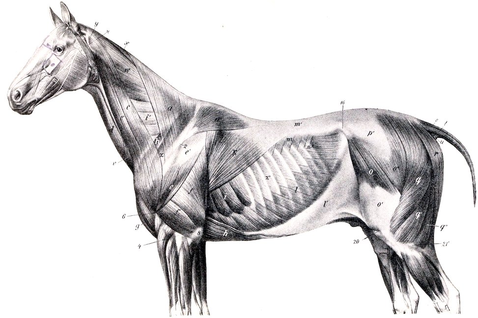
Fig. 178.—Superficial Muscles of Horse. The panniculus, except the cervical part, has been removed. a, Trapezius cervicalis; a′, trapezius thoracalis; c, c′, mastoido-humeralis; d, sterno-cephalicus; e, deltoid; f, long head of triceps; f′, external head of triceps; g, anterior superficial pectoral; h, posterior deep pectoral; h′, anterior deep pectoral; i, serratus thoracis; i′, serratus cervicis; k, latissimus dorsi; l, obliquus abdominis externus; l′, aponeurosis of l; m, serratus posticus; m′, lumbo-dorsal fascia; o, tensor fasciæ latæ; o′, fascia lata; o″, gluteus superficialis; q, q′, q″, biceps femoris; r, semitendinosus; s, sacro-coccygeus superior; t, sacro-coccygeus lateralis; u, coccygeus; v, cervical panniculus; w, splenius; x, rhomboideus; y, tendon of trachelo-mastoideus and mastoido-humeralis; z, suprasinatus; X, wing of atlas; 2, spine of scapula; 4′, external epicondyle of humerus; 6, deltoid tuberosity; 8, olecranon; 16, external angle of ilium; 20, patella; 21′, external condyle of tibia. (After Ellenberger-Baum, Anat. für Künstler.)
231Action.—Acting together, to extend the head and neck; acting singly, to flex the head and neck laterally or to rotate the atlas.
Structure.—The origin from the thoracic vertebræ is by aponeurotic slips which blend with the complexus. The succeeding fleshy portion, in passing along the neck, receives fasciculi from each of the cervical vertebræ except the first two. The dorsal division of the muscle is inserted into the mastoid process by a flat tendon which fuses with that of the splenius; the ventral division is inserted into the wing of the atlas by a ribbon-like tendon in common with the splenius and mastoido-humeralis.
Relations.—Superficially, the splenius muscle and dorsal branches of the cervical nerves; deeply, the complexus, the spinalis colli, and the oblique muscles of the head. The superior cervical vessels cross the deep face of the muscle obliquely at the level of the sixth and seventh cervical vertebræ.
Blood-supply.—Vertebral and superior cervical arteries.
Nerve-supply.—Dorsal branches of the last six cervical nerves.
6. Complexus (M. semispinalis capitis).—This is a large triangular muscle which lies chiefly on the ligamentum nuchæ, under cover of the splenius and trachelo-mastoideus muscles.
Origin.—(1) The second, third, and fourth thoracic spines, in common with the splenius and serratus anticus; (2) the transverse processes of the first six or seven thoracic vertebræ; (3) the articular processes of the cervical vertebræ.
Insertion.—The posterior surface of the occipital bone, external to the funicular portion of the ligamentum nuchæ.
Action.—It is the chief extensor of the head and neck. Acting singly, the muscle inclines the head to the same side.
Structure.—The origin of the muscle at the withers is aponeurotic. In the neck the bundles arising from the articular processes run obliquely upward and forward, giving the lower part of the muscle a distinct pennate character. The upper part of the belly is crossed obliquely by four or five tendinous intersections. The insertion is by a strong tendon.
Relations.—Superficially, the rhomboideus, serratus magnus, splenius, and trachelo-mastoideus muscles; deeply, the ligamentum nuchæ, the multifidus cervicis, longissimus, and the oblique and posterior straight muscles of the head, the deep or superior cervical vessels, and the dorsal cutaneous branches of the cervical nerves.
Blood-supply.—Deep cervical, vertebral, and occipital arteries.
Nerve-supply.—Dorsal branches of the last six cervical nerves.
7. Multifidus cervicis (M. semispinalis colli; transverse spinous muscle of neck).—This muscle lies on the arches of the last five cervical vertebræ. It consists of five or six segments.
Origin.—The articular processes of the last five (or four) cervical and the first thoracic vertebræ.
Insertion.—The spinous processes of the cervical vertebræ.
232
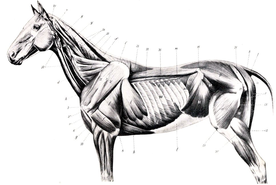
Fig. 179.—Deeper Muscles of Horse. f, Long head of triceps; f′, external head of triceps; g, anterior superficial pectoral; h, posterior deep pectoral; h′, anterior deep pectoral; i, serratus thoracis; i′, serratus cervicis; l, obliquus abdominis externus, and l′, its aponeurosis, the posterior part of which has been removed; m, serratus posticus; p, gluteus medius; r, semitendinosus; s, sacro-coccygeus superior; t, sacro-coccygeus lateralis; u, coccygeus; v′, biceps brachii; x, rhomboideus; y, y′, trachelo-mastoideus; z, supraspinatus; z′, infraspinatus; 1, cartilage of scapula; 2, spine of scapula; 5, external tuberosity of humerus; 6, deltoid tuberosity; 8, olecranon; 16, external angle of ilium; 19, trochanter major; 20, patella; 21′, external condyle of tibia; 26, articular processes of cervical vertebræ; 27, parotido-auricularis; 28, vastus externus; 28′, rectus femoris; 28″, trochanter tertius; 29, semimembranosus; 30, gastrocnemius; 31, sacro-sciatic ligament; 32, omo-hyoideus; 33, complexus; 34, rectus capitis anterior major; 35, spinalis dorsi; 36, longissimus dorsi; 37, transversalis costarum; 38, teres minor; 39, brachialis; 40, external intercostal; 41, obliquus abdominis internus; 42, iliacus; 43, transversus abdominis. (After Ellenberger-Baum, Anat. für Künstler.)
233Action.—Acting together, to extend the neck; acting singly, to flex and rotate the neck to the opposite side.
Structure.—The muscle is composed of two sets of bundles. The superficial bundles are directed obliquely forward and inward, each passing from an articular process to the spine of the preceding vertebra. The deep bundles are shorter and run straight from an articular process to that of the preceding vertebra.
Relations.—Superficially, the complexus, longissimus, trachelo-mastoideus, and great oblique muscles; deeply, the spinalis muscle, the ligamentum nuchæ, and the arches of the vertebræ.
Blood-supply.—Deep cervical and vertebral arteries.
Nerve-supply.—Dorsal branches of the last six cervical nerves.
8. Spinalis.—Described with the longissimus on p. 237.
9. Obliquus capitis posterior (s. caudalis) (great oblique muscle of the head).—This is a strong, quadrilateral muscle, which covers the dorso-lateral aspect of the atlas and axis.
Origin.—The side of the spine and the posterior articular process of the axis.
Insertion.—The dorsal surface of the wing of the atlas.
Action.—Chiefly to rotate the atlas, and with it the head, to the same side; also to assist in extending and fixing the atlanto-axial joint.
Structure.—The muscle is composed almost entirely of parallel fleshy fibers directed obliquely forward and outward.
Relations.—Superficially, the skin, the splenius, complexus, trachelo-mastoideus, and mastoido-humeralis muscles; deeply, the arch and spine of the axis, the wing of the atlas, the atlanto-axial joint, the multifidus cervicis, the posterior straight muscles, the occipital and vertebral vessels, and the first and second cervical nerves. The terminal part of the vertebral artery joins the posterior branch of the occipital artery under cover of the muscle.
Blood-supply.—Occipital and vertebral arteries.
Nerve-supply.—Dorsal branch of the second cervical nerve.
10. Obliquus capitis anterior (s. cranialis) (small oblique muscle of the head).—A short, thick, quadrilateral muscle which lies on the side of the occipito-atlantal articulation.
Origin.—The anterior edge of the wing of the atlas and the fossa atlantis.
Insertion.—The styloid process and crest of the occipital bone, and the mastoid process.
Action.—Acting together, to extend the head on the atlas; acting singly, to flex the head laterally.
Structure.—The muscle contains a good deal of tendinous tissue. The direction of its fibers is forward, upward, and inward.
Relations.—Superficially, the posterior auricular muscles, artery, and nerve, the aponeurosis of the splenius, trachelo-mastoideus and mastoido-humeralis, and the parotid gland; deeply, the straight muscles, the complexus, the occipito-hyoideus, the occipito-atlantal articulation, branches of the occipital artery, and a branch of the occipital nerve.
234Blood-supply.—Occipital artery.
Nerve-supply.—Dorsal branch of the first cervical nerve.
11. Rectus capitis posterior major (s. dorsalis major) (great posterior straight muscle of the head).—This muscle extends from the axis to the occipital bone, in contact with the ligamentum nuchæ.
Origin.—The edge of the spinous process of the axis.
Insertion.—The occipital bone, below and external to the complexus and ligamentum nuchæ.
Action.—To extend the head.
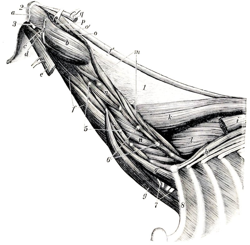
Fig. 180.—Deepest Layer of Muscles of Neck of Horse. a, Obliquus capitis anterior; b, obliquus capitis posterior; c, rectus capitis lateralis; d, rectus capitis anterior minor; e, rectus capitis anterior major; f, longus colli; g, g′, scalenus; h, transversalis costarum; i, longissimus dorsi; k, spinalis et semispinalis; l, multifidus dorsi; m, multifidus cervicis; n, intertransversales; o, o′, rectus capitis posterior major; p, rectus capitis posterior minor; q, tendon of insertion of complexus; 1, lamellar part, 1′, funicular part of ligamentum nuchæ; 2, occipital crest; 3, paramastoid process; 4, edge of wing of atlas; 5, transverse, and 6, articular, processes of cervical vertebræ; 7, nerves of brachial plexus (cut); 8, first rib. (Ellenberger-Baum, Anat. d. Haustiere.)
Structure.—The muscle is fleshy and may be divided into two parallel portions, superficial and deep. The former blends somewhat with the terminal part of the complexus. The deep portion may be termed the rectus capitis posterior medius.
Relations.—Superficially, the anterior oblique, splenius, and complexus; internally, the ligamentum nuchæ; deeply, the atlas, the occipito-atlantal articulation, and the rectus capitis posterior minor. The dorsal branch of the first cervical nerve appears between this muscle and the small oblique.
Blood-supply.—Occipital artery.
Nerve-supply.—Dorsal branch of the first cervical nerve.
12. Rectus capitis posterior minor (s. dorsalis minor) (small posterior straight muscle of the head).—This small muscle lies under cover of the preceding.
235Origin.—The dorsal surface of the atlas.
Insertion.—The occipital bone beneath the preceding muscle.
Action.—To assist the preceding muscle.
Structure.—It is fleshy and varies a good deal in volume, being sometimes small and difficult to recognize.[44] On the other hand, it is sometimes double.
Relations.—Superficially, the preceding muscle and the small oblique; deeply, the atlas and the occipito-atlantal articulation.
Blood-supply.—Occipital artery.
Nerve-supply.—Dorsal branch of the first cervical nerve.
The Fasciæ and Muscles of the Back and Loins
The superficial fascia presents no special features. The lumbo-dorsal fascia (Fascia lumbo-dorsalis) closely invests the muscles, but is easily stripped off the longissimus. It is attached medially to the supraspinous ligament and the spinous processes of the vertebræ; laterally, it divides into two layers. The superficial layer is practically the aponeurosis of the latissimus dorsi. The deep layer gives origin to the serratus anticus and posticus, the lumbar part of the obliquus externus abdominis, the transversus abdominis, and the retractor costæ. Its lateral edge curves under the longissimus and is attached to the ribs and lumbar transverse processes. Posteriorly, it is continuous with the gluteal fascia. At the withers it forms an important structure, the dorso-scapular ligament. This is a strong tendinous sheet, attached to the third, fourth, and fifth thoracic spines. Its upper part is very thick and gives origin by its superficial surface to the rhomboideus dorsi, by its anterior part to the splenius. The lower part is thin and elastic, and furnishes numerous lamellæ which intersect the scapular part of the serratus and are attached with it to the scapula. Three lamellæ are detached from the deep face of the ligament. The deepest of these passes between the longissimus and spinalis and is attached to the transverse processes of the first seven thoracic vertebræ; it gives attachment to the complexus. The middle one dips in between the longissimus and transversalis costarum. The superficial one gives origin to the serratus anticus. A strong fascial layer, the ilio-lumbar ligament, extends from the last rib to the external angle of the ilium.
There are nine pairs of muscles in this region, arranged in four layers.
First Layer
1. Trapezius thoracalis.
2. Latissimus dorsi.
Second Layer
3. Rhomboideus thoracalis.
The foregoing are described with the other muscles which attach the thoracic limb to the trunk (p. 251).
4. Serratus anticus (M. serratus dorsalis inspiratorius).—This is a thin quadrilateral muscle, named from its toothed ventral border. It lies beneath the rhomboideus, serratus magnus, and latissimus dorsi.
Origin.—The lumbo-dorsal fascia and dorso-scapular ligament.
Insertion.—The external surfaces of the fifth to the eleventh or twelfth ribs inclusive.
Action.—To draw the ribs on which it is inserted forward and outward, thus assisting in inspiration.
236Structure.—The muscle arises by means of a thin aponeurosis which blends with the lumbo-dorsal fascia and the aponeurosis of the latissimus dorsi. The muscle-fibers pass downward and backward to be attached to the ribs by seven or eight digitations below the outer edge of the transversalis costarum.
Relations.—Superficially, the rhomboideus, serratus magnus, latissimus dorsi, and serratus posticus; deeply, the longissimus dorsi, transversalis costarum, external intercostal muscles, and the ribs.
Blood-supply.—Intercostal arteries.
Nerve-supply.—Thoracic nerves.
5. Serratus posticus (M. serratus dorsalis exspiratorius).—This muscle resembles the preceding one, which it partly covers.
Origin.—The lumbo-dorsal fascia.
Insertion.—The outer surfaces of the last seven or eight ribs.
Action.—To draw the ribs backward, thus assisting in expiration.
Structure.—Similar to the preceding muscle. The fibers are directed downward and forward and terminate in seven or eight digitations, one or two of which cover the posterior teeth of the anticus. The aponeurosis blends with that of the latissimus dorsi.
Relations.—Superficially, the latissimus dorsi and external oblique; deeply, the longissimus dorsi, transversalis costarum, external intercostals, serratus anticus, and the ribs.
Blood-supply.—Intercostal and lumbar arteries.
Nerve-supply.—Thoracic nerves.
Third Layer
6. Transversalis costarum (M. ilio-costalis).—This long, segmental muscle extends, as its name indicates, across the series of ribs, in contact with the outer edge of the longissimus dorsi.
Origin.—(1) The transverse processes of the second and third lumbar vertebræ and the deep layer of the lumbo-dorsal fascia. (2) The anterior borders and external surface of the last fifteen ribs.[45]
Insertion.—The posterior borders of the ribs and the transverse processes of the last two or three cervical vertebræ.
Action.—Chiefly to depress and retract the ribs and so help in expiration. Acting together, they may assist in extending this spine, acting singly in inclining it laterally.
Structure.—This muscle presents a distinct segmental arrangement. It is composed of a series of bundles, the fibers of which are directed forward and a little downward and outward. From these are detached two sets of tendons. The superficial tendons spring from the outer edge of the muscle. They are flat and are about half an inch in width. Each crosses two or three intercostal spaces, to be inserted on the posterior border of a rib. The deep tendons are detached from the dorsal part of the deep face of the muscle. Each passes backward across one or two intercostal spaces to its origin on the anterior border or outer surface of a rib. Small bursæ may be found between the ribs and tendons.
Relations.—Superficially, the dorsal serrati and the complexus; deeply, the external intercostals and the ribs. The lumbar origin is covered by the longissimus. The superior (deep) cervical and dorsal vessels cross the surface of the muscle at the first and second intercostal spaces respectively, and branches of the intercostal vessels and nerves emerge between the transversalis and longissimus; here a fascial layer dips in between the two.
237Blood-supply.—Intercostal arteries.
Nerve-supply.—Dorsal branches of the thoracic nerves.
7. Longissimus.[46]—This is the largest and longest muscle in the body. It extends from the sacrum and ilium to the neck, filling up the space between the spinous processes medially and the lumbar transverse processes and the upper ends of the ribs below; consequently it has the form of a three-sided prism.
Origin.—(1) The internal angle, crest, and adjacent part of the ventral surface of the ilium; (2) the first three sacral spines; (3) the lumbar and thoracic spines and the supraspinous ligament.
Insertion.—(1) The lumbar transverse and articular processes; (2) the thoracic transverse processes; (3) the spinous and transverse processes of the last four cervical vertebræ; (4) the outer surfaces of the ribs, except the first.
Action.—Acting with its fellow, it is the most powerful extensor of the back and loins; by its cervical attachment it assists in extending the neck. By its costal attachment it may also assist in expiration. Acting singly, it flexes the spine laterally.
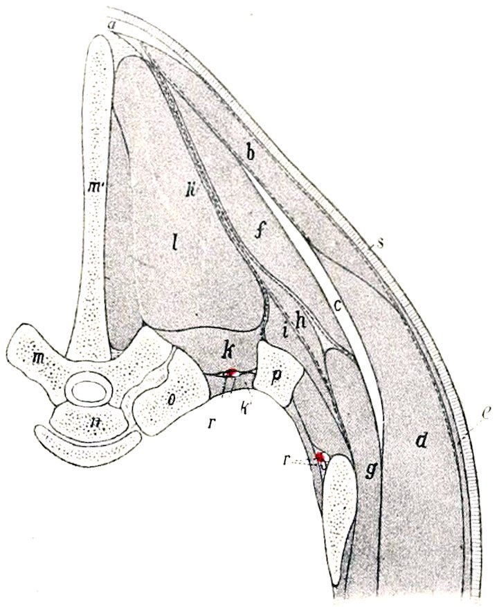
Fig. 181.—Right Portion of Cross-section of Back of Horse. Section is Cut Through Seventh Thoracic Vertebra. a, Ligamentum nuchæ; b, trapezius muscle; c, cartilage of scapula; d, latissimus dorsi; e, panniculus carnosus; f, rhomboideus dorsi; g, serratus magnus; h, serratus anticus; h′, lumbo-dorsal fascia, which divides below into three layers; i, transversalis costarum; k, levator costæ; k′, internal intercostal muscle; l, longissimus dorsi; m, m′, seventh thoracic vertebra; n, head of eighth thoracic vertebra; o, head of eighth rib; p, seventh rib; r, intercostal artery and nerve; s, skin. The fasciæ are indicated by dotted lines. (After Ellenberger, in Leisering’s Atlas.)
Structure.—This is quite complex. The posterior part of the muscle is greatly developed and constitutes the common mass of the loins. This is covered by a strong aponeurosis which blends with the supraspinous and sacro-iliac ligaments, and is attached to the crest and inner (sacral) angle of the ilium and the first and second sacral spines; it furnishes origin to the lumbar portion of the middle gluteus. In its course further forward the muscle receives fasciculi from the lumbar and thoracic spines, but diminishes somewhat in volume. At the withers it divides into two parts. The dorsal division (spinalis et semispinalis), reinforced by bundles from the first four thoracic spines, passes forward under the complexus to be inserted into the spines of the last four cervical vertebræ. The ventral division passes forward and downward underneath the serratus magnus to be inserted into the ribs and the transverse processes of the last four cervical vertebræ. Three sets of fasciculi may be distinguished, viz.: (1) spinal, which are superficial and internal; (2) transverse, attached to the transverse and articular processes, which are internal and deep; (3) costal, which are external.
Relations.—Superficially, the middle gluteus, the lumbo-dorsal fascia, the latissimus dorsi, dorsal serrati, serratus magnus, and complexus; deeply, the multifidus, intertransversales, external intercostals, levatores costarum, the ligamentum nuchæ, and its fellow of the opposite side (in the neck).
Blood-supply.—Dorsal, superior cervical, intercostal, and lumbar arteries.
Nerve-supply.—Dorsal branches of the thoracic and lumbar nerves.
2388. Multifidus dorsi (semispinalis of the back and loins).—This is a long segmental muscle which covers the sides of the spinous processes of the vertebræ from the sacrum to the neck.
Origin.—(1) The lateral portion of the sacrum; (2) the articular processes of the lumbar vertebræ; (3) the transverse processes of the thoracic vertebræ.
Insertion.—The spinous processes of the first two sacral, the lumbar, thoracic, and last cervical vertebræ.
Action.—Acting with its fellow, it is an extensor of the spine; acting singly, it flexes it laterally.
Structure.—It is composed of a series of bundles which are directed obliquely forward and upward. Each fasciculus passes over several vertebræ to its insertion. In the posterior part of the series the bundles cross two or three vertebræ and are inserted into the summits of the spines. Anteriorly, the bundles have a more horizontal direction, cross three to five vertebræ, and are inserted into the sides of the spines considerably below their summits. A further complication consists in the fusion of several bundles into a common insertion.
Relations.—Superficially, the longissimus; deeply, the vertebral spines.
Blood-supply.—Intercostal and lumbar arteries.
Nerve-supply.—Dorsal branches of the thoracic and lumbar nerves.
9. Intertransversales lumborum.—These are very thin muscular and tendinous strata, which occupy the spaces between the transverse processes of the lumbar vertebræ except the fifth and sixth.
Action.—To assist in flexing the loins laterally.
Relations.—Superficially, the longissimus; deeply, the quadratus lumborum.
Blood-supply.—Lumbar arteries.
Nerve-supply.—Lumbar nerves.
The Fascia and Muscles of the Tail
The muscles of the tail are inclosed in the strong coccygeal fascia, which is continuous in front with the gluteal fascia and blends with the lateral sacro-iliac ligament. At the root of the tail it is loosely attached to the subjacent muscles, but further back it is intimately adherent to them. From its deep face are detached septa which pass between the muscles to become attached to the vertebræ.
1. Coccygeus (Compressor coccygis; ischio-coccygeus).—This is a flat, triangular muscle which lies chiefly between the sacro-sciatic ligament and the rectum.
Origin.—The pelvic surface of the sacro-sciatic ligament near the ischiatic spine.
Insertion.—The first four coccygeal vertebræ and the coccygeal fascia.
Action.—Acting together, to depress (flex) the tail, compressing it over the perineum; acting singly, to depress and incline it to the same side.
Structure.—The origin of the muscle is aponeurotic. Becoming fleshy, its fibers pass upward and backward and divide into two layers. The outer layer is attached to the vertebræ, the inner to the fascia; included between the two lie the intertransversales. When the tail is raised, the ventral edges of the muscles produce a distinct ridge at either side of the anus.
Relations.—Outwardly, the sacro-sciatic ligament and the semimembranosus; inwardly, the rectum and the sacro-coccygeus ventralis muscle. The internal pudic artery crosses the outer face of the origin of the muscle.
2. Sacro-coccygeus superior (M. sacro-coccygeus dorsalis medialis; erector s. extensor coccygis).—This muscle lies along the dorso-median aspect of the tail, in contact with its fellow.
Origin.—The last three sacral spines and some of the coccygeal spines.
239Insertion.—The dorsal surface of the coccygeal vertebræ.
Action.—Acting together, to elevate (extend) the tail; acting singly, to elevate and incline it laterally.
Structure.—The muscle has a strong rounded belly. It is inserted by means of short tendons which fuse with those of the next muscle.
Relations.—Superficially, the coccygeal fascia; internally, its fellow; laterally, the sacro-coccygeus lateralis; deeply, the vertebræ.
3. Sacro-coccygeus lateralis (M. sacro-coccygeus dorsalis lateralis; curvator coccygis).—This muscle lies immediately lateral to the preceding.
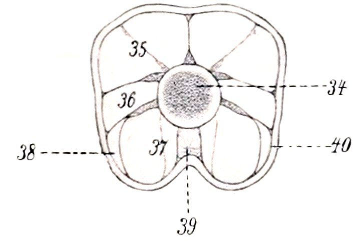
Fig. 182.—Cross-section of Tail of Horse Close to Anus. 34, Coccygeal vertebra; 35, sacro-coccygeus superior; 36, intertransversalis; 37, sacro-coccygeus inferior; 38, coccygeus; 39, recto-coccygeus; 40, coccygeal fascia. (After Ellenberger-Baum, Anat. für Künstler.)
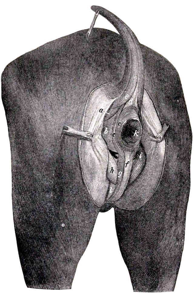
Fig. 183.—Muscles of Perineum of Horse. a, Coccygeus; b, retractor ani; c, c′, sphincter and externus; d, recto-coccygeus; e, sacro-coccygeus inferior lateralis; f, retractor penis; g, bulbo-cavernosus; h, ischio-cavernosus; i, internal pudic artery; k, anus; l, penis. (After Ellenberger-Baum, Top. Anat. d. Pferdes.)
Origin.—The sides of the sacral spines, with the multifidus, and the transverse processes of the sacral and coccygeal vertebræ.
Insertion.—The lateral surface of the coccygeal vertebræ, except the first four.
Action.—Acting with its fellow, to assist the preceding muscle in elevating the tail; acting singly, to incline it to the same side.
Structure.—This muscle appears to be a direct continuation of the multifidus dorsi. The belly is fusiform and receives reinforcing fasciculi from the transverse processes of the sacrum. This is succeeded by bundles of tendons, as many as four lying alongside of each other.
Relations.—Superficially, the lateral sacro-iliac ligament and the coccygeal fascia; dorsally, the sacro-coccygeus superior; ventrally, the intertransversales; deeply, the vertebræ and a branch of the lateral coccygeal artery and accompanying vein and nerve.
4. Intertransversales caudæ (Mm. intertransversarii caudæ).—These consist of muscular bundles which lie on the lateral aspect of the tail, between the preceding muscle and the sacro-coccygeus inferior. They begin on the lateral edge 240of the sacrum and occupy the spaces between the transverse processes, to which they are attached. They are, however, not arranged in a strict segmental manner.
Action.—Acting together, to fix the coccygeal vertebræ; acting singly, to assist in lateral flexion.
5. Sacro-coccygeus inferior (Mm. sacro-coccygei ventrales; depressor coccygis).—This muscle lies on the ventral aspect of the sacrum and coccyx. It is composed of two portions, described by Bourgelat and the German anatomists as separate muscles.
(a) The outer portion (M. coccygeus ventralis lateralis) is much the larger of the two. It arises from the outer part of the ventral surface of the sacrum, about as far forward as the third foramen, and is inserted into the transverse processes and ventral surface of the coccygeal vertebræ.
(b) The inner portion (M. sacro-coccygeus ventralis medialis) arises from the ventral surface of the sacrum internal to the preceding muscle and the first eight coccygeal vertebræ, and is inserted into the ventral surfaces of the coccygeal vertebræ.
Action.—Acting together, to depress (flex) the tail; acting singly, to incline it laterally also.
Structure.—The outer portion has a somewhat compressed belly, and receives bundles from the transverse processes of the coccygeal vertebræ. The inner portion is much smaller and shorter, reaching only about to the middle of the tail.
Relations.—Ventrally, the pelvic and coccygeal fasciæ; dorsally, the sacrum, coccygeal vertebræ, and the intertransversales; laterally, the sacro-sciatic ligament, the coccygeus, and the coccygeal fascia; medially, its fellow, the recto-coccygeus, and the middle coccygeal vessels. Branches of the lateral coccygeal vessels and nerves lie between the outer division of the muscle and the intertransversales.
Blood-supply.—Middle and lateral coccygeal arteries.
Nerve-supply.—Coccygeal nerves.
The Muscles of the Thorax
These consist of seven muscles or sets of muscles, which are attached to the thoracic vertebræ, to the ribs and their cartilages, and to the sternum. Functionally, they are muscles of respiration.
1. Levatores costarum.—These constitute a series of small muscles which occupy and overlie the upper ends of the intercostal spaces.
Origin.—The transverse processes of the thoracic vertebræ.
Insertion.—The external surfaces of the upper ends of the ribs posterior to the vertebral origin.
Action.—To draw the ribs forward in inspiration.
Structure.—Arising by tendinous fibers, each muscle passes backward and outward and expands at its insertion. Some fibers pass over one rib and are inserted on a succeeding one. At the first and last spaces the muscle cannot be distinguished from the external intercostal, of which it is in reality only a specially developed part.
Relations.—Superficially, the longissimus dorsi; deeply, the ribs, internal intercostal muscles, and the intercostal vessels and nerves.
Blood-supply.—Intercostal arteries.
Nerve-supply.—Intercostal nerves.
2. External intercostals (Mm. intercostales externi).—Each of these occupies an intercostal space, from the levatores to the sternal extremity of the rib. They do not occupy the intercartilaginous spaces.
Origin.—The posterior borders of the ribs.
241Insertion.—The anterior borders and external surfaces of the succeeding ribs.
Action.—To draw the ribs forward in inspiration.
Structure.—The fibers are directed downward and backward. There is a considerable admixture of tendinous tissue. The thickness of the muscles gradually diminishes toward the lower ends of the spaces.
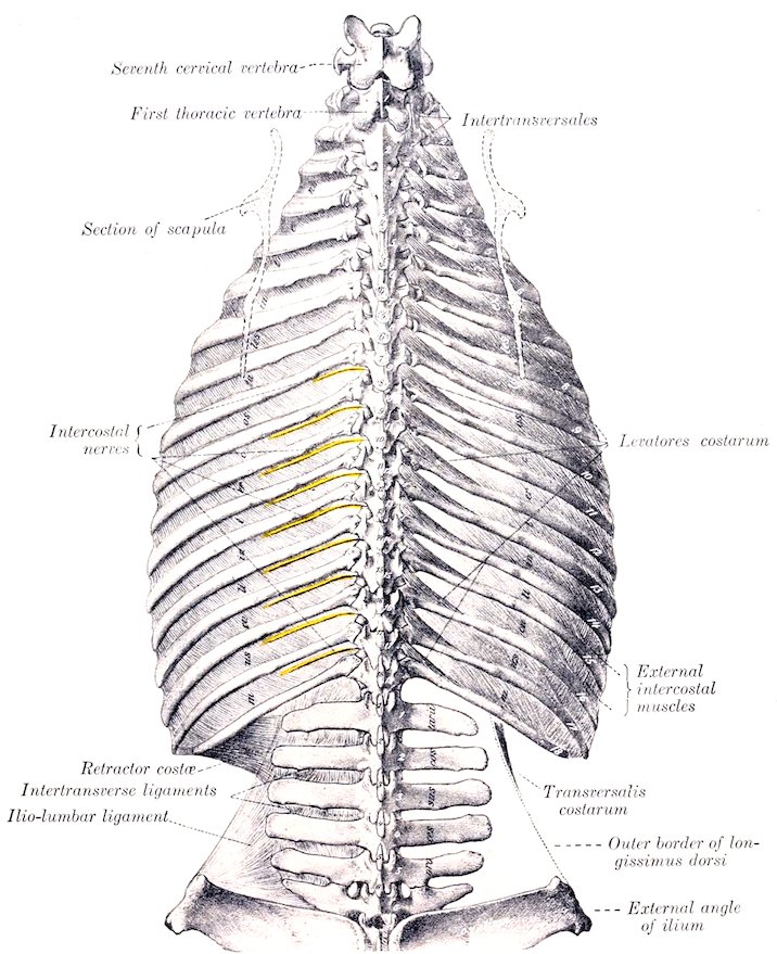
Fig. 184.—Dorsal and Lumbar Regions of Horse, Dorsal View. (After Schmaltz, Atlas d. Anat. d. Pferdes.)
Relations.—Superficially, the serratus magnus, latissimus dorsi, serratus anticus and posticus, longissimus dorsi, transversalis costarum, rectus thoracis, deep pectorals, obliquus abdominis externus, and panniculus; deeply, the internal intercostals and (in the upper part of the spaces) the intercostal vessels and nerves.
Blood-supply.—Intercostal and internal thoracic arteries.
Nerve-supply.—Intercostal nerves.
2423. Internal intercostals (Mm. intercostales interni).—These occupy the entire length of the intercostal spaces, including their interchondral portion.
Origin.—The anterior borders of the ribs and their cartilages.
Insertion.—The posterior borders of the preceding ribs and cartilages.
Action.—To draw the ribs backward in expiration. It seems probable, however, that the intercartilaginous portion is inspiratory.[47]
Structure.—The direction of the fibers is oblique downward and forward. There is a smaller amount of tendinous tissue than in the external set, and the thickness diminishes from below upward. In the upper part of the spaces fibers sometimes cross a rib in a fashion similar to the subcostals of man. A thin aponeurosis separates the internal from the external intercostal muscle in each space.
Relations.—Superficially, the levatores costarum and the external intercostals; deeply, the endothoracic fascia and pleura, the transversus thoracis, diaphragm, transversus abdominis, and the internal thoracic and asternal vessels. In the upper part of the intercostal spaces the intercostal vessels and nerves lie between the internal and external intercostal muscle, but below they lie chiefly on the deep face of the inner muscle.
Blood-supply.—Intercostal and internal thoracic arteries.
Nerve-supply.—Intercostal nerves.
4. Retractor costæ.—This is a small triangular muscle which lies behind the last rib, chiefly under cover of the serratus posticus.
Origin.—The transverse processes of the first three or four lumbar vertebræ by means of the lumbar fascia.
Insertion.—The posterior border of the last rib.
Action.—To retract the last rib.
Structure.—The muscle arises by a thin aponeurosis. Its fibers are parallel to those of the adjacent internal oblique.
Relations.—Superficially, the serratus posticus and external oblique; deeply, the transversus abdominis.
Blood-supply.—Lumbar arteries.
Nerve-supply.—Lumbar nerves.
5. Rectus thoracis (M. transversus costarum; lateralis sterni).—This is a thin muscle which lies under cover of the deep pectoral muscles. It is directed obliquely backward and downward, and crosses the lower part of the first three intercostal spaces.
Origin.—The outer surface of the first rib, below the scalenus.
Insertion.—The cartilage of the third or fourth rib. The aponeurosis usually joins the rectus abdominis. It may reach the sternum.
Action.—It may assist in inspiration or concur with the rectus abdominis.
Relations.—Superficially, the deep pectoral muscles; deeply, the intercostal muscles and the ribs.
6. Transversus thoracis (Triangularis sterni).—This is a flat muscle situated on the thoracic surface of the sternum and the cartilages of the sternal ribs.
Origin.—The sternal ligament.
Insertion.—The cartilages of the ribs, from the second to the eighth inclusive.
Action.—It draws the costal cartilages inward and backward, thus assisting in expiration.
Structure.—Each muscle has the form of a scalene triangle, of which the base is the strongly serrated external border. The muscle contains a good deal of tendinous tissue. The anterior bundles are directed forward and outward; the posterior backward and outward.
243Relations.—Superiorly, the endothoracic fascia and pleura; inferiorly, the costal cartilages, the internal intercostal muscles, and the internal thoracic vessels.
Blood-supply.—Internal thoracic artery.
Nerve-supply.—The intercostal nerves.
7. Diaphragm.—This is a broad, unpaired muscle which forms a partition between the thoracic and abdominal cavities.[48] In outline it has some resemblance to a palm-leaf fan. In form it is dome-shaped, compressed laterally. On a median section it is seen to have a general direction downward and forward from the lumbar vertebræ to the xiphoid cartilage. The thoracic surface is strongly convex, and is covered by the pleura. The abdominal surface is deeply concave, and is covered for the most part by the peritoneum. The muscle consists of a peripheral fleshy portion, two muscular crura, and a tendinous center.
Attachments.—(1) Costal part: The cartilages of the ninth to the fifteenth ribs, and the last three ribs at an increasing distance from their sternal ends.
(2) Sternal part: The upper surface of the xiphoid cartilage.
(3) Lumbar part: (a) The right crus is attached to the inferior common ligament, and by this means to the first four or five lumbar vertebræ. (b) The left crus is attached in a similar fashion to the first and second lumbar vertebræ.
Action.—It is the principal muscle of inspiration and increases the longitudinal diameter of the chest. The contraction produces a general lessening of the curvature of the diaphragm. In the expiratory phase the costal part and crura lie directly on the body walls, so that the bases of the lungs are in contact with the tendinous center and sternal portion only. In ordinary inspiration the fleshy rim recedes from the chest-wall, so that the bases of the lungs move backward to a line about parallel with the curve formed by the cartilages of the asternal ribs, and about four or five inches (ca. 10 to 12 cm.) therefrom. It is stated that the inspiratory movement affects the tendinous center much less than the fleshy part, since the posterior vena cava is firmly attached to the former. It should be noted, however, that the direction of the thoracic part of the vena cava in the expiratory phase is oblique upward and backward. Thus it would seem that there is no anatomical reason why the diaphragm should not move as a whole in ordinary inspiration at least.
Structure.—The costal part (Pars costalis) consists of a series of digitations which meet, or are separated by a very narrow interval from, the transversus abdominis; between the two are the asternal vessels. From the tenth rib backward the attachments are to the ribs at an increasing distance above the costo-chondral junctions. Thus at the last rib the attachment is four to five inches (10 to 12 cm.) from the lower end. Anteriorly, the origin extends along the ninth costal cartilage to the xiphoid cartilage. From these points of origin the fibers curve inward and forward to join the tendinous center. The right crus (Crus dextrum) is about twice as thick as the left one and is also longer. It arises by a strong tendon from the lumbar vertebræ (by means of the inferior common ligament). The tendon is succeeded by a rounded belly which leaves the vertebral column, at the last thoracic vertebra. Passing downward and forward, its fibers spread out and join the tendinous center. The left crus (Crus sinistrum) arises by a thin tendon from the inferior common ligament at the first and second lumbar vertebræ. This is succeeded by a triangular belly which joins the central tendon. Between the crura and the attachment to the last rib the edge of the muscle crosses the ventral surface of the psoas muscles without attachment, forming the so-called lumbo-costal arch; here the thoracic and abdominal cavities are separated only by the serous membranes and some areolar tissue. The tendinous center (Centrum tendineum) resembles the periphery in outline, but is more elongated. It is partially 244divided into right and left halves by the descent of the crura into it. It is composed largely of radiating fibers, but many interlace in various directions; this is specially evident around the foramen venæ cavæ, which is encircled by fibers. A strong tendinous layer extends across below the hiatus œsophageus.
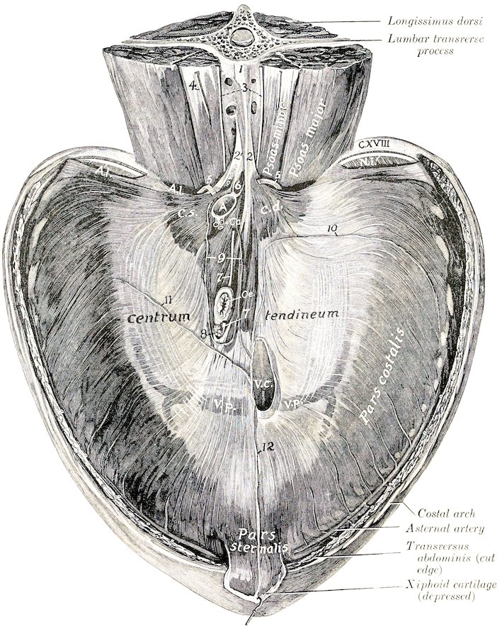
Fig. 185.—Diaphragm of Horse, Abdominal Surface. 1, Inferior common ligament; 2, 2′, tendons of crura; 3, lumbar sympathetic trunks; 4, external spermatic nerve; 5, 5′, great splanchnic nerves; 6, cisterna chyli (opened); 7, 7′, œsophageal continuations of vagus nerves; 8, lymph gland; 9, coronary ligament of liver (cut); 10, right lateral ligament of liver (cut); 11, left lateral ligament of liver (cut); 12, falciform ligament of liver (cut); A.l., lumbo-costal arch; N.i., intercostal nerve; C.d., right crus; C.s., left crus; A, aorta; Ca, cœliac artery; Oe., œsophagus; V.c., posterior vena cava; V.p., phrenic veins. (After Schmaltz, Atlas d. Anat. d. Pferdes.)
The diaphragm is pierced by three foramina. (1) The hiatus aorticus is an interval between the two crura and below the last thoracic vertebra. It contains the posterior aorta, vena azygos, and cisterna chyli. (2) The hiatus œsophageus (or foramen sinistrum) perforates the right crus near its junction with the tendinous 245center. It is situated a little to the left of the median plane and two or three inches below the thirteenth thoracic vertebra (in expiration). It transmits the œsophagus, the vagus nerves, and the œsophageal branch of the gastric artery. (3) The foramen venæ cavæ (s. dextrum) pierces the tendinous center about an inch to the right of the median plane, and about six inches below the twelfth thoracic vertebra (in expiration). The vena cava is firmly attached to the margin of the opening.[49]
Relations.—The thoracic surface is related to the endothoracic fascia, pleuræ, pericardium, the bases of the lungs, and the ribs in part. The abdominal surface is in great part covered by the peritoneum, and is related chiefly to the liver, stomach, spleen, pancreas, kidneys and adrenals, and the anterior flexures of the colon. The sympathetic and splanchnic nerves pass between the crus and the psoas muscles on each side. The asternal vessels perforate the edge of the muscle at the ninth costo-chondral joint.
Blood-supply.—Phrenic and asternal arteries.
Nerve-supply.—Phrenic nerves (from the fifth, sixth, and seventh cervical nerves).
The Abdominal Muscles
The superficial fascia covering the lateral and ventral walls of the abdomen is continuous dorsally with the lumbo-dorsal fascia, in front with the thoracic fascia, and behind with the gluteal fascia. In the inguinal region it forms part of the fascia of the penis or of the mammary glands. At the lower part of the flank it forms a fold which is continuous with the fascia of the thigh near the stifle joint. In this fold are the precrural lymph glands. Medially it blends with the linea alba. It contains the abdominal portion of the panniculus carnosus.
The abdominal panniculus (M. cutaneus maximus) covers a large part of the lateral surface of the abdomen and thorax. The general direction of its fibers is longitudinal. Its posterior extremity forms the basis of the fold of the flank. Its anterior extremity is inserted by a thin tendon into the internal tuberosity of the humerus, with the posterior deep pectoral muscle. Its dorsal edge may be indicated by a line drawn from the upper end of the thirteenth rib to the fold of the flank. Its ventral limit corresponds to a line drawn from the fold to a point about a handbreadth external to the umbilicus, and from here to a point a little above the level of the elbow. Behind the shoulder the fibers become oblique and blend with the scapular portion. The aponeurosis extends ventrally to the linea alba, dorsally to the supraspinous ligament. The muscle is intimately adherent to the skin, so that special care is necessary in removing the latter. Its deep face, on the other hand, is loosely attached to the underlying structures by a quantity of areolar tissue which is more or less loaded with fat (panniculus adiposus) in animals in good condition. The large external thoracic (“spur”) vein is partially embedded in the lower part of the muscle. Its action is to twitch the skin.
The deep fascia is represented chiefly by the abdominal tunic (Tunica flava abdominis). This is a sheet of elastic tissue which assists the muscles in supporting the great weight of the abdominal viscera. It is practically coextensive with the obliquus externus, which it covers. Ventrally it is thick, and is intimately adherent to the aponeurosis of the muscle. Laterally it becomes thinner and is more easily separated, although fibers from it dip in between the muscle-bundles. It is continued for some distance upon the intercostals and serratus magnus. 246Traced forward, it passes as a thin layer beneath the posterior deep pectoral muscle. Posteriorly it is attached to the external angle of the ilium. In the inguinal region it forms the deep fascia of the prepuce or of the mammary glands.
The linea alba is a median fibrous raphé which extends from the xiphoid cartilage to the symphysis pubis. It is formed chiefly by the junction of the aponeuroses of the oblique and transverse muscles, but partly by longitudinal fibers. A little behind its middle is a cicatrix which indicates the position of the umbilical opening of the fœtus.
1. Obliquus abdominis externus (great oblique; external oblique of the abdomen).—This is the most extensive of the abdominal muscles. It is a broad sheet, irregularly triangular in shape, widest behind. Its fibers are directed chiefly downward and backward.
Origin.—(1) The outer surfaces of the last fourteen ribs, and the fascia over the external intercostal muscles; (2) the lumbo-dorsal fascia.
Insertion.—(1) The linea alba and the prepubic tendon; (2) the external angle and shaft of the ilium; (3) the internal femoral fascia.
Action.—(1) To compress the abdominal viscera, as in defecation, micturition, parturition, and expiration; (2) to flex the trunk (arch the back); (3) acting singly, to flex the trunk laterally.
Structure.—The muscle is composed of a fleshy portion and an aponeurosis. The muscular portion lies on the lateral wall of the thorax and abdomen. It arises by a series of digitations, the anterior four of which alternate with those of the serratus magnus. The origin may be indicated by a slightly curved line (concave above) drawn from the lower part of the fifth rib to the external angle of the ilium. The fibers are directed downward and backward and terminate on the aponeurosis, except in the flank, where they are almost horizontal in direction. The line of junction is a curve (concave above) extending from the upper edge of the posterior deep pectoral muscle toward the external angle of the ilium. The aponeurosis is intimately attached to the abdominal tunic, and its fibers are largely interwoven ventrally with those of the aponeurosis of the internal oblique. By this fusion is formed the outer sheath of the rectus abdominis, which blends at the linea alba with that of the opposite side. In the inguinal region the aponeurosis divides into two chief layers; one of these curves upward and backward and is inserted into the external angle of the ilium and the prepubic tendon. Between these points the aponeurosis is much strengthened and is called the inguinal (Poupart’s) ligament (Ligamentum inguinale). This curves upward and somewhat forward, becomes thin, and blends with the iliac fascia. It forms the posterior wall of the inguinal canal. About an inch (ca. 2 to 3 cm.) in front of the pubis and about two inches (ca. 4 to 5 cm.) from the median plane the aponeurosis is pierced by a slit-like opening,[50] the external inguinal ring (Annulus inguinalis subcutaneus). This is the external orifice of the inguinal canal. Its long axis is directed outward and forward, and is about four inches (ca. 10 cm.) in length. The inner angle is rounded and is well defined by the junction of the inguinal ligament with the prepubic tendon, but the outer angle is not so sharply defined. The borders or pillars are constituted by arciform fibers of the aponeurosis of the external oblique (Crus mediale, laterale). The femoral layer of the aponeurosis (Lamina femoralis) passes on to the inner surface of the thigh, where it blends with the femoral fascia. A thin iliac layer (Lamina iliaca) passes over the outer margin of the iliacus to the external border of the ilium.
Relations.—Superficially, the skin, the panniculus carnosus, the abdominal tunic, and the posterior deep pectoral muscle; deeply, the ribs and their cartilages, the intercostal muscles, the internal oblique, the contents of the inguinal canal, and the sartorius and gracilis.
247Blood-supply.—Intercostal and lumbar arteries.
Nerve-supply.—Intercostal and lumbar nerves.
2. Obliquus abdominis internus (small oblique; internal oblique of the abdomen).—This muscle is situated under the preceding one. Its fibers are directed downward, forward, and inward. It forms a triangular curved sheet with the base behind.
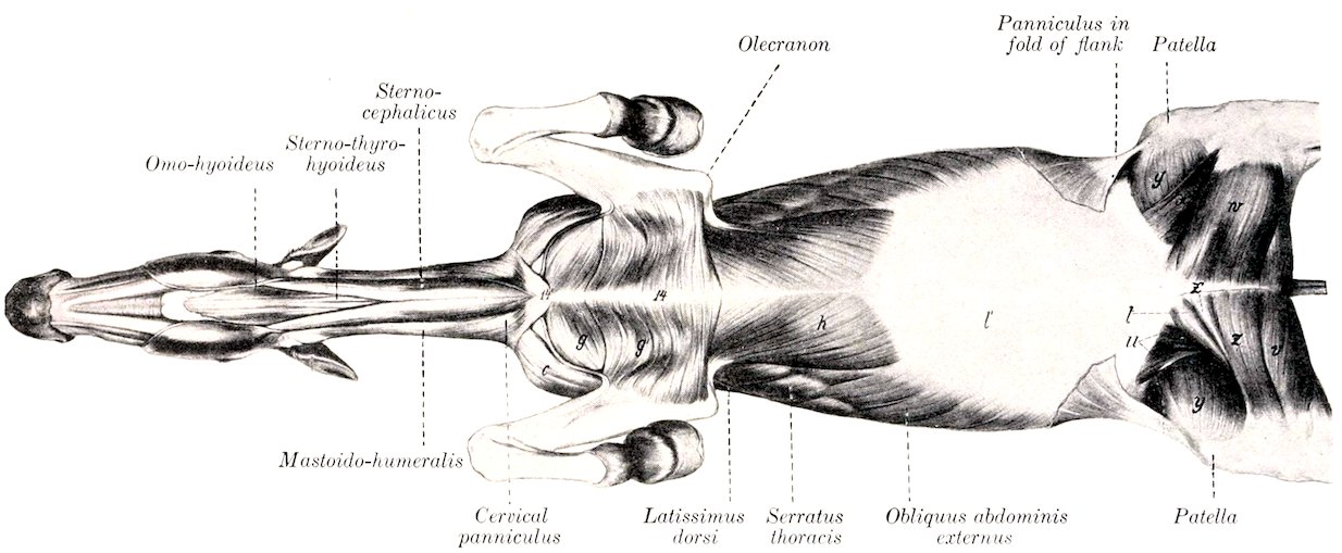
Fig. 186.—Ventral Muscles of Horse, After Removal of Greater Part of Panniculus Carnosus and Abdominal Tunic. Sartorius and Gracilis Removed from Right Thigh. c, Mastoido-humeralis; g, anterior superficial pectoral; g′, posterior superficial pectoral; h, posterior deep pectoral; l′, aponeurosis of obliquus abdominis externus; t, pectineus; u, ilio-psoas; v, semimembranosus; w, gracilis; x, sartorius; y, vastus internus; z, z′, adductor; 14, sternum; 14′, cariniform cartilage. (After Ellenberger-Baum, Anat. f. Künstler.)
248Origin.—The external angle of the ilium and the adjacent part of the inguinal (Poupart’s) ligament.
Insertion.—(1) The cartilages of the last four or five ribs; (2) the linea alba and the prepubic tendon.
Action.—Similar to that of the preceding muscle.
Structure.—Like the external oblique, it is composed of a fleshy portion and an aponeurosis. The fleshy portion is fan-shaped, and is situated chiefly in the flank. At its iliac origin it is covered by a glistening aponeurosis. Traced inward and downward along the abdominal surface of the inguinal ligament, the muscular origin is found to become much thinner, and also becomes loosely attached to the ligament. About four or five inches (ca. 10 to 12 cm.) from the linea alba the muscle separates from the ligament and forms the anterior wall of the inguinal canal. The abdominal orifice of the canal, the internal inguinal ring[51] (Annulus inguinalis abdominalis), is found here. It is normally a narrow slit, bounded in front by the edge of the internal oblique, and behind by the inguinal ligament. The aponeurosis is to a great extent blended with that of the external oblique, being, indeed, considerably interwoven with it ventrally. Where it covers the rectus abdominis it is attached to the tendinous inscriptions of that muscle.
Relations.—Superficially, the external oblique; deeply, the rectus abdominis, transversus abdominis, and the peritoneum.
Blood-supply.—Circumflex iliac, lumbar, and intercostal arteries.
Nerve-supply.—Ventral branches of the lumbar nerves.
3. Rectus Abdominis.—This muscle is confined to the ventral part of the abdominal wall; it extends from the lower part of the chest-wall to the pubis.
Origin.—The cartilages of the fifth to the ninth ribs inclusive, and the adjacent surface of the sternum.
Insertion.—The pubis, by means of the prepubic tendon.
Action.—Similar to that of the oblique muscles. It is specially adapted to flex the lumbo-sacral joints and the lumbar and thoracic parts of the spine.
Structure.—The fibers of the muscle are directed longitudinally. Nine to eleven transverse bands of fibrous tissue extend in an irregular manner across the muscle. These are termed inscriptiones tendineæ. They strengthen the muscle and serve to prevent separation of its fibers. The width of the muscle is greatest about its middle.
Relations.—Superficially, the aponeuroses of the oblique muscles (which constitute the external rectus sheath), and the posterior deep pectoral; deeply, the transversus, intercostals, the cartilages of the ribs, and the sternum. The posterior abdominal artery runs along the outer edge of the muscle posteriorly, and the anterior abdominal artery on or in its anterior part.
Blood-supply.—Anterior and posterior abdominal arteries.
Nerve-supply.—Intercostal and lumbar nerves.
4. Transversus abdominis.—This muscle, named from the general direction of its fibers, is a triangular curved sheet. Its lateral part is muscular, its ventral aponeurotic.
Origin.—(1) The inner surfaces of the distal ends or the cartilages of the asternal ribs, meeting the costal attachment of the diaphragm; (2) the transverse processes of the lumbar vertebræ, by means of the deep layer of the lumbo-dorsal fascia.
Insertion.—The xiphoid cartilage and the linea alba.
249Action.—Similar to that of the oblique muscles.
Structure.—The muscular part is a sheet of parallel bundles of fibers, directed downward and inward. It is thickest over the cartilages of the ribs, and from here it thins out greatly toward the aponeurosis and the lumbar region. The fibers of the aponeurosis directly continue those of the fleshy part. Posteriorly it becomes extremely thin and blends with the aponeuroses of the oblique muscles. It covers the deep face of the rectus, so forming the internal rectus sheath.
Relations.—Superficially, the oblique and straight muscles, the retractor costæ, the cartilages of the asternal ribs, and the internal intercostal muscles; deeply, the transversalis fascia and the peritoneum. The transversalis fascia is little developed in the horse, and is very thin in emaciated subjects, but in animals in good condition it contains a good deal of fat. It blends with the iliac fascia and descends into the inguinal canal. The asternal artery runs along the interval between the origin of the transversus and the costal part of the diaphragm. The intercostal nerves pass down over the external surface of the muscle, to which they give branches. Branches of the first three lumbar nerves are similarly disposed further back.
Blood-supply.—Intercostal, lumbar, and asternal arteries.
Nerve-supply.—Intercostal and lumbar nerves.
5. Cremaster externus.—This small muscle may be regarded as a detached portion of the internal oblique, with which it blends at its origin (Figs. 272, 450).
Origin.—The iliac fascia, near the origin of the sartorius.
Insertion.—The tunica vaginalis communis.
Action.—To raise the tunica vaginalis, and with it the testicle.
Structure.—The muscle arises by a thin aponeurosis which is succeeded by a flat muscular belly about one and a half to two inches (ca. 4 to 5 cm.) in width.[52] It passes down the inguinal canal on the postero-external surface of the tunica vaginalis, to which it is rather loosely attached. On reaching the point where the tunic is reflected on to the tail of the epididymis, the muscle is firmly attached to the outer surface of the tunic by short tendinous fibers.
Relations.—The muscle lies between the peritoneum and the fascia transversalis in front and the iliac fascia and inguinal ligament behind. On reaching the internal ring it descends the inguinal canal on the postero-external surface of the tunica vaginalis communis.
Blood-supply.—External spermatic or cremasteric artery.
Nerve-supply.—External spermatic nerve.
The Inguinal Canal.—This term (Canalis inguinalis) is applied to an oblique passage through the posterior part of the abdominal wall.[53] It begins at the internal inguinal or abdominal ring, and extends obliquely downward, inward, and somewhat forward, to end at the external inguinal or subcutaneous ring. Its anterior wall is formed by the fleshy posterior part of the internal oblique muscle, and the posterior wall by the strong tendinous inguinal (Poupart’s) ligament. The average length of the canal, measured along the spermatic cord, is about four inches (ca. 10 cm.). The internal inguinal ring (Annulus inguinalis abdominalis) is bounded in front by the thin margin of the internal oblique muscle, and behind by the inguinal ligament. It is directed approximately from the edge of the prepubic tendon toward the external angle of the ilium. Its length is about four or five inches (ca. 10 to 12 cm.). The edge of the muscle is attached to the 250surface of the ligament here by delicate connective tissue, except where structures intervene between the walls of the canal. Consequently the limits of the ring are not very clearly defined. The external inguinal ring (Annulus inguinalis subcutaneus) is a well defined slit in the aponeurosis of the external oblique muscle, situated lateral to the prepubic tendon. Its long axis is directed from the edge of the prepubic tendon outward and forward, and its average length is about four inches (ca. 10 cm.). The canal contains in the male the spermatic cord, the tunica vaginalis, the external cremaster muscle, the external pudic artery and a small satellite vein, and the inguinal lymph vessels and nerves. In the female it contains the mammary vessels and nerves; in the bitch it also lodges the round ligament of the uterus, inclosed in a tubular process of peritoneum.
The two rings do not correspond in direction, so that the length of the canal varies greatly when measured at different points. The inner angle of the internal ring lies almost immediately above that of the external ring, but the outer angle is situated five to six inches (ca. 12 to 15 cm.) from that of the external ring. The inner angles of the external rings are well defined and distinctly palpable in the living subject; they are about three to four inches (ca. 8 to 10 cm.) apart.