THE MALE GENITAL ORGANS (continued 2)
Reading edition
Vessels and Nerves.—The arteries are derived from the external pudic artery, which enters the gland at the posterior part of its base. The veins form a 517plexus on either side of the base of the gland, which is drained by the external pudic vein chiefly. The lymphatics are numerous and pass chiefly to the superficial inguinal (or supramammary) lymph glands. The nerves are derived from the inguinal nerves and the posterior mesenteric plexus of the sympathetic system.
GENITAL ORGANS OF THE COW
The ovaries of the cow are much smaller than those of the mare, measuring usually about one to one and a half inches (ca. 3 to 4 cm.) in length, and a little less than an inch (ca. 2 cm.) in thickness in their largest part. They are oval in form, pointed at the uterine end, and have no ovulation fossa. They are situated usually a little above the middle of the lateral margin of the pelvic inlet in the non-pregnant subject, but may be found a little further forward. They are thus about 16 to 18 inches (ca. 40 to 45 cm.) from the vulva in a cow of medium size. The greater part of the surface of the gland is covered with germinal epithelium, the peritoneal epithelium being limited to a narrow zone along the attached border. Follicles of various sizes are often seen projecting from the surface, as well as corpora lutea; a corpus luteum verum has a pronounced yellow color, and may reach a width of half an inch or more (ca. 1 to 1.5 cm.).
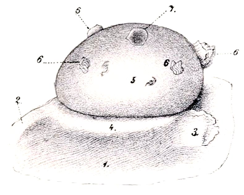
Fig. 411.—Ovary of Cow. 1, Broad ligament of uterus; 2, ligament of ovary; 3, fimbria ovarica; 4, attachment of broad ligament; 5, surface of ovary covered with germinal epithelium; 6, corpora lutea; 7, Graafian follicle. (From Leisering’s Atlas.)
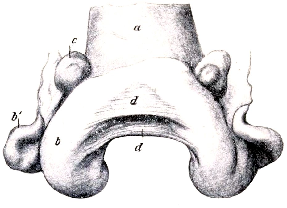
Fig. 412.—Uterus of Cow, Contracted, Dorsal View. a, Body of uterus; b, b′, horn of uterus; c, ovary; d, d′, triangular folds connecting horns of uterus (Lig. intercornualia). (After Zieger.)
The Fallopian or uterine tubes are long and less flexuous than in the mare. The junction with the cornu of the uterus is not so abrupt as in the mare, since the extremities of the horns are pointed. The uterine orifice of the tube is rather large and funnel-shaped. The fimbriæ are not so extensive as in the mare.
The uterus lies almost entirely within the abdominal cavity in the adult. The body is only about one inch (ca. 2 to 3 cm.) in length, although externally it appears to be much longer. This false impression is due to the fact that the posterior parts of the cornua are united by connective and muscular tissue and have a common peritoneal covering. The cornua are, therefore, really longer than they appear externally. They taper gradually toward the free end, so that the junction with the Fallopian tubes is not abrupt, as in the mare. The free part of the horn curves at first downward, forward, and outward, and then turns upward, forming a spiral coil. The cervix is about four or five inches (ca. 8 to 10 cm.) long; 518its wall is remarkably dense, and may be nearly an inch (ca. 2 cm.) in thickness. Its lumen, the cervical canal, is spiral, and is ordinarily tightly closed and very difficult to dilate. The muscular coat of the uterus is thicker than in the mare. It consists of an external longitudinal layer and two circular strata. The inner circular layer is about a fourth of an inch (ca. 6 mm.) thick in the cervix. The other layers are continued in the vagina. The mucous membrane of the horns and body presents as a characteristic feature the uterine cotyledons (Cotyledones uterinæ). These are oval prominences, about a hundred in number, which are either irregularly scattered over the surface or arranged in rows of about a dozen.
In the non-gravid uterus they average about ½ to ¾ inch (ca. 15 to 18 mm.) in length, ⅓ inch (ca. 8 mm.) in width, and ⅛ inch (ca. 2 to 4 mm.) in thickness. During pregnancy they become greatly enlarged and pedunculated. The larger ones then measure about 4 to 5 inches (10 to 12 cm.) in length, 1 to 1½ inches (3 to 4 cm.) in width, and 1 inch (2 to 2.5 cm.) in thickness. The deep face has a hilus at which the vessels enter. The rest of the surface has a spongy appearance, due to numerous crypts which receive the villi of the chorion.
The uterine glands are long and branched. The mucous membrane of the cervix is pale and forms numerous folds. The latter are arranged in several series which obliterate the lumen. At the external orifice (os uteri) the folds (Plicæ palmatæ) form rounded prominences arranged circularly, which project into the cavity of the vagina. There are no glands in the cervix, but a thick mucus is secreted by goblet cells.
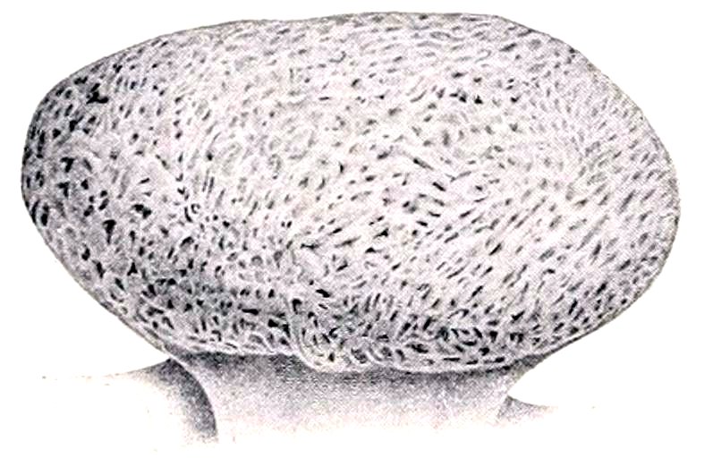
Fig. 413.—Uterine Cotyledon of Cow. The figure represents a cotyledon of medium size. (After Ellenberger, in Leisering’s Atlas.)
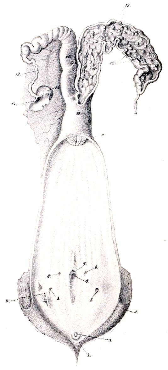
Fig. 414.—Genital Organs of Cow, Dorsal View. The right uterine cornu and the vagina and vulva are opened up: 1, Labium vulvæ; 2, ventral commissure; 3, glans clitoridis; 4, glandula vestibularis major, exposed by slit in mucous membrane; 5, opening of duct of preceding; 6, suburethral diverticulum; 7, external urethral opening; 8, openings of canals of Gartner; 9, os uteri; 10, corpus uteri; 11, cornu uteri; 12, cotyledons; 13, Fallopian tube; 14, ostium abdominale tubæ; 15, ovary (From Leisering’s Atlas, reduced.)
The broad ligaments are not attached in the sublumbar region as in the mare, but to the upper part of the flanks, about a handbreadth below the level of the external angle of the ilium. They contain a conspicuous amount of unstriped muscle. The round ligaments are well developed, and can be traced distinctly to the vicinity of the internal inguinal ring.
The vagina is somewhat longer and more roomy than that of the mare; its 519wall is also thicker. Its average length in the non-pregnant animal is about eight inches (ca. 20 cm.); but in the pregnant cow the length increases to twelve inches (30 cm.) or more. The recto-genital pouch of peritoneum extends backward about five inches (ca. 12 cm.) on the dorsal surface, while ventrally the serous coat only extends backward about two inches (ca. 5 cm.).
This gives ample space dorsally for the incision of the fornix vaginæ in oöphorectomy.
In the ventral wall of the vagina, between the muscular and mucous coats, there are frequently present the two canals of Gartner (Ductus epoöphori longitudinales). When well developed they may attain the diameter of a goose quill, and may be traced forward to the anterior part of the vagina or even further. They open posteriorly near the external urethral orifice.
These tubes are remnants of the Wolffian ducts, and, like other fœtal vestiges, are very variable. Röder states that the right canal was absent in over 52 per cent., the left in only 22 per cent., of the cows examined by him. In some cases they may be traced in the broad ligaments for a variable distance toward the ovary. They are of clinical interest in that cysts frequently form along their course.
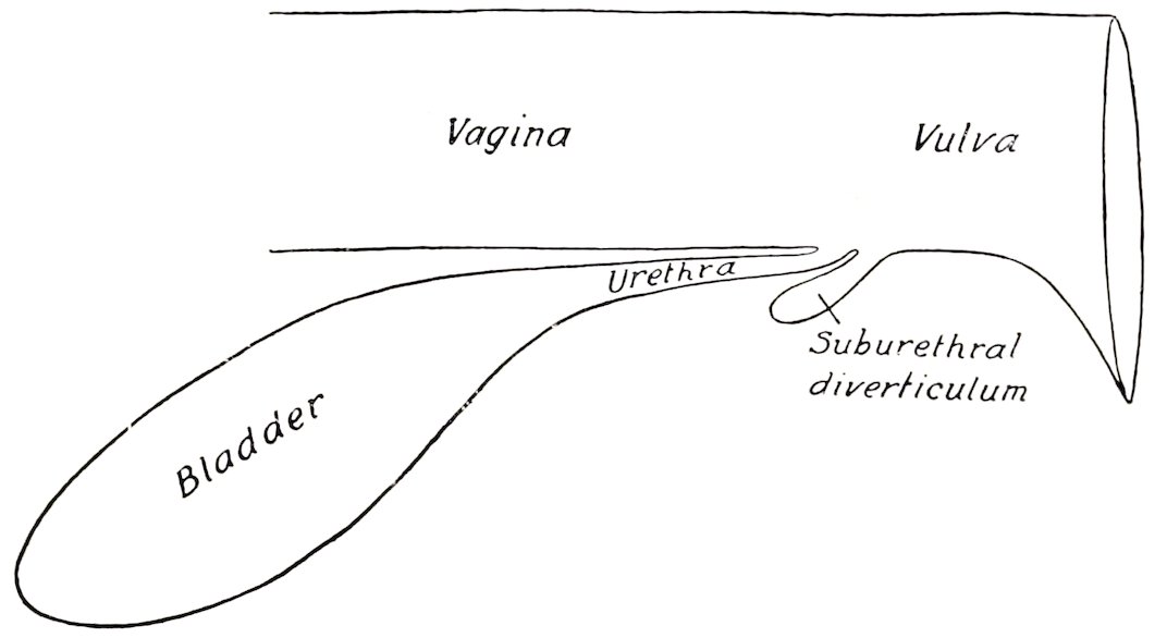
Fig. 415.—Diagrammatic Sagittal Section of Part of Urogenital Tract of Cow, Showing Suburethral Diverticulum.
The vulva has thick wrinkled labia, and both commissures are acute; the lower one is pointed, and has on it a number of long hairs; it lies about two inches (5 cm.) behind, and about the same distance below, the level of the ischial arch. The glans clitoridis is small; the crura are about four inches (ca. 10 cm.) long and are flexuous. The external urethral orifice or meatus urinarius is four or five inches (10 to 12 cm.) from the ventral commissure. Beneath it is a blind pouch, the suburethral diverticulum, which is more than an inch (ca. 3 to 4 cm.) long, and readily admits the end of a finger.
The form and position of this pouch should be carefully noted on account of the difficulty it causes in catheterizing the bladder. If the catheter is passed along the ventral wall of the vulva (as in the mare), it will always enter the pouch instead of the urethra.
The glandulæ vestibulares majores (or glands of Bartholin) are situated on the lateral walls of the vulva, under the constrictor vulvæ. They are little over an inch (ca. 3 cm.) long and about half an inch (ca. 1.5 cm.) in width. Each has a single duct which opens about an inch and a half (ca. 3 to 4 cm.) lateral to and behind the external urethral orifice.
The urethra of the cow is about four inches (10 cm.) in length; it is narrower and much less dilatable than that of the mare. It is fused dorsally with the wall of the vagina, while laterally and ventrally it is covered by the constrictor vaginæ muscle.
The mammary glands, normally two in number, are popularly termed the udder. They are very much larger than in the mare, and the body of each is 520somewhat ellipsoidal in form, but flattened transversely. The base (or dorsal surface) of each gland is slightly concave and slopes obliquely downward and forward in adaptation to the abdominal wall, to which it is adherent; posteriorly it is in relation to the large supramammary lymph glands and a quantity of fat. The inner surface is flat, and is separated from the other gland by a well-developed double septum. The outer surface is convex. Four well-developed teats are present; they average about three inches (ca. 7 to 8 cm.) in length. It is customary to consider the udder to consist of four “quarters.” There is no septum nor visible division between the two quarters of the same side, but, on the other hand, injections of fluids of different colors into the two teats of the gland demonstrate that the cavities drained by them do not communicate.
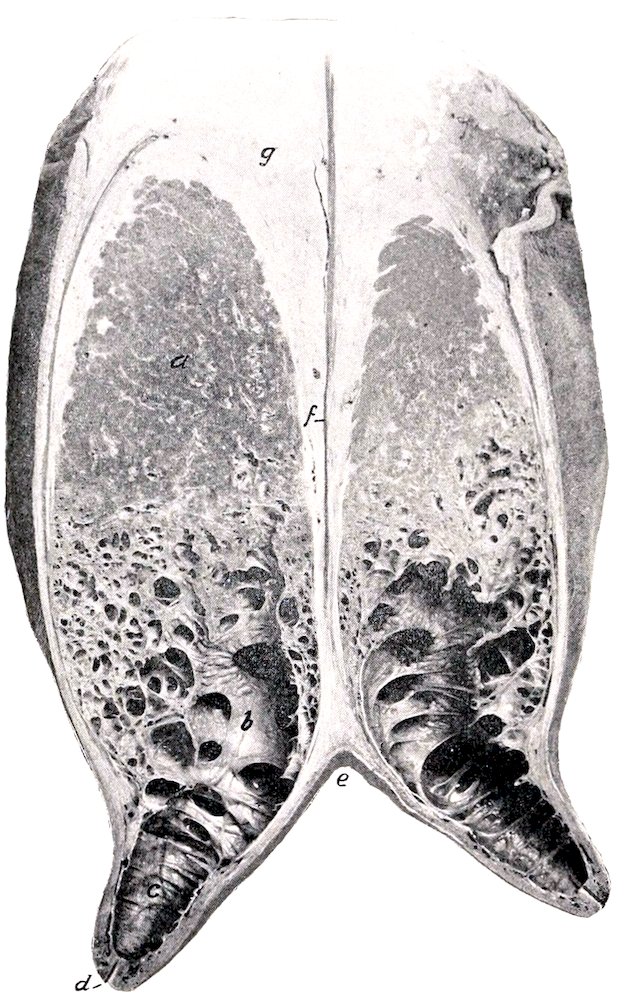
Fig. 416.—Cross-section of Mammary Glands of Cow. a, Body of gland; b, lactiferous sinus; c, cavity of teat; d, duct of teat; e, intermammary groove; f, septum between glands; g, supramammary fat.
The size and form of the glands vary greatly; in the highly specialized dairy breeds (e. g., Holsteins) they often attain enormous dimensions. The size of the udder is not a certain index of its productivity, since some so-called “fleshy” udders contain a very large amount of interstitial tissue and relatively little parenchyma. Small accessory teats often occur posteriorly; commonly these are rudimentary and imperforate (polythelia), but sometimes they drain a small amount of gland substance (polymastia). Sanson described a case in which there were seven teats, all giving milk.
521Each teat has a single duct which widens superiorly and opens freely into a roomy lactiferous sinus or milk cistern (Sinus lactiferus). The lactiferous duct or teat canal is lined by a glandless mucous membrane which is covered with stratified squamous epithelium; the lower part (ca. 1 cm.) of the canal is narrow, and is closed by a sphincter of unstriped muscle. The mucous membrane of the sinus forms numerous folds which render the cavity multilocular.
Vessels and Nerves.—The blood-supply is derived from the external pudic arteries. The veins form a circle at the base of the udder, from which the blood is drained by three trunks, viz., the very large subcutaneous abdominal (“milk vein”), the external pudic, and the perineal vein. The lymphatics are numerous, and pass to the supramammary glands chiefly. The nerves are derived from the inguinal nerves and the posterior mesenteric plexus of the sympathetic.
GENITAL ORGANS OF THE SOW
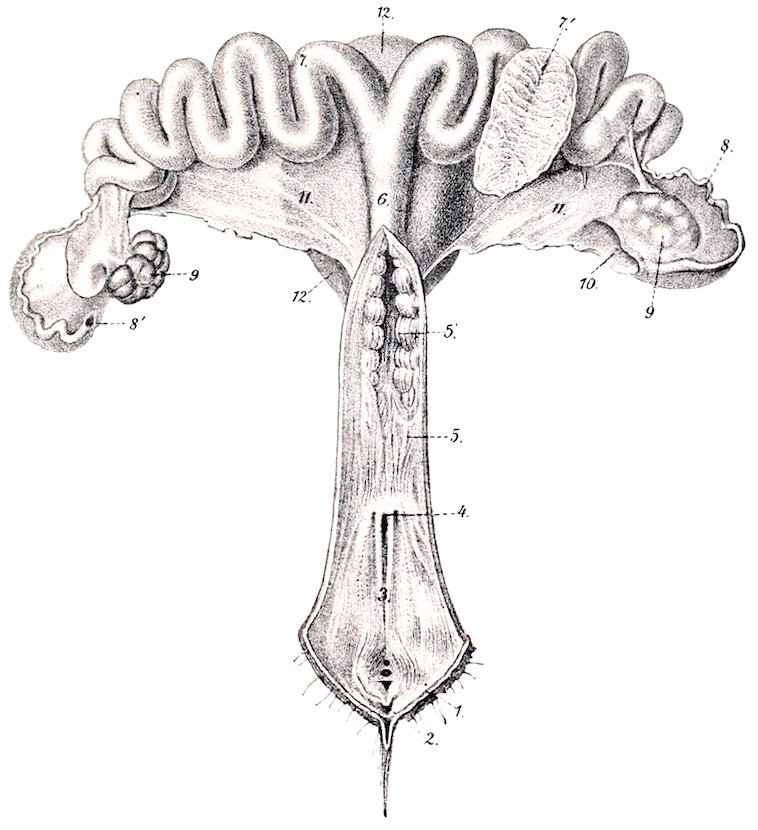
Fig. 417.—Genital Organs of Sow, Dorsal View. The Vagina and Cervix Uteri are Slit Open. 1, Labia vulvæ; 2, glans clitoridis; 3, vulva; 4, external urethral orifice; 5, vagina; 5′, cervix uteri; 6, corpus uteri; 7, cornua uteri, one of which is opened at 7′ to show folds of mucous membrane; 8, Fallopian tube; 8′, abdominal opening of tube; 9, ovaries; 10, ovarian bursa; 11, broad ligaments of uterus; 12, urinary bladder. (From Leisering’s Atlas.)
The ovaries are concealed in the bursa ovarii, owing to the large extent of the mesosalpinx. They are more rounded than in the bitch, and have a distinct hilus. They are situated usually as in the cow, but their position is quite variable in animals which have borne young. The surface commonly presents rounded prominences, 522so that sometimes the gland has an irregular, mulberry-like aspect; this is due to the fact that there is very little stroma and the larger Graafian follicles project from the surface.
The Fallopian or uterine tubes are long (ca. 15 to 30 cm.), and less flexuous than in the mare. The fimbriated extremity forms an ampulla and has a large abdominal opening. The uterine end shades insensibly into the small extremity of the cornu of the uterus.
The uterus presents several striking features. The body is only about two inches (ca. 5 cm.) long. The horns are extremely long and flexuous, and are freely movable, on account of the large extent of the broad ligaments. The extremities of the horns taper to about the diameter of the Fallopian tubes.[154] The neck is remarkable for its length (ca. 15 to 20 cm.) and the fact that it is directly continued by the vagina without forming any intravaginal projection (os uteri). It can be distinguished by the transverse folds of its mucous membrane and the thickness of its wall.
The changes in form and position of the uterus during pregnancy are similar to those mentioned later in the case of the bitch.
The vagina is about four or five inches (ca. 10 to 12 cm.) long in a sow of medium size. It is small in caliber, and has a thick muscular coat which consists mainly of circular fibers. The mucous membrane is plicated, and is intimately united with the muscular coat.
The vulva is relatively long. The inferior commissure is pointed and dependent. The mucous lining is plicated. There is a small suburethral diverticulum. The vestibular glands are small and variable in number. The vestibular bulbs are little developed. The canals of Gartner may sometimes be traced as far as the ovary, but are often difficult to find.
The clitoris is long, flexuous, and pointed; beneath it is a cul-de-sac.
The urethra is very long; its vulvar orifice is not concealed by a valvular fold.
The mammary glands are usually ten or twelve in number, and are arranged in two rows, as in the bitch. Each teat has commonly two excretory ducts.
GENITAL ORGANS OF THE BITCH
The ovaries (Fig. 336) are small, elongated-oval in outline, and flattened. Each ovary is situated close to or in contact with the posterior pole of the corresponding kidney, and thus lies ventral to the third or fourth lumbar vertebræ, or about half-way between the last rib and the crest of the ilium. Each is concealed in a peritoneal pouch, the bursa ovarii, which has a slit-like opening ventrally. The two folds which form this pouch contain a quantity of fat and unstriped muscle. They are continued to the cornu of the uterus, constituting the mesosalpinx and the ovarian ligament. The surface of the ovary presents prominences caused by projecting Graafian follicles. There is no distinct hilus.
The Fallopian or uterine tubes are small and average two or three inches (ca. 5 to 8 cm.) in length. Each passes at first forward and then turns backward, having a straight or only slightly flexuous course. The fimbriated extremity lies in the bursa ovarii, and has a rather large opening. The uterine orifice is very small.
The uterus has a very short body and two long narrow horns. In a bitch of medium size the body is about an inch (ca. 2 to 3 cm.) and the cornua five or six 523inches (ca. 12 to 15 cm.) long. The horns are of uniform diameter, are nearly straight, and lie entirely within the abdomen. They diverge from the body in the form of a V toward each kidney. Their posterior parts are united by the peritoneum. The neck is very short and is fused dorsally with the vagina. It has a thick muscular coat. The mucous membrane of the uterus has long uterine glands and also short tubular crypts.
The broad ligaments contain fat and unstriped muscle. They are much longer in the middle than at either end. The posterior part is attached to the anterior part of the vagina. The round ligaments are very long, extending through the inguinal canals, and traceable almost to the vulva.
The horns of the gravid uterus present dilatations or ampullæ, which contain the fœtuses, and are separated by constrictions. The gravid uterus lies on the ventral abdominal wall, and toward the end of gestation extends forward to the stomach and liver.
The vagina is relatively long. It is narrow anteriorly, and has no distinct fornix. The muscular coat is thick and consists chiefly of circular fibers. The mucous membrane forms longitudinal folds. The canals of Gartner are usually absent.
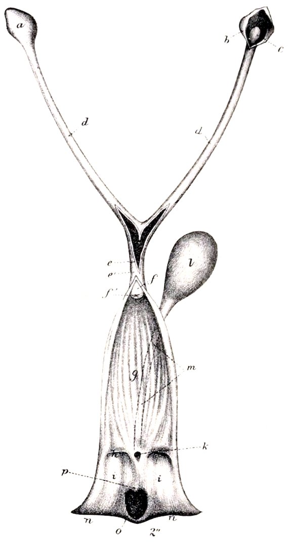
Fig. 418.—Genital Organs of Bitch. Vulva, vagina, and uterus (in part) are slit open. a, Ovarian bursa; b, same opened to show the right ovary, c; d, d, horns of uterus; e, e′, body of uterus; f, neck of uterus; f′, os uteri; g, vagina; h, hymen; i, vulva; k, external urethral orifice; l, urinary bladder; m, urethra; n, n, labia vulvæ; o, fossa clitoridis; p, clitoris. (After Ellenberger, in Leisering’s Atlas.)
The vulva has thick labia which form a pointed inferior commissure. The mucous lining is smooth. On either side of the urethral orifice there is a small depression. The glandulæ vestibulares majores are absent, but the smaller glands are present, and their ducts open ventrally on either side of a median ridge. The vestibular bulbs are relatively large and join dorsally. The clitoris has a small pointed glans, beneath which is a diverticulum. The corpus cavernosum is a little more than an inch (ca. 3 to 4 cm.) long in a subject of medium size, and is infiltrated with fat.
The mammary glands are usually ten in number, and are arranged in two series extending from the posterior part of the pectoral region to the inguinal region; they are, therefore, designated according to location as pectoral, abdominal, and inguinal. The teats are short, and present on their apices six to twelve small orifices of the excretory ducts.
524