ARTHROLOGY
Reading edition
THE ARTICULATIONS OR JOINTS
An articulation or joint is formed by the union of two or more bones or cartilages by other tissue. Bone is the fundamental part of most joints; in some cases a bone and a cartilage, or two cartilages, form a joint. The uniting medium is chiefly fibrous tissue or cartilage, or a mixture of these. Union of parts of the skeleton by muscles (Synsarcosis), as in the attachment of the thoracic limb in the horse, will not be considered in this section.
Joints may be classified—(a) anatomically, according to their mode of development, the nature of the uniting media, and the form of the joint surfaces; (b) physiologically, with regard to the amount and kind of movement or the absence of mobility in them; (c) by a combination of the foregoing considerations.
The classification of joints is still in a very unsatisfactory state, and, unfortunately, the same term is used in various senses by different authors. The two main subdivisions proposed by Hepburn are: (1) Those in which the uniting medium is coextensive with the opposed joint surfaces, and in which a direct union of these surfaces is thereby effected. (2) Those in which the uniting medium has undergone interruption in its structural continuity, and in which a cavity of greater or less extent is thus formed in the interior of the joint. This distinction is of considerable importance clinically.
Three chief subdivisions of joints are usually recognized—viz., synarthroses, diarthroses, and amphiarthroses.
SYNARTHROSES
In this group the segments are united by fibrous tissue or cartilage, or a mixture of the two in such a manner as practically to preclude movement; hence they are often termed fixed or immovable joints. There is no joint cavity. Many of these joints are temporary, the uniting medium being invaded by the process of ossification, with a resulting ankylosis or synostosis. The chief classes in this group of joints are as follows:
(1) Suture.—This term (Sutura) is applied to those joints in the skull in which the adjacent bones are closely united by fibrous tissue—the sutural ligament. In many cases the edges of the bones have irregular interlocking margins, forming the sutura serrata, e. g., the frontal suture. In others the edges are beveled and overlap, forming the sutura squamosa, e. g., the parieto-temporal suture. If the edges are plane or slightly roughened, the term sutura harmonia is applied to the joint, e. g., the nasal suture.
(2) Synchondrosis.—In these the two bones are united by cartilage, e. g., the joint between the basioccipital and the sphenoid bone. Very few of these joints are permanent.
(3) Symphysis.—This term is usually limited to a few median joints which connect symmetrical parts of the skeleton, e. g., symphysis pelvis, symphysis mandibulæ. The uniting media are cartilage and fibrous tissue. In some cases a cleft-like rudimentary joint cavity occurs.
(4) Gomphosis.—This term is applied to the implantation of the teeth in the alveoli.
170
DIARTHROSES
These joints are characterized by the presence of a joint cavity and by their mobility. They are often called movable or true joints. The structures which enter into their formation are:
1. The joint surfaces (Facies articulares), which are usually more or less expanded. They are in most cases smooth, and vary much in form. They are formed of specially dense bone, which differs histologically from ordinary compact substance. In certain cases (vide Osteology) the surface is interrupted by non-articular cavities known as synovial fossæ.
2. The articular cartilages (Cartilagines articulares), usually hyaline in type, form a covering over the articular surfaces of the bones. They vary in thickness in different joints; they are thickest on the areas of the greatest pressure, and usually tend to accentuate the curvature of the bone, i. e., on a concave surface the peripheral part is the thickest, while on a convex surface the central part is the thickest. The articular cartilages are non-vascular, very smooth, and have a bluish tinge in the fresh state. They diminish the effects of concussion and greatly reduce friction.
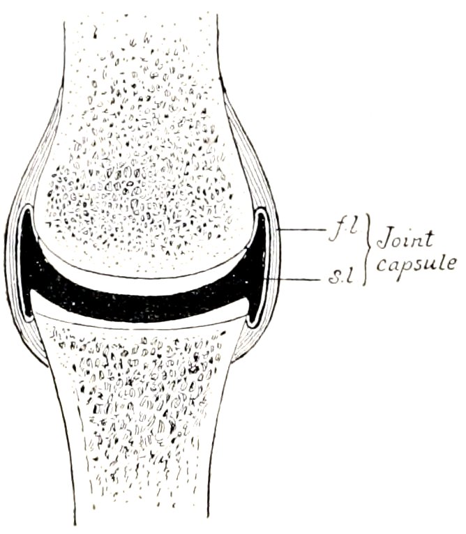
Fig. 138.—Diagram of Section of Diarthrosis. f.l., Fibrous layer, s.l., synovial layer of joint capsule. The articular cartilages are white, bones dotted, and the joint cavity black in the figure.
3. The joint capsule (Capsula articularis) is, in its simplest form, a sac, the margins of which are attached around the articulating surfaces. It consists of two layers—an external one, composed of fibrous tissue (Stratum fibrosum), and an internal one, the synovial layer or membrane (Stratum synoviale). The fibrous layer, sometimes termed the capsular ligament, is attached either close to the margins of the articular surfaces or at a variable distance from them. Its thickness varies greatly in different situations: in certain places it is extremely thick, and sometimes cartilage or bone develops in it; in other places it is practically absent, the capsule then consisting only of the synovial membrane. Parts of the capsule may undergo thickening and so form ligaments, which are not separable, except artificially, from the rest of the capsule. The synovial layer lines the joint cavity except where this is bounded by the articular cartilages; it stops normally at the margin of the latter. It is a thin membrane, and is richly supplied by close networks of vessels and nerves. It frequently forms folds (Plicæ synoviales) and villi (Villi synoviales), which project into the cavity of the joint. The folds commonly contain pads of fat which fill up interstices and vary in form and position in various phases of movement. The synovial membrane secretes a fluid, the synovia, which resembles white-of-egg and lubricates the joint.[28] In many places the membrane forms extra-articular pouches, which facilitate the play of muscles and tendons.
The joint cavity (Cavum articulare) is inclosed by the synovial membrane and the articular cartilages. Normally, it is, strictly speaking, only a potential cavity, which contains nothing but a small amount of synovia.
The student must guard against a false conception of the joint cavity which may result from dissections and diagrams, in which an actual cavity of considerable extent appears to exist. A correct idea of the intimate apposition of the parts is best obtained from the study of frozen sections. It is also instructive to examine joints which have been injected so as to distend the capsule fully. It is then seen that the cavity is often of much greater potential extent than 171one might suppose, and that the capsule is often very irregular in form, i. e., forms a variety of sacculations.
The foregoing are constant and necessary features in all diarthroses. Other structures which enter into the formation of these joints are ligaments, articular discs or menisci, and marginal cartilages.
4. Ligaments.—These are strong bands or membranes, usually composed of white, fibrous tissue, which bind the bones together. They are pliable, but practically inelastic. In a few cases, however, e. g., ligamenta flava, ligamentum nuchæ, they are composed of elastic tissue. They may be subdivided, according to position, into periarticular and intraarticular. Periarticular ligaments are frequently blended with or form part of the fibrous capsule; in other cases they are quite distinct. Strictly speaking, intraarticular ligaments, though within the fibrous capsule, are not in the joint cavity; the synovial membrane is reflected over them. The term seems justifiable, however, on practical grounds. Ligaments which connect directly opposed surfaces of bones are termed interosseous. The special names are based usually on their position, form, and attachments, e. g., lateral or collateral, cruciate, sacro-iliac, etc. In many places muscles, tendons, and thickenings of the fasciæ function as ligaments and increase the security of the joint. Atmospheric pressure and cohesion play a considerable part in keeping the joint surfaces in apposition.
5. Articular discs or menisci are plates of fibro-cartilage or dense fibrous tissue placed between the articular cartilages, and divide the joint cavity partially or completely into two compartments. They render certain surfaces congruent, e. g., femoro-tibial joint, allow greater range or variety of movement, and diminish concussion.
6. A marginal cartilage (Labrum glenoidale) is a ring of fibro-cartilage which encircles the rim of an articular cavity. It enlarges the cavity and tends to prevent fracture of the margin.
Vessels and Nerves.—The arteries form anastomoses around the larger joints, and give off branches to the extremities of the bones and to the joint capsule. The synovial membrane has a close-meshed network of capillaries; the latter form loops around the margins of the articular cartilages, but do not usually enter them. The veins form plexuses. The synovial membrane is also well supplied with lymphatics. Nerve-fibers are especially numerous in and around the synovial membrane and there are special nerve-endings, e. g., Pacinian bodies and the articular end-bulbs described by Krause.
Movements.—The movements of a joint are determined chiefly by the form and extent of the joint surfaces and the arrangement of the ligaments. They are usually classified as follows:
1. Gliding.—This refers to the sliding of one practically plane surface on another, as in the joints between the articular processes of the cervical vertebræ.
2. Angular Movements.—In these cases there is movement around one or more axes. Motion which diminishes the angle included by the segments forming the joint is termed flexion, while that which tends to bring the segments into line with each other is called extension.
With reference to the joints of the lower parts of the limbs, it seems advisable to employ the terms dorsal and volar or plantar flexion, since these joints can be “overextended.” Similarly, the terms dorsal and ventral flexion should be applied to the corresponding movements of the spinal column. The meaning of the term lateral flexion is evident. These movements are all rotations around axes which are approximately either transverse or vertical. Depression, elevation, and lateral movement of the lower jaw fall in this category.
3. Circumduction.—This designates movements in which the distal part of the limb describes a circle or a segment of one. In man such movement is easily 172performed, but in quadrupeds it is possible to a limited degree only, and is to be regarded usually as an indication of disease.
4. Rotation.—As a matter of convenience, this term is reserved to indicate rotation of one segment around the longitudinal axis of the other segment forming the joint. It is seen typically in the atlanto-axial joint.
5. Adduction and abduction designate respectively movement of a limb toward and away from the median plane, or of a digit toward and away from the axis of the limb.
Classification.—This is based on the form of the joint surfaces and the movements which occur. The following chief classes may be recognized:
1. Arthrodia, or gliding joint. In these the surfaces are practically flat, admitting of gliding movement. Examples: carpo-metacarpal joints; joints between the articular processes of the cervical and thoracic vertebræ.
2. Ginglymus, or hinge-joint. In this class the joint surfaces consist usually of two condyles, or of a segment of a cylinder or cone, which are received by corresponding cavities. In typical cases the movements are flexion and extension, i. e., around a single transverse axis. Examples: occipito-atlantal and elbow joints.
3. Trochoid, or pivot joint. In these the movement is limited to rotation of one segment around the longitudinal axis of the other. Example: atlanto-axial joint.
4. Enarthrosis, or ball-and-socket joint. These are formed by a surface of approximately spherical curvature, received into a corresponding cavity. They are multiaxial, and allow of the greatest variety of movement, e. g., flexion, extension, rotation, abduction, adduction, circumduction. Examples: hip and shoulder joints.[29]
AMPHIARTHROSES
These joints, as the name indicates, share some characters with both of the preceding groups. In them the segments are directly united by a plate of fibro-cartilage, and usually by ligaments also. The amount and kind of movement are determined by the shape of the joint surfaces and the amount and pliability of the uniting medium.[30] These joints are all medial in position, and are best illustrated by the joints between the bodies of the vertebræ. There is usually no joint cavity, but in certain situations a rudimentary one exists.
THE ARTICULATIONS OF THE HORSE
Joints and Ligaments of the Vertebræ
The movable vertebræ form two sets of articulations, viz., those formed by the bodies, and those formed by the articular processes of adjacent vertebræ; the former are termed intercentral, and the latter, interneural. Associated with these are ligaments uniting the arches and processes; some of these are special, i. e., confined to a single joint, while others are common, i. e., extend along the entire 173vertebral column or a considerable part of it. The joints between the atlas and axis and between the former and the skull require separate consideration.
INTERCENTRAL ARTICULATIONS
These are amphiarthroses, formed by the junction of the extremities of the bodies of adjacent vertebræ. The articular surfaces in the cervical region consist of a cavity on the posterior end of the body of the anterior vertebra, and a corresponding convexity or head of the succeeding vertebra. In the other regions the surfaces are much flattened. The uniting media are:
1. The intervertebralfibro-cartilages (Fibrocartilagines intervertebrales). Each of these is a disc which fits into the space between the bodies of two adjacent vertebræ, to which it is intimately attached. The discs are thinnest in the middle of the thoracic region, thicker in the cervical and lumbar regions, and thickest in the coccygeal region. Each consists of a dense fibrous peripheral part (Annulus fibrosus), and a soft pulpy central part (Nucleus pulposus).
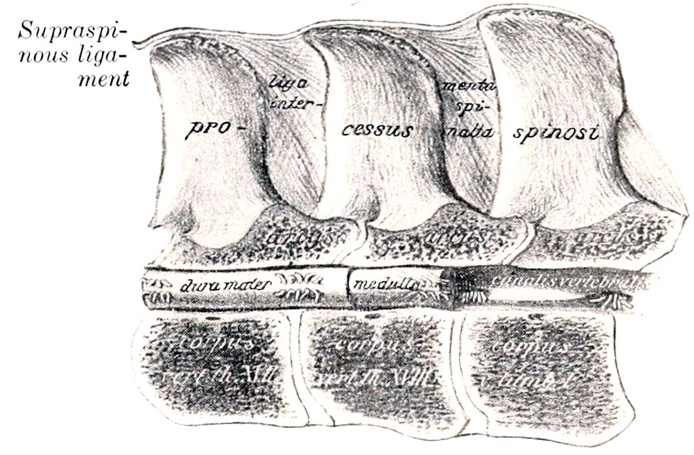
Fig. 139.—Sagittal Section of Last Two Thoracic and First Lumbar Vertebræ, showing Ligaments and Spinal Cord (Medulla). (After Schmaltz, Atlas d. Anat. d. Pferdes.)
The fibrous ring consists of laminæ of fibrous tissue and fibro-cartilage, which pass obliquely between the two vertebræ and alternate in direction, forming an X-shaped arrangement. The central part of the ring is largely cartilaginous, and gradually assumes the character of the pulpy center. The latter is very elastic and is compressed, so that it bulges considerably from the surface of sections; it consists of white and elastic fibers, connective-tissue cells, and peculiar clear, transparent cells of various sizes. It is a remnant of the notochord. There are joint cavities in the cervical intercentral joints, and in those between the last cervical and the first thoracic, and between the last lumbar and the sacrum. In the latter the cavity is coextensive with the extremities of the bodies; in the former, it is usually not so extensive.
2. The inferior common ligament (Ligamentum longitudinale ventrale) lies on the ventral surface of the bodies of the vertebræ and the intervertebral fibro-cartilages, to which it is firmly attached. It begins about the fourteenth or fifteenth thoracic vertebra, and is at first a narrow, thin band. Further back it becomes gradually thicker and wider, and terminates on the pelvic surface of the sacrum by spreading out and blending with the periosteum. It is strongest in the lumbar region, where the tendons of the crura of the diaphragm fuse with it.
3. The superior common ligament (Ligamentum longitudinale dorsale) lies on the floor of the vertebral canal from the dens or odontoid process to the sacrum. It is narrow over the middles of the vertebral bodies, and widens over the intervertebral fibro-cartilages, to which it is very firmly attached.
This ligament is in relation with the spinal veins on either side, and in the middle of each vertebra a transverse anastomotic branch passes under the ligament.
INTERNEURAL ARTICULATIONS
Each typical vertebra presents two pairs of articular processes, which form diarthroses with the two adjacent vertebræ. The articular surfaces are extensive, 174flat, and oval in the cervical region, small and flat in the thoracic region, while in the lumbar region the anterior ones are concave and the posterior convex. The joint capsule is strong and ample in the cervical region, in conformity with the large size and greater mobility of these joints in the neck. In the thoracic and lumbar regions the capsule is small and close. These joints are arthrodia.
Associated with these joints are the ligamenta flava, which connect the arches of adjacent vertebræ. They are membranous and consist largely of elastic tissue.
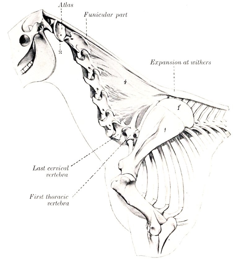
Fig. 140.—Ligamentum Nuchæ of Horse. 1, Scapula; 1′, cartilage of scapula; 4, lamellar part of ligamentum nuchæ; x, wing of atlas. (After Ellenberger-Baum, Anat. für Künstler.)
The supraspinous ligament extends medially from the occipital bone to the sacrum. From the withers backward it consists of a strong cord of fibrous tissue, attached to the summits of the vertebral spines. In the neck and withers it is remarkably modified to form the ligamentum nuchæ, which requires more extended notice.
The ligamentum nuchæ is a powerful elastic apparatus, the principal function of which is to assist the extensor muscles of the head and neck. It extends from the occipital bone to the withers, where it is directly continuous with the supraspinous ligament. It consists of two parts—funicular and lamellar. The funicular part (Pars occipitalis) arises from the external occipital protuberance and the fossa below it, and is inserted into the summits of the vertebral spines at the withers. Two bursæ are usually found under it in the adult. The supra-atloid bursa lies between the ligament and the dorsal arch of the atlas. The supraspinous 175bursa occurs usually over the fourth thoracic spine, but may be over the third and may extend to the fifth.[31] In the neck the funicular part consists for the greater part of two bands closely applied and attached to each other. Near and at the withers it broadens greatly, forming an expansion three to five inches (ca. 8 to 12.5 cm.) in width, the lateral margins of which are thin and overlie the trapezius and rhomboideus muscles. Behind the higher spines it becomes narrower and thinner, and is continued by the white fibrous lumbo-dorsal ligament.[32] A mass of fat and elastic tissue lies upon the ligament as far back as the withers. It varies greatly in amount in different subjects, and is most developed in stallions of draft breeds, in which it forms the basis of the “crest.” The lamellar portion (Pars cervicalis) consists of two laminæ separated medially by a layer of loose connective tissue. Each lamina is formed of digitations which arise from the second and third thoracic spines and from the funicular portion, are directed downward and forward, and end on the spines of the cervical vertebræ, except the first and last. The digitation which is attached to the spine of the axis is very thick and strong. Behind this they diminish in size and strength; that to the sixth cervical is quite thin and feeble, or may be absent.
The interspinous ligaments (Ligamenta interspinalia) extend between the spines of contiguous vertebræ. In the cervical region they are narrow elastic bands, and in the thoracic and lumbar regions they consist of white fibers directed obliquely downward and forward.
The intertransverse ligaments (Ligamenta intertransversaria) are membranes which connect adjacent transverse processes in the lumbar region.
INTERTRANSVERSE ARTICULATIONS
These joints (peculiar to equidæ) are diarthroses formed by the transverse processes of the last two lumbar vertebræ and the alæ of the sacrum. A similar joint between the fourth and fifth lumbar processes is frequently present. The articular surfaces have an elongated oval form, the anterior one being concave and the posterior one convex. The capsule is tight, and is reinforced ventrally.
SACRAL AND COCCYGEAL ARTICULATIONS
In the foal the bodies of the five sacral vertebræ form joints which resemble somewhat those in the posterior part of the lumbar region. These joints are invaded by the process of ossification early, so that the consolidation of the sacrum is usually complete, or nearly so, at three years.
The coccygeal vertebræ are united by relatively thick intervertebral fibro-cartilages, which have the form of biconcave discs. Special ligaments are not present, but there is a continuous sheath of fibrous tissue. The movement in this region is extensive and varied. In old horses the first coccygeal vertebra is often fused with the sacrum.
MOVEMENTS OF THE VERTEBRAL COLUMN
The movements of the spine, exclusive of those at the atlanto-axial joint, are dorsal, ventral, and lateral flexion, and rotation. The range of movement at a single joint is very small, but the sum of the movements is considerable. The 176movements are freest in the cervical and coccygeal regions. Rotation is extremely limited in the thoracic and lumbar regions.
ATLANTO-AXIAL ARTICULATION
This is a trochoid or pivot joint, of a rather peculiar character. The articular surfaces are: (1) On the lateral masses of the atlas, two somewhat saddle-shaped facets, which are usually confluent ventrally; (2) on the axis, reciprocal saddle-shaped surfaces which extend upon the odontoid process and are confluent on its ventral aspect. It will be observed that the joint surfaces are not at all accurately adapted to each other, so that only limited areas are in contact at any time.
The joint capsule is attached around the margins of the articular surfaces. It is loose and ample enough laterally to allow extensive movement.
The superior atlanto-axial ligament (Ligamentum interarcuale) is membranous and reinforces the capsule dorsally.
The interspinous ligament (Ligamentum inter-spinale) consists of two elastic bands which extend from the dorsal arch of the atlas to the spine of the axis.
The inferior atlanto-axial ligament (Ligamentum dentis externum) arises from the ventral tubercle of the atlas and is attached by two branches on the ventral spine of the axis.
The odontoid ligament (Ligamentum dentis internum) is short, very strong, and somewhat fan-shaped. It extends from the rough concave dorsal surface of the dens or odontoid process, widens in front, and is attached to the transverse rough area on the inner surface of the ventral arch of the atlas.
Movements.—The atlas and the head rotate upon the axis; the axis of rotation passes through the center of the odontoid process and body of the axis.
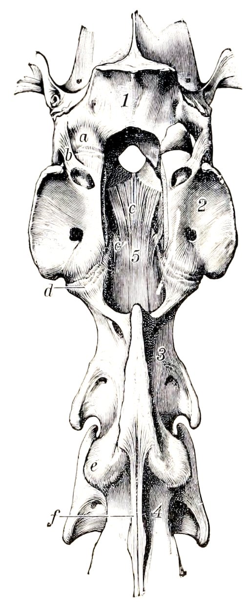
Fig. 141.—Atlanto-occipital and Atlanto-axial Joints of Horse, Dorsal View after Removal of Dorsal Arch of Atlas. a, Joint capsule of left part of atlanto-occipital joint; b, lateral ligament of same; c, c′, odontoid ligament; d, atlanto-occipital joint capsule; e, joint capsule of articulation between axis and third cervical vertebra; f, interspinous ligament; 1, occipital bone; 2, atlas; 3, axis; 4, third cervical vetrebra. (Ellenberger-Baum, Anat. d. Haustiere.)
THE ATLANTO-OCCIPITAL ARTICULATION
This joint may be classed as a ginglymus. The articular surfaces of this joint are: (1) On the atlas, two deep oval cavities; (2) the corresponding condyles of the occipital bone.
The joint surfaces are oblique, coming very close to the median line ventrally, but separated by a considerable interval dorsally. Posteriorly, a triangular rough area cuts into the middle of each articular surface on the atlas.
There are two roomy joint capsules, which sometimes communicate ventrally, especially in old subjects.
The superior atlanto-occipital membrane extends from the dorsal arch of the atlas to the upper margin of the foramen magnum. It is blended with the capsules and contains many elastic fibers.
The inferior atlanto-occipital membrane extends from the ventral arch of the 177atlas to the lower margin of the foramen magnum. It is narrower and thinner than the superior membrane, and also fuses with the joint capsules.
The lateral ligaments are two short bands which are partially blended with the capsules. Each is attached to the border of the wing of the atlas near the intervertebral foramen, and to the outer surface of the paramastoid or styloid process of the occipital bone.
Movements.—These are chiefly flexion and extension. A small amount of lateral oblique movement is also possible.
Articulations of the Thorax
COSTO-VERTEBRAL ARTICULATIONS
Each typical rib forms two joints with the vertebral column, one by its head, and one by its tubercle. They are termed respectively costo-central and costo-transverse joints.
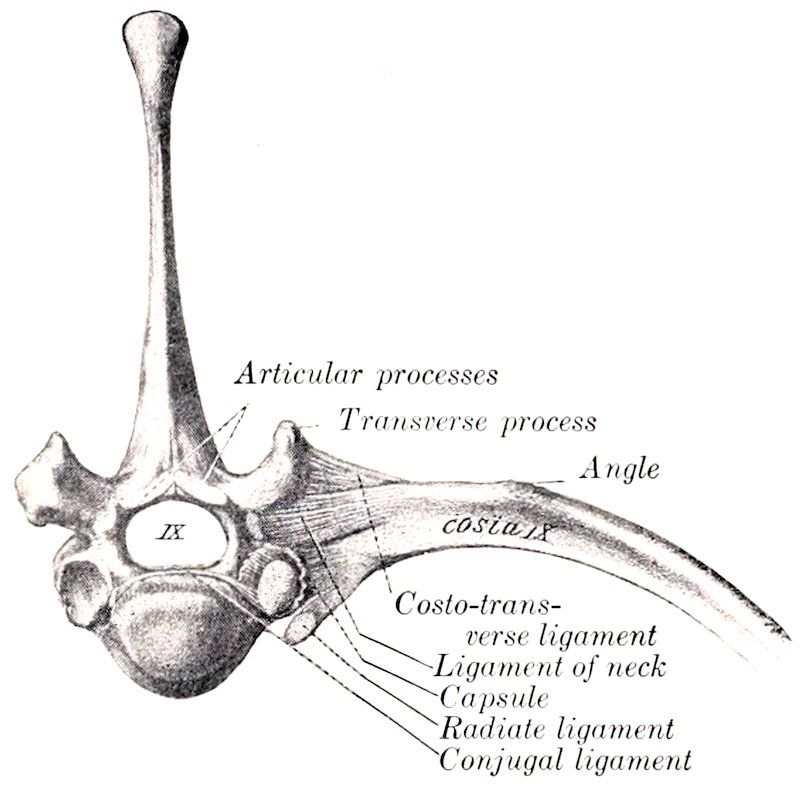
Fig. 142.—Costo-vertebral Articulation, Anterior View. (After Schmaltz, Atlas d. Anat. d. Pferdes.)
I. The costo-central articulations (Articulationes capitulorum) are trochoid or rotatory joints, formed by the junction of the head of the rib with the bodies of two adjacent vertebræ and the intervertebral fibro-cartilage. The two facets on the head of the rib are separated by a non-articular groove, and correspond to the two concave facets (Foveæ costales) on the vertebral bodies. The capsules are rather tight, and are covered by the accessory ligaments, which are as follows: 1. The radiate ligament (Ligamentum capituli costæ radiatum) extends ventrally from the neck of the rib to spread out on the vertebral bodies and the intervertebral fibro-cartilage. 2. The conjugal ligament (Ligamentum conjugale)—absent from the first joint—is attached to the groove on the head of the rib, passes transversely into the vertebral canal, and divides under the superior common ligament into two branches; one of these is attached to the body of the anterior vertebra; the other is continued across to the head of the opposite rib, and is attached to the intervertebral fibro-cartilage. The joint cavity is divided into two compartments by the conjugal ligament. 3. The ligament of the neck of the rib (Ligamentum colli costæ) is a strong band which crosses the joint dorsally. It is attached on the vertebra above the costal facet and on the neck of the rib.
II. The costo-transverse articulations. These occur between the facets on the tubercles of the ribs and those on the transverse processes of the vertebræ. They are gliding joints. The capsule is reinforced by the superior costo-transverse ligament (Ligamentum costo-transversarium dorsale), a distinct strong band which arises on the transverse process and ends on the non-articular part of the tubercle. It is covered by the levator costæ muscle, and begins to be quite distinct at the fifth joint.
178The cavity for the head of the first rib is formed by concave facets on the bodies of the last cervical and first thoracic vertebræ. The conjugal ligament is absent, but the ligament of the neck is short and strong. The radiate ligament is very strong, and consists of two bands. In the case of the last two or three ribs the costo-central and costo-transverse joints are confluent, and the various structures are correspondingly modified.
Movements.—The chief movement is rotation around an axis which connects the centers of the head and tubercle of the rib. The movement is very limited in the anterior part of the series of joints, but very considerable in the posterior part.
In the case of the first rib, the movement is evidently extremely limited. The facet for the tubercle of the rib is deeply concave, and the axis of rotation is almost transverse. Further back the facets on the transverse processes become flat, and the axis of rotation gradually approaches a longitudinal direction. This, in connection with the mobility of the ventral ends of the asternal ribs and their elasticity, allows a great increase here in the range of movement, the effect of which is to enlarge (chiefly) the transverse diameter of the thorax.
COSTO-CHONDRAL ARTICULATIONS
The costo-chondral junctions are synarthroses. The rib has a concave surface which receives the convex end of the cartilage. They are united by the continuity of the strong periosteum and perichondrium.
CHONDRO-STERNAL ARTICULATIONS
These joints (Articulationes sternocostales) are diarthroses formed by the junction of the cartilages of the sternal ribs with the sternum. The articular ends of the cartilages (except the first) are somewhat enlarged, and present surfaces of cylindrical curvature. The articular surfaces on the sternum for the first pair of cartilages are placed close together on the dorsal border of the cariniform cartilage (Manubrium sterni); the other seven are placed laterally at the junction of the segments. The capsules are strong and tight; the first pair of joints has a common capsule, and the cartilages articulate with each other medially. The lower ends of the first pair of ribs are firmly attached to each other by dense fibrous tissue, which is prolonged forward along the upper margin of the cariniform cartilage and is continuous behind with the sternal ligament. Each of the other capsules is reinforced dorsally by the superior costo-sternal ligament (Ligamentum sternocostale radiatum), composed of radiating fibers which blend with the sternal ligament. The movement is rotation around a nearly vertical axis, except in the case of the first pair of joints.
INTERCHONDRAL LIGAMENTS
The eighth and ninth costal cartilages are firmly united by fibrous tissue. The chondro-xiphoid ligament attaches the ninth costal cartilage to the xiphoid cartilage. The remaining cartilages are rather loosely attached to each other by elastic tissue.
STERNAL ARTICULATIONS
In the new-born foal the sternum has seven bony segments (Sternebræ), united by persisting cartilage. The last two segments coalesce within a few weeks after birth. In old subjects there is more or less ossification of the intersegmental cartilage, which may lead to partial fusion of adjacent segments, especially posteriorly. The sternal ligament (Ligamentum sterni proprium internum) lies on the thoracic surface of the sternum. It arises on the first segment, and divides opposite the second chondro-sternal joint into three bands. The median band passes backward and spreads out on the last segment and the xiphoid cartilage. The 179lateral branches—thicker and wider—lie along the lateral borders above the chondro-sternal joints, and end at the cartilage of the eighth rib; they are covered by the transversus thoracis muscle.
The Articulations of the Skull
TEMPORO-MANDIBULAR ARTICULATION
This joint (Articulatio mandibularis) is the only diarthrosis formed between bones of the skull.
The articular surfaces are dissimilar in form and size. That on the squamous temporal bone is concavo-convex, and the long axis is directed outward and somewhat forward; it consists of a glenoid cavity, which is continued upon the postglenoid process behind, and a condyle in front. The mandible presents a transversely elongated condyle.
The articular disc is placed between the joint surfaces, which it renders congruent. Its upper and lower surfaces are molded upon the temporal and mandibular surfaces respectively, and its circumference is attached to the joint capsule; thus it divides the joint cavity into upper and lower compartments, the former being the more roomy.
The joint capsule is strong and tight. It is reinforced by two ligaments. The external ligament (Ligamentum laterale) extends obliquely across the anterior part of the outer surface of the capsule, from which it is not distinctly separable. The posterior ligament (Ligamentum posterius) is an elastic band which is attached above to the postglenoid process, and below to a line on the posterior face of the neck of the mandible.
Movements.—The chief movements take place around a transverse axis passing through both joints. Associated with this hinge-like action is slight gliding movement, as in opening and shutting the mouth. When the mouth is shut, the condyle lies under the glenoid cavity. When the mandible is depressed, the condyle moves forward under the articular eminence of the temporal bone, carrying the disc with it. In protrusion and retraction of the lower jaw the gliding movement just described occurs without the hinge-like rotation of the condyle. These movements are similar in both joints. In the lateral movements (as usually performed in mastication) the action consists of rotation of the condyles around a vertical axis, while the disc glides forward on one side and backward on the other.
THE SYNARTHROSES OF THE SKULL
Most of the bones of the skull are united with the adjacent bones by sutures; a few are united by cartilage. The difference in the uniting medium depends on the fact that most of these bones are developed in membrane, but some are preformed in cartilage. Most of these joints are temporary, and are obliterated at various periods during development and growth. Their importance rests on the fact that so long as they persist, continuous growth is possible. They are usually designated according to the bones which enter into their formation, e. g., spheno-squamous, naso-frontal, internasal, etc. Special names (borrowed from human anatomy) are sometimes used; thus the interparietal, the parieto-occipital, and the parieto-frontal sutures are often called the sagittal, lambdoid, and coronal respectively.
Detailed description of the sutures has not sufficient clinical value to justify much addition to the statements made in the osteology in this connection. The obliteration or closure of the sutures is, however, worthy of brief mention. The cranial sutures are usually all closed at seven years, but the apex only of the petrous temporal is fused with the occipital and squamous temporal. Most of the facial sutures are practically closed at ten years, although complete synostosis may in 180some be delayed for years or may not occur at all; the nasal suture, for example, often persists even in advanced age, so far as its anterior part is concerned.
The principal synchondroses are: (1) That between the basioccipital and the body of the sphenoid (Synchondrosis sphenooccipitalis); (2) that between the presphenoid and postsphenoid (Synchondrosis intersphenoidalis); (3) those between the parts of the occipital bone (Synchondroses intraoccipitales). The first is usually ossified at four years, the second at three years, and the occipital bone is consolidated at two years.
The symphysis of the lower jaw (Symphysis mandibulæ) ossifies at one to six months.
THE HYOIDEAN ARTICULATIONS
The temporo-hyoid articulation is an amphiarthrosis, in which the dorsal angle of the proximal end of the great cornu (Stylo-hyal) is attached by a short bar of cartilage to the hyoid process of the petrous temporal bone. The cartilage (Arthrohyal) is about half an inch (ca. 1 to 1.5 cm. ) in length. The chief movement is hinge-like, the axis of motion passing transversely through both joints.
The intercornual articulation is an amphiarthrosis formed by the junction of the distal extremity of the great cornu with the proximal end of the small cornu (kerato-hyal). They are united by a very short piece of cartilage, in which there is usually a small nodule of bone in the young subject. This nodule, the epihyal or middle cornu, is usually fused with the great cornu in the adult. The chief movement here is also hinge-like, the angle between the cornua being increased or diminished.
The basi-cornual articulation is a diarthrosis formed by the junction of each small cornu (kerato-hyal) with the body (basi-hyal). The small cornu has a concave facet which articulates with the convex facet on either end of the dorsal surface of the body. The capsule is ample enough to allow considerable movement, which is chiefly hinge-like. The movements of the hyoid bone are concerned chiefly in the acts of mastication and swallowing. In the latter the distal parts of the hyoid bone are moved forward and upward, carrying the root of the tongue and the larynx with them, and then return to their former position.
The Articulations of the Thoracic Limb
In the absence of the clavicle the thoracic limb forms no articulation with the trunk, unless we regard as such the union by muscles. The movement of the shoulder on the chest-wall is chiefly rotation around a transverse axis passing through the scapula behind the upper part of the spine.
THE SHOULDER JOINT
The shoulder or scapulo-humeral joint (Articulatio scapulo-humeralis) is formed by the junction of the distal end of the scapula with the proximal end of the humerus. The articular surfaces are: (1) On the scapula, the glenoid cavity; (2) on the humerus, the head. Both surfaces are approximately spherical and similar in curvature, but the humeral surface is much more extensive than that of the scapula.
The joint capsule is ample enough to allow the bones to be drawn apart about an inch (ca. 2 to 3 cm.); but this requires a very considerable amount of force unless air is admitted into the joint cavity. The fibrous layer (or capsular ligament) is not attached to the margin of the joint surfaces, but at a distance of one to two centimeters from it. It is strengthened in front by two diverging elastic bands, 181which arise on the scapular tuberosity and end on the lips of the bicipital groove. A pad of fat is interposed between the capsule and the tendon of the biceps.
Ligaments are absent from this joint, but the muscles and tendons around it afford remarkable security, so that dislocation seldom or never occurs. The large extent of the head of the humerus is also of importance in this regard.
The principal muscles which are attached around the joint and act as ligaments are: externally, the supraspinatus, infraspinatus, and teres minor; internally, the subscapularis; in front, the biceps and supraspinatus; behind, the triceps.
Movements.—While it is a typical enarthrosis in structure, and capable of the various movements of the ball-and-socket joint, the chief normal movements are flexion and extension. In the position of rest the angle formed between the scapula and humerus posteriorly is about 110° to 120°; in flexion it is reduced to about 80°, and in extension it is increased to about 145°. Adduction and abduction are very restricted, the former being limited chiefly by the infraspinatus, the latter by the subscapularis and the low insertion of the superficial pectoral muscles. Rotation is somewhat freer, but does not exceed 33°, when all the muscles are removed (Franck).
THE ELBOW JOINT
This, the cubital articulation (Articulatio cubiti), is a ginglymus formed between the distal extremity of the humerus and the proximal ends of the bones of the forearm.
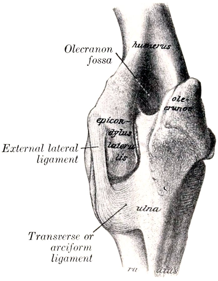
Fig. 143.—Left Elbow Joint of Horse, Posterior View. The Capsule is Removed. (After Schmaltz, Atlas d. Anat. d. Pferdes.)
The articular surfaces are: (1) A trochlear surface formed by the condyles of the humerus and the groove between them; (2) the corresponding glenoid cavities and ridge on the proximal extremity of the radius, together with the semilunar notch of the ulna.
The articular surface of the condyles does not extend upon the back of the extremity, but the groove which receives the semilunar notch of the ulna extends up into the olecranon fossa. In the fore part of the groove there is a synovial fossa. The surface on the outer condyle is smaller than that of the inner one, and is subdivided into two unequal parts by a shallow furrow. On the lower part of the semilunar notch and the adjacent part of the ridge on the radius are synovial fossæ.
The joint capsule is extremely thin behind, where it forms a pouch in the olecranon fossa under the anconeus muscle and a pad of fat. In front it is strengthened by oblique fibers (Ligamentum obliquum or anterior ligament), and laterally it fuses with the lateral ligaments. Small pouches of the synovial membrane lubricate the origins of the flexors of the carpus and digit and the small radio-ulnar joints. There are two lateral ligaments.
The internal lateral ligament (Ligamentum collaterale radiale) is attached above to an eminence on the internal epicondyle of the humerus, and divides into two parts: the long, superficial part ends on the inner border of the radius, just below the level of the interosseous space; the deep, short part is inserted into the internal tuberosity of the radius. (The superficial part represents the pronator teres muscle, which is only exceptionally present in the horse.)
182The external lateral ligament (Ligamentum collaterale ulnare) is short and strong. It is attached above to a depression on the external epicondyle of the humerus, and below to the external tuberosity of the radius, just below the margin of the articular surface.
Movements.—This joint is a typical ginglymus, the only movements being flexion and extension around an axis which passes through the upper attachments of the lateral ligaments. In the standing position the articular angle (in front) is about 140° to 150°. The range of movement is about 55° to 60°. Complete extension is prevented chiefly by the tension of the lateral ligaments and the biceps muscle. (The axis of movement is slightly oblique, so that in flexion the forearm is carried somewhat outward.)
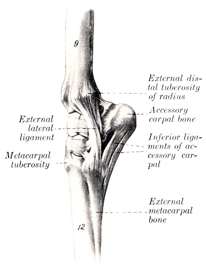
Fig. 144.—Left Carpal Joints of Horse, External View. The capsule has been removed, g, Radius; 12, large metacarpal bone. (After Ellenberger-Baum, Anat. f. Künstler.)
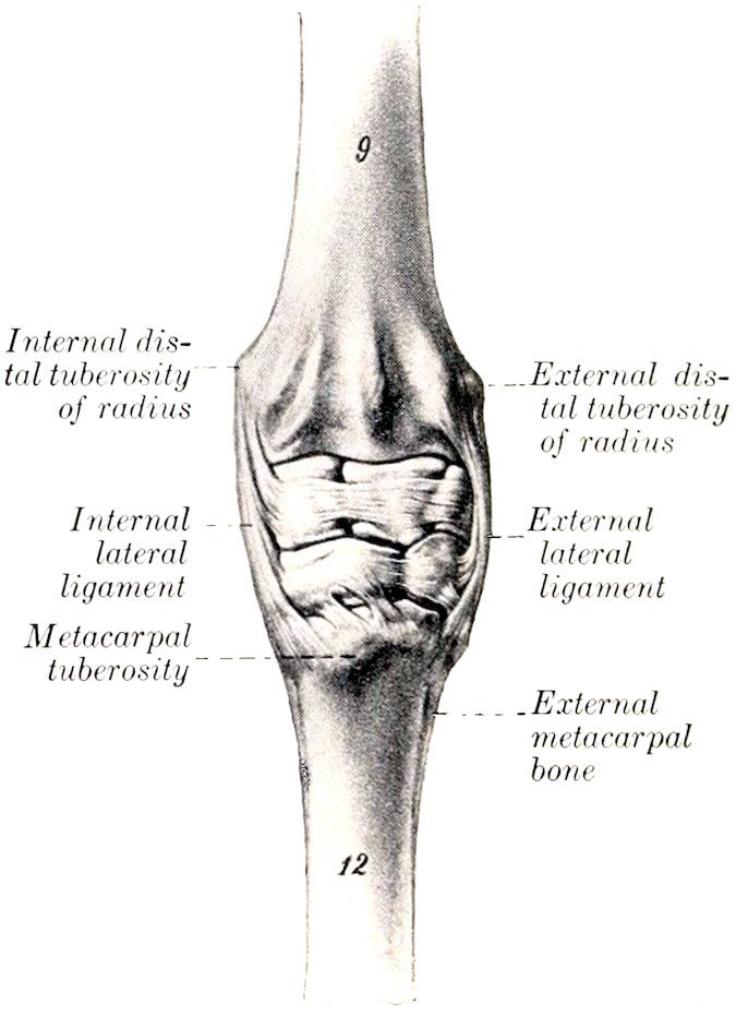
Fig. 145.—Left Carpal Joints of Horse, Anterior View. The capsule has been removed. The smaller ligaments are shown. (After Ellenberger-Baum, Anat. f. Künstler.)
THE RADIO-ULNAR ARTICULATION
In the foal the shaft of the ulna is attached to the radius above and below the interosseous space by the interosseous ligament. Below the space the two bones become fused before adult age is reached. Above the space the ligament usually persists, but may undergo more or less ossification in extreme old age. The transverse or arciform ligaments (Ligamentum transversum ulnare et radiale ulnæ et radii) consist of fibers which pass above the interosseous space from either border of the shaft of the ulna to the posterior surface of the radius. The proximal radio-ulnar articulation, formed by two small convex facets on the ulna and the corresponding facets on the posterior surface of the proximal extremity of the radius, is inclosed in the capsule of the elbow joint and does not require separate consideration. The distal extremity of the ulna fuses early with the radius, and is, therefore, regarded usually as a part of the latter.
Movement.—This is inappreciable, the forearm being fixed in the position of pronation.
183
THE CARPAL JOINTS
These joints taken together constitute the composite articulatio carpi, or what is popularly termed the “knee-joint” in animals.[33] This consists of three chief joints, viz., (1) The radio-carpal joint, formed by the distal end of the radius and the proximal row of the carpus; (2) the intercarpal joint, formed between the two rows of the carpus; (3) the carpo-metacarpal joint, formed between the distal row of the carpus and the proximal ends of the metacarpal bones. The proximal and middle joints may be regarded as ginglymi, although they are not typical or pure examples of hinge-joints. The distal joint is arthrodial. In addition there are arthrodial joints formed between adjacent bones of the same row (Articulationes interosseæ). All these constitute a very composite joint, with numerous ligaments. The articular surfaces have been described in the Osteology.
The joint capsule may be regarded, so far as the fibrous part is concerned, as being common to all three joints. It is attached close to the margin of the articular surface of the radius above and the metacarpus below; its deep face is also attached to a considerable extent to the carpal bones and to the small ligaments. Its anterior part, the dorsal or anterior common ligament, is rather loose, and assists in forming the fibrous canals for the extensor tendons. Its posterior part, the volar or posterior common ligament, is very thick and dense, and is closely attached to the carpal bones. It levels up the irregularities of the skeleton here, and forms the smooth anterior wall of the carpal canal. It is continued downward to form the subcarpal or inferior check ligament, which blends with the tendon of the flexor perforans about the middle of the metacarpus.
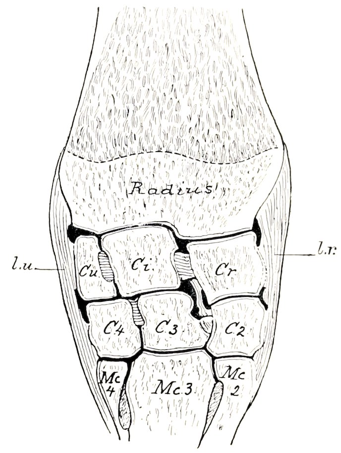
Fig. 146.—Frontal Section of Carpal Joints of Horse (Right Side). l.u., External, l.r., internal, lateral ligament; Cr, radial carpal; Ci, intermediate carpal; Cu, ulnar carpal; C2, second carpal; C3, third carpal; C4, fourth carpal; Mc2, second (internal) metacarpal; Mc3, third (large) metacarpal; Mc4, fourth (external) metacarpal.
The synovial membrane forms three sacs corresponding to the three joints. The radio-carpal sac is the most voluminous; it includes the joints formed by the accessory carpal bone, and also those between the proximal carpal bones as far as the interosseous ligaments. The intercarpal sac sends extensions upward and downward between the bones of the two rows as far as the interosseous ligaments; it communicates between the third and fourth carpal bones with the carpo-metacarpal sac. The latter is very limited in extent, and is closely applied to the bones; it incloses the carpo-metacarpal joint, and lubricates also the lower parts of the joints between the distal carpal bones and the intermetacarpal joints.
The external lateral ligament (Ligamentum carpi collaterale ulnare) is attached above to the external tuberosity of the distal end of the radius. Its long superficial part is attached below to the proximal end of the external small metacarpal chiefly, 184but some fibers end on the large metacarpal bone. A canal for the lateral extensor tendon separates a short deep band which ends on the ulnar carpal bone. Other deep fibers connect the latter with the fourth carpal bone, and the fourth carpal with the metacarpus.
The internal lateral ligament (Ligamentum carpi collaterale radiale) resembles the preceding in general, but is stronger and wider distally. It is attached above to the internal tuberosity of the distal end of the radius and ends below on the proximal ends of the large and inner small metacarpal bones. Deep fasciculi are detached to the radial and second carpal bones. The first carpal bone, when present, is usually embedded in the posterior part of the distal end of the ligament. The posterior part of the ligament is fused with the posterior annular ligament (Ligamentum carpi transversum), and concurs in the formation of a canal for the tendon of the flexor carpi internus.
A number of special short ligaments connect two or more adjacent bones; only the most distinct of these will be described here.
The accessory carpal bone is connected with adjacent bones by three ligaments (Fig. 444). The proximal one is a short band which extends from the accessory carpal in front of the groove on its outer face and is inserted into the distal end of the radius behind the groove for the lateral extensor tendon. A middle band connects the accessory with the ulnar carpal. The distal ligament consists of two strong bands which pass from the lower margin of the accessory to the fourth carpal and the proximal end of the outer metacarpal bone; these bands transmit the action of the muscles, which are inserted into the accessory carpal bone. The other bones of the proximal row are connected by two anterior or dorsal ligaments, which are transverse in direction, and two interosseous ligaments. An oblique ligament passes from an eminence on the posterior surface of the radial carpal bone to a small depression on the radius internal to the facet for the accessory carpal bone.
Two ligaments connect the proximal and distal rows posteriorly. The inner one joins the radial to the second and third carpal, and the outer one attaches the ulnar to the third and fourth carpals.
The bones of the distal row are connected by two strong transverse anterior or dorsal ligaments and two interosseous ligaments.
There are four carpo-metacarpal ligaments. Two oblique anterior bands connect the third carpal with the large metacarpal. Two interosseous ligaments pass downward from the interosseous ligaments of the distal row to end in depressions in the interstices between the proximal ends of the metacarpal bones.
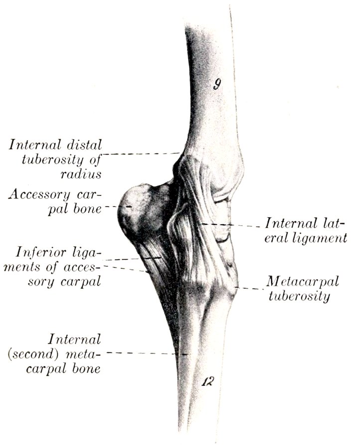
Fig. 147.—Left Carpal Joints of Horse, Inner View. g, Radius; 12, large (third) metacarpal bone. (After Ellenberger-Baum, Anat. für Künstler.)
Movements.—Taking the joint as a whole, the chief movements are flexion and extension. In the standing position the joint is extended. When the joint is flexed, slight lateral movement and rotation can be produced by manipulation. The anterior part of the capsule is, of course, tense during flexion, the posterior part in extension.
The movement practically all occurs at the radio-carpal and intercarpal joints, the articular surfaces of which are widely separated in front during flexion, but remain in contact behind. The distal row remains in contact with the metacarpus. The intermediate and ulnar carpals move together as one piece, but the radial does not move so far as the intermediate, so that the anterior and interosseous ligaments connecting these bones become tense and oblique in direction.
185
THE FETLOCK JOINT
This, the metacarpo-phalangeal articulation (Articulatio metacarpo-phalangea), is a ginglymus formed by the junction of the distal end of the large (third) metacarpal bone, the proximal end of the first phalanx, and the proximal sesamoid bones.
Articular Surfaces.—The surface on the large metacarpal bone is approximately cylindrical in curvature, but is divided into two slightly unequal parts by a sagittal ridge. This is received into a sort of socket formed by the first phalanx below and the two sesamoids together with the intersesamoid ligament behind. The latter is a mass of fibro-cartilage in which the sesamoid bones are largely embedded. It extends above the level of the sesamoids, and is grooved to receive the ridge on the metacarpal bone; its posterior surface forms a smooth groove for the deep flexor tendon.
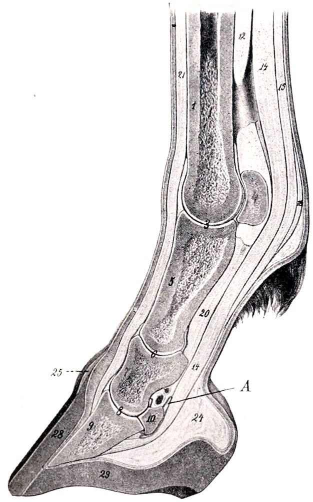
Fig. 148.—Sagittal Section of Distal Part of Limb of Horse. 1, Large metacarpal bone; 3, fetlock joint; 4, proximal sesamoid bone; 5, first phalanx; 6, pastern joint; 7, second phalanx; 8, coffin joint; 9, third phalanx; 10, distal sesamoid (navicular bone); 12, suspensory ligament; 14, deep flexor tendon; 15, superficial flexor tendon; 16, posterior annular ligament of fetlock; 20, inferior sesamoidean ligaments; 21, extensor tendon; 24, plantar cushion; 25, periople; 28, wall of hoof; 29, sole of hoof; A, navicular bursa, proximal part. (After Ellenberger-Baum, Anat. für Künstler.)
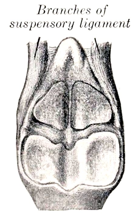
Fig. 149.—Articular Surfaces of First Phalanx and Sesamoids at Fetlock, with Intersesamoid and Suspensory Ligaments. (After Schmaltz, Atlas d. Anat. d. Pferdes.)
The joint capsule is attached around the margin of the articular surfaces. It is thick and ample in front; here a bursa is interposed between it and the extensor tendons, but the tendons are also attached to the capsule. Posteriorly it forms a thin-walled pouch which extends upward between the metacarpal bone and the suspensory ligament about as high as the point of bifurcation of the latter. The capsule is reinforced by two lateral ligaments.
The lateral ligaments, external and internal (Ligamentum collaterale ulnare, radiale) are partially divided into two layers: the superficial layer arises from the eminence on the side of the distal end of the large metacarpal bone, and passes straight to the rough lateral area below the margin of the articular surface of the first phalanx; the deep layer, shorter and much stronger, arises in the lateral depression on the distal end of the metacarpal bone, and passes obliquely downward and backward to be inserted into the outer surface of the sesamoid and the proximal end of the first phalanx.
186The capsule is further strengthened by a layer of oblique fibers which pass over the lateral ligament on either side and end on the extensor tendon and the proximal extremity of the first phalanx. It may properly be regarded as fascia rather than ligament.
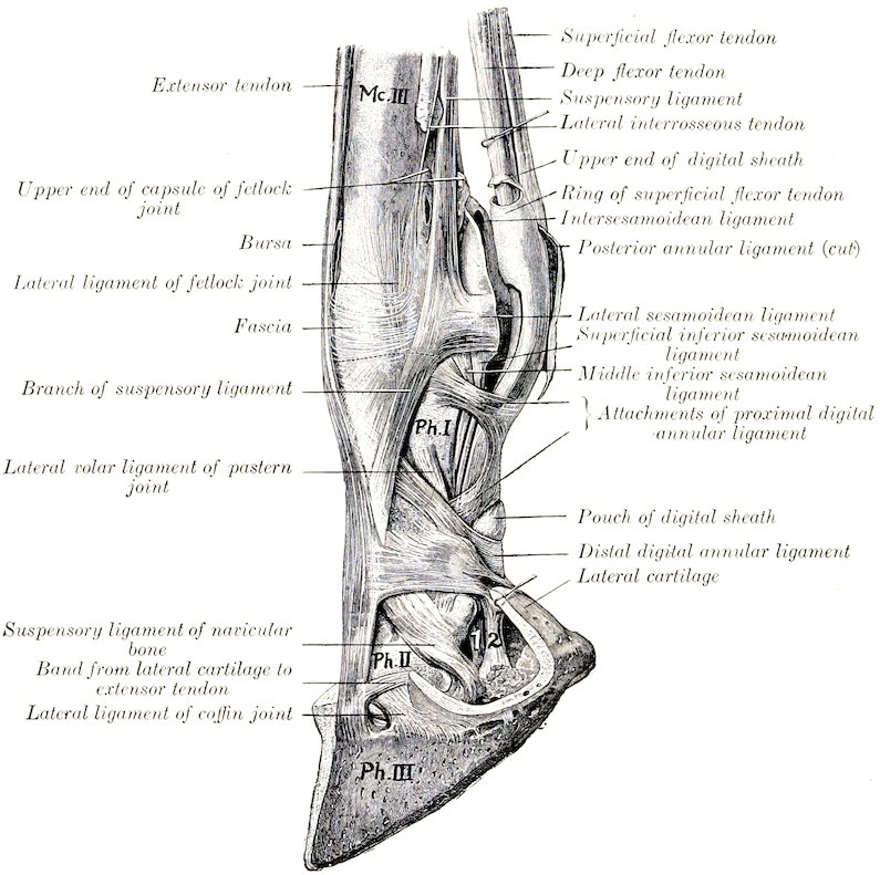
Fig. 150.—Ligaments and Tendons of Distal Part of Limb of Horse. Mc.III, Large metacarpal bone; Ph.I, first phalanx; Ph.II, second phalanx; Ph.III, third phalanx; 1, deep flexor tendon; 2, band from first phalanx to plantar cushion. (After Schmaltz, Atlas d. Anat. d. Pferdes.)
Movements.—These are of the nature of flexion and extension, the axis of motion passing through the upper attachments of the lateral ligaments. In the ordinary standing position the joint is in a state of partial dorsal flexion, the articular angle (in front) being about 140° to 150°. (In the hind limb it is about 5° greater.) Diminution of this angle (sometimes termed “overextension”) is normally very limited on account of the resistance offered by the sesamoidean apparatus, but it varies considerably in amount in different subjects. Volar flexion is limited only by contact of the heels with the metacarpus. During volar flexion a small amount of lateral flexion is possible.
THE SESAMOIDEAN LIGAMENTS
Under this head will be described a number of important ligaments which are connected with the sesamoid bones and form a sort of stay apparatus or brace.
187The intersesamoidean ligament (Ligamentum intersesamoideum) not only fills the space between and unites the sesamoid bones, but also extends above them, entering into the formation of the articular surface of the fetlock joint. Other facts in regard to it have been given above.
The lateral sesamoidean ligaments, outer and inner (Ligamenta sesamoidea ulnare et radiale), arise on the abaxial surface of each sesamoid bone, pass forward, and divide into two branches, one of which ends in the depression on the distal end of the large metacarpal bone, the other on the eminence on the proximal end of the first phalanx. They are partly covered by the branches of the suspensory or superior sesamoidean ligament.
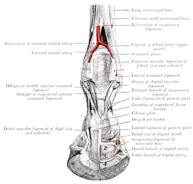
Fig. 151.—Deep Dissection of Distal Part of Right Fore Limb of Horse, Showing Joints and Ligaments, Posterior View. 1, Lateral cartilage; 2, tendon surface of navicular bone; 3, inferior navicular or interosseous ligament; 4, insertion of deep flexor tendon. Small arrows point to openings made in capsules of pastern and coffin joints. (After Schmaltz, Atlas d. Anat. d. Pferdes.)
The suspensory or superior sesamoidean ligament (Musculus interosseus medius) lies for the greater part in the metacarpal groove, where it has the form of a wide, thick band. It is attached above to the upper part of the posterior surface of the large metacarpal bone and to the distal row of carpal bones. At the lower fourth of the metacarpus it divides into two diverging branches. Each branch passes to the abaxial face of the corresponding sesamoid, on which a considerable 188part, is attached. The remainder passes obliquely downward and forward to the anterior surface of the first phalanx, where it joins the extensor tendon. This ligament possesses considerable elasticity, and is the highly modified interosseous medius muscle. It consists mainly of tendinous tissue, but contains a variable amount of striped muscular tissue, especially in its deep part and in young subjects. Its principal function is to support the fetlock, i. e., to prevent excessive dorsal flexion of the joint when the weight is put on the limb. The branches which join the common extensor tendon limit volar flexion of the interphalangeal joints in certain phases of movement.
The inferior sesamoidean ligaments are three in number—superficial, middle, and deep. The superficial or straight ligament (Ligamentum sesamoideum rectum) is a flat band and is somewhat wider above than below.[34] It is attached above to the bases of the sesamoid bones and the intersesamoid ligament, below to the complementary fibro-cartilage of the proximal end of the second phalanx. The middle ligament is triangular, with thick, rounded margins (Ligamenta obliqua) and a thin central portion.[35] Its base is attached to the sesamoid bones and intersesamoid ligament, and its deep face to the triangular rough area on the posterior surface of the first phalanx. The deep or cruciate ligament (Ligamenta sesamoidea cruciata) consists of two thin layers of fibers which arise on the base of the sesamoid bones, cross each other, and end on the opposite eminence on the proximal end of the first phalanx.
The short sesamoidean ligaments (Ligamenta sesamoidea brevia) are best seen by opening the joint in front and pushing the sesamoid bones backward; they are covered by the synovial membrane. Each is a short band which extends from the anterior part of the base of the sesamoid bone outward to the posterior margin of the articular surface of the first phalanx.
The inferior sesamoidean ligaments may be regarded as digital continuations of the suspensory ligament, the sesamoid bones being intercalated in this remarkable stay apparatus by which the fetlock is supported and concussion diminished.
THE PASTERN JOINT
This, the proximal interphalangeal articulation (Articulatio phalangis secundæ), is a ginglymus formed by the junction of the distal end of the first phalanx and the proximal end of the second phalanx.
The articular surfaces are: (1) On the first phalanx, two slightly unequal convex areas with an intermediate shallow groove; (2) on the second phalanx, a corresponding surface, completed behind by a plate of fibro-cartilage.
The joint capsule is close fitting in front and laterally, where it blends with the extensor tendon and the lateral ligaments respectively. Behind it pouches upward a little and is reinforced by the straight sesamoidean ligament and the branches of the superficial flexor tendon.
There are two lateral and four volar ligaments.
The lateral ligaments, internal and external (Ligamentum collaterale radiale, ulnare) are very short and strong bands which are attached above on the eminence and depression on each side of the distal end of the first phalanx, and below on the eminence on either side of the proximal end of the second phalanx. The direction of the ligaments is about vertical and, therefore, does not correspond to the digital axis.
The volar or posterior ligaments consist of central and lateral pairs of bands which are attached below to the posterior margin of the proximal end of the second 189phalanx and its complementary fibro-cartilage. The lateral pair is attached above to the middle of the borders of the first phalanx, the central pair lower down and on the margin of the triangular rough area.
These ligaments are very commonly thickened as a result of chronic inflammation, and then are not well defined. The central ones blend below with the branches of the superficial flexor tendon and with the straight sesamoidean ligament.
Movements.—These are very limited, and consist of flexion and extension. The axis of motion passes transversely through the distal end of the first phalanx. In the standing position the joint is extended. A small amount of volar flexion is possible, and in this position slight lateral flexion and rotation can be produced by manipulation. Dorsal flexion is prevented by the lateral, volar, and straight sesamoidean ligaments.
THE COFFIN JOINT
This joint, technically termed the distal interphalangeal articulation (Articulatio phalangis tertiæ), is a ginglymus formed by the junction of the second and third phalanges and the third sesamoid bone.
Articular Surfaces.—The surface on the distal end of the second phalanx is convex from before backward, concave transversely. The articular surface of the third phalanx slopes sharply upward and forward; its central part is prominent, and is flanked by two glenoid cavities. It is completed behind by the articular surface of the third sesamoid or navicular bone.
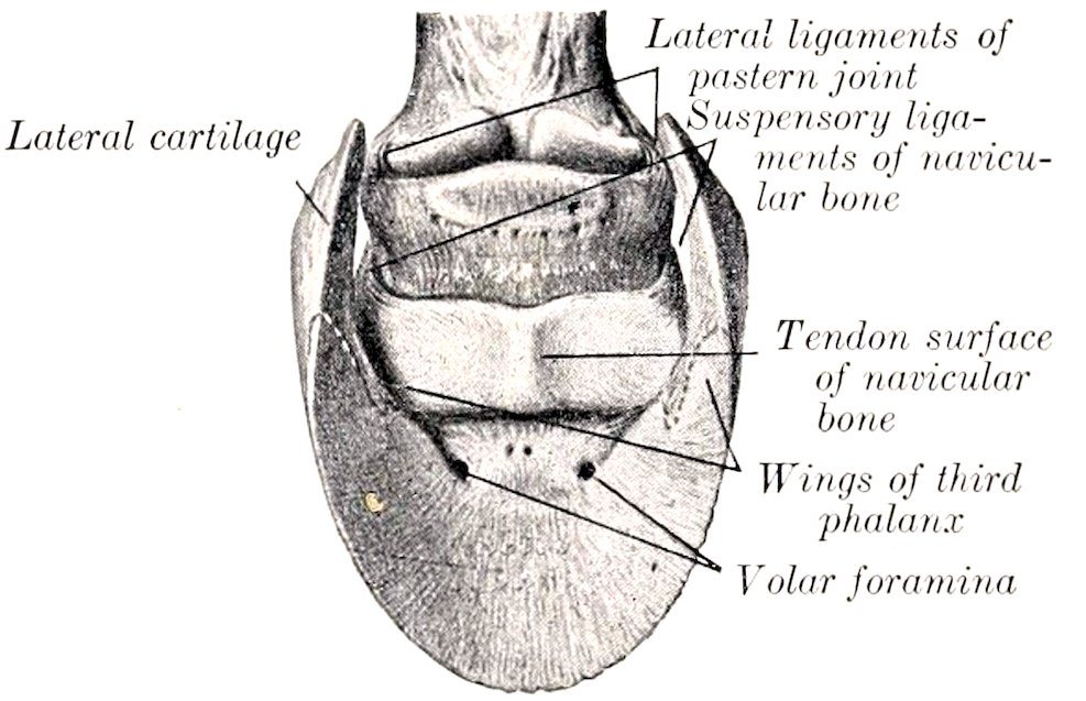
Fig. 152.—Lateral Ligaments of Pastern Joint and Suspensory Ligaments of Navicular Bone. (After Schmaltz, Atlas d. Anat. d. Pferdes.)
Joint Capsule.—This is attached around the margins of the articular surfaces. In front and laterally it is tight, and is blended with the extensor tendon and the lateral ligaments respectively. Posteriorly, it forms a considerable pouch which extends upward to about the middle of the second phalanx, where it is separated by a fibrous membrane from the digital synovial sheath. Laterally small pouches project outward (especially during volar flexion) against the lateral cartilages, just behind the lateral ligaments.[36]
Ligaments.—The lateral ligaments, external and internal (Ligamentum collaterale ulnare, radiale), are short strong bands which are attached above in the depressions on either side of the lower part of the second phalanx, under cover of the lateral cartilage. They widen below and end in the depressions on either side of the extensor process and on the anterior end of the lateral cartilages.
The suspensory navicular ligaments, external and internal (Ligamentum sesamoideum collaterale ulnare, radiale),[37] are strong, somewhat elastic bands, which form a sort of suspensory apparatus for the third sesamoid. They are attached superiorly in and above the depressions on either side of the distal end of the first 190phalanx and are here partly blended with the lateral ligaments of the pastern joint. They are directed obliquely downward and backward, and end chiefly on the ends and proximal border of the third sesamoid, but detach a branch to the inner surface of each lateral cartilage and wing of the third phalanx.
The inferior navicular ligament (Ligamentum phalangeo-sesamoideum) reinforces the capsule interiorly. It is a strong layer of fibers which extend from the distal border of the third sesamoid to the tendon surface of the third phalanx, near the posterior margin of the articular surface.
Movements.—The chief movements are flexion and extension. In the standing position the joint is extended. During volar flexion a very small amount of lateral movement and rotation can be produced by manipulation. Dorsal flexion is very limited.
Dorsal flexion appears to be checked mainly by the deep flexor tendon, since in cases of rupture of the latter the toe turns up. The slight mobility of the posterior part of the socket for the second phalanx (formed by the third sesamoid) diminishes concussion when the weight comes on the foot.
LIGAMENTS OF THE LATERAL CARTILAGES
In addition to the bands mentioned above, which attach the lateral cartilages to the extremities of the navicular bone, there are three ligaments on either side which attach the cartilages to the phalanges.
An ill-defined elastic band passes from the middle part of the border of the first phalanx to the upper part of the cartilage, detaching a branch to the plantar cushion.
A short strong band connects the anterior extremity of the cartilage with the rough eminence on the second phalanx in front of the attachment of the lateral ligament of the coffin joint.
The lower border of the cartilage is covered externally by fibers which attach it to the wing of the third phalanx.
The Articulations of the Pelvic Limb
THE SACRO-ILIAC ARTICULATION
This joint (Articulatio sacro-iliaca) is a diarthrosis formed between the auricular surfaces of the sacrum and ilium. These surfaces are not smooth in the adult, but are marked by eminences and depressions, and are covered by a thin layer of cartilage. The joint cavity is a mere cleft, and is often crossed by fibrous bands.
The capsule is very close fitting, and is attached around the margins of the articular surfaces. It is reinforced by the ventral sacro-iliac ligament (Ligamentum sacro-iliacum ventrale), which surrounds the joint, and is exceedingly strong above.
The movements are inappreciable in the adult—stability, not mobility, being the chief desideratum.
The following ligaments may be regarded as accessory to the joint, although not directly connected with it.
The dorsal sacro-iliac ligament (Ligamentum sacro-iliacum dorsale breve) is a strong band which is attached to the internal angle (Tuber sacrale) of the ilium and the summits of the sacral spines.
The lateral sacro-iliac ligament (Ligamentum sacro-iliacum dorsale longum) is a triangular, thick sheet which is attached in front to the internal angle and border of the ilium above the great sciatic notch, and below to the lateral border of the sacrum. It blends above with the dorsal sacro-iliac ligament, below with the sacro-sciatic ligament, and behind with the coccygeal fascia.
The sacro-sciatic ligament (Ligamentum sacrospinosum et tuberosum) is a 191quadrilateral sheet which completes the lateral pelvic wall. Its upper border is attached to the border of the sacrum and the transverse processes of the first and second coccygeal vertebræ. Its lower border is attached to the superior ischiatic spine and tuber ischii. Between these it bridges over the external border of the ischium and completes the lesser sciatic foramen. The anterior border is concave, and completes the greater sciatic foramen. The posterior border is fused with the vertebral head of the semimembranosus muscle.
The ilio-lumbar ligament (Ligamentum ilio-lumbale) is a triangular sheet which attaches the ends of the lumbar transverse processes to the ventral surface of the ilium below the attachment of the longissimus muscle (Fig. 184).
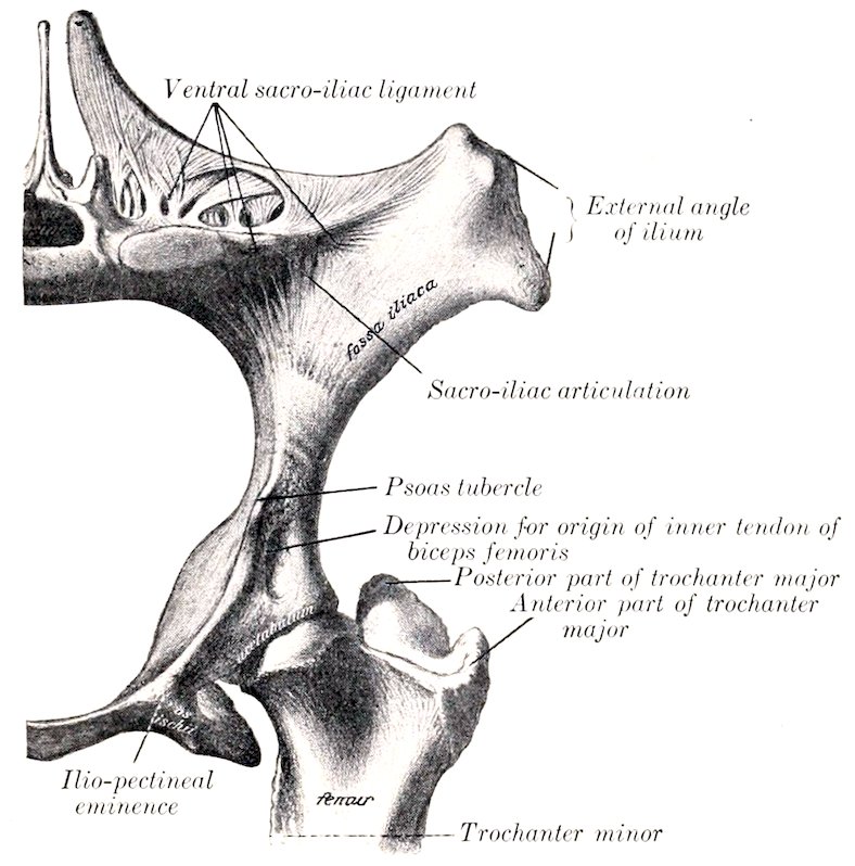
Fig. 153.—Left Os Coxæ and Adjacent Parts of Sacrum and Femur of Horse. (After Schmaltz, Atlas d. Anat. d. Pferdes.)
SYMPHYSIS PELVIS
The symphysis pelvis is formed by the junction of the two ossa coxarum at the ventral median line. In the young subject the bones are united by a layer of cartilage; in the adult the latter is gradually replaced by bone, the process beginning in the pubic portion and extending backward. The union is strengthened by white fibrous tissue above and below, and a transverse band also covers the anterior border of the pubis (pecten). No appreciable movement occurs even before synostosis takes place.
OBTURATOR MEMBRANE
This (Membrana obturatoria) is a thin layer of fibrous tissue which covers the obturator foramen, leaving, however, a passage (Canalis obturatorius) for the obturator vessels and nerve.
192
THE HIP JOINT
This joint (Articulatio coxæ) is an enarthrosis formed by the proximal end of the femur and the acetabulum.
Articular Surfaces.—The head of the femur presents an almost hemispherical articular surface, which is continued a short distance on the upper surface of the neck. It is more extensive than the socket which receives it. Internally it is cut into by a deep notch for the attachment of the round and pubo-femoral ligaments. The acetabulum is a typical cotyloid cavity. Its articular surface is somewhat crescentic, being deeply cut into internally by the acetabular notch and fossa. It is increased and deepened by a ring of fibro-cartilage, the cotyloid ligament (Labrum glenoidale), which is attached to the bony margin; that part of the ligament which crosses the notch is called the transverse ligament (Fig. 456).
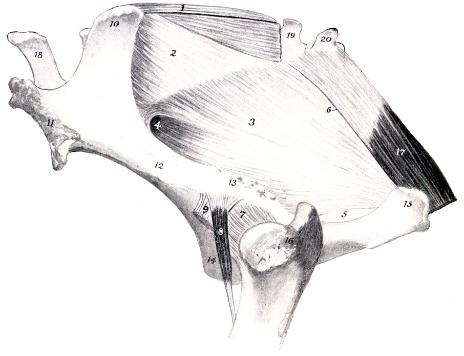
Fig. 154.—Pelvic Ligaments and Hip Joint. 1, Dorsal sacro-iliac ligament; 2, lateral sacro-iliac ligament; 3, sacro-sciatic ligament; 4, greater sciatic foramen; 5, lesser sciatic foramen; 6, line of attachment of intermuscular septum between biceps femoris and semitendinosus; 7, capsule of hip joint; 8, rectus parvus or capsularis muscle; 9, outer tendon of origin of biceps femoris; 10, internal, 11, external, angle of ilium; 12, shaft of ilium; 13, superior ischiatic spine; 14, pubis; 15, tuber ischii; 16, trochanter major; 17, semimembranosus; 18, fifth lumbar spine; 19, 20, first and second coccygeal vertebræ.
The joint capsule is roomy. It is attached around the margin of the acetabulum and the neck of the femur. It is thickest externally.
The attachment on the femur is about 1 cm. from the margin of the articular surface, except above, where 2 to 3 cm. of the neck is intracapsular. A thin oblique band corresponding in direction with the rectus parvus muscle reinforces the antero-external part of the capsule; this appears to be the feeble homologue of the very strong ilio-femoral ligament of man. The capsule is very thin under the ilio-psoas, and is adherent to the muscle. Internally, its fibrous part is perforated by the pubo-femoral and round ligaments and the articular vessels.
193The round ligament (Ligamentum teres) is a strong hand which is attached in the subpubic groove close to the acetabular notch, passes outward, and ends in the notch on the head of the femur (Fig. 456).
The pubo-femoral ligament (Ligamentum accessorium) does not occur in the domestic animals other than the equidæ. It is a strong band detached from the prepubic tendon of the abdominal muscles (Fig. 456). It is directed outward, backward, and upward, passes through the acetabular notch above the transverse ligament, and ends behind the round ligament in the notch on the head of the femur. The origin of the pectineus muscle is perforated by the ligament, which furnishes attachment to the greater part of the fibers of the muscle.
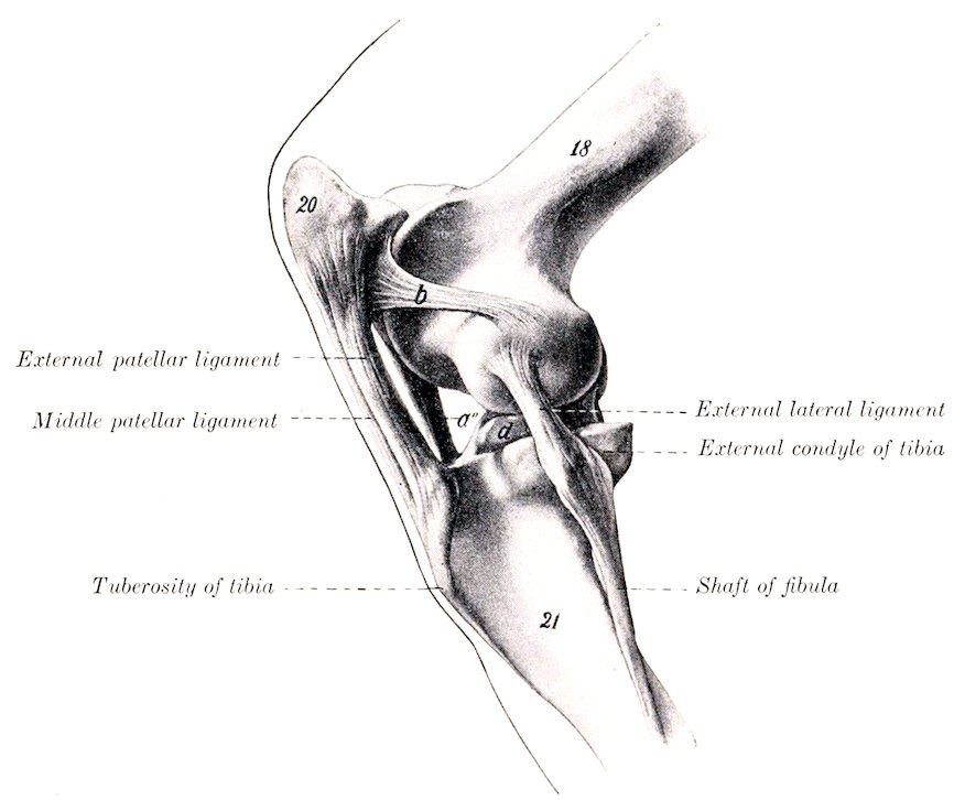
Fig. 155.—Left Stifle Joint of Horse, External View, in Partial Flexion. The capsules have been removed. 18, Femur; 20, patella; 21, tibia; a″, internal patellar ligament; b, external femoro-patellar ligament; d, external semilunar cartilage. (After Ellenberger-Baum, Anat. für Künstler.)
The synovial membrane is reflected over the intracapsular parts of these ligaments and covers the fossa acetabuli. A pouch also extends from the acetabular notch for a variable distance along the subpubic groove above the pubo-femoral ligament.
Movements.—This joint is capable of all the movements of a ball-and-socket joint, viz., flexion, extension, abduction, adduction, rotation, and circumduction. The greatest range of movement is displayed in flexion and extension. When standing at rest, the joint is partially flexed, the articular angle (in front) being about 100° to 115°. The other movements occur to a very limited extent in normal action.
Abduction appears to be checked by tension of the round ligament. The pubo-femoral ligament is tensed most promptly by inward rotation of the thigh.
194
THE STIFLE JOINT
This joint (Articulatio genu), which corresponds to the knee-joint of man, is the largest and most elaborate of all the articulations. Taken as a whole, it may be classed as a ginglymus, although it is not a typical example of the group. In reality it consists of two joints—the femoro-patellar and the femoro-tibial.
The femoro-patellar articulation (Articulatio femoro-patellaris) is formed between the trochlea of the femur and the articular surface of the patella.
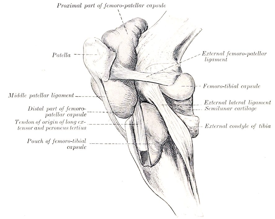
Fig. 156.—Left Stifle Joint of Horse, Lateral View. The capsules are distended and the external patellar ligament is removed.
Articular Surfaces.—The trochlea consists of two slightly oblique ridges, with a wide and deep groove between them. The inner ridge is much the larger of the two, especially at its upper part, which is wide and rounded. The outer ridge is much narrower, and is more regularly curved; its upper part lies about an inch behind a frontal plane tangent to the inner ridge. The articular surface of the patella is much smaller than that of the trochlea. It is completed internally by a supplementary plate of fibro-cartilage (Fibrocartilago patellæ), which curves over the internal surface of the inner lip of the trochlea. A narrow strip of cartilage is found along the outer border also. The articular cartilage on the trochlea completely covers both surfaces of the inner ridge, but only a narrow marginal area on the external surface of the outer ridge.
Joint Capsule.—This is thin and is very capacious. On the patella it is attached around the margin of the articular surface, but on the femur the line of attachment is at a varying distance from the articular surface. On the inner side it is an inch or more from the articular cartilage; on the outer side and above, 195about half an inch. It pouches upward under the quadriceps femoris for a distance of two or three inches, a pad of fat separating the capsule from the muscle. Below the patella it is separated from the straight ligaments by a thick pad of fat, but inferiorly it is in contact with the femoro-patellar capsules. The joint cavity is the most extensive in the body. It usually communicates with the inner femoro-tibial joint cavity by a slit-like opening situated at the lowest part of the inner ridge of the trochlea. A similar, but smaller, communication with the outer femoro-patellar capsule is often found at the lowest part of the outer ridge.
The inner communication appears to be constant in adult horses, but is liable to be overlooked on account of the fact that it is covered by a valvular fold of the synovial membrane. It is about half an inch wide, and lies under the narrow articular area which connects the trochlea and internal condyle. The outer communication occurs in 18 to 25 per cent. of cases, according to Baum. It is instructive to distend this capsule and thus obtain an idea of its potential capacity and relations ([Fig. 156]).
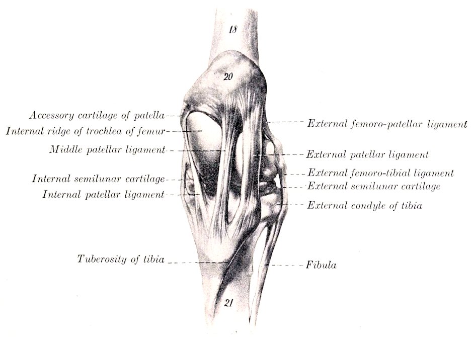
Fig. 157.—Stifle Joint of Horse, Front View, in Extension. The capsules are removed. 18, Femur; 20, patella; 21, tibia. (After Ellenberger-Baum, Anat. für Künstler.)
Ligaments.—The lateral femoro-patellar ligaments, external and internal (Ligamentum femoro-patellare fibulare, tibiale), are two thin bands which reinforce the capsule on either side. The external ligament is fairly distinct; it arises from the external epicondyle of the femur just above the lateral femoro-tibial ligament, and ends on the external border of the patella. The internal ligament is thinner and is not distinct from the capsule; it arises above the internal epicondyle, and ends on the patellar fibro-cartilage.
The patellar ligaments (Ligamenta patellæ), also called the straight ligaments of the patella, are three very strong bands which attach the patella to the tuberosity of the tibia. The external patellar ligament is attached above to the outer part of the anterior surface of the patella, and below to the outer part of the tuberosity. It receives a strong tendon from the biceps femoris muscle. The middle patellar ligament extends from the front of the apex of the patella to the lower part of 196the groove on the tuberosity of the tibia, a bursa being interposed between the ligament and the upper part of the groove. The internal patellar ligament is attached above to the patellar fibro-cartilage, and ends on the tuberosity of the tibia, internally to the groove. It is joined by the common aponeurosis of the gracilis and sartorius. These so-called ligaments are, in reality, the tendons of insertion of the quadriceps femoris muscle, and transmit the action of the latter to the tibia; they also function similarly for the other muscles attached to them as noted above.
It will be noticed that the upper attachments are further apart than the lower ones, so that the ligaments converge below. The inner ligament is especially oblique. The middle ligament is more deeply placed than the others, and therefore cannot usually be felt distinctly in the living animal.
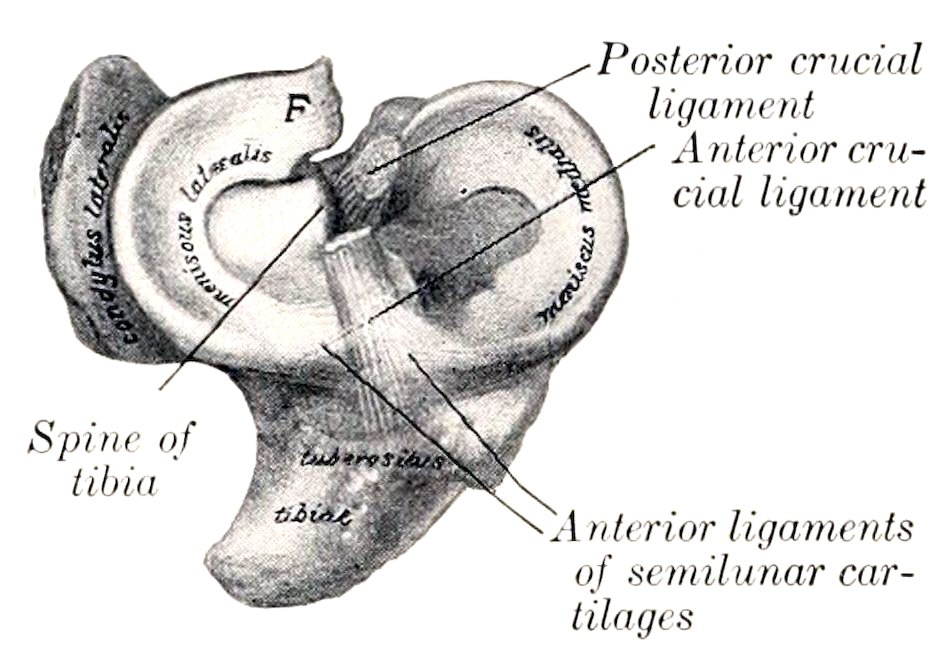
Fig. 158.—Semilunar Cartilages and Crucial Ligaments of Right Stifle of Horse. F, Femoral ligament of external cartilage. (After Schmaltz, Atlas d. Anat. d. Pferdes.)
The femoro-tibial articulation (Articulatio femoro-tibialis) is formed between the condyles of the femur, the proximal end of the tibia, and the interposed semilunar cartilages.
Articular Surfaces.—The condyles of the femur are slightly oblique in direction. The articular surface of the outer one is more strongly curved than that of the inner one; the latter is confluent below with the inner ridge of the trochlea, while the narrow ridge which connects the external condyle with the trochlea is usually non-articular. The saddle-shaped surfaces of the condyles of the tibia are not adapted to the femoral condyles, and are in contact with only a small part of them.
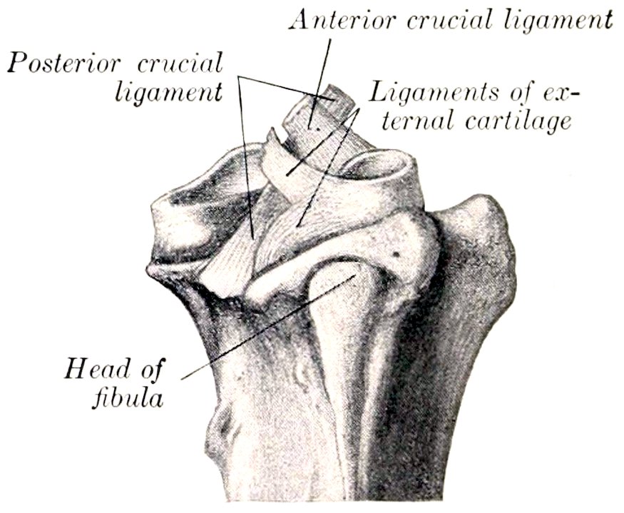
Fig. 159.—Proximal Ends of Right Tibia and Fibula of Horse, with Semilunar Cartilages and Crucial Ligaments, External View. (After Schmaltz, Atlas d. Anat. d. Pferdes.)
The semilunar cartilages (Meniscus lateralis, medialis) are two C-shaped or crescentic discs of fibro-cartilage which produce congruence in the articular surfaces. Each has an upper concave surface adapted to the condyle of the femur, and a lower surface which fits the corresponding condyle of the tibia. The external cartilage does not cover the outer and posterior part of the condyle, over which the tendon of origin of the popliteus muscle plays. The peripheral border is thick and convex, the central one very thin and concave. The fibrous ends or cornua are attached to the tibia in front of and behind the spine. The external cartilage has a third attachment by means of an oblique band (Ligamentum femorale menisci lateralis) which passes from the posterior cornu to the posterior part of the intercondyloid fossa of the femur.
The cornua of the internal cartilage (Ligamenta tibiæ anterius et posterius menisci medialis) are attached in front of and behind the inner eminence of the spine. The anterior cornu of the external cartilage (Ligamentum tibiæ anterius menisci lateralis) is attached in front of the outer eminence of the spine. The posterior cornu bifurcates; the lower branch (Ligamentum tibiæ 197posterius menisci lateralis) is inserted at the popliteal notch, the upper (Ligamentum femorale menisci lateralis) in a small fossa in the extreme posterior part of the intercondyloid fossa.
The joint capsule is attached to the margin of the tibial articular surface, but on the femur the line of attachment is for the greater part about half an inch from the articular margin. It is also attached to the convex borders of the semilunar cartilages and to the crucial ligaments. It is strong posteriorly, but in front it practically consists only of the synovial layer. There are two synovial sacs, corresponding to the double nature of the articular surfaces; they do not usually communicate, and each is partially divided into an upper and a lower compartment by the semilunar cartilage. The inner sac pouches upward about half an inch over the condyle of the femur. The external sac invests the tendon of origin of the popliteus muscle, and also pouches downward about three or four inches (ca. 8 to 10 cm.) beneath the peroneus tertius and long extensor muscles (Fig. 156). As stated above, the outer sac sometimes communicates with the femoro-patellar joint cavity, and the inner sac usually, if not always, does so in the adult.
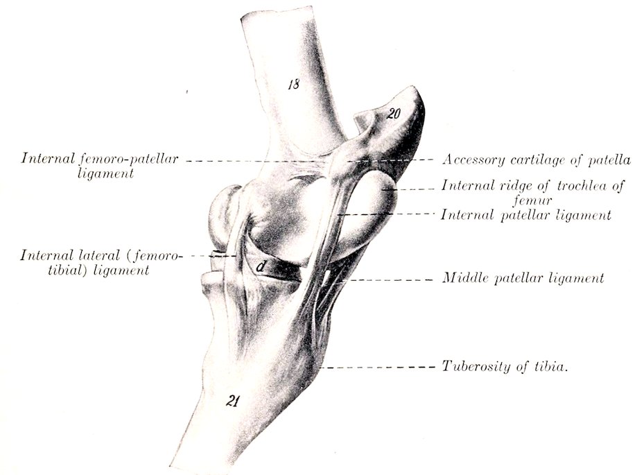
Fig. 160.—Left Stifle Joint of Horse, Internal View in Extreme Extension.[38] 18, Femur; 20, patella (base); 21, tibia; d, internal semilunar cartilage. (After Ellenberger-Baum, Anat. für Künstler.)
Ligaments.—There are four of these—two lateral and two crucial.
The internal lateral ligament (Ligamentum collaterale tibiale) is attached above to the prominent internal epicondyle of the femur, and below to a rough area below the margin of the internal condyle of the tibia.
The external lateral ligament (Ligamentum collaterale fibulare) is somewhat thicker; it arises from the upper depression on the external epicondyle, and ends on the head of the fibula. It covers the tendon of the origin of the popliteus muscle, and a bursa is interposed between the lower part of the ligament and the margin of the external condyle of the tibia.
198The crucial ligaments are two strong rounded bands situated mainly in the intercondyloid fossa of the femur, between the two synovial sacs. They cross each other somewhat in the form of an X, and are named according to their tibial attachments. The anterior crucial ligament (Ligamentum cruciatum anterius) arises in the central fossa on the tibial spine, extends upward and backward, and ends on the upper part of the inner surface of the external condyle of the femur. The posterior crucial ligament (Ligamentum cruciatum posterius) is internal to the preceding, and is somewhat larger. It is attached to an eminence at the popliteal notch of the tibia, is directed upward and forward, and ends in the anterior part of the intercondyloid fossa of the femur.
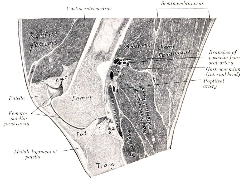
Fig. 161.—Right Stifle Joint of Horse; Sagittal Section Passing Through Outer Part of Inner Ridge of Trochlea and Intercondyloid Fossa. 1, Anterior crucial ligament; 2, posterior crucial ligament; 3, posterior cornu of internal semilunar cartilage; 4, femoral ligament of external semilunar cartilage; 5, articular artery; 6, part of vastus internus.
It may be added that these ligaments do not lie in a sagittal plane, but are somewhat twisted across each other; outward rotation of the leg untwists and slackens them.
Movements.—The principal movements of the stifle joint as a whole are flexion and extension. In the ordinary standing position the articular angle (behind) is about 140° to 150°. Flexion is limited only by contact of the leg with the thigh if the hock is also flexed. Extension is incomplete, i. e., the femur and tibia cannot be brought into the same straight line. Rotation is limited, and is freest during semiflexion. The patella glides on the femoral trochlea upward in extension, downward in flexion.
Extension is checked mainly by tension of the crucial and lateral ligaments. In extreme extension, which is accompanied by slight outward rotation of the leg, the patella can be pushed upward and inward so that its fibro-cartilage hooks over the upper end of the inner ridge of the trochlea, but it will not remain there unless held in position. When pressure is removed, the base of the patella tips forward and the cartilage lies upon the most prominent part of the trochlear 199ridge. During flexion, which is accompanied by slight inward rotation of the leg, the condyles of the femur and the semilunar cartilages glide backward on the tibia; the movement of the external condyle and cartilage is greater than that of the inner one. In extreme flexion the patellar and posterior crucial ligaments are tense; the other ligaments are relaxed. The movement of the patella is gliding with coaptation, i. e., different parts of the opposing articular surfaces come into contact successively. Only a narrow transverse strip (ca. 1.5 to 2 cm. wide) of the patella is in contact with the trochlea at a time.
TIBIO-FIBULAR ARTICULATION
The head of the fibula articulates with a crescentic facet just below the outer margin of the external condyle of the tibia. The joint capsule is strong and close. The shaft of the fibula is attached to the external border of the tibia by the interosseous membrane of the leg (Membrana interossea cruris); this is perforated about an inch from its proximal end by an opening which transmits the anterior tibial vessels to the front of the tibia. A fibrous cord usually extends from the distal end of the shaft of the fibula to the external malleolus. The latter is the distal end of the fibula which has fused with the tibia. No appreciable movement occurs in this joint.
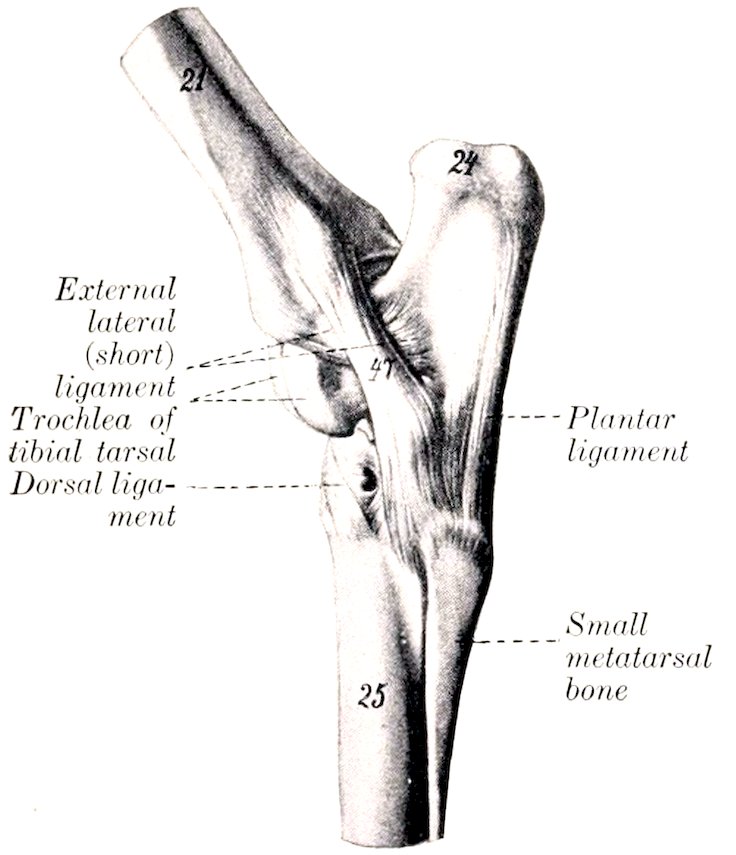
Fig. 162.—Left Hock Joint of Horse, External View.
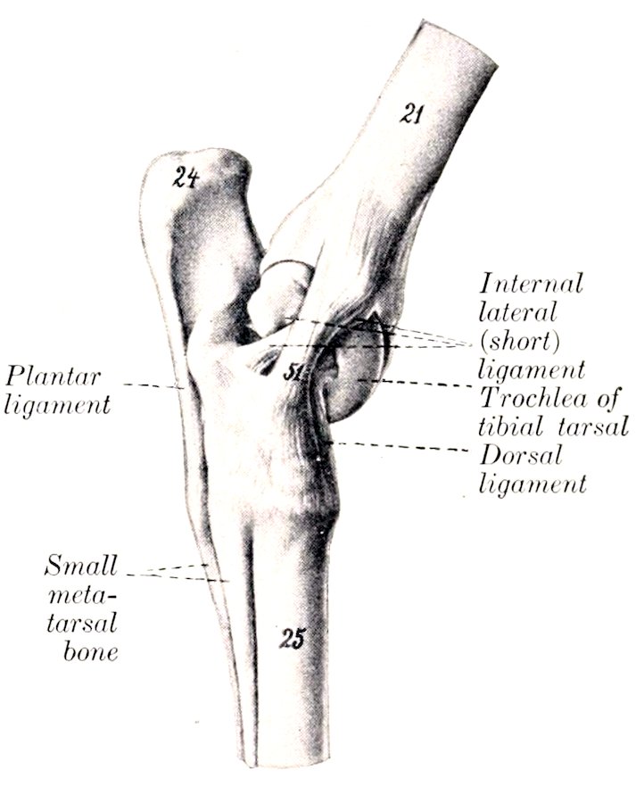
Fig. 163.—Left Hock Joint of Horse, Internal View. 21, Tibia; 24, tuber calcis; 25, large metatarsal bone; 47, long external lateral ligament; 51, long internal lateral ligament. (After Ellenberger-Baum, Anat. für Künstler.)
THE HOCK JOINT
This is a composite joint made up of a number of articulations (Articulationes tarsi). These are: (1) The tibio-tarsal articulation; (2) the intertarsal articulations; (3) the tarso-metatarsal articulation.
The tibio-tarsal articulation (Articulatio talo-cruralis) is a typical ginglymus formed by the trochlea of the tibial tarsal bone (astragalus or talus) and the corresponding surface of the distal end of the tibia. The ridges and grooves of these surfaces are directed obliquely forward and outward at an angle of about 12° to 15°, with a sagittal plane. The trochlear surface is about twice as extensive as that on the tibia, and its ridges have a spiral curvature. The other articulations are 200arthrodia, which have joint surfaces and ligaments of such a nature as to allow only a minimal amount of gliding motion.
As in the case of the carpal joints, it is convenient to describe first the common capsule and ligaments, which are the more important practically, and then to consider very briefly the special ligaments.
The fibrous part of the joint capsule is attached around the margin of the tibial articular surface above and the metatarsal surfaces below; it is also attached in part to the free surface of the bones which it covers, and blends with the lateral ligaments. Its anterior part (anterior ligament) is rather thin; in distention of the capsule, as in “bog-spavin,” its antero-internal part, which is not bound down by the tendons passing over the joint, forms a fluctuating swelling over the inner ridge of the trochlea. The posterior part (posterior and tarso-metatarsal ligaments) is very thick, and is intimately attached to the tarsal bones. Its superficial face is in part cartilaginous, and forms a smooth surface for the perforans tendon. Superiorly, it pouches upward behind the distal end of the tibia for a distance of about two inches (ca. 5 cm.). Interiorly, it is continued downward, forming the subtarsal or check ligament, which unites with the perforans tendon about the middle of the metatarsus.
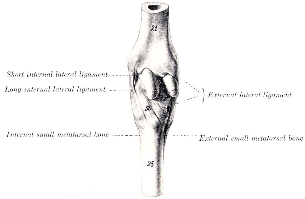
Fig. 164.—Left Hock Joint of Horse, Anterior View. 21, Tibia; 22, trochlea of tibial tarsal bone; 25, large metatarsal bone; 50, dorsal or oblique ligament. (After Ellenberger-Baum, Anat. für Künstler.)
There are four synovial sacs: 1. The tibio-tarsal sac lubricates the proximal joint, and is much the largest and most important. It is chiefly involved in the swelling produced by excess of fluid in the joint cavity, when the capsule bulges antero-internally and postero-superiorly. 2. The first intertarsal sac lines the joints formed by the tibial and fibular tarsal bones above, and the central and fourth tarsals below; it communicates in front with the proximal synovial capsule. 3. The second intertarsal sac lubricates the joints formed between the central tarsal and the bones below and on either side. 4. The tarso-metatarsal sac lubricates the joints formed between the tarsal and metatarsal bones, those between the proximal ends of the metatarsal bones, and those formed by the third tarsal with the bones on either side.
Common Ligaments.—The external lateral ligament (Ligamentum collaterale 201fibulare longum et breve) consists of two distinct bands which cross each other. The long (superficial) ligament arises on the posterior part of the external malleolus, is directed almost straight downward, and is attached to the fibular and fourth tarsal bones and the large and external small metatarsal bones. It forms a canal for the lateral extensor tendon. The short (deep) ligament arises on the anterior part of the external malleolus, is directed chiefly backward, and ends on the rough excavation on the external surface of the tibial tarsal and the adjacent surface of the fibular tarsal bone.
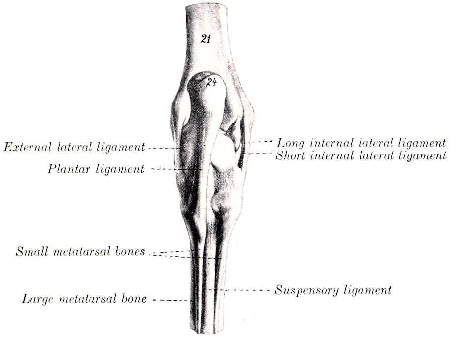
Fig. 165.—Left Hock Joint of Horse, Posterior View. 21, Tibia; 24, tuber calcis. (After Ellenberger-Baum, Anat. für Künstler.)
The internal lateral ligament (Ligamentum collaterale tibiale longum et breve) also consists of two parts which cross each other. The long ligament arises on the posterior part of the internal malleolus, becomes wider below, and is attached on the lower tuberosity of the tibial tarsal, the large and outer small metatarsal bones, and the inner surface of the lower tarsal bones which it covers. The short ligament lies largely under cover of the long one. It extends from the anterior part of the internal malleolus, runs backward and somewhat downward, and divides into two branches; one of these ends on the upper tuberosity on the inner surface of the tibial tarsal bone, the other on the sustentaculum tali.
The plantar or calcaneo-metatarsal ligament (Ligamentum tarsi plantare) is a very strong flat band which covers the outer part of the posterior surface of the tarsus. It is attached to the posterior surface of the fibular and fourth tarsal bones and the proximal end of the external metatarsal bone.
The dorsal or oblique ligament (Ligamentum tarsi dorsale) is a triangular sheet which is attached above to the lower tuberosity on the inner face of the tibial tarsal bone, and spreads out below on the central and third tarsal bones, and the proximal ends of the large and inner small metatarsal bones, to all of which it is attached.
Special Ligaments.—A considerable number of short bands which connect adjacent bones of the tarsus and metatarsus are described by various authors; some of these are quite distinct; others are difficult to isolate. Most of them are not of sufficient importance to justify detailed description.
202(1) The tibial and fibular tarsal bones are united by four bands (astragalo-calcaneal ligaments). The internal ligament extends from the sustentaculum tali to the adjacent part of the tibia tarsal, blending with the short lateral ligament. The external ligament extends from the anterior process of the fibular tarsal to the adjacent part of the external ridge of the trochlea. The superior ligament extends from the posterior margin of the trochlea to the fibular tarsal. The interosseous ligament is deeply placed in the sinus tarsi between the two bones, and is attached in the rough areas of the opposed surfaces.
(2) The smaller bones are attached to each other as follows: The central and third tarsal are united by an interosseous and an oblique anterior ligament (scaphoido-cunean ligaments). The central and fourth tarsal are united by an interosseous and a transverse external ligament (cuboido-scaphoid ligament). The third and fourth tarsals are similarly connected (cuboido-cunean ligaments). The third tarsal is joined by an interosseous (intercunean) ligament to the (fused) first and second tarsals; the latter are connected with the fourth tarsal by a transverse posterior ligament.
(3) The smaller bones are connected with the upper row as follows: The central is attached to the tibial tarsal by posterior and interosseous (astragalo-seaphoid) ligaments, and to the fibular tarsal by a short oblique (calcaneo-scaphoid) band. The fourth is attached to the fibular tarsal by interosseous and posterior (calcaneo-cuboid) ligaments. The (fused) first and second tarsals are connected with the fibular tarsal by a posterior (calcaneo-cunean) ligament.
(4) The lower tarsal bones are connected with the metatarsus by tarso-metatarsal ligaments, which are not distinct from the common ligaments, except in the case of the interosseous ligament between the third tarsal and metatarsal bones.
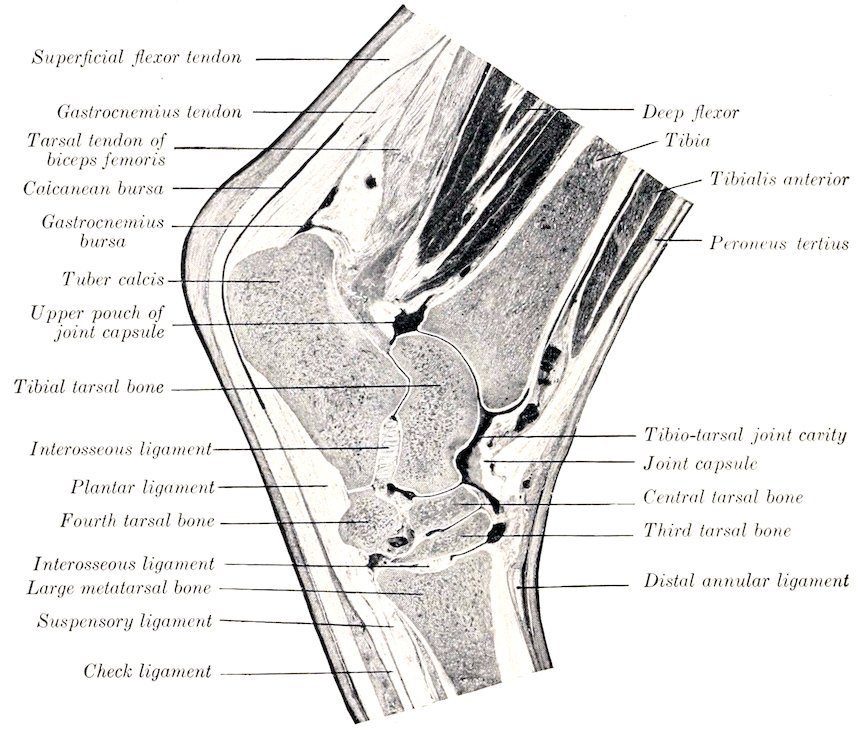
Fig. 166.—Sagittal Section of Hock of Horse.
Movements.—These are flexion and extension, which take place at the tibio-tarsal joint. The movements between the tarsal bones, and between the latter and the metatarsus, are so limited as to be negligible so far as the action of the joint as a whole is concerned. In the standing position the articular angle (in front) is about 150° to 160°. Complete extension is prevented by tension of the lateral ligaments. Flexion is checked only by contact of the metatarsus with the leg, 203provided the stifle joint is also flexed. Owing to the fact that the axis of motion is slightly oblique, the lower part of the limb deviates somewhat outward during flexion.
The movements of the hock joint must correspond with those of the stifle on account of the tendinous bands in front and behind (peroneus tertius and flexor perforatus), which extend from the lower part of the femur to the tarsus and metatarsus.
The remaining joints differ in no material respect from those of the thoracic limb.
COMPARATIVE ARTHROLOGY[39]
JOINTS AND LIGAMENTS OF THE VERTEBRÆ
Ox.—The ligamentum nuchæ is better developed than in the horse. The funicular part is clearly divided into two lateral halves, which are round at their occipital attachment, but from the axis backward become rapidly wider and flat. This wide portion is almost sagittal, lies on either side of the vertebral spines, and is covered by the trapezius and rhomboideus muscles. From the highest part of the withers (third thoracic spine) it gradually diminishes in size and fades out in the lumbar region. The lamellar part is thick, and consists of anterior and posterior parts. The anterior part is double; its fibers proceed from the funicular part to the second, third, and fourth cervical spines. The posterior part is single; its fibers extend from the first thoracic spine to the fifth, sixth, and seventh cervical spines.
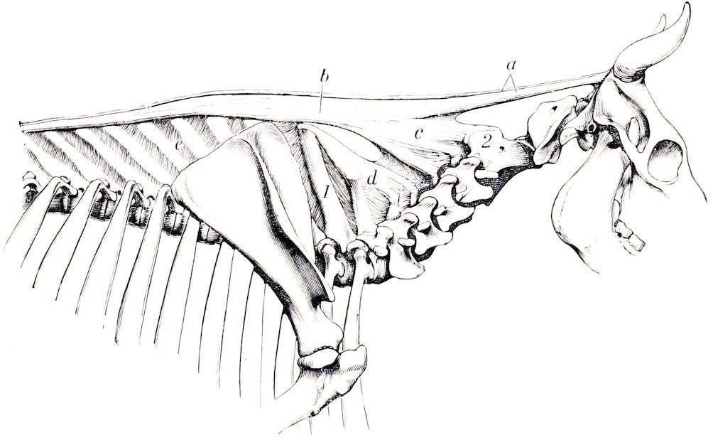
Fig. 167.—Ligamentum Nuchæ of Ox. a, Funicular part; b, wide portion; c, d, lamellar part; e, interspinous ligaments; 1, spinous process of first thoracic vertebra; 2, axis. (Ellenberger-Baum, Anat. d. Haustiere.)
The inferior common ligament is very strong in the lumbar region.
The intervertebral fibro-cartilages are thicker than in the horse.
The interspinous ligaments of the back and loins consist largely of elastic tissue.
204There are no intertransverse joints in the lumbar region.
Pig.—The ligamentum nuchæ is represented by a fibrous raphé and thin layers of elastic tissue which extend between the cervical spines.
The atlanto-occipital and atlanto-axial joints resemble those of the dog.
The interspinous ligaments of the neck are elastic.
Dog.—The ligamentum nuchæ consists of a small fibrous band which extends from the spine of the axis to the anterior thoracic spines; it may be regarded as a mere fibrous raphé between the right and left muscles.
There are interspinous muscles instead of ligaments in the neck.
There are three ligaments in connection with the odontoid process. The two alar ligaments (Ligamenta alaria) arise on either side of the odontoid process, diverge, and end on either side of the foramen magnum. The transverse ligament of the atlas (Ligamentum transversum atlantis) stretches across the dorsal surface of the odontoid process and binds it down on the ventral arch of the atlas, a bursa being interposed. It is attached on either side to the lateral masses of the atlas.
The two capsules of the atlanto-occipital joint communicate with each other, and usually with the capsule of the atlanto-axial joint also.
Articulations of the Thorax
Ox.—The second to the eleventh costo-chondral joints inclusive are diarthroses with close capsules, reinforced externally. (They are synchondroses in the sheep.) The upper parts of the cartilages are attached to each other by distinct elastic ligaments (Ligamenta intercostalia).
The first pair of chondro-sternal joints are separate from each other; intercrossing fibers unite the costo-chondral junctions above the joints.
The first segment of the sternum forms a diarthrosis with the body. The anterior joint surface is concave, the posterior convex. The joint (Articulatio intersternalis) is surrounded by a close capsule, and the joint surfaces are attached to each by a small intraarticular ligament. Limited lateral movement is possible. (In the sheep the joint is a synchondrosis.)
Both surfaces of the sternum are covered by a layer of fibrous tissue.
Pig.—The second to the fifth or sixth costo-chondral joints are diarthroses. The intersternal articulation and the sternal ligaments resemble those of the ox.
Dog.—The first chondro-sternal joints do not coalesce.
The internal sternal ligament divides into three bands.
TEMPORO-MANDIBULAR ARTICULATION
Ox.—The articular surfaces are of such a character as to permit more extensive lateral movement than in the horse.
The condyle of the mandible is relatively small and is concave transversely. The temporal articular surface is extensive and is convex in both directions. The postglenoid process is small.
Pig.—The considerable longitudinal diameter of the temporal articular surfaces and the very small size of the postglenoid process allow great freedom of protraction and retraction of the lower jaw. Lateral movement is limited. The posterior ligament is absent.
Dog.—The articular surfaces allow extremely little lateral or gliding movement. They are cylindrical in curvature, and the interarticular disc is very thin. The posterior ligament is absent.
The other articulations of the skull are sufficiently described in the Osteology.
205
Articulations of the Thoracic Limb
SHOULDER JOINT
Ox.—The articular angle is about 100°.
Pig and Dog.—The joint capsule communicates freely with the bicipital bursa. There is a rudimentary marginal cartilage around the rim of the glenoid cavity. In the dog there is usually a strong band extending from the acromion to the outer part of the capsule; another band (Ligamentum coraco-acromiale) often stretches between the scapular tuberosity and the acromion.
ELBOW JOINT
Ox.—No important differences exist. The upper part of the interosseous radio-ulnar ligament is commonly ossified in the adult.
Pig.—There are no important differences. The radius and ulna are so firmly united by the interosseous ligament as to prevent any appreciable movement between them.
Dog.—The joint capsule is reinforced in front by an oblique ligament which arises on the front of the external condyle of the humerus above the joint surface, and joins the terminal part of the biceps and brachialis below. The external lateral ligament is thick and divides into two parts; the anterior part is attached to the radius and blends with the annular ligament; the posterior part widens below, forming a sort of cap, and is attached to the ulna. The internal lateral ligament also divides into two branches; the anterior branch ends on the inner surface of the neck of the radius; the posterior enters the interosseous space and is attached to both bones. An elastic band (Ligamentum olecrani) extends from the outer surface of the internal epicondyle to the anterior border of the ulna.
There are two radio-ulnar joints. The proximal radio-ulnar joint is included in the capsule of the elbow, but is provided with an annular ligament which extends from the lower part of the external ligament across the front of the proximal end of the radius to the ulna, blending with the biceps and brachialis tendons. The distal joint is formed by a concave facet on the radius and a convex one on the radius, and is surrounded by a tight capsule. The interosseous membrane unites the shafts of the two bones. The movements consist of limited rotation of the radius (ca. 20°), carrying the paw with it. The ordinary position is termed pronation; outward rotation is supination.[40]
THE CARPAL JOINTS
These have the same general arrangement as in the horse. Numerous minor differences naturally exist, but must be excluded from this brief account, which contains only important special features.
The lateral movements are freer, especially in the dog, but flexion is not so complete: the anatomical explanation of these facts lies in the nature of the articular surfaces and certain ligamentous differences. The lateral ligaments are much weaker, the long external one being especially small in the ox. Two oblique, somewhat elastic, bands cross the front of the radio-carpal and intercarpal joints. The proximal one is attached to the distal end of the radius and passes downward and outward to the ulnar carpal bone; the other one connects the radial and fourth carpal bones in a similar fashion.
In the ox the short lateral ligaments are well defined, a ligament connects 206the accessory carpal with the distal end of the ulna, and strong bands connect the distal bones with the metacarpus.
The interosseous and interordinal ligaments vary with the number of carpal bones present in the different species.
INTERMETACARPAL JOINTS
In the ox the small (fifth) metacarpal bone articulates with the large metacarpal, but not with the carpus. The joint cavity is connected with that of the carpo-metacarpal sac. The proximal end of the small metacarpal bone is attached by a ligament to the fourth carpal, and another band extends from its distal part to the side of the large metacarpal. There is also an interosseous ligament, which is permanent and allows a small amount of movement.