OSTEOLOGY (continued 3)
Reading edition
The proximal extremity (Basis) bears an undulating articular surface adapted to the lower row of carpal bones. The greater part supports the third carpal bone; the oblique outer part, separated from the preceding by a ridge, articulates with the fourth, and a small facet for the second is usually found at the postero-internal angle. On either side is a notch separating two small facets which articulate with the proximal ends of the small metacarpal bones. Toward the inner side of the anterior surface is the metacarpal tuberosity, into which the extensor carpi radialis is inserted. The posterior surface is roughened for the attachment of the suspensory ligament.
The distal extremity (Trochlea s. Capitulum) presents an articular surface for the first phalanx and the proximal sesamoid bones, which is composed of two condyles, separated by a sagittal ridge; the inner condyle is slightly the larger. On either side is a small fossa, surmounted by a tubercle, for the attachment of the lateral ligaments of the fetlock joint.
The large metacarpal is one of the strongest bones in the skeleton. The compact substance is specially thick in front and internally. The medullary canal extends further toward the ends than in most of the long bones of the horse and there is little spongy bone.
The Small Metacarpal Bones
These are situated on either side of the posterior surface of the large metacarpal bone, and form the sides of the metacarpal groove. Each consists of a shaft and two extremities.
The shaft (Corpus) is three-sided and tapers to the distal end. It is variably curved, convex toward the middle line of the limb. The anterior surface is flattened and is rough, except in its lower part; it is attached to the large metacarpal bone by an interosseous ligament, except near the distal end. The abaxial surface is smooth and rounded from side to side above, grooved below. The axial surface is smooth and concave from edge to edge, except below, where it forms a rounded edge.
The proximal extremity or head (Basis) is relatively large. In the case of the inner bone it usually bears two facets above which support the second and third carpal bones, while the outer bone has here a single facet for articulation with the fourth carpal bone. Each has also two facets for articulation with the large metacarpal, and is elsewhere roughened for the attachment of ligaments and muscles. The inner bone may present a small facet behind for the first carpal bone.
The distal extremity (Capitulum) is usually a small nodule, which projects to a variable extent in different subjects, and is easily felt in the living animal. It is situated two-thirds to three-fourths of the way down the region.
The small metacarpal bones vary much in length, thickness, and curvature. In the majority of cases the inner bone is the longer; in other subjects the outer one is the longer or there is no material difference. Sometimes the curvature is very pronounced, so that the distal end causes a decided projection. The distal end is very variable in size and may be a mere point.
Development.—The large metacarpal bone ossifies from three centers. The proximal extremity unites with the shaft before birth, the distal extremity toward the middle of the second year. The small metacarpal bones ossify from two centers, one of which is for the proximal extremity. Their distal ends are cartilaginous 89at birth. Fusion of the middle part of the shaft with the large metacarpal bone is common.
THE PHALANGES
The First Phalanx
The first phalanx (Phalanx prima)[22] is a long bone, situated between the large metacarpal bone above and the second phalanx below. It is directed obliquely downward and forward, forming an angle of 50 to 55 degrees with the horizontal plane in well-formed limbs. It consists of a shaft and two extremities.
The shaft (Corpus) is wider and much thicker above than below, and presents two surfaces and two borders. The anterior or dorsal surface is convex from side to side and smooth. The posterior or volar surface is flattened, and bears a triangular rough area, bounded laterally by ridges which begin at the tuberosities above and converge below; this area furnishes attachment to the inferior sesamoidean ligaments. The borders, internal and external, are rounded and have a rough area or a tubercle on their middle parts.
The proximal extremity (Basis) is relatively large. It bears an articular surface adapted to the distal end of the large metacarpal bone, consisting of two glenoid cavities separated by a sagittal groove; the inner cavity is a little larger than the outer one. The posterior angles are formed by buttress-like tuberosities for ligamentous attachment. The anterior surface has a slight elevation for the attachment of the lateral extensor tendon.
The distal extremity is smaller, especially in its antero-posterior diameter. It presents a trochlea for articulation with the second phalanx, consisting of a shallow central groove and two lateral convex areas or condyles; the inner area is a little the larger. On either side, just above the margin of the articular surface, is a depression surmounted by a tubercle, to both of which the lateral ligament is attached. Behind the tubercle is a distinct facet to which the superficial flexor tendon is attached.
Development. The first phalanx ossifies from three centers. The distal end unites with the shaft before birth, the proximal end early in the first year.
The first phalanx contains a small medullary canal in the middle of the shaft. It may be remarked that the bone is twisted slightly; when placed volar surface down on the table, it touches the latter by three points only, the proximal tuberosities and the internal condyle.
The Second Phalanx
The second phalanx (Phalanx secunda)[23] is situated between the first and third phalanges, its direction corresponding to that of the first phalanx. It is flattened from before backward, and its width is greater than its height. It may be described as possessing four surfaces.
The upper or proximal surface presents two glenoid cavities separated by a low ridge, and articulates with the first phalanx. The middle of the anterior border is elevated and roughened in front for the attachment of the anterior or common extensor tendon. The posterior border is thick and overhanging; in the fresh state its middle part is covered with cartilage, over which the deep flexor tendon passes. On either side there is an eminence, to which the lateral ligament and the superficial flexor tendon are attached.
The inferior or distal surface is trochlear, and articulates with the third phalanx and third sesamoid bone. It resembles somewhat the trochlea of the first phalanx, but is more extensive and encroaches more on the anterior and posterior surfaces.
The anterior or dorsal surface is convex from side to side and smooth in its 90middle; on its lower part are lateral rough depressions, surmounted by tuberosities, to both of which ligaments are attached.
The posterior or volar surface is smooth, flattened, and slopes obliquely downward and forward. The borders which separate the anterior and posterior surfaces are concave from above downward, rounded from before backward.
Development.—The second phalanx ossifies like the first, but the proximal end unites with the shaft two or three months earlier.
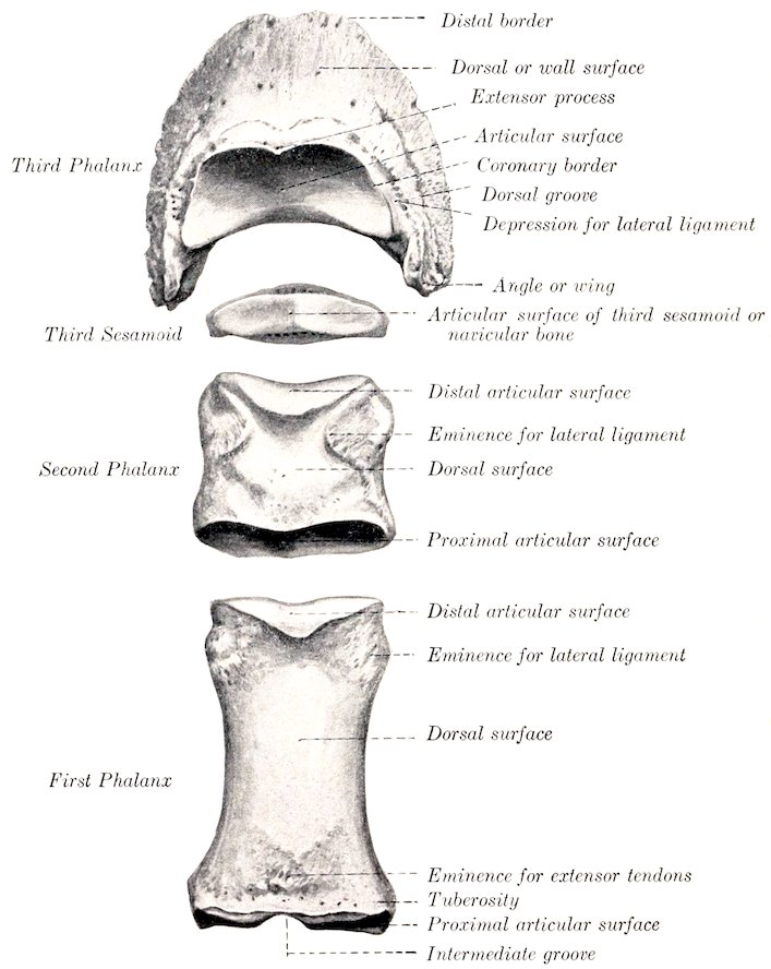
Fig. 58.—Phalanges and Third Sesamoid of Horse, Dorsal Aspect.
The Third Phalanx
The third or ungual phalanx (Phalanx tertia)[24] is entirely inclosed by the hoof, to which it conforms in a general way. It presents for examination three surfaces, three borders, and two angles or wings.
The articular surface (Facies articularis) faces upward and backward, and is chiefly adapted to the distal surface of the second phalanx, but a narrow flattened area along the posterior border articulates with the third sesamoid. The anterior 91or coronary border forms a central eminence, the extensor (or pyramidal) process (Processus extensorius), to the front of which the anterior extensor tendon is attached. On either side is a depression for the attachment of the lateral ligament.
The dorsal or wall surface (Facies dorsalis) slopes downward and forward. The angle of inclination on the ground plane is about 45 to 50 degrees in front. Laterally the height diminishes, and the slope becomes steeper, especially on the inner side. From side to side the curvature is almost semicircular. The surface is rough and porous, resembling pumice stone somewhat. It is perforated by numerous foramina of various sizes; a series of larger ones is situated on or near the lower border. On either side the dorsal (or preplantar) groove (Sulcus dorsalis) passes forward from the wing and ends at one of the larger foramina. In the fresh state this surface is covered by the matrix of the wall of the hoof. The inferior or distal border is thin, sharp, and irregularly notched; there is commonly a wider notch in front.
The volar or inferior surface (Facies volaris) is arched, and divided into two unequal parts by a curved rough line, the semilunar crest (Crista semilunaris). The larger anterior area is crescent-shaped, concave, and comparatively smooth; it corresponds to the sole of the hoof, and may be termed the sole surface. The posterior part is much smaller, and is semilunar; it is related to the deep flexor tendon, and is hence called the flexor or tendon surface (Facies flexoria). It presents a central prominent rough area, on either side of which is the volar (or plantar) foramen (Foramen volare), to which the volar (or plantar) groove (Sulcus volaris) conducts from the wing. The foramina lead into the semilunar canal within the bone, from which small canals lead to some of the foramina of the wall surface. The deep flexor tendon is inserted into the semilunar crest and the central rough area behind it.
The volar grooves and foramina transmit the terminations of the digital arteries into the semilunar canal, where they meet and form a terminal arch, from which branches pass through canals in the bone and emerge through the foramina on the wall surface.
The angles or wings (Anguli) are prismatic masses which project backward on either side; the inner one is usually the shorter. Each is divided into upper and lower parts by a notch, or is perforated by a foramen which leads to the vascular groove on the side of the wall surface.[25] The upper border carries the lateral cartilage.
The lateral cartilages (Cartilagines ungulæ) are rhomboid curved plates, which surmount the wings on either side. They are relatively large and extend above the margin of the hoof sufficiently to be distinctly palpable. The abaxial surface is convex, the axial concave. The upper border is convex and thin; the lower is thicker and is in part attached to the wing. The anterior end is attached by ligament to the side of the second phalanx. The posterior end curves toward its fellow at the heel, and is perforated by numerous foramina for the passage of veins. The central part is mainly hyaline, the periphery mainly fibrous.
It will be noted that the size and form of the wings vary much in different specimens. In the new-born foal the wing is a small, pointed projection. Later the process of ossification invades the lower part of the cartilage to a varying extent. In some cases the greater part of the cartilage is ossified—a condition commonly termed “sidebone.”
Development.—The ossification of the terminal phalanx is peculiar. While the proximal articular part is still cartilaginous, a perichondrial cap of bone is formed in relation to the hoof. Later the process extends into the upper part.
Structure.—The interior of this bone is channeled by numerous canals for 92vessels, most of which radiate from the semilunar canal to the wall surface; these are not canals for nutrient vessels of the bone, but transmit arteries to the matrix of the hoof. Thick layers of compact substance are found at the articular and flexor surfaces and the extensor process, i. e., at the points of greatest pressure and traction.
THE SESAMOID BONES
The two proximal or great sesamoids (Ossa sesamoidea phalangis primæ) are situated behind the distal end of the large metacarpal bone, and are closely attached to the first phalanx by strong ligaments. Each has the form of a three-sided pyramid. The anterior or articular surface conforms to the corresponding part of the distal end of the large metacarpal bone. The posterior or flexor surface is flattened and oblique; in the fresh state it is covered by a layer of cartilage which also fills the interval between the opposed borders of the two bones, and forms a smooth groove for the deep flexor tendon. The abaxial surface is concave, and gives attachment to part of the suspensory ligament; it is separated from the posterior surface by a rough everted border. The base faces downward, and furnishes attachment to the inferior sesamoidean ligaments. The apex is directed upward and is rounded.
The third sesamoid or navicular bone (Os sesamoideum phalangis tertiæ) is shuttle-shaped, and is situated behind the junction of the second and third phalanges. Its long axis is transverse, and it possesses two surfaces, two borders, and two extremities. The articular surface (Facies articularis) faces upward and forward; it consists of a central eminence, flanked by concave areas, and articulates with the distal end of the second phalanx. The flexor or tendon surface (Facies flexoria) is directed downward and backward. It resembles the articular surface in form, but is more extensive and not so smooth. In the fresh state it is coated with cartilage and the deep flexor tendon plays over it. The proximal border (Margo liber) is wide and grooved in its middle, narrower and rounded on either side. The distal border (Margo ligamenti) bears in front a narrow facet for articulation with the third phalanx. Behind this is a groove, which contains a number of relatively large foramina, and is bounded behind by a prominent edge. The extremities are blunt-pointed.
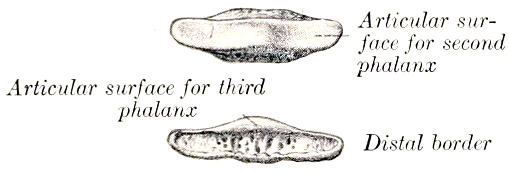
Fig. 59.—Third Sesamoid or Navicular Bone of Horse. (After Schmaltz, Atlas d. Anat. d. Pferdes.)
Development.—It ossifies from a single center.
The Bones of the Pelvic Limb
The pelvic girdle consists of the ossa coxæ, which unite ventrally at the symphysis pelvis, and articulate with the sacrum dorsally.
OS COXÆ
The os coxæ (or os innominatum) forms the skeleton of the hip or haunch, and is the largest of the flat bones. It consists primarily of three parts, the ilium, ischium, and pubis, which meet to form the acetabulum, a large cotyloid cavity for articulation with the head of the femur. These parts are fused at about one year of age, but it is convenient to describe them separately.
93
THE ILIUM
The ilium (Os ilium) is the largest of the three parts. It is irregularly triangular and presents two surfaces, three borders, and three angles.
The gluteal surface (Facies glutæa) faces upward, backward, and outward. It is wide and concave in front, narrower and convex behind. The wide part is crossed by the curved gluteal line (Linea glutæa), which extends from the middle of the inner border toward the external angle. This surface gives attachment to the middle and deep gluteal muscles.
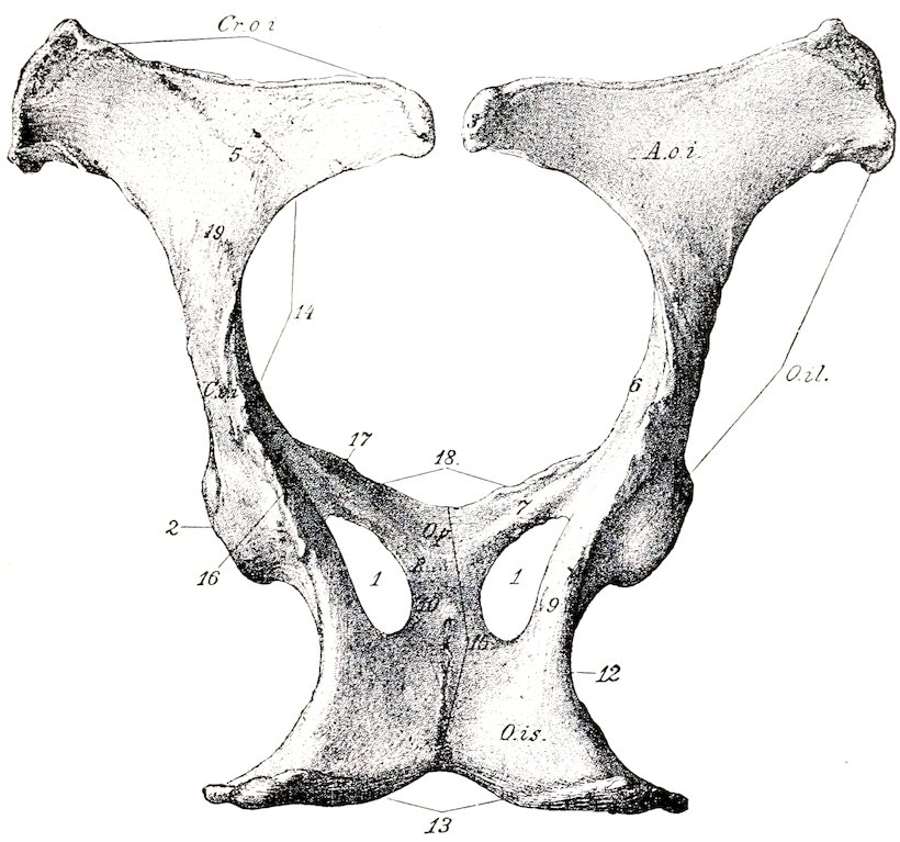
Fig. 60.—Ossa Coxarum of Mare, Dorsal View. O.il., Ilium; O.p., pubis; O.is., ischium; A.o.i., wing of ilium; C.o.i., shaft of ilium; Cr.o.i., anterior border (crest) of ilium; 1, obturator foramen; 2, acetabulum; 3, internal angle of ilium; 4, external angle of ilium; 5, gluteal line; 6, psoas tubercle; 7, 8, acetabular and symphyseal branches of pubis; 9, 10, acetabular and symphyseal branches of ischium; 12, lesser sciatic notch; 13, ischial arch; 14, great sciatic notch; 15, symphysis pelvis; 17, ilio-pectineal eminence; 18, anterior borders of pubic bones; 19, posterior gluteal line. (Struska, Anat. d. Haustiere.)
The pelvic surface (Facies pelvina) faces in the opposite direction; it is convex, and consists of two distinct parts. The inner triangular part (Pars articularis) is roughened for ligamentous attachment, and bears an irregular facet, the auricular surface (Facies auricularis), for articulation with the sacrum. The outer quadrilateral part (Pars iliaca) is in general smooth. It is crossed by the ilio-pectineal line (Crista iliopectinea), which begins below the auricular surface and is continued on the shaft of the bone to join the anterior border of the pubis. The line is interrupted by furrows for the iliaco-femoral vessels, and below these it bears the psoas tubercle (Tuberculum psoadicum), which gives attachment to the psoas minor muscle. The iliacus muscle is attached to the surface external to the ilio-pectineal line.
94The anterior border or crest (Crista iliaca) is concave, thick, and rough.
The internal border is deeply concave; its middle part forms the lower boundary of the great sciatic foramen, and it is continuous behind with the superior ischiatic spine.
The external border is concave and in great part rough. Its fore part is crossed by grooves for the ilio-lumbar vessels, which are continued on the pelvic surface. The nutrient foramen is usually situated on or near the posterior part of this border.
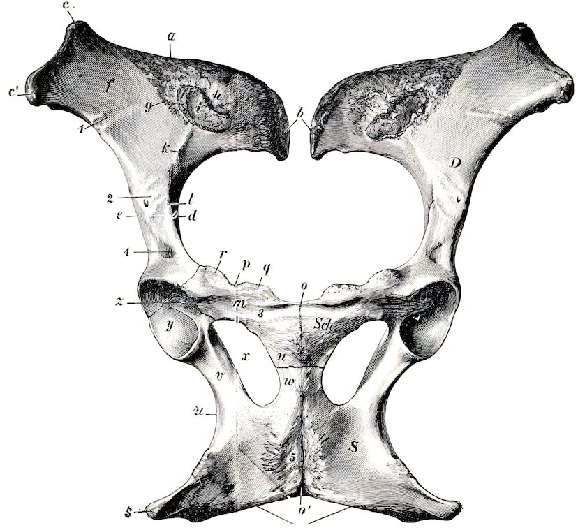
Fig. 61.—Ossa Coxarum of Mare, Ventral View. D, Ilium; Sch., pubis; S, ischium; a, anterior border (crest) of ilium; b, internal angle of ilium; c′, external angle of ilium; d, great sciatic notch; e, external border of ilium; f, iliac surface; g, linea arcuata; h, rough ligamentous area; i, auricular surface; k, ilio-pectineal line; l, psoas tubercle; m, transverse branch, and n, symphyseal branch of pubis; o, o′, symphysis pelvis; p, anterior border of pubis (pecten); q, tuberculum pubicum; r, ilio-pectineal eminence; s, tuber ischii; t, ischial arch; u, lesser sciatic notch; v, acetabular branch, and w, symphyseal branch of ischium; x, obturator foramen; y, articular surface of acetabulum; z, fossa acetabuli; 1, groove for ilio-lumbar artery; 2, groove for iliaco-femoral artery; 3, subpubic groove; 4, depression for inner tendon of origin of rectus femoris; 5, rough area for attachment of adductor muscles. (Ellenberger-Baum, Anat. d. Haustiere.)
The internal or sacral angle (Tuber sacrale) curves upward and a little backward close to the first sacral spine, and forms here the highest point of the skeleton. It is somewhat thickened and rough.
The external or coxal angle (Tuber coxæ) forms the basis of the point of the hip. It is a large quadrangular mass, narrow in its middle, and enlarged at either end, where it bears a pair of tuberosities. It is roughened for muscular attachment.
The posterior or acetabular angle meets the other two bones at the acetabulum, of which it forms about two-fifths. Its prominent upper border forms part of the superior ischiatic spine, which is roughened externally, smooth internally. Two depressions above and in front of the acetabulum give attachment to the 95tendons of origin of the rectus femoris muscle. This angle is connected with the ala or wide part of the bone by a constricted part, often termed the shaft. The latter is of three-sided prismatic form. Its external surface is convex and rough, and gives attachment to the deep gluteus muscle. Its pelvic surface is smooth and is grooved for the obturator vessels and nerves. Its ventral surface is crossed by vascular grooves, below which is a rough area, bounded internally by the psoas tubercle.
THE ISCHIUM
The ischium (Os ischii) forms the posterior part of the ventral wall or floor of the bony pelvis. It slopes a little downward and inward, but is practically horizontal in the longitudinal direction. It is irregularly quadrilateral, and may be described as having two surfaces, four borders, and four angles.
The superior or pelvic surface (Facies pelvina) is smooth and slightly concave from side to side.
The inferior surface (Facies externa) is nearly flat, and is in great part roughened for the attachment of the adductor muscles.
The anterior border forms the posterior margin of the obturator foramen.
The posterior border is thick and rough. It slopes obliquely inward and forward to meet the border of the other side, forming with it the ischial arch (Arcus ischiadicus).
The internal border meets the opposite bone at the symphysis.
The external border is thick and rounded, but concave in its length; it forms the lesser sciatic notch, the lower boundary of the lesser sciatic foramen.
The antero-internal angle or symphyseal branch (Ramus symphyseos) meets the pubis, with which it forms the inner boundary of the obturator foramen.
The antero-external angle or acetabular branch joins the other two bones at the acetabulum, of which it forms more than half. Superiorly it bears part of the superior ischiatic spine (Spina ischiadica), and internally it is grooved for the obturator vessels.
The postero-external angle is a thick three-sided mass, the tuber ischii (Tuber ischiadicum); its lower border is the inferior ischiatic spine, to which the biceps femoris and semitendinosus muscles are attached.
THE PUBIS
The pubis (Os pubis) is the smallest of the three parts of the os coxæ. It forms the anterior part of the pelvic floor, and may be described as having two surfaces, three borders, and three angles.
The superior or pelvic surface (Facies pelvina) is convex in the young subject and the stallion, concave and smooth in the mare and usually in the gelding also. The urinary bladder rests on it.
The inferior or ventral surface (Facies externa) is convex, and in great part rough for muscular attachment. Near the anterior border it is crossed by the subpubic groove, the inner part of which is occupied by a large vein, the outer part by the pubo-femoral ligament.
The anterior border is thin in its inner part (except in the young subject and the stallion), forming the pecten ossis pubis. Externally it bears the rough ilio-pectineal eminence (Eminentia iliopectinea), beyond which it is continuous with the ilio-pectineal line.
The internal border joins the opposite bone at the symphysis pubis.
The posterior border forms the anterior margin of the obturator foramen, and is marked externally by the obturator groove.
The internal angle meets its fellow at the anterior end of the symphysis. This 96part is very thick in the young subject and the stallion, but in the mare, and usually in the gelding also, it becomes thin with advancing age.
The external or acetabular angle joins the ilium and ischium at the acetabulum.
The posterior angle joins the ischium, with which it forms the inner boundary of the obturator foramen.
The pubis may conveniently be regarded as consisting of two branches which meet at a right angle; these are termed the transverse or acetabular branch (Ramus acetabularis) and the longitudinal or symphyseal branch (Ramus symphysecs).
The acetabulum is a cotyloid cavity which lodges the head of the femur. It faces downward and outward, and consists of an articular and a non-articular part. The articular part (Facies lunata) is crescentic, and is cut into internally by the non-articular part, which lies at a lower level, and is termed the acetabular fossa (Fossa acetabuli). The inner part of the rim is correspondingly cut into by the acetabular notch (Incisura acetabuli), which is converted into a foramen by the transverse ligament in the fresh state, and transmits the pubo-femoral and round ligaments to the head of the femur.
The obturator foramen (Foramen obturatum) is situated between the pubis and ischium. It is oval in outline, the longer axis being directed forward and outward. Its margin is grooved antero-externally for the obturator nerve and vessels.
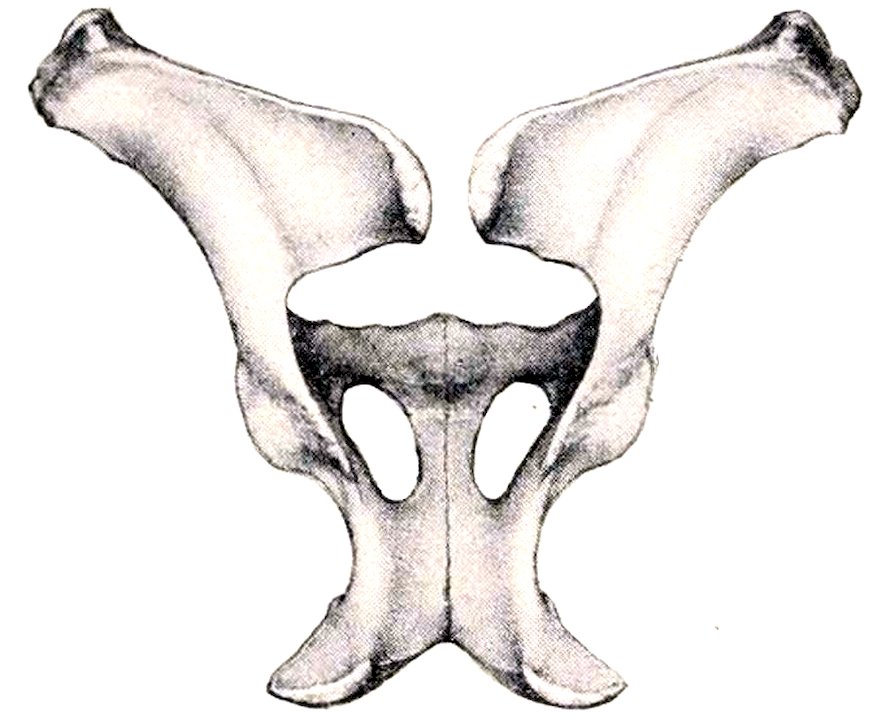
Fig. 62.—Ossa Coxarum of Stallion, Dorsal View. (After Ellenberger-Baum, Anat. für Künstler.)
Development.—Each division of the os coxæ ossifies from one chief center. The center for the ilium appears first near the acetabulum, followed quickly by one for the ischium, and a little later by the pubic center. Secondary centers appear for the crest and external angle of the ilium, the tuber and posterior border of the ischium, and the acetabular part of the pubis. The pubis and ischium are united at birth or soon after, but are not fused with the ilium until the second year. The epiphyseal parts fuse with the main mass at four and a half to five years of age.
The acetabular part of the pubis ossifies from a separate center. It is most distinct in the embryo at three months, and is often called the os acetabuli. Martin says that the ilium has a center for the acetabular part, one for the shaft and wing, and a third for the crest. He also states that there is a special center for the acetabular part of the ischium, and a transitory nucleus in the symphyseal part of the pubis.
THE PELVIS
The bony pelvis is composed of the ossa coxarum, the sacrum, and the first three coccygeal vertebræ. The dorsal wall or roof is formed by the sacrum and first three coccygeal vertebræ, and the ventral wall or floor by the pubic and ischial bones. The lateral walls are formed by the ilia and the acetabular part of the ischia. The defect in the skeleton here is supplied in the fresh state by the sacro-sciatic ligaments and semimembranosus muscles.
The anterior aperture or inlet (Apertura pelvis cranialis) is bounded by the terminal line (Linea terminalis) or brim, composed of the base of the sacrum dorsally, the ilio-pectineal lines laterally, and the anterior border of the pubis ventrally. 97It is almost circular in the mare, semi-elliptical in the stallion, and faces obliquely downward and forward. It has two principal diameters. Of these, the conjugate or sacro-pubic diameter (Conjugata) is measured from the sacral promontory to the anterior end of the symphysis. The transverse diameter (Diameter transversa) is measured at the greatest width, i. e., just above the psoas tubercle.
The posterior aperture or outlet (Apertura pelvis caudalis) is much smaller and is very incomplete in the skeleton. It is bounded above by the third coccygeal vertebra and below by the ischial arch; in the fresh state it is completed laterally by the sacro-sciatic ligament and the semimembranosus muscle.
The axis of the pelvis is an imaginary line drawn through the centers of the inlet, cavity, and outlet.
Sexual Differences.—Marked differences exist in the size and form of the pelvis in the two sexes. The average conjugate diameter is about 9½ inches (ca. 23 to 24 cm.) in the mare, 7½ inches (ca. 18 to 20 cm.) in the stallion. The transverse diameter of the inlet averages about 9 inches (ca. 22 to 23 cm.) in the mare, and 8 inches (ca. 20 cm.) in the stallion. The obliquity of the inlet is greater in the female; the difference is indicated by the fact that a vertical plane from the pecten cuts the fourth sacral segment in the female, the second in the male. The outlet is also larger in the mare, the ischial arch being about one-third wider than in the stallion. The cavity is much more roomy in the female; the transverse diameter between the middles of the superior ischiatic spines is about 8 inches (20 cm.) in the mare, 6 inches (15 cm.) in the stallion. The pubic part of the floor in the female is concave and lies considerably lower than the ischiatic part, which is wide and relatively flat. In the stallion the pubis is very thick centrally, and this part of the floor is convex, while the ischial part is relatively narrow, and is concave from side to side. The obturator foramina are correspondingly larger in the female. The ilium is shorter, and the greater sciatic notch deeper and narrower in the male. The pelvis of the gelding, when castration has been performed early, resembles that of the mare; otherwise the male characters appear to be retained to a large degree.
THE FEMUR
The femur or thigh bone (Os femoris) is the largest and most massive of the long bones. It extends obliquely downward and forward, articulating with the acetabulum above and the tibia and patella below. It presents for examination a shaft and two extremities.
The shaft or body (Corpus femoris) is in general cylindrical, but flattened behind, and larger above than below. The anterior and lateral surfaces are continuous and strongly convex from side to side; there is often a central vertical rough line on the proximal part, but otherwise these surfaces are smooth. They are covered by the quadriceps femoris muscle. The posterior surface is wide, flat, and smooth in its proximal fourth. Below this part there is a rough elevation externally for the attachment of the femoral tendon of the biceps femoris, and a rough line internally to which the quadratus femoris is attached. The middle third is narrower, and is rough for the attachment of the adductor muscle. Just below this area an oblique groove crosses the surface, indicating the position of the femoral vessels. The internal border bears on its proximal part the internal trochanter or trochanter minor, a thick rough ridge, to which the ilio-psoas muscle is attached. From this a rough line curves up to the front of the neck and indicates the limit of the attachment of the vastus internus muscle. A narrow rough area about the middle gives attachment to the pectineus muscle, and the nutrient foramen is usually found just in front of this mark. The supracondyloid crest is situated below the groove for the femoral vessels, and gives origin to the inner head of the gastrocnemius. The external border is prominent in its upper part, and bears at the junction of its proximal and middle thirds the external or third trochanter (Trochanter tertius); this process is curved forward, and furnishes insertion to the tendon of the superficial gluteus muscle. At the lower part is found the supracondyloid or plantar fossa (Fossa plantaris), in which the superficial flexor arises; it is bounded externally by a thick rough margin, to which the outer head of the gastrocnemius muscle is attached.
98
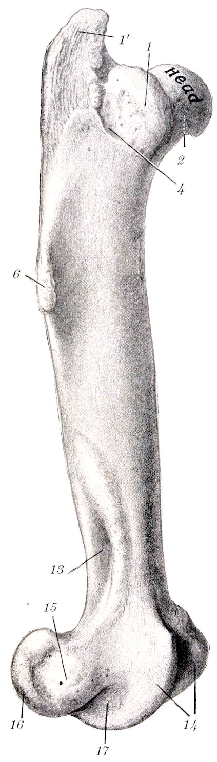
Fig. 63.—Right Femur of Horse, External View.
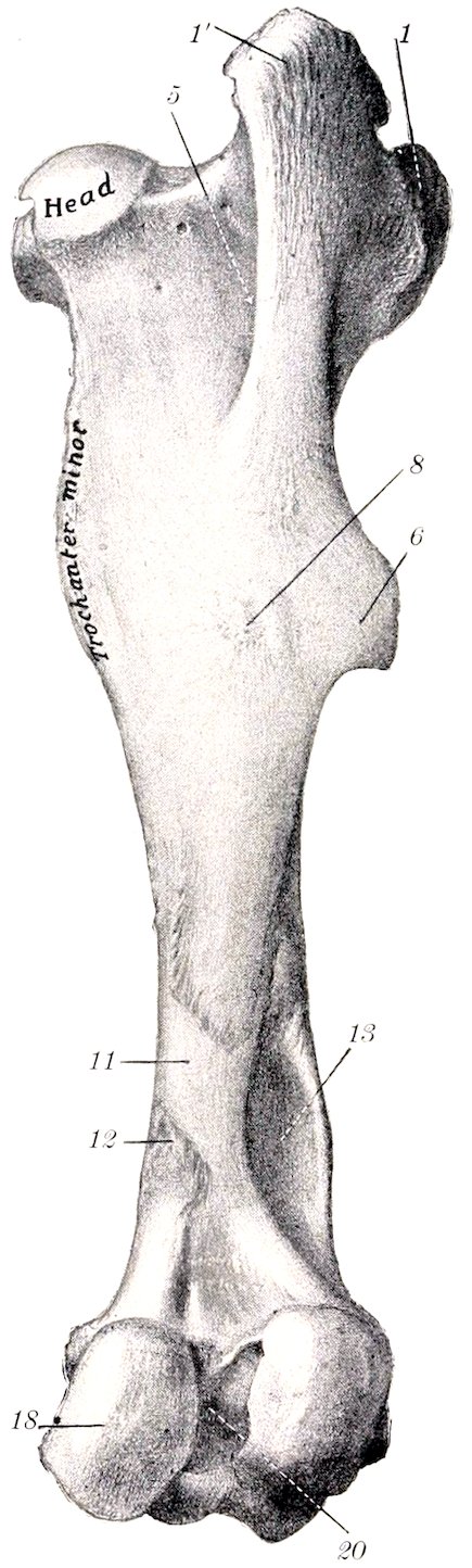
Fig. 64.—Right Femur of Horse, Posterior View.
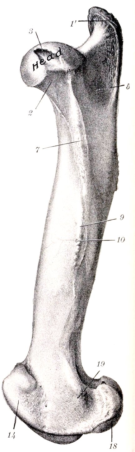
Fig. 65.—Right Femur of Horse, Internal View. 1, Anterior part, 1′, posterior part of trochanter major; 2, neck; 3, fovea capitis; 4, crest; 5, trochanteric fossa; 6, external or third trochanter; 7, trochanter minor; 8, eminence for attachment of biceps femoris; 9, internal border; 10, nutrient foramen; 11, groove for femoral vessels; 12, supracondyloid crest; 13, supracondyloid fossa; 14, trochlea; 15, external epicondyle; 16, external condyle; 17, extensor fossa; 18, internal condyle; 19, internal epicondyle; 20, intercondyloid fossa. (After Schmaltz, Atlas d. Anat. d. Pferdes.)
99The proximal extremity (Extremitas proximalis) is large and consists of the head, neck, and great trochanter. The head (Caput femoris) is placed at the inner side and is directed inward, upward, and somewhat forward. It is approximately hemispherical and articulates with the acetabulum. It is cut into internally by a deep notch, the fovea capitis, in which the pubo-femoral and round ligaments are attached. The articular surface is surrounded by a distinct margin. The neck (Collum femoris) is most distinct in front and internally. The great trochanter (Trochanter major) is situated externally; it presents three features. The anterior part or convexity is situated opposite to the head and rises little above the level of the latter; it gives attachment to the deep gluteus muscle, and in the fresh state its outer surface is coated with cartilage, over which a tendon of the middle gluteus passes, to be inserted into the crest, which is placed below and behind the convexity. The posterior part or summit is separated from the convexity by a notch; it is situated behind the plane of the head and rises to a much greater height. It furnishes insertion to part of the middle gluteus muscle. Its posterior border is continued downward as the trochanteric ridge, which forms the outer wall of the trochanteric fossa. A number of foramina are found in the concave area internal to the convexity.
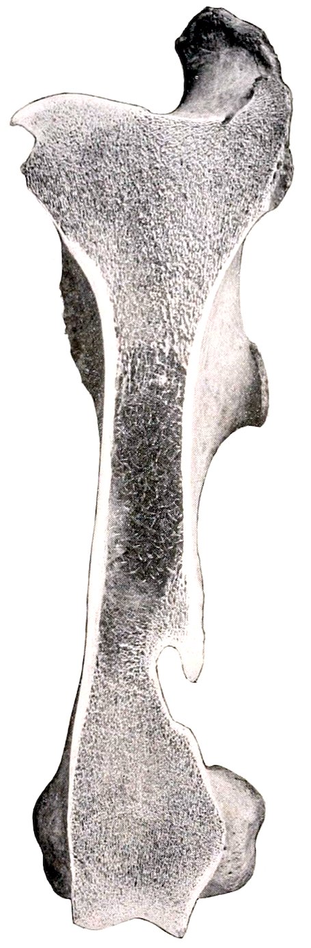
Fig. 66.—Frontal Section of Left Femur of Horse, Anterior View. The figure shows that the medullary cavity is traversed for the most part by fine bony trabeculæ.
The distal extremity (Extremitas distalis) is large in both directions and comprises the trochlea in front and two condyles behind. The trochlea consists of two ridges separated by a groove, and forms an extensive surface (Facies patellaris) for articulation with the patella. It is very unsymmetrical; the inner ridge or lip is much wider, more prominent, and extends up higher than the external one, and the two converge below. The condyles, internal and external (Condylus medialis, lateralis), are separated by the deep intercondyloid fossa (Fossa intercondyloidea), and articulate with the condyles of the tibia and the semilunar cartilages of the stifle joint. A ridge connects each condyle with the lower part of the corresponding lip of the trochlea. The intercondyloid fossa lodges the spine of the tibia and the crucial ligaments of the stifle joint, which are attached here.
The condyles are obliquely placed, with their long axes directed downward, forward, and inward. The articular surface of the external condyle is more strongly convex from side to side than that of the inner one, and the ridge which connects it with the trochlea is much narrower.
100The internal epicondyle (Epicondylus medialis) is a rounded prominence on the internal surface of the internal condyle, to which the internal lateral ligament and the adductor muscle are attached. The corresponding external epicondyle (Epicondylus lateralis) is less distinct; it presents a mark where the lateral ligament is attached, below and behind which there is a depression (Fossa musculi poplitei) in which the popliteus muscle arises. Between the external condyle and trochlea is the extensor fossa (Fossa extensoria), in which the tendon of origin of the anterior extensor and peroneus tertius is attached.
Development.—The shaft and the distal end each ossify from one center, but the proximal end has two centers, one of which is for the great trochanter. The edge of the external trochanter also has a separate center. The proximal end fuses with the shaft at three to three and a half years, the distal at about three and a half years.
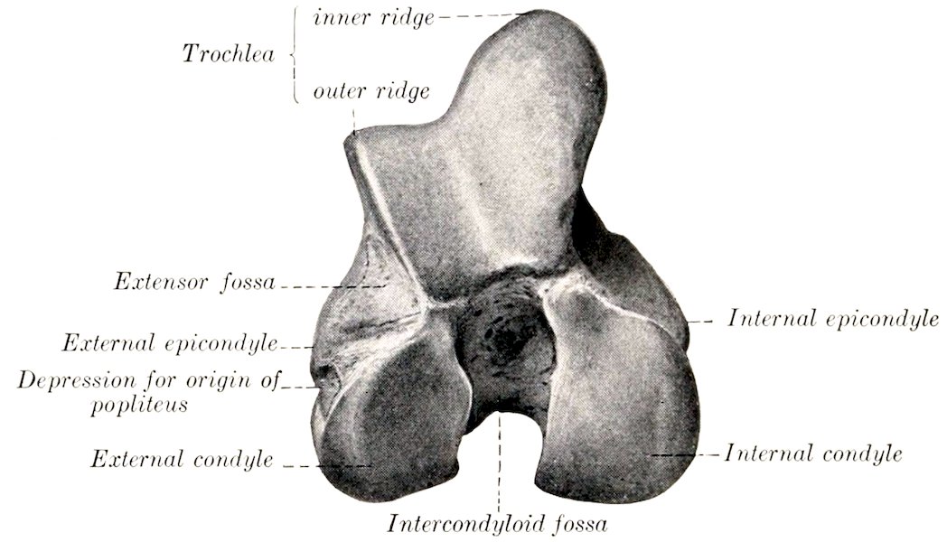
Fig. 67.—Distal Extremity of Right Femur of Horse, End View.
THE TIBIA
The tibia is a long bone which extends obliquely downward and backward from the stifle to the hock. It articulates above with the femur, below with the tarsus, and externally with the fibula. It possesses a shaft and two extremities.
The shaft or body (Corpus tibiæ), large and three-sided above, becomes smaller and flattened in the sagittal direction below, but widens a little at the distal end. It presents for notice three surfaces and three borders. The internal surface (Facies medialis) is broad above, where it furnishes insertion to the internal lateral ligament and the sartorius and gracilis muscles; below this it is convex and subcutaneous. The external surface (Facies lateralis) is smooth and somewhat spiral. It is wide and concave in its upper part, below which it becomes narrower and slightly convex, and winds gradually to the front of the bone; near the distal end it widens a little, becomes flat, and faces forward. The posterior surface (Facies posterior) is flattened, and is divided into two parts by the rough popliteal line, which runs obliquely from the upper part of the external border to the middle of the internal border. The triangular area above the line is occupied by the popliteus muscle, while the area below is marked by rough lines (Lineæ musculares) to which the deep flexor muscle of the digit is attached; the lines fade out below, where the surface is smooth and flat. The nutrient foramen is situated on or near the popliteal line. The anterior border is very prominent in its upper third, forming the tibial crest (Crista tibiæ); below it is reduced to a rough line, which ends at a 101small elevation near the distal end of the bone. The internal border (Margo medialis) is rounded in its upper half, to which the popliteus muscle is attached, and a tubercle is found on this part. The lower part is a rough line on well-marked bones. The external border (Crista interossea) is concave in its upper part and concurs with the fibula in the formation of the interosseous space of the leg; a smooth impression indicates the course of the anterior tibial vessels through the space to the front of the leg. Lower down the border divides and incloses a narrow triangular surface.
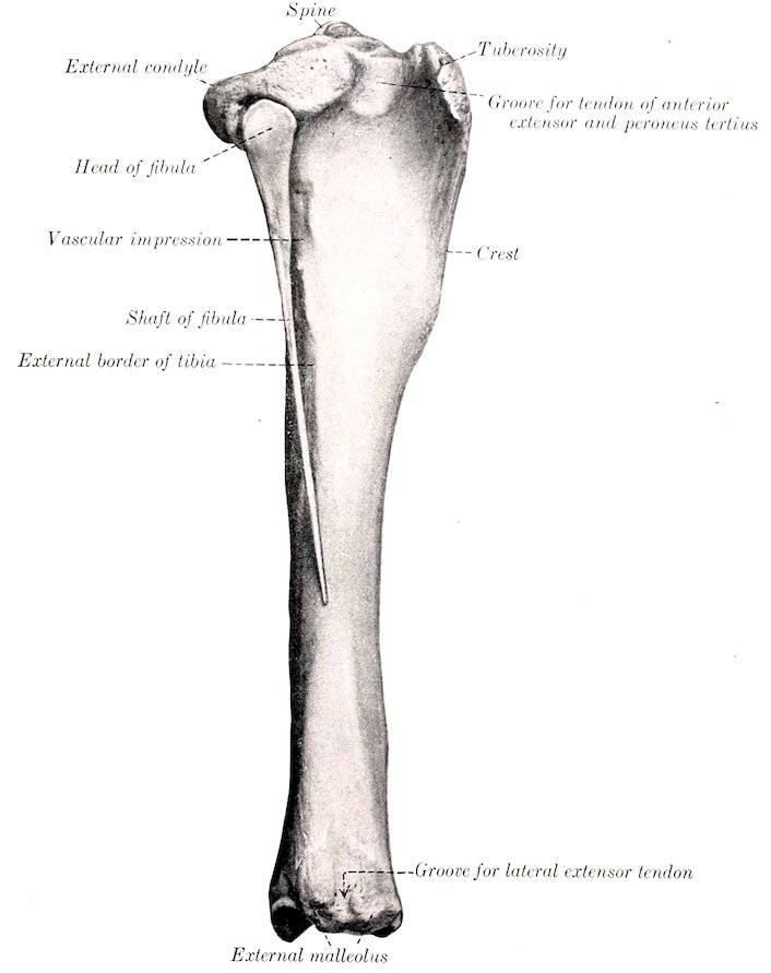
Fig. 68.—Right Tibia and Fibula of Horse, External View.
The proximal extremity (Extremitas proximalis) is large and three-sided. It bears two lateral eminences, the internal and external condyles (Condylus medialis, lateralis). Each presents a somewhat saddle-shaped surface for articulation with the condyle of the femur and the semilunar cartilage. The spine or intercondyloid eminence (Eminentia intercondyloidea) is the central prominence, upon which the articular surfaces are continued; it consists of a high inner part and a lower 102outer part (Tuberculum intercondyloideum mediale, laterale). On, before, and behind the spine are the anterior and posterior intercondyloid fossæ, in which the anterior crucial ligament and the semilunar cartilages are attached. The condyles are separated behind by the deep popliteal notch (Incisura poplitea), on the inner side of which is a tubercle for the attachment of the posterior crucial ligament. The external condyle has an overhanging outer margin, below which is a facet for articulation with the fibula. The large anterior eminence is the tuberosity of the tibia (Tuberositas tibiæ). It is marked in front by a groove, the lower part of which gives attachment to the middle patellar ligament, and the groove is flanked by rough areas for the attachment of the internal and external patellar ligaments. A semicircular smooth notch (Sulcus muscularis) separates the tuberosity from the external condyle, and gives passage to the tendon of origin of the anterior extensor and the peroneus tertius.
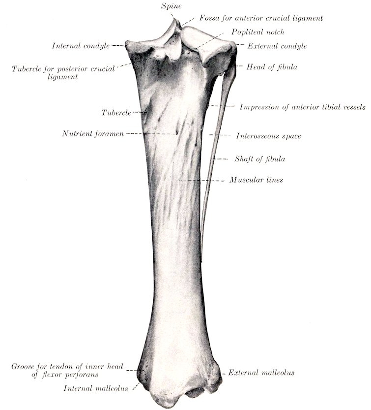
Fig. 69.—Right Tibia and Fibula of Horse, Posterior View.
103The distal extremity (Extremitas distalis) is much smaller than the proximal; it is quadrangular in form and larger internally than externally. It presents an articular surface (Cochlea tibiæ), which is adapted to the trochlea of the tibial tarsal bone (astragalus), and consists of two grooves separated by a ridge. The ridge and grooves are directed obliquely forward and outward, and are bounded laterally by the malleoli, to which the lateral ligaments of the hock joint are attached. The internal malleolus (Malleolus tibialis) is the more prominent of the two, and forms the anterior boundary of a groove for the tendon of the inner head of the flexor perforans. The external malleolus (Malleolus fibularis) is broader, and is marked by a vertical groove for the passage of the lateral extensor tendon.
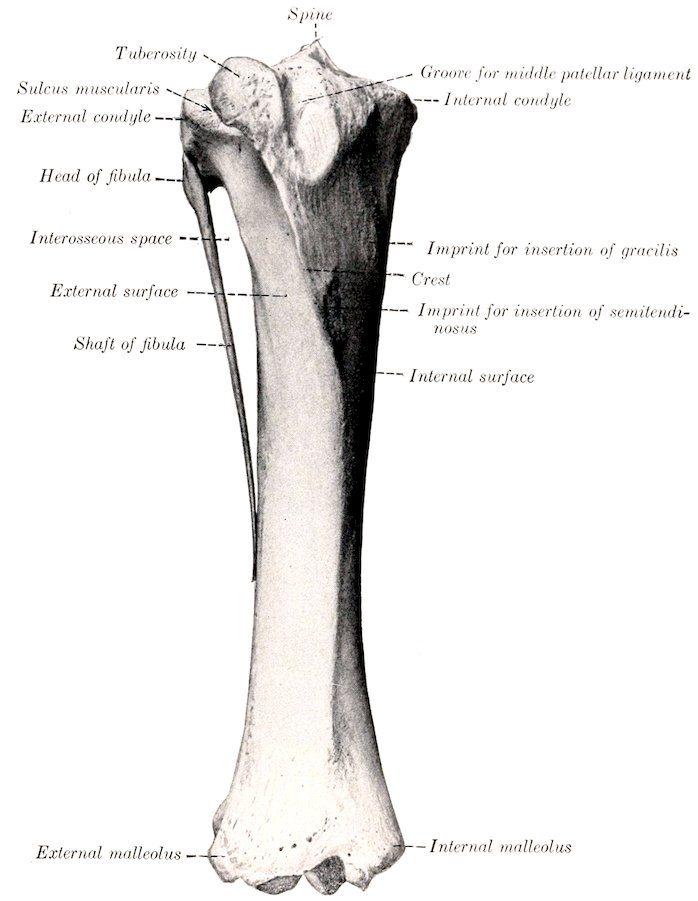
Fig. 70.—Right Tibia and Fibula of Horse, Anterior View.
Development.—The tibia has the usual three chief centers of ossification and supplementary ones for the tuberosity and the external malleolus. The latter is 104really the distal end of the fibula; it is a separate piece at birth, and the line of union is commonly quite evident in the adult in the external articular groove. The proximal end unites with the shaft at about three and a half years, and the distal end at about two years of age.
THE FIBULA
The fibula of the horse is a much reduced long bone, situated along the outer side of the tibia.
The shaft or body (Corpus fibulæ) is a slender rod which forms the outer boundary of the interosseous space; it usually terminates below in a pointed end about one-half to two-thirds of the way down the external border of the tibia.
The proximal extremity or head (Capitulum fibulæ) is relatively large, and is flattened transversely. Its internal surface presents a narrow area along the upper border for articulation with the tibia. The external surface is rough and gives attachment to the external lateral ligament of the stifle joint. It has rounded anterior and posterior borders.
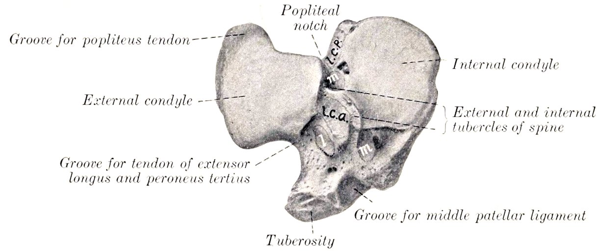
Fig. 71.—Proximal Extremity of Right Tibia of Horse, End View. l.c.a., l.c.p., Depressions for attachment of anterior and posterior crucial ligaments; l, m, m, depressions for attachment of semilunar cartilages.
The distal extremity is fused with the tibia, constituting the external malleolus.
Development.—This resembles that of the ulna. The embryonic cartilaginous fibula extends the entire length of the leg, but does not articulate with the femur. The lower part of the shaft is usually reduced to a fibrous band. Three centers of ossification appear, one each for the shaft and the extremities. The distal end unites early with the tibia, forming the external malleolus.
It is interesting to note that in some cases the entire shaft of the fibula develops, a reversion to the condition in the Miocene ancestors of the present horse.
THE PATELLA
The patella is a large sesamoid bone which articulates with the trochlea of the femur. It presents for description two surfaces, two borders, a base, and an apex.
The anterior or free surface (Facies libera) is irregularly quadrilateral, convex, and rough for muscular and ligamentous attachment.
The posterior or articular surface is smaller and is triangular in outline. It presents a vertical rounded ridge, which corresponds to the groove on the trochlea of the femur, and separates two concave areas. Of the latter, the inner cavity is 105much the larger, and is not very well adapted to the inner lip of the trochlea; in the fresh state, however, it is completed and rendered more congruent by the curved accessory fibro-cartilage.
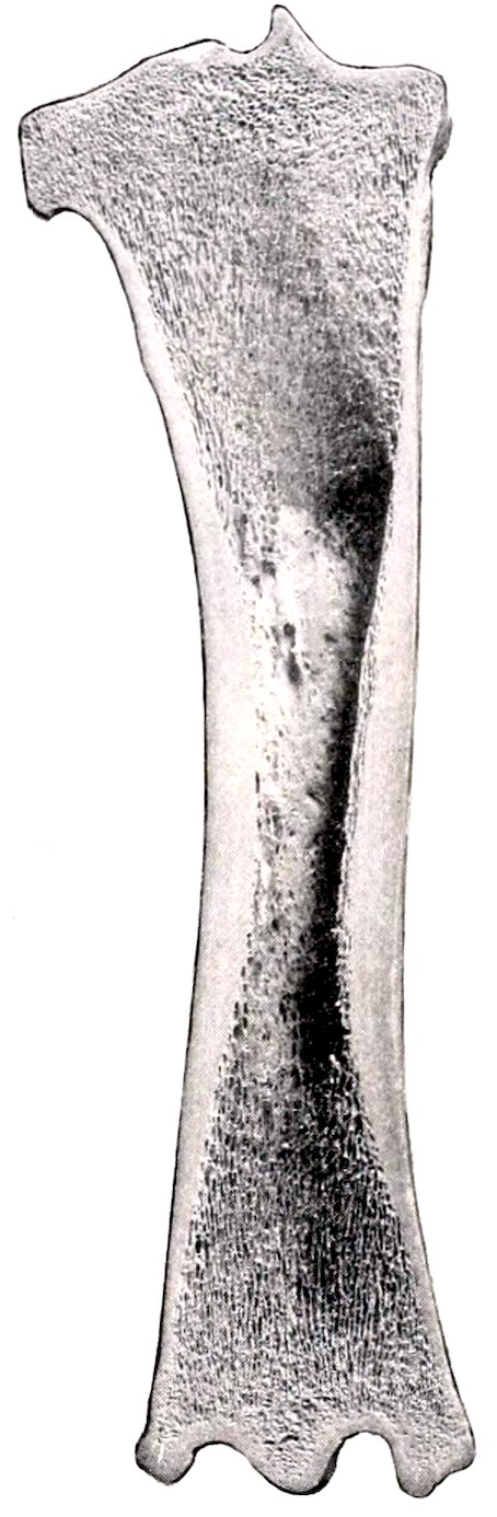
Fig. 72.—Frontal Section of Right Tibia of Horse, Anterior View.
The lateral borders converge to the apex below, and each forms a prominence or angle at the base. The inner angle and the adjacent part of the border give attachment to the fibro-cartilage of the patella, which increases the articular surface. The external border is rounded and its angle is less prominent.
The base (Basis patellæ) faces upward and backward, and is convex transversely, concave from before backward.
The apex forms a blunt point directed downward.
Development.—The patella develops as a sesamoid bone from a single center in a cartilaginous deposit in the tendon of the quadriceps femoris muscle.
THE TARSUS
The tarsus or hock of the horse usually comprises six short bones (Ossa tarsi), but exceptionally seven are present.
The Tibial Tarsal Bone
The tibial tarsal bone (Os tarsi tibiale, astragalus, or talus) is the inner bone of the proximal row. It is extremely irregular in form, but may be considered as offering six surfaces for description.
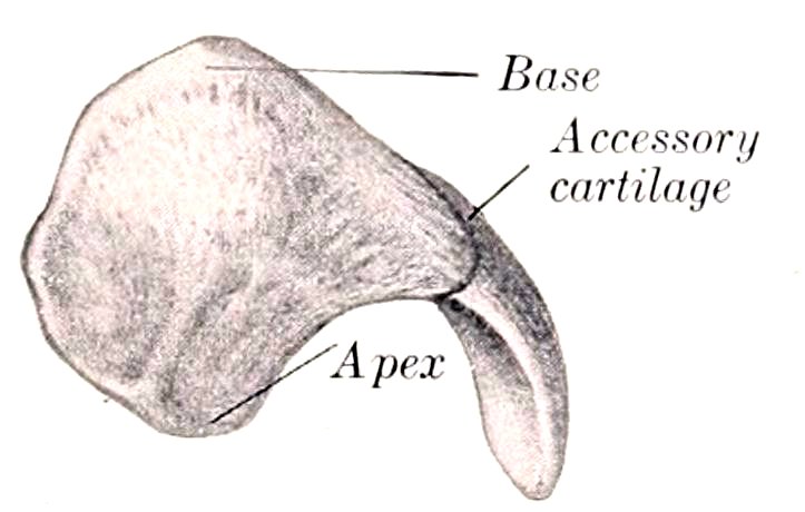
Fig. 73.—Right Patella of Horse, Anterior View.
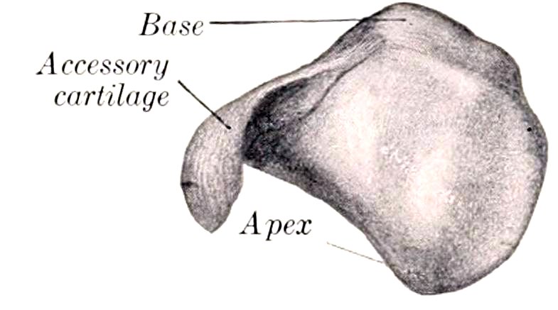
Fig. 74.—Right Patella of Horse, Posterior View.
The superior and the anterior or dorsal surface are continuous, and form a trochlea for articulation with the distal end of the tibia. The trochlea consists of two oblique ridges with a deep groove between them, which curve spirally forward, downward, and outward. There is usually a shallow synovial fossa in the groove. The inferior surface is convex from before backward, and most of it articulates with the central tarsal; externally it has an oblique facet for the fourth tarsal, and a non-articular groove cuts into the surface to its middle. The posterior or plantar surface is oblique and extremely irregular; it presents four facets for articulation with the fibular tarsal bone; the facets are separated by rough excavated areas, and the largest fossa (Sulcus tali) forms with a corresponding one on the fibular tarsal a cavity termed the sinus tarsi. The internal surface bears on its lower part a large tuberosity, and on its upper part a small one for the attachment of the 106internal lateral ligament. The external surface is smaller than the internal, and is marked by a wide rough fossa in which the external ligament is attached.
The Fibular Tarsal Bone
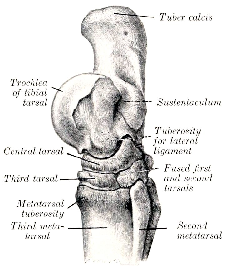
Fig. 75.—Right Tarsus and Upper Part of Metatarsus of Horse, Internal View. (After Schmaltz, Atlas d. Anat. d. Pferdes.)
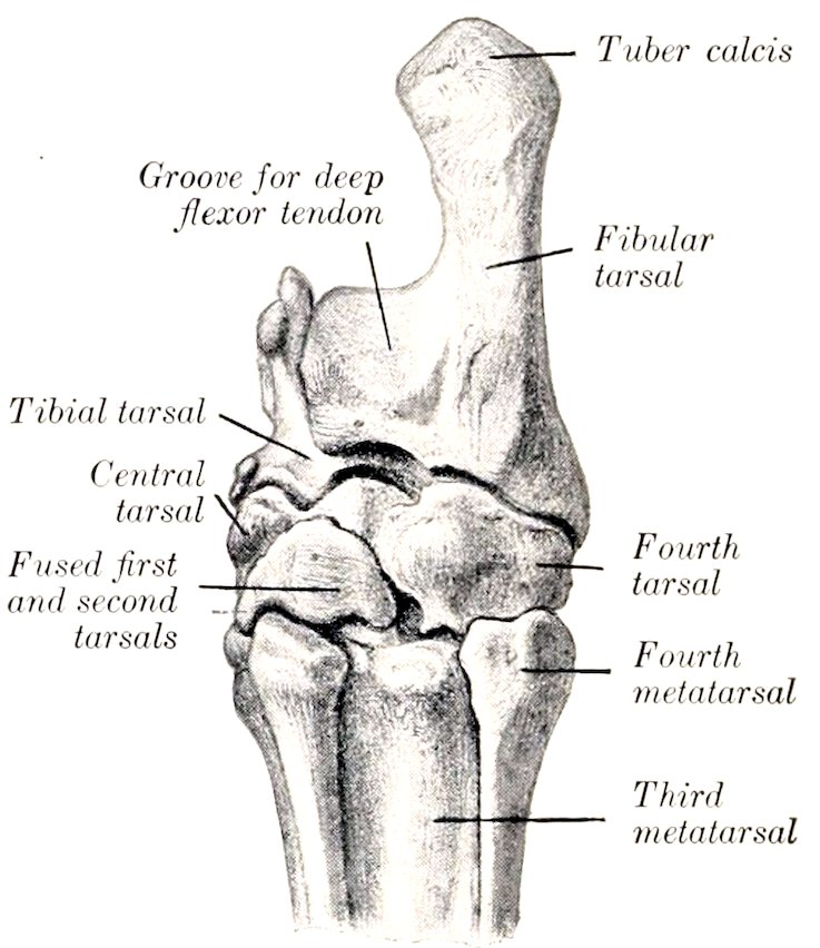
Fig. 76.—Right Tarsus and Upper Part of Metatarsus of Horse, Posterior (Plantar) View. (After Schmaltz, Atlas d. Anat. d. Pferdes.)
The fibular tarsal bone (Os tarsi fibulare, calcaneum, os calcis) is the largest bone of the hock. It is elongated, flattened from side to side, and forms a lever for the muscles which extend the hock joint. It consists of a body and an inner process, the sustentaculum tali.
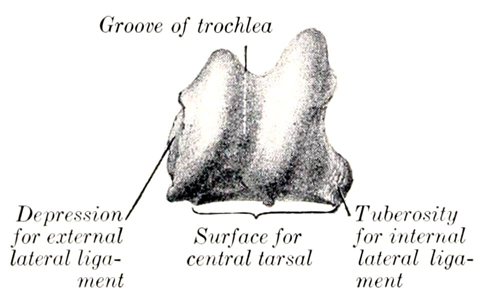
Fig. 77.—Right Tibial Tarsal Bone of Horse, Anterior View. (After Schmaltz, Atlas d. Anat. d. Pferdes.)
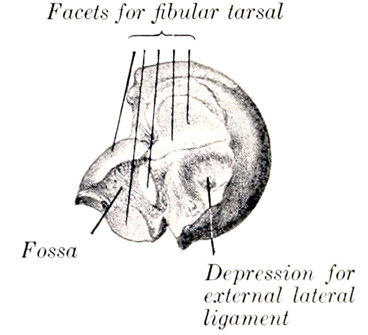
Fig. 78.—Right Tibial Tarsal Bone of Horse, External View. (After Schmaltz, Atlas d. Anat. d. Pferdes.)
The body (Corpus calcanei) is enlarged at its proximal end to form the tuber calcis or “point of the hock.” The posterior part of this eminence gives attachment to the tendon of the gastrocnemius, while in front and laterally it furnishes insertion to tendons of the flexor perforatus, biceps, and semitendinosus muscles. The inferior extremity bears a concave facet for articulation with the fourth tarsal bone. The internal surface has on its lower part a strong process, the sustentaculum tali, which projects inward. The process has a large, oval, slightly concave 107facet in front for articulation with the tibial tarsal, and sometimes a small articular surface below for the central tarsal bone. Its plantar surface forms with the smooth inner surface of the body a groove for the deep flexor tendon (Sulcus musculi flexoris hallucis longi). Its inner surface has a prominence on the lower part for the attachment of the lateral ligament. The external surface of the body is flattened, except below, where there is a rough prominence for the attachment of the lateral ligament. The anterior or dorsal border is concave in its length, smooth and rounded in its upper part. About its middle is a blunt-pointed projection (Processus cochlearis) which bears facets on its inner and lower surfaces for articulation with the tibial tarsal bone, and is roughened outwardly for ligamentous attachment. Below this are two facets for the tibial tarsal, and an extensive rough fossa which concurs in the formation of the sinus tarsi. The posterior or plantar border is straight and thick, and widens a little at either end; it is rough, and gives attachment to the long plantar ligament.
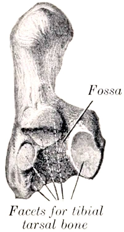
Fig. 79.—Right Fibular Tarsal Bone of Horse, Anterior View. (After Schmaltz, Atlas d. Anat. d. Pferdes.)
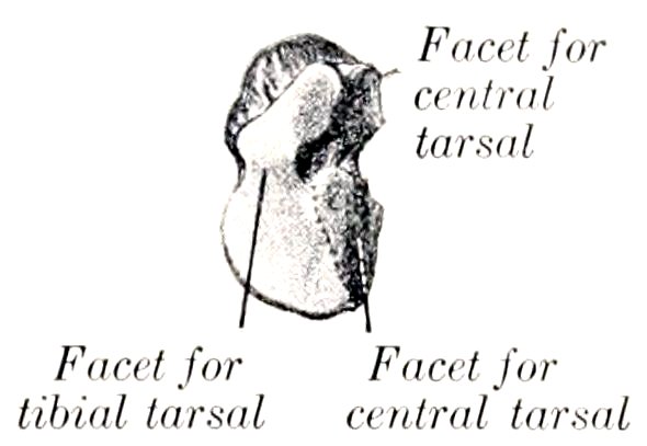
Fig. 80.—Right Fourth Tarsal Bone of Horse, Upper Surface. (After Schmaltz, Atlas d. Anat. d. Pferdes.)
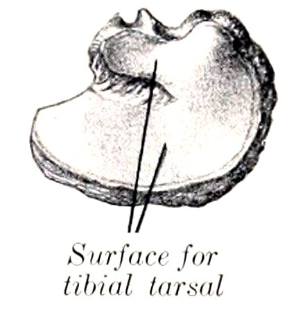
Fig. 81.—Right Central Tarsal Bone of Horse, Upper Surface. (After Schmaltz, Atlas d. Anat. d. Pferdes.)
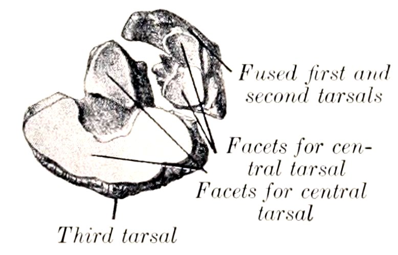
Fig. 82.—Right First, Second, and Third Tarsal Bones of Horse, Upper Surface. (After Schmaltz, Atlas d. Anat. d. Pferdes.)
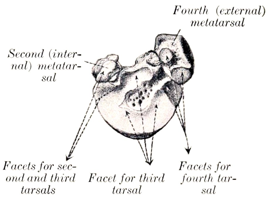
Fig. 83.—Proximal Articular Surfaces of Left Metatarsal Bones of Horse. (After Schmaltz, Atlas d. Anat. d. Pferdes.)
The Central Tarsal Bone
The central tarsal bone (Os tarsi centrale, scaphoid, or navicular) is irregularly quadrilateral, and is situated between the tibial tarsal above and the third tarsal below. It is flattened from above downward, and may be described as having two 108surfaces and four borders. The superior surface is concave from before backward, and almost all of it articulates with the tibial tarsal; a non-articular depression cuts into its outer part, and sometimes there is a facet for the fibular tarsal bone on the posterior angle. The inferior surface is convex, and is crossed by a non-articular groove, which separates facets for articulation with the third and the first and second (fused) tarsals. The anterior or dorsal border and the internal border are continuous, convex, and rough. The posterior or plantar border bears two prominences, separated by a notch. The external border is oblique, and bears anterior and posterior facets for articulation with the fourth tarsal, between which it is excavated and rough.
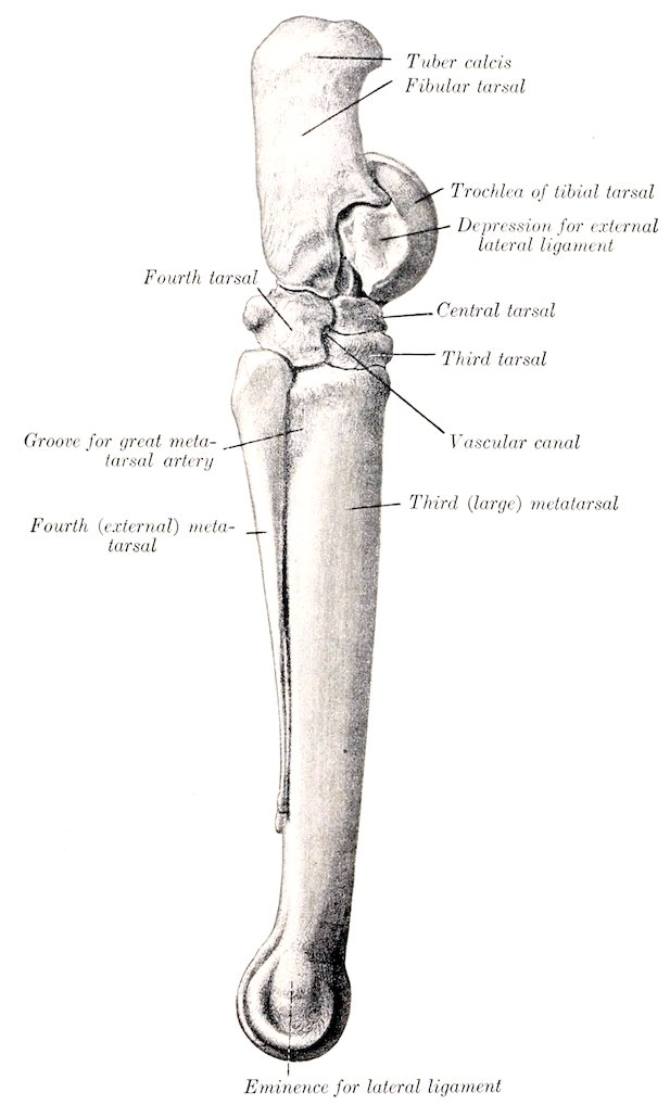
Fig. 84.—Right Tarsal and Metatarsal Bones of Horse. External View. (After Schmaltz, Atlas d. d. Anat. d. Pferdes.)
109
First and Second Tarsal Bones
The first and second tarsal bones (Os tarsale primum et secundum, cuneiform parvum) are usually fused in the horse, forming a bone of very irregular shape, situated in the inner and posterior part of the lower row, below the central and behind the third tarsal. It is the smallest of the tarsal bones, and may be described as having two surfaces, two borders, and two extremities. The internal surface faces backward and inward, and is convex. Its anterior part is ridged, and gives attachment to the internal lateral ligament, and its posterior part bears an imprint where the inner tendon of the tibialis anterior is inserted. The external surface is marked by a deep notch which indicates the division between the first and second tarsal elements; it bears on its anterior part a large concave facet for the central tarsal. The superior border is convex. The inferior border is broad in front, where it articulates with the large and inner small metatarsal bones. The anterior extremity has a small facet for articulation with the third tarsal, and bears internally a ridge or tubercle. The posterior extremity is a blunt point.
In some cases the first and second tarsal bones remain separate—a remarkable reversion to the condition in the early ancestors of the horse. In such specimens the first tarsal is a discoid bone, articulating above with the central, below with the small metacarpal bone. The second tarsal is quadrangular, equivalent to the thick anterior part of the bone as described above, and overlapped by the anterior part of the first tarsal.
The Third Tarsal Bone
The third tarsal bone (Os tarsale tertium, third or great cuneiform) resembles the central, but is smaller and triangular in outline. It is situated between the central above and the large metatarsal bone below. It possesses two surfaces and three borders.
The superior surface is concave, and is crossed by a non-articular depression which divides it into two unequal facets; it articulates with the central tarsal. The inferior surface is slightly convex, and rests on the large metatarsal bone; it has an extensive central rough excavation. The anterior or dorsal border is convex and bears a rounded ridge on its inner part. The internal border is deeply notched and has a small facet for the second tarsal on its anterior part. The external border is also divided by a notch into two parts, and bears two diagonally opposite facets for articulation with the fourth tarsal. In some cases there is a facet for the inner small metatarsal bone.
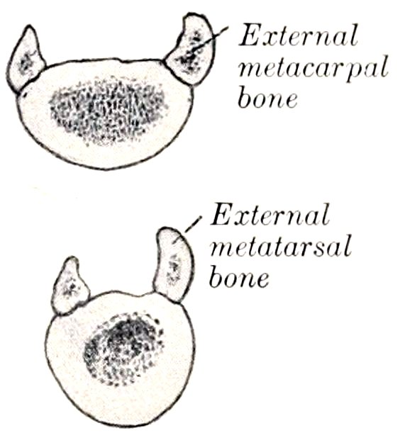
Fig. 85.—Cross-sections of Metacarpal and Metatarsal Bones of Horse. (After Schmaltz, Atlas d. Anat. d. Pferdes.)
The Fourth Tarsal Bone
The fourth tarsal bone (Os tarsale quartum, cuboid) is the outer bone of the lower row, and is equal in height to the central and third together. It is cuboid in shape and presents six surfaces.
The superior surface is convex from side to side, and articulates chiefly with the fibular tarsal, but to a small extent with the tibial tarsal also. The inferior surface rests on the large and external small metatarsal bones. The internal surface bears four facets for articulation with the central and third tarsal bones. It is crossed from before backward by a smooth groove, which by apposition with the adjacent bones forms the canal of the tarsus (Canalis tarsi) for the passage of the perforating tarsal vessels. The anterior or dorsal, external, and posterior or plantar surfaces are continuous and rough. A tuberosity behind gives attachment to the plantar ligament.
110Development.—The fibular tarsal bone has two centers of ossification, one for the main mass and the other for the tuber calcis; the latter fuses with the rest of the bone at about three years of age. The first and second tarsals have separate centers, but fusion usually occurs before birth. Each of the other bones ossifies from a single center.
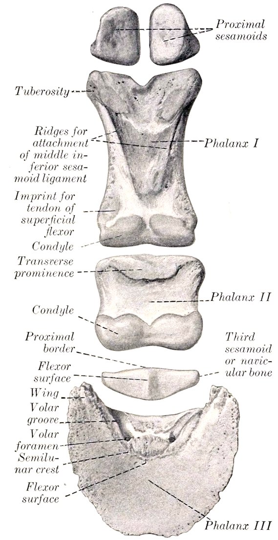
Fig. 86.—Digital Bones of Fore Limb of Horse, Volar Aspect.
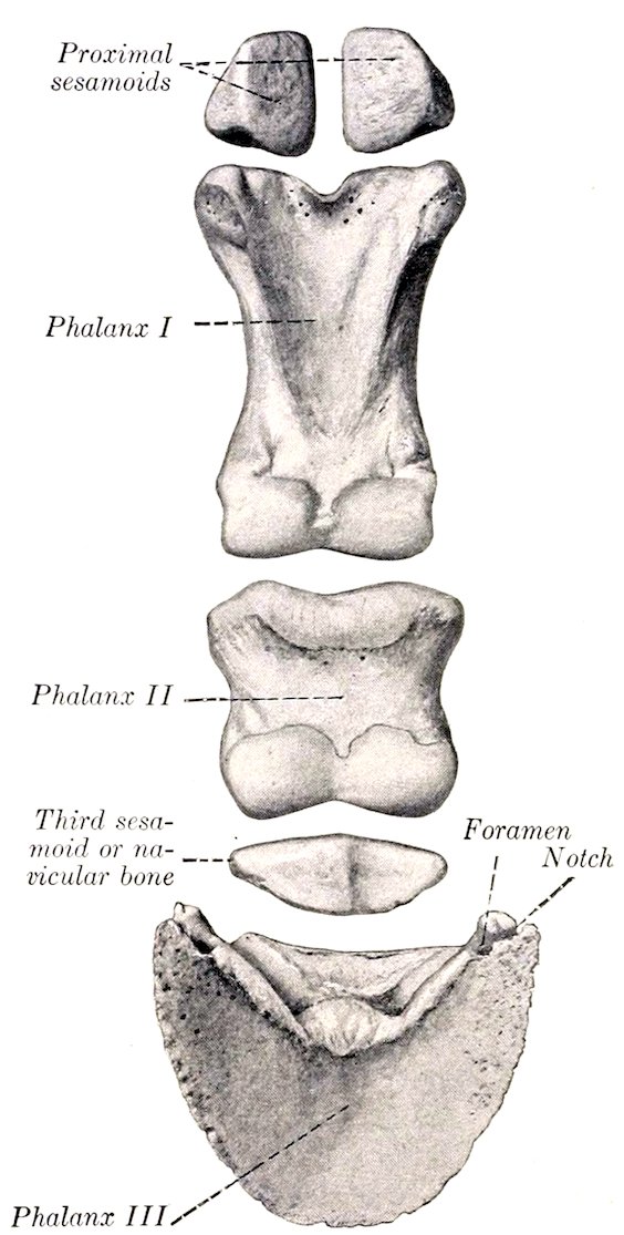
Fig. 87.—Digital Bones of Hind Limb of Horse, Volar Aspect.
THE METATARSUS
The metatarsal bones (Ossa metatarsalia), three in number, have the same general arrangement as the metacarpal bones, but present some important differences. Their direction is slightly oblique, downward and a little forward.
The large or third metatarsal bone is about one-sixth longer than the corresponding metacarpal; in an animal of medium size the difference is about two inches. The shaft is more cylindrical, and is almost circular on cross-section, except in its lower part. At the upper part of its external surface there is a groove, which is directed obliquely downward and backward, and is continued by the furrow formed by the apposition of the external metatarsal bone; it indicates the course of the great metatarsal artery. A faint impression in a similar place on the inner side 111marks the position of the corresponding vein. The nutrient foramen is relatively higher than on the metacarpal bone. The proximal extremity is much wider from before backward than that of the metacarpal bone. Its articular surface is slightly concave, and is marked by a large central non-articular depression, continued outward by a deep notch. The greater part of the surface articulates with the third tarsal, but there is an outer facet for the fourth, and usually a small facet posterointernally for the second tarsal bone. Posteriorly there are two pairs of facets for articulation with the small metatarsal bones. The front is crossed by a rough ridge for insertion, which becomes larger and turns downward on the outer side behind the vascular groove. The distal extremity closely resembles that of the corresponding metacarpal bone.
In some cases the lower part of the shaft is bent backward somewhat. The articular surface extends a little higher behind than in the case of the metacarpal bone. The large metatarsal bone is even more strongly constructed than the metacarpal. The shell of compact substance is very thick in the middle of the shaft, especially in front and internally.
The small metatarsal bones are a little longer than the corresponding metacarpals. The external (fourth) metatarsal is relatively massive, especially in its upper part. The head is large and outstanding, and bears two facets above for the fourth tarsal, and two in front and internally for articulation with the large metatarsal; elsewhere it is roughened for attachment. The internal (second) metatarsal is much more slender than the outer one, especially in its upper part. The head bears two facets above for the first and second tarsals, and sometimes one for the third tarsal.
THE PHALANGES AND SESAMOIDS
The axis of the phalanges of the hind limb is about five degrees less oblique than that of the fore limb, and the chief differences in the form and size of the bones are as follows:
The first phalanx is a little shorter, wider above, and narrower below.
The second phalanx is narrower and slightly longer.
The third phalanx is narrower, the angle of inclination of the wall surface is a little (ca. 5 degrees) greater, the sole surface is more concave, and the wings are less prominent and closer together.
The proximal sesamoids are a little smaller, except in thickness. The third sesamoid or navicular bone is narrower and shorter.
112
SKELETON OF THE OX
VERTEBRAL COLUMN
The vertebral formula is C7T13L6S5Cy18–20.
The cervical vertebræ are much shorter than those of the horse and are smaller in their other dimensions. The articular processes are smaller than in the horse, and a plate of bone connects each two of the same side. The transverse processes of the third, fourth, and fifth are double; the upper part projects backward, and is short and stout; the lower part is directed downward and forward, and is longer and more plate-like. The lower part of the sixth transverse process is a large, thick, quadrilateral and almost sagittal plate, directed downward. The seventh transverse process is single, short, and thick, and presents no foramen transversarium; it is in series with the upper part of the preceding processes. The spinous processes are well developed, and increase in height from before backward. They are directed upward and forward, with the exception of the last, which is nearly vertical and is about four or five inches (ca. 10 to 12 cm.) in height. The summit of that of the third bone is usually bifid. The ventral spines are prominent and thick in their posterior part; they are absent on the last two.
The atlas has a large rough tuberosity on its dorsal arch. The ventral arch is very thick. The wings are less curved than in the horse, and the foramen transversarium (posterior foramen) is absent. The cavities for the occipital condyles are divided into upper and lower parts by a non-articular area, and are separated by a narrow interval below. The posterior articular surfaces are flattened behind and are continued into the vertebral canal, forming an extensive area for the odontoid process of the axis.
The axis is short. The spine projects a little in front, and increases in height and thickness behind; its posterior border descends abruptly. The odontoid process (dens) is wide, and its upper surface is deeply concave from side to side. The intervertebral foramen is circular and not so close to the anterior border of the arch as in the horse. The posterior notches are not so deep. The transverse processes are stouter, but the foramen transversarium is small and sometimes absent.
The thoracic vertebræ, thirteen in number, are larger than those of the horse. The bodies are longer and are distinctly constricted in the middle. They bear a thin-edged ventral crest. The arches—in addition to the usual notches, which are shallow—are perforated in the posterior part by a foramen. The transverse processes are thick and strong, and bear rounded mammillary processes (except at the posterior end of the series); the last two, although prominent, do not always articulate with the ribs. The spinous processes are long. The first is much higher than in the horse, the next two are usually the most prominent, and behind this there is a very gradual diminution in height. The backward slope, slight at first, increases to the tenth; the last is vertical and lumbar in character. The summit is usually pointed on the first, and the thickening on those further back is less than in the horse. The width diminishes from the fifth to the eleventh usually. Both borders of the spines are in general thin and sharp, but the last three or four sometimes have thick posterior margins.
113
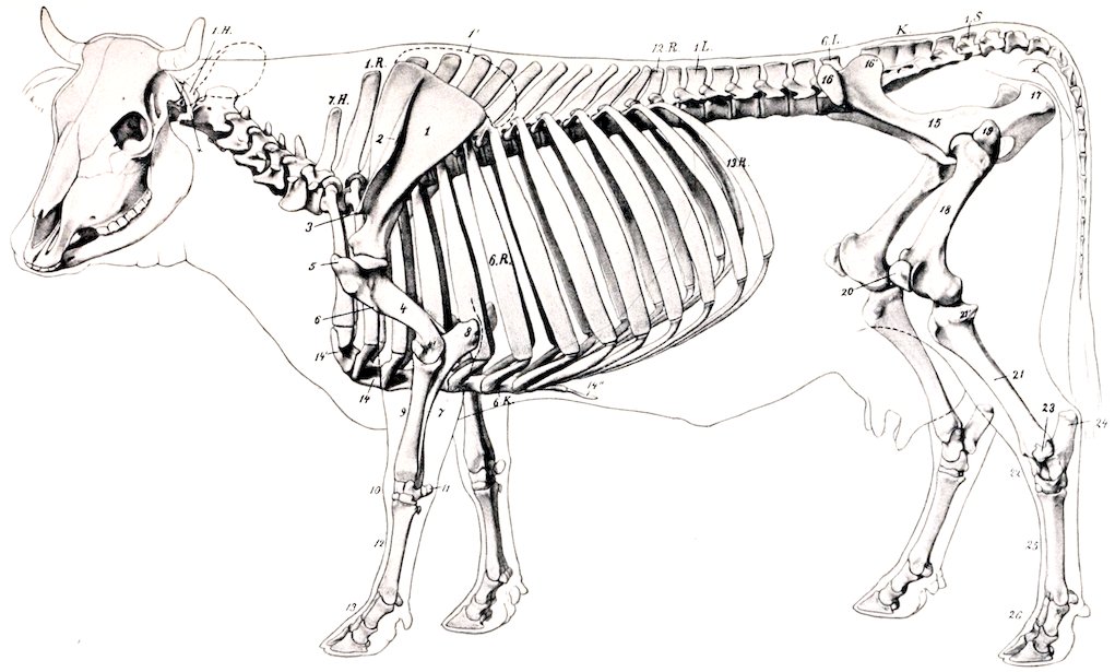
Fig. 88.—Skeleton of Cow. 1.H., Atlas; 7.H., seventh cervical vertebra; 1.R., first thoracic vetrebra; 6.R., sixth rib; 12.R., twelfth thoracic vertebra; 13.R., last rib; 1.L., first, 6.L., last lumbar vertebra; K, sacrum; 1.S., first coccygeal vertebra; 6.K., sixth costal cartilage; x, wing of atlas; 1, scapula; 1′, cartilage of scapula; 2, spine of scapula; 3, acromion; 4, humerus; 4′, external condyle of humerus; 5, external tuberosity of humerus; 6, deltoid tuberosity; 7, ulna; 8, olecranon; 9, radius; 10, carpus; 11, accessory carpal bone; 12, metacarpus; 13, phalanges; 14, sternum; 14′, manubrium; 14″, xiphoid cartilage; 15, ilium; 16, external angle of ilium; 16′, internal angle of ilium; 17, tuber ischii; 18, femur; 19, trochanter major; 20, patella; 21, tibia; 21′, external condyle of tibia; 22, tarsus; 23, distal end of fibula; 24, tuber calcis; 25, metatarsus; 26, phalanges. (After Ellenberger-Baum, Anat. für Künstler.)
114The lumbar vertebræ, six in number, are much longer than in the horse. The bodies are much constricted in the middle, expanded at either end, and bear rudimentary ventral crests. The fourth and fifth are usually the longest. The intervertebral foramina are often double in the anterior part of the series, and are very large further back. The articular processes are large, and their facets are more strongly curved than in the horse. The transverse processes all curve forward. They are separated by considerable intervals, and form no articulations with each other or with the sacrum. Their borders are thin and irregular, and often bear projections of variable size and form. The first is the shortest and the length increases to the fifth, the last being considerably shorter. The spinous processes are relatively low and wide, the last being the smallest; their summits are moderately thickened.
The sacrum is longer than that of the horse. It consists originally of five segments, but fusion is more complete and involves the spinous processes, which are united to form a median sacral crest, with a convex thick and rough margin. A lateral crest is formed by the fusion of the articular processes. The pelvic surface is concave in both directions, and is marked by a central groove, which indicates the course of the middle sacral artery. The inferior sacral foramina are large. The wings are quadrangular, short, compressed from before backward, and high dorso-ventrally. They have an extensive anterior surface, which is concave from side to side, and non-articular. The posterior surface is rough, and bears a triangular area below for articulation with the ilium. The body of the first segment is very wide, and the entrance to the sacral canal correspondingly wide and low. The anterior articular processes are large, and are concave and semicylindrical in curvature internally. The lateral borders are thin, sharp, and irregular. The apex is wider than in the horse, and the posterior end of the crest forms a pointed projection over the opening of the sacral canal.
The coccygeal vertebræ are longer and better developed than in the horse. The first five or six have complete arches and spinous processes. The transverse processes are relatively large in the anterior part of the series, in which there are also anterior articular processes (which do not articulate), and a pair of ventral spines which form a hæmal groove for the middle coccygeal artery.
Vertebral curves: The cervical curve is very slight and is concave dorsally. The thoracic and lumbar regions form a gentle curve, concave ventrally. The promontory is more pronounced than in the horse, especially in subjects in which the sacrum is inclined upward behind. Another promontory occurs at the junction of the sacrum and first coccygeal vertebræ.
Length: The following table gives the lengths of the regions of a shorthorn cow of medium size:
Cervical 47 cm.
Thoracic 75 cm.
Lumbar 40 cm.
Sacral 24 cm.
Coccygeal 75 cm.
261 cm.
Variations: Sometimes fourteen thoracic vertebræ and fourteen pairs of ribs are present; reduction to twelve with the normal number of lumbar vertebræ is very rare. According to Franck, there are sometimes seven lumbar vertebræ with the normal number in the thoracic region. The number of coccygeal vertebræ may vary from sixteen to twenty-one.
THE RIBS
Thirteen pairs of ribs are normally present, of which eight are sternal and five asternal. They are in general longer, wider, flatter, less curved, and less regular in form than in the horse. The eighth, ninth, and tenth are the longest and widest. The width of most of the ribs increases considerably in the middle, and the breadth of the intercostal spaces is correspondingly diminished. The neck is long, and forms (except in the posterior part of the series) a smaller angle with the shaft than in the horse. The articular surface of the tubercle is concave transversely, except on the last two or three, where the facet is small and flat or absent. The lower ends of the second to the tenth or eleventh inclusive form diarthrodial joints with the costal 115cartilages. The first costal cartilages are very short; they articulate by their internal surfaces with the sternum, but not with each other.
The presence of a fourteenth rib is not very rare. It is usually floating and may correspond to an additional thoracic vertebra or to the first lumbar. Reduction of the thirteenth is more common. The eighth cartilage often does not reach the sternum, but articulates with the seventh.
The sternum consists of seven sternebræ, most of which are developed from two lateral centers. It is wider, flatter, and relatively longer than in the horse, and the ventral crest or “keel” is absent. The manubrium is somewhat wedge-shaped and laterally compressed. Its base forms a diarthrodial joint with the body of the bone, and laterally it bears extensive facets for articulation with the first pair of costal cartilages. The body widens from before backward, but behind the last pair of costal facets it becomes much narrower. The ventral surface is prominent on the second and third segments, concave further back. The lateral borders are notched for the passage of vessels. The cariniform cartilage is absent. The xiphoid cartilage is like that of the horse but is smaller.
THE THORAX
The bony thorax is shorter than in the horse. The inlet is higher. The roof is short, and the floor wider and relatively longer. The transverse diameter is wider in the posterior part. The summits of the spinous processes are almost in a straight line from the second thoracic to the middle of the lumbar region.
Bones of the Skull
Bones of the Cranium
The occipital bone is situated in the lower part only of the posterior surface of the skull, and is separated from the highest part (the frontal eminence) by the parietal and interparietal bones. The supraoccipital, interparietals, and parietals fuse before birth or soon after, and the mass so formed is separated from the lateral parts of the occipital bone by a transverse suture in the skull of the calf. Above this suture is a central tuberosity, to which the ligamentum nuchæ is attached, and the surface on either side is depressed and rough for muscular attachment. Below the suture the bone is much wider than that of the horse. The foramen magnum is wide, so that the condyles are further apart except below. The paramastoid processes are short and wide and are bent inward. Two constant foramina are found in the condyloid fossa; the anterior one is the hypoglossal, the other (sometimes double) conducts a vein from the condyloid canal. The latter passes upward from a foramen on the inner side of the condyle and opens into the parieto-temporal canal. The cranial surface of the supraoccipital presents a central depression, and above this is a variable but never very pronounced eminence, which corresponds to the tentorium osseum of the horse. On either side is a groove leading to the parieto-temporal canal. The basilar process is short and wide; its cranial surface is deeply concave, and the internal spheno-occipital crest is prominent. Two large tubercles below mark the junction with the sphenoid. The foramen lacerum is short and very narrow. In the adult animal the bone is excavated to contain an air-cavity which is regarded as a part of the frontal sinus.
The sphenoid bone is short. The cranial surface of the body presents a deep sella turcica, in front of which it rises abruptly. The high anterior part bears a central ridge (Rostrum sphenoidale), which joins the crista galli of the ethmoid. Two foramina occur on either side. Of these, the large anterior one is equivalent to the foramen rotundum, lacerum orbitale, and patheticum of the horse. The posterior one is the foramen ovale, which transmits the inferior maxillary nerve. 116The orbital wing is thick and is overlapped by the frontal in such manner as to appear externally to divide into two branches; the anterior part joins the ethmoid close to the sphenopalatine foramen, and contains a small sinus which communicates with an ethmoidal meatus. The temporal wing is small, but forms a prominent thick pterygoid crest. The pterygoid process is wide, and the pterygoid or alar foramen is absent. The sphenoidal sinus is absent in the calf and small in the adult; it communicates by one or two small openings with an ethmoidal meatus, and so with the nasal cavity.
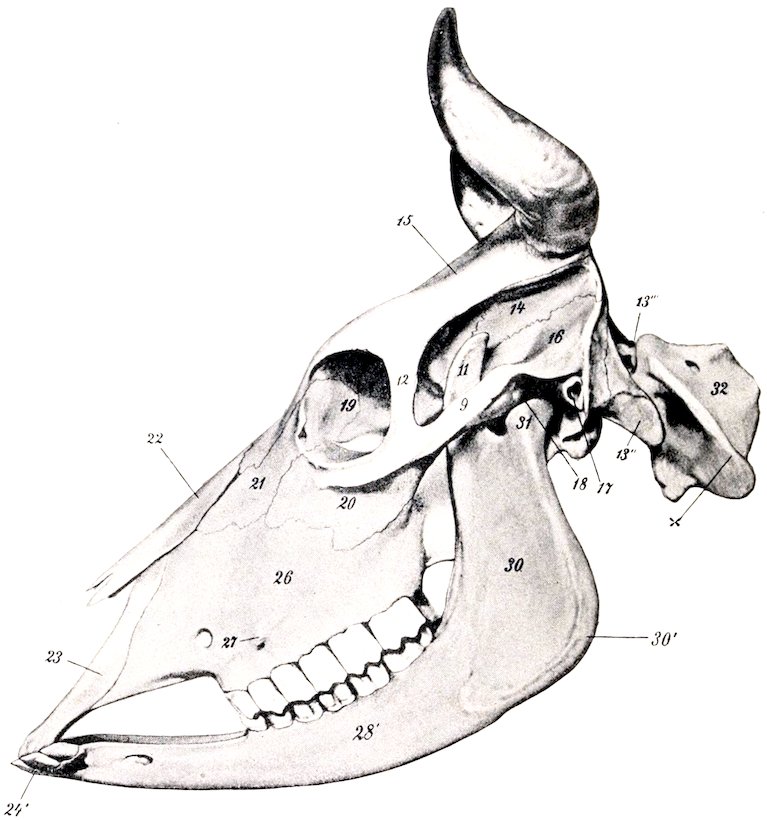
Fig. 89.—Skull and Atlas of Ox, Lateral View. 9, Zygomatic arch; 11, coronoid process; 12, supraorbital process; 13″, paramastoid or styloid process; 13‴, occipital condyle; 14, parietal bone; 15, frontal bone; 16, squamous temporal bone; 17, external auditory meatus; 18, temporal condyle; 19, orbital surface of lacrimal bone; 20, malar bone; 21, facial surface of lacrimal bone; 22, nasal bone; 23, nasal process of premaxilla; 24′, incisor teeth; 26, maxilla; 27, facial tuberosity; infraorbital foramen in front of 27; 28′, molar part of ramus of mandible; 30, broad vertical part of ramus; 30′, angle of jaw; 31, condyle of mandible; 32, atlas; x, wing of atlas. (After Ellenberger-Baum, Anat. für Künstler.)
The ethmoid bone has an extensive perpendicular plate. The lateral mass consists of five endoturbinals and eighteen ectoturbinals (Paulli). The largest ethmoturbinal is so extensive as to be termed a third or middle turbinal bone; it projects forward between the upper and lower turbinals. The lamina papyracea appears to a small extent externally in the pterygo-palatine fossa, forming part of the upper margin of the sphenopalatine foramen.
The interparietals are primitively paired, but unite before birth. As already 117mentioned, fusion occurs before or shortly after birth with the parietals and supraoccipital. No distinct tentorium osseum is present.
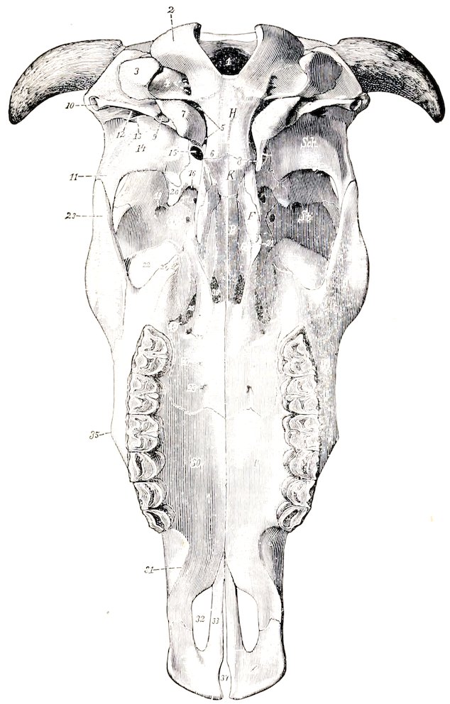
Fig. 90.—Skull of Ox, Basal View, Without Mandible. F, Pterygoid bone; G, horizontal and G′, perpendicular part of palate bone; H, occipital bone; J, malar bone; K, sphenoid bone; O, maxilla; P, vomer; Sch., squamous temporal; St., frontal bone; Z, premaxilla; 1, for. magnum; 2, occipital condyle; 3, paramastoid (styloid) process of occipital; 4, hypoglossal and condyloid foramina; 5, for. lacerum; 6, pharyngeal tubercles; 7, bulla ossea; 8, muscular process (left one is removed to expose for. ovale); 9, hyoid process; 10, ext. auditory meatus; 11, zygomatic process; 12, external opening of parieto-temporal canal; 13, postglenoid process; 14, temporal condyle; 15, for. ovale; 16, for. lac. orbitale + for. rotundum; 17, optic foramen; 18, ethmoidal foramen; 19, orbital opening of supraorbital canal; 20, pterygoid crest; 21, hamulus of pterygoid bone; 22, lacrimal bulla; 23, temporal process of malar; 24, pterygoid process of maxilla; 25, sphenopalatine foramen; 26, maxillary foramen; 27, posterior palatine foramen; 28, anterior palatine foramen; 29, accessory palatine foramina; 30, palate process of maxilla; 31, interalveolar border; 32, palatine cleft; 33, palate process of premaxilla; 34, fissura incisiva; 35, facial tuberosity; 36, posterior nares. (Ellenberger-Baum, Anat. d. Haustiere.)
The parietal bones do not enter into the formation of the roof of the cranium. They constitute the upper part of the posterior wall, and bend sharply forward along the lateral wall, forming part of the wall of the temporal fossa. The line of inflection is marked by the prominent parietal crest, which is continuous with the temporal crest below. The parietals are excavated to form part of the frontal sinuses in the adult animal.
118The condition in the young subject is as follows: The two parietals are united with each other and also with the interparietal and supraoccipital. The resulting mass is somewhat horseshoe-shaped. Its occipital part (Planum occipitale) forms the greater part of the posterior wall of the cranium and bears about its center the tuberosity for the attachment of the ligamentum nuchæ. From either side of this a line curves outward, and divides the surface into an upper smooth area and a lower area which is rough for muscular attachment. The upper border joins the frontal bone and concurs in the formation of the frontal eminence. The temporal parts (Plana temporalia) are much smaller and are concave externally; they join the frontal above and the squamous temporal below.
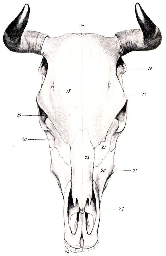
Fig. 91.—Skull of Ox, Dorsal View (with Mandible). 12, Supraorbital process; 14, parietal bone; 15, frontal bone; 16, squamous temporal bone; 19, orbit; 20, malar bone; 21, lacrimal bone; 22, nasal bone; 23, premaxilla; 26, maxilla; 27, facial tuberosity; 24, incisor teeth. (After Ellenberger-Baum, Anat. f. Künstler.)
119
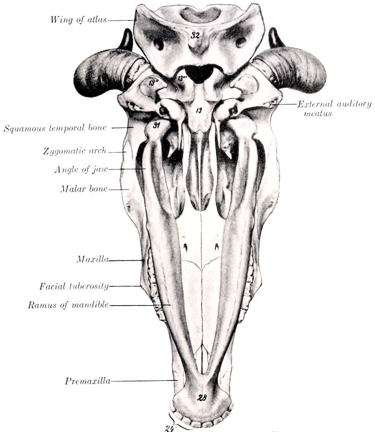
Fig. 92.—Skull and Atlas of Ox, Ventral View. 13, Basi-occipital; 13″, paramastoid or styloid process; 13‴, occipital condyle; 24′, incisor teeth; 28, body of mandible; 31, condyle of mandible; 32, ventral tubercle of atlas. (After Ellenberger-Baum, Anat. für Künstler.)
The frontal bones are very extensive, forming about one-half of the entire length of the skull, and all of the roof of the cranium. The posterior borders form with the parietals a large central frontal eminence (Torus frontalis), the highest point of the skull. At the junction of the posterior and the lateral borders are the processus cornu or “horn-cores,” for the support of the horns. These processes are of elongated conical form, and vary greatly in size, length, curvature, and direction. The external surface is rough and porous, marked by numerous grooves and foramina; in the fresh state they are covered by the matrix of the horns. The base has a constriction, the neck. The interior is excavated to form a number of irregular intercommunicating spaces, divided by bony septa, and communicating with the frontal sinus. In the polled breeds these processes are absent, the skull is narrower in this region, and the frontal eminence more pronounced. The supraorbital process is situated about half-way between the anterior and posterior margins; it is short and joins the frontal process of the malar bone. The supraorbital foramen (often double) is situated about an inch inward from the root of the process; it is the external orifice of the supraorbital canal (Canalis supraorbitalis), which passes downward and forward into the orbit. The foramen is in the course of the supraorbital groove (Sulcus supraorbitalis), which marks the course of the frontal vein. The anterior ends of the naso-frontal parts form a notch which receives the nasal bones, and sutural or Wormian bones are often found at this junction (naso-frontal suture). The orbital part is extensive; it is perforated behind by the orbital opening of the supraorbital canal, and below by the ethmoidal or internal orbital foramen. It does not articulate with the palatine, from which it is separated by the orbital wing of the sphenoid. The temporal part is also more 120extensive than in the horse. The frontal sinus is very extensive, being continued into the parietals and occipital, and the horn processes when present.
The squamous and petrous parts of the temporal bone fuse early—in fact, union is nearly complete at birth. The squamous part is relatively small. Its external surface is divided into two parts by the prominent temporal crest, which is continuous with the parietal crest above and turns forward below, ending at a tubercle above the external auditory meatus. The part behind the crest faces backward, and is partly free, partly united with the occipital. The area in front of the crest is concave and enters into the formation of the temporal fossa; it is perforated by foramina which communicate with the parieto-temporal canal. The zygomatic process is much shorter and weaker than in the horse, and articulates with the malar only. The condyle is convex in both directions. The postglenoid process is less prominent, and behind it is the chief external opening of the parieto-temporal canal. The internal surface is almost completely overlapped by the parietal and sphenoid. The petrous part proper is small, but the tympanic part is extensive. The external auditory meatus is smaller than in the horse and is directed outward. From it a plate projects downward and helps to inclose the deep depression in which the hyoid process is placed. Behind this plate is the stylo-mastoid foramen. The muscular process is large and often bifid at its free end. The bulla ossea is large and laterally compressed. It is separated from the occipital bone by a narrow opening which is equivalent to part of the foramen lacerum basis cranii of the horse. The parieto-temporal canal is formed entirely in the temporal bone. The facial canal, on the other hand, is bounded partly by the occipital bone.
Bones of the Face
The maxilla is shorter but broader and relatively higher than in the horse. Its external surface bears instead of the facial crest a rough facial tuberosity (Tuber malare), placed above the third cheek tooth; a rough curved line often extends from it to the upper part of the malar bone. The infraorbital foramen—often double—is situated above the first cheek tooth. The tuber maxillare is small, laterally compressed, and usually bears a small pointed process (Processus pterygoideus). The zygomatic process is very small. The interalveolar border is concave, and there is no alveolus for a canine tooth. The palatine process is wider, but somewhat shorter than in the horse. It incloses a large air-space, which is continuous behind with a like cavity in the horizontal part of the palate bone, forming the palatine sinus. This communicates externally (over the infraorbital canal) with the maxillary sinus; in the macerated skull it communicates with the nasal cavity by a large oval opening, which is closed by mucous membrane in the fresh state. A median septum separates the two palatine sinuses. The alveoli for the cheek teeth increase in size from before backward. The maxillary sinus proper is small and is undivided. The maxillary foramen is a narrow fissure, deeply placed at the inner side of the lacrimal bulla. The maxilla takes no part in the formation of the palatine canal. Sutural (or Wormian) bones may be found at its junction with the lacrimal and malar bones.
The body of the premaxilla is thin and flattened, and has no alveoli, since the canine and upper incisor teeth are absent. A deep notch takes the place of the foramen incisivum. The nasal process is short, convex externally, and is not closely attached to the nasal bone; the space between the two processes is greater than in the horse. The palatine process is narrow and is grooved on its nasal surface for the septal cartilage and the vomer. The palatine cleft is very wide.
The palatine bone is very extensive. The horizontal part forms one-fourth or more of the hard palate. The anterior palatine foramen opens near the junction with the maxilla, about half an inch from the median palatine suture and crest. 121Accessory palatine foramina are also present. The palatine groove is usually not very distinct. The palatine canal is formed entirely in this part, and there is no articulation with the vomer. A rounded ridge occurs on the nasal side of the median suture. The interior is hollow, forming part of the palatine sinus. The perpendicular part is an extensive, quadrilateral, thin plate, which forms the posterior part of the lateral wall of the nasal cavity and in part bounds the narrow posterior nares. Its internal surface is nearly flat, and is smooth and free, except behind, where it is overlapped by the pterygoid bone. The external surface is attached to a small extent to the pterygoid process behind, and is free elsewhere. The sphenopalatine foramen is a long elliptical opening, formed by a deep notch in the upper edge of the palate bone and completed by the ethmoid and sphenoid. The edge behind this foramen articulates with the orbital wing of the sphenoid, not the frontal as in the horse.
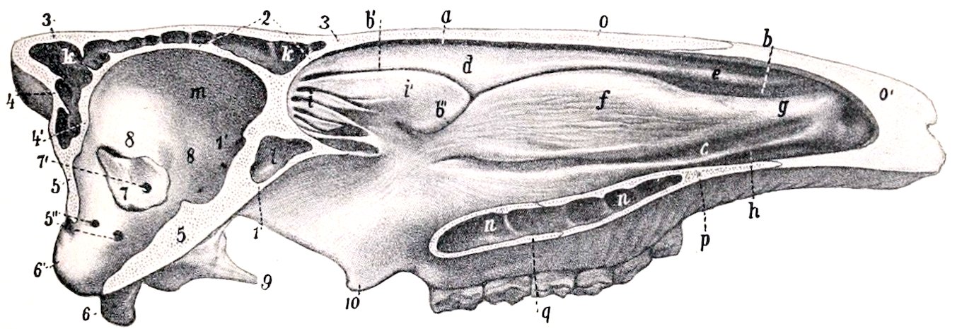
Fig. 93.—Median Section of Skull of Ox, Without the Mandible. The mucous membrane is retained. The septum nasi is removed. a, Superior meatus; b, middle meatus, with b′ and b″, its upper and lower divisions; c, inferior meatus; d, superior turbinal; e, superior turbinal fold; f, inferior turbinal; g, alar fold; h, inferior turbinal fold; i, lateral mass of ethmoid (ethmoturbinals); i′, large ethmoturbinal or middle turbinal; k, frontal sinus; l, sphenoidal sinus; m, cranial cavity; n, palatine sinus; o, nasal bone; o′, parietal cartilage; p, palate process of maxilla; q, palate bone (horizontal part); 1, sphenoid bone; 2, 3, inner and outer plates of frontal bone; 4, 4′, outer and inner plates of parietal bone; 5, 5′, occipital bone; 5″, condyloid and hypoglossal foramina; 6, paramastoid or styloid process; 6′, occipital condyle; 7, petrous temporal bone; 7′, internal auditory meatus; 8, squamous temporal bone; 9, muscular process; 10, pterygoid bone (hamulus). (After Ellenberger, in Leisering’s Atlas.)
The pterygoid bone is wider than in the horse, and forms the greater part of the lateral boundary of the posterior nares. Its outer surface is almost entirely united to the palate bone and the pterygoid process, but a small part is free in the pterygo-palatine fossa. The hamulus is distinctly hook-like, thin, and sharp.
The nasal bone is little more than half the length of that of the horse. It is straight in its length, but strongly curved from side to side. It does not fuse laterally with the adjacent bones, even in old age. The posterior extremity is pointed and fits into the notch between the frontal bones. The anterior end is broader, and is divided into two parts by a deep notch. In old animals there is a small extension of the frontal sinus into this bone.
The lacrimal bone is very large. The extensive facial part is concave in its length, and bears no lacrimal tubercle. The orbital margin is marked by several notches. The orbital part forms below the remarkable lacrimal bulla; this is a large and very thin-walled protuberance, which bulges backward into the lower part of the orbit, and contains an extension of the maxillary sinus. The lacrimal fossa is small, and is just behind the orbital margin.
The malar bone is relatively long. The facial surface is extensive; it bears a curved crest (Crista facialis) just below the orbital margin, and below this it is concave from above downward. The zygomatic process divides into two branches; 122of these, the frontal process turns upward and backward and joins the supraorbital process of the frontal bone; the temporal process continues backward, and is overlapped by the zygomatic process of the temporal bone, completing the zygomatic arch.
The superior turbinal bone is less cribriform and fragile than in the horse, and is thickest in its middle, small at either end. It is attached to the turbinal crest of the nasal bone, and curves downward, outward, and upward to be applied outwardly to the frontal and lacrimal bones. It thus incloses a cavity which communicates with the middle meatus nasi. (In the macerated skull it opens into the frontal sinus, but this communication is closed by mucous membrane in the fresh state.)
The inferior turbinal bone is shorter but much broader than in the horse. It is attached to the maxilla by a basal lamella about an inch (ca. 2 to 3 cm.) wide, which slopes downward and inward. At the inner edge of this it splits into two plates which are rolled in opposite directions, and inclose two separate cavities, subdivided by several septa. The upper one opens into the middle meatus, the lower one into the inferior meatus nasi.
The vomer forms a wider and deeper groove than in the horse. Its anterior end rests in a groove formed by the ends of the palatine processes of the premaxillæ. The anterior third of its thin lower edge fits into the nasal crest of the maxilla; behind this it is free and separated by a considerable interval from the nasal floor.
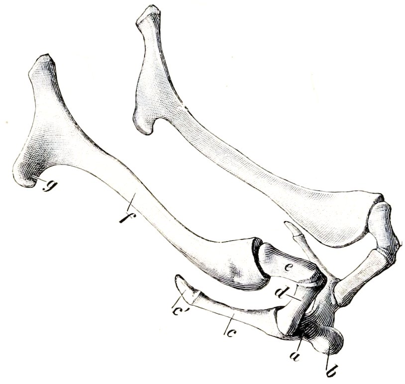
Fig. 94.—Hyoid Bone of Ox. a, Body; b, lingual process; c, thyroid cornu and cartilage, c′; d, small cornu; e, middle cornu; f, great cornu; g, muscular angle of f. (Ellenberger-Baum, Anat. d Haustiere.)
The two halves of the mandible do not fuse completely even in advanced age. The symphyseal surfaces are extremely rough and are marked by reciprocal projections and cavities. The body is shorter, wider, and flatter than in the horse, and has eight round and relatively shallow alveoli for the lower incisors. The interalveolar border is long, curved, thin and sharp. There are no alveoli for the canine teeth, which are absent. The anterior part of the ramus constitutes a long narrow neck. The mental foramen is further forward than in the horse, and is in the posterior end of a fossa. The rami diverge more, so that the submaxillary space is wider than in the horse. They are also more strongly curved, and the angle is more pronounced. The molar part is not so high, especially in its anterior part. Its lower border is convex in its length. Its upper border bears six alveoli for the lower cheek teeth; the first is quite small, and they increase in size from before backward. The vertical part is much smaller than in the horse and its posterior border is relatively thin below, concave and wider above. The mandibular foramen is about in the middle of its inner surface, and a groove for the lingual nerve curves downward and forward from it. The condyle projects inward further than in the horse, and is concave from side to side. The coronoid process is extensive and curves backward.
The hyoid bone has a short tuberous lingual process. The middle cornua are almost, as large as the small cornua. The great cornua are narrow, except at the ends. The upper end divides into two branches, which correspond 123to the two angles of that of the horse. The thyroid cornua do not fuse with the body.
SKULL OF THE OX AS A WHOLE
The skull of the ox is more clearly pyramidal than that of the horse, and is shorter and relatively wider. The cranium is quadrangular and larger externally than in the horse; its large size is due mainly to the great extent of the frontal sinus, and does not affect the cranial cavity, which is smaller than in the horse.
The superior or frontal surface is formed by the frontals, nasals, and premaxillæ. The frontal part is quadrilateral and very extensive, the greatest width being at the orbits. It presents a central depression on its anterior part, and on either side are the supraorbital grooves and foramina. Behind is the median frontal eminence, and at the lateral angles the “horn-cores” project in horned cattle. The nasal part is very short. The osseous nasal aperture is wide. The premaxillæ do not bend downward as in the horse; they are relatively thin and weak, and are separated by an interval which has a wide anterior part in place of the foramen incisivum.
The lateral surface is more triangular than in the horse. The temporal fossa is confined to this surface. It is deep and narrow, and its boundaries are more complete. It is limited above by a crest which extends from the postero-external angle of the frontal to the supraorbital process, and is analogous to the sagittal crest of the horse. Behind it is bounded by the temporal crest. It is clearly marked off from the orbit by the rounded posterior orbital ridge and the pterygoid crest. The zygomatic process is short, weak, and flattened, and is formed by the temporal and malar only. Its condyle is convex and is wide from before backward. The glenoid cavity and postglenoid process are small. The orbit is encroached upon below by the lacrimal bulla, and presents the orifice of the supraorbital canal behind. The margin is completed behind by the frontal process of the malar; its lower part is prominent and rough, not smooth and rounded as in the horse. The pterygo-palatine fossa is much larger, deeper, and more clearly defined. It has a long narrow forward extension (maxillary hiatus) between the vertical plate of the palate bone internally and the maxilla and lacrimal bulla externally; thus the sphenopalatine and maxillary foramina are deeply placed. The preorbital region is short but relatively high. A tuberosity and curved line take the place of the facial crest. The infraorbital foramen is situated above the first cheek tooth and is often double.
The basal surface is short and wide, especially in its cranial part. The occipital condyles are limited in front by transverse ridges. The tubercles at the junction of the occipital and sphenoid are large. The condyloid fossæ contain two foramina, the hypoglossal below and in front, and the condyloid above and behind; other inconstant ones occur. The paramastoid processes are short and convergent. The foramen lacerum is slit-like. The bulla ossea is a large, laterally compressed prominence. The muscular processes are usually long and narrow triangular plates, with one or two sharp points. The external auditory process is directed almost straight outward. A curved plate extends downward from it and joins the bulla ossea internally, completing the deep cavity which receives the articular angle of the hyoid bone. The chief external opening of the temporal canal is in front of this plate, and an accessory one lies behind it. The infratemporal fossa is small, and presents the foramen ovale. The posterior nares are very narrow, and the vomer does not reach to the level of their margin. The hard palate is wide, and forms about three-fifths of the entire length of the skull. A small central part only of its posterior border enters into the formation of the posterior nares; the lateral parts are notched and just above them are the posterior palatine foramina. 124The anterior palatine foramina are an inch or more from the posterior margin, and about the same distance apart. The palatine grooves are distinct for a short distance only. Just beyond the cheek teeth the palate narrows and becomes concave; beyond this it widens and flattens.
The posterior or nuchal surface is extensive and somewhat pentagonal in outline in the adult. About its center is the eminence for the attachment of the ligamentum nuchæ. From this a median crest extends toward the foramen magnum, and laterally two lines (Lineæ nuchæ superiores) curve outward, marking the upper limit of the area which is roughened for muscular attachment. The surface above the lines is relatively smooth, and is covered only by the skin and the thin auricular muscles in the living animal. It is separated from the cranial roof by a thick border, which forms centrally the frontal eminence, and bears at its extremities the processus cornu—except in the polled breeds. The condyles are further apart, and the articular surfaces are more clearly divided into upper and lower parts than in the horse.
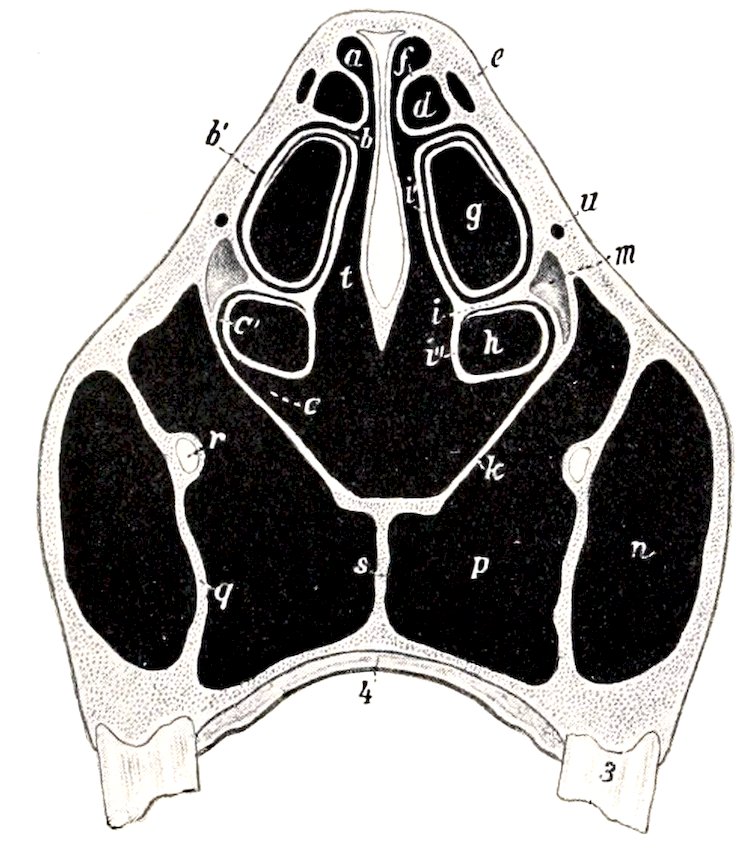
Fig. 95.—Cross-section of Skull of Ox. Section Passes Through Fifth Cheek Tooth.
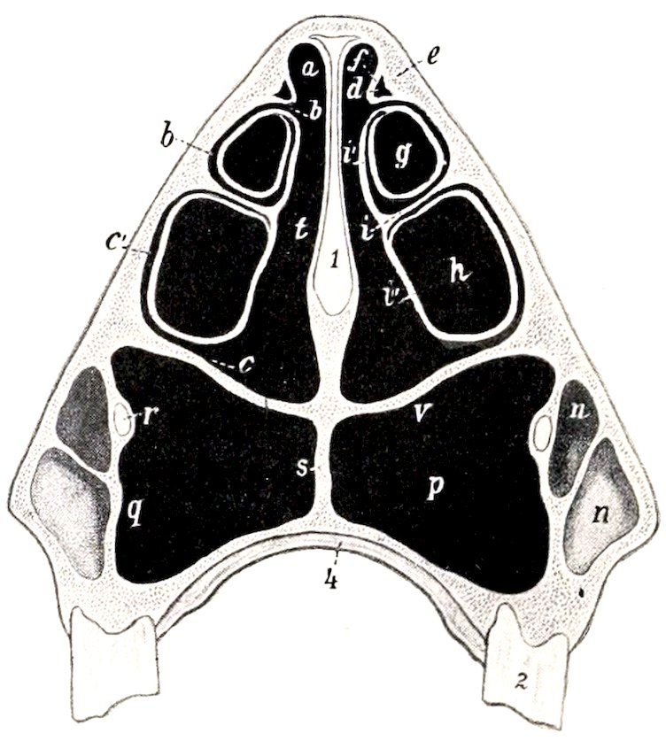
Fig. 96.—Cross-section of Skull of Ox. Section Passes through Second Cheek Tooth. a, b, c, Superior, middle, inferior meatus; b′, communication between middle meatus and cavity of upper part of inferior turbinal; c′, communication between inferior meatus and lower part of inferior turbinal; d, cavity of superior turbinal; e, f, outer and inner walls of d; g, h, upper and lower cavities of inferior turbinal bone; i, basal lamella of inferior turbinal; i′, i″, upper and lower divisions of inferior turbinal bone; k, floor of nasal cavity; m, lacrimal sinus; n, maxillary sinus; p, palatine sinus; q, septum between maxillary and palatine sinuses; r, infraorbital canal; s, septum between palatine sinuses; t, common meatus; u, naso-lacrimal canal; v, floor of nasal cavity and roof of palatine sinus; 1, septum nasi; 2, posterior part of second cheek tooth; 3, posterior part of fifth cheek tooth; 4, hard palate. (After Ellenberger, in Leisering’s Atlas.)
The cranial cavity is shorter and its long axis is more oblique than in the horse, but it is relatively high and wide. The anterior fossa lies at a much higher level than the rest of the floor. The olfactory fossæ are smaller, and the sella turcica is much deeper than in the horse. A deep groove leads from the petrous temporal forward over the foramen ovale to the foramen rotundum. Behind the sella there is often a distinct prominence (Dorsum sellæ). The internal sagittal crest is prominent anteriorly, but absent further back. A faintly marked elevation takes the place of the tentorium osseum. The small petrous temporal bone projects into the cavity laterally. The ridges and digital impressions are very pronounced. The parieto-temporal canal is formed entirely in the temporal bone, and opens at 125the apex of the petrous, where it is joined by the condyloid canal. The foramen lacerum is divided into two parts (For. lacerum anterius et posterius).
The nasal cavity is incompletely divided by the septum, which does not reach the floor posteriorly. The floor is relatively long, and is more concave from side to side than in the horse. In the dry skull it has a large oval opening into the palatine sinus, which is closed during life by mucous membrane. The middle meatus is divided behind into upper and lower branches by the great ethmoturbinal. The posterior nares are narrow and oblique.
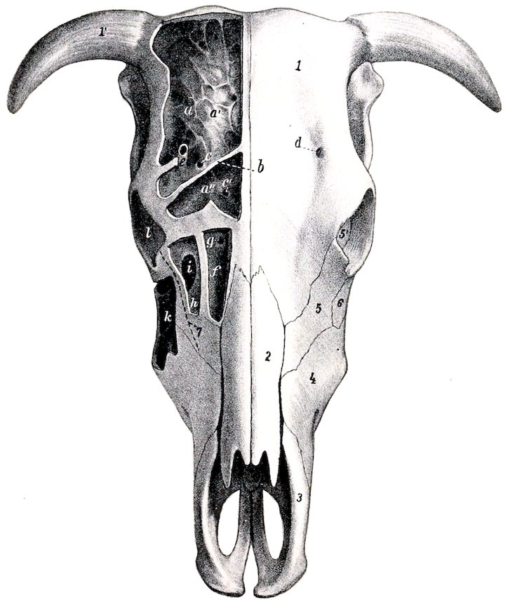
Fig. 97.—Skull of Ox, Dorsal View. The outer plate of bone has been removed to show the sinuses. a, Frontal sinus; a′, cranial plate of frontal bone; a″, anterior part of frontal sinus, which is separated from remainder by a septum (b); c, c′, communications between frontal sinus and nasal cavity; d, supraorbital foramen; e, supraorbital canal; f, cavity of superior turbinal bone, and g, its opening into the nasal cavity; h, lacrimal sinus, i, its communication with the maxillary sinus; k, maxillary sinus; l, orbit; 1, frontal bone; 1′, processus cornu; 2, nasal bone; 3, premaxilla (nasal process); 4, maxilla; 5, lacrimal bone; 6, malar bone; 7, dotted line indicating course of naso-lacrimal duct. (After Ellenberger, in Leisering’s Atlas.)
The frontal sinus is very large. It involves almost all of the frontal bone and a large part of the posterior wall of the cranium. It also extends for a variable distance into the horn processes when these are present. A complete median septum separates the right and left sinuses. The anterior limit is indicated by a transverse plane through the middle of the orbits. It extends externally to the crest which limits the temporal fossa above, and into the root of the supraorbital process. At the highest part of the cranial cavity and at the tuberosity on the posterior surface 126the two plates of the bone come together. The cavity is very irregular and is subdivided into numerous spaces by ridges and partial septa. This multilocular character is most marked in the anterior part, and here several small spaces appear to be cut off from the main cavity (Baum). The supraorbital canal passes through the sinus. Several small openings lead from the sinus to the ethmoidal meatuses, and thus indirectly to the upper division of the middle meatus nasi. The communications with the cavity of the superior turbinal and with the lacrimal part of the maxillary sinus which are seen in the macerated skull are closed in the fresh state by mucous membrane.
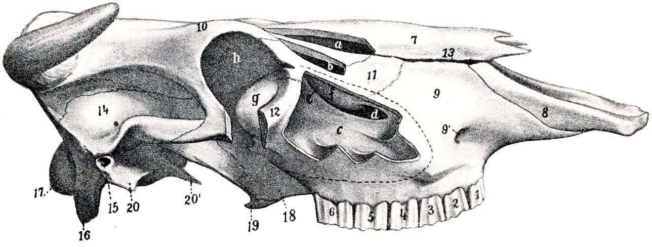
Fig. 98.—Skull of Ox, Lateral View without Mandible. The maxillary, lacrimal, and turbinal sinuses have been opened, and a portion of the orbit removed, a, Cavity of superior turbinal bone; b, lacrimal sinus; c, maxillary sinus; d, communication between maxillary and palatine sinuses; e, opening between maxillary and lacrimal sinuses; f, thin osseous bulla; g, lacrimal bulla; h, orbit; 1–6, cheek teeth; 7, nasal bone; 8, premaxilla (nasal process); 9, maxilla; 9′, infraorbital foramen; 10, frontal bone; 11, lacrimal bone; 12, malar bone; 13, fissure between nasal bone and maxilla; 14, temporal bone (squamous); 15, external auditory meatus; 16, styloid or paramastoid process; 17, occipital condyle; 18, palate bone (perpendicular part); 19, pterygoid bone (hamulus); 20, tympanic part of temporal; 20′, muscular process of petrous temporal. (After Ellenberger, in Leisering’s Atlas.)
The maxillary sinus is excavated chiefly in the maxilla, lacrimal, and malar, and is not divided by a septum as in the horse. It extends forward as far as the facial tuberosity, or a little further in old animals. Its upper limit is indicated approximately by a line drawn from the infraorbital foramen to the upper margin of the orbit. It is continued directly backward into the lacrimal bulla to a point nearly opposite to the bifurcation of the zygomatic process of the malar. It also extends upward and backward through a large opening into a cavity formed by the lacrimal, frontal, ethmoid, and turbinal bones, at the inner side of the orbit.[26] The floor of the cavity is irregular and the roots of the last three or four cheek teeth project up into it, covered by a plate of bone. The sinus communicates with the palatine sinus freely over the infraorbital canal through an oval opening about two to three inches (ca. 5 to 7.5 cm.) long. Above this it communicates by a shorter and much narrower opening with the middle meatus nasi.
The palatine sinus is excavated in the hard palate, and is separated from that of the opposite side by a median septum. It extends from the posterior border of the palate to a plane an inch or more (2.5 to 3 cm.) in front of the first cheek tooth. As mentioned above, there is a large communication with the maxillary sinus over the infraorbital canal, so that the cavity is sometimes regarded as a part of that sinus. The large defect in the bony roof of the sinus is closed by two layers of mucous membrane in the fresh state. The palatine canal passes obliquely through the posterior part of the sinus.