THE MUSCULAR SYSTEM (continued 3)
Reading edition
Blood-supply.—Obturator artery.
Nerve-supply.—Sciatic nerve.
III. ANTERIOR MUSCLES OF THE THIGH
This group consists of the sartorius, quadriceps femoris, and capsularis.
1. Sartorius.—This is described on p. 281.
2. Quadriceps femoris (Figs. 179, 186, 200) (Crural triceps).—This constitutes the large muscular mass which covers the front and sides of the femur. It has four heads, one of which, the rectus, arises from the ilium; the other three arise from the femur. All are inserted into the patella.
(1) Rectus femoris.—This is fusiform and rounded. It arises by two tendons.
Origin.—Two depressions on the shaft of the ilium above and in front of the acetabulum.
Insertion.—The base and anterior surface of the patella.
Action.—To extend the stifle joint and to flex the hip joint.
Structure.—It has two short strong tendons of origin; beneath the outer one is a bursa. The belly is rounded and rests in a groove formed by the other portions of the quadriceps. Its sides are covered by a strong tendinous layer which furnishes insertion to fibers of the vasti. The tendon of insertion is formed by the fusion of these tendinous layers on the lower part of the muscle. The lower portion of the muscle is pennate, the fibers on either side converging on the tendon at an acute angle.
Relations.—Internally, the iliacus, sartorius, and vastus internus; externally, the tensor fasciæ latæ, glutei, and vastus externus; posteriorly, the hip joint and the vastus intermedius; anteriorly, the fascia lata and the skin. The anterior 285femoral artery and branches of the femoral nerve descend into the interspace between the upper part of the rectus and the vastus internus; similarly, the iliaco-femoral artery dips in between the rectus femoris and vastus externus.
Blood-supply.—Femoral and iliaco-femoral arteries.
Nerve-supply.—Femoral nerve.
(2) Vastus externus (s. lateralis).—This lies on the outer surface of the thigh, extending from the great trochanter to the patella. It is thick and wide in its upper part, and becomes much thinner and narrow below.
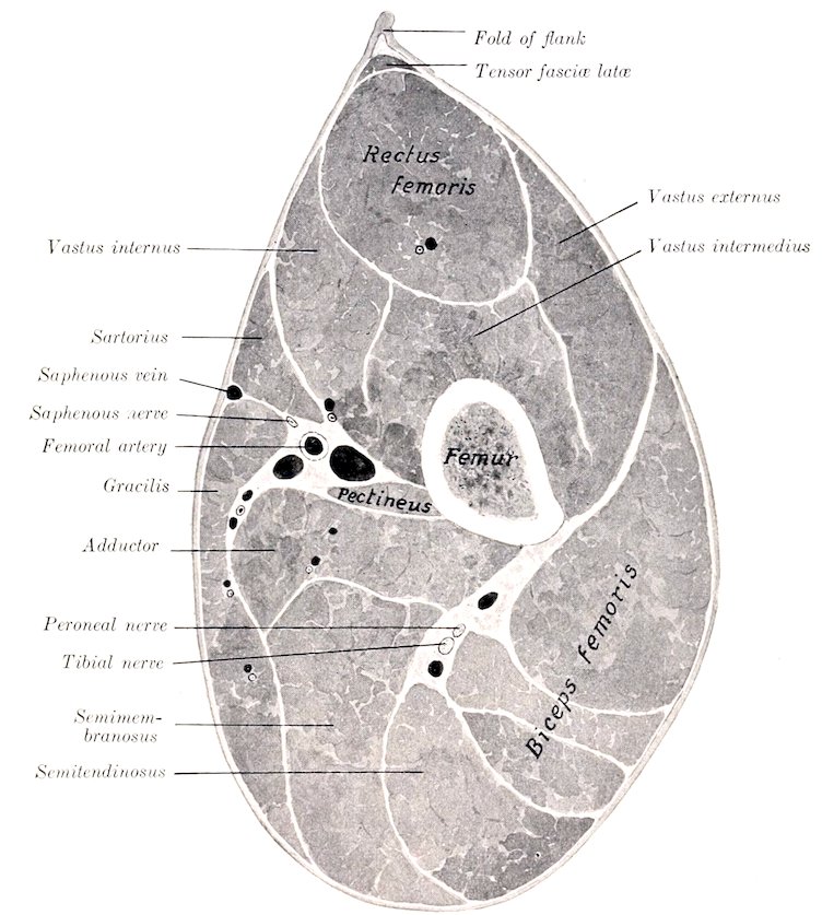
Fig. 200.—Cross-section of Middle of Right Thigh of Horse.
Origin.—The external border and surface of the femur, from the great trochanter to the supracondyloid fossa.
Insertion.—(1) The outer part of the anterior surface of the patella; (2) the tendon of the rectus femoris.
Action.—To extend the stifle joint.
Structure.—The fibers are directed downward and forward, many being inserted into the tendinous sheet which covers the side of the rectus. A bursa is often found between the distal end and the patella.
286Relations.—Externally, the fascia lata and skin, tensor fasciæ latæ, superficial gluteus, and biceps femoris; internally, the femur and femoro-patellar joint capsule, the rectus femoris, vastus intermedius, and the iliaco-femoral artery.
Blood-supply.—Iliaco-femoral artery.
Nerve-supply.—Femoral nerve.
(3) Vastus internus (s. medialis).—This is smaller than the preceding muscle, and lies in a similar position on the inner side of the thigh.
Origin.—The internal surface of the femur, from the neck to the distal third.
Insertion.—(1) The upper part of the inner border of the patella and its cartilage; (2) the tendon of the rectus femoris.
Action.—To extend the stifle joint.
Structure.—This is very similar to that of the vastus externus. It is, however, more difficult to separate from the intermedius, because many fibers of the latter arise on the tendinous sheet which covers the contact surface of the inner vastus. Its insertion into the patella is chiefly by means of a broad strong tendon. From the deep face fleshy fibers are inserted also into the femoro-patellar capsule.
Relations.—Internally, the skin and fascia lata, the iliacus, sartorius, pectineus, and adductor, the femoral vessels and saphenous nerve; externally, the femur, femoro-patellar joint capsule, rectus femoris, and vastus intermedius, the anterior femoral artery, and branches of the femoral nerve.
Blood-supply.—Femoral and anterior femoral arteries.
Nerve-supply.—Femoral nerve.
(4) Vastus intermedius (Crureus).—This muscle is deeply situated on the anterior face of the femur, and is entirely covered by the preceding heads.
Origin.—(1) The anterior and external surfaces of the femur, from the proximal to the distal fourth; (2) the aponeurosis of the vastus internus.
Insertion.—(1) The base of the patella; (2) the femoro-patellar joint capsule.
Action.—(1) To extend the stifle joint; (2) to tense (raise) the femoro-patellar capsule during extension of the joint.
Structure.—The muscle is usually quite difficult to isolate from the other vasti, so that many since Günther have declared it an artefact.[71] It is entirely fleshy, and is small at its proximal end, but when traced downward increases in bulk by the accession of fibers arising on the femur and the tendinous covering of the vastus internus. The terminal part is intimately adherent to the femoro-patellar joint capsule, where the latter bulges upward above the level of the patella.
Relations.—Internally, the vastus internus; externally, the vastus externus; anteriorly, the rectus; posteriorly, the femur and femoro-patellar capsule.
Blood-supply.—Iliaco-femoral and anterior femoral arteries.
Nerve-supply.—Femoral nerve.
The straight ligaments of the patella are to be regarded as tendons of the quadriceps which communicate the action of the latter to the tibia, the patella being intercalated as a sesamoid bone.
3. Capsularis (Fig. 154) (Rectus parvus).—This is a small muscle (scarcely as large as one’s finger), which arises on the ilium immediately above the outer tendon of the rectus femoris, and passes down between the vastus internus and externus to be inserted into the anterior surface of the femur. It passes over the front of the hip joint, to the capsule of which some fibers are attached. Sometimes the muscle has two distinct heads, in which case the additional head arises between the two tendons of origin of the rectus femoris. Its action may be to raise the capsule during flexion of the joint.
287
IV. THE MUSCLES OF THE LEG AND FOOT
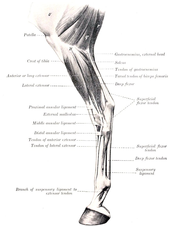
Fig. 201.—Muscles of Lower Part of Thigh, Leg, and Foot of Horse, External View. o′, Fascia lata; q, q′, q″, biceps femoris; r, semitendinosus; 21′, external condyle of tibia. (After Ellenberger-Baum, Anat. für Künstler.)
The muscles of this region cover almost all of the tibia except its internal face, which is largely subcutaneous. As in the forearm, the muscles fall into two groups, an anterior or dorso-lateral, and a posterior or plantar. The muscles of the first group are extensors of the digit and flexors of the hock, those of the second have the opposite action.
288
A. Anterior Group
1. Anterior or long digital extensor (M. extensor digitalis longus; extensor pedis; anterior extensor of the phalanges).—This muscle is situated superficially on the antero-external aspect of the leg, and is provided with a long tendon which passes down over the front of the tarsus, metatarsus, and digit.
Origin.—The small fossa (Fossa extensoria) between the external condyle and the trochlea of the femur.
Insertion.—(1) The extensor process of the third phalanx; (2) the anterior surface of the proximal extremities of the first and second phalanges.
Action.—To extend the digit and flex the hock. It also assists in fixing the stifle joint.
Structure.—The origin is by means of a strong tendon in common with the peroneus tertius, on which also many fibers arise. The common tendon passes downward in the groove between the outer condyle and the tuberosity of the tibia, where a pouch from the femoro-tibial capsule descends four or five inches (ca. 10 to 12 cm.) beneath the tendon. The belly is fusiform and somewhat flattened. The long tendon of insertion begins in the belly about its middle, and is clear of the fleshy part near the tarsus. It passes downward over the front of the hock, bound down by the three annular ligaments already described (see crural fascia), and enveloped by a synovial sheath which begins a little above the level of the external malleolus, and extends nearly to the junction with the lateral extensor tendon. This union occurs usually about a hand’s breadth below the tarsus. In the angle of union the extensor brevis also joins the principal tendon. Beyond this point the arrangement is the same as in the fore limb.
Relations.—Superficially, the skin and fascia; deeply, the femoro-tibial capsule, peroneus tertius, and tibialis anterior; behind, the lateral extensor and the superficial and deep peroneal nerves. In front of the tarsus the anterior tibial artery crosses the deep face of the tendon (Fig. 460).
Blood-supply.—Anterior tibial artery.
Nerve-supply.—Peroneal nerve.
2. Lateral digital extensor (M. extensor digitalis lateralis; peroneus; lateral extensor of the phalanges).—This muscle lies on the outer surface of the leg, behind the preceding one.
Origin.—The external lateral ligament of the stifle joint, the fibula, the external border of the tibia, and the interosseus ligament.
Insertion.—The tendon of the anterior extensor, about a third of the way down the metatarsus.
Action.—To assist the anterior extensor.
Structure.—The belly is fusiform, flattened, and pennate. The tendon runs through the entire length of the belly and becomes free from it at the lower fourth of the tibia. It passes downward through the groove on the external malleolus, bound down by an annular ligament, and, inclining forward, blends (usually) with the tendon of the anterior extensor. It is provided with a synovial sheath, which begins about one inch (ca. 2 to 3 cm.) above the external malleolus and ends about one and one-half inches (ca. 3 to 4 cm.) above the junction. Sometimes the fusion does not occur, and the tendon then passes down the metatarsus, alongside of that of the long extensor, to lie inserted into the first phalanx like the corresponding muscle of the thoracic limb.
Relations.—Externally, the skin and fascia and the superficial peroneal nerve; internally, the tibia and fibula; anteriorly, the intermuscular septum, the long extensor, and the tibialis anterior; posteriorly, the deep flexor and the soleus.
Blood-supply.—Anterior tibial artery.
Nerve-supply.—Peroneal nerve.
2893. Peroneus tertius (Tendinous part of the flexor metatarsi; tendo femorotarseus [Schmaltz]).—This consists in the horse of a strong tendon which lies between the anterior extensor and the tibialis anterior.[72]
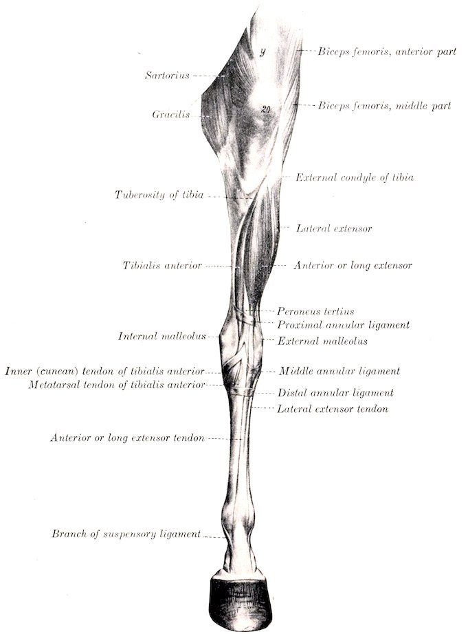
Fig. 202.—Muscles of Pelvic Limb of Horse, Anterior View. y, Rectus femoris; 20, patella. (After Ellenberger-Baum, Anat. für Künstler.)
Origin.—The fossa extensoria (between the external condyle and the trochlea of the femur), in common with the anterior extensor.
290Insertion.—(1) The proximal extremities of the large (third) and external small (fourth) metatarsal bones, and the third tarsal bone; (2) the fibular and fourth tarsal bones.
Action.—Mechanically to flex the hock when the stifle joint is flexed.
Structure.—This is entirely tendinous. The proximal end and the underlying prolongation of the synovial membrane of the femoro-tibial joint have been mentioned in the description of the anterior extensor. Fibers of the latter muscle and of the tibialis anterior arise on the tendon as it passes downward to the front of the hock. At the distal end of the tibia the tendon divides into two branches, between which the tendon of the tibialis anterior emerges. The anterior branch is attached to the third tarsal and third and fourth metatarsal bones, while the outer one curves outward, bifurcates, and is inserted into the fibular and fourth tarsal bones (Fig. 460).
Relations.—Superficially, the anterior extensor; deeply, the tibialis anterior. The anterior tibial vessels cross the deep face of the outer branch.
4. Tibialis anterior (Muscular portion of the flexor metatarsi).—This lies on the antero-external face of the tibia; it is wide and flattened above, pointed below.
Origin.—The external condyle and border of the tibia and a small area on the external surface of the tuberosity.
Insertion.—(1) The metatarsal tuberosity; (2) the first and second tarsal bones.
Action.—To flex the hock joint.
Structure.—The origin is fleshy, and is divided by the groove in which lie the common tendon of the long extensor and peroneus tertius and a synovial pouch. Passing downward on the tibia, the belly is united by tendinous and fleshy fibers with the peroneus tertius, and terminates close to the tarsus in a point on the tendon of insertion. The latter emerges between the branches of the peroneus tertius and bifurcates, the anterior branch being inserted into the metatarsal tuberosity, the inner one (cunean tendon) into the fused first and second tarsal bones (cuneiform parvum). The tendon is provided with a synovial sheath at its emergence, and a bursa (cunean bursa) is interposed between the inner branch and the internal lateral ligament.
Relations.—Superficially, the anterior and lateral extensors, the peroneus tertius, and the deep peroneal nerve; deeply, the tibia, the deep flexor, and the anterior tibial vessels.
Blood-supply.—Anterior tibial artery.
Nerve-supply.—Peroneal nerve.
B. Posterior Group
1. Gastrocnemius (Figs. 179, 201, 203).—This muscle extends from the lower third of the femur to the point of the hock. It arises by two heads.
Origin.—(1) Outer head, from the external rough margin of the supracondyloid fossa (Fossa plantaris); (2) inner head, from the supracondyloid crest.
Insertion.—The posterior part of the tuber calcis.
Action.—To extend the hock.
Structure.—The two bellies are thick, fusiform, and somewhat flattened. They are covered by a strong aponeurosis and contain tendinous intersections. They terminate toward the middle of the leg on a common tendon (Tendo calcaneus s. Achillis), which at first lies posterior to that of the superficial flexor, but, by a twist in both, comes to lie in front of the latter. The deep fascia blends with the tendon throughout its length, and the soleus muscle is inserted into its anterior edge. A small bursa (Bursa tendinis calcanei) lies in front of the insertion on the tuber calcis, and a large bursa is interposed between the two tendons from the twist 291downward. The superficial flexor lies between the two heads and is adherent to the outer one (Fig. 459).
Relations.—Anteriorly, the stifle joint, the superficial flexor, popliteus, deep flexor, popliteal vessels, and tibial nerve; internally (above), the semitendinosus, semimembranosus, and adductor, (below) the fascia and skin; externally (above), the biceps femoris and peroneal (anterior tibial) nerve, (below) the fascia and skin.
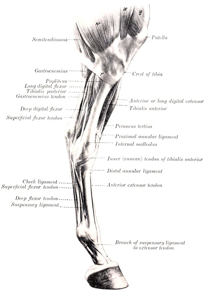
Fig. 203.—Muscles of Leg and Foot of Horse, Internal View. r′, Tendon of semitendinosus; w, gracilis; x, sartorius; y, vastus internus. (After Ellenberger-Baum, Anat. für Künstler.)
Blood-supply.—Popliteal artery.
Nerve-supply.—Tibial nerve.
2922. Soleus (Fig. 201).—This muscle is very small in the horse. It lies immediately under the deep fascia, on the proximal half of the outer surface of the leg, and is directed obliquely downward and backward.
Origin.—The head of the fibula.
Insertion.—The tendon of the gastrocnemius, about the middle of the leg.
Action.—To assist the gastrocnemius.
Structure.—It is a thin, fleshy band, about an inch (ca. 2 to 3 cm.) in width, terminating on a thin tendon which fuses with that of the gastrocnemius.[73]
Relations.—Superficially, the skin, fascia, and peroneal nerve; deeply, the lateral extensor and deep flexor.
Blood-supply.—Posterior tibial artery.
Nerve-supply.—Tibial nerve.
3. Superficial digital flexor or flexor pedis perforatus (Figs. 199, 201, 203) (M. flexor digitalis pedis sublimis; superficial flexor of the phalanges).—The proximal part of this muscle lies between and under cover of the two heads of the gastrocnemius (Fig. 459). It consists almost entirely of a strong tendon, the belly being very little developed.
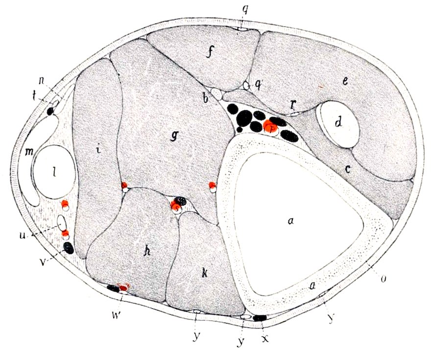
Fig. 204.—Cross-section of Left Leg of Horse; Section is Cut a Little Above Middle of Region. a, Tibia; b, fibula; c, tibialis anterior; d, peroneus tertius; e, anterior or long digital extensor; f, lateral extensor; g, deep head of deep flexor (flexor hallucis longus); h, inner head of deep flexor (flexor accessorius s, digitorum longus); i, superficial head of deep flexor (tibialis posterior); k, popliteus; l, superficial flexor tendon; m, gastrocnemius tendon; n, soleus; o, skin; p, anterior tibial artery; q, superficial peroneal nerve; q′, deep peroneal nerve; r, branch of q; s, posterior tibial artery; t, cutaneous nerve; u, tibial nerve; v, recurrent tibial vein; w, saphenous artery; x, saphenous vein; y, branches of saphenous nerve. (After Ellenberger, inl Leisering’s Atlas.)
Origin.—The supracondyloid fossa (Fossa plantaris) of the femur.
Insertion.—(1) The front and sides of the tuber calcis; (2) the eminences on the proximal extremity of the second phalanx, and the distal extremity of the first phalanx behind the lateral ligament.
Action.—To flex the digit and extend the hock joint. On account of the exceedingly small amount of muscular tissue the action is to be regarded chiefly as a mechanical effect, resulting from the action of other muscles on the stifle joint.
Structure.—The origin is by means of a strong round tendon which is incompletely covered with fleshy fibers as far as the upper third of the leg. It is pretty intimately attached to the gastrocnemius, especially to the outer head. At the distal third of the tibia it winds around the inner surface of the gastrocnemius tendon, and then occupies a position behind the latter. At the point of the hock it widens out, forming a sort of cap over the tuber calcis, detaching on either side a strong band which is inserted into the tuber calcis with the tarsal tendon of the biceps and semitendinosus. It then passes downward over the plantar (Calcaneo-metatarsal) ligament, becomes narrower, and is arranged below as in the thoracic limb. A large synovial bursa lies under the tendon from the distal fourth of the 293tibia to the middle of the tarsus. A subcutaneous bursa is sometimes found on the wide part of the tendon at the point of the hook. (Either or both of these bursæ may be involved in so-called “capped hock.”)
Relations.—Posteriorly, the gastrocnemius, fascia, and skin; anteriorly, the femoro-patellar capsule, the popliteus, the deep flexor, and the popliteal vessels; internally, the tibial nerve.
Blood-supply.—Femoro-popliteal artery.
Nerve-supply.—Tibial nerve.
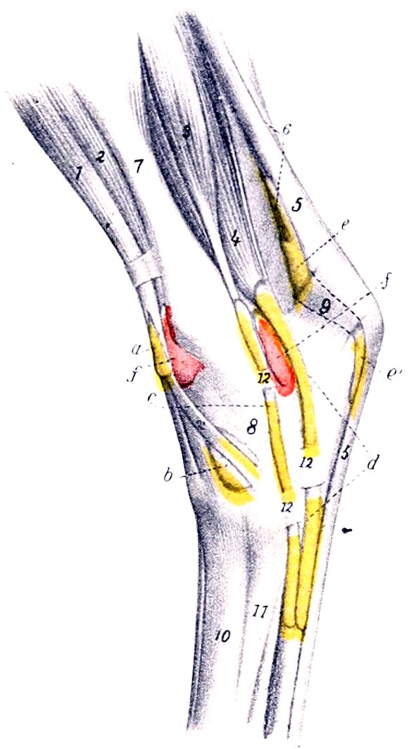
Fig. 205.—Injected Synovial Sheaths and Bursæ of Tarsal Region of Horse, Inner View. a, Synovial sheath of peroneus tertius and tibialis anterior; b, bursa under inner (cunean) tendon of tibialis anterior; c, synovial sheath of flexor longus s. accessorius; d, tarsal sheath of deep flexor; e, e′, bursa under superficial flexor tendon; f, f′, tibio-tarsal joint capsule; 1, anterior extensor; 2, tibialis anterior; 2′, inner (cunean) tendon of 2; 3, flexor longus; 4, deep digital flexor; 5, superficial flexor tendon; 6, gastrocnemius tendon; 7, tibia; 8, tarsus; 9, tuber calcis; 10, large metatarsal bone; 11, inner small metatarsal bone; 12, 12′, fascial bands. (After Ellenberger, in Leisering’s Atlas.)
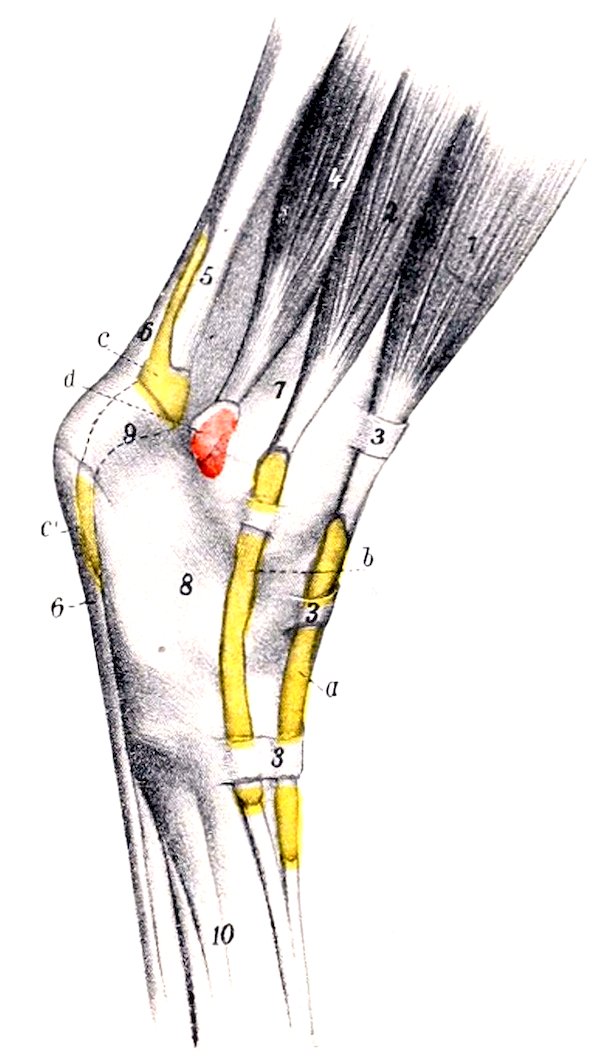
Fig. 206.—Injected Synovial Sheaths and Bursæ of Tarsal Region of Horse, External View. a, Synovial sheath of anterior or long digital extensor; b, synovial sheath of lateral digital extensor; c, c′, bursa under superficial flexor tendon; d, capsule of hock joint; 1, anterior or long extensor; 2, lateral extensor; 3, 3, 3, annular ligaments; 4, deep digital flexor; 5, tendon of gastrocnemius; 6, superficial flexor tendon; 7, tibia; 8, tarsus; 9, tuber calcis; 10, metatarsus. (After Ellenberger, in Leisering’s Atlas.)
4. Deep digital flexor or flexor pedis perforans (M. flexor digitalis pedis profundus; deep and oblique flexors of the phalanges).—The belly of this muscle lies on the posterior surface of the tibia, and is divisible into three parts or heads, which, however, finally unite on a common tendon of insertion.
Origin.—(1) The posterior edge of the external condyle of the tibia; (2) the external border of the external condyle of the tibia, just behind the facet for the fibula; (3) the middle third of the posterior surface and the upper part of the external border of the tibia, the posterior border of the fibula, and the interosseous ligament.[74]
294Insertion.—The semilunar crest of the third phalanx and the adjacent surface of the lateral cartilage.
Action.—To flex the digit and to extend the hock joint.
Structure.—(1) The inner head (M. flexor digitalis longus s. flexor accessorius) is easily isolated (Figs. 203, 459). It has a fusiform belly, which crosses the leg obliquely and lies in a groove formed by the other heads and the popliteus. This terminates near the lower third of the tibia on a round tendon which passes downward, partly embedded in the internal lateral ligament of the hock, and joins the common tendon about a third of the way down the metatarsus. In its course over the inner surface of the hock the tendon lies in a canal formed by the strong tarsal fascia, the tibia, and the lateral ligament, and is provided with a synovial sheath which extends from the distal fourth of the tibia to the junction with the principal tendon. (2) The superficial head (M. tibialis posterior) is only partially separable. It has a flattened belly, terminating near the lower third of the tibia on a flat tendon which soon fuses with the principal tendon. (3) The deep head (M. flexor hallucis longus) is much the largest. It lies on the posterior surface of the tibia, from the popliteal line outward and downward. The belly contains much tendinous tissue, and terminates behind the distal end of the tibia on a strong round tendon. The latter receives the tendon of the tibialis posterior, passes downward in the tarsal groove, bound down by the strong tarsal fascia (Ligamentum laciniatum) and enveloped in a synovial sheath, receives the tendon of the inner head below the hock, and, a little further down, the so-called check ligament (Caput tendineum [Schmaltz]). The tarsal sheath (Vagina tarsea) begins about two to three inches (ca. 5 to 7.5 cm.) above the level of the internal malleolus, and extends about one-fourth of the way down the metatarsus. (Distention of the sheath, as in “thoroughpin,” affects chiefly its proximal end.) The check ligament resembles that of the fore limb, except that it is longer and very much weaker; it may be absent. The remainder of the tendon is arranged like that of the thoracic limb.
Relations.—Anteriorly, the tibia and fibula, the popliteus, lateral extensor, tibialis anterior, and the tibial vessels; posteriorly, the gastrocnemius, superficial flexor, and the tibial nerve; externally, the fascia, skin, and the soleus; internally, the fascia and skin.
Blood-supply.—Posterior tibial artery.
Nerve-supply.—Tibial nerve.
5. Popliteus (Fig. 203).—This thick and triangular muscle lies on the posterior surface of the femoro-tibial articulation and the posterior surface of the tibia above the popliteal line.
Origin.—A small depression on the external epicondyle of the femur, close to the articular surface and under the lateral ligament.
Insertion.—A triangular area on the posterior surface of the tibia, above and internal to the popliteal line; also the proximal half of the inner border and a narrow adjacent part of the internal surface of the tibia.
Action.—To flex the femoro-tibial joint and to rotate the leg inward.
Structure.—The strong tendon of origin lies at first under the lateral ligament, and curves backward and inward over the external condyle of the tibia and its semilunar cartilage, being invested by a reflection of the synovial capsule of the joint (Fig. 459). The tendon is succeeded by a thick triangular belly, the fibers of which are directed obliquely downward and inward.
Relations.—Superficially, the fascia and skin, semitendinosus, gastrocnemius, superficial flexor; deeply, the femoro-tibial joint, the tibia, the popliteal vessels and their divisions. The saphenous vessels and nerve lie along the inner border of the muscle, separated from it, however, by the deep fascia.
Blood-supply.—Popliteal and posterior tibial arteries.
Nerve-supply.—Tibial nerve.
295
MUSCLES OF THE METATARSUS AND DIGIT
Extensor pedis brevis (M. extensor digitalis brevis).—This small muscle lies in the angle of union of the long and lateral extensors of the digit. (Shown in Fig. 201, but not marked.)
Origin.—The outer tendon of the peroneus tertius, the middle annular ligament, and the outer lateral ligament of the hock.
Insertion.—The tendon of the anterior or long extensor.
Action.—To assist the anterior or long extensor.
Structure.—It is principally fleshy, having a superficial origin from the annular ligament, and a deep one (by a thin tendon) from the outer tendon of the peroneus tertius. The insertion is by a thin tendon.
Relations.—Superficially, the skin and fascia and the tendons of the anterior and lateral extensors; deeply, the joint capsule, the great metatarsal artery, and the deep peroneal nerve.
Blood-supply.—Great metatarsal artery.
Nerve-supply.—Deep peroneal nerve.
The interossei and lumbricales are arranged like those of the thoracic limb, the only noticeable difference being the greater development of the lumbricales in the pelvic limb.
THE MUSCLES OF THE OX
Muscles of the Face
The panniculus is much more developed than in the horse, presenting as a special feature the extensive frontalis muscle, which covers the frontal and nasal regions.
The orbicularis oris does not form a complete ring, the defect being in the middle of the upper lip.
The levator nasolabialis is extensive, thin, and not very distinct from the frontalis; it divides into two layers, between which the levator labii superioris proprius and the lateral dilator of the nostril pass. The superficial layer ends in the nostril and upper lip, the deep layer on the accessory (lateral) nasal cartilages and on the nasal process of the premaxilla.
The levator labii superioris proprius arises on and before the facial tuberosity and terminates by several tendons in the muzzle. It passes between the two layers of the preceding muscle, blending in part with the deep layer.
The zygomaticus is much stronger than in the horse. It arises on the masseteric fascia, and ends chiefly in the upper lip.
The depressor labii superioris does not resemble the muscle of the same name in the horse. It arises just in front of the facial tuberosity, and divides usually into two branches, which terminate in a number of tendons that form a network in the muzzle and upper lip.
The incisivus inferior is a small, rounded muscle, which arises on the body of the mandible below the second and third incisors, and ends in the lower lip, blending with the orbicularis.
The depressor labii inferioris is thin, and does not extend as far backward as in the horse; only the anterior end is distinct from the buccinator.
The buccinator shows no marked variation, but its superficial layer is well developed.
The dilatator naris lateralis arises in front of the facial tuberosity, passes forward 296between the branches of the levator nasolabialis, and terminates in the outer wing of the nostril.
The dilatator naris transversus is replaced by the dilatator naris apicalis, which is situated in the muzzle and joins its fellow at a median raphé. It arises on the border and upper surface of the body of the premaxilla, the fibers passing obliquely upward and outward to the inner wing of the nostril.
The dilatator naris superior arises from the alar cartilage of the nostril and ends in the inner wing of the nostril.
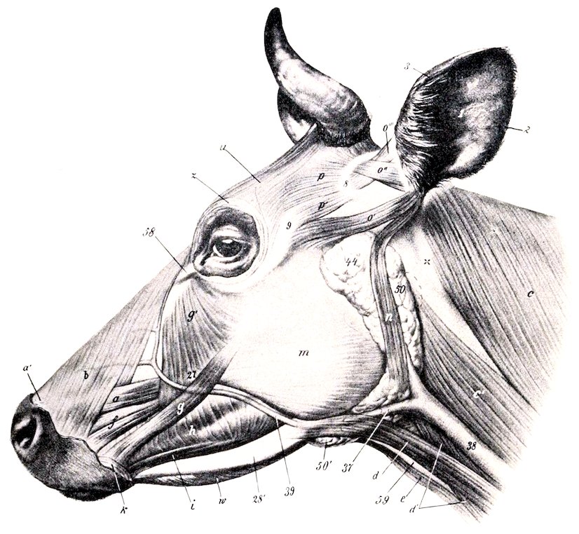
Fig. 207.—Muscles of Head of Ox, Lateral View. a, Levator labii superioris proprius; b, levator nasolabialis; c, trapezius; c′, mastoido-humeralis; d, d′, sterno-cephalicus; e, omo-hyoideus; f, dilatator naris lateralis; g, zygomaticus; g′, malaris; h, buccinator; i, depressor labii inferioris; k, orbicularis oris; m, masseter; n, parotido-auricularis; o′, zygomatico-auricularis and scutulo-auricularis superficialis inferior; o″, scutulo-auricularis superficialis superior; o‴, scutulo-auricularis superficialis accessorius; p, p′, scutularis; u, frontalis; w, mylo-hyoideus; 1, concha; 2, 3, posterior and anterior borders of 1; 8, scutiform cartilage; 9, zygomatic arch; 28′, ramus of mandible; 37, external maxillary vein; 38, jugular vein; 39, facial vein; 44, parotid gland; 50, 50′, submaxillary gland; dotted line at 50 indicates position of large lymph gland, and another lies partly under parotid gland, just in front of 44; 58, internal palpebral ligament; 59, laryngeal prominence. (After Ellenberger-Baum, Anat. für Künstler.)
The dilatator naris inferior consists of two layers which arise on the nasal process of the premaxilla and the lateral nasal cartilage and end in the outer wing of the nostril.
The orbicularis oculi is well developed.
The corrugator supercilii is not present as a separate muscle, its place and function being taken by the frontalis.
The malaris is broad, and spreads out below on the fascia over the buccinator and masseter; it is divided into two parts.
297
MUSCLES OF MASTICATION
The masseter is not so large as in the horse; a considerable part of it arises on the facial tuberosity and is directed obliquely backward and downward, so that it would draw the lower jaw forward as well as upward.
The temporalis conforms to the temporal fossa, and is therefore longer and entirely lateral in position.
The pterygoidei are not quite clearly separated; their direction is more oblique, and the origin of the pterygoideus internus is nearer the median plane than in the horse.
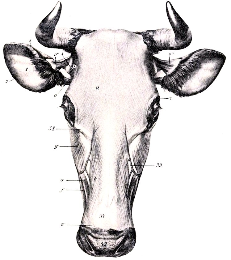
Fig. 208.—Muscles of Head of Ox, Dorsal View. a, a′, Levator labii superioris proprius; b, levator nasolabialis; f, dilatator naris lateralis; g′, malaris; o, zygo-inatico-auricularis and scutulo-auricularis superficialis inferior; o″, scutulo-auricularis superficialis superior; o‴, scutulo-auricularis superficialis accessorius; p, scutularis; u, frontalis; z, orbicularis oculi; 1, concave surface of concha; 3, 2, anterior and posterior borders of concha; 8, scutiform cartilage; 34, parietal cartilage; 39, facial vein; 49, muzzle; 58, internal palpebral ligament. (After Ellenberger-Baum, Anat. für Künstler.)
The stylo-mandibularis is absent.
The digastricus has a tendinous origin on the paramastoid or styloid process of the occipital bone; its bellies are short and thick. It does not perforate the stylo-hyoideus. The two digastrici are connected beneath the root of the tongue by a layer of transverse muscle-fibers (Transversus mandibulæ).
HYOID MUSCLES
The mylo-hyoideus is thicker and more extensive than in the horse.
The stylo-hyoideus has a long thin tendinous origin and is not perforated by the digastricus.
298The genio-hyoideus is more developed.
The kerato-hyoideus has an additional attachment on the middle cornu of the hyoid bone.
The hyoideus transversus is bifid.
The sterno-thyro-hyoideus has no intermediate tendon and is thicker.
The omo-hyoideus arises as a thin band from the fascia over the third and fourth cervical vertebræ. It blends here with the rectus capitis anterior major. The occipito-hyoideus is thick.
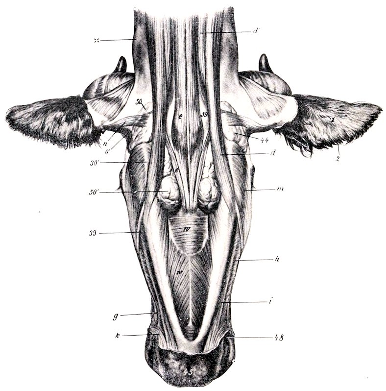
Fig. 209.—Muscles of Head of Ox, Ventral View. d, d′, Sterno-cephalicus; e, omo-hyoideus; g, zygomaticus; h, buccinator; i, depressor labii inferioris; k, orbicularis oris; m, masseter; n, parotido-auricularis; o′, zygomatico-auricularis; w, mylo-hyoideus; 1, concha, convex surface; 2, anterior border of concha; 30′, angle of jaw; 39, facial vein; 44, parotid gland; 45, lower lip; 48, angle of mouth; 50, 50′, submaxillary gland; 59, larynx; x, wing of atlas. (After Ellenberger-Baum, Anat. für Künstler.)
Muscles of the Neck
A. VENTRAL GROUP
The sterno-cephalicus consists of two muscles. They arise from the manubrium sterni and first rib. The superficial muscle (Sterno-mandibularis) is inserted on the anterior border of the masseter, the ramus of the mandible, and the buccal fascia. The deep muscle (Sterno-mastoideus) crosses under the preceding and ends on the mastoid process, the mandible, and, in common with the rectus capitis anterior major, on the basilar process of the occipital bone.
299
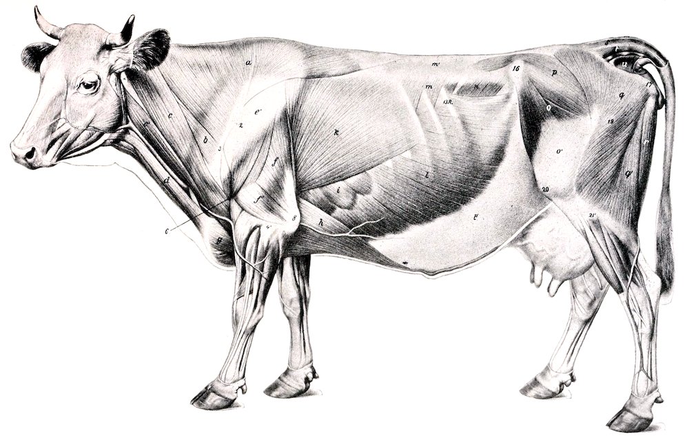
Fig. 210.—Superficial Muscles of Ox, After Removal of Panniculus. a, Trapezius; b, omo-transversarius; c, c′, mastoido-humeralis (c, cleido-mastoideus; c′, cleido-occipitalis); d, sterno-cephalicus; e, deltoid; f, long head, f′, external head of triceps; g, superficial pectoral; h, posterior deep pectoral; i, serratus magnus; k, latissimus dorsi; l, obliquus abdominis externus; l′, aponeurosis of l; m, serratus posterior; m′, lumbo-dorsal fascia; n, obliquus abdominis internus; o, tensor fasciæ latæ; o′, fascia lata; p, gluteus medius; q, q′, biceps femoris; r, semitendinosus; s, sacro-coccygeus superior; t, sacro-coccygeus lateralis; u, coccygeus. (After Ellenberger-Baum, Anat. f Künstler.)
300There are two scaleni. The scalenus ventralis (s. primæ costæ) arises on the first rib and ends on the transverse processes of the third to the seventh cervical vertebræ. It is traversed by the roots of the brachial plexus, which partially divide it into a small dorsal and a large ventral part. The brachial vessels lie below the latter. The scalenus dorsalis (s. supracostalis) arises usually on the second, third, and fourth ribs, and ends on the transverse processes of the third to the sixth cervical vertebræ.
The rectus capitis anterior major arises on the third to the sixth cervical transverse processes, and blends at its insertion with the sterno-mastoideus and the mastoid portion of the mastoido-humeralis.
The rectus capitis anterior minor is larger than in the horse.
The rectus capitis lateralis and longus colli resemble those of the horse.
The intertransversales are large. From the sixth cervical vertebra forward they form a muscular mass (M. intertransversarius longus) which is inserted into the wing of the atlas.
B. LATERAL GROUP
The splenius is thin. It arises directly from the first three or four thoracic spines, and ends by a thin tendon on the occipital bone, the wing of the atlas, and the transverse process of the axis, blending with the mastoido-humeralis, trachelo-mastoideus, and omo-transversarius. The remaining muscles present no very marked differential features.
Muscles of the Thorax
The levatores costarum number ten or eleven pairs.
The diaphragm presents several important differential features. Its slope is much steeper and its width is greater than in the horse. The costal attachment extends almost in a straight line from the upper fourth of the last rib to the junction of the eighth rib with its cartilage, and along the latter to the sternum. The mid-line slopes from the twelfth thoracic vertebra obliquely as far as the vena cava, beyond which it is almost vertical. The right crus divides into two branches, which circumscribe the œsophageal opening, unite below, and then spread out in the tendinous center. The left crus is small. The œsophageal opening is situated about four to five inches (10 to 12 cm.) below the eighth thoracic vertebra, a little to the left of the median plane. The foramen venæ cavæ is a little more ventral and almost in the median plane. The other muscles resemble those of the horse.
Muscles of the Back and Loins
The serratus anticus is very thin. It is inserted on the fifth to the eighth ribs. It may be reduced to two or three digitations or may be absent. The serratus posticus is usually inserted on the last three or four ribs.
The transversalis costarum (Iliocostalis) has a distinct lumbar portion which is attached to the lumbar transverse processes and the external angle of the ilium.
The longissimus resembles that of the horse, but it is more fleshy anteriorly, and the spinalis dorsi is clearly distinguishable from the common mass. In the lumbar region the tendons meet across the summits of the spines.
Intertransversales are present in the back, and interspinales in the back and loins.
Muscles of the Tail
These resemble those of the horse; the coccygeus is, however, much more developed.
301
Abdominal Muscles
The obliquus abdominis externus is somewhat thinner and has a less extensive origin, which begins at the lower part of the fifth intercostal space and ends on the last rib above its middle. The direction of the fibers in the flank is horizontal, and they do not reach to the external angle of the ilium, nor as high as the lumbar transverse processes. (In this region the abdominal tunic has a strong attachment to the point of the hip and the lumbo-dorsal fascia.) The aponeurosis is intimately united with the abdominal tunic, and does not detach a layer on the inner surface of the thigh.
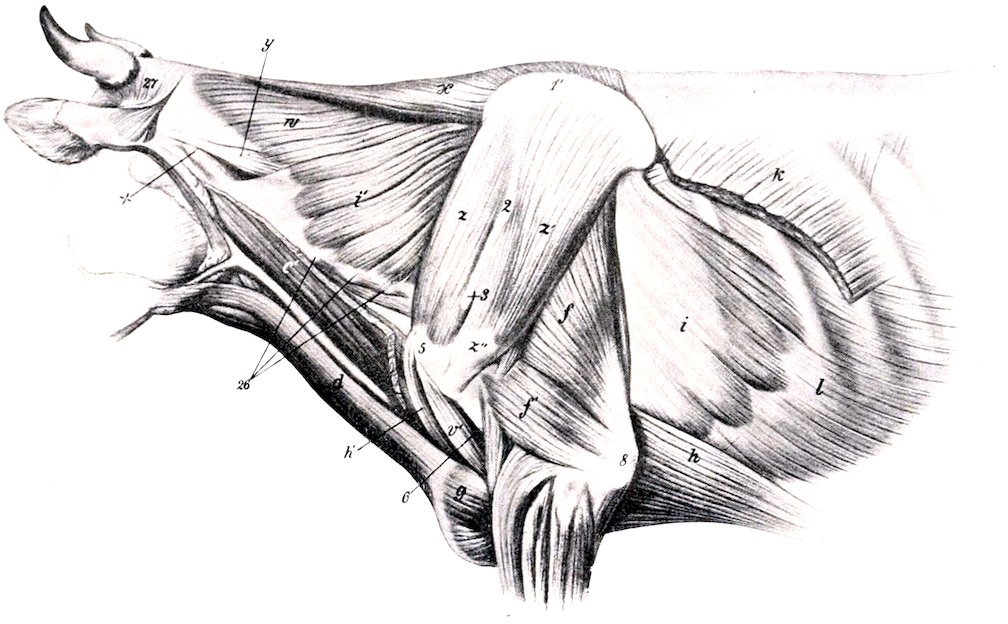
Fig. 211.—Deeper Muscles of Neck, Shoulder, and Thorax of Ox. c′, Cleido-occipitalis muscle; d, sterno-cephalicus; f, f′, long and external heads of triceps; g, superficial pectoral muscle; h, h′, posterior and anterior deep pectoral muscles; i, i′, serratus magnus; k, latissimus dorsi; l, obliquus abdominis externus; v′, biceps brachii; w, splenius; x, rhomboideus; y, trachelo-mastoideus; z, supraspinatus; z′, infraspinatus; z″, tendon of insertion of z′; 1′, cartilage of scapula; 2, tuberosity of spine of scapula; 3, acromion; 5, external tuberosity of humerus; 6, deltoid tuberosity; 8, olecranon; 26, transverse processes of cervical vertebræ; 27, posterior auricular muscles; X, wing of atlas. (After Ellenberger-Baum, Anat. für Künstler.)
The obliquus abdominis internus is more developed and has an additional origin from the lumbo-dorsal fascia. The aponeurosis blends with that of the external oblique near the linea alba, and detaches a layer which assists in the formation of the internal sheath of the rectus.
The rectus abdominis arises on the lateral border of the sternum as far forward as the third costal cartilage. The two muscles are separated, except near the pelvis, by an interval varying from two to four inches (ca. 5 to 10 cm.), so that this part of the abdominal wall is entirely fibrous. (The umbilicus is in a transverse plane through the third lumbar vertebra.) There are five tendinous inscriptions, on the third of which is a foramen for the passage of the subcutaneous abdominal vein (milk vein). The prepubic tendon has, in addition to branches inserted into the ilio-pectineal eminences, a strong attachment to the median common tendon of the adductors of the thigh, so that the abdominal wall is strongly retracted and almost vertical at its junction with the pelvis.
302The transversus abdominis presents no striking differential features. The transversalis fascia is strong and distinct except over the diaphragm.
Muscles of the Thoracic Limb
I. MUSCLES OF THE SHOULDER GIRDLE
The trapezius is much thicker and broader than in the horse, and is undivided. It arises on the ligamentum nuchæ and supraspinous ligament, from the atlas to the twelfth thoracic vertebra.
The omo-transversarius is a muscle (not present in the horse) which arises on the wing of the atlas, and, inconstantly, the transverse process of the second cervical vertebra, and is inserted into the scapular spine and fascia.
The rhomboideus is clearly divided into cervical and dorsal portions, the latter extending further than in the horse.
The latissimus dorsi has a broad tendon of origin, which blends with the lumbo-dorsal fascia; it is also attached to the eleventh and twelfth ribs, the fascia over the external intercostal and oblique abdominal muscles, and by a tendinous slip to the deep pectoral muscle.
The mastoido-humeralis has two distinct parts. The dorsal division (M. cleido-occipitalis) arises on the occipital bone and the ligamentum nuchæ. The ventral part (M. cleido-mastoideus) is smaller and arises by a round tendon on the mastoid process and the rectus capitis anterior major, and by a thin tendon on the mandible. At the shoulder a small muscle which arises on the first rib blends with the deep face of the mastoido-humeralis.[75]
The superficial pectoral muscle does not present any striking difference.
The deep pectoral arises as far forward as the second rib and is undivided. However, the scapular portion may be considered to be represented by a small branch extending over the lower part of the supraspinatus. A tendon is detached from the dorsal edge, which blends with the latissimus dorsi and coraco-brachialis.
The serratus magnus is clearly divided into cervical and thoracic portions. The former is large and extends from the third (or second) cervical vertebra to the fifth rib, being overlapped behind by the thoracic part. The thoracic part is relatively thin and is covered by a very strong aponeurosis; it is attached to the fourth to the ninth ribs by six digitations, and is inserted by a flat tendon into the inner face of the dorsal angle of the scapula.
II. MUSCLES OF THE SHOULDER
The deltoid is clearly divided into acromial and scapular portions. The former arises on the acromion, the latter on the posterior border of the scapula and the aponeurotic covering of the infraspinatus. The scapular part of the muscle is largely inserted into the fascia covering the triceps.
The supraspinatus, infraspinatus, and teres minor do not differ materially from those of the horse.
The subscapularis consists of three portions with a common tendon of insertion.
The teres major and coraco-brachialis resemble those of the horse.
III. MUSCLES OF THE ARM
The biceps is smaller and less tendinous, and is situated more internally than in the horse. The tendon of origin is flat, and is bound down in the bicipital groove 303by a fibrous band. (In the sheep the tendon is round and passes through the shoulder joint.)
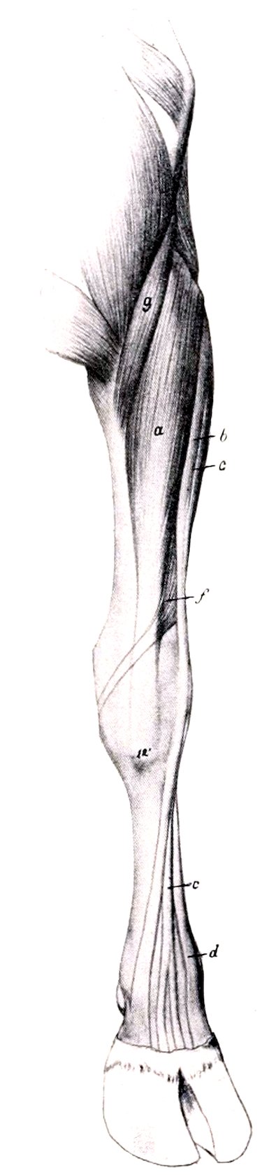
Fig. 212.—Muscles of Antibrachium and Manus of Ox, Anterior View. a, Extensor carpi radialis; b, extensor digiti tertii; c, extensor digitalis communis; d, extensor digiti quarti (tendon); f, extensor carpi obliquus; g, brachialis; 12′, metacarpal tuberosity. (After Ellenberger-Baum, Anat. für Künstler.)
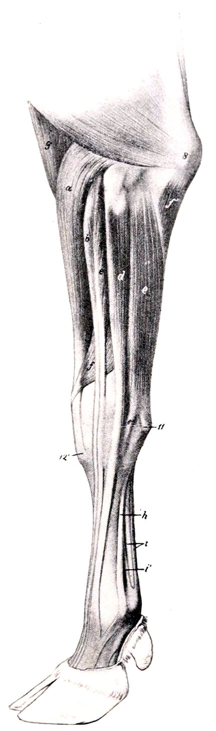
Fig. 213.—Muscles of Antibrachium and Manus of Ox, External View. a, Extensor carpi radialis; b, extensor digiti tertii; c, common or anterior digital extensor; d, extensor digiti quarti; e, flexor carpi externus; f, extensor carpi obliquus; f′, ulnar head of flexor carpi medius; g, brachialis; h, interosseus medius or suspensory ligament; i, flexor tendons; i′, branch of h, to superficial flexor tendon; 8, olecranon; 11, accessory carpal bone; 12′, metacarpal tuberosity. (After Ellenberger-Baum, Anat. für Künstler.)
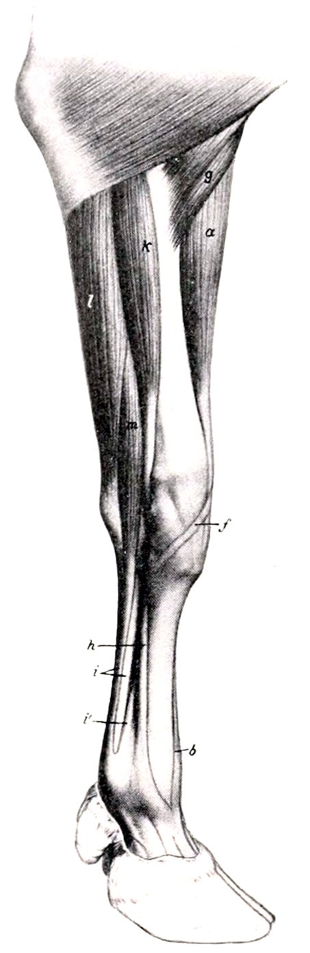
Fig. 214.—Muscles of Antibrachium and Manus of Ox, Internal View. a, Extensor carpi radialis; b, tendon of extensor digiti tertii; f, tendon of extensor carpi obliquus; g, brachialis; h, interosseus medius or suspensory ligament; i, flexor tendons; i′, branch of h; k, flexor carpi internus; l, flexor carpi medius; m, superficial digital flexor. (After Ellenberger-Baum, Anat. für Künstler.)
The internal head of the triceps is more developed than in the horse.
The tensor fasciæ antibrachii is a slender muscle.
304
IV. MUSCLES OF THE FOREARM
A. Extensor Division
The extensor carpi radialis is like that of the horse. There is sometimes a small muscle lying along its inner border, which may represent the extensores pollicis.
There are three digital extensors: 1. The common or anterior digital extensor (M. extensor digitalis communis) arises by two heads from the extensor epicondyle and the ulna. The heads fuse about the middle of the forearm, and terminate soon on a tendon which passes over the carpus and metacarpus, gradually inclining forward. At the fetlock joint it divides into two branches, which are inserted into the third phalanges. 2. The internal digital extensor (M. extensor digiti tertii proprius) arises on the extensor epicondyle, and is inserted by two branches into the second and third phalanges of the inner digit. The tendon receives two reinforcing slips from the suspensory ligament. 3. The lateral digital extensor (M. extensor digitalis lateralis s. digiti quarti proprius) arises from the external lateral ligament of the elbow joint, the external tuberosity of the radius, and the ulna. The tendon terminates like that of the preceding muscle.
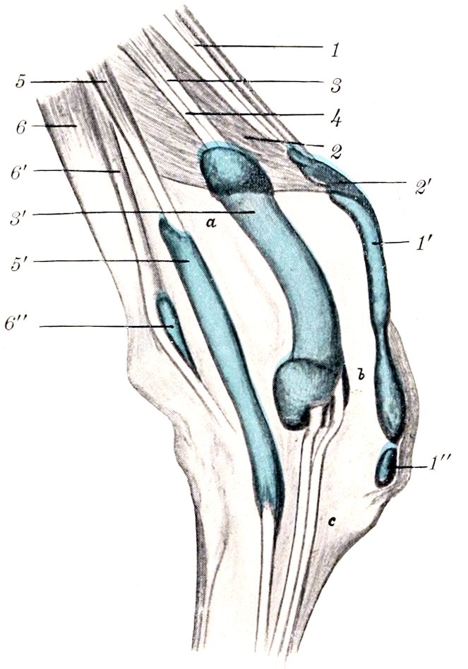
Fig. 215.—Right Carpus of Ox with Bursæ and Synovial Sheaths Injected, External View.
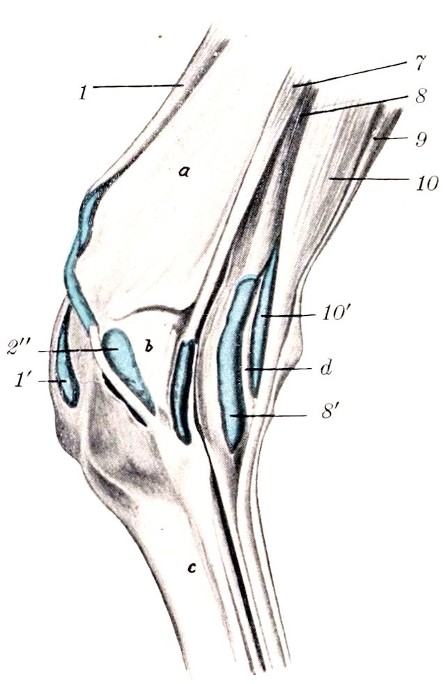
Fig. 216.—Right Carpus of Ox with Bursæ and Synovial Sheaths Injected, Internal View. 1, Extensor carpi radialis, with synovial sheath (1′) and bursa (1″); 2, extensor carpi obliquus, with synovial sheath (2′) and bursa (2″); 3, extensor digiti tertii proprius; 4, extensor digitalis communis; 3′, common synovial sheath of 3 and 4; 5, extensor digiti quarti proprius, with synovial sheath (5′); 6, 6′, flexor carpi externus, with bursa (6″); 7, flexor carpi internus, with synovial sheath; 8, deep digital flexor, with synovial sheath (8′); 9, flexor carpi medius; 10, superficial digital flexor, with synovial sheath 10′; a, radius; b, carpus; c, metacarpus; d, cut edge of annular ligament. (After Schmidtchen.)
The extensor carpi obliquus resembles that of the horse.
The extensor tendons are bound down at the carpus by an annular ligament, and are furnished with synovial sheaths (Figs. 215, 216).
305
B. Flexor Division
The three flexors of the carpus are like those of the horse.
The superficial digital flexor is somewhat blended at its origin with the middle flexor of the carpus. It divides into two bellies, superficial and deep, terminating on tendons at the distal part of the forearm. The superficial tendon passes over the posterior annular ligament (Ligamentum carpi transversum), perforates the metacarpal fascia, and joins the deep tendon about the middle of the metacarpus. The deep belly is connected with the deep flexor by a strong fibrous band. Its tendon passes under the annular ligament in a groove on the deep flexor, from which it receives fibers. The conjoined tendon soon bifurcates, each branch receiving a reinforcing band from the suspensory ligament, and forming near the fetlock a ring for the corresponding branch of the deep flexor tendon. Passing under two digital annular ligaments, they are inserted into the volar surfaces of the second phalanges by three slips.
The deep digital flexor has the same heads as in the horse, the humeral head, as before mentioned, being connected with the deep portion of the superficial flexor. The tendon divides near the distal end of the metacarpus into two branches which are inserted into the volar surfaces of the third phalanges.
The synovial sheaths at the carpus present the following special features: One is found in connection with the tendon of the superficial part of the superficial flexor of the digits. There is a common sheath for the tendons of the common extensor and the inner extensor.
Bursæ may occur under the tendons of the proper extensors of the digits at the fetlock; they are constant only in old animals (Schmidtchen). The branches of the tendon of the common extensor are provided with synovial sheaths from their origin to the middle of the second phalanx. There are two digital synovial sheaths for the flexor tendons; they may communicate at their upper part, and extend from the distal third of the metacarpus nearly to the distal sesamoids. Bursæ occur between the latter and the branches of the deep flexor tendon.
METACARPAL MUSCLES
The lumbricales are absent, unless we regard as such the muscular bundles which arise on the deep flexor and are inserted into the superficial flexor tendon at the carpus.
The interosseus medius or suspensory ligament is somewhat more muscular than in the horse; indeed, in the young subject it may be almost entirely fleshy. Its arrangement is somewhat complex. Single at its origin, it detaches about the middle of the metacarpus a band which joins the tendon of the superficial flexor and concurs near the fetlock in the formation of the ring for the deep flexor tendon. A little lower down it divides into three and then into five branches. The lateral branches (two pairs) are attached to the corresponding sesamoids and tendons of the proper extensors, while the middle branch passes through the sulcus at the distal end of the metacarpus and bifurcates, each division fusing with the tendon of the corresponding proper extensor.
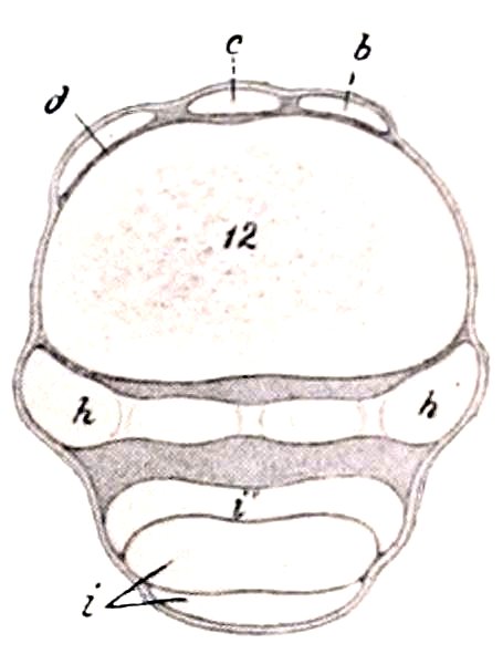
Fig. 217.—Cross-section of Distal Third of Metacarpus of Ox. b, Tendon of extensor digiti tertii; c, tendon of common extensor; d, tendon of extensor digiti quarti; h, interosseus medius or suspensory ligament; i, tendons of digital flexors; i′, branch of h; 12, metacarpal bone. (After Ellenberger-Baum, Anat. für Künstler.)
It is to be noted that the fascia on the posterior face of the metacarpus and digit is very thick. It is continuous above with the ligamentum carpi transversum, and is attached on either side to the metacarpal bone. At the fetlock it forms the fibrous basis for the small claws, and below this it detaches two strong bands which 306diverge to be inserted into the second and third phalanges, blending with the lateral ligaments.
The Muscles of the Pelvic Limb
I. THE SUBLUMBAR MUSCLES
The psoas minor begins at the disc between the twelfth and thirteenth thoracic vertebræ.
The psoas major has a fleshy origin on the posterior border of the last rib, and a thin tendon attached to the twelfth rib.
The iliacus begins under the body of the sixth lumbar vertebra, and is more closely united with the psoas major than in the horse.
The quadratus lumborum extends as far forward as the body of the tenth or eleventh thoracic vertebra.
II. EXTERNAL MUSCLES OF THE HIP AND THIGH
The tensor fasciæ latæ is large, and the fleshy part extends further down than in the horse.
The gluteus superficialis is not present as such; apparently its anterior part has fused with the tensor fasciæ latæ and its posterior with the biceps femoris.
The gluteus medius is small, the lumbar portion being insignificant and extending forward only to the fourth lumbar vertebra. Its deep portion (Gluteus accessorius) is easily separable, and its strong tendon is inserted into the femur below the trochanter major, under cover of the upper part of the vastus externus.
The gluteus profundus is thin, but extensive, arising as far forward as the external angle of the ilium, and from the lower part of the sacro-sciatic ligament. The fibers converge on a tendon which passes under the upper part of the vastus externus, and is inserted into a tubercle a short distance below the great trochanter.
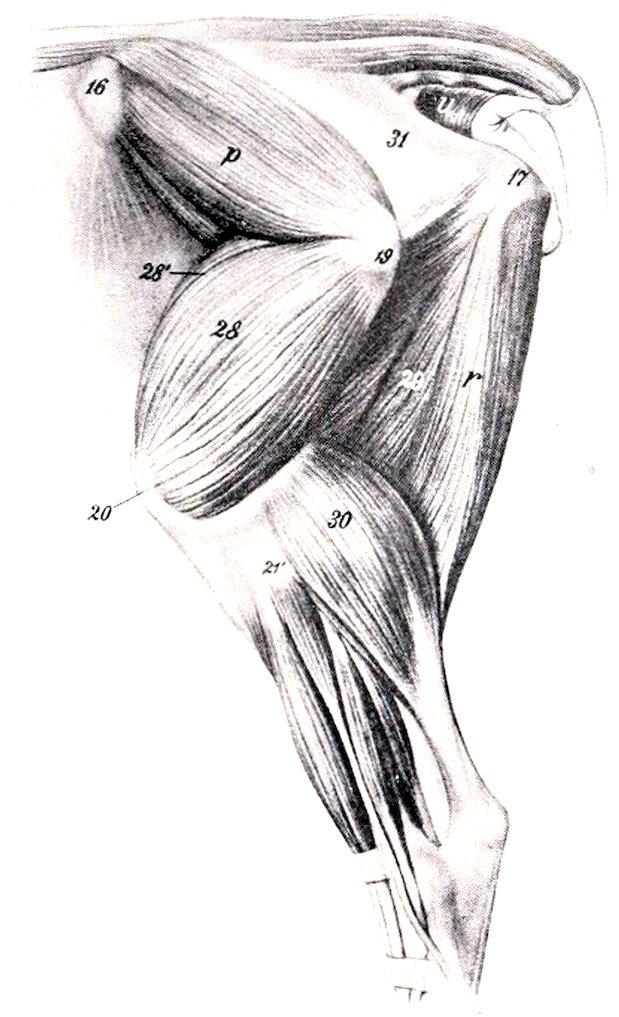
Fig. 218.—Gluteal and Femoral Regions of Ox, After Removal of Superficial Muscles. p, Gluteus medius; r, semitendinosus; u, coccygeus; 28, vastus externus; 28′, rectus femoris; 29, semimembranosus; 30, gastrocnemius; 31, sacro-sciatic ligament; 16, tuber coxæ; 17, tuber ischii; 19, trochanter major; 20, patella; 21′, external condyle of tibia. (After Ellenberger-Baum. Anat. für Künstler.)
The biceps femoris is very wide at its upper part, having apparently absorbed the posterior part of the superficial gluteus. It is divided in the thigh into two portions, which end on a wide aponeurosis. There is no femoral attachment. A large bursa occurs between the muscle and the great trochanter in the adult. The part of the tendon which fuses with the external patellar ligament presents a fibro-cartilaginous thickening, and an extensive bursa is interposed between it and the external condyle of the femur.
The semitendinosus and semimembranosus arise on the ischium only. The latter has a branch attached to the internal condyle of the tibia.
307
III. ANTERIOR MUSCLES OF THE THIGH
The quadriceps femoris resembles that of the horse; the vastus intermedius is more clearly separable, and consists of two parts. Bursæ occur under the insertions of the internal and external vasti, and often under the end of the biceps in the adult.
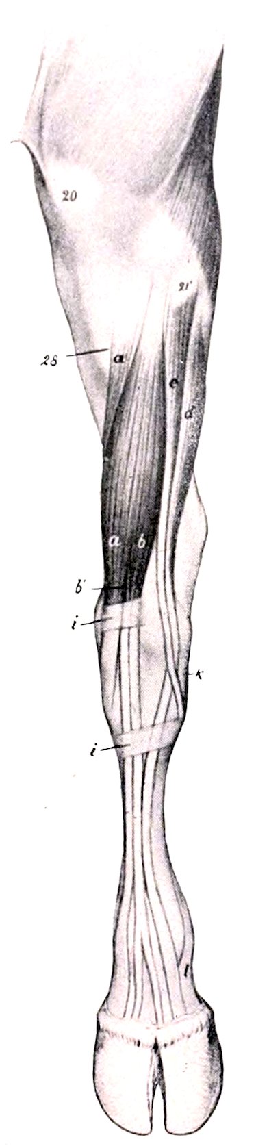
Fig. 219.—Muscles of Left Leg and Foot of Ox, Anterior View. a, Peroneus tertius; a′, tibialis anterior; b, anterior or long digital extensor; b′, extensor digiti tertii; c, peroneus longus; d, extensor digiti quarti; i, i, annular ligaments; k, external lateral ligament of hock joint; l, branch of suspensory ligament; 20, patella; 21′, external condyle of tibia; 28, tuberosity of tibia. (After Ellenberger-Baum, Anat. für Künstler.)
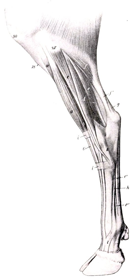
Fig. 220.—Muscles of Leg and Foot of Ox, External View. a, Peroneus tertius; a′, tibialis anterior; b, anterior or long digital extensor; b′, tendon of b; c, peroneus longus; d, extensor digiti quarti; e, deep digital flexor; e′, tendon of e; e″, branch of interosseus medius or suspensory ligament; f, gastrocnemius (the soleus lies just in front of f); f′, tendon of f; g, tendon of superficial digital flexor; h, interosseus medius or suspensory ligament; i, i, annular ligaments; 20, patella; 21′, external condyle of tibia; 28, crest of tibia. (After Ellenberger-Baum, Anat. für Künstler.)
The articularis genu or subcrureus is a small muscle which lies under the lower part of the vastus intermedius, and is inserted on the suprapatellar cul-de-sac of the synovial membrane.
The capsularis is absent.
308
IV. INTERNAL MUSCLES OF THE THIGH
The sartorius arises by two heads, one from the tendon of the psoas minor and the iliac fascia, the other from the shaft of the ilium. The femoral vessels pass between them.
The gracilis is more extensively united with its fellow than in the horse.
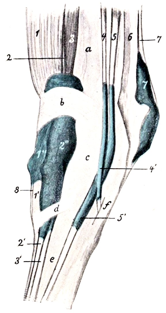
Fig. 221.—Left Tarsus of Ox with Synovial Sheaths and Bursæ Injected, External View. 1, Peroneus tertius; 1′, tendon of 1 and its sheath, 1″; 2, extensor digiti tertii proprius; 2′, tendon of 2; 2″, common sheath of tendons of extensor digiti tertii and extensor digitalis longus (3, 3′); 4, peroneus longus and its sheath, 4′; 5, extensor digiti quarti and its sheath, 5′; 6, tendon of gastrocnemius; 7, superficial digital flexor and its sheath, 7′; 8, tendon of tibialis anterior; a, tibia; b, proximal annular ligament; c, tarsus; d, distal annular ligament; e, metatarsus; f, external lateral ligament. (After Schmidtchen.)
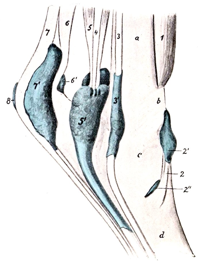
Fig. 222.—Left Tarsus of Ox with Synovial Sheaths and Bursæ Injected, Internal View. 1, Peroneus tertius; 2, tendon of tibialis anterior with sheath, 2′, and bursa, 2″; 3, long digital flexor and 3′, its sheath; 4, tibialis posterior; 5, deep digital flexor and its sheath, 5′; 6, tendon of gastrocnemius and bursa, 6′; 7, superficial digital flexor and its sheath, 7′; 8, subcutaneous bursa; a, tibia; b, proximal annular ligament; c, tarsus; d, metatarsus. (After Schmidtchen.)
The pectineus is large, and arises by a single head from the pubic crest and prepubic tendon. It divides into two branches, one of which is inserted as in the horse, while the other extends to the internal epicondyle of the femur.
The adductor resembles that of the horse, but does not reach to the internal condyle.
The quadratus femoris and obturator externus resemble those of the horse.
The obturator internus arises from the ischium only, and its tendon passes through the obturator foramen.
The gemellus is large.
309
V. MUSCLES OF THE LEG AND FOOT
There are four digital extensors, two of which are fused with each other and the peroneus tertius in the upper third of the leg.
1. The anterior or long digital extensor (M. extensor digitalis longus) arises by the common tendon in the extensor fossa of the femur, and separates from the other muscles near the middle of the leg. At the distal end of the tibia it terminates on a tendon which passes down over the hock (bound down by two annular ligaments) and ends like that of the thoracic limb.
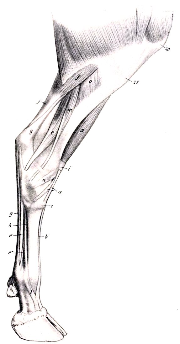
Fig. 223.—Muscles of Leg and Foot of Ox, Inner View. a, Peroneus tertius; b′, tendon of anterior or long extensor; e, deep digital flexor; e′, tendon of e; e″, branch of h to superficial flexor tendon; f, gastrocnemius; g, tendon of superficial digital flexor; h, interosseus medius or suspensory ligament; i, annular ligaments; l, branch of h; m, long digital flexor; o, popliteus; 20, patella; 28, tuberosity of tibia. (After Ellenberger-Baum, Anat. für Künstler.)
2. The internal digital extensor (M. extensor digiti tertii proprius) arises in common with the preceding muscle and the peroneus tertius, and is covered by them to the distal third of the tibia. Its tendon passes under the annular ligaments between those of its cogeners and ends on the second phalanx of the inner digit.
3. The lateral digital extensor (M. extensor digitalis lateralis s. digiti quarti proprius) arises on the external lateral ligament of the stifle joint and the external condyle of the tibia. Its tendon passes over the outer surface of the hock, and terminates on the anterior surface of the second phalanx of the external digit.
310The reinforcing bands from the suspensory ligament are arranged as in the fore limb.
4. The extensor brevis resembles that of the horse, but is inserted on the tendon of the anterior extensor only.
The peroneus longus (not present in the horse) is situated in front of the lateral extensor. It arises on the external condyle of the tibia and the fibrous band which represents the shaft of the fibula. Its tendon passes downward and backward over the outer surface of the hock, crosses over that of the lateral extensor and under the lateral ligament, and ends on the first tarsal bone (Cuneiform parvum). It is enveloped by a synovial sheath. It would apparently act as an inward rotator at the hock joint.
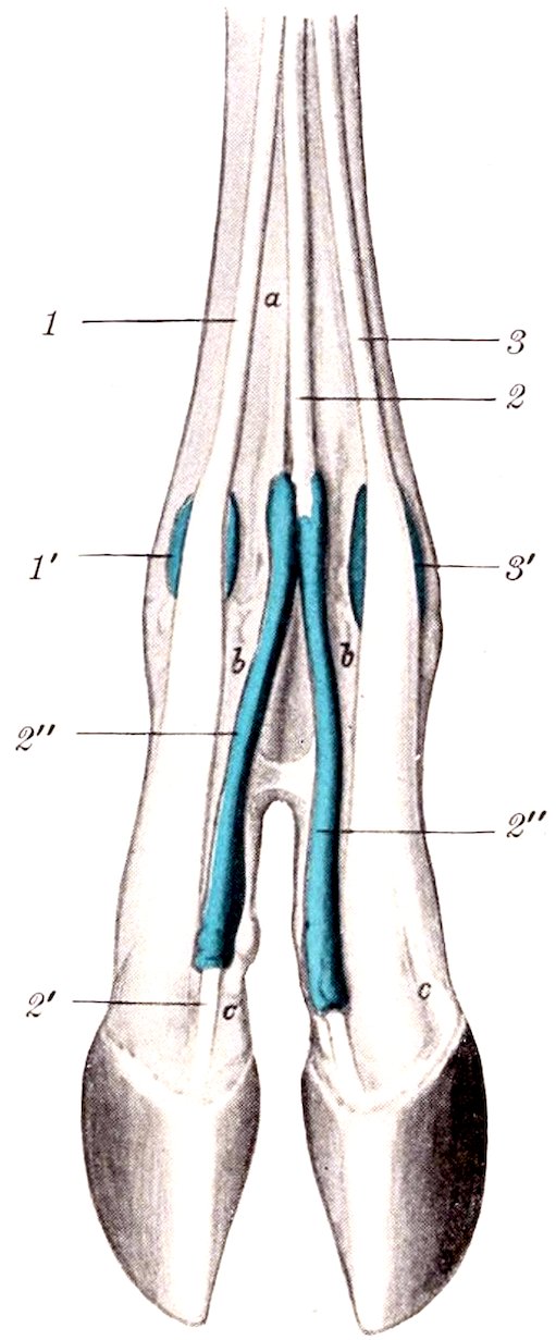
Fig. 224.—Distal Part of Limb of Ox with Synovial Sheaths and Bursæ Injected, Anterior View. 1, Tendon of extensor digiti tertii, with bursa (1′); 2, tendon of anterior extensor; 2′, branch of 2, with synovial sheath (2″); 3, tendon of extensor digiti quarti, with bursa (3′); a, metatarsus; b, first phalanx; c, second phalanx. (After Schmidtchen.)
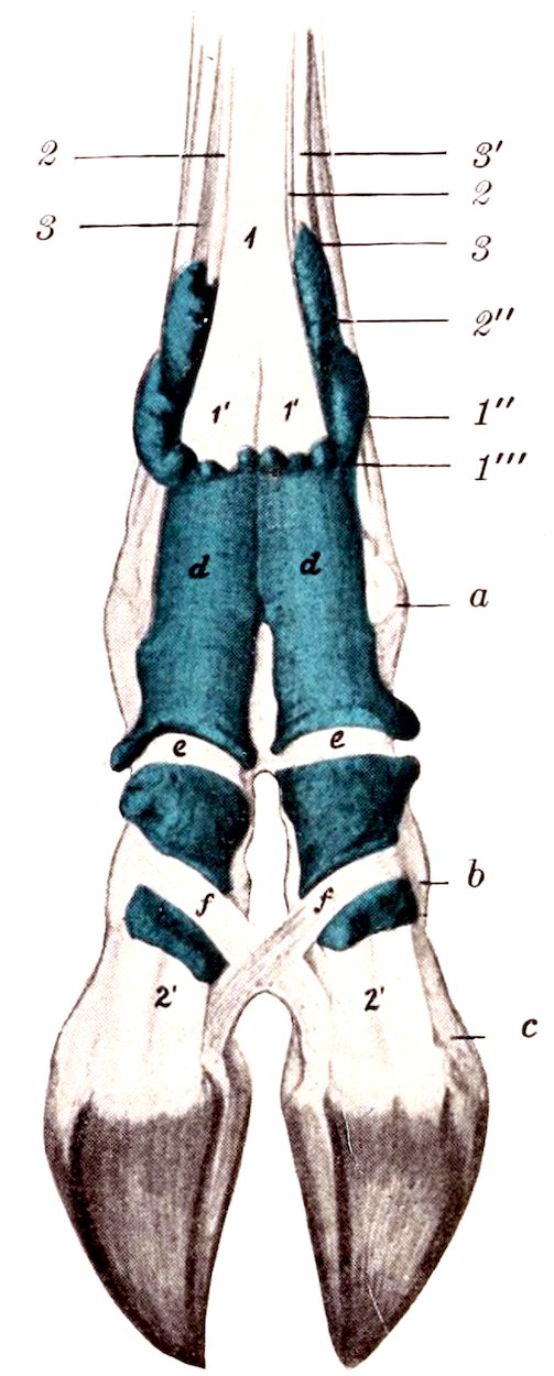
Fig. 225.—Distal Part of Limb of Ox with Synovial Sheaths Injected, Posterior View. 1, Superficial flexor tendon; 1′, 1′, branches of 1; 1″, 1‴, upper part of synovial sheaths of 1′, 1′; 2, deep flexor tendon; 2′, 2″, branches of 2; 2″, upper part of synovial sheath of 2′; 3, 3, lateral branches of interosseus medius or suspensory ligament; 3′, branch of same to superficial flexor tendon; a, fetlock joint; b, pastern joint; c, coffin joint; d, e, annular ligaments; f, crucial interdigital ligament. (After Schmidtchen.)
The peroneus tertius is a well-developed muscle which arises on the common tendon with the long and internal extensors. It has a large, fusiform belly, which is superficially situated on the front of the leg. Close to the tarsus it ends on a flat tendon, which is perforated by that of the tibialis anterior, and ends on the large metatarsal and second and third (fused) tarsal bones (Cuneiform magnum).
The tibialis anterior is smaller, and arises by two heads. The larger head 311springs from the outer surface of the tuberosity of the tibia; the outer, smaller one (M. extensor hallucis longus), from the upper part of the external border of the tibia and the fibrous band which replaces the shaft of the fibula. The tendon perforates that of the preceding muscle and ends on the metatarsal and second and third tarsal bones.
B. Posterior Group
The gastrocnemius and soleus resemble those of the horse. The superficial flexor is more fleshy than in the horse. Its tendon terminates as in the fore limb.
The deep flexor has in its upper part a close resemblance to that of the horse, but the superficial head (tibialis posterior) is larger and distinct. The tendon ends like that on the fore limb.
MUSCLES OF THE PIG
Muscles of Head
The facial panniculus is pale, thin, and difficult to separate from the skin.
The orbicularis oris is little developed.
The levator nasolabialis is thin and pale, and is undivided.
The levator labii superioris proprius may well be termed the levator rostri. It has a large penniform belly, which arises in the fossa on the lacrimal bone and maxilla. The tendon ends on the anterior part of the os rostri. A muscular slip connects it with the premaxilla.
The zygomaticus arises on the fascia over the masseter and ends at the angle of the mouth.
The depressor labii inferioris separates from the buccinator only near the angle of the mouth; it ends by a number of tendinous branches in the lower lip.
The dilatator naris lateralis is well developed. It arises under the levator rostri and ends by a tendinous network around the nostril.
The dilatator naris transversus is represented only by a few fibers which cross over the os rostri.
The depressor rostri arises on the facial crest. It has a long strong tendon which passes below the nostril and turns up and inward to meet the tendon of the opposite side and end in the skin of the snout. It depresses the snout and contracts the nostril.
The malaris is absent, and the other palpebral muscles present no special features.
MUSCLES OF MASTICATION
The digastricus has only one belly. It ends on the inner and lower surface of the mandible, in front of the groove for the facial vessels.
The other muscles have no important differential features.
The same is true of the oral hyoid muscles.
Muscles of the Neck
The panniculus carnosus consists of two layers which cross each other obliquely. The fibers of the superficial layer are directed nearly vertically, those of the deep layer toward the face, on which they are continued to form the facial portion.
The mastoido-humeralis is described on p. 314.
312The sterno-cephalicus (Sterno-mastoideus) arises on the sternum and is inserted by a long round tendon on the mastoid process.
The thyroid portion of the sterno-thyro-hyoideus has a peculiar arrangement. It arises (separately from the opposite muscle) on the manubrium sterni. About the middle of the neck it has an oblique tendinous intersection, beyond which it divides into two branches; one of these is inserted in the usual fashion, the other ends on the laryngeal prominence. The hyoid portion is well developed.
The omo-hyoideus is thin. It arises as in the horse, but has no connection with the mastoido-humeralis nor with the opposite muscle.
The omo-transversarius arises on the first or second cervical vertebra (under cover of the mastoido-humeralis), and is inserted into the lower part of the scapular spine.
There are two scaleni. The scalenus ventralis (s. primæ costæ) resembles that of the ox, is attached to the last four cervical vertebræ, and is perforated by the nerves of the brachial plexus. The scalenus dorsalis (s. supracostalis) arises on the transverse processes of the third to the sixth cervical vertebra, and ends on the third rib.
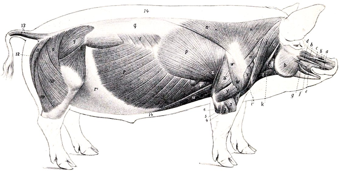
Fig. 226.—Superficial Muscles of Pig, After Removal of Panniculus Carnosus. a, Levator nasolabialis; b, levator labii superioris proprius; b′, fleshy slip of b which comes from premaxilla; c, dilatator naris lateralis; d, depressor rostri; e, orbicularis oris; f, depressor labii inferioris; g, zygomaticus; h, masseter; i, i′, i″, brachiocephalicus (cleido-occipitalis, cleido-mastoideus, pars clavicularis); k, sterno-cephalicus; l, sterno-hyoideus; m, omo-transversarius; n, n′, trapezius; o, anterior deep pectoral; p, latissimus dorsi; q, lumbo-dorsal fascia; r, obliquus abdominis externus; r′, aponeurosis of r; s, serratus posticus; t, serratus magnus; u, posterior deep pectoral; v, supraspinatus; w, w′, deltoideus; x, long head of triceps; y, external head of triceps; z, tensor fasciæ antibrachii; 1, brachialis; 2, extensor carpi radialis; 3, extensor digiti quarti; 4, extensor digiti quinti; 5, extensor carpi ulnaris; 6, ulnar head of deep flexor; 7, gluteus medius; 8, tensor fasciæ latæ; 9, 10, 10′, biceps femoris; 11, semitendinosus; 12, semimembranosus; 13, caudal muscles; 14, panniculus adiposus in section. (After Ellenberger, in Leisering’s Atlas.)
The straight muscles of the head present no special features.
The longus colli is separated from the opposite muscle, so that part of the bodies of the cervical vertebræ is exposed as in man.
The intertransversales resemble those of the ox.
The splenius is thick and extensive. It ends in three parts on the occipital, the temporal, and the wing of the atlas (inconstant).
The trachelo-mastoideus is small, and its atlantal portion is blended with the longissimus.
The complexus is large, and is clearly divided into two portions: the dorsal 313portion (Biventer cervicis) is marked by several tendinous intersections; the ventral part is the complexus proper.
The obliquus capitis posterior is relatively thin.
The recti capitis are thick and more or less fused.
Muscles of the Thorax
The levatores costarum and rectus thoracis present no special features.
The external intercostal muscles are absent under the serratus anticus and posticus and the digitations of the external oblique.
The internal intercostals are thick between the cartilages of the sternal ribs.
The retractor costæ and the transversus thoracis resemble those of the horse; the latter extends back to the eighth cartilage and fuses with the transversus abdominis.
The diaphragm has seven costal digitations on each side, the posterior ones being attached to the ribs at some distance (ca. one-third to one-fourth of rib-length) from the costo-chondral junction. The line of attachment reaches the latter at the tenth rib, and passes along the eighth cartilage to the xiphoid process. The tendinous center is more rounded than in the horse. The crura are well developed. The right crus is very large, and is perforated by the extensive slit-like opening for the œsophagus, which is median in position, and lies about two and one-half to three inches (ca. 6 to 8 cm.) below the twelfth thoracic vertebra. The hiatus aorticus is between the crura.
Abdominal Muscles
The abdominal tunic is little developed.
The obliquus abdominis externus has an extensive fleshy portion and a correspondingly narrow aponeurosis; the latter does not detach a femoral lamina, but is reflected in toto to form the inguinal (Poupart’s) ligament.
The obliquus abdominis internus resembles that of the ox; a small fusiform muscle, which crosses the inguinal canal obliquely and is attached on the abdominal surface of the inguinal ligament, is apparently a detached slip of the internal oblique.
The rectus abdominis is extensive and thick. It has seven to ten intersections. Its tendon of insertion fuses largely with the common tendon of the gracilis, and does not give off an accessory band to the head of the femur.
The fleshy part of the transversus abdominis is well developed. It blends in front with the transversus thoracis.
The cremaster externus is present in the female as well as in the male.
Muscles of the Back and Loins
The serratus anticus is inserted into the fifth to the eighth ribs inclusive, the serratus posticus into the last four or five ribs. There are usually no digitations attached to the ninth and tenth ribs.
The transversalis costarum (Iliocostalis) extends to the wing of the atlas.
The spinalis et semispinalis can be separated without much difficulty from the longissimus dorsi, the division from the common mass of the loins beginning about the first lumbar vertebra.
The multifidus resembles that of the horse.
Interspinales are present, as well as distinct intertransversales of the back and loins.
314
Muscles of the Tail
The superior and lateral sacro-coccygei arise as far forward as the last lumbar vertebra. Gurlt explains the twist of the tail as being due to the spiral arrangement of the insertions of the tendons.
Muscles of the Thoracic Limb
MUSCLES OF THE SHOULDER GIRDLE
The trapezius is very wide, its line of origin extending from the occipital bone to the tenth thoracic vertebra. There is no clear division between its two parts, which are both inserted into the scapular spine.
The omo-transversarius resembles that of the ox.
The rhomboideus consists of three portions. The cervical portion (Rhomboideus cervicalis) is greatly developed, its origin extending from the second cervical to the sixth thoracic vertebra. The cephalic portion (Rhomboideus capitis) arises with the splenius on the occipital bone, and is inserted with the cervical part. The dorsal portion (Rhomboideus thoracalis) extends as far back as the ninth or tenth thoracic vertebra.
The latissimus dorsi is attached to the four ribs preceding the last. It is inserted into the internal lip of the bicipital groove.
The mastoido-humeralis divides into two parts, the cleido-mastoideus and cleido-occipitalis, which arise on the mastoid process and occipital crest respectively, and unite at the fibrous vestige of the clavicle.
The anterior superficial pectoral is thin. The posterior superficial pectoral is divided into parts, one of which ends on the humerus, the other on the fascia of the forearm.
The deep pectoral is clearly divided into two parts. The scapular portion resembles that of the horse, but its origin does not extend behind the first two chondro-sternal joints. The posterior deep pectoral is very long.
The cervical portion of the serratus magnus is greatly developed, its origin extending from the wing of the atlas to the upper part of the fifth rib, and passing under the thoracic portion; the latter resembles that of the ox.
MUSCLES OF THE SHOULDER
The deltoid is undivided; it arises from the aponeurosis covering the infraspinatus, and it ends largely on the deltoid ridge, but partly on the fascia of the arm.
The supraspinatus is large; it ends chiefly on the external tuberosity of the humerus.
The infraspinatus is inserted into a depression below the posterior division of the external tuberosity.
The teres minor is well developed; it ends on a tubercle between the external and deltoid tuberosities of the humerus.
The subscapularis and teres major have no remarkable features.
The coraco-brachialis is short and undivided.
The capsularis is very small and frequently absent.
MUSCLES OF THE ARM
The biceps brachii is fusiform and not greatly developed. Its tendon of origin is rounded and passes through the capsule of the shoulder joint. The tendon of insertion is bifid, ending on the radius and ulna.
315The brachialis is well developed, and also ends on the radius and ulna.
The tensor fascias antibrachii resembles that of the horse.
The long head of the triceps is divided into two parts, except at its distal end, under which there is a synovial bursa. The external head is inserted into a crest on the outer surface of the olecranon by a thin tendon, under which there is a bursa. The internal head arises near the head of the humerus, beneath the coraco-brachialis.
There are two anconei.
MUSCLES OF THE FOREARM AND DIGITS
The extensor carpi radialis is a strong, fleshy muscle, the tendon of which is inserted into the proximal end of the third metacarpal bone. It may be divided into two parts (M. extensor carpi radialis longus, brevis).
The extensor carpi obliquus is well developed; it ends on the second metacarpal bone.
There are three (or four) digital extensors.
1. The common or anterior digital extensor (M. extensor digitalis communis) arises on the extensor epicondyle of the humerus and the lateral ligament of the elbow, and divides into three parts. The tendon of the inner part divides into two branches which end on the second and third digits; the tendon to the second digit is small and often absent. The tendon of the middle part divides lower down into two branches for the third and fourth (chief) digits; above this bifurcation it detaches a small branch to the second digit, which usually unites with the tendon of the extensor indicis. The tendon of the deep head divides into two branches, the inner one joining the tendon of the middle head, while the outer one ends on the fifth digit.
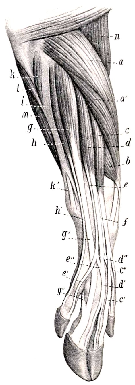
Fig. 227.—Muscles of Antibrachium and Manus of Pig, Antero-External View. a, a′, Extensor carpi radialis; b, extensor carpi obliquus (s. abductor pollicis longus); c, d, e, common or anterior digital extensor; c′, c″, tendons of insertion of c; d′, d″, tendons of d; e′, e″, tendons of e; f, tendon of extensor indicis; g, extensor digiti quarti; h, extensor digiti quinti; h′, tendon of h; i, tendinous, and k, fleshy part of flexor carpi externus; k′, tendon of k; l, ulnar head of deep digital flexor; m, superficial digital flexor; n, brachialis. (After Ellenberger, in Leisering’s Atlas.)
2. The extensor of the second digit (M. extensor indicis proprius) is covered by the preceding muscle, with which it is partially fused. It arises on the ulna. Its delicate tendon usually unites with the tendon of the middle head of the common extensor which goes to the second digit.
3. The lateral digital extensor (M. extensor digitalis lateralis) consists of two distinct parts: (1) The large dorsal (anterior) muscle (M. extensor digiti quarti proprius) has a long tendon which ends on the fourth digit, and often sends a slip to the fifth digit. (2) The small volar (posterior) muscle (M. extensor digiti quinti proprius) ends by a long tendon on the lateral aspect of the fifth digit.
The supinator has been found by Arloing and Lesbre, but is usually absent in the pig.
The pronator teres is a small muscle which lies on the internal lateral ligament of the elbow. It arises from the internal epicondyle of the humerus, and reaches about to the middle of the inner border of the radius.
The flexor carpi internus is well developed. It arises on the flexor epicondyle of the humerus, and is inserted into the third metacarpal bone.
The flexor carpi medius is narrow and has no ulnar head.
316The flexor carpi externus (M. extensor carpi ulnaris) is covered by a thick, tendinous band, which extends from the extensor epicondyle to the accessory carpal bone and outer aspect of the carpus. The tendon of the muscle perforates this band in the distal part of the forearm and ends on the proximal end of the fifth metacarpal bone.
The superficial digital flexor has two bellies. The tendon of the superficial head passes down behind the posterior annular ligament of the carpus (bound down by a special annular ligament), forms a ring at the fetlock for a tendon of the deep flexor, and ends by two branches on the second phalanx of the fourth digit. The tendon of the deep head, after detaching a slip to the tendon of the deep flexor, passes down with the latter (for which it forms a ring), and ends on the third digit.
The deep digital flexor has three heads—humeral, ulnar, and radial. The common tendon divides into four branches, the larger central pair ending on the third phalanges of the principal digits, the smaller pair on the accessory digits. The latter are bound down by a peculiar spiral band. There is no check ligament.
The lumbricales are represented by bundles which extend from the deep flexor tendon to the tendon of the deep head of the superficial flexor.
The third and fourth interossei are present. Each sends two slips to the corresponding sesamoid bones and extensor tendon.
There are flexors, adductors, and abductors of the second and fifth digits.
Muscles of the Pelvic Limb
SUBLUMBAR MUSCLES
The psoas minor is intimately united with the psoas major in front, and has a long small tendon which ends on the psoas tubercle. It has no thoracic part.
The psoas major is large and rounded. It begins at the last rib.
The quadratus lumborum is well developed, and extends forward as far as the twelfth rib.
MUSCLES OF THE HIP AND THIGH
The tensor fasciæ latæ is broad, and its fleshy part reaches almost to the patella.
The gluteus superficialis has a sacral head only; it blends with the biceps femoris.
The gluteus medius has a small lumbar part which does not extend so far forward as in the horse. The deep part (Gluteus accessorius) is pretty clearly marked.
The gluteus profundus is extensive, reaching nearly to the external angle of the ilium.
The biceps femoris has a narrow origin from the sacro-sciatic ligament and tuber ischii. It ends below like that of the ox.
The semitendinosus resembles that of the horse.
The semimembranosus has two insertions as in the ox.
The sartorius has two heads of origin, between which the external iliac vessels are situated. The inner arises from the tendon of the psoas minor, the outer one from the iliac fascia.
The graciles are united at their origin even more than in the ox.
The pectineus is well developed and is flattened from before backward.
The adductor shows no division and is partially fused with the gracilis. It ends on the femur just above the origin of the gastrocnemius.
317The quadratus femoris is large.
The obturator externus resembles that of the horse.
The obturator internus is extensive and strong; its tendon emerges through the obturator foramen.
The gemellus is fused in part with the obturator internus.
The quadriceps femoris is more clearly divided than in the horse, and its action is transmitted by a single patellar ligament.
The capsularis is absent.
MUSCLES OF THE LEG AND FOOT
The peroneus tertius resembles that of the ox, but its tendon is not perforated by that of the tibialis anterior, and ends on the inner (second) large metatarsal bone, detaching a strong branch to the first and second tarsal bones.
The tibialis anterior arises on the tibial crest. Its tendon passes over the hock along the inner border of the tendon of the peroneus tertius (both being bound down by an annular ligament), and is inserted into the inner (second) metatarsal and first tarsal bones.
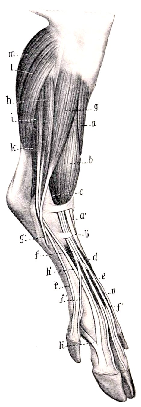
Fig. 228.—Muscles of Leg and Foot of Pig, Antero-external View. a, Tibialis anterior; a′, tendon of preceding; b, peroneus tertius; b′, tendon of b; c, anterior or long digital extensor; d, e, f, f′, f″, tendons of c; g, peroneus longus; g′, tendon of g; h, extensor digiti quarti; h′, tendon of h, which receives h″, from the interosseus medius; i, extensor digiti quinti; k, deep digital flexor; l, soleus; m, gastrocnemius; n, extensor brevis. (After Ellenberger, in Leisering’s Atlas.)
The peroneus longus lies partly on the outer surface of the peroneus tertius. Its tendon crosses those of the lateral extensor, passes along a groove on the plantar surface of the fourth tarsal bone, and ends on the first tarsal and second metatarsal bones.
The anterior or long digital extensor arises with the peroneus tertius and is largely covered by it and the peroneus longus. It divides into three parts. The tendon of the inner division (M. extensor digiti tertii) ends on the inner chief (third) digit; that of the middle division by two branches on the principal digits; and that of the outer division by three branches on the second, fourth, and fifth digits.
The lateral digital extensor divides into proper extensors of the fourth and fifth digits (Extensores digiti quarti et quinti).
The extensor hallucis longus arises on the fibula and ends on the inner small (second) digit.
The extensor digitalis brevis is well developed and consists of three parts. The inner and outer tendons end on the first phalanges of the chief digits, the middle one joins the common extensor tendon of the same.
The gastrocnemius presents nothing special.
The soleus is wide, and is partly blended with the external head of the gastrocnemius.
The superficial digital flexor has a large belly. Its tendon ends on the two chief digits, and is also connected with the ligaments of the accessory digits.
The inner head of the deep digital flexor (flexor digitalis longus) is relatively small. The common tendon ends as in the thoracic limb.
The popliteus presents no special features.
The plantar muscles resemble their homologues of the fore limb, except that the adductors of the accessory digits and the lumbricales are absent.
318
THE MUSCLES OF THE DOG
Muscles of the Face
The panniculus carnosus is well developed and presents two distinct bands, one of which is inserted into the lower lip, the other into the scutiform cartilage of the ear.
The orbicularis oris is poorly developed. In the upper lip it is divided centrally, and in the lower it is distinct only near the angles of the mouth.
The levator nasolabialis is wide and undivided, and ends in the upper lip.
The levator labii superioris proprius arises behind the infraorbital foramen, runs forward under the preceding muscle, and ends in numerous small tendons around the nostril, some blending with those of the opposite side.
The zygomaticus, long and narrow, arises on the scutiform cartilage, and ends at the angle of the mouth.
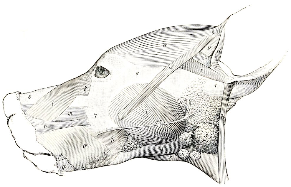
Fig. 229.—Muscles of Head of Dog a, Scutularis; b, c, anterior auricular muscles; d, helicis; e, antitragicus; f, f, zygomaticus, out of which a portion is cut; g, slip of panniculus; h, parotido-auricularis; i, masseter; k, malaris; l, levator nasolabialis; m, levator labii superioris proprius; n, dilatator naris lateralis; o, p, buccinator (buccalis, molaris); q, retractor anguli oris; r, digastricus; s, mylo-hyoideus; 1, base of concha; 2, parotid gland; 2′, parotid duct; 3, submaxillary gland; 4, submaxillary lymph glands; 5, buccal glands; 6, zygomatic arch; 7, maxilla; 8, dorsum nasi; 9, parotid lymph gland. (Ellenberger-Baum, Anat. d. Hundes.)
The depressor labii inferioris is absent, unless we recognize as such a thin stratum arising on the body of the mandible and spreading out in the orbicularis.
There are no special nasal muscles, the homologue of the lateral dilator ending almost entirely in the upper lip.
The buccinator is wide and very thin, and the two planes of fibers cross each other.
The palpebral muscles present no very noteworthy special characters.
MUSCLES OF MASTICATION
The masseter is large. It arises from the zygomatic arch, and extends beyond the branch of the jaw below and behind. Three strata may be recognized in its structure.
The temporalis is very large and strong, and contains much tendinous tissue. It arises from the temporal fossa and the orbital ligament.
319The digastricus is usually not digastric, but is a strong, round, fleshy muscle, which arises on the paramastoid or styloid process and is inserted into the border and inner surface of the ramus at the level of the last molar teeth.
The stylo-mandibularis is absent.
HYOID MUSCLES
The mylo-hyoideus is well developed.
The stylo-hyoideus is very slender, and is inserted into the body of the hyoid bone: it is not perforated by the digastricus.
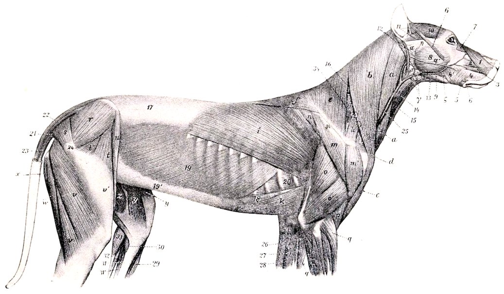
Fig. 230.—Superficial Muscles of Dog, After Removal of Panniculus Carnosus. 1, Levator nasolabialis; 2, levator labii superioris proprius; 3, dilatator naris lateralis; 4, 4′, buccinator; 5, retractor anguli oris s. risorius; 6, zygomaticus; 7, malaris; 8, masseter; 9, digastricus; 10, scutularis; 11, other auricular muscles; 12, parotido-auricularis; 13, mylo-hyoideus; 14, sterno-hyoideus; 15, sterno-thyroideus; 16, splenius; 17, lumbo-dorsal fascia; 18, rectus abdominis; 19, obliquus abdominis externus; 19′, aponeurosis of preceding; 20, intercostal muscle; 21, sacro-coccygeus accessorius; 22, sacro-coccygeus superior; 23, sacro-coccygeus inferior; 24, great trochanter; 25, jugular vein; a, b, c, brachiocephalicus; d, clavicle; e, f, trapezius; g, serratus cervicis; h, omo-transversarius; i, latissimus dorsi; k, posterior deep pectoral; l, supraspinatus; m, m′, deltoid; n, infraspinatus; o, triceps, long head; o′, triceps, external head; p, brachialis; q, extensor carpi radialis; r, gluteus medius; s, gluteus superficialis; t, t′, tensor fascia latæ; u, sartorius; v, biceps femoris; v′, fascia lata; w, semitendinosus; x, semimembranosus; y, sartorius; z, gracilis; 26, pronator teres; 27, flexor carpi radialis; 28, flexor carpi ulnaris; 29, tibialis anterior; 30, popliteus; 31, 31′, deep digital flexor; 32, superficial digital flexor; 33, gastrocnemius; 34, spine of scapula; α, parotid gland, with α′, its duct; β, submaxillary gland; δ, submaxillary lymph glands; η, parotid lymph gland; ζ, inferior buccal glands. (After Ellenberger, in Leisering’s Atlas.)
The hyoideus transversus and omo-hyoideus are absent.
The sterno-thyro-hyoideus is large and arises chiefly on the first costal cartilage.
Muscles of the Thoracic Limb
The trapezius is thin, and is not clearly divided into cervical and thoracic portions. Its line of origin extends from about the middle of the neck to the ninth or tenth thoracic spine, the right and left muscles meeting on a median fibrous raphé.
The omo-transversarius arises by a tendon on the lower part of the spine of the scapula (often partially blended with the trapezius), and is inserted into the wing of the atlas.
320The rhomboideus consists of three parts. The dorsal portion (Rhomboideus thoracalis) is small; it arises from the fourth to the sixth or seventh thoracic spine, and is inserted into the inner surface (chiefly) of the dorsal angle of the scapula. The cervical portion (Rhomboideus cervicalis) arises from the ligamentum nuchæ as far forward as the second or third cervical vertebra, and is inserted into the inner surface of the cervical angle of the scapula. The cephalic portion (Rhomboideus capitis) is a continuation of the preceding which is inserted into the occipital crest.
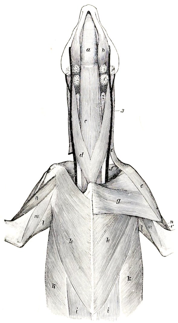
Fig. 231.—Ventral Muscles of Head, Neck, and Thorax of Dog. a, Mylo-hyoideus; b, digastricus; c, sterno-hyoideus; c′, sterno-thyroideus; d, sterno-cephalicus; e, brachiocephalicus; f, subscapularis; g, superficial pectoral; h, deep pectoral; i, rectus abdominis; k, obliquus abdominis externus; l, long head of triceps; m, internal head of triceps; n, biceps brachii; o, brachialis; 1, 1′, 1″, submaxillary lymph glands; 2, thyroid gland; 3, external jugular vein. (Ellenberger-Baum, Anat. d. Hundes.)
The latissimus dorsi is extensive, and has a fleshy attachment to the last two ribs. Its lower edge blends near the shoulder with the panniculus.
The mastoido-humeralis contains in front of the shoulder a fibrous mass in which the clavicle is embedded. Anterior to this it separates into two diverging portions. The dorsal portion (M. cleido-cervicalis) is attached to the median raphé of the neck and to the occipital bone. The ventral portion (M. cleido-mastoideus) is attached to the mastoid process. The common mass posterior to the clavicle and the fibrous intersection, which is attached to the humerus, is homologous with the clavicular portion of the deltoid of man.
The superficial pectoral muscle is small. It arises on the sternum from the first to the third costal cartilage, and is inserted into the anterior surface of the humerus. A superficial slip detached from it is inserted into the fascia of the forearm. The deep pectoral has no prescapular portion. It arises on the sternum and costal cartilages from the second costal to the xiphoid cartilage. It is inserted chiefly into the internal tuberosity of the humerus, but also by small slips into the external tuberosity and the fascia of the arm.
The serratus magnus shows no clear division into cervical and thoracic portions. It arises from the last five cervical vertebræ and the first seven or eight ribs, and is inserted into the upper part of the costal surface of the scapula.
The deltoid is clearly divided into scapular and acromial parts, the former arising on the spine, the latter on the acromion of the scapula. Both end on the deltoid tuberosity.
321The supraspinatus ends chiefly on the external tuberosity of the humerus, but has a small attachment to the internal tuberosity also.
The infraspinatus is inserted into the outer surface of the external tuberosity of the humerus.
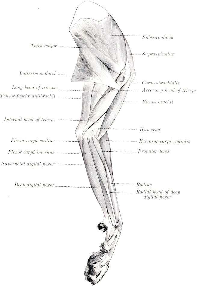
Fig. 232.—Muscles of Thoracic Limb of Dog, Internal View. (Ellenberger-Baum, Anat. des Hundes.)
The teres minor arises on a tubercle on the posterior border of the scapula, just above the glenoid cavity, and is inserted into the deltoid ridge.
The subscapularis is wide and is somewhat multipennate in structure, being intersected by fibrous septa which are attached to the rough lines on the costal surface of the scapula.
322The teres major is thick. It arises on the upper part of the posterior border of the scapula and on the subscapularis.
The coraco-brachialis is short and undivided, and is inserted into the upper part of the postero-internal surface of the humerus.
The capsularis is absent.
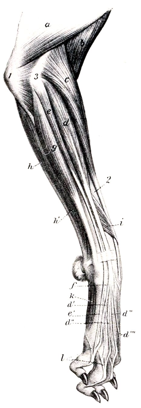
Fig. 233.—Muscles of Antibrachium and Manus of Dog, External View. a, Triceps brachii; b, brachialis; c, extensor carpi radialis; d, common or anterior digital extensor; d′, d″, d‴, d⁗, tendons of preceding; e, lateral digital extensor; e′, f, tendons of preceding; g, extensor carpi ulnaris; h, h′, flexor carpi ulnaris; i, extensor carpi obliquus (s. abductor pollicis longus); k, interossei; l, branches from preceding to extensor tendons; 1, olecranon; 2, radius; 3, extensor epicondyle of humerus. (After Ellenberger, in Leisering’s Atlas.)
The biceps brachii lies almost entirely on the inner surface of the humerus. It is long and fusiform. The tendon of origin is round, and passes through the capsule of the shoulder joint. The tendon of insertion is bifid, one branch being attached to the ulnar, the other to the radial tuberosity.
The brachialis is very little curved, and is inserted chiefly into the tuberosity and inner border of the ulna.
The tensor fasciæ antibrachii is thin and narrow. It arises on the outer surface of the latissimus dorsi, and ends on the olecranon and the fascia of the forearm.
The triceps has an additional deep head (Caput accessorium), which arises just below the head of the humerus.
The brachio-radialis is a long, narrow, delicate muscle, situated superficially on the anterior surface of the forearm. It arises with the extensor carpi on the crest above the extensor epicondyle of the humerus, and is inserted into the distal part of the inner border of the radius. It is often much reduced, and is sometimes absent. It rotates the forearm and paw outward.
The extensor carpi divides into two parts. The larger outer part, the extensor carpi radialis brevis, ends on the proximal end of the third metacarpal bone. The inner and more superficial part, the extensor carpi radialis longus, ends on the second metacarpal bone. (A tendon to the fourth metacarpal may occur.)
The extensor carpi obliquus or abductor pollicis longus arises from the outer border and anterior surface of the ulna, the interosseous ligament, and the outer border of the radius. It is inserted into the first metacarpal bone by a tendon which contains a small (sesamoid?) bone. It abducts the first digit.
There are three (or four) extensors of the digits.
1. The common digital extensor (M. extensor digitalis communis) arises on the extensor epicondyle of the humerus and the lateral ligament of the elbow joint. It has four bellies, each terminating in a tendon. These are inserted into the third phalanges of the second, third, fourth, and fifth digits.
2. The extensor of the first and second digits (M. extensor pollicis longus et extensor indicis proprius) is small, and is covered by the common and lateral extensors. It arises on the proximal part of the ulna. Its tendon passes down with that of the common extensor and divides into two branches. The delicate inner branch ends on the first digit, while the other blends with the tendon of the common extensor for the second digit.
3. The lateral digital extensor (M. extensor digitalis lateralis) consists of two 323muscles which are not rarely fused. They arise on the extensor epicondyle of the humerus and the lateral ligament of the elbow joint. The larger, superficial belly (M. extensor digiti tertii et quarti) terminates on a tendon which divides at or near the carpus into two branches, which are inserted into the third phalanges of the third and fourth digits, blending with the corresponding tendons of the common extensor. The posterior belly (M. extensor digiti quinti) terminates by a tendon which fuses with that of the common extensor for the fifth digit.
The ulnaris lateralis or extensor carpi ulnaris corresponds to the flexor carpi externus of the horse, but is an extensor of the carpus. It is a large flat muscle which lies on the outer surface of the ulna. It arises on the extensor epicondyle of the humerus, and is inserted into the proximal end of the fifth metacarpal and the accessory carpal bone.
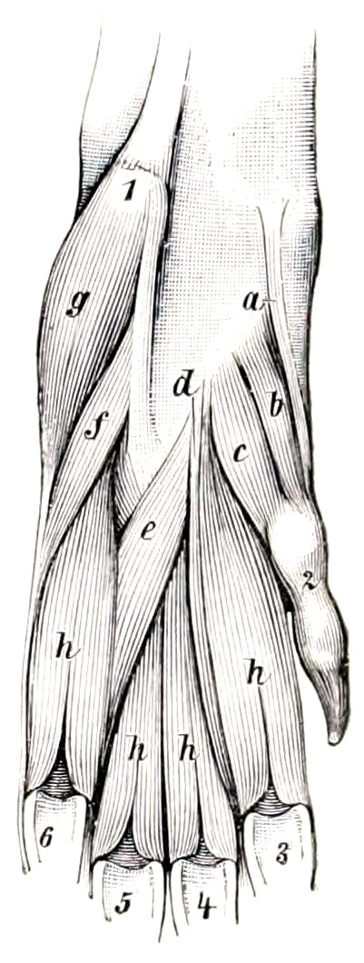
Fig. 234.—Volar Muscles of Fore Paw of Dog. a, Abductor pollicis brevis et opponens pollicis; b, flexor pollicis brevis; c, adductor pollicis; d, adductor digiti secundi; e, adductor digiti quinti; f, flexor digiti quinti brevis; g, abductor digiti quinti; h, interossei; 1, accessory carpal bone; 2, first digit; 3–6, sesamoids of metacarpo-phalangeal joints. (Ellenberger-Baum, Anat. d. Hundes.)
The ulnaris medialis or flexor carpi medius (s. ulnaris) consists of two quite distinct heads. The larger, humeral head arises on the flexor epicondyle, while the smaller, superficial ulnar head arises on the posterior border of the ulna. The tendons of the two end together on the accessory carpal bone.
The radialis volaris or flexor carpi internus (s. radialis) arises on the flexor epicondyle of the humerus and is inserted by a bifid tendon into the second and third metacarpal bones.
The tendons of the foregoing muscles are provided with synovial sheaths at the carpus.
The pronator teres is a round muscle, which is situated superficially on the inner border of the proximal part of the radius. It arises on the flexor epicondyle of the humerus, and is inserted into the internal border of the radius. Its action is to flex the elbow and rotate the forearm inward.
The superficial digital flexor is situated superficially on the posterior and inner surfaces of the forearm. It arises on the flexor epicondyle of the humerus and terminates near the carpus on a tendon which passes downward outside of the carpal canal and receives below the carpus two reinforcing bands, one from the accessory carpal, the other from the sesamoid bone at the inner side of the carpus. Below this it divides into four branches, which are inserted into the second phalanges of the second, third, fourth, and fifth digits.
The deep digital flexor has humeral, ulnar, and radial heads, the latter arising from the inner border of the radius. They unite on a common tendon which passes down through the carpal canal, gives off a branch to the first digit, and divides into four branches. These perforate the tendons of the superficial flexor and are inserted into the third phalanges of the second to the fifth digits. The tendons are provided with synovial sheaths from the middle of the metacarpus downward, and are held in place by three digital annular ligaments.
The palmaris longus accessorius (?) is a small muscle which arises from the deep flexor below the middle of the forearm and ends by two tendons which unite with those of the superficial flexor for the third and fourth digits.
The supinator is a short, wide muscle which arises on the humerus beneath the external lateral ligament, crosses the anterior surface of the radius, and is inserted into the inner border of the radius. (A pouch of the capsule of the elbow joint lies under the tendon of origin.) Its action is to rotate the forearm outward (supination).
324The pronator quadratus consists of fibers which cross the inner surface of the interosseous ligament of the forearm, except at the two extremities of the latter. It is attached to the inner border of the ulna and the anterior surface of the radius. It rotates the forearm inward (pronation).
The palmaris brevis (?) is a very small muscle, which arises on the tendon of the superficial digital flexor for the fifth digit, and is inserted at the fifth metacarpo-phalangeal joint into the sheath and annular ligament of the deep flexor.
The lumbricales are three very delicate muscles, which arise on the tendons of the deep flexor, and are inserted into the first phalanges of the third, fourth, and fifth digits.
The abductor pollicis brevis et opponens pollicis, a very small pale muscle, arises on the fibrous band which connects the superficial flexor tendon with the inner carpal sesamoid, and ends on the distal end of the first metacarpal bone and the first phalanx of the first digit. It abducts the first digit.[76]
The flexor pollicis brevis arises on the posterior carpal ligament over the second metacarpal bone, and ends on the volar sesamoid of the first digit.
The adductor pollicis, situated externally to the preceding, is the largest of the thumb muscles. It arises between the preceding and the second interosseous muscle, and is inserted into the first phalanx of the first digit.
The adductor digiti secundi is situated between the second interosseous muscle and the adductor digiti quinti. It arises on the carpal ligament, and ends on the first phalanx of the second digit.
The adductor digiti quinti arises close to the preceding muscle, and passes outward to end on the first phalanx of the fifth digit.
The flexor digiti quinti arises on the ligament connecting the accessory carpal to the third and fourth metacarpal bones, crosses the corresponding interosseous muscle, and ends on the fifth digit with the next muscle.
The abductor digiti quinti is larger than the two preceding muscles; it arises on the accessory carpal bone, and ends on the outer sesamoid of the fifth digit and on the lateral ligament.
There are four interossei which lie on the volar (posterior) surface of the metacarpus. They are well developed and fleshy. They arise on the distal row of the carpus and on the proximal ends of the metacarpals. Each divides distally into two branches, which are inserted by small tendons on the corresponding sesamoid bones, and detach slips to the extensor tendons.
Muscles of the Neck
The sterno-cephalicus is well developed. It arises on the manubrium sterni and ends on the mastoid process, blending with the cleido-cervicalis.
The scalenus ventralis (s. primæ costæ) arises on the last four cervical transverse processes, and is inserted into the first rib.
The scalenus dorsalis (s. supracostalis) is large. Anteriorly it blends with the preceding muscle, while posteriorly it divides into two parts. The upper part is inserted on the third and fourth ribs, the lower part by a long, thin tendon on the eighth rib.
The longus colli resembles that of the horse.
The rectus capitis anterior major arises on the transverse processes of the second to the sixth cervical vertebræ and ends as in the horse. The rectus minor and lateralis resemble those of the horse.
The intertransversales resemble those of the ox.
The splenius is strong and extensive. It arises on the first four or five thoracic 325spines and the median raphé of the neck, and is inserted into the occipital crest and mastoid process.
The complexus is composed of two parts—the biventer cervicis and the complexus major proper. The biventer cervicis arises from the transverse processes of the fifth and sixth, and the spines of the second to the fifth (or sixth) thoracic vertebræ, from the ligamentum nuchæ, and the median raphé. It has four tendinous intersections. The complexus major arises on the transverse processes of the first three or four thoracic vertebræ and the articular processes of the last five cervical. Both end on a strong common tendon which is inserted into the occipital crest and the depression below it.
The trachelo-mastoideus consists of two unequal portions. The large dorsal part (M. longissimus capitis) arises on the transverse processes of the first four thoracic and the articular process of the last three or four cervical vertebræ, and ends with the splenius on the mastoid process of the temporal bone. The small ventral part (M. longissimus atlantis) arises on the articular processes of the third, fourth, and fifth cervical vertebræ, and ends on the wing of the atlas.
The other muscles present no striking differential features, but it may be noted that distinct interspinales are present.
Muscles of the Thorax
There are twelve pairs of levatores costarum.