Chapter VIII
Reading edition
PORIFERA (CONTINUED): FORMS OF SPICULES—CALCAREA—HOMOCOELA—HETEROCOELA—HEXACTINELLIDA—DEMOSPONGIAE—TETRACTINELLIDA—MONAXONIDA—CERATOSA—KEY TO BRITISH GENERA OF SPONGES
Sponges fall naturally into two branches differing in the size of their choanocytes: in the Megamastictora these cells are relatively large, varying from 5µ to 9µ in diameter; in Micromastictora they are about 3µ in diameter.[212] For further subdivision of the group the spicules are such important weapons in the hands of the systematist that it is convenient to name them according to a common scheme. This has been arrived at by considering first the number of axes along which the main branches of the spicules are distributed, and secondly whether growth has occurred in each of these axes in one or both directions from a point of origin.[213]
I. Monaxons.—Spicules of rod-like form, in which growth is directed from a single origin in one or both directions along a single axis. The axis of any spicule is not necessarily straight, it may be curved or undulating. The ray or rays are known as actines.
Biradiate monaxon spicules are termed "rhabdi" (Fig. 76, a). A rhabdus pointed at both ends is an "oxea," rounded at both ends a "strongyle," knobbed at both ends a "tylote." By branching a rhabdus may become a "triaene" (Fig. 110, k, l).
Uniradiate monaxon spicules are termed "styli."
II. Tetraxons.—Spicules in which growth proceeds from an origin in one direction only, along four axes arranged as normals to the faces of a regular tetrahedron. Forms produced by growth from an origin in one direction along three axes lying in one plane are classed with tetraxons.
III. Triaxons.—Spicules in which growth is directed from an origin in both directions along three rectangular axes. One or more actines or one or two axes may be suppressed.
IV. Polyaxons.—Spicules in which radiate growth from a centre proceeds in several directions.
V. Spheres.—Spicules in which growth is concentric about the origin.
A distinction more fundamental than that of form is afforded by the chemical composition: all sponges having spicules composed of calcium carbonate belong to a single class, Calcarea, which stands alone in the branch Megamastictora.
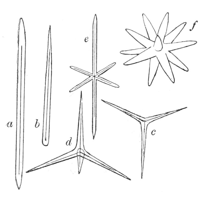
Fig. 76.—Types of megascleres. a, Rhabdus (monaxon diactine); b, stylus (monaxon monactine); c, triod (tetraxon triactine); d, calthrop (tetraxon tetractine); e, triaxon hexactine; f, euaster.
BRANCH I. MEGAMASTICTORA
CLASS CALCAREA
Calcarea are marine shallow-water forms attached for the most part directly by the basal part of the body or occasionally by the intervention of a stalk formed of dermal tissue. They are almost all white or pale grey brown in colour. Their spicules are either monaxon or tetraxon or both. The tetraxons are either quadriradiate and then called "calthrops," or triradiate when the fourth actine is absent. The triradiates always lie more or less tangentially in the body-wall; similarly three rays of a calthrop are tangentially placed, the fourth lying across the thickness of the wall. It is convenient to include the triradiate and the three tangentially placed rays of a calthrop under the common term "triradiate system" (Minchin). The three rays of one of these systems may all be equal in length and meet at equal angles: in this case the system is "regular." Or one ray or one angle may differ in size from the other rays or angles respectively, which are equal: in either of these two cases the system is bilaterally symmetrical and is termed "sagittal." A special name "alate" is given to those systems which are sagittal in consequence of the inequality in the angles. Thus all equiangular systems whether sagittal or not are opposed to those which are alate. This is the natural classification.[214]
Sub-Class I. Homocoela.
The Homocoela or Ascons possess the simplest known type of canal system, and by this they are defined. The body is a sac, branched in the adult, but simple in the young; its continuous cavity is everywhere lined with choanocytes, its wall is traversed by inhalant pores, and its cavity opens to the exterior at the distal end by an osculum. The simple sac-like young is the well-known Olynthus of Haeckel—the starting-point from which all sponges seem to have set out. Two processes are involved in the passage from the young to the adult, namely, multiplication of oscula and branching of the original Olynthus tube or sac. If the formation of a new osculum is accompanied by fission of the sac, and the branching of the latter is slight, there arises an adult formed of a number of erect, well separated main tubes, each with one osculum and lateral branches. Such is the case in the Leucosoleniidae. In the Clathrinidae, on the other hand, branching of the Olynthus is complicated, giving rise to what is termed reticulate body form, that is, a sponge body consisting of a network of tubules with several oscula, but with no external indication of the limits between the portions drained by each osculum. These outward characters form a safe basis for classification, because they are correlated with other fundamental differences in structure and development.[215]
As in Halichondria, and in fact all sponges, the body-wall is formed of two layers; the gastral layer, as we have said, forming a continuous lining to the Ascon tube and its branches. The dermal layer includes a complete outer covering of pinacocytes, which is reflected over the oscular rim to meet the gastral layer at the distal end of the tube; a deeper gelatinous stratum in which lie scleroblasts and their secreted products—calcareous spicules; and finally porocytes.[216] These last are cells which traverse the whole thickness of the thin body-wall, and are perforated by a duct or pore. The porocytes are contractile, and so the pores may be opened or closed; they are a type of cell which is known only in Calcarea. It will be noticed that the fusiform or stellate "connective tissue cells" are absent. The layer of pinacocytes as a whole is highly contractile, and is capable of diminishing the size of the sponge to such an extent as quite to obliterate temporarily the gastral cavity.[217]
The choanocytes show certain constant differences in structure in the families Clathrinidae and Leucosoleniidae respectively. In the former, the nucleus of the choanocyte is basal; in the latter, it is apical, and the flagellum can be traced down to it (Fig. 77).
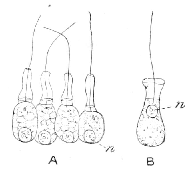
Fig. 77.—The two types of Asconid collar cells. A, of Clathrina, nucleus basal; B, of Leucosolenia, nucleus not basal, flagellum arising from the nuclear membrane. (A, after Minchin; B, after Bidder.)
The tetraxon spicules have "equiangular" triradiate systems in the Clathrinidae, while in Leucosoleniidae they are "alate." Finally, the larva of Clathrinidae is a "parenchymula" (see p. 226), that of Leucosoleniidae an "amphiblastula."
The fact that it is possible to classify the Calcarea Homocoela largely by means of histological characters is in accordance with the importance of the individual cell as opposed to the cell-layers generally throughout the Porifera, and is interesting in serving to emphasise the low grade of organisation of the Phylum. The organs of sponges are often unicellular (pores), or the products of the activity of a single cell (many skeletal elements); and even in the gastral layer, which approaches nearly to an epithelium, comparable with the epithelia of Metazoa, the component cells still seem to assert their independence, the flagella not lashing in concert,[218] but each in its own time and direction.
Sub-Class II. Heterocoela.
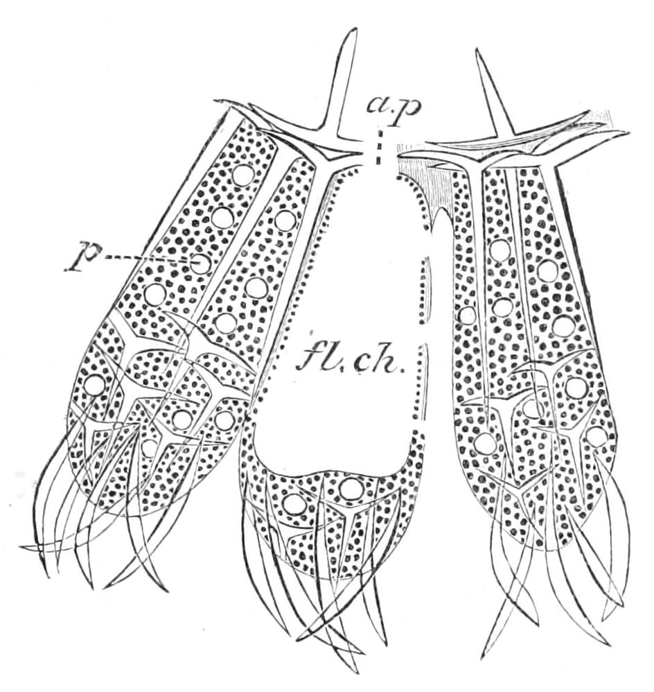
Fig. 78.—Transverse section of the body-wall of Sycon carteri, showing articulate tubar skeleton, gastric ostia (a.p), tufts of oxeas at the distal ends of the chambers (fl.ch), and pores (p). (After Dendy.)
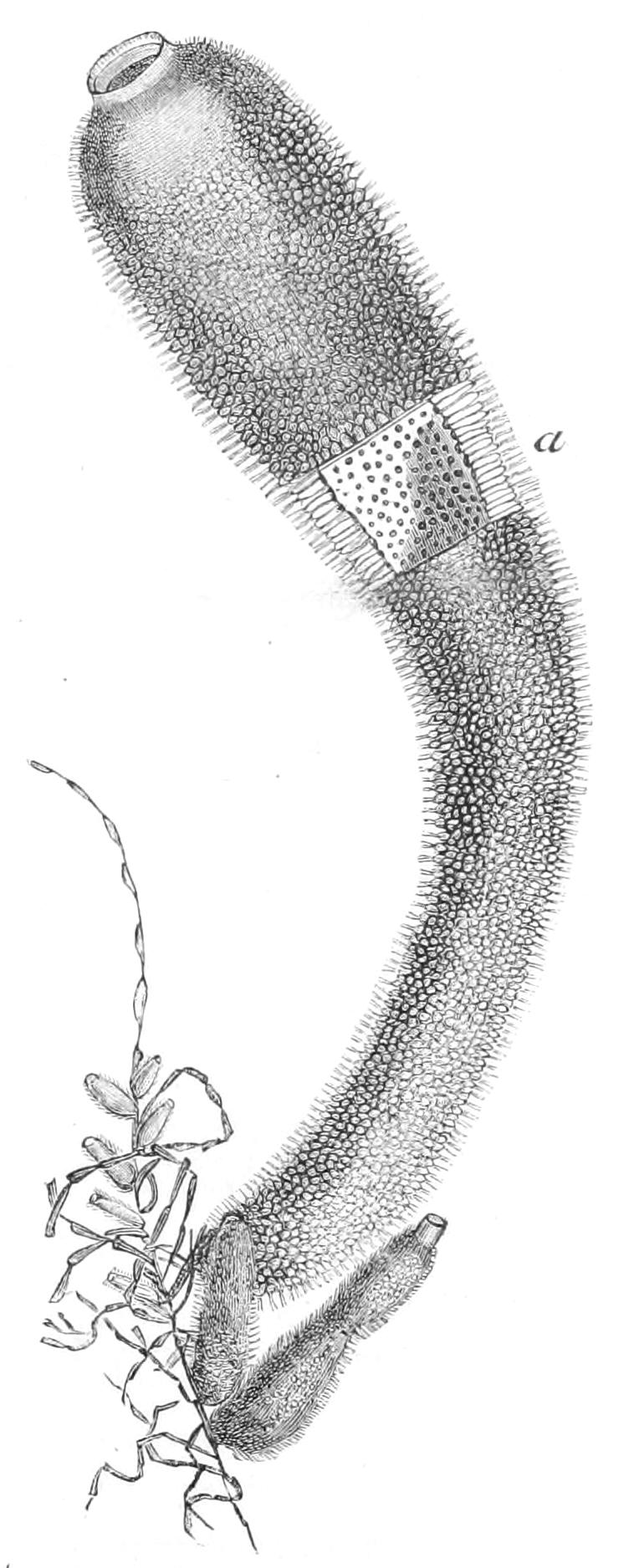
Fig. 79.—Sycon coronatum. At a a portion of the wall is removed, exposing the paragaster and the gastric ostia of the chambers opening into it.
The Heterocoela present a series of forms of successive grades of complexity, all derivable from the Ascons, from which they differ in having a discontinuous gastral layer. The simplest Heterocoela are included in the family Sycettidae, of which the British representative is Sycon (Fig. 79). In Sycon numerous tubular flagellated chambers are arranged radially round a central cavity, the "paragaster," into which they open (Figs. 78, 79). The chambers, which are here often called radial tubes, are close set, leaving more or less quadrangular tubular spaces, the inhalant canals, between them; and where the walls of adjacent chambers come in contact, fusion may take place. Pores guarded by porocytes put the inhalant canals into communication with the flagellated chambers. The paragaster is lined by pinacocytes; choanocytes are confined to the flagellated chambers.
The skeleton is partly defensive, partly supporting; one set of spicules strengthens the walls of the radial tubes and forms collectively the "tubar skeleton." It is characteristic of Sycettidae that the tubar skeleton is of the type known as "articulate"—i.e. it is formed of a number of successive rings of spicules, instead of consisting of a single ring of large spicules which run the whole length of the tube.
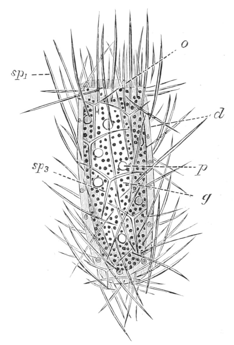
Fig. 80.—Sycon setosum. Young Sponge. × 200. d, Dermal cell; g, gastral cell; o, osculum; p, pore cell; sp1, monaxon; sp3, triradiate spicule. (After Maas.)
The walls of the paragaster are known as the "gastral cortex"; they contain quadriradiate spicules, of which the triradiate systems lie tangentially in the gastral cortex, while the apical ray projects into the paragaster, and is no doubt defensive. The distal ends of the chambers bristle with tufts of oxeate spicules, and the separate chambers are distinguishable in surface view. It is interesting to notice that in some species of Sycon, the gaps between the distal ends of the chambers are covered over by a delicate perforated membrane, thus leading on, as we shall see presently, to the next stage of advance.[219] The larva of Sycon is an amphiblastula (see p. 227). Fig. 80 is a drawing of the young sponge soon after fixation; it would pass equally well for an ideally simple Ascon or, neglecting the arrangement of the spicules, for an isolated radial tube of Sycon. Figs. 81, 82 show the same sponge, somewhat older. From them it is seen that the Sycon type is produced from the young individual, in what may be called its Ascon stage, by a process of outgrowth of tubes from its walls, followed by restriction of choanocytes to the flagellated chambers. Minute observation has shown[220] that this latter event is brought about by immigration of pinacocytes from the exterior. These cells creep through the jelly of the dermal layer and line the paragaster as fast as its original covering of choanocytes retreats into the newly formed chambers.
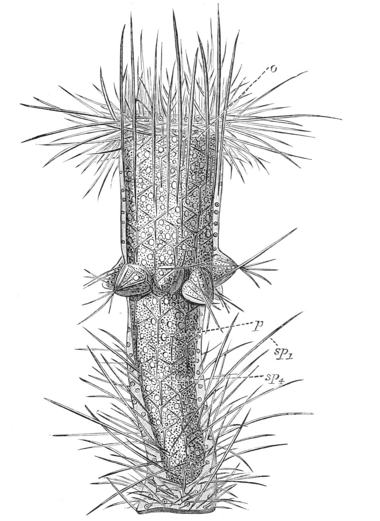
Fig. 81.—S. setosum. Young Sponge, with one whorl of radial tubes. o, Osculum; p, pore; sp1, monaxon; sp4, quadriradiate spicule. (After Maas.)
With a canal system precisely similar to that of Sycon, Ute (Fig. 83) shows an advance in structure in the thickening of the dermal layers over the distal ends of the chambers. The dermal thickenings above neighbouring chambers extend laterally and meet; and there results a sheet of dermal tissue perforated by dermal ostia, which open into the inhalant canals, and strengthened by stout spicules running longitudinally. This layer is termed a cortex; it covers the whole sponge, compacting the radial tubes so that they form, together with the cortex, a secondary wall to the sponge, which is once more a simple sac, but with a complex wall. The cortex may be enormously developed, so as to form more than half the thickness of the wall (Fig. 84). The chambers taken together are spoken of as the chamber layer.
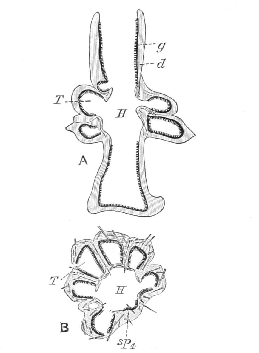
Fig. 82.—Sycon raphanus. A, Longitudinal section of young decalcified Sponge at a stage somewhat later than that shown in Fig. 81. B, Transverse section of the same through a whorl of tubes. d, Dermal membrane; g, gastral membrane; H, paragaster; sp4, tetraradiate spicule; T, radial tube. (After Maas.)
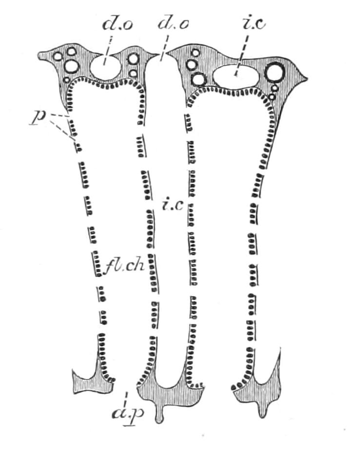
Fig. 83.—Transverse section of the body-wall of Ute, passing longitudinally through two chambers. a.p, Apopyle; d.o, dermal ostium; fl.ch, flagellated chamber or radial tube; i.c, inhalant canal; p, prosopyle. (After Dendy.)
We have already alluded to the resemblance between a young Ascon person and a radial tube of Sycon—a comparison which calls to mind the somewhat strange view of certain earlier authors, that the flagellated chambers are really the sponge individuals. If now we suppose each Ascon-like radial tube of Sycon to undergo that same process of growth by which the Sycon itself was derived from the Ascon, we shall then have a sponge with a canal system of the type seen in Leucandra among British forms, but more diagrammatically shown in the foreign genus Leucilla (Fig. 85). The foregoing remarks do not pretend to give an account of the transition from Sycon to Leucilla as it occurred in phylogeny. For some indication of this we must await embryological research.
In Leucandra the fundamental structure is obscured by the irregularity of its canal system. It shows a further and most important difference from Leucilla in the smaller size and rounded form of its chambers. This change of form marks an advance in efficiency; for now the flagella converge to a centre, so that they all act on the same drop of water, while in the tubular chamber their action is more widely distributed and proportionately less intense (see p. 236).
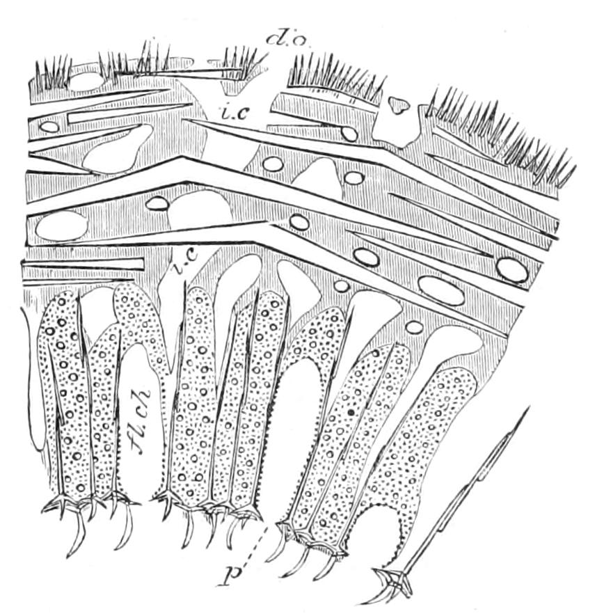
Fig. 84.—Transverse section through the body-wall of Grantiopsis. d.o, Dermal ostium; fl.ch, flagellated chamber; i.c, long incurrent canal traversing the thick cortex to reach the chamber layer; p, apopyle. (After Dendy.)
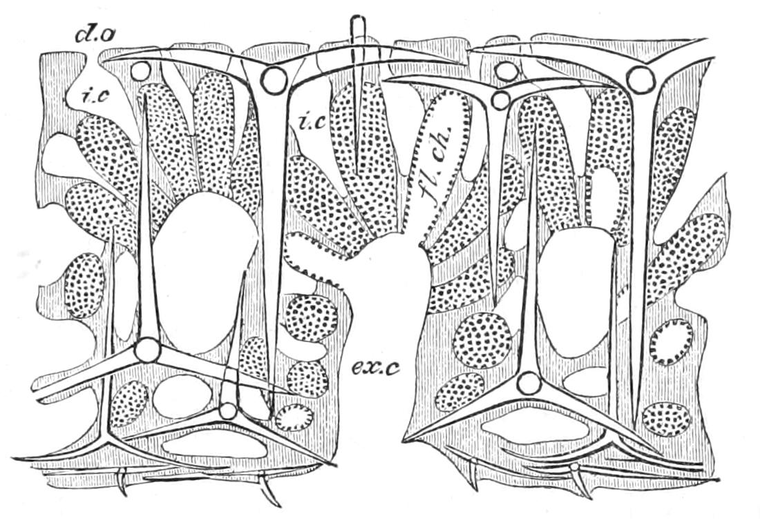
Fig. 85.—Transverse section through the body-wall of Leucilla. d.o, Dermal ostium; ex.c, exhalant canal; fl.ch, chamber; i.c, inhalant canal. (After Dendy.)
Above are described three main types of canal system—that of Homocoela, of Sycon, and of Leucandra and Leucilla. These are conveniently termed the first, second, and third types respectively, and may be briefly described as related to one another somewhat in the same way as a scape, umbel, and compound umbel among inflorescences. These types formed the basis of Haeckel's famous classification.[221] It has, however, been concluded[222] that the skeleton is a safer guide in taxonomy, at any rate for the smaller subdivisions; and in modern classifications genera with canal systems of the third type will be found distributed among various families; while in the Grantiidae, Ute and Leucandra stand side by side. This treatment implies a belief that the third type of canal system has been independently and repeatedly evolved within the Calcarea—an example of a phenomenon, homoplasy, strikingly displayed throughout the group. It is, remarkably enough, the case that all the canal systems found in the remainder of the Porifera are more or less modified forms of one or other of the second two types of canal system above described.
The families Grantiidae, Heteropidae, and Amphoriscidae, all possessing a dermal cortex, are distinguished as follows:—The Grantiidae by the absence of subdermal sagittal triradiate spicules and of conspicuous subgastral quadriradiates; the Heteropidae by the presence of sagittal triradiates; the Amphoriscidae by the presence of conspicuous subgastral quadriradiates.
Two families of Calcarea, possibly allied, remain for special mention—the Pharetronidae, a family rich in genera, and containing almost all the fossil forms of the group, and the Astroscleridae.
The Pharetronidae are with one, or perhaps two exceptions, fossil forms, having in common the arrangement of the spicules of their main skeletal framework in fibres. The family is divided into two sub-families:—
I. Dialytinae.—The spicules are not fused to one another; the exact mode of their union into fibres is unknown, but an organic cement may be present.
Lelapia australis, a recent species, should probably be placed here as the sole living representative. Dendy has shown[223] that this remarkable species has a skeleton of the same fibrous character as is found in typical Dialytinae, and that the triradiate spicules in the fibres undergo a modification into the "tuning-fork" type (Fig. 86, C), to enable them to be compacted into smooth fibres. "Tuning-forks," though not exclusively confined to Pharetronids, are yet very characteristic of them.
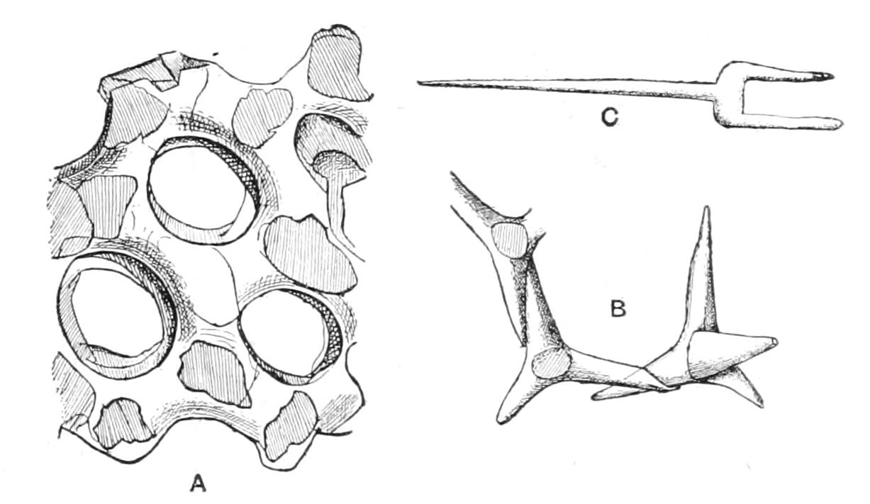
Fig. 86.—Portions of the skeleton of Petrostroma schulzei. A, Framework with ensheathing pellicle; B, quadriradiate spicules with laterally fused rays; C, a "tuning-fork." (After Doederlein.)
II. Lithoninae.—The main skeletal framework is formed of spicules fused together, and is covered by a cortex containing free spicules.
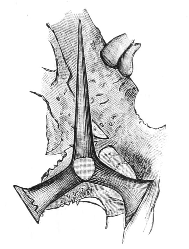
Fig. 87.—A spicule from the skeleton framework of Plectroninia, showing the terminally expanded rays. (After Hinde.)
The sub-family contains only one living genus and a few recently described fossil forms. Petrostroma schulzei[224] lives in shallow water near Japan; Plectroninia halli[225] and Bactronella were found in Eocene beds of Victoria; Porosphaera[226] long known from the Chalk of England and of the Continent, has recently been shown by Hinde[226] to be nearly allied to Plectroninia; finally, Plectinia[227] is a genus erected by Počta for a sponge from Cenomanian beds of Bohemia. Doederlein, in 1896, expressed his opinion that fossil representatives of Lithoninae would most surely be discovered. The fused spicules are equiangular quadriradiates; they are united in Petrostroma by lateral fusion of the rays, in Plectroninia (Fig. 87) and Porosphaera by fusion of apposed terminal flat expansions of the rays, and in some, possibly all, genera a continuous deposit of calcium carbonate ensheaths the spicular reticulum. Thus they recall the formation of the skeleton on the one hand of the Lithistida and on the other of the Dictyonine Hexactinellida (see pp. 202, 211). "Tuning-forks" may occur in the dermal membrane.
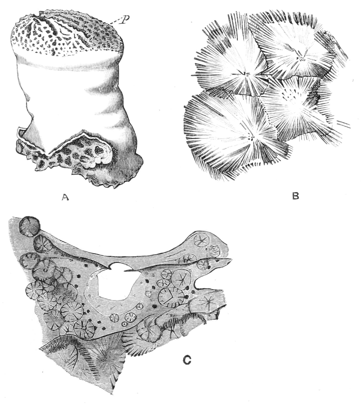
Fig. 88.—Astrosclera willeyana, Lister. A, the Sponge, × about 3. p, The ostia on its distal surface. B, a portion of the skeleton showing four polyhedra with radiating crystalline fibres. C, an ostium; the surrounding tissue contains young stages of polyhedra. (After Lister.)
The Astroscleridae, as known at present, contain a single genus and species, apparently the most isolated in the phylum. Astrosclera willeyana[228] was brought back from the Loyalty Islands, and from Funafuti of the Ellice group. Its skeleton is both chemically and structurally aberrant. In other Calcarea the calcium carbonate of the skeleton is present as calcite, in Astrosclera as aragonite, and the elements are solid polyhedra, united by their surfaces to the total exclusion of soft parts (Fig. 88). Each element consists of crystalline fibres radially disposed around a few central granules, and terminating peripherally in contact with the fibres of adjacent elements. Young polyhedra are to be found free in the soft parts at the surface. The chambers are exceptionally minute, especially for a calcareous sponge, comparing with those of other sponges as follows:—
Astrosclera chambers, 10µ × 8µ to 18µ × 11µ. Smallest chambers in Silicea, 15µ × 18µ to 24µ × 31µ. Smallest chambers in Calcarea, 60µ × 40µ.
In its outward form Astrosclera resembles certain Pharetronids. The minute dimensions of the ciliated chambers relegate Astrosclera to the Micromastictora, and the fortunate fact that the calcium carbonate of its skeleton possesses the mineral characters not of calcite, but of aragonite, renders it less difficult to conceive that its relations may be rather with the non-calcareous than the calcareous sponges.
BRANCH II. MICROMASTICTORA
All sponges which do not possess calcareous skeletons are characterised by choanocytes, which, when compared with those of Calcarea, are conspicuous for their smaller size. The great majority (Silicispongiae) of the non-calcareous sponges either secrete siliceous skeletons or are connected with siliceous sponges by a nicely graded series of forms. The small remainder are entirely askeletal. All these non-calcareous sponges are included, under the title Micromastictora, in a natural group, opposed to the Megamastictora as of equal value.
The subdivision of the Micromastictora is a matter of some difficulty. The Hexactinellida alone are a well circumscribed group. After their separation there remains, besides the askeletal genera, an assemblage of forms, the Demospongiae, which fall into two main tribes. These betray their relationship by series of intermediate types, but a clue is wanting which shall determine decisively the direction in which the series are to be read. The askeletal genera are the crux of the systematist. It is perhaps safest, while recognising that many of them bear a likeness of one kind or another to various Micromastictora, to retain them together in a temporary class, the Myxospongiae.
CLASS I. MYXOSPONGIAE
The class Myxospongiae is a purely artificial one, containing widely divergent forms, which possess a common negative character, namely, the absence of a skeleton. As a result of this absence they are all encrusting in habit.
One genus, Hexadella, has been regarded by its discoverer Topsent[229] as an Hexactinellid. The same authority places Oscarella with the Tetractinellida; it is more difficult to suggest the direction in which we are to seek the relations of the remaining type, Halisarca.
Hexadella, from the coast of France, is a remarkable little rose-coloured or bright yellow sponge, with large sac-like flagellated chambers and a very lacunar ectosome.
Oscarella is a brightly coloured sponge, with a characteristic velvety surface; it is a British genus, but by no means confined to our shores. Its canal system has been described by some authors as diplodal, by others as eurypylous. Topsent[230] has shown, and we can confirm his statement, that though the chambers have usually the narrow afferent and efferent ductules of a diplodal system, yet since each one may communicate with two or three canals, the canal system cannot be described as diplodal. The hypophare attains a great development, and in it the generative products mature. The pinacocytes, like those of Plakinidae, and perhaps of Aplysilla, are flagellated.
Halisarca, also British, is easily distinguished from Oscarella by the presence of a mucus-like secretion which oozes from it, and by the absence of the bright coloration characteristic of Oscarella. It naturally suggests itself that the coloration in the one case and the secretion in the other are protective, and in this respect perform one of the functions of the skeleton of other sponges. The chambers are long, tubular, and branched. There is no hypophare.
CLASS II. HEXACTINELLIDA[231]
Silicispongiae, defined by their spicules, of which the rays lie along three rectangular axes. The canal system is simple, with thimble-shaped chambers. The body-wall is divided into endosome, ectosome, and choanosome.
Some authors would elevate the Hexactinellida to the position of a third main sub-group of Porifera, thus separating them from other siliceous sponges. In considering this view it is important to realise at the outset that they are deep-water forms. They bear evident traces of the influence of their habitat, and like others of the colonists of the deep sea, are impressed with marked archaic features. Yet they are still bound to other Micromastictora, first by the small size of their choanocytes, and secondly by the presence of siliceous spicules. This second character is really a double link, for it involves not merely the presence of silica in the skeleton, but also the presence in each spicule of a well-marked axial filament. Now this axial filament is a structure which is gaining in importance, for purposes of classification, in proportion as its absence in Calcarea is becoming more probable. The Hexactinellida are the only sponges, other than the bath sponge, which are at all generally known. They have won recognition by their beauty, as the bath sponge by its utility, and, like it, one of their number—the Venus's Flower-Basket—forms an important article of commerce, the chief fishery being in the Philippine Islands. This wonderful beauty belongs to the skeleton, and is greatly concealed when the soft parts are present.
We have said that the Hexactinellids are deep-sea forms; they are either directly fixed to the bottom or more often moored in the ooze by long tufts of rooting spicules. In the "glass-rope sponge," the rooting tuft of long spicules, looking like a bundle of spun glass, is valued by the Japanese, who export it to us. In Monorhaphis the rooting tuft is replaced by a single giant spicule,[232] three metres in length, and described as "of the thickness of a little finger"! Probably it is as a result of their fixed life in the calm waters of the deep sea[233] that Hexactinellids contrast with most other sponges by their symmetry. It should not, however, be forgotten that many of the Calcarea which inhabit shallow water exhibit almost as perfect a symmetry.
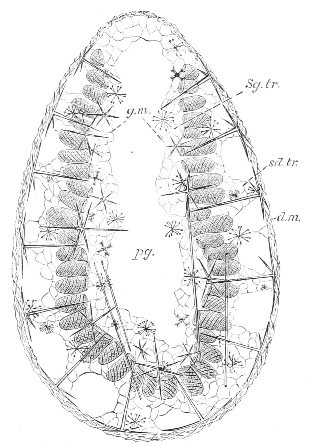
Fig. 89.—Longitudinal section of a young specimen of Lanuginella pupa O.S., with commencing formation of the oscular area. × 35. d.m, Dermal membrane; g.m, gastral membrane; pg, paragaster; sd.tr, subdermal trabeculae; Sg.tr, subgastral trabeculae. (After F. E. Schulze.)
The structure of the body-wall in Hexactinellida is so constant as to make it possible to give a general description applicable to all members of the group. It is of considerable thickness, but a large part is occupied by empty spaces, for the actual tissue is present in minimum quantity. In the wall the chamber-layer is suspended by trabeculae of soft tissue, between a dermal membrane on the outside and a similar gastral membrane on the inner side (Fig. 89). Thus the water entering the chambers through their numerous pores has first passed through the ostia in the dermal membrane and traversed the subdermal trabecular space; on leaving the chambers it flows through the subgastral trabecular space and the ostia in the gastral membrane, to enter the paragaster and leave the body at the osculum. The trabeculae and the dermal and gastral membranes together constitute the dermal layer. This conclusion is based on comparison with adults of the other groups, for in the absence of embryological knowledge no direct evidence is available. According to the Japanese investigator, Isao Ijima,[234] the dermal and gastral membranes are but expansions of the trabeculae, and the trabeculae themselves are entirely cellular, containing none of the gelatinous basis met with in the dermal layer of all other sponges. There is no surface layer of pinacocytes, the cells forming the trabeculae being all of one type, namely, irregularly branching cells, connected with one another by their branches to form a syncytium. In the trabeculae are found scleroblasts and archaeocytes.
The chambers have a characteristic shape: they are variously described as "thimble-shaped," "tubular," or "Syconate," and they open by wide mouths into the subgastral trabecular space. Their walls have been named the membrana reticularis from the fact that, when preserved with only ordinary precautions, they are seen as a regular network of protoplasmic strands, with square meshes and nuclei at the nodes. This appearance recently found an explanation when Schulze, for the first time, succeeded in preserving the collared cells of Hexactinellids.[235] Schulze was then able to show that the choanocytes are not in contact with one another at their bases, where the nuclei are situated, but communicate with one another by stout protoplasmic strands. The form of the choanocyte can be seen in Fig. 91.
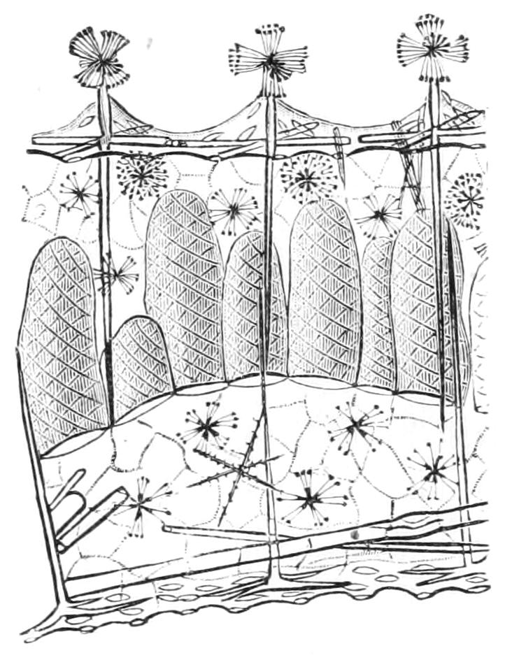
Fig. 90.—Portion of the body-wall of Walteria sp., showing the thimble-shaped flagellated chambers, above which is seen the dermal membrane. (After F. E. Schulze.)
To Schulze's description of the chamber, Ijima has added the important contributions that every mesh in the reticulum functions as a chamber pore or prosopyle; and that porocytes, such as are found in Calcarea, are wanting. This structure of the chamber-walls, the absence of gelatinous basis in the dermal layer, and the slight degree of histological differentiation in the same layer, added to the more obvious character of thimble-shaped chambers, are the chief archaic features of Hexactinellid morphology.
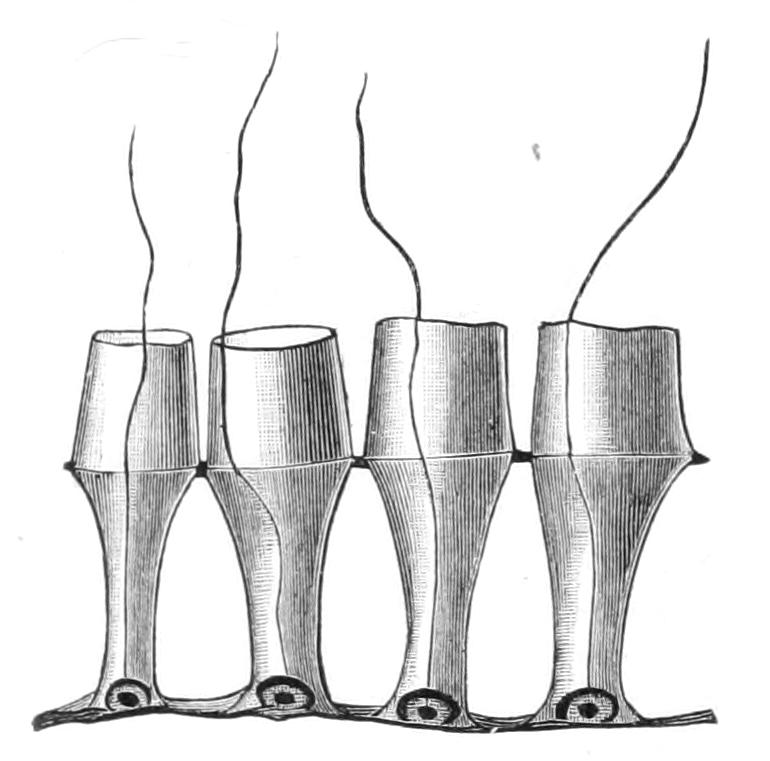
Fig. 91.—Portion of a section of the membrana reticularis or chamber-wall of Schaudinnia arctica, × 1500. (After F. E. Schulze.)
The skeleton which supports the soft parts is, like them, simple and constant in its main features. It is secreted by scleroblasts, which lie in the trabeculae, and is made up of only one kind of spicule and its modifications. This is the hexactine, a spicule which possesses six rays disposed along three rectangular axes. Each ray contains an axial thread, which meets its fellow at the centre of the spicule, where they together form the axial cross. Modifications of the hexactine arise either by reduction or branching, by spinulation or expansion of one or more of the rays. The forms of spicule arising by reduction are termed pentactines, tetractines, and so on, according to the number of the remaining rays. Those rays which are suppressed leave the proximal portion of their axial thread as a remnant marking their former position (Fig. 94). Octactine spicules seem to form an exception to the above statements, but Schulze has shown that they too are but modifications of the hexactine arising by (1) branching of the rays of a hexactine, followed by (2) recombination of the secondary rays (Fig. 92).
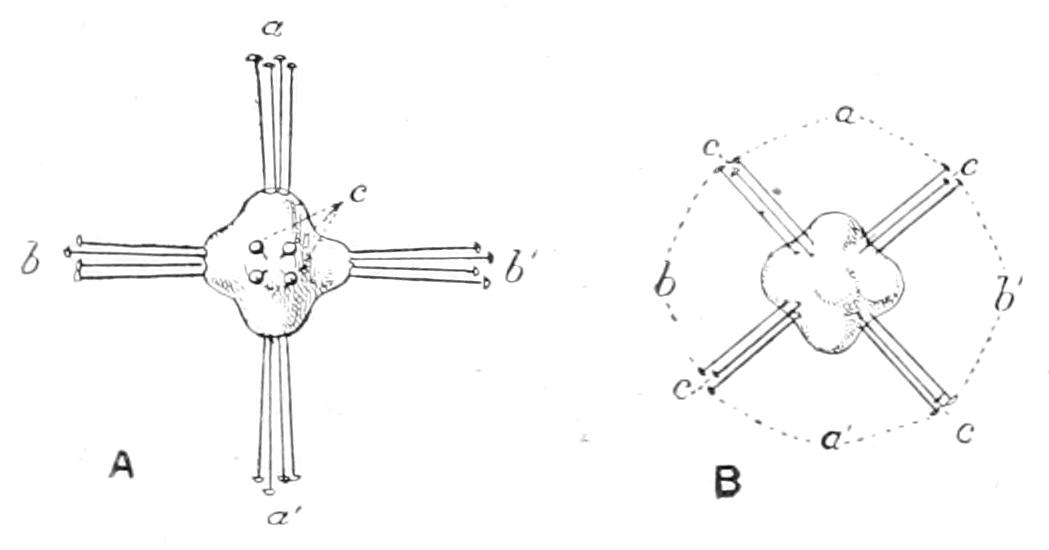
Fig. 92.—A, discohexaster, in which the four cladi a, a', b, b', c of each ray start directly from a central nodule. B, disco-octaster, resulting from the redistribution of the twenty-four cladi of A into eight groups of three. (After Schulze, from Delage.)
The various spicules are named, irrespective of their form, according to their position and corresponding function. The arrangement of the spicules is best realised by means of a diagram (Fig. 93).
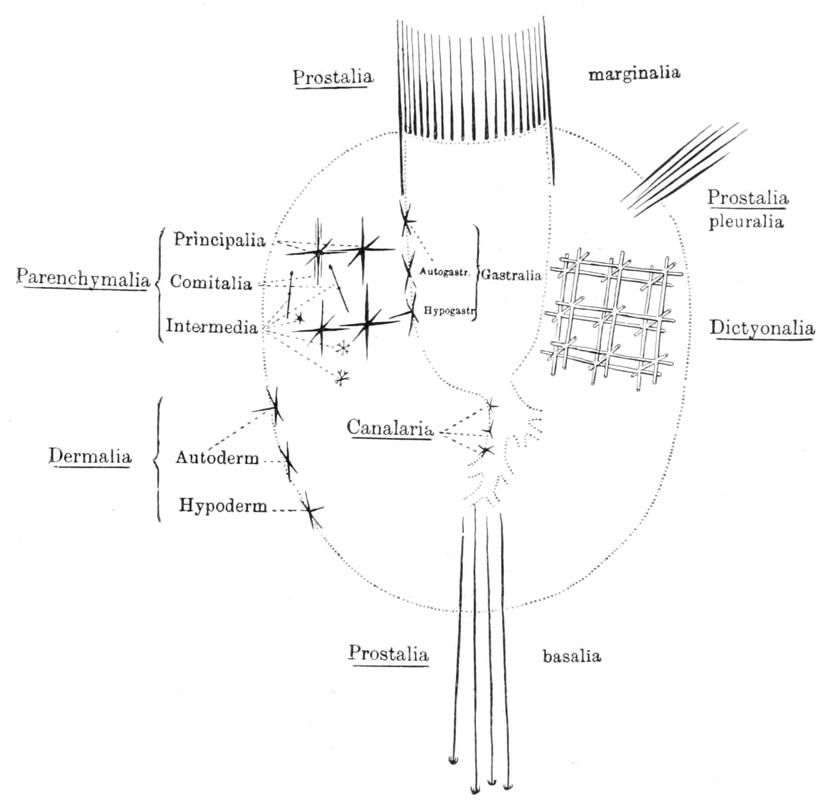
Fig. 93.—Scheme to show the arrangement of spicules in the Hexactinellid skeleton. Canalaria, microscleres in the walls of the excurrent canals; Dermalia Autoderm[alia], microscleres in the dermal membrane; D. Hypoderm[alia], more deeply situated dermalia; Dictyonalia, parenchymalia which become fused to form the skeletal framework of Dictyonina; Gastralia Autogastr[alia], microscleres in the gastral membrane; Gastralia Hypogastr[alia], more deeply situated gastralia; Parenchymalia Principalia, main supporting spicules between the chambers; P. Comitalia, slender diactine or triactine spicules accompanying the last; P. Intermedia, microscleres between the P. principalia; Prostalia, projecting spicules; P. basalia, rooting spicules, from the base; P. marginalia, defensive spicules, round the oscular rim; P. pleuralia, defensive spicules, from the sides. (From Delage and Hérouard, after F. E. Schulze.)
The deviations from this ground-plan of Hexactinellid structure are few and simple. They are due to folding of the chamber-layer, or to variations in the shape of the chambers, and to increasing fusion of the spicules to form rigid skeletons. A simple condition of the chamber-layer, like that of the young sponge of Fig. 89, occurs also in some adult Hexactinellids, e.g. in Walteria of the Pacific Ocean (Fig. 90). Thus is represented in this order the second type of canal system described among Calcarea. More frequently, however, instead of forming a smooth sheet, the chamber-layer grows out into a number of tubular diverticula, the cavities of which are excurrent canals; these determine a corresponding number of incurrent canals which lie between them. In this way there arises a canal system resembling the third type of Calcarea. By still further pouching so as to give secondary diverticula, opening into the first, a complicated canal system is formed, as, for example, in Euplectella suberea.
To return to the skeleton, the most complete fusion is attained by the deposit of a continuous sheath of silica round the apposed parallel rays of neighbouring spicules. This may be termed the dictyonine type of union, for it occurs in all those forms originally included under the term Dictyonina, in which the cement is deposited pari passu with the formation of the spicules. In other cases connecting bridges of silica unite the spicules, or there may be a connecting reticulum of siliceous threads, or, again, rays crossing obliquely may be soldered together at the point of contact. These more irregular methods occur in species where the spicules are free at their first formation. Spicules originally free may later be united in a true Dictyonine fashion. The terms Lyssacina and Dictyonina are useful to denote respectively: the former all those Hexactinellida in which the spicules are free at their first formation, and the latter those in which the deposit of the cementing layer goes hand in hand with the formation of the spicules. But the terms do not indicate separateness of origin of the groups denoted by them, for there is evidence that Dictyonine types have been derived repeatedly from Lyssacine types, and that in fact every Dictyonine was once a Lyssacine.
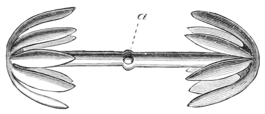
Fig. 94.—Amphidisc, at a are traces of the four missing rays.
The real or natural cleft in the class lies between those genera possessing amphidiscs (Figs. 94, 97) among their microscleres, and all the remainder of the Hexactinellida which bear hexasters (Fig. 96). The former set of genera constitute the sub-class Amphidiscophora, the latter the Hexasterophora.
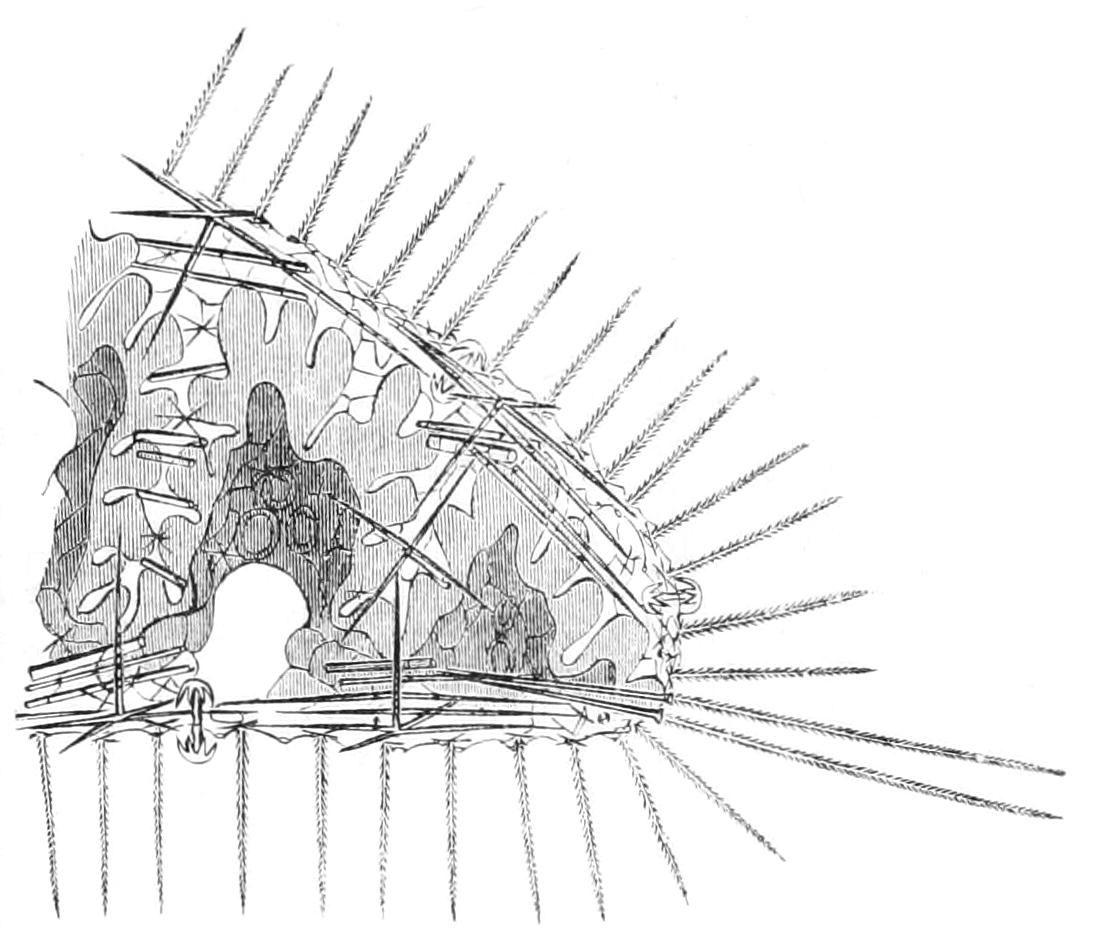
Fig. 95.—Portion of body-wall of Hyalonema, in section, showing the irregular chambers.
Sub-Class 1. Amphidiscophora.—Amphidiscs are present, hexasters absent. A tuft of rooting spicules or basalia is always present. The ciliated chambers deviate more or less from the typical thimble shape, and the membrana reticularis is continuous from chamber to chamber (Figs. 94, 95, 97).
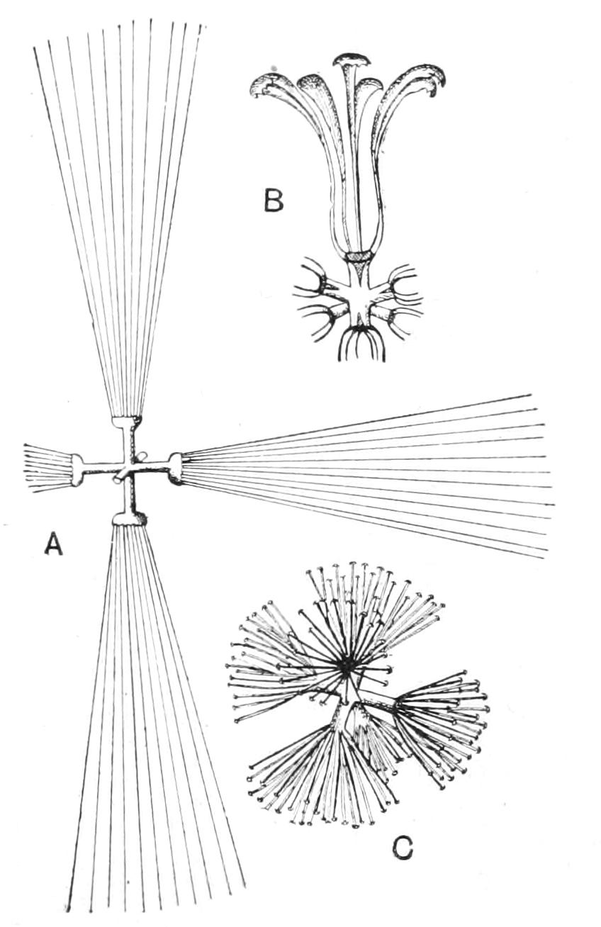
Fig. 96.—Hexasters. A, Graphiohexaster; B, floricome; C, onychaster.
Sub-Class 2. Hexasterophora.—Hexasters are present, amphidiscs absent. The chambers have the typical regular form, and are sharply marked off from one another (Figs. 90, 96).
All the Amphidiscophora have Lyssacine skeletons; in the Hexasterophora both types of skeleton occur. The subdivision of the Hexasterophora is determined by the presence or absence of uncinate spicules. An "uncinatum" is a diactine spicule, pointed at both ends and bearing barbs all directed towards one end. This method of classification gives us a wholly Dictyonine order, Uncinataria, and an order consisting partly of Dictyonine, partly of Lyssacine genera, which may be distinguished as the Anuncinataria. Ova have rarely been found, and sexually produced larvae never; but Ijima has found archaeocyte clusters in abundance, and his evidence is in favour of the view that they give rise asexually to larvae, described by him in this class for the first time (see p. 231).
Both sub-classes are represented in British waters: the Amphidiscophora by Hyalonema thomsoni and Pheronema carpenteri; the Hexasterophora by Euplectella suberea and Asconema setubalense, and of course possibly by others.
Hyalonema thomsoni, one of the glass-rope sponges, was dredged by the Porcupine off the Shetland Islands in water of about 550 fathoms. The spindle-shaped body of the sponge is shown in Fig. 97. Its long rooting tuft is continued right up its axis, to end in a conical projection, which is surrounded by four apertures leading into corresponding compartments of the paragaster.
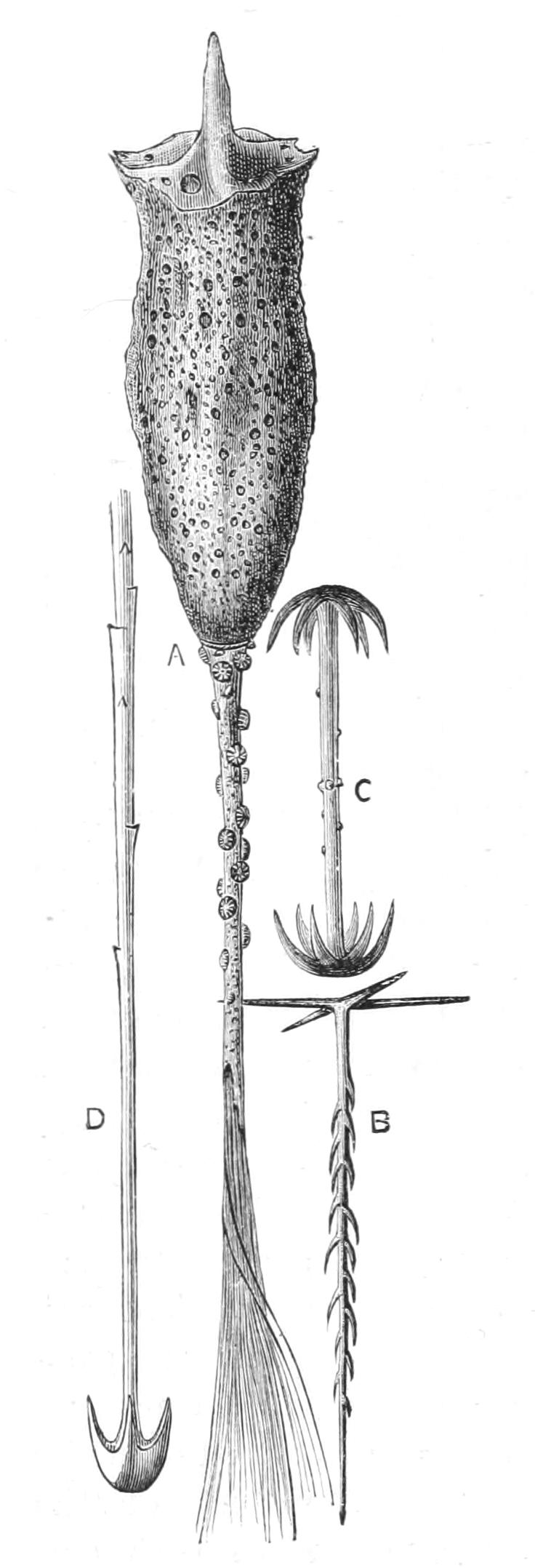
Fig. 97.—Hyalonema thomsoni. A, Whole specimen with rooting tuft and Epizoanthus crust; B, pinulus, a spicule characteristic of but not peculiar to the Amphidiscophora, occurring in the dermal and gastral membranes; C, amphidisc with axial cross; D, distal end of rooting spicule with grapnel. (After F. E. Schulze.)
The crust of Anthozoa of the genus Epizoanthus (p. 406) on the rooting tuft is a constant feature in this as in other species of Hyalonema. It contributed to make the sponge a puzzle, which long defied interpretation. The earliest diagnosis the genus received was the "Glass Plant." Then the root tuft was thought to be part of the Epizoanthus, which was termed a "most aberrant Alcyonarian with its base inserted in a sponge"; next we hear of the sponge as parasitic on the Sea Anemone. Finally, the root tuft was shown to be proper to the sponge, which was, however, figured upside down, till some Japanese collectors described the natural position, or that in which they were accustomed to find it.
Pheronema carpenteri was found by the Lightning off the north of Scotland in 530 fathoms. The goblet shaped, thick walled body and broad, ill-defined root tuft are shown in Fig. 98, but no figure can do justice to the lustre of its luxuriant prostalia and delicate dermal network with stellate knots at regular intervals. The basalia are two-pronged and anchor-like.
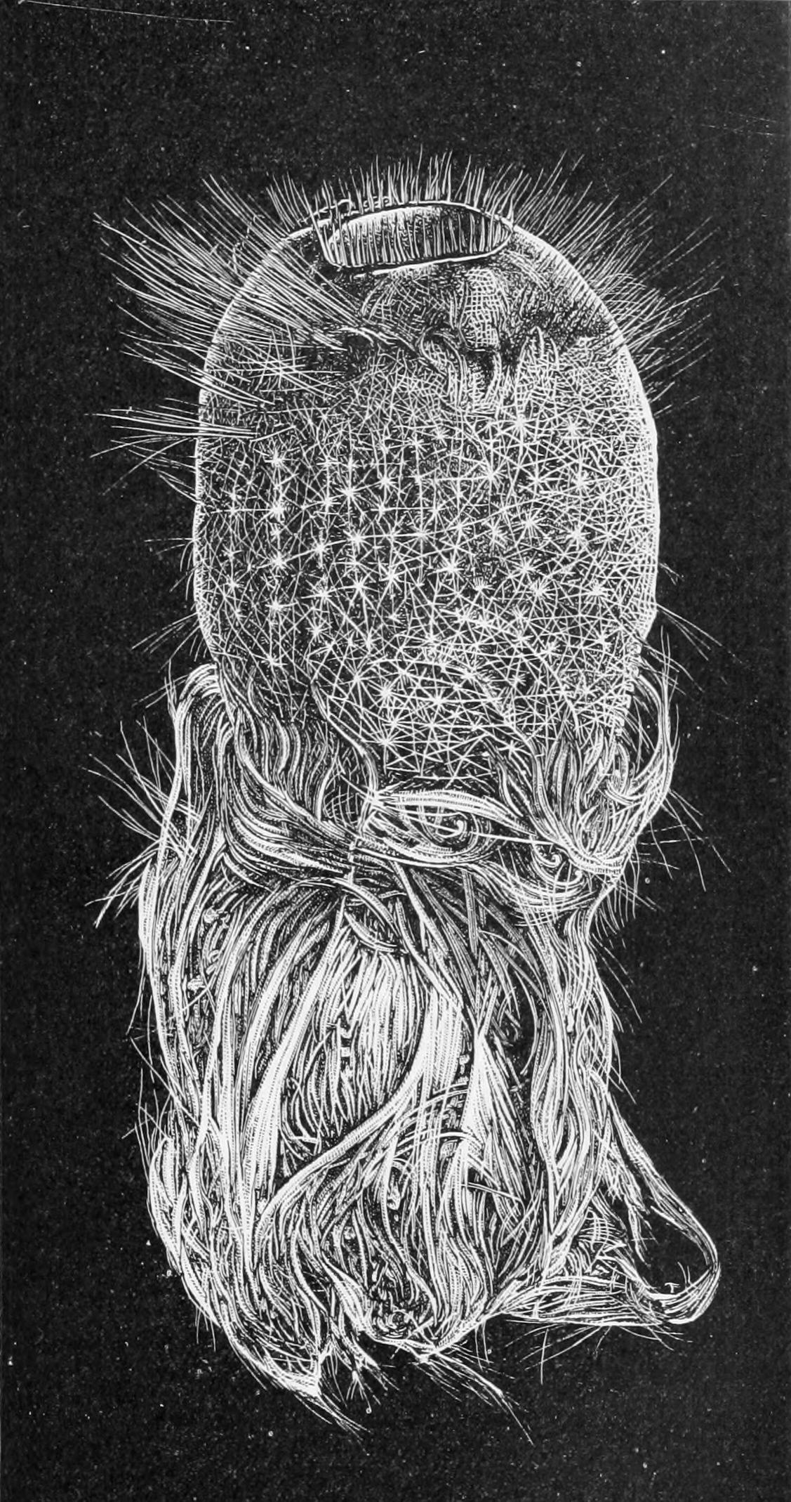
Fig. 98.—Pheronema carpenteri. × ½. (From Wyville Thomson.)
Both the Hexasterophoran genera were dredged off the north of Scotland, and both conform to the Lyssacine type without uncinates. Euplectella suberea is a straight, erect tube, anchored by a tuft of basalia. The upper end of the tube is closed by a sieve plate, the perforations in which are oscula, while the beams contain flagellated chambers, so that the sieve is simply a modified portion of the wall. It is a peculiarity of this as of one or two other allied genera that the lateral walls are perforated by oscula. They are termed parietal gaps, and are regularly arranged along spiral lines encircling the body.
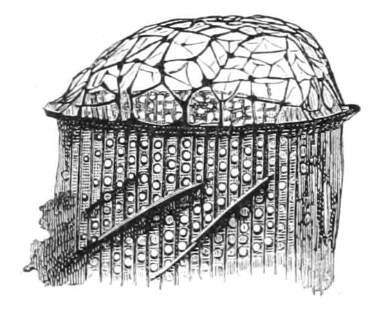
Fig. 99.—Sieve plate of Euplectella imperialis. (After Ijima.)
Ijima, who has dredged Euplectellids from the waters near Tokyo, finds that in young specimens oscula are confined to the sieve plate; parietal gaps are secondary formations. The groundwork of the skeleton is a lattice similar to that shown in Fig. 100. The chamber-layer is much folded. Various foreign species of Euplectella afford interesting examples of association with a Decapod Crustacean, Spongicola venusta, of which a pair lives in the paragaster of each specimen. The Crustacean is light pink, the female distinguished by a green ovary, which can be seen through the transparent tissues. It is not altogether clear what the prisoner gains, nor what fee, if any, the host exacts.
Ijima relates that the skeleton of Euplectella is in great demand in Japan for marriage ceremonies. He also informs us that the Japanese name means "Together unto old age and unto the same grave," while by a slight alteration it becomes "Lobsters in the same cell," and remarks that the Japanese find this an amusing pun.
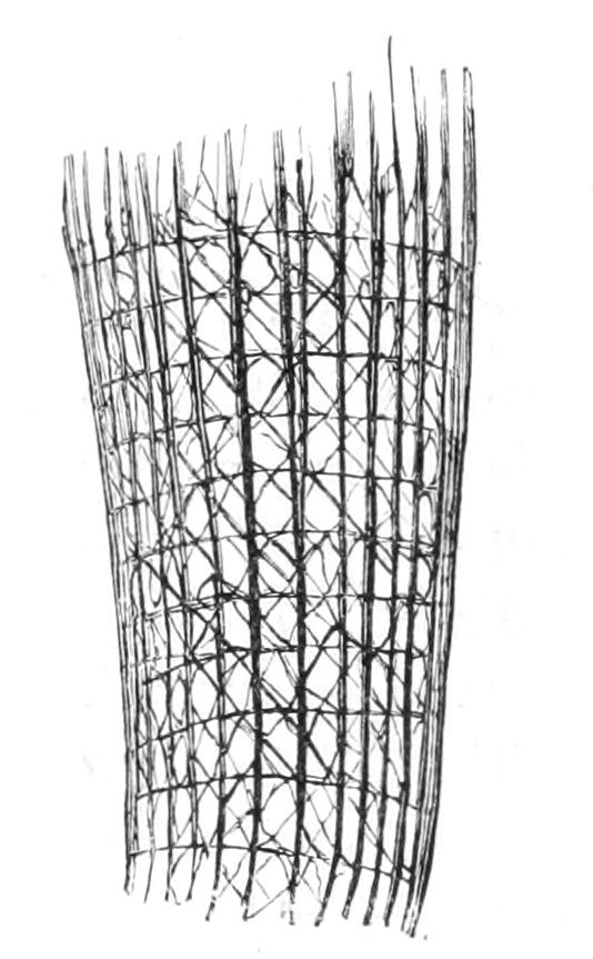
Fig. 100.—Skeletal lattice of Euplectella imperialis. (After Ijima.)
The same Spongicola lives in pairs in Hyalonema sieboldi. Another case of apparently constant association is that of the Hydroid stocks which inhabit Walteria. F. E. Schulze describes Stephanoscyphus mirabilis (see p. 318) in a specimen of Walteria flemmingi; the presence of the polyp causes the sponge to grow out into little dome-shaped elevations, each of which shelters one polyp; while in W. leuckarti Ijima finds a similar association in every specimen examined.
Fossil Hexactinellida.
This group has the distinction of including among its Lyssacine members the oldest known sponge, Protospongia fenestrata, of Cambrian age (Salter). As preserved it consists of a single layer of quadriradiate, or possibly quinqueradiate spicules, which, arranged as a square meshed lattice, supported the superficial layer of the sponge (Fig. 101). Whether or not the fossil represents the whole of the sponge-skeleton does not appear.[236]
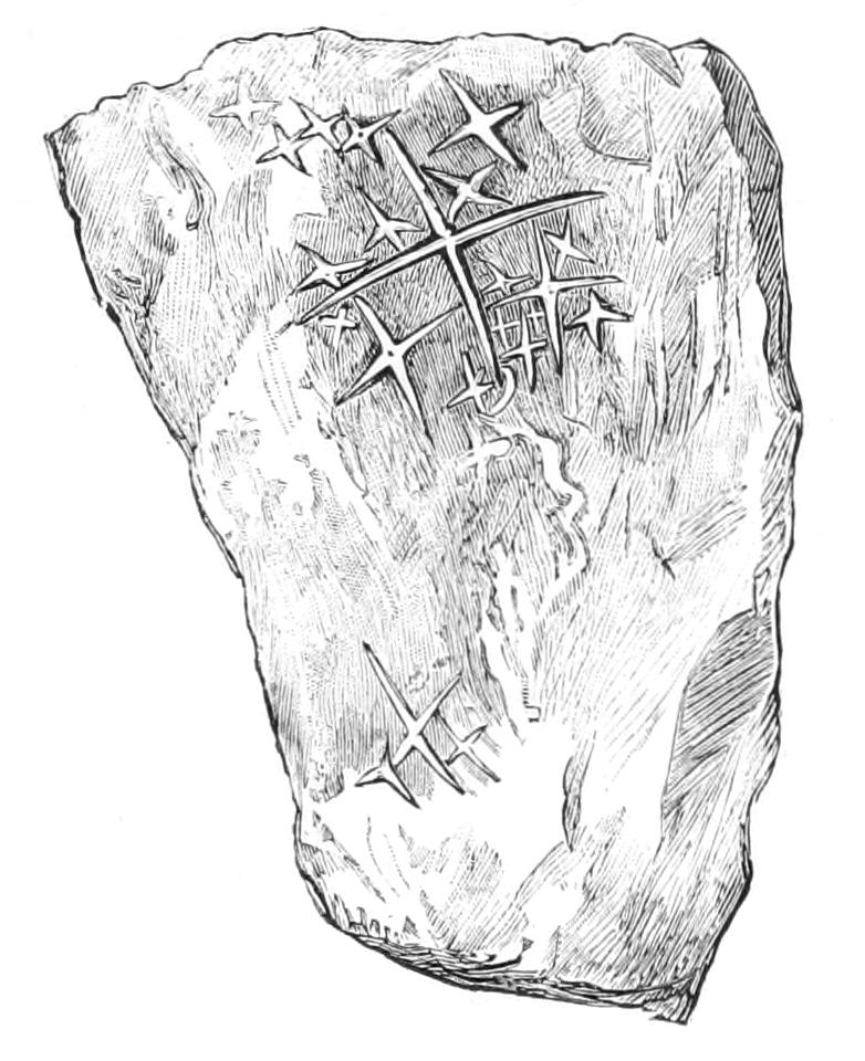
Fig. 101.—Part of the specimen of Protospongia fenestrata in the Sedgwick Museum, Cambridge. Nat. size. (After Sollas.)
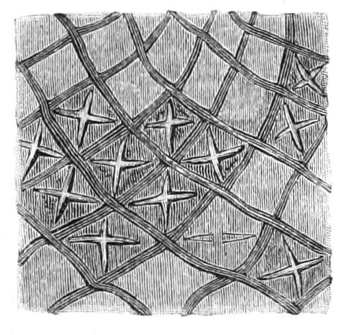
Fig. 102.—A portion of the outer surface of a Receptaculitid, Acanthoconia barrandei, in which the expanded outer rays of the spicules are partially destroyed, revealing the four tangential rays beneath, × 3. (After Hinde.)
The extraordinary Receptaculitidae are probably early Lyssacine forms: they are cup- or saucer-shaped fossils, abundant in Silurian and above all in Devonian strata, and have been "assigned in turn to pine cones, Foraminifera, Sponges, Corals, Cystideans," and Tunicata. Hinde[237] brings forward important arguments for retaining them among Hexactinellida. The only elements in the skeleton of the simpler genera, e.g. Ischadites, are structures comparable to Hexactinellid spicules. The surface of the fossil presents a series of lozenges forming a regular mosaic. Each lozenge is the expanded end of one of the rays of a spicule; it conceals four rays in one plane, tangential to the wall of the cup-shaped fossil, while the sixth ray projects vertically to the wall into the cavity of the cup. In the genus Receptaculites itself there is an inner layer of plates abutting against the inner ends of the sixth rays, and at present problematic. An axial canal is present in each of the rays—the six canals meeting at the centre of the spicule. Special chinks between the spicules appear to have provided a passage for the water current.
The beautiful Ventriculites, so common in the Chalk and present in the Cambridge Greensand, are historically interesting, for the fact that they are fossil Hexactinellida of which the general and skeletal characters were very minutely described by Toulmin Smith long before recent representatives of the group were known. In common with a number of fossil Dictyonine species they are distinguished by the perforation of the nodes, a character due to the fact that the siliceous investment which unites the spicules together stops short before reaching the centre of each spicule, and bridges across the rays so as to form a skeleton octahedron. This character is rare in recent Hexactinellids, but, as first pointed out by Carter, it is presented by one or two forms, of which Aulocystis grayi Bwk is best known. The majority of the fossil Hexactinellida belong to the Dictyonine section, a fact attributable to the greater coherence of their skeleton. The "Dictyonina" are to be reckoned among the rock-builders of Jurassic and Cretaceous times.
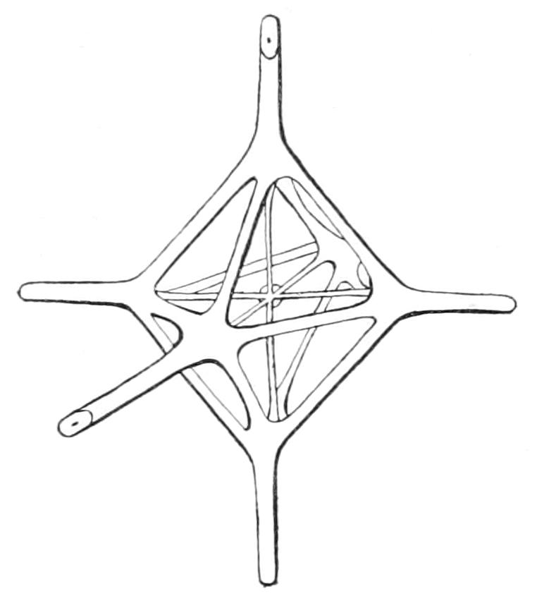
Fig. 103.—A node of the skeleton of Ventriculites from the Cambridge Greensand. (After Sollas.)
The Octactinellida and Heteractinellida are two classes created by Hinde[238] to contain certain little-known Devonian and Carboniferous sponges, possessing in the one case 8-rayed spicules, of which 6 rays lie in one plane and 2 are perpendicular to this plane; in the other case, spicules with a number of rays varying from 6 to 30. Bearing in mind the manner in which octactine spicules are known to arise in recent Hexactinellida (p. 200), it is clearly possible to derive these 8-rayed spicules from hexactines by some similar method; while the typical spicule of the Heteractinellida is a euaster. Hence we may refer the Octactinellid fossils to the class Hexactinellida, and the Heteractinellid forms either to the Monaxonida or Tetractinellida.
CLASS III. DEMOSPONGIAE
Silicispongiae in which triaxonid spicules are absent.
This class has attained the highest level of organisation known among Porifera; the most efficient current-producing apparatus is met with here, so, too, are protective coverings, stout coherent skeletons, and the highest degree of histological differentiation found in the phylum.
Correspondingly it is the most successful group, the majority of existing sponges coming within its boundaries. A few genera and species are exceedingly specialised, for example, Disyringa dissimilis (p. 215). These, however, contribute only a very small contingent to the Demosponge population, those species which are really prolific and abundant being, as we should expect, the less exaggerated types.
Canal System.—With a few exceptions the representatives of the Demospongiae may be said to have taken up the evolution of the canal system at the stage where it was left in Leucandra aspera—a stage which the ancestral Demosponges must have reached quite independently of the Calcarea. These commoner members are thus already gifted with the advantages pertaining to a spherical form of ciliated chamber, and so, too, is the Rhagon (Fig. 105), an immature stage noteworthy as the simplest form of Demosponge, and thus the starting-point for the higher types of canal system. The exceptions above alluded to are not without interest: they are the Dendroceratina, of doubtful affinities, (p. 220), which possess small tubular Syconate chambers. They may be regarded either as of independent origin from other Demospongiae, thus making the group polyphyletic, or more simply as representing the ancestral condition, and in this case we must look on the possession of spherical chambers by the Rhagon as a secondary feature. Occupying as it does the important position above indicated, the Rhagon merits a brief description. It is a small discoid or hemispherical body attached by a flat base. It contains a central paragaster, with a single osculum at the free end. Into the paragaster open directly a few spherical flagellated chambers, which lie in the lateral walls of the body. The basal wall of the paragaster, the parts of its lateral walls between the openings of neighbouring chambers, and the entire outer surface of the body are covered with pinacocytes. It is convenient to call the basal part of the sponge from which chambers are absent the hypophare, the upper chamber-bearing part the spongophare. In some of the deeper dermal cells spicules may be already present. In the Rhagon, then, the canal system is of the second type, but all the adult Demosponges have advanced to the third type, and the further evolution in this system is in the direction of improving the mode of communication of the chambers with the canal system. The changes involved go hand in hand with increasing bulk of the dermal layer. A glance at the accompanying figures will show at once the connexion between the phenomena. The increase in the dermal layer (1) greatly reduces the extent of the lumen of the excurrent canals; and (2) results in the intervention of a narrow tube or aphodus between the mouth of each chamber and the excurrent canal. The chamber system is then converted from an "eurypylous" to an "aphodal" type. When the incurrent canal also opens into the chamber by way of narrow tubes, one proper to each chamber and termed "prosodus," the canal system is of the "diplodal" type.
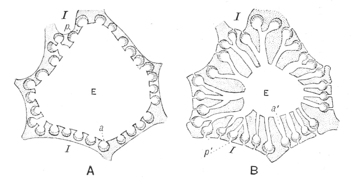
Fig. 104.—Diagram of (A) eurypylous and (B) aphodal canal systems. a, Apopyle; a', aphodus; E, excurrent canal; I, incurrent canal; p, prosopyle; p', short prosodus. (After Sollas.)
Cortex.—All the stages in the formation of a cortex are to be seen among the adult members of the group. Certain species (e.g. Plakina monolopha, F.E.S.) are destitute even of an ectosome, others have a simple dermal membrane (Halichondria panicea, Tetilla pedifera) and various others are provided with a cortex, either of simple structure or showing elaboration in one or more particulars. Thus a protective armature of special spicules may be present in the cortex, e.g. in Geodia, or to a less extent in Tethya, or there may be an abundance of contractile elements, and these may be arranged in very definite ways, forming valve-like apparatus that will respond to stimuli.
Everywhere among sponges the goal of the skeleton appears to have been coherence. We have seen how in Calcarea and in Hexactinellida this has been attained by the secretion around the separate elements of a continuous mineral sheath, calcareous in the one case and siliceous in the other. Here we had an excellent instance of the attainment of one end by similar means in two different groups, after their separation from the common stock, and therefore independently. In Demospongiae, on the other hand, the same end—coherence—has been secured by two new methods, each distinct from the former: first the spicules may be united in strands by an organic deposit, spongin; secondly, the spicules may assume irregular shapes and interlock closely with one another, forming dense and stout skeletons. The latter method is that characteristic of the Lithistid Tetractinellida.
Classification.—It is not of great moment which scheme of classification we maintain, seeing that all hitherto proposed are confessedly more or less artificial, and sufficient data for framing a natural one are not yet forthcoming. For convenience, we accept three subdivisions and define them thus:—
I. Tetractinellida.—Demospongiae possessing tetraxon or triaene spicules or Lithistid desmas.
II. Monaxonida.—Demospongiae possessing monaxon but not tetraxon spicules.
III. Ceratosa.—Demospongiae in which the main skeleton is formed of fibres of spongin. The fibres may have a core of sand-grains or of foreign spicules, but not of spicules proper to the sponge.
But at the same time we admit that some of the Ceratosa are probably descended from some of the families of Monaxonida, so that we should perhaps be justified in separating these families of Monaxonida from the rest, and associating them with the allied families of Ceratosa—a method of classification due to Vosmaer. Again, some Monaxonida approximate to Tetractinellida, and we might, with Vosmaer, unite them under the title Spiculispongiae. This proceeding, though it has the advantage of being at least an attempt to secure a natural classification, involves too much assumption when carried out in detail to be wholly satisfactory.
Sub-Class I. Tetractinellida.[239]
Tetractinellida appear to flourish best in moderate depths from 50 to 200 fathoms, but they are found to be fairly abundant also in shallower water right up to the coast line, and in deep water up to and beyond the 1000 fathom line. Occasionally they lie free on the bottom, but are far more commonly attached; fixation may be direct or by means of rooting spicules; the occurrence of a stalk is rare. There is great variety in the root tuft, which may be a long loose wisp of grapnel-headed spicules, as in many species of Tetilla, or a massive tangle, as in Cinachyra barbata; in these cases the sponge is merely anchored, so that it rests at the level of the surface of the ooze; in other cases, e.g. Thenea wyvillei, the root tuft consists of a number of pillars of spicules which raise the sponge above the level of the ooze, into which they descend and there become continuous with a large dense and confused mass of spicules. The parachute-like base of Tetilla casula invites comparison with the "Crinorhiza" forms of some Monaxonids (p. 216).
Two Orders are distinguished thus:—
I. Choristida.—Tetractinellida with quadriradiate spicules, which are never articulated together into a rigid network.
II. Lithistida.—Tetractinellida with branching scleres (desmas), which may or may not be modified tetrad spicules, articulated together to form a rigid network. Triaene spicules may or may not be present in addition.
Order I. Choristida.
Plakina monolopha, from the Adriatic and Mediterranean, furnishes a connecting link between the Rhagon stage and other Tetractinellida. The choanosome is simply folded; there is no distinct ectosome; the chambers are eurypylous. The skeleton consists of microcalthrops and their derivatives. The hypophare is well developed. Plakina thus shows a certain amount of resemblance to Oscarella (p. 196), with which it shares the very remarkable possession of flagellated pinacocytes.
One of the species of Tetilla, T. pedifera, continues the series. The folds of its choanosome are more complicated than in P. monolopha, and their outer ends are bridged together by a thin layer of ectosome (cf. species of Sycon among Calcarea); the chambers are still eurypylous.
The skeleton reaches a high level: it includes oxeas and triaenes radiately disposed and microscleres (sigmata) scattered throughout the dermal layer. The British Poecillastra compressa from the north of Scotland and Orkney and Shetland is at about the same stage of development, being without cortex and having eurypylous chambers, but it is not so good an example, as the folds of its choanosome are confused.
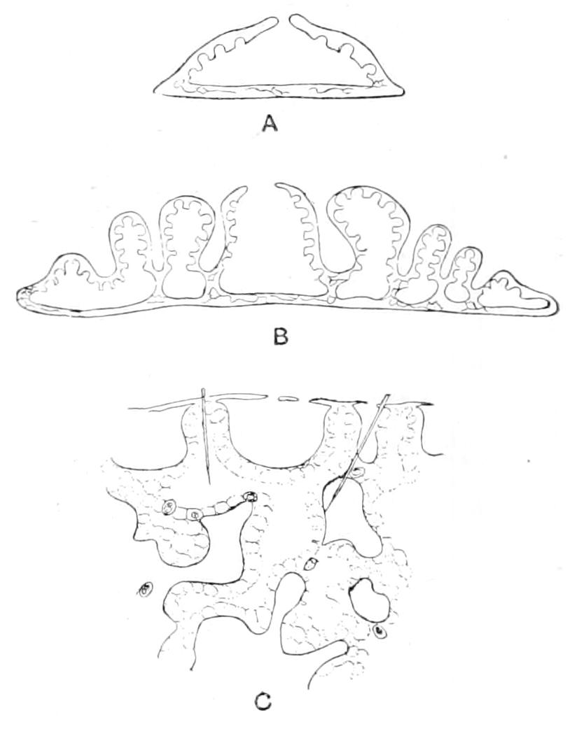
Fig. 105.—Diagrammatic vertical sections of A, Rhagon; B, Plakina; C, Tetilla pedifera.
From T. pedifera we pass to the other species of Tetilla and all the higher genera of Choristida; these possess a cortex not of homologous origin in the various cases, but probably to be classified under one of two heads, typified by Stelletta and Craniella respectively (Fig. 106).
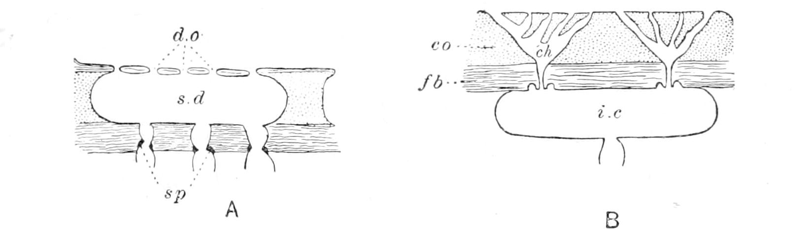
Fig. 106.—A, Craniella type; B, Stellettid type. ch, Chone; co, collenchyma; d.o, dermal ostia; fb, fibrous tissue; i.c, intercortical cavity; sd, subdermal cavity; sp, sphincter. (After Sollas.)
In the Stellettids the cortex arises by the centrifugal growth of a dermal membrane such as that of Tetilla pedifera; in Craniella directly from the dermal tissue of the distal ends of the choanosomal folds.
In both cases the end result, after completion of cell differentiation, is a cortex either fibrous throughout or collenchymatous in its outer portion and fibrous in the deeper layers. In the Stellettid type the centrifugal growth of the dermal membrane involves the addition of secondary distal portions to the ends of the inhalant passages. These are the intercortical cavities or canals. Their most specialised form is the "chone." A chone is a passage through the cortex opening to the exterior by one or more ostia, and communicating with the deeper parts of the inhalant system by a single aperture provided with a sphincter (Fig. 106, B).
In the Craniella type the intercortical cavities are parts of the primary inhalant system. They communicate with its deeper parts by sphinctrate apertures. Without any knowledge of the development one would certainly have supposed that the subdermal cavity, pore-sieve and sphinctrate passages of Craniella represented a number of chones, of which the outer portions had become fused (Fig. 106, A).
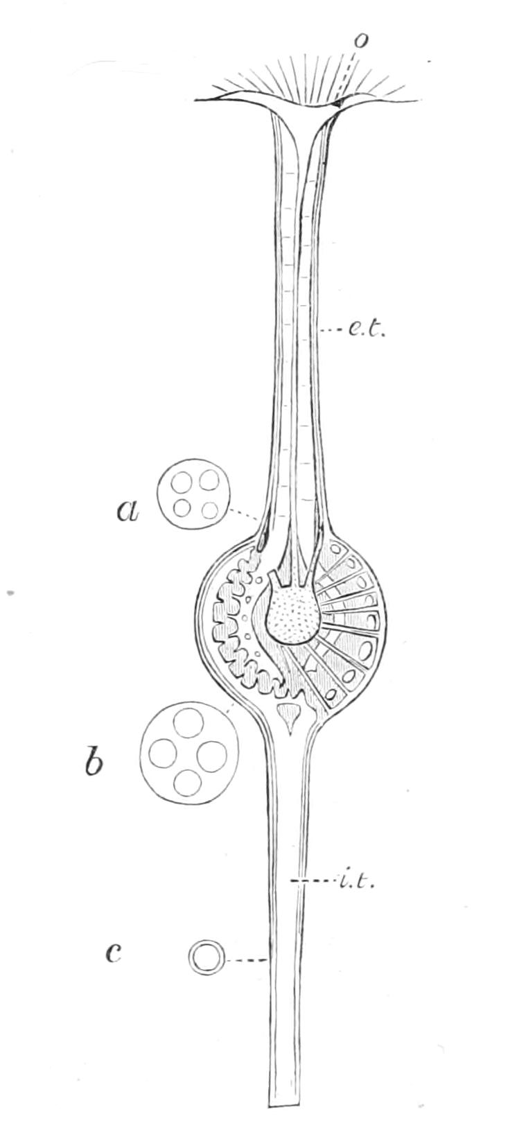
Fig. 107.—Disyringa dissimilis. Diagrammatic longitudinal section of the Sponge. × ½. a, b, c, Transverse sections at the levels indicated to show subdivision of the lumina of the excurrent and incurrent tubes; e.t, excurrent tube; i.t, incurrent tube; o, osculum. (After Sollas.)
In both Craniella and Stelletta the chamber system is aphodal, and these genera may fairly be taken as representatives of the average level reached by Tetractinellida. The skeleton is of the radiate type: the type which prevails in the Choristida, but which has an erratic distribution, appearing in some genera of each family but not in others. The genus Pachymatisma, of which we have the species P. johnstonia and P. normani in these islands, exemplifies this; it belongs to the highly differentiated family Geodiidae, possesses an elaborate cortex with chones, but its main skeleton is non-radiate.
Disyringa dissimilis is remarkable for the perfection of its symmetry, and for the absence of that multiplication of parts which is so common among sponges. It possesses a single inhalant tube and a single osculum (Fig. 107). Until quite recently it stood alone in the restriction of its inhalant apertures to a single area. Kirkpatrick, however, has now described a sponge—Spongocardium gilchristi[240]—from Cape Colony, in which the dermal ostia are concentrated in one sieve-like patch at the opposite pole to the single osculum. Disyringa is still without companions in the possession of an inhalant tube. The concentration of ostia into sieve areas occurs again in Cinachyra, each sponge possessing in this case several inhalant areas with or without scattered ostia also.
Order II. Lithistida.
The characteristic spicule of Lithistida—the desma—may be a modified calthrop (tetracrepid desma), or it may be produced by the growth of silica over a uniaxial spicule (rhabdocrepid desma) (Fig. 110, q), or it may be of the polyaxon type. It is probable that the group is polyphyletic,[241] and that some of its members should remain associated with Tetractinellida, while others should be removed to Monaxonida. Forms with tetracrepid desmas, and those forms with rhabdocrepid desmas which possess triaenes, have Tetractinellid affinities, while forms possessing rhabdocrepid desmas but lacking triaenes, and again those in which the desmas are polyaxon, are probably descendants of Monaxonida.
Owing to the consistency of the skeleton Lithistida are frequently found as fossils. The commonest known example is Siphonia.[242] As in the case of so many other fossil sponges the skeleton is often replaced by carbonate of lime, a fact which misled some of the earlier investigators but was established by the researches of Sollas and Zittel.
Sub-Class II. Monaxonida.[243]
The Monaxonida inhabit for the most part shallow water, but they also extend through deep water into the abysses, thirteen species having been dredged from depths of over 2000 fathoms by the "Challenger" Expedition alone. In some cases, e.g. Cladorhiza, Chondrocladia, all the species of a genus may live in deep water, while in others the genus, or in others, again, the species, may have a wide bathymetrical range. Thus Axinella spp. occur in shallow water and in various depths down to 2385 fathoms, Axinella erecta ranges from 90 to 1600 fathoms, Stylocordyla stipitata from 7 to 1600, and so on. The symmetry of the deep-water forms contrasts strikingly with the more irregular shape of their shallow-water allies.[244] The shallow-water species are almost always directly attached, some few are stalked; those from deep water have either a long stalk or some special device to save them from sinking in the soft ooze or mud. Thus the deep-sea genus Trichostemma has the form of a low inverted cone, round the base of which a long marginal fringe of spicules projects, continuing the direction of the somal spicules, and so forming a supporting rim. The same form has been independently evolved in Halicnemia patera, and an approach to it in Xenospongia patelliformis. A similar and more striking case of homoplasy is afforded by the Crinorhiza form, which has been attained in certain species of the deep-sea genera Chondrocladia, Axoniderma, and Cladorhiza; here the sub-globular body is supported by a vertical axis or root, and by a whorl of stout processes radiating outwards and downwards from it, and formed of spicular bundles together with some soft tissue.
There is recognisable in the order Monaxonida a cleft between one set of genera, typically corticate, and suggesting by their structure a relationship, whether of descent or parentage, with the Tetractinellida, and a second set typically non-corticate: these latter are the Halichondrina, the former are the Spintharophora.
Order I. Halichondrina.
We have already seen typical examples of the Halichondrina in Halichondria panicea and Ephydatia fluviatilis. Within the Halichondrina the development of spongin reaches its maximum among spiculiferous sponges, and accordingly the Ceratosa take their multiple origin here (p. 220). Among Halichondrina spongin co-operates with spicules to form a skeleton in various ways, but always so as to leave some spicules bare or free in the flesh. It may bind the spicules end to end in delicate networks (as in Reniera or Gellius), or into strands, sometimes reaching a considerable thickness (as in Chalina and others). In a few cases there appears to be a kind of division of labour between the spicules and spongin, the latter forming the bulk of the fibre, i.e. fulfilling the functions of support, while the spicules merely beset its surface as defensive organs, rendering the sponge unfit for food. Fibres formed on this pattern are called plumose, and are typical of Axinellidae. The distinctive fibre of the Ectyoninae is as it were a combination of the Axinellid and Chalinine types: a horny fibre both cored with spicules and beset with them. Spicules besetting the surface of a fibre are termed "echinating." Whenever its origin has been investigated, spongin has proved to be the product of secretion of cells; in the great majority of cases it is poured out at the surface of the cell, and Evans showed,[245] at any rate in one species of Spongilla, that the spongin fibres are continuous with a delicate cuticle at the surface of the sponge. In Reniera spp. occurs a curious case of formation of spongin as an intracellular secretion. A number of spherical cells each secrete within themselves a short length of fibre; they then place themselves in rows, so orientated that their contained rods lie end to end in one line. The rods then fuse and make up continuous threads; the cells diminish in breadth, ultimately leaving the fibre free.[246]
Order II. Spintharophora.
These corticate forms are further characterised by the arrangement of their megascleres, which is usually, like that of most Tetractinellida, radial, or approximating to radial. The microscleres are, when present, some form of aster. The cortex resembles that of Tetractinellida, and v. Lendenfeld has described chones in Tethya lyncurium.[247]
The existence of the above points of resemblance between Spintharophora and Tetractinellida suggests a relationship between the two groups as its cause. In judging this possibility the following reflections occur to us. A cortex exists in various independent branches of Tetractinellida. It has in all probability had a different phylogenetic history in each—why not then in these Monaxonida also? Within single genera of Tetractinellida some species are corticate, others not, witness Tetilla. The value of a cortex for purposes of classification may easily be overestimated. If we are to uphold the relationship between these two groups, we must base our argument on the conjunction of similar characters in each.
The genus Proteleia[248] is interesting for its slender grapnel-like spicules, which project beyond the radially disposed cortical spicules, and simulate true anatriaenes of minute proportions. That they are not anatriaenes is shown by the absence of an axial thread in their cladi. It is not surprising that a form of spicule of such obvious utility as the anatriaene should arise more than once.
Of exceptional interest, on account of their boring habit, are the Clionidae. How the process of boring is effected is not known; the presence of an acid in the tissues was suspected, but has been searched for in vain. The pieces of hard substance removed by the activity of the sponge take their exit through the osculum and have a fixed shape[249] (Fig. 108).
As borers into oyster shells, Clionidae may be reckoned as pests of practical importance, and in some coasts they even devastate the rocks, penetrating to a depth of some feet, and causing them to crumble away.[250]
Sponges, however, as agents in altering the face of the earth do not figure as destroyers merely. On the contrary, it has been calculated[251] that sponge skeletons may give rise with considerable rapidity to beds of flint nodules; in fact, it appears that a period so short as fifty years is sufficient for the formation of a bed of flints out of the skeletons of sponges alone.
Suberites domuncula is well known for its constant symbiosis with the Hermit crab. The young sponge settles on a Whelk or other shell inhabited by a Pagurus, and gradually envelops it, becoming very massive, and completely concealing the shell, without however closing its mouth. The aperture of this always remains open to the exterior, however great the growth of the sponge, a tubular passage being left in front of it, which continues the lumen of the shell and maintains its spiral direction. When the crab has grown too big for the shell, it merely advances a little down this passage. The shell is never absorbed, as was once supposed.[252] The crab, besides being provided with a continually growing house, and being thus spared the great dangers attending a shift of lodgings, benefits continually by the concealment and protection afforded by the massive sponge; the latter in return is conveyed to new places by the crab.
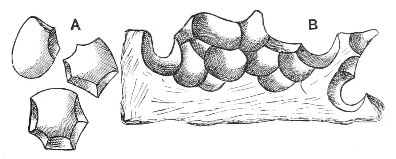
Fig. 108.—A, calcareous corpuscle detached by Cliona; B, view of the galleries excavated by the Sponge. (After Topsent.)
Ficulina ficus is sometimes, like S. domuncula, found in symbiosis with Pagurus, but the constancy of the association is wanting in this case. The sponge has several metamps, one of which, from its fig-like shape, gives it its name.
Sub-Class III. Ceratosa.
The Ceratosa are an assemblage of ultimate twigs shorn from the branches of the Monaxonid tree. They are therefore related forms, but many of them are more closely connected with their Monaxonid relatives than with their associates in their own sub-class.
The genera Aulena and Phoriospongia, placed by v. Lendenfeld among Ceratosa, by Minchin among Monaxonida, show each in its own way how close is the link between these two sub-classes.
Aulena possesses in its deeper parts a skeleton of areniferous spongin fibres, in fact a typical Ceratose skeleton; but this is continuous with a skeleton in the more superficial parts, which is composed of spongin fibres echinated by spicules proper to the sponge, and precisely comparable to the ectyonine fibres of some Monaxonida.
Phoriospongia, as far as its main skeleton is concerned, is a typical Ceratose sponge, with fibres of the areniferous type, but it possesses sigmata free in the flesh.
The sub-class is confined to shallow water, no horny sponge having been dredged from depths greater than 410 fathoms.[253] The greatest number occur at depths between 10 and 26 fathoms.
In the majority of the Ceratosa the skeletal fibres are homogeneous, formed of concentric lamellae of spongin, deposited by a sheath of spongoblasts around a filiform axis. In others, however, the axis attains a considerable diameter, so as to form a kind of pith to the fibre, which is then distinguished as heterogeneous. In one or two cases some of the spongoblasts of a heterogeneous fibre are included in the fibre between the spongin lamellae. Ianthella is the best-known example in which this occurs.
Ceratosa are divided into Dictyoceratina and Dendroceratina, distinguished, as their names express, by the nature of the skeleton—net-like, with many anastomoses, in the one; tree-like, without anastomoses between its branches, in the other.
The Dictyoceratina comprise by far the larger number of Ceratosa. They fall into two main families, the Spongidae and Spongelidae, both represented in British waters. The Spongidae are characterised by a granular ground substance and aphodal chamber system; the Spongelidae by a clear ground substance and sac-like eurypylous chambers.
The bath sponge, Euspongia officinalis, belongs to the Spongidae. The finest varieties come from the Adriatic, the coarser ones from the Dalmatian and North African coasts of the Mediterranean, from the Grecian Archipelago, from the West Indies, and from Australian seas. The softer species of the genus Hippospongia also form a source of somewhat inferior bath sponges.
Among Dendroceratina, Darwinella is unique and tempts to speculation, in that it possesses isolated spongin elements, resembling in their forms triaxon spicules.
Key to British Genera of Sponges.
1. (a) Skeleton calcareous 2
(b) Skeleton siliceous 6
(c) Skeleton horny, or without free spicules 53
(d) Skeleton absent 55
2. (a) Gastral layer continuous 3
(b) Gastral layer discontinuous, confined to chambers 4
3. (a) Equiangular triradiate systems present Clathrina
(b) Triradiate systems all alate Leucosolenia
4. (a) Chambers tubular, radially arranged 5
(b) Chambers spherical, irregularly scattered Leucandra
5. (a) Tufts of oxeate spicules at the ends of the chambers Sycon
(b) Oxeate spicules lying longitudinally in the cortex Ute
6. (a) All the spicules hexradiate or spicules easily derived from hexradiate type 7
(b) Some of the spicules calthrops or triaenes 10
(c) Megascleres uniaxial 15
7. (a) Amphidiscs present 8
(b) Amphidiscs absent 9
8. (a) Rooting spicules a well-defined wisp; four apertures lead into the gastric cavity Hyalonema thomsoni
(b) Rooting tuft diffuse; sponge oval; osculum single Pheronema carpenteri
9. (a) Sponge tubular, dermal and gastral pinuli absent Euplectella suberea
(b) Sponge a widely open cup; dermal and gastral pinuli present Asconema setubalense
10. (a) Tetractine spicule, a calthrop or triaene with short rhabdome; microsclere a spined microxea Dercitus bucklandi
(b) Triaenes with fully developed rhabdome 11
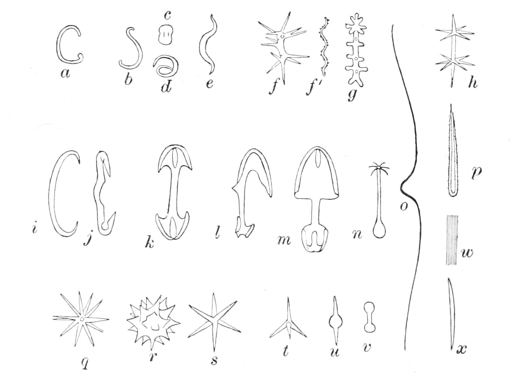
Fig. 109.—Microscleres of Demospongiae. a, b, Sigmaspires viewed in different directions; c, d, bipocilli viewed in different directions; e, toxaspire; f, f', spiraster; g, sanidaster; h, amphiaster; i, sigma; j, diaucistra; k, isochela; l, m, anisochelae viewed in different directions; n, cladotyle; o, toxa; p, forceps; q, oxyaster; r, spheraster; s, oxyaster with 6 actines; t, another with 4 actines; u, another with rays reduced to two (centrotylote microxea); v, tylote microrhabdus; w, trichodragmata; x, oxeate microrhabdus or microxea.
11. (a) Microscleres sigmata Craniella cranium
(b) Sigmata absent, asters present 12
12. (a) Microscleres include spirasters Poecillastra compressa
(b) Microscleres include sterrasters 14
(c) Microscleres include euasters: spirasters and sterrasters absent 13
13. (a) Two kinds of euaster present Stelletta
(b) Microscleres include a euaster and a sanidaster or amphiaster Stryphnus ponderosus
14. (a) Microscleres include microrhabdi Pachymatisma johnstonia
(b) Microscleres include many-rayed euasters Cydonium milleri
15. (a) Some of the microscleres asters 16
(b) Microscleres absent, or not asters 17
16. (a) Skeleton radiate; asters of more than one kind Tethya
(b) Sponge encrusting; asters of one kind only Hymedesmia
(c) Skeleton fibrous Axinella spp.
17. (a) Megascleres all diactinal; chelae present Desmacidon
(b) Megascleres all diactinal; chelae absent 18
(c) Some or all of the megascleres monactinal 19
18. (a) Habitat fresh water 56
(b) Habitat marine 22
19. (a) Megascleres include cladotyles Acarnus
(b) Megascleres include dumb-bell or sausage-shaped spicules forming the main reticulum Plocamia
(c) Microscleres include bipocilli 20
(d) Microscleres include diancistra Hamacantha
(e) Megascleres include forceps Forcepia
(f) Skeleton formed of isolated monactines vertically placed Hymeraphia
(g) None of the above peculiarities present 21
20. (a) Skeleton fibre not echinated Iophon
(b) Skeleton fibre echinated Pocillon
21. (a) Skeleton with echinating spicules 28
(b) Skeleton without echinating spicules 30
22. (a) Spongin abundant 23
(b) Spongin scanty 25
23. (a) Fibre not echinated 24
(b) Fibre echinated Diplodemia
24. (a) Fibre with a single axial series of spicules Chalina
(b) Fibres with numerous spicules arranged polyserially Pachychalina
25. (a) Microscleres absent 26
(b) Microscleres sigmata and/or toxa 27
26. (a) Skeleton confused Halichondria
(b) Skeleton reticulate Reniera
27. (a) Rind and fistulous appendages present; microscleres sigmata Oceanapia
(b) No rind; skeleton reticulate; microscleres sigmata and/or toxa Gellius
28. (a) Skeleton confused or formed of bundles of spicules with echinating spined styles 29
(b) Skeleton fibrous or reticulate, or formed of short columns 45
(c) Skeleton formed of a dense central axis, and columns radiating from it to the surface 52
29. (a) Spicules of the ectosome styles Pytheas
(b) Spicules of the ectosome oxeas or absent Clathrissa
(c) Main skeleton confused. Special ectosomal skeleton absent Spanioplon
30. (a) Megascleres of the choanosome not differing from those of the ectosome 31
(b) Megascleres of the choanosome differing from those of the ectosome 32
31. (a) Chelae absent. 33
(b) Chelae present 44
32. (a) Trichodragmata present Tedania
(b) Trichodragmata absent 42
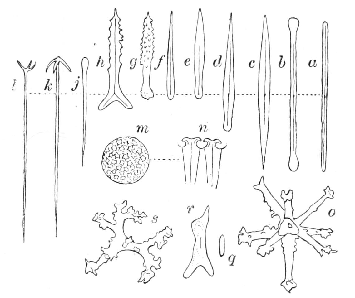
Fig. 110.—Megascleres. a-l and q-s, Modifications of monaxon type. a, Strongyle; b, tylote; c, oxea; d, tylotoxea; e, tylostyle; f, style; g, spined tylostyle; h, sagittal triod (a triaxon form derived from monaxon); j, oxytylote; k, anatriaene; l, protriaene; m, sterraster (polyaxon); n, radial section through the outer part of m, showing two actines soldered together by intervening silica; o, desma of an Anomocladine Lithistid (polyaxon); q, crepidial strongyle, basis of rhabdocrepid Lithistid desma; r, young form of rhabdocrepid desma, showing crepidial strongyle coated with successive layers of silica; s, rhabdocrepid desma.
33. (a) Skeleton reticulate or fibrous 34
(b) Skeleton radiate or diffuse 37
(c) Skeleton with radiating fibres forming a reticulum with others crossing them at right angles Quasillina
34. (a) No microscleres 35
(b) Microscleres sigmata and/or toxa with or without trichodragmata Desmacella
35. (a) Sponge fan- or funnel-shaped 36
(b) Sponge not fan- or funnel-shaped Hymeniacidon
36. (a) Megascleres slender and twisted Phakellia
(b) Megascleres somewhat stout, not twisted Tragosia
37. (a) Sigmata present, skeleton diffuse Biemma
(b) Sigmata absent 38
38. (a) Skeleton more or less radiate 39
(b) Skeleton diffuse; sponge boring Cliona
39. (a) Sponge discoid with marginal fringe Halicnemia
(b) Sponge massive or stipitate without marginal fringe 40
40. (a) Sponge body prolonged into mammiform projections Polymastia
(b) Sponge body without mammiform projections 41
41. (a) No microscleres. Megascleres tylostyles with or without styles Suberites
(b) Microscleres centrotylote. Megascleres styles or tylostyles Ficulina
42. (a) Choanosomal megascleres smooth 43
(b) Choanosomal megascleres spined Dendoryx
43. (a) Microscleres chelae and sigmata of about the same size Lissodendoryx
(b) Chelae, if present, smaller than the sigmata Yvesia
44. (a) Isochelae Esperiopsis
(b) Anisochelae Esperella
45. (a) Fibres or columns plumose 46
(b) Fibres or columns ectyonine 47
46. (a) Microscleres toxa Ophlitaspongia
(b) Microscleres absent Axinella
47. (a) Skeleton reticulate 48
(b) Skeleton not reticulate 49
48. (a) Microscleres present. Spicules of the fibre core spined Myxilla
(b) Microscleres absent. Spicules of the fibre core smooth Lissomyxilla
49. (a) Main skeleton formed of plume-like columns 50
(b) Main skeleton formed of horny fibres (ectyonine). Special dermal skeleton wanting Clathria
50. (a) Dermal skeleton contains styles only Microciona
(b) Dermal skeleton contains diactine spicules with or without styli 51
51. (a) Main skeleton columns with a core of smooth oxeas Plumohalichondria
(b) Main skeleton columns with a core of spined styles Stylostichon
52. (a) Central axis contains much spongin. Echinating spined styli present Raspailia
(b) Central axis with little or no spongin. Spined styles absent. Pillars radiating from the axis support dermal skeleton Ciocalypta
53. (a) Ground substance between chambers clear; chambers pear-shaped or oval; eurypylous Spongelia
(b) Ground substance granular. Chambers spherical with aphodi 54
54. (a) Fibres not pithed; sponge fan-shaped Leiosella
(b) Fibres pithed; sponge massive Aplysina
55. (a) Chambers long, tubular, branched Halisarca
(b) Chambers not much longer than broad; not branched Oscarella
56. (a) Amphidiscs present Ephydatia
(b) Amphidiscs absent Spongilla