Chapter VII
Reading edition
PORIFERA (SPONGES)[185]
INTRODUCTION—HISTORY—DESCRIPTION OF HALICHONDRIA PANICEA AS AN EXAMPLE OF BRITISH MARINE SPONGES AND OF EPHYDATIA FLUVIATILIS FROM FRESH WATER—DEFINITION—POSITION IN THE ANIMAL KINGDOM.
Sponges occupy, perhaps, a more isolated position than any other animal phylum. They are not only the lowest group of multicellular animals, but they are destitute of multicellular relatives. They are all aquatic and—with the exclusion of a few genera found in fresh water—marine, inhabiting all depths from between tide marks to the great abysses of the ocean. They depend for their existence on a current of water which is caused to circulate through their bodies by the activity of certain flagellated cells. This current contains their food, it is their means of respiration, and it carries away effete matters. Consequently sponges cannot endure deprivation of oxygenated water except for short periods, and only the hardiest inhabit regions where the supply is intermittent, as between tide marks. This also renders useless attempts to keep specimens in tanks, unless the water is frequently renewed.
The outward appearance of sponges has an exceptionally wide range, so that it is difficult to give a novice any very definite picture of what he is to expect when searching for these animals. This diversity is in part due to the absence of organs of sufficient size to determine the shape of the whole or limit its variation, that is to say, the separate organs are of an order of size inferior to that of the entire body. The animals are fixed or lie loose on the sea bottom; there are in no case organs of locomotion, and again no sense-organs, no segregated organs of sex, and as a rule no distinction into axis and lateral members. It is by these negative characters that the collector may easily recognise a sponge.
History.—Sponges are, then, in many of their characters unique; and they present a variety of problems for solution, both of special and general interest, they are widely distributed in time and space, and they include a host of forms. It therefore causes no little surprise to learn that they have suffered from a long neglect, even their animal nature having been but recently established. Though known to naturalists from the time of Aristotle, sponges have been left for modern workers as a heritage of virgin soil: it has yielded to them a rich harvest, and is as yet far from exhausted.
The familiar bath sponge was naturally the earliest known member of the phylum. It is dignified by mention in the Iliad and in the Odyssey, and Homer, in his choice of the adjective "full of holes," πολύτρητος, shows at least as much observation as many a naturalist of the sixteenth and seventeenth centuries. Aristotle based his ideas of sponges entirely upon the characters of the bath sponge and its near allies, for these were the only kinds he knew. With his usual perspicuity he reached the conclusion that sponges are animals, though showing points of likeness to plants.
The accounts of sponges after Aristotle present little of scientific interest until the last century. Doubtless this is in part due to the absence of organs which would admit of dissection, and the consequent necessity of finer methods of study. Like other attached forms, sponges were plant or animal as it pleased the imagination of the writer, and sometimes they were "plant animals" or Zoophyta: those who thought them animal were frequently divided among themselves as to whether they were "polypous" or "apolypous." An opinion which it is somewhat difficult to classify was that of Dr. Nehemiah Grew,[186] who says: "No Sponge hath any Lignous Fibers, but is wholly composed of those which make the Pith and all the pithy parts of a Plant, ... So that a Sponge, instead of being a Zoophyton, is but the one-half of a Plant."
Sponges figure in herbals beside seaweeds and mushrooms, and Gerarde says:[187] "There is found growing upon rockes near unto the sea a certaine matter wrought together of the foame or froth of the sea which we call Spunges ... whereof to speak at any length would little benefit the reader ... seeing the use thereof is so well known." About the middle of the eighteenth century, authors, especially Peyssonnel, suggested that sponges were but the houses of worms, which built them much as a bee or wasp builds nests and cells. This was confuted by Ellis in 1765,[188] when he pointed out that the sponge could not be a dead structure, as it gave proof of life by "sucking and throwing out water." To Ellis, then, is due the credit of first describing, though imperfectly, a current set up by sponges. He mentions that Count Marsigli[189] had already made somewhat similar observations.
It was not till 1825 that attention was again turned to the current, when Robert Grant approached the group in a truly scientific manner, and was ably supported by Lieberkühn. It would be impossible to do justice to Grant in the brief summary to which we must limit ourselves. The most important of his contributions was the discovery that water enters the sponge by small apertures scattered over the surface, and leaves it at certain larger holes, always pursuing a fixed course. He made a few rough experiments to estimate the approximate strength of the current, and, though he failed to detect its cause, he supposed that it was probably due to ciliary action. Grant's suggestion was afterwards substantiated by Dujardin (1838), Carter (1847), Dobie (1852), and Lieberkühn (1857). These five succeeded in establishing the claims of sponges to a place in the animal kingdom, claims which were still further confirmed when James-Clark[190] detected the presence of the protoplasmic collar of the flagellated cells (see pp. 171, 176). Data were now wanted on which to base an opinion as to the position of sponges within the animal kingdom. In 1878 Schulze[191] furnished valuable embryological facts, in a description agreeing with an earlier one of Metschnikoff's, of the amphiblastula larva (p. 226) and its metamorphosis. Then Bütschli[192] (1884) and Sollas[193] on combined morphological and embryological evidence (1884) concluded that sponges were remote from all the Metazoa, showing bonds only with Choanoflagellate Protozoa (p. 121). This the exact embryological work of Maas, Minchin, and Delage has done much to prove, but it has to be admitted that unanimity on the exact position of the phylum has not yet been attained, some authorities, such as Haeckel, Schulze, and Maas still wishing to include sponges in the Metazoa.
In this short history we have been obliged to refer only to work helping directly to solve the problem of the nature of a sponge, hence many names are absent which we should have wished to mention.
Halichondria panicea.
One of the commonest of British sponges, which may be picked up on almost any of our beaches, and which has also a cosmopolitan distribution, is known by the clumsy popular name of the "crumb of bread sponge," alluding to its consistency; or by the above technical name, with which even more serious fault may be found.[194]
In its outward form H. panicea affords an excellent case of a peculiarity common among sponges. Its appearance varies according to the position in which it has lived. In fact, Bowerbank remarks that it has no specific form. It may grow in sheets of varying thickness closely attached to a rock, when it is "encrusting," or it is frequently massive and lying free on the sea bottom; again, it may be fistular, consisting of a single long tube, or it may be ridge-like, apparently in this case consisting of a row of long tubes fused laterally. In this last form it used to be called the "cockscomb sponge," having been taken for a distinct species.
Bidder has proposed to call the different forms of the same species "metamps" of the species. Figures of the metamps of H. panicea will be found in Bowerbank's useful Monograph.[195]
The colour of the species is as inconstant as its form, ranging from green to light brown and orange. MacMunn concludes from spectroscopic work that H. panicea, contains at least three pigments, a chlorophyll, a lipochrome, and a histohaematin.[196] Lipochromes vary from red to yellow, chlorophyll is always associated with one or more of them. Histohaematin is a respiratory pigment. Proof has not yet been adduced that the chlorophyll is proper to the sponge and is not contained in symbiotic algae.
In spite of all this inconstancy H. panicea, is one of the most easily determined species. It is only necessary to dry a small fragment, including the upper surface; a beautiful honeycomb-like structure is then visible on this surface, and among British sponges this is a property peculiar to the species (Bowerbank). Whatever the form of the sponge, one or more large rounded apertures are always present on the exterior; these are the "oscula." In the encrusting metamp the oscula are flush with the general surface, while in the other cases they are raised on conical projections; fistular specimens carry the osculum at the distal end, and the cockscomb has a row of them along its upper edge. Much more numerous than the oscula are smaller apertures scattered over the general surface of the sponge, and known as "ostia."
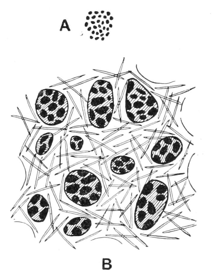
Fig. 63.—Portion of the surface of H. panicea, from dried specimen. A, natural size; B, magnified. The large shaded patches are ostia.
If the sponge be placed in a shallow glass dish of sea water the function of the orifices can be made out with the naked eye, especially if a little powdered chalk or carmine be added to the water. If the specimen has been gathered after the retreating tide has left it exposed for some time, this addition is unnecessary, for as soon as it is plunged into water its current bursts vigorously forth, and is rendered visible by the particles of detritus that have accumulated in the interior during the period of exposure and consequent suspended activity. The oscula then serve for the exit of currents of water carrying particles of solid matter, while the entrance of water is effected through the ostia.
Sections show that the ostia lead into spaces below the thin superficial layer or "dermal membrane"; these are continued down into the deeper parts of the sponge as the "incurrent canals," irregular winding passages of lumen continually diminishing as they descend. They all sooner or later open by numerous small pores—"prosopyles"—into certain subspherical sacs termed flagellated chambers. Each chamber discharges by one wide aperture—"apopyle"—into an "excurrent canal." This latter is only distinguishable from an incurrent canal by the difference in its mode of communication with the chambers.
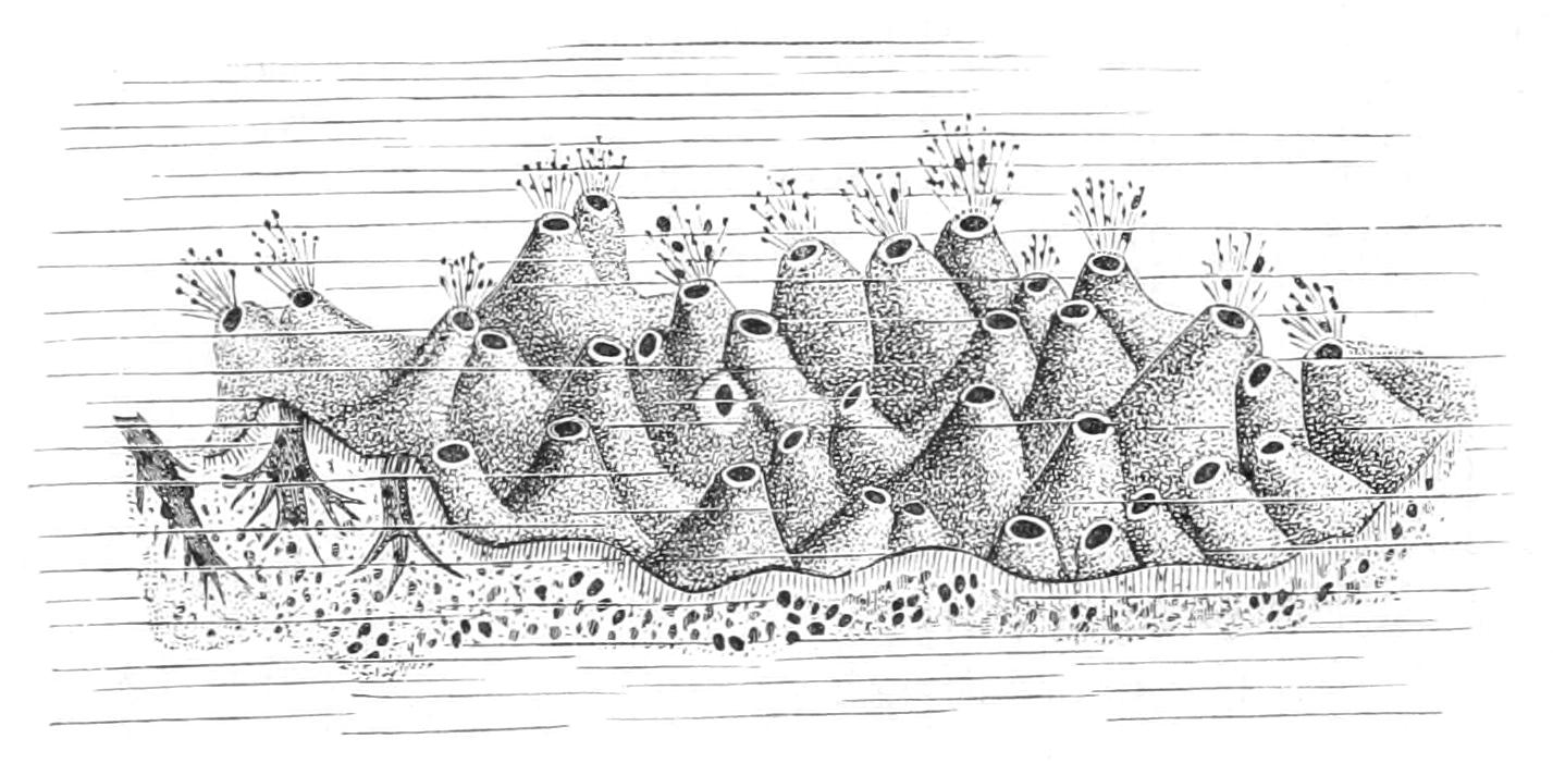
Fig. 64.—H. panicea: the arrows indicate the direction of the current, which is made visible by coloured particles. (After Grant.)
The excurrent canals convey to the osculum the water which has passed through the ostia and chambers. All the peripheral parts of the sponge from which chambers are absent are termed the "ectosome," while the chamber-bearing regions are the "choanosome."
The peculiar crumb-of-bread consistency is due to the nature of the skeleton, which is formed of irregular bundles and strands of minute needles or spicules composed of silica hydrate, a substance familiar to us in another form as opal: they are clear and transparent like glass. They are scattered through the tissues in great abundance.
The classes of cellular elements in the sponge are as follows: Flattened cells termed "pinacocytes" cover all the free surfaces, that is to say, the external surface and the walls of the excurrent and incurrent canals. The flagellated chambers are lined by "choanocytes" (cf. Fig. 70, p. 176); these are cells provided at their inner end with a flagellum and a collar surrounding it. They resemble individuals of the Protozoan sub-class Choanoflagellata, and the likeness is the more remarkable because no other organisms are known to possess such cells. Taken together the choanocytes constitute the "gastral layer," and they are the active elements in producing the current. The tissue surrounding the chambers thus lying between the excurrent and incurrent canals consists of a gelatinous matrix colonised by cells drawn from two distinct sources. In the first place, it contains cells which have a common origin with the pinacocytes, and which together with them make up the "dermal layer"; these are the "collencytes" and "scleroblasts"; secondly, it contains "archaeocytes," cells of independent origin.
Collencytes are cells with clear protoplasm and thread-like pseudopodial processes; they are distinguished as stellate or bipolar, according as these processes are many or only two. Scleroblasts or spicule cells are at first rounded, but become elongated with the growth of the spicule they secrete, and when fully grown are consequently fusiform.
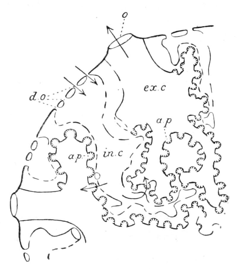
Fig. 65.—Diagrammatic section of a siliceous Sponge. a.p, Apopyle; d.o, dermal ostia; ex.c, excurrent, or exhalant canal; in.c, incurrent canal; o, osculum. (Modified from Wilson.)
Each spicule consists of an organic filamentar axis or axial fibre around which sheaths of silica hydrate are deposited successively by the scleroblast. Over the greater length of the spicule the sheaths are cylindrical, but at each end they taper to a point. The axial canal in which the axial fibre lies is open at both ends, and the fibre is continuous at these two points with an organic sheath, which invests the entire spicule. From this structure we may conclude that the spicule grows at both ends—i.e. it grows in two opposite directions along one line—it has two rays lying in one axis, and is classed among uniaxial diactinal spicules. Being pointed at both ends it receives the special name oxea. The lamination of the spicule is rendered much more distinct by heating or treatment with caustic potash.[197]
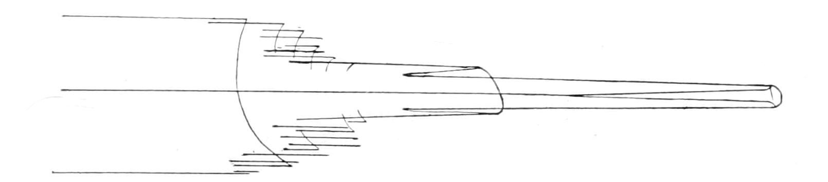
Fig. 66.—Cut end of a length of a siliceous spicule from Hyalonema sieboldii, with the lamellar structure revealed by solution. × 104. (After Sollas.)
The archaeocytes are rounded amoeboid cells early set apart in the larva; they are practically undifferentiated blastomeres. Some of them become reproductive elements, and thus afford a good instance of "continuity of germ plasm," others probably perform excretory functions.[198]
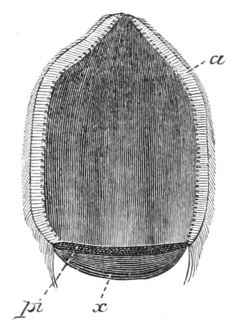
Fig. 67.—Free-swimming larva of Gellius varius, in optical section. a, Outer epithelium; pi, pigment; x, hinder pole. (After Maas.)
The reproductive elements are ova and spermatozoa, and are to be found in all stages in the dermal jelly. Dendy states that the eggs are fertilised in the inhalant canals, to which position they migrate by amoeboid movements, and there become suspended by a peduncle.
The larva has unfortunately not been described, but as the course of development among the near relatives of H. panicea is known to be fairly constant, it will be convenient to give a description of a "Halichondrine type" of larva based on Maas' account of the development of Gellius varius.[199] The free-swimming larvae escape by the osculum; they are minute oval bodies moving rapidly by means of a covering of cilia. The greater part of the body is a dazzling white, while the hinder pole is of a brown violet colour. This coloured patch is non-ciliate, the general covering of cilia ending at its edge in a ring of cilia twice the length of the others. Forward movement takes place in a screw line; when this ceases the larva rests on its hinder pole, and the cilia cause it to turn round on its axis.
Sections show that the larva is built up of two layers:—
1. "The inner mass," consisting of various kinds of cells in a gelatinous matrix.
2. A high flagellated epithelium, which entirely covers the larva with the exception of the hinder pole.
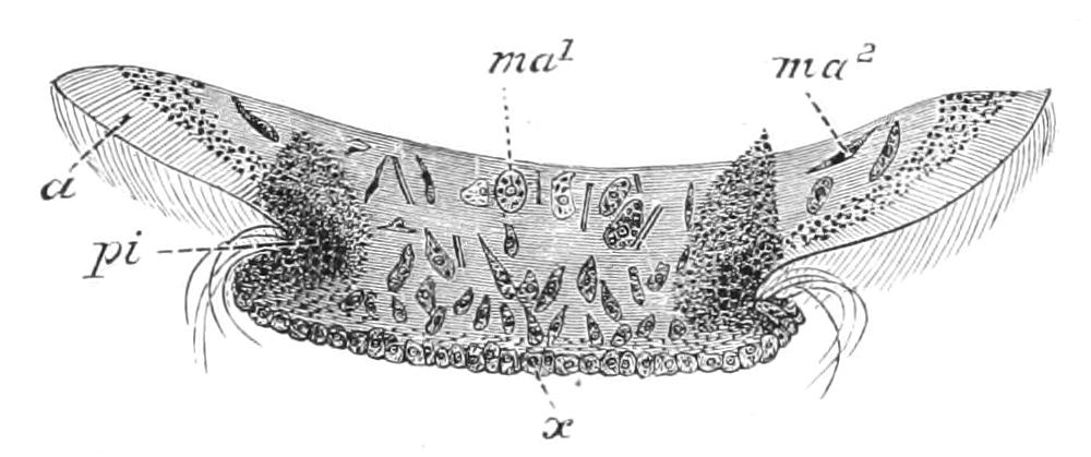
Fig. 68.—Longitudinal section through the hinder pole of the larva of G. varius. a, Flagellated cells; ma1, undifferentiated cell; ma2, differentiated cell; pi, pigment; x, surface of hinder pole. (After Maas.)
The cells in the inner mass are classified into (1) undifferentiated cells, recognised by their nucleus, which possesses a nucleolus; these are the archaeocytes; (2) differentiated cells, of which the nucleus contains a chromatin net; these give rise to pinacocytes, collencytes, and scleroblasts. Some of them form a flat epithelium, which covers the hinder pole. Some of the scleroblasts already contain spicules. Fixation occurs very early. The front pole is used for attachment, the pigmented pole becoming the distal end (Fig. 69). The larva flattens out, the margin of the attached end is produced into radiating pseudopodial processes. The flagellated cells retreat to the interior, leaving the inner mass exposed, and some of its cells thereupon form a flat outer epithelium. This is the most important process of the metamorphosis; it is followed by a pause in the outward changes, coinciding in time with rearrangements of the internal cells to give rise to the canal system; that is to say, lacunae arise in the inner mass, pinacocytes pass to the surface of the lacunae, and form their lining; the flagellated cells, which have lain in confusion, become grouped in small clusters. These become flagellated chambers, communications are established between the various portions of the canal system, and its external apertures arise. There is at first only one osculum. The larvae may be obtained by keeping the parent sponge in a dish of sea water, shielded from too bright a light, and surrounded by a second dish of water to keep the temperature constant. They will undergo metamorphosis in sea water which is constantly changed, and will live for some days.
We have said that the young sponge has only one osculum. This is the only organ which is present in unit number, and it is natural to ask whether perhaps the osculum may not be taken as a mark of the individual; whether the fistular specimens, for example, of H. panicea may not be solitary individuals, and the cockscomb and other forms colonies in which the individuals are merged to different degrees. Into the metaphysics of such a view we cannot enter here. We must be content to refer to the views of Huxley and of Spencer on Individuality.
But it is advisable to avoid speaking of a multi-osculate sponge as a colony of many individuals, even in the sense in which it is usual to speak of a colony of polyps as formed of individuals. The repetition of oscula is probably to be regarded as an example of the phenomenon of repetition of parts, the almost universal occurrence of which has been emphasised by Bateson.[200] Delage[201] has shown that when two sponge larvae fixed side by side fuse together, the resulting product has but one osculum. This, though seeming to bear out our point of view, loses weight in this connexion, when it is recalled that two Echinoderm larvae fused together give rise in a later stage to but one individual.
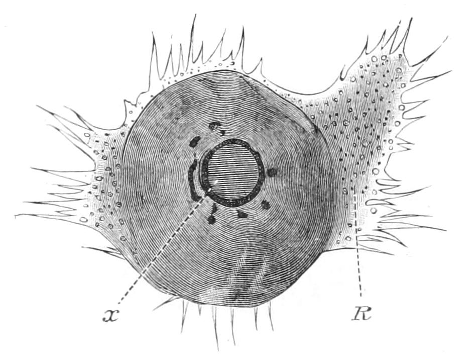
Fig. 69.—Larva of Gellius varius shortly after fixation. The pigmented pole, originally posterior, is turned towards the reader. R, Marginal membrane with pseudopodia; x, hinder pole. (After Maas.)
Ephydatia fluviatilis.
In the fresh water of our rivers, ponds, and lakes, sponges are represented very commonly by Ephydatia (Spongilla) fluviatilis, a cosmopolitan species. The search for specimens is most likely to be successful if perpendicular timbers such as lock-gates are examined, or the underside of floating logs or barges, or overhanging branches of trees which dip beneath the surface of the water.
The sponge is sessile and massive, seldom forming branches, and is often to be found in great luxuriance of growth, masses of many pounds weight having been taken off barges in the Thames. The colour ranges from flesh-tint to green, according to the exposure to light. This fact is dealt with in a most interesting paper by Professor Lankester,[202] who has shown not only that the green colour is due to the presence of chlorophyll, but that the colouring matter is contained in corpuscles similar to the chlorophyll corpuscles of green plants, and, further, that the flesh-coloured specimens contain colourless corpuscles, which, though differing in shape from those which contain the green pigment, are in all probability converted into these latter under the influence of sufficient light. The corpuscles, both green and colourless, are contained in amoeboid cells of the dermal layer;[203] and in the same cells but not in the corpuscles are to be found amyloid substances.
The anatomy of Ephydatia fluviatilis is very similar to that of Halichondria panicea, differing only in one or two points of importance. The ectosome is an aspiculous membrane of dermal tissue covering the whole exterior of the sponge and forming the roof of a continuous subdermal space. This dermal membrane is perforated by innumerable ostia, and is supported above the subdermal cavity by means of skeletal strands, which traverse the subdermal cavity and raise the dermal membrane into tent-like elevations, termed conuli. The inhalant canals which arise from the floor of the subdermal cavity are as irregular as in H. panicea, and interdigitate with equally irregular exhalant canals; these latter communicate with the oscular tubes. Between the two sets of canals are the thin folds of the choanosome with its small subspherical chambers provided with widely open apopyles (Fig. 70). The soft parts are supported on a siliceous skeleton of oxeas, which may have a quite smooth surface or may be covered in various degrees with minute conical spines (Fig. 72, a, b). These spicules are connected by means of a substance termed spongin deposited around their overlapping ends, so as to form an irregular network of strands, of which some may be distinguished as main strands or fibres, others as connecting fibres. In the main fibres several spicules lie side by side, while in the connecting fibres fewer or frequently single spicules form the thickness of the fibre. The fibres are continuous at the base with a plate or skin of spongin, which is secreted over the lower surface of the sponge and intervenes between it and the substratum. Of the chemical composition of spongin we shall speak later (see p. 237). It is a substance which reaches a great importance in some of the higher sponges, and forms the entire skeleton of certain kinds of bath sponge. Lying loose in the soft parts and hence termed flesh spicules, or microscleres, are minute spicules of peculiar form. These are the amphidiscs, consisting of a shaft with a many-rayed disc at each end (Fig. 72).
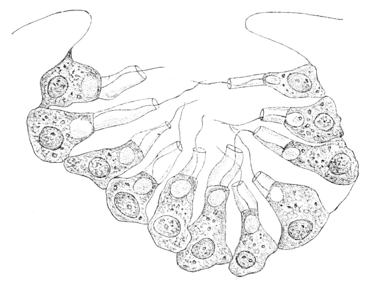
Fig. 70.—Ephydatia fluviatilis. Section of flagellated chamber, showing the choanocytes passing through the apopyle. (After Vosmaer and Pekelharing.)
In addition to its habitat the fresh-water sponge is worthy of attention on account of its methods of reproduction, which have arisen in adaptation to the habitat. A similar adaptation is widespread among fresh-water members of most aquatic invertebrates.[204]
Ephydatia fluviatilis normally produces not only free-swimming larvae of sexual origin, but also internal gemmules arising asexually. These bodies appear in autumn, distributed throughout the sponge, often more densely in the deeper layers, and they come into activity only after the death of the parent, an event which happens in this climate at the approach of winter.
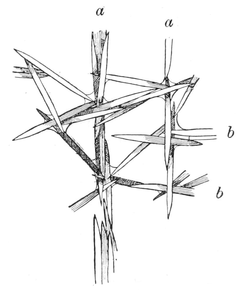
Fig. 71.—Portion of the skeletal framework of E. fluviatilis. a, Main fibres; b, connecting fibres. (After Weltner.)
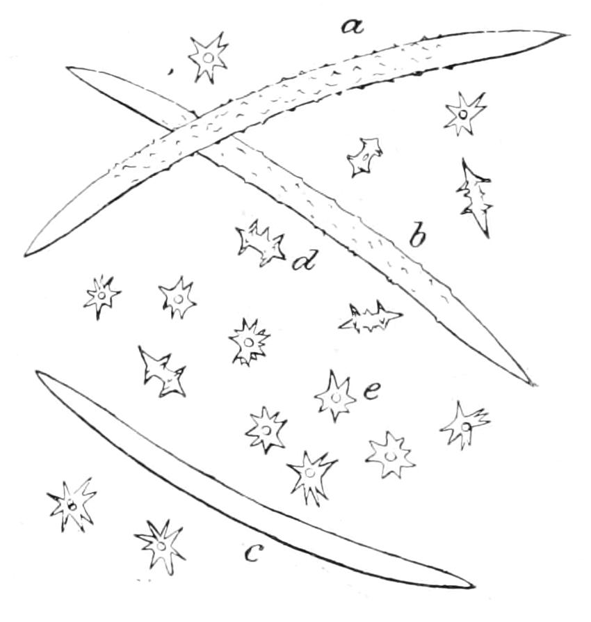
Fig. 72.—Spicules of E. fluviatilis. a. b. c. Oxeas, spined and smooth; d. e, amphidiscs, side and end views. (After Potts.)
Weltner[205] has shown that on the death and disintegration of the mother sponge some of the gemmules remain attached to the old skeleton, some sink and some float. Those which remain attached are well known to reclothe the dead fibres with living tissue. They inherit, as it were, the advantages of position, which contributed to the survival of the parent, as one of the selected fittest. The gemmules which sink are doubtless rolled short distances along the bottom, while those which float have the opportunity of widely distributing the species with the risk of being washed out to sea. But even these floating gemmules are exposed to far less dangers than the delicate free-swimming larvae, for their soft parts are protected from shocks by a thick coat armed with amphidiscs.
The gemmules are likewise remarkable for their powers of resistance to climatic conditions, powers which must contribute in no small way to the survival of a species exposed to the variable temperatures of fresh water. Thus, if the floating gemmules or the parent skeleton with its attached and dormant offspring should chance to be included in the surface layer of ice during the winter, so far from suffering any evil consequences they appear to benefit by these conditions. Both Potts and Weltner have confirmed the truth of this statement by experiments. Weltner succeeded in rearing young from gemmules which had suffered a total exposure of 17 days to a temperature "under 0° C."
Of important bearing on the question of the utility of the gemmules are certain instances in which E. fluviatilis has been recorded as existing in a perennial condition.[206] The perennial individuals may or may not bear gemmules, which makes it evident that, with the acquisition of the power to survive the winter cold, the prime necessity of forming these bodies vanishes.
The perennial specimens are described as exhibiting a diminished vegetative activity in winter, the flagellated chambers may be absent (Lieberkühn), or present in unusually small numbers (Weltner), the entire canal system may be absent (Metschnikoff), or, on the other hand, it may be complete except for the osculum.
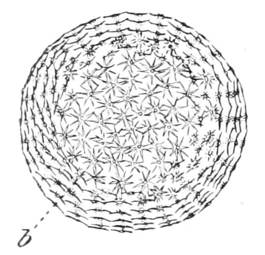
Fig. 73.—Gemmule of E. fluviatilis. b, Amphidisc. (After Potts.)
In tropical countries gemmulation occurs as a defence against the ravages caused by the dry season when the waters recede down their banks, exposing all or most of their sponge inhabitants to the direct rays of the sun. The sponges are at once killed, but the contained gemmules being thoroughly dried, become efficient distributing agents of the species; they are light enough to be carried on the wind. It is probable that those individual sponges which escape desiccation survive the dry season without forming gemmules.
It has been shown experimentally that gemmules are not injured by drying—Zykoff found that gemmules kept dry for a period of two years had not lost the power of germination.
The mature gemmules consist of a more or less spherical mass of cells, which we shall refer to as yolk cells, and of a complex coat. The latter is provided with a pore or pore tube (Fig. 74) which is closed in winter by an organic membrane.
There are three layers in the coat: an inner chitinous layer surrounded by an air-chamber layer, which is finely vesicular, showing a structure recalling plant tissue, and containing amphidiscs arranged along radii passing through the centre of the gemmule. One of the discs of each amphidisc lies in the inner chitinous coat, while the other lies in a similar membrane which envelopes the air-chamber layer and is termed the outer chitinous coat.
Marshall has suggested that one function of the amphidiscs is to weight the gemmules and thus protect them against the force of the river current; and no doubt the sinking or floating of individual gemmules depends on the relative degree of development of the air-chambers and of the amphidiscs.
A study of the development of Ephydatia gemmules vividly illustrates various characters of the inner processes of sponges. Specially noteworthy are the migrations of cells and the slight extent to which division of labour is carried: one and the same cell will be found to perform various functions.
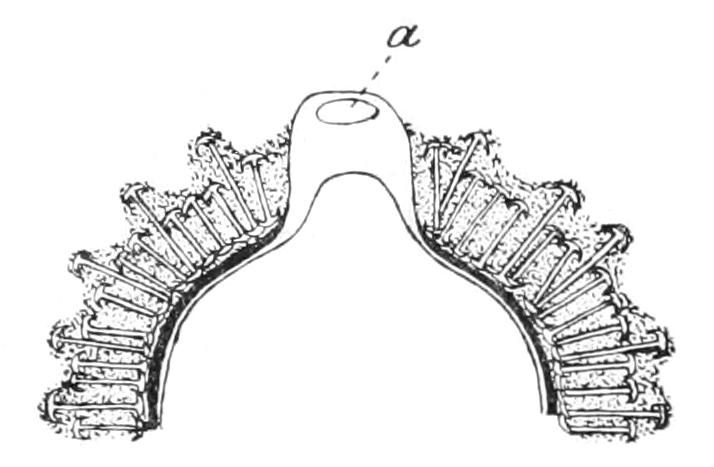
Fig. 74.—Part of a longitudinal section of a gemmule of Ephydatia sp. passing through the pore (a). (After Potts.)
The beginning of a gemmule is first recognisable[207] as a small cluster of amoeboid archaeocytes in the dermal membrane. These move into the deeper parts of the sponge to form larger groups. They are the essential part of the gemmule, the yolk cells, which, when germination takes place, give rise to a new sponge. They are followed by two distinct troops of actively moving cells. Those forming the first troop arrange themselves round the yolk cells and ultimately assume a columnar form so that they make an epithelioid layer. They then secrete the inner chitinous coat. The cells of the second troop are entrusted with the nutrition of the gemmule. Consequently they pass in among the yolk cells, distribute their food supplies, and make their escape by returning into the tissues of the mother sponge, before the columnar cells have completed the chitinous coat. Yet another migration now occurs, the cells—"scleroblasts"—which have been occupied in secreting amphidiscs at various stations in the sponge, carry the fully formed spicules to the gemmules and place them radially round the yolk cells between the radially lying cells of the columnar layer. The scleroblasts themselves remain with the amphidiscs, and becoming modified, contribute to the formation of the air-chamber layer. The columnar cells now creep out between the amphidiscs till their inner ends rest on the outer ends of these spicules. They then secrete the outer chitinous coat and return to the mother sponge.
Carter gives directions[208] for obtaining young sponges from the gemmules. The latter should be removed from the parent, cleaned by rolling in a handkerchief, and then placed in water in a watch-glass, protected with a glass cover and exposed to sunlight. In a few days the contents of the gemmule issue from the foramen and can be seen as a white speck. A few hours later the young sponge is already active and may be watched producing aqueous currents. At this age the sponge is an excellent object for studying in the living condition: being both small and transparent it affords us an opportunity of watching the movements of particles of carmine as they are carried by the current through the chambers.
Potts[209] describes how he has followed the transportal of spicules by dermal cells, the end of each spicule multiplying the motion, swaying like an oscillating rod.
In E. fluviatilis reproduction also occurs during the warmer months in this climate by means of sexual larvae. These are interesting for certain aberrant features in their metamorphosis.[210] While some of the flagellated chambers are formed in the normal way from the flagellated cells of the larva, others arise each by division of a single archaeocyte. This, it is suggested, is correlated with the acquisition of the method of reproduction by gemmules, the peculiarities (i.e. development of organs from archaeocytes) of which are appearing in the larvae.
Definition.—We may now define sponges as multicellular, two-layered animals; with pores perforating the body-walls and admitting a current of water, which is set up by the collared cells of the "gastral" layer.
Position in the Animal Kingdom.—Sponges are the only multicellular animals which possess choanocytes, and their mode of feeding is unique. Since they are two-layered it has been sought to associate them with the Metazoan phylum Coelenterata, but they are destitute of nematocysts or any other form of stinging cell, and their generative cells arise from a class of embryonic cells set apart from the first, while the generative cells of Coelenterata are derived from the ectoderm, or in other cases from the endoderm. These weighty differences between sponges and that group of Metazoa to which they would, if of Metazoan nature at all, be most likely to show resemblance, suggest that we should seek a separate origin for sponges and Metazoa. We naturally turn to the Choanoflagellate Infusorian stock (see p. 121) as the source of Porifera, leaving the Ciliate stock as the progenitors of Metazoa.
That both Porifera and Metazoa are reproduced by ova and spermatozoa is no objection to this view, seeing that the occurrence of similar reproductive cells has been demonstrated in certain Protozoa (see pp. 100, 128).
Let us now see which view is borne out by facts of embryology. Suppose, for the moment, we regard sponges as Metazoa, then if the sponge larva be compared with the Metazoan larva we must assign the large granular cells to the endoderm; the flagellated cells to the ectoderm; and we are led to the anomalous statement that the digestive cells in the adult are ectodermal, the covering, outer cells endodermal; or conversely, if we start our comparisons with the adults, then it follows that the larval ectoderm has the characters of an endoderm, and the larval endoderm those of an ectoderm.
Thus both embryology and morphology lead us to the same point, they both show that in the absence of any fundamental agreement between Porifera and Metazoa it is necessary to regard the two stocks as independent from the very first, and hence the name Parazoa (Sollas) has been given to the group which contains the Porifera as its only known phylum.
Interesting in connexion with the phylogeny of Parazoa is the Choanoflagellate genus Proterospongia (Fig. 75), described by Saville Kent, and since rediscovered both in England and abroad.[211] This is a colony of unicellular individuals embedded in a common jelly. The individuals at the surface are choanoflagellate, while in the interior the cells are rounded or amoeboid, and some of them undergo multiple fission to form reproductive cells. This is just such a creature as we might imagine that ancestral stage to have been of which the free-swimming sponge larva is a reminiscence: for we have seen that the flagellated cells of the larva are potential choanocytes.
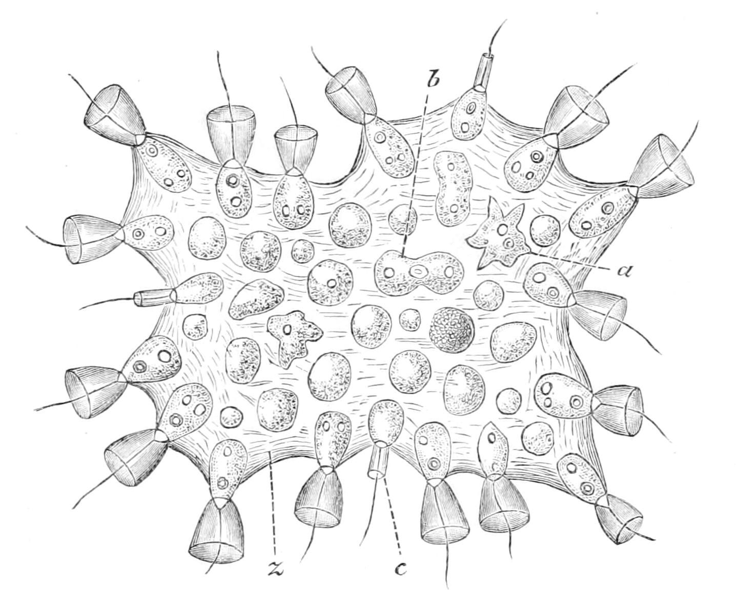
Fig. 75.—Proterospongia haeckeli. a, Amoeboid cell; b, a cell dividing; c, cell with small collar; z, jelly. × 800. (After S. Kent.)