Chapter VI
Reading edition
PROTOZOA (CONTINUED): INFUSORIA (CILIATA AND SUCTORIA)
IV. Infusoria.
Complex Protozoa, never holophytic save by symbiosis with plant commensals, never amoeboid, with at some period numerous short cilia, of definite outline, with a double nuclear apparatus consisting of a large meganucleus and a small micronucleus (or several),[149] the latter alone taking part in conjugation (karyogamy), and giving rise after conjugation to the new nuclear apparatus.
The name Infusoria was formerly applied to the majority of the Protozoa, and included even the Rotifers. For the word signifies organisms found in "infusions" of organic materials, including macerations. Such were made with the most varied ingredients, pepper and hay being perhaps the favourites. They were left for varying periods exposed to the air, to allow the organisms to develop therein, and were then examined under the microscope.[150] With the progress of our knowledge, group after group was split off from the old assemblage until only the ciliate or flagellate forms were left. The recognition of the claims of the Flagellates to independent treatment left the group more natural;[151] while it was enlarged by the admission of the Acinetans (Suctoria), which had for some time been regarded as a division of the Rhizopoda.
I. Ciliata
Infusoria, with a mouth, and cilia by which they move and feed; usually with undulating membranes, membranellae, cirrhi, or some of these. Genera about 144: 27 exclusively marine, 50 common to both sea and fresh water, 27 parasitic on or in Metazoa, the rest fresh water. Species about 500.
We divide the Ciliata thus:[152]—
(I.) Mouth habitually closed, opening by retraction of its circular or slit-like margin; cilia uniform Order 1. Gymnostomaceae.
Lacrymaria, Ehrb.; Loxodes, Ehrb.; Loxophyllum, Duj.; Lionotus, Wrez.; Trachelius, Schrank; Amphileptus, Ehrb.; Actinobolus, St.; Didinium, St.; Scaphiodon, St; Dysteria, Huxl.; Coleps, Nitzsch.; Dileptus, Duj.; Ileonema, Stokes; Mesodinium, St.
(II.) Mouth permanently open, usually equipped with one or more undulating membranes, receiving food by ciliary action (Trichostomata, Bütschli)
(a) Cilia nearly uniform, usually extending over the whole body, without any special adoral wreath of long cilia or membranellae; mouth with one or two undulating membranes at its margin or extending into the short pharynx. Order 2. Aspirotrichaceae.
Paramecium, Hill; Colpoda, O. F. Müll.; Colpidium, St.; Leucophrys, Ehrb.; Cyclidium, Cl. and L.; Lembadion, Perty; Cinetochilum, Perty; Pleuronema, Duj.; Ancistrum, Maup.; Glaucoma, Ehrb.; Uronema, Duj.; Lembus, Cohn; Urocentrum, Nitzsch; Icthyophtheirius, Fouquet.
(b) Strong cilia or membranellae forming an adoral wreath, and bounding a more or less enclosed area, the "peristome," at one point of which the mouth lies.
(i.) Body more or less equally covered with fine cilia; adoral wreath an open spiral Order 3. Heterotrichaceae
Spirostomum, Ehrb.; Bursaria, O. F. Müll.; Stentor, Oken; Folliculina, Lamk.; Conchophtheirus, St.; Balantidium, Cl. and L.; Nyctotherus, Leidy; Metopus, Cl. and L.; Caenomorpha, Perty; Discomorpha, Levander; Blepharisma, Perty.
(ii.) Body cilia limited in distribution or absent; peristome anterior, nearly circular, sinistrorse. Order 4. Oligotrichaceae.
Halteria, Duj.; Maryna, Gruber; Tintinnus, Schrank; Dictyocystis, Ehrb.; Strombidium, Cl. and L. (= Torquatella, Lank.).
(iii.) Peristome extending backwards along the ventral face, which alone is provided with motile cirrhi, etc.; dorsal cilia fine, motionless. Order 5. Hypotrichaceae.
Stylonychia, Ehrb.; Kerona, O. F. Müll.; Oxytricha, Ehrb.; Euplotes, Ehrb.; Stichotricha, Perty; Schizotricha, Gruber.
(iv.) Body cilia reduced to a posterior girdle, or temporarily or permanently absent; peristome anterior, nearly circular, edged by the adoral wreath,[153] bounded by a gutter edged by an elevated rim or collar. Order 6. Peritrichaceae.
Lichnophora, Cl.; Trichodina, Ehrb.; Vorticella, L.; Zoothamnium, Bory; Carchesium, Ehrb.; Epistylis, Ehrb.; Opercularia, Lamk.; Vaginicola, Lamk.; Pyxicola, Kent; Cothurnia, Ehrb.; Scyphidia, Lachmann; Ophrydium, Bory; Spirochona, St.
The Ciliata have so complex an organisation that, as with the Metazoa, it is well to begin with the description of a definite type. For this purpose we select Stylonychia mytilus, Ehrb. (Fig. 49), a species common in water rich in organic matter, and relatively large (1⁄75" = ⅓ mm.). It is broadly oval in outline, with the wide end anterior, truncate, and sloping to the left side behind; the back is convex, thinning greatly in front; the belly flat. It moves through the water either by continuous swimming or by jerks, and can either crawl steadily over the surface of a solid or an air surface such as an air bubble, or advance by springs, which recall those of a hunting spider. The boundary is everywhere a thin plasmic pellicle, very tender, and readily undergoing diffluence like the rest of the cell. From the pellicle pass the cilia, which are organically connected with it, though they may be traced a little deeper; they are arranged in slanting longitudinal rows, and are much and variously modified, according to their place and function. On the edge of the dorsal surface they are fine and motionless, probably only sensory (s.h.); except three, which protrude well over the hinder end (c.p.), stout, pointed, and frayed out at the ends, and possibly serving as oars or rudders for the darting movements. These are distinguished from simple cilia as "cirrhi."
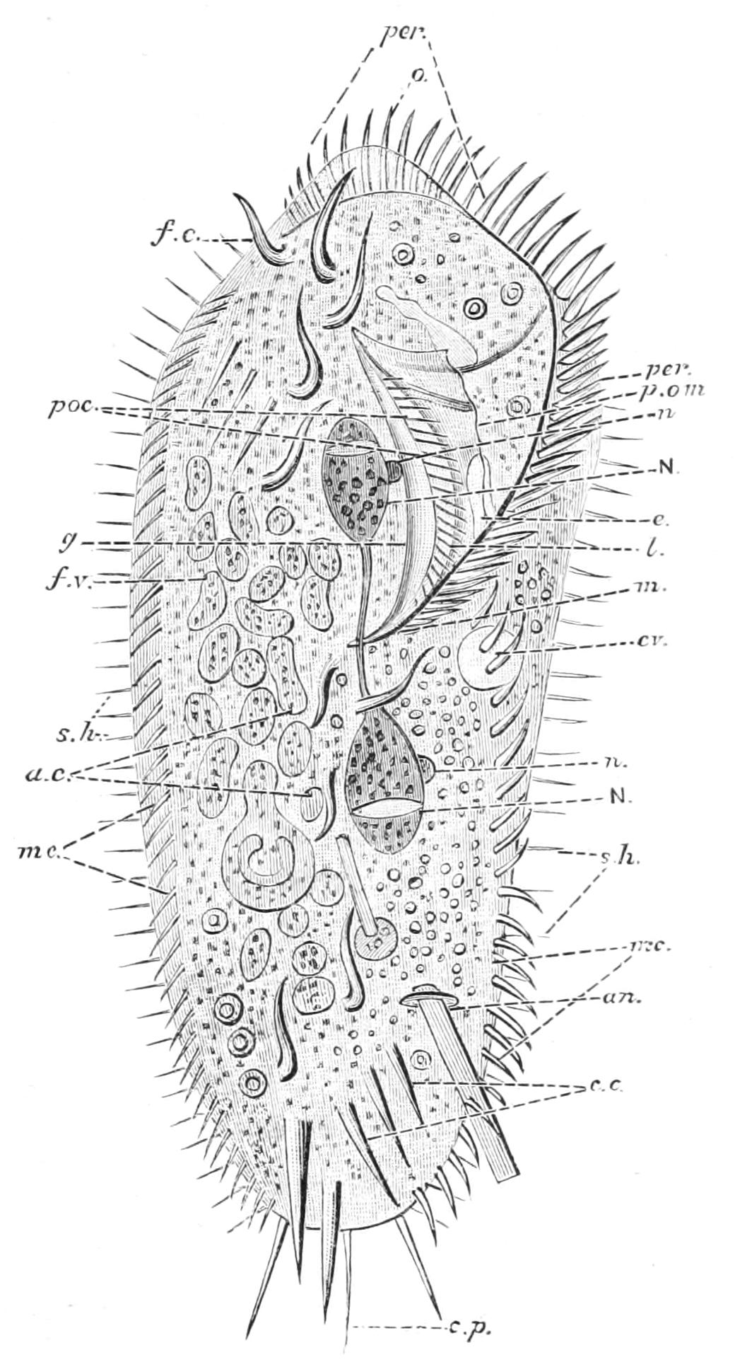
Fig. 49.—Ventral view of Stylonychia mytilus. a.c, Abdominal cirrhi; an, anus discharging the shell of a Diatom; c.c, caudal cirrhi; c.p, dorsal cirrhi; cv, contractile vacuole; e, part of its replenishing canal; f.c, frontal cirrhi; f.v, food vacuoles; g, internal undulating membrane; l, lip; m, mouth or pharynx; mc, marginal cirrhi; N, N, lobes of meganucleus; n, n, micronuclei; o, anterior end; per, adoral membranellae; poc, preoral cilia; p.om, preoral undulating membrane; s.h, sense hairs. (Modified from Lang.)
At the right hand of the frontal area there begins, just within the dorsal edge, a row of strong cilium-like organs (Fig. 49, per); these, on careful examination, prove to be transverse triangular plates, which after death may fray into cilia.[154] They are the "adoral membranellae." This row passes to the left blunt angle, and there crosses over the edge of the body to the ventral aspect, and then curves inwards towards the median line, which it reaches about half-way back, where it passes into the pharynx (m). It forms the front and left-hand boundary of a wedge-shaped depression, the "peristomial area," the right-hand boundary being the "preoral ridge" or lip (l), which runs nearly on the median line, projecting downward and over the depression. This ridge bears on its inner and upper side a row of fine "preoral cilia" (poc) and a wide "preoral undulating membrane" (p.om), which extends horizontally across, below the peristomial area. The roof of this area bears along its right-hand edge an "internal undulating membrane" (g), and then, as we pass across to the left, first an "endoral membrane" and then an "endoral" row of cilia. In some allied genera (not in Stylonychia), at the base and on the inner side of each adoral membranella, is a "paroral" cilium. All these motile organs, with the exception of the preoral cilia, pass into the pharynx; but the adoral membranellae soon stop short for want of room. There are some seventy membranellae in the adoral wreath.
The rest of the ventral surface is marked by longitudinal lines, along which the remaining appendages are disposed. On either side is a row of "marginal cirrhi" (mc.), which, like the membranellae, may fray out into cilia, but are habitually stiff spine-like, and straight in these rows; these are the chief swimming organs. Other cirrhi, also arranged along longitudinal rows, with so many blank spaces that the arrangement has to be carefully looked for, occur in groups along the ventral surface. On the right of the peristome are a group which are all curved—the "frontal cirrhi" (f.c.). Behind the mouth is a second group—the "abdominal cirrhi" (a.c.), also curved hooks; and behind these again the straight spine-like "caudal" or "anal" cirrhi (c.c), which point backwards. These three sets of ventral cirrhi are the organs by which the animal executes its crawling and darting movements. Besides the mouth there are two other openings, both indistinguishable save at the very moment of discharge; the anus (an) which is dorsal, and the pore of the contractile vacuole, which is ventral.
The protoplasm of the body is sharply marked off into a soft, semi-fluid "endoplasm" or "endosarc," and a firmer "ectoplasm" or "ectosarc." The former is rich in granules of various kinds, and in food-vacuoles wherein the food is digested. The mode of ingestion, etc., is described below (p. 145). The ectoplasm is honeycombed with alveoli of definite arrangement, the majority being radial to the surface or elongated channels running lengthwise; inside each of these lies a contractile plasmic streak or myoneme. The contractile vacuole (cv) lies in this layer, a little behind the mouth, and is in connexion with two canals, an anterior (e) and a posterior, from which it is replenished.
The nuclear apparatus lies on the inner boundary of the ectoplasm; it consists of (1) a large "meganucleus" formed of two ovoid lobes (N, N), united by a slender thread; and (2) two minute "micronuclei" (n, n), one against either lobe of the meganucleus.
Stylonychia multiplies by transverse fission, the details of which are considered on pp. 144, 147.
The protoplasm of Ciliata is the most differentiated that we find in the Protista, and we can speak without exaggeration of the "organs" formed thereby.
The form of the body, determined by the firm pellicle or plasmic membrane, is fairly constant for each species, though it may be subject to temporary flexures and contractions. The pellicle varies in rigidity; where the cilia are abundant it is proportionately delicate, and scarcely differs from the ectoplasm proper, save for not being alveolate. In the Peritrichaceae it is especially resistant and proof against decay. In Coleps (Gymnostomaceae) it is hardened and sculptured into the semblance of plate-armour, and the prominent points of the plates around the mouth serve as teeth to lacerate other active Protista, its prey; but, like the rest of the protoplasm, this disappears by decay soon after the death of the Coleps. Where, as in certain Oligotrichaceae, cilia are absent over part of the body, the pellicle is hardened; and on the dorsal face and sides of Dysteria it even assumes the character of a bivalve shell, and forms a tooth-like armature about the mouth.
From the pellicle protrude the cilia, each of which is continued inwards by a slender basal filament to end in a "basal granule" or "blepharoplast." The body-cilia are fine, and often reversible in action, which is exceptional in the organic world. They may be modified or combined in various ways. We have seen that in Stylonychia some are motionless sensory hairs. The cirrhi and setae sometimes fray out during life, and often after death, into a brush at the tip, and have a number of blepharoplasts at their base. The same holds good for the membranellae and undulating membranes. They are thus comparable to the "vibratile styles" of Rotifers (Vol. II. p. 202) and the "combs" or "Ctenophoral plates" of the Ctenophora (p. 412 f.).[155]
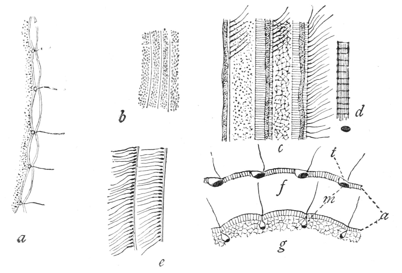
Fig. 50.—Ectosarc of Ciliata. a-f, from Stentor coeruleus; g, Holophrya discolor. a, Transverse section, showing cilia, pellicle, canals, and myonemes; b, surface view below pellicle, showing myonemes alternating with blue granular streaks; c, more superficial view, showing rows of cilia adjacent to myonemes; d, myoneme, highly magnified, showing longitudinal and transverse striation; e, two rows of cilia; f, g, optical sections of ectosarc, showing pellicle, alveolar layer (a), myonemes (m), and canals in ectosarc. (From Calkins, after Metschnikoff, Bütschli, and Johnson.)
The ectosarc has a very complex structure. Like other protoplasm it has a honeycombed or alveolate structure, but in this case the alveoli are permanent in their arrangement and position. Rows of these alveoli run under the surface; and the cilia are given off from their nodal points where the vertical walls of several unite, and wherein the basal granule or blepharoplast is contained. Longitudinal threads running along the inner walls of the alveoli of the superficial layer are differentiated into muscular fibrils or "myonemes," to which structures so many owe their marked longitudinal striation on the one hand, and their power of sudden contraction on the other. The appearance of transverse striation may be either due to transverse myonemes, or produced by the folds into which the contraction of longitudinal fibrils habitually wrinkles the pellicles, when it is fairly dense (Peritrichaceae); circular muscular fibrils, however, undoubtedly exist in the peristomial collar of this group. Embedded in the ectosarc are often found trichocysts,[156] analogous to the nematocysts of the Coelenterata (p. 247), and doubtless fulfilling a similar purpose, offensive and defensive. A trichocyst is an oblong sac (4 µ long in Paramecium) at right angles to the surface, which on irritation, chemical (by tannin, acids, etc.) or mechanical, emits or is converted into a thread several times the length of the cilia (33 µ), often barbed at the tip. In the predaceous Gymnostomaceae, such as Didinium, the trichocysts around (or even within) the mouth are of exceptional size, and are ejected to paralyse, and ultimately to kill, the active Infusoria on which they feed. In most of the Peritrichaceae they are, when present, limited to the rim around the peristome, while in the majority of species of Ciliata they have not been described. Fibrils, possibly nervous,[157] have been described in the deepest layer of the ectosarc in Heterotrichaceae.
The innermost layer of the ectosarc is often channelled by a system of canals,[158] usually inconspicuous, as they discharge continuously into the contractile vacuole; but by inducing partial asphyxia (e.g. by not renewing the limited supply of air dissolved in the drop of water on the slide under the cover-glass), the action of the vacuole is slackened, and these canals may be more readily demonstrated. The vacuole, after disappearance, forms anew either by the coalescence of minute formative vacuoles, or by the enlargement of the severed end of the canal or canals. The pore of discharge to the surface is visible in several species, even in the intervals of contraction.[159] The pore is sometimes near that of the anus, but is only associated with it in Peritrichaceae, where it opens beside it into the vestibule or first part of the long pharynx, often through a rounded reservoir (Fig. 60, r) or elongated canal.
The endosarc, in most Ciliates well differentiated from the ectosarc, is very soft; though it is not in constant rotation like that of a Rhizopod, it is the seat of circulatory movements alternating with long periods of rest. Thus it is that the food-vacuoles, after describing a more or less erratic course, come to discharge their undigested products at the one point, the anus. In a few genera (Didinium, for instance) the course from mouth to anus is a direct straight line, and one may almost speak of a digestive tract. In Loxodes and Trachelius (Fig. 56) the endosarc, as in the Flagellate Noctiluca (Fig. 48, p. 133), has a central mass into which the food is taken, and which sends out lobes, which branch as they approach and join the ectoplasm. The endosarc contains excretory granules, probably calcium phosphate, droplets of oil or dissolved glycogen, proteid spherules, paraglycogen grains, etc.
The nuclear apparatus lies at the inner boundary of the ectoplasm. The "meganucleus" may be ovoid, elongated, or composed of two or more rounded lobes connected by slender bridges (Stentor, Stylonychia). The "micronucleus" may be single; but even when the meganucleus is not lobed it may be accompanied by more than one micronucleus, and when it is lobed there is at least one micronucleus to each of its lobes.[160] The meganucleus often presents distinct granules of more deeply staining material, varying with the state of nutrition; these are especially visible in the band-like meganuclei of the Peritrichaceae (Figs. 51, 60). At the approach of fission it is in many cases distinctly fibrillated.[161] But all other internal differentiation, as well as any constriction, then disappears; and the ovoid or rounded figure becomes elongated and hour-glass shaped, and finally constricts into two ovoid daughter-meganuclei, which, during and after the fission of the cell, gradually assume the form characteristic of the species. The micronuclei (each and all when they are multiple) divide by modification of karyokinesis (or "mitosis") as a prelude to fission: in this process the chromatin is resolved into threads which divide longitudinally, but the nuclear wall remains intact. If an Infusorian be divided into small parts, only such as possess a micronucleus and a fragment of the meganucleus are capable of survival. We shall see how important a part the micronuclei play in conjugation, a process in which the old meganuclei are completely disorganised and broken up and their débris expelled or digested.
The mouth of the Gymnostomaceae is habitually closed, opening only for the ingestion of the living Protista that form their prey. It usually opens into a funnel-shaped pharynx, strengthened with a circle of firm longitudinal bars, recalling the mouth of an eel-trap or lobster-pot ("Reusenapparat" of the Germans); and this is sometimes protrusible. In Dysteria the rods are replaced by a complicated arrangement of jaw- or tooth-like thickenings, which are not yet adequately described. We have above noted the strong adoral trichocysts in this group.
In all other Ciliates[162] the "mouth" is a permanent depression lined by a prolongation of the pellicle, and containing cilia and one or more undulating membranes, and when adoral membranellae are present, a continuation of these. In some species, such as Pleuronema (Fig. 57), one or two large membranes border the mouth right and left. In Peritrichaceae the first part of the pharynx is distinguished as the "vestibule," since it receives the openings of the contractile vacuole or its reservoir and the anus. The pharynx at its lower end (after a course exceptionally long and devious in the Peritrichaceae; Figs. 51, 60) ends against the soft endosarc, where the food-particles accumulate into a rounded pellet; this grows by accretion of fresh material until it passes into the endosarc, which closes up behind it with a sort of lurch. Around the pellet liquid is secreted to form the food-vacuole. If the material supplied be coloured and insoluble, like indigo or carmine, the vacuoles may be traced in a sort of irregular, discontinuous circulation through the endosarc until their remains are finally discharged as faeces through the anus. No prettier sight can be watched under the microscope than that of a colony of the social Bell-animalcule (Carchesium) in coloured water—all producing food-currents brilliantly shown up by the wild eddies of the pigment granules, and the vivid blue or crimson colour of the food-vacuoles, the whole combining to present a most attractive picture. Ehrenberg fancied that a continuous tube joined up the vacuoles, and interpreted them as so many stomachs threaded, as it were, along a slender gut; whence he named the group "Polygastrica."
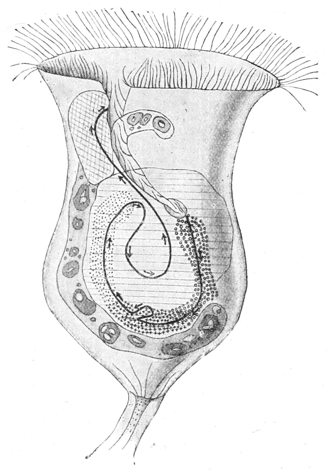
Fig. 51.—Carchesium polypinum. Scheme of the path taken by the ingested food in digestion and expulsion of the excreta. The food enters through the pharynx and is transported downward (small circles), where it is stored in the concavity of the sausage-shaped meganucleus (the latter is recognised by its containing darker bodies). It remains here for some time at rest (small crosses). Then it passes upward upon the other side (dots) and returns to the middle of the cell, where it undergoes solution. The excreta are removed to the outside, through the vestibule and cell mouth. The black line with arrows indicates the direction of the path. (From Verworn, after Greenwood.)
We owe to Miss Greenwood[163] a full account of the formation and changes of the food-vacuoles in Carchesium polypinum. The vacuole passes steadily along the endosarc for a certain time after its sudden admission into it, and then enters on a phase of quiescence. A little later the contents of the vacuole aggregate together in the centre of the vacuole, where they are surrounded by a zone of clear liquid; this takes place in the hollow of the meganucleus, in this species horseshoe-shaped. The vacuole then slowly passes on towards the peristome, lying deep in the endosarc, and the fluid peripheral zone is absorbed. For some time no change is shown in the food-material itself: this is the stage of "storage." Eventually a fresh zone of liquid, the true digestive vacuole, forms again round the food-pellet, and this contains a peptic juice, of acid reaction. The contents, so far as they are capable of being digested, liquefy and disappear. Ultimately the solid particles in their vacuole reach the anal area of the vestibule, and pass into it, to be swept away by the overflow of the food-current. The anus is seated on a transverse ridge about a third down the tube, the remaining two-thirds being the true pharynx.
Fission is usually transverse; but is oblique in the conical Heterotrichaceae, and longitudinal in the Peritrichaceae. It involves the peristome, of which one of the two sisters receives the greater, the other the lesser part; each regenerates what is missing. When there are two contractile vacuoles, as in Paramecium, either sister receives one, and has to form another; where there is a canal or reservoir divided at fission, an extension of this serves to give rise to a new vacuole in that sister which does not retain the old one. In some cases the fission is so unequal as to have the character of budding (Spirochona). We have described above (p. 144) the relations of the nuclear apparatus in fission.
Several of the Ciliata divide only when encysted, and then the divisions are in close succession, forming a brood of four, rarely more. This is well seen in the common Colpoda cucullus. In the majority, however, encystment is resorted to only as a means of protection against drought, etc., or for quiet rest after a full meal (Lacrymaria).
Maupas[164] has made a very full study of the life-cycles of the Ciliata. He cultivated them under the usual conditions for microscopic study, i.e. on a slide under a thin glass cover supported by bristles to avoid pressure, preserved in a special moist chamber; and examined them at regular intervals.
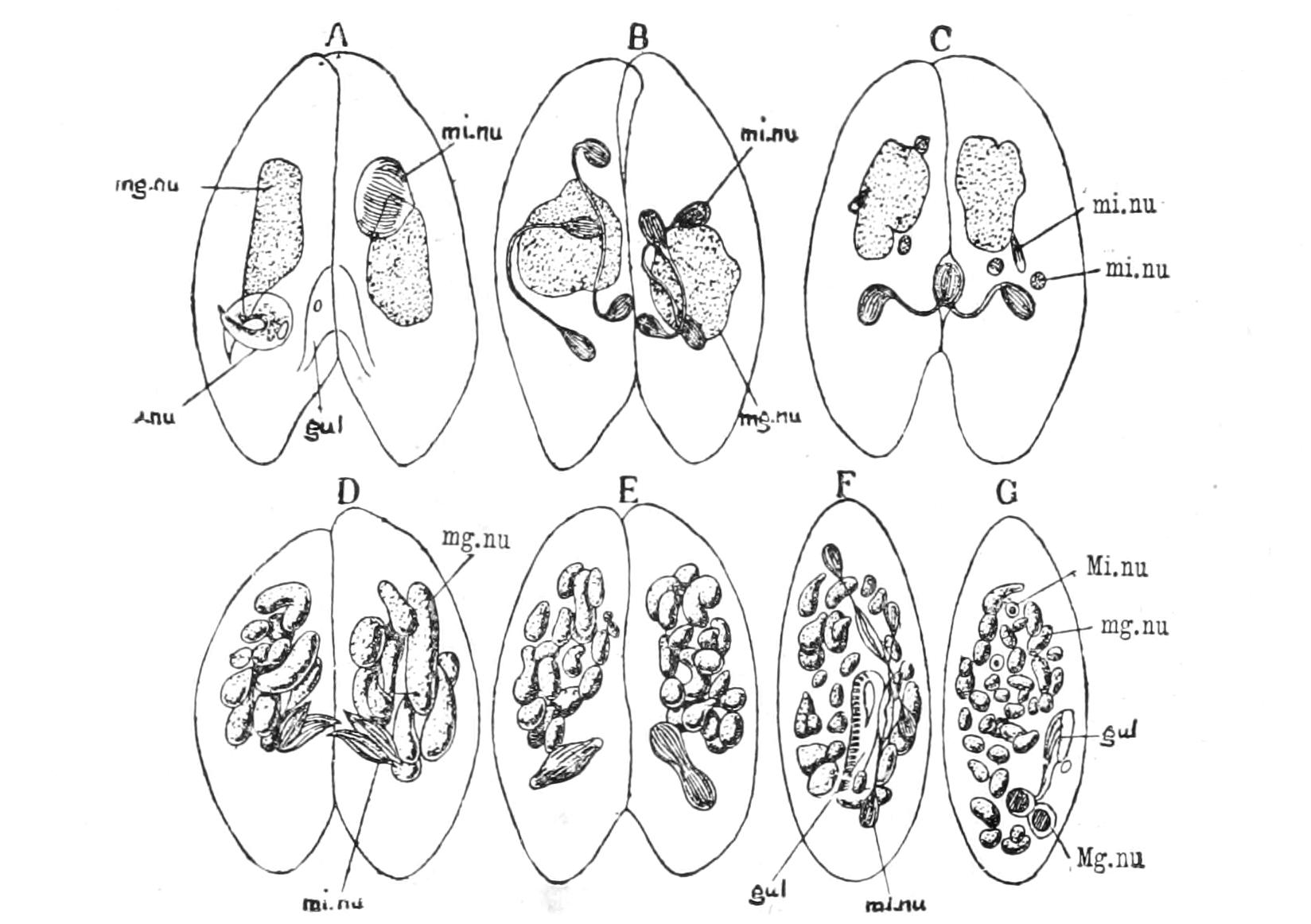
Fig. 52.—Paramecium caudatum, stages in conjugation. gul, Gullet; mg.nu, meganucleus; Mg.nu, reconstructed meganucleus; mi.nu, micronucleus; Mi.nu, reconstructed micronucleus; o, mouth. (From Parker and Haswell, after Hertwig.)
The animals collect at that zone where the conditions of aeration are most suitable, usually just within the edge of the cover, and when well supplied with food are rather sluggish, not swimming far, so that they are easily studied and counted. When well supplied with appropriate food they undergo binary fission at frequent intervals, dividing as often as five times in the twenty-four hours at a temperature of 65-69° F. (Glaucoma scintillans), so that in this period a single individual has resolved itself into a posterity of 32; but such a rapid increase is exceptional. At a minimum and a maximum temperature multiplication is arrested, the optimum lying midway. If the food-supply is cut off, encystment occurs in those species capable of the process; but when there is a mixture of members of different broods of the same species, subject to the limitations that we shall learn, conjugation ensues. Under the conditions of Maupas' investigations he found a limit to the possibilities of continuous fissions, even when interrupted by occasional encystment. The individuals of a series ultimately dwindle in size, their ciliary apparatus is reduced, and their nuclear apparatus degenerates. Thus the ultimate members of a fission-cycle show a progressive decay, notably in the nuclear apparatus, which Maupas has aptly compared to "senility" or "old age" in the Metazoan. If by the timely mixture of broods conjugation be induced, these senile degenerations do not occur.[165] In Stylonychia mytilus the produce of a being after conjugation died of senility after 336 fissions; in Leucophrys after 660.
Save in the Peritrichaceae (p. 151) conjugation takes place between similar mates, either of the general character and size of the species, or reduced by fissions, in rapid succession, induced by the same conditions as those of mating. The two mates approach, lying parallel and with their oral faces or their sides (Stentor) together, and partially fuse thereby; though no passage of cytoplasm is seen it is probable that there is some interchange or mixture.[166]
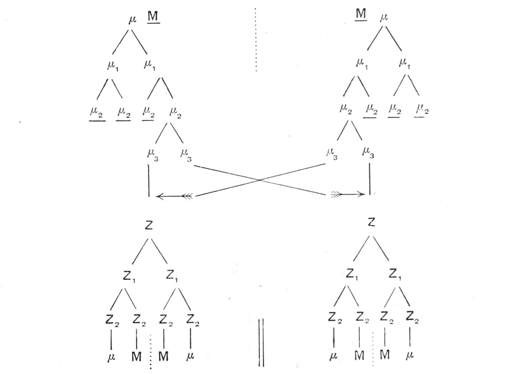
Fig. 53.—Diagram of conjugation in Colpidium colpoda. Horizontal line means degeneration; parallel vertical lines, separation of gametes; broken lines (above), boundary between pairing animals; (below), first fission; single vertical line, continuity or enlargement. M, Meganucleus; µ, micronucleus; Z, zygote-nucleus.
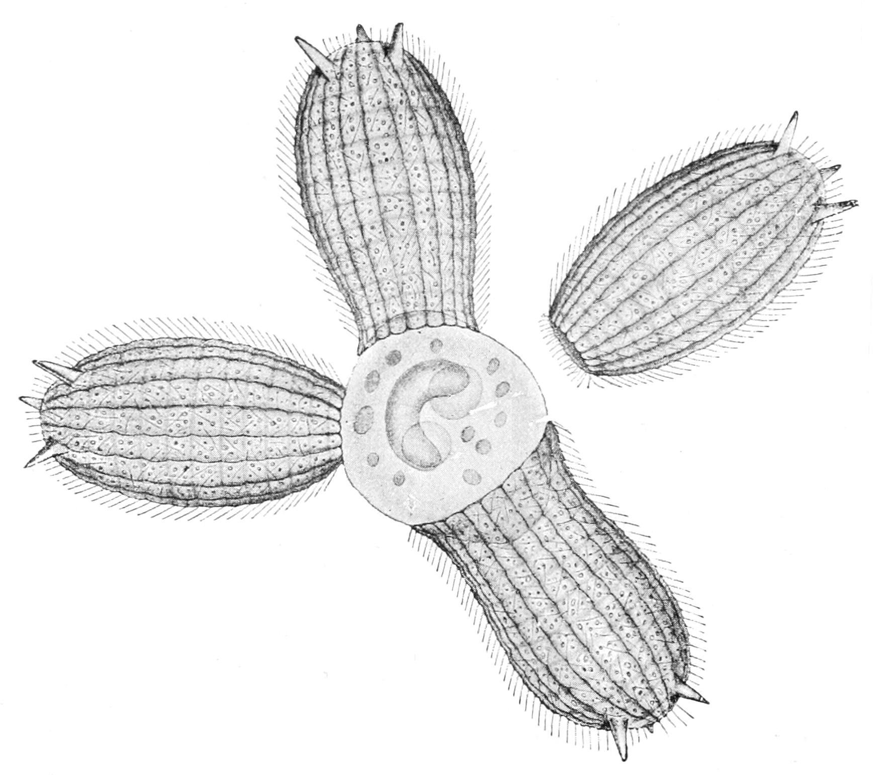
Fig. 54.—Four individuals of Coleps hirtus (Gymnostomaceae) swarming about and ingesting a Vorticella (?) (From Verworn.)
The meganucleus lengthens, becomes irregularly constricted, and breaks up into fragments, which are ultimately extruded or partially digested. The micronucleus enlarges (Fig. 52, A) and undergoes three successive divisions, or, strictly speaking, two fissions producing four nuclei, of which one only undergoes the third. The other three nuclei of the second fission degenerate like the meganucleus.[167] Of the two micronuclei of this last division one remains where it is as a "stationary" pairing nucleus, while its sister passes as a "migratory" pairing-nucleus into the other mate, and fuses with its stationary pairing-nucleus. Thus in either mate is formed a "zygote-nucleus," or "fusion-nucleus." All these processes are simultaneous in the two mates; and the migratory nuclei cross one another on the bridge of junction of the two mates (Fig. 52, C). Each mate now has its original cytoplasm (subject to the qualification above), but its old nuclear apparatus is replaced by the fusion-nucleus. This new nucleus undergoes repeated fissions; its offspring enlarge unequally, the larger being differentiated as mega-, the smaller as micro-nuclei. The mates now separate (Fig. 52, F, G), and by the first (or subsequent) fission of each, the new mega- and micro-nuclei are distributed to the offspring. Colpidium colpoda offers the simplest case, on which we have founded our diagram showing the nuclear relations. During conjugation the oral apparatus often atrophies, and is regenerated; and in some cases the pellicle and ciliary apparatus are also "made over."
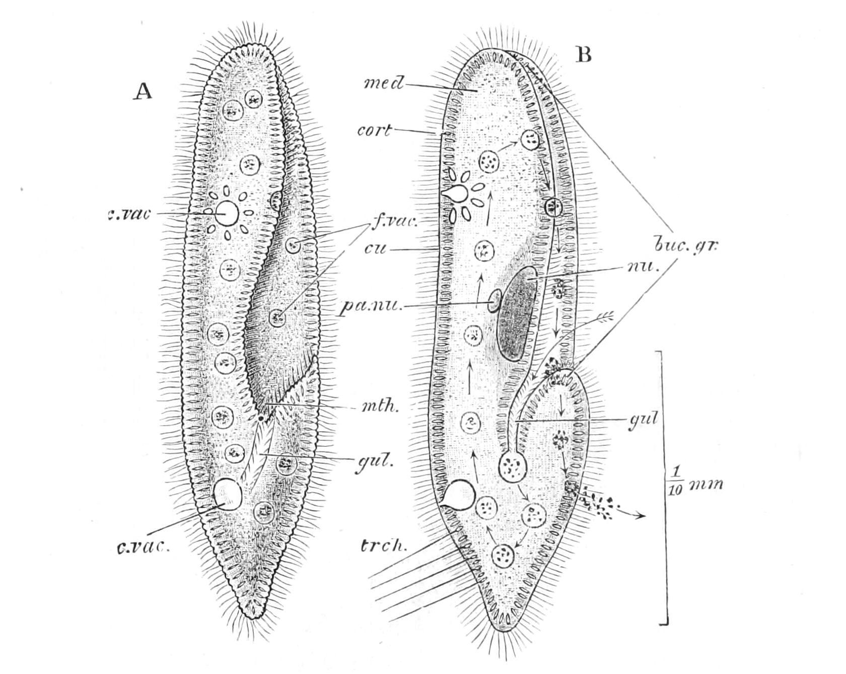
Fig. 55.—Paramecium caudatum (Aspirotrichaceae). A, The living animal from the ventral aspect; B, the same in optical section, the arrows show the course taken by food-particles. buc.gr, Buccal groove; cort, cortex; cu, cuticle; c.vac, contractile vacuole; f.vac, food vacuole; gul, gullet; med, medulla; mth, mouth; nu, meganucleus; pa.nu, micronucleus; trch, trichocysts discharged. (From Parker's Biology.)
In the Peritrichaceae the mates are unequal; the larger is the normal cell, and is fixed; the smaller, mobile, is derived from an ordinary individual by brood-divisions, which only occur under the conditions that induce conjugation (Fig. 60). Here, though the two pairs of nuclei are formed, it is only the migratory nuclei that unite, the stationary ones aborting in both mates. During the final processes of conjugation the smaller mate is absorbed into the body of the larger, and so plays the part of male there. But this process, though one of true binary sex, is clearly derived from the peculiar type of equal reciprocal conjugation of the other Infusoria.
The Ciliata are almost all free-swimming animals with the exception of most of the Peritrichaceae, and of the genera we now cite. Folliculina forms a sessile tube open at either end; and Schizotricha socialis inhabits the open mouths of a branching gelatinous tubular stem, obviously secreted by the hinder end of the animal, and forking at each fission to receive the produce. A similar habit to the latter characterises Maryna socialis; all three species are marine, and were described by Gruber.[168] Stentor habitually attaches itself by processes recalling pseudopodia, and often forms a gelatinous sheath.
The majority of the Oligotrichaceous Tintinnidae inhabit free chitinous tests often beautifully fenestrated, as in Dictyocystis.
Many genera are parasitic in the alimentary canal of various Metazoa, but none appear to be seriously harmful except Ichthyophtheirius, which causes an epidemic in fresh-water fish. Quite a peculiar fauna inhabit the paunch of Ruminants. Nyctotherus and Balantidium are occasionally found in the alimentary canal of Man.[169]
The Gymnostomaceae are predaceous, feeding for the most part on smaller Ciliates. We have described the peculiar character of the mouth and pharynx in this group, and the mail-like pellicle of Coleps (Fig. 54). Loxophyllum is remarkable for the absence of cilia from one of the sides of its flattened body, and the tufts of trichocysts studding its dorsal edge at regular intervals. Actinobolus has numerous tentacles, exsertile and retractile, each bearing a terminal tuft of trichocysts, which serve to paralyse such active prey as Halteria. Ileonema has one tentacle overhanging the mouth; and Mesodinium has four short sucker-like projections around it.[170] It has only two girdles of cilia, which are stout and resemble fine-pointed cirrhi. In Dysteria the cilia are exclusively ventral, and the naked dorsal surface has its pellicle condensed into a bivalve shell; a posterior motile process ("foot") and a complex pharyngeal armature add to the exceptional characters of the genus.
The Aspirotrichaceae are well known to every student of "Elementary Biology" by the "type" Paramecium (Fig. 55), so common in infusions, especially when containing a little animal matter. P. bursaria often contains in its endosarc the green symbiotic Flagellate Zoochlorella. Colpoda cucullus, very frequent in vegetable infusions, usually only divides during encystment, and forms a brood of four. Pleuronema chrysalis (Fig. 57) is remarkable for its habit of lying for long periods on its side and for its immense undulating membrane, forming a lip on the left of its mouth; Glaucoma has two, right and left.
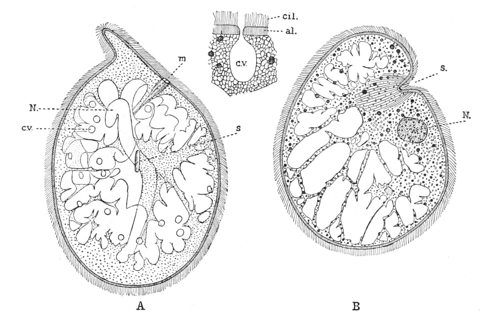
Fig. 56.—Trachelius ovum. A, general view; B, section through sucker; C, section through contractile vacuole and its pore of discharge. al, Alveolar layer of ectoplasm; cil, cilia; c.v, contractile vacuole; m, mouth; N, meganucleus; s, sucker, from which pass inwards retractile myonemes. (After Clara Hamburger.)
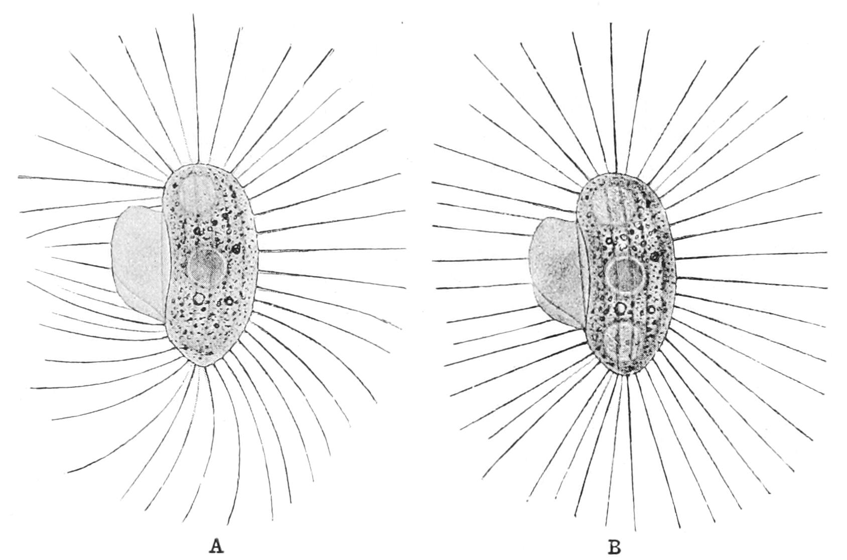
Fig. 57.—Pleuronema chrysalis (Aspirotrichaceae). A, Unstimulated, lying quiet; B, stimulated, in the act of springing by the stroke of its cilia. (From Verworn.)
The Heterotrichaceae present very remarkable forms. Spirostomum is nearly cylindrical, and, a very giant, may attain a length of 4 mm. (1⁄6"). Stentor can attach itself by its hinder end, which is then finely tapered and prolonged into a few pseudopodia; its body is trumpet-shaped, with a spiral peristome forming a coil round its wide end, and leading on the left side into the mouth. Many species when attached secrete a gelatinous sheath or tube. S. polymorphus is often coloured green by Zoochlorella (p. 125); S. coeruleus[171] and S. igneus owe their names to the brilliant pigment, blue or scarlet, deposited in granules in lines between the conspicuous longitudinal myonemes. From their large size and elongated meganucleus accompanied by numerous micronuclei, these two genera have frequently been utilised for experiments on regeneration. In Metopus sigmoides the peristomial area forms a dome above its wreath of membranellae; and in M. pyriformis this is so great as to form the larger part of the cell, which is top-shaped, tapering behind to a point. Caenomorpha (Fig. 58) has the same general form, with a peg-like tail, and possesses a girdle of cirrhi.[172] The converse occurs in Bursaria; the cell is a half ellipse, something like a common twin tobacco-pouch when closed: a deep depression thus occupies the whole ventral surface, and opens by a wide slit extending along the anterior end. The peristomial area occupies the dorsal side of the pocket so formed, and the mouth is in the hinder left-hand corner. Blepharisma sp. is parasitic in the Heliozoon Raphidiophrys viridis (Fig. 20, 1, p. 74).
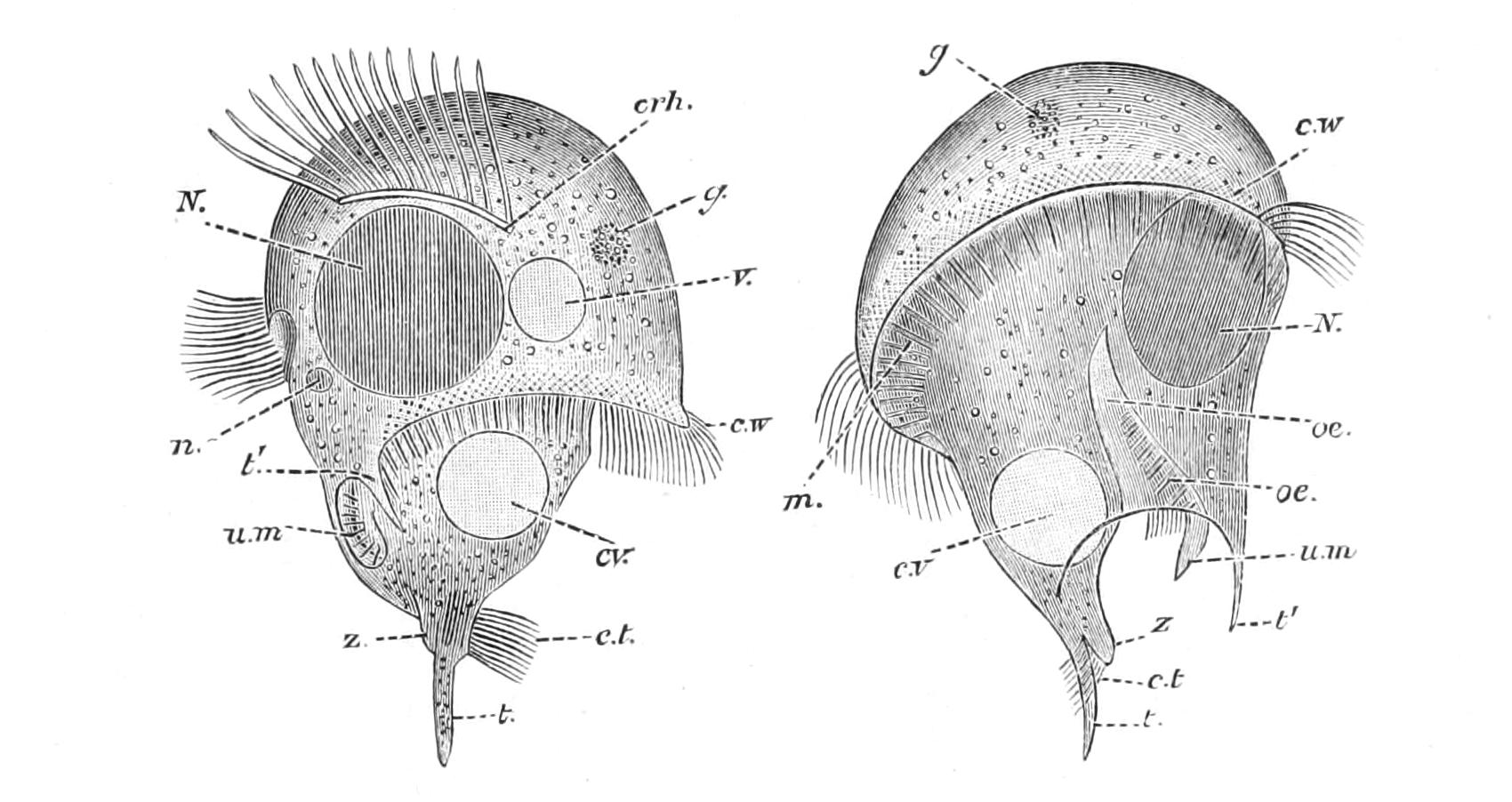
Fig. 58.—Caenomorpha uniserialis. crh, Zone of cirrhi; c.t, cilia of tail; c.v, contractile vacuole; c.w, ciliary wreath; g, granular aggregate; m, zone of membranellae; N, meganucleus; n, micronucleus; oe, pharynx; t, tail-spine; t1, accessory spine; u.m, undulating membrane; v, vacuole; z, precaudal process. (After Levander.)
Among Oligotrichaceae, Halteria, common among the débris at the bottom of pools in woods containing dead leaves, is remarkable for an equatorial girdle of very long fine setae, and for its rapid erratic darting movements, alternating with a graceful bird-like hover. The Tintinnidae are mostly marine, pelagic, with the general look of a stalkless Vorticella; some have a latticed chitinous shell.[173]
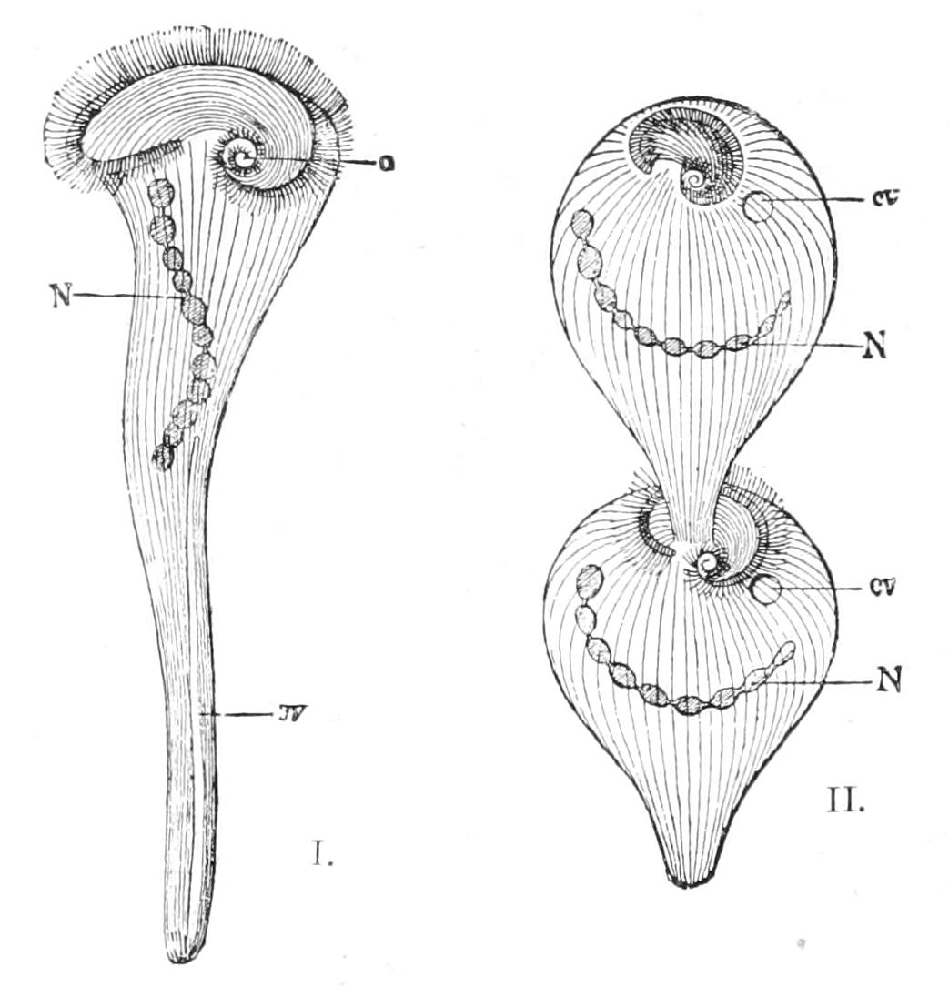
Fig. 59.—Stentor polymorphus. I, Young individual attached, extended; II, adult in fission, contracted; cv in I, afferent canal of contractile vacuole; in II, contractile vacuole; N, moniliform meganucleus (micronuclei omitted); o, mouth; the fine lines are the myoneme fibrils. (From Verworn.)
Among Peritrichaceae, Vorticella (Fig. 60) and its allies have long been known as Bell-animalcules to every student of pond-life. The body has indeed the form of an inverted bell, closed at its mouth by the "peristome," or oral disc; this is a short, inverted truncate cone set obliquely so that its wide base hardly projects at one side, but is tilted high on the other; the edge of the bell is turned out into a rim or "collar," separated from the disc by a deep gutter. The collar, habitually everted, or even turned down, contracts over the retracted disc when the animal is retracted (E2), which is brought about by any sort of shock, or when it swims freely backwards. For the latter purpose a posterior ring of cilia (or rather membranellae) is developed round the hinder end of the bell (A, cr, E3). The cilia of the adoral wreath are very strong, united at the base into a continuous membrane, and indeed themselves partake of the composite nature of membranellae. The wreath forms more than one turn of a right-handed spiral, the innermost turn ending abruptly on the disc, the outer leading down into the mouth at the point where the disc is most tilted and the groove deepest.[174] The pharynx (p) is long, and contains an undulating membrane (u.m) on its inner side projecting out through the mouth, and numerous cilia; it leads deep into the body (p). The first part is distinguished as the "vestibule" (v), as into it opens the anus, and the contractile vacuole (c.v.), the latter sometimes opening by a reservoir (r). The body contains in the ectoplasm myonema-fibrils which, by their contraction, withdraw the disc, and at the same time circular fibrils close the peristome over it. In the type-genus the pellicle is continued into a long, slender elastic stalk (s), of which the longitudinal myoneme fibrils of the ectoplasm converge to the stalk, and are prolonged into it as a spirally winding fibre, sometimes transversely striated.[175] The effect of the contraction of this is to pull the stalk into a helicoid spiral (like a coil-spring), with the line of insertion of the muscle along the inner side of the coils, which is, of course, the shortest path from one end to the other (Fig. 60, B).
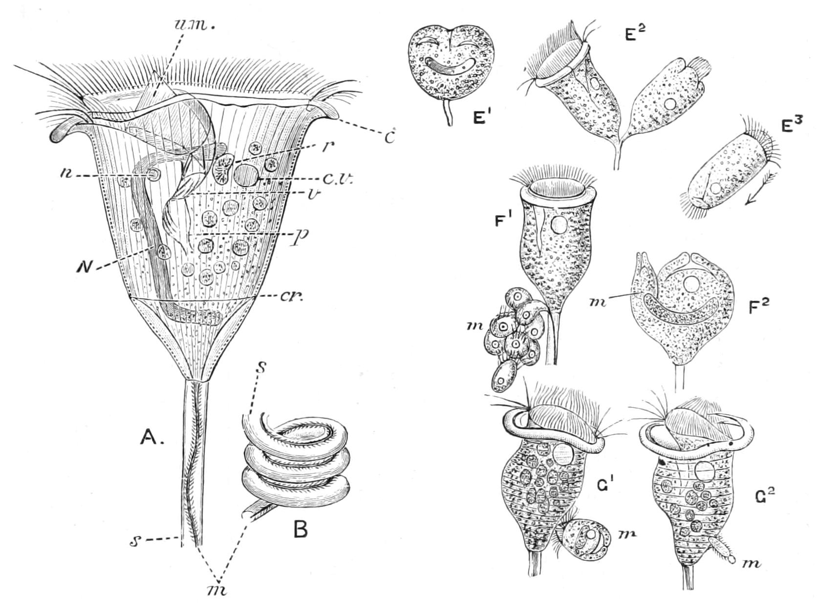
Fig. 60.—Vorticella. A, expanded; B, stalk in contraction; c, eversible collar below peristome; cr, line of posterior ciliary ring; c.v, contractile vacuole; m, muscle of stalk; N, meganucleus; n, micronucleus; p, pharynx; r, reservoir of contractile vacuole; s, tubular stalk; u.m, undulating membrane in vestibule; v, hinder end of vestibule. E1, E2, two stages in binary fission; E3, free zooid, with posterior wreath; F1, F2 division into mega- and micro-zooids (m); G1, G2, conjugation; m, microzooid. (Modified from Bütschli, from Parker and Haswell.)
The members of the Vorticellidae are very commonly attached to weeds or to various aquatic Metazoa, each species being more or less restricted in its haunts. Vorticella, the type, is singly attached to a contractile stalk; fission takes place in the vertical plane, and one of the two so formed retains the original stalk, while the other swims off (Fig. 60, E1-E3), often to settle close by, so that the individuals are found in large social aggregates, side by side, fringing water-weeds with a halo visible to the naked eye, which disappears on agitation by the sudden contraction of all the stalks. Carchesium and Zoothamnium differ from Vorticella in the fact that the one daughter-cell remains attached by a stalk coming off a little below the body of the other, so as to give rise to large branching colonies.
In Carchesium (Fig. 51) the muscular threads of each cell are separate, while in Zoothamnium they are continuous throughout the colony. Epistylis has a solid, rigid stalk, and may give rise to branching colonies, which often infest the body of the Water-Fleas (Copepoda) of the genus Cyclops. Opercularia is characterised by the depth of the gutter, the height of the collar, and the tapering downward of the elongated disc. Vaginicola, Pyxicola, Cothurnia, Scyphidia, all inhabit tubes, some of extreme elegance. Ophrydium is a colonial form, found in ponds and ditches, resembling Opercularia, but inhabiting tubes of jelly[176] that coalesce by their outer walls into a large floating sphere; it usually contains the green symbiotic Flagellate Zoochlorella. Trichodina is free, short, and cylindrical, with both wreaths permanently exposed, and is provided with a circlet of hooks within the aboral wreath. It is often parasitic, or perhaps rather epizoic, on the surface of Hydra (see p. 254), gliding over its body[177] with a graceful waltzing movement; it occurs also in the bladder and genito-urinary passages of Newts, and even in their body-cavity and kidneys.
II. Suctoria = Tentaculifera
Infusoria with cilia only in the young state,[178] without mouth or anus, but absorbing food (usually living Ciliates) by one or more tentacles, perforated at the apex; mostly attached, frequently epizoic, rarely parasitic in the interior of other Protozoa.
Acineta, Ehrb. (Fig. 61, 2); Amoebophrya, Koppen; Choanophrya, Hartog (Fig. 62); Dendrocometes, St. (Fig. 61, 4); Dendrosoma, Ehrb. (Fig. 61, 9); Endosphaera, Engelm.; Ephelota, Str. Wright (Fig. 61, 5, 8); Hypocoma, Gruber; Ophryodendron, Cl. and L. (Fig. 61, 7); Podophrya, Ehrb. (Fig. 61, 1); Rhyncheta, Zenker (Fig. 61, 3); Sphaerophrya, Cl. and L. (Fig. 61, 6), Suctorella, Frenzel; Tokophrya, Bütschli.
This group, despite a superficial resemblance to the Heliozoa, show a close affinity to the Ciliata; the nuclear apparatus is usually double though a micronucleus is not always seen; the young are always ciliated, and the mode of conjugation is identical in all cases hitherto studied. Most of the genera are attached by a chitinous stalk (Fig. 61), continued in Acineta into a cup or "theca" surrounding the cell. The pellicle is firm, often minutely shagreened or "milled" in optical section by fine radial processes, whether superficial rods or the expression of the meeting edges of radial alveoli is as yet uncertain. The pellicle closely invests the ectosarc, is continued down into a tubular sheath, from the base of which the tentacle rises, and upwards to invest the tentacle, and is even prolonged into its cavity in Choanophrya, the only genus where the tentacles are large enough for satisfactory demonstration. These organs may be one or more, and vary greatly in character. They may be (1) pointed for prehension, puncture, and suction (Ephelota, Fig. 61, 5); (2) nearly cylindrical, with a slightly "flared" truncate apex (Podophrya, Fig. 61, 1a); (3) filiform with a terminal knob; (4) "capitate" (Acineta, Fig. 61, 2); (5) bluntly truncate and capable of opening into a wide funnel for the suction of food[179] (Choanophrya, Fig. 62; Rhyncheta, Fig. 61, 3). Their movements, too, are varied, including retraction and protrusion, and a degree of flexion which reaches a maximum in Rhyncheta (Fig. 61, 3), whose tentacle is as freely motile as an elephant's trunk might be supposed to be were it as slender in proportion to its length. They are continued into the body, and in Choanophrya may extend right across it. In Podophrya trold the pellicle rises into a conical tube about the base of the tentacle, which is retracted through it completely with the prey in deglutition. In Dendrocometes, Dendrosoma, and Ophryodendron (Fig. 61, 4, 9, 7), the tentacles arise from outgrowths of the cell-body.
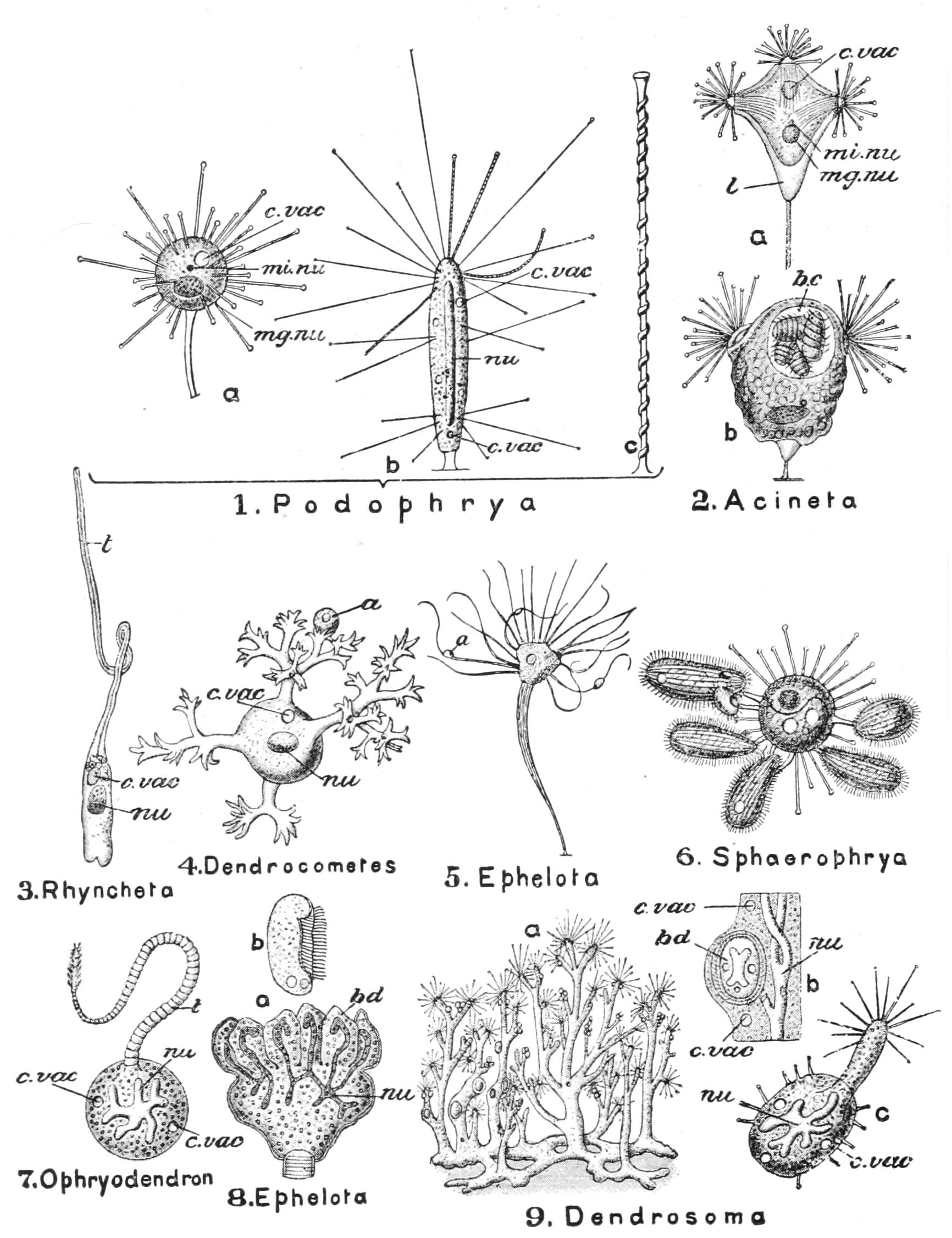
Fig. 61.—Various forms of Suctoria. 1, a and b, two species of Podophrya; c, a tentacle much enlarged; 2, a, Acineta jolyi; 2, b, A. tuberosa, with four ciliated buds; in 6 the animal has captured several small Ciliata; 8, a, a specimen multiplying by budding; 8, b, a free ciliated bud; 9, a, the entire colony; 9, b, a portion of the stem; 9, c, a liberated bud. a, Organism captured as food; b.c, brood-cavity; bd, bud; c.vac, contractile vacuole; l, test; mg.nu, meganucleus; mi.nu, micronucleus; nu, nucleus; t, tentacle. (From Parker and Haswell, after Bütschli and Saville Kent.)
The mechanism of suction is doubtful; but from the way particles from a little distance flow into the open funnels of Choanophrya, it may be the result of an increase of osmotic pressure. The external pellicle of the tentacles is marked by a spiral constriction,[180] which may be prolonged over the part included in the sheath. The endosarc is rich in oil-drops, often coloured, and in proteid granules which sometimes absorb stains so readily as to have been named "tinctin bodies." It usually contains at least one contractile vacuole.
In Dendrocometes (and perhaps others) the whole cell may become ciliated, detach itself and swim off; this it does when its host (Gammarus) moults its cuticle.
In fission or budding we have to distinguish many modes. (1) In the simplest, after the nuclear apparatus has divided, the cell divides transversely; the distal half acquires cilia and swims off to attach itself elsewhere, while the proximal remains attached. The tentacles have previously disappeared and have to be formed afresh in both. (2) More commonly fission passes into budding on the distal face; a sort of groove deepens around a central prominence which becomes the ciliated larva (Fig. 62, em); the tentacles of the "parent" are retained. This process passes into (3) "internal budding," where a minute pit leads into a bottle-shaped cavity.[181] (4) Again, the budding may be multiple, the meganucleus protruding a branch for each bud, while the micronucleus, by successive divisions, affords the supply requisite. Sphaerophrya (Fig. 61, 6) and Endosphaera multiply freely by fission within their Ciliate hosts, and were indeed described by Stein as stages in their life-cycle. Conjugation of the same type as in most Ciliates has been fully worked out in Dendrocometes alone, by Hickson,[182] who has found the meganuclei (though destined to disorganisation) conjugate for a short time by the bridge of communication before the reciprocal conjugation of the micronuclei.
We have referred to the endoparasitism of two genera. Amoebophrya lives in several Acanthometrids, and in the aberrant Radiolarian Sticholonche (see p. 86). The attached species are some of them indifferent to their base; others are only found on Algae, or again only epizoic on special Metazoan hosts, or even on special parts of these. Thus Rhyncheta is only found on the couplers of the thoracic limbs of Cyclops, and Choanophrya on the ventral surface of its head and the adjoining appendages.
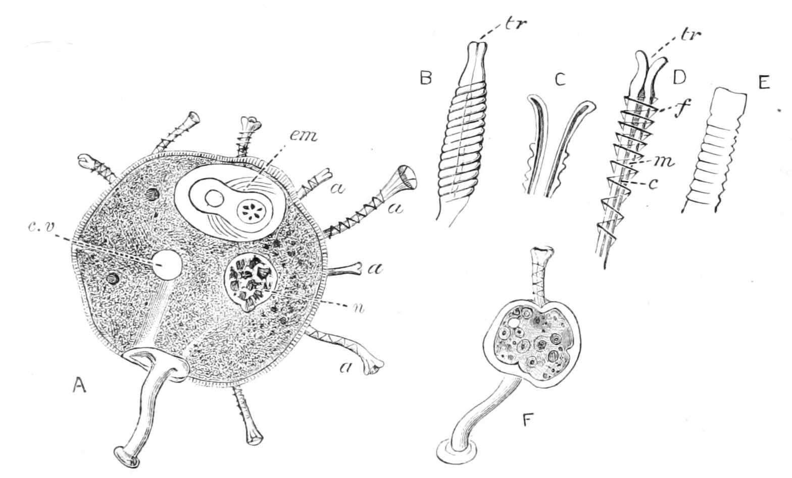
Fig. 62.—Choanophrya infundibulifera. A, adult; B-D, tentacles in action in various stages; E, tentacle at rest; F, young, just settled down, a, a, a, Tentacles in various stages of activity; c, central cavity; c.v, contractile vacuole; em, ciliated embryo showing contractile vacuole and nucleus; f, spiral ridge; m, muscular wall of funnel; n, nucleus; tr, opening of funnel. (A-D, F, modified after Zenker; E, original.)
We owe our knowledge of this group to the classical works of Ehrenberg, Claparède and Lachmann, Stein, R. Hertwig, and Bütschli. Plate has shed much light on Dendrocometes, and Hickson has studied its conjugation. Ischikawa[183] has utilised modern histological methods for the cytological study of Ephelota bütschliana. René Sand has written a useful, but unequal, and not always trustworthy monograph of the Order,[184] containing an elaborate bibliography.