Chapter IV
Reading edition
PROTOZOA (CONTINUED): SPOROZOA[100]
II. Sporozoa.
Protozoa parasitic in Metazoa, usually intracellular for at least part of their cycle, rarely possessing pseudopodia, or flagella (save in the sperms), never cilia; reproduction by brood-formation, often of alternating types; syngamy leading up to resting spores in which minute sickle-germs are formed, or unknown (Myxosporidiaceae).
This group, of which seven years ago no single species was known in its complete cycle, has recently become the subject of concentrated and successful study, owing to the fact that it has been recognised to contain the organisms which induce such scourges to animals as malarial fevers, and various destructive murrains. Our earliest accurate, if partial knowledge, was due to von Siebold, Kölliker, and van Beneden. Thirty years ago Ray Lankester in England commenced the study of species that dwell in the blood, destined to be of such moment for the well-being of man and the animals in his service; and since then our knowledge has increased by the labours of Manson, Ross and Minchin at home, Laveran, Blanchard, Thélohan, Léger, Cuénot, Mesnil, Aimé Schneider in France, Grassi in Italy, Schaudinn, Siedlecki, L. and R. Pfeiffer, Doflein in Central Europe, and many others.
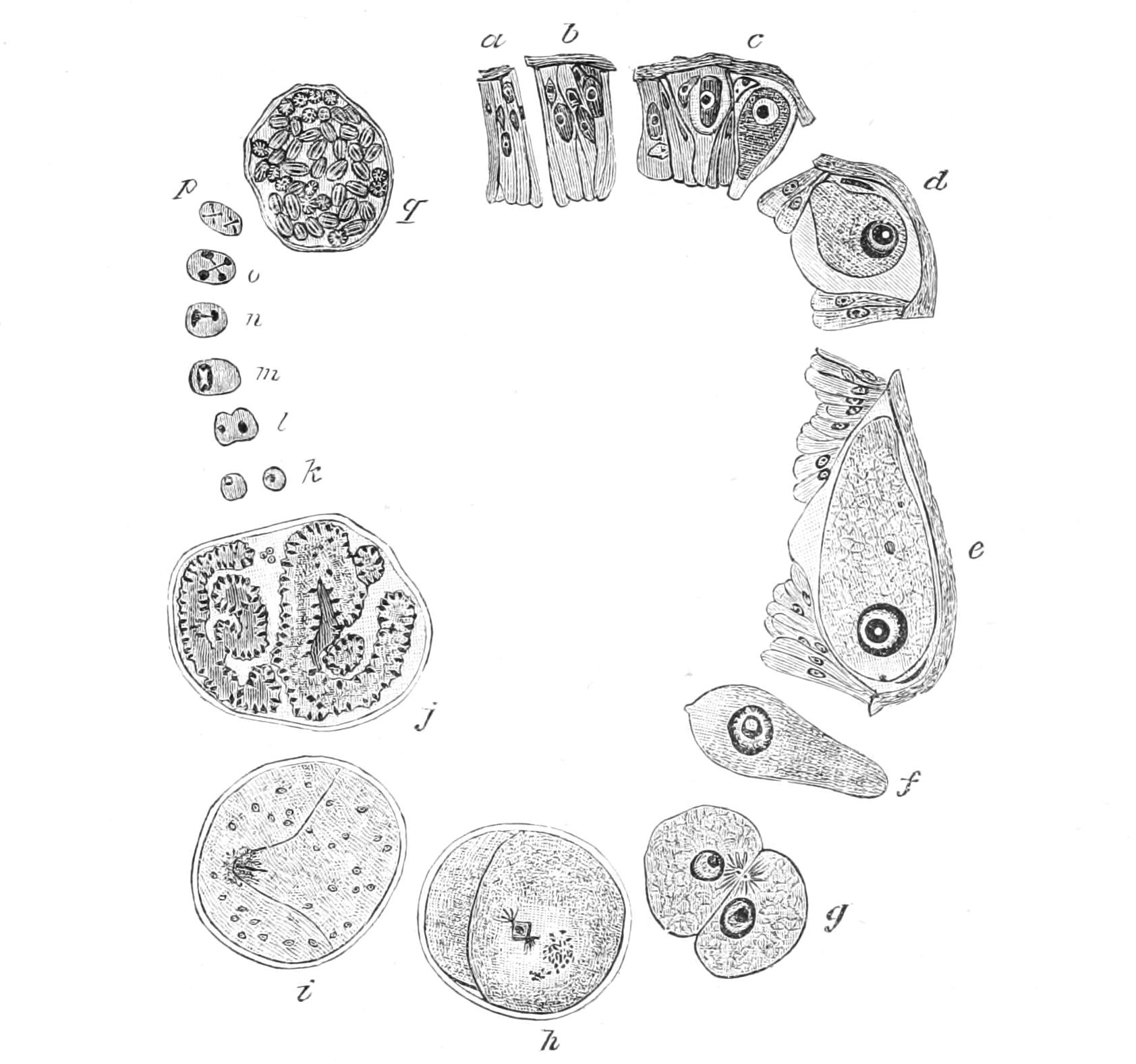
Fig. 31.—Lankesteria ascidiae, showing life-cycle. a, b, c, Sporozoites in digestive epithelium cells of host; d, e, growth stages; f, free gregarine; g, association; h, encystment; i, j, brood-divisions in associated mates; k, pairing-cells; l, syngamy; m, zygote; n, o, p, nuclear divisions in spores; q, cyst with adult spores, each containing 8 sickle-germs. (After Luhe, modified from Siedlecki.)
As a type we will take a simple form of the highest group, the Gregarinidaceae, Monocystis, which inhabits the seminal vesicles of the earthworm. In its youngest state, the "sporozoite," it is a naked, sickle-shaped cell, which probably makes its way from the gut into one of the large radial cells of the seminal funnel, where it attains its full size, and then passes out into the vesicles or reservoirs of the semen, to lie among the sperm morulae and young spermatozoa. The whole interior is formed of the opaque endosarc, which contains a large central nucleus, and is full of refractive granules of paramylum or paraglycogen,[101] a carbohydrate allied to glycogen or animal starch, so common in the liver and muscles of Metazoa; besides these it contains proteid granules which stain with carmine, and oil-drops. The ectosarc is formed of three layers: (1) the outer layer or "cuticle"[102] is, in many cases if not here, ribbed, with minute pores in the furrows, and is always porous enough to allow the diffusion of dissolved nutriment; (2) a clear plasmatic layer, the "sarcocyte"; (3) the "myocyte," formed of "myonemes," muscular fibrils disposed in a network with transverse meshes, which effect the wriggling movements of the cell. The endosarc contains the granules and the large central nucleus. The adult becomes free in the seminal vesicles; here two approximate, and surround themselves with a common cyst: a process which has received the name of "association" (Fig. 31, g-i). Within this, however, the protoplasms remain absolutely distinct. The nucleus undergoes peculiar changes by which its volume is considerably reduced. When this process of "nuclear reduction" is completed, each of the mates undergoes brood-divisions (j), so as to give rise to a large number of rounded naked 1-nucleate cells—the true pairing-cells. These unite two and two, and so form the 1-nucleate spores (k-m), which become oat-shaped, form a dense cyst-wall, and have been termed "pseudonavicellae" from their likeness to the Diatomaceous genus Navicella. Some of the cytoplasm of the original cells remains over unused, as "epiplasm," and ultimately degenerates, as do a certain number of the brood-cells which presumably have failed to pair. It is believed that the brood-cells from the same parent will not unite together. The contents of each spore have again undergone brood-division to form eight sickle-shaped zoospores, or "sporozoites" (n-q), and thus the developmental cycle is completed. Probably the spores, swallowed by birds, pass out in their excrement, and when eaten by an earthworm open in its gut; the freed sickle-germs can now migrate through the tissues to the seminal funnels, in the cells of which they grow, ultimately becoming free in the seminal vesicles.[103]
We may now pass to the classification of the group.
A. Telosporidia.—Cells 1-nucleate until the onset of brood-formation, which is simultaneous.
1. Gregarinidaceae.—Cells early provided with a firm pellicle and possessing a complex ectosarc; at first intracellular, soon becoming free in the gut or coelom of Invertebrates. Pairing between adults, which simultaneously produce each its brood of gametes, isogamous or bisexual, which pair within the common cyst; zygotospores surrounded by a firm cyst, and producing within a brood of sickle-shaped zoospores.
(i.) Schizogregarinidae.—Multiplying by simple fission in the free state as well as by brood-formation; the brood-cells conjugating in a common cyst, but producing only one pairing nucleus in each mate (the rest aborting), and consequently only one spore.
Ophryocystis A. Schn.
(ii.) Acephalinidae.—Cell one-chambered, usually without an epimerite for attachment.
Monocystis F. Stein; Lankesteria Mingazzini.
(iii.) Dicystidae.—Cell divided by a plasmic partition; epimerite usually present.
Gregarina Dufour; Stylorhynchus A. Schn.; Pterocephalus A. Schn.
2. Coccidiaceae.—Cells of simple structure, intracellular in Metazoa. Pairing between isolated cells usually sexually differentiated as oosphere and sperm, the latter often flagellate. Brood-formation of the adult cell giving rise to sickle-shaped zoospores (merozoites), or progamic and producing the gametes. Oosperm motile or motionless, finally producing a brood of spores, which again give rise to a brood of sickle-spores.
(i.) Coccidiidae.—Cell permanently intracellular, or very rarely coelomic, encysting or not before division; zoospores always sickle-shaped; oosperm encysting at once, producing spores with a dense cell-wall producing sickle-germs.
(ii.) Haemosporidae.—Cells parasitic in the blood corpuscles or free in the blood of cold-blooded animals, encysting before brood-formation; zoospores sickle-shaped; oosperm at first motile.
Lankesterella Labbé; (Drepanidium Lank.;) Karyolysus Labbé; Haemogregarina Danilewski.
(iii.) Acystosporidae.—Cells parasitic in the blood and haematocytes of warm-blooded Vertebrates; never forming a cyst-wall before dividing; zoospores formed in the corpuscles, amoeboid. Gametocytes only forming gametes when taken into the stomach of insects. Oosperm at first active, passing into the coelom, producing naked spores which again produce a large brood of sickle zoospores, which migrate to the salivary gland, and are injected with the saliva into the warm-blooded host.
Haemamoeba Grassi and Feletti; Laverania Grassi and Feletti; Haemoproteus Kruse; Halteridium Labbé.[104]
B. Neosporidia.—Cells becoming multinucleate apocytes before any brood-formation occurs. Brood-formation progressive through the apocyte, not simultaneous.
1. Myxosporidiaceae.—Naked parasites in cold-blooded animals. Spore-formation due to an aggregation of cytoplasm around a single nucleus to form an archespore, which then produces a complex of cells within which two daughter-cells form the spores and accessory nematocysts.
Myxidium Bütsch.; Myxobolus Bütsch.; Henneguya Thélohan; Nosema Nageli (= Glugea Th.).
2. Actinomyxidiaceae.[105]—Apocyte resolved into a sporange, containing eight secondary sporanges (so-called spores), of ternary symmetry and provided with three polar nematocysts.
3. Sarcosporidiaceae.—Encysted parasites in the muscles of Vertebrates, with a double membrane; spores simple.
Sarcocystis Lankester.
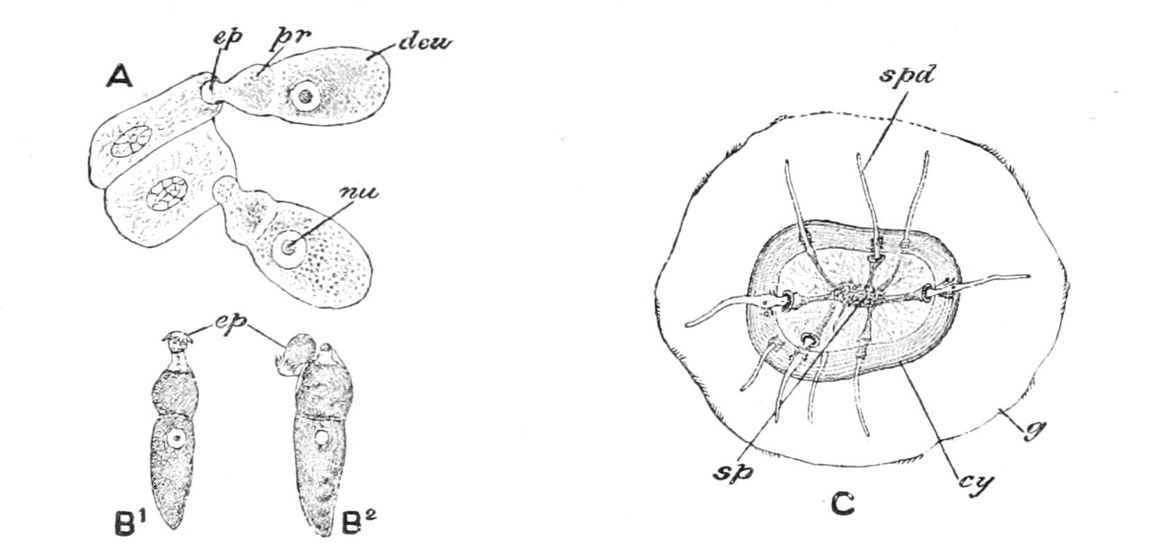
Fig. 32.—Gregarina blattarum Sieb. A, two cephalonts, embedded by their epimerite (ep), in cells of the gut-epithelium; deu, deutomerite; nu, nucleus; pr, protomerite; B1, B2, two free specimens of an allied genus; the epimerite is falling off in B2, which is on its way to become a sporont; C, cyst (cy) of A, with sporoducts (spd) discharging the spores (sp), surrounded by an external gelatinous investment (g). (From Parker and Haswell.)
Monocystis offers us the simplest type of Gregarinidaceae. In most Gregarines (Figs. 31, 32) the sporozoite enters the epithelium-cell of the gut of an Arthropod, Worm or Mollusc, and as it enlarges protrudes the greater part of its bulk into the lumen, and may become free therein, or pass into the coelom. The attached part is often enlarged into a sort of grapple armed with spines, the "epimerite"; this contains only sarcocyte, the other layers being absent. The freely projecting body is usually divided by an ingrowth of the myocyte into a front segment ("protomerite"), and a rear one ("deutomerite"), with the nucleus usually in the latter. In this state the cell is termed a "cephalont." Conjugation is frequent, but apparently is not always connected with syngamy or spore-formation; sometimes from two to five may be aggregated into a chain or "syzygy." The number of cases in which a syngamic process between two cells has been observed is constantly being increased. In Stylorhynchus (Fig. 33) the conjugation at first resembles that of Monocystis, but the actual pairing-cells are bisexually differentiated into sperms in the one parent, and oospheres in the other; it is remarkable that here the pear-shaped sperms are apparently larger than the oospheres. In Pterocephalus the chief difference is that the sperms are minute.[106] In all cases of spore-formation the epimerite is lost and the septum disappears; in this state the cell is termed a sporont. Sometimes the epiplasm of the sporont forms tubes ("sporoducts"), which project through the cyst-wall and give exit to the spores, as in Gregarina (Fig. 32, C), a parasite in the beetle Blaps.
Gregarines infest most groups of Invertebrates except Sponges and perhaps Coelenterates, the only exception cited being that of Epizoanthus glacialis, a Zoantharian (p. 406). They appear to be relatively harmless and are not known to induce epidemics.
The Coccidiaceae never attain so high a degree of cellular differentiation as the Gregarines, which may be due to their habitat; for in the growing state they are intracellular parasites. Their life-history shows a double cycle, which has been most thoroughly worked out in Coccidiidae by Schaudinn and Siedlecki in parasites of our common Centipedes. We take that of Coccidium schubergi (in Lithobius forficatus[107]), beginning with the sporozoite, which is liberated from the spores taken in with the food, in the gut of the Centipede. This active sickle-shaped cell (Fig. 34, l) enters an epithelial cell of the mid-gut, and grows therein till it attains its full size (a), when it is termed a "schizont"; for it segments (Gk. σχίζω, "I split") superficially into a large number of sickle-shaped zoospores, the "merozoites" (c), resembling the sporozoites. The segmentation is superficial, so that there may remain a large mass of residual epiplasm. The merozoites are set free by the destruction of the epithelium-cell in which they were formed, and which becomes disorganised, like the residual epiplasm. Each merozoite may repeat the behaviour of the sporozoite, so that the disease spreads freely, and becomes acute after several reinfections. After a time the adult parasites, instead of becoming schizonts and simply forming merozoites by division, differentiate into cells that undergo a binary sexual differentiation. Some cells, the "oocytes" (d, e), escape into the gut, and the nucleus undergoes changes by which some of its substance (or an abortive daughter-nucleus) is expelled to the exterior (f), such a cell is now an "oogamete" or oosphere. Others, again, are spermatogones (h): each when full grown on escaping into the gut commences a division (i, j), like that of the schizonts. The products of this division or segment-cells are the flagellate sperms (s): they are more numerous and more minute than the merozoites produced by the schizonts, and are attracted to the oosphere by chemiotaxy (p. 23), and one enters it and fuses with it (g). The oosperm, zygote or fertilised egg, thus formed invests itself with a dense cyst-wall, as a "oospore" (k), its contents form one or more (2, 4, 8, etc.) spores; and each spore forms again one, two, or four sickle-shaped zoospores ("sporozoites"), destined to be liberated for a fresh cycle of parasitic life when the spores are swallowed by another host.
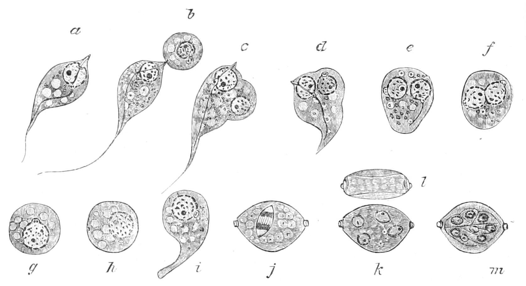
Fig. 33.—Bisexual pairing of Stylorhynchus. a, Spermatozoon; b-e, fusion of cytoplasm of spermatozoon and oosphere; f, g, fusion of nuclei; h-j, development of wall to zygote; k, l, formation of four sporoblasts; l, side view of spore; m, mature sporozoites in spore. (After Léger.)
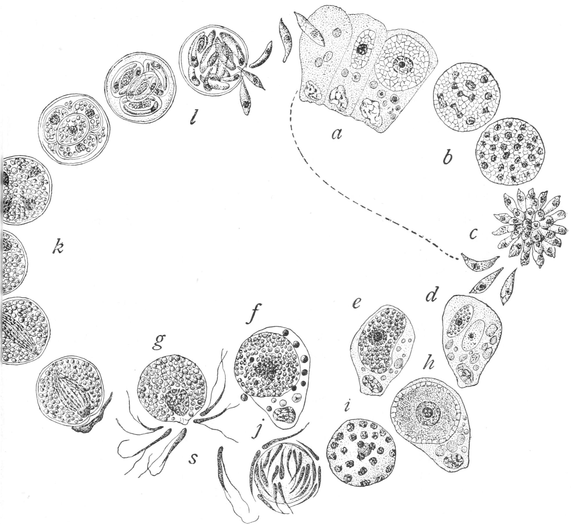
Fig. 34.—Life-history of Coccidium schubergi. a, Penetration of epithelium-cell of host by sporozoite; b-d, stages of multiple cell-formation in naked state (schizogony); e, f, formation of oogamete; g, conjugation; h-j, formation of sperms (s); k, development of zygote (fertilised ovum) to form four spores; l, formation of two zoospores (or sickle germs) in each spore. (From Calkins's Protozoa, after Schaudinn.)
In some cases the oogametes are at first oblong, like ordinary merozoites, and round off in the gut. The microgametocyte, or spermatogone, has the same character, but is smaller; it applies itself like a cap to one pole of the oogamete, which has rounded off; it then divides into four sperms, whose cytoplasm is not sharply separated; one of these then separates from the common mass, enters the oogamete, and so conjugation is effected, with an oosperm as its result. This latter mode of conjugation is that of Adelea ovata and Coccidium lacazei: the former is probably the more primitive and the commoner. The sperms of Coccidiidae, when free, usually possess two long flagella, either both anterior, or a very long one in front and a short one behind, both turned backwards.
The genus Coccidium affects many animals, and one species in particular, C. cuniculi Rivolta, attacks the liver of young rabbits,[108] giving rise to the disease "coccidiosis." Coccidium may also produce a sort of dysentery in cattle on the Alpine pastures of Switzerland; and cases of human coccidiosis are by no means unknown. Coccidium-like bodies have been demonstrated in the human disease, "molluscum contagiosum," and the "oriental sore" of Asia; similar bodies have also been recorded in smallpox and vaccinia, malignant tumours and even syphilis, but their nature is not certainly known; some of these are now referred to Flagellata (see p. 121).
Closely allied to the Coccidiidae are the Haemosporidae, dwellers in the blood of various cold-blooded Vertebrates,[109] and entering the corpuscles as sporozoites or merozoites to attain the full size, when they divide by schizogony; they are freed like those of the next family by the breaking up of the corpuscle. The merozoites were described by Gaule (1879) as "vermicles" ("Würmchen"), and regarded by him as peculiar segregation-products of the blood; though Lankester had described the same species in the Frog's blood as early as 1871, with a full recognition of its true character. His name, Drepanidium, has had to give way, having been appropriated to another animal, and has been aptly replaced by that of Lankesterella. The sexual process of Karyolysus has been found to take place in a Tick, that of Haemogregarina in a Leech, thus presenting a close analogy to the next group, which only differs in its less definite form in the active state, and in the lack of a cell-wall during brood-formation.
Laveran was the first to describe a member of the Acystosporidae, in 1880, as an organism always to be found in the blood of patients suffering from malarial fever; this received the rather inappropriate name of Plasmodium, which, by a pedantic adherence to the laws of priority, has been used by systematists as a generic name. Golgi demonstrated the coincidence of the stages of the intermittent fever with those of the life-cycle of the parasite in the patient, the maturation of the schizont and liberation of the sporozoites coinciding with the fits of fever. Manson, who had already shown that the Nematodes of the blood that give rise to Filarial haematuria (see Vol. II. p. 149) have an alternating life in the gnats or mosquitos of the common genus Culex,[110] in 1896 suggested to Ronald Ross that the same might apply to this parasite, and thus inspired a most successful work. The hypothesis had old prejudices in its favour, for in many parts there was a current belief that sleeping under mosquito-netting at least helped other precautions against malaria. Ross found early in his investigations that Culex was a good host for the allied genus Haemoproteus or Proteosoma, parasitic in birds, but could neither inoculate man with fever nor be inoculated from man. He found, however, that the malaria germs from man underwent further changes in the stomach of a "dappled-wing mosquito," that is, as we have since learned, a member of the genus Anopheles. Thenceforward the study advanced rapidly, and a number of inquirers, including Grassi, Koch, MacCallum (who discovered the true method of sexual union in Halteridium[111]), and Ross himself, completed his discovery by supplying a complete picture of the life-cycles of the malaria-germs. Unfortunately, there has been a most unhappy rivalry as to the priority of the share in each fragment of the discovery, whose history is summarised by Nuttall, we believe, with perfect fairness.[112]
The merozoite is always amoeboid, and in this state enters the blood corpuscle; herein it attains its full size, as a schizont, becoming filled with granules of "melanin" or black pigment, probably a decomposition product of the red colouring matter (haemoglobin).
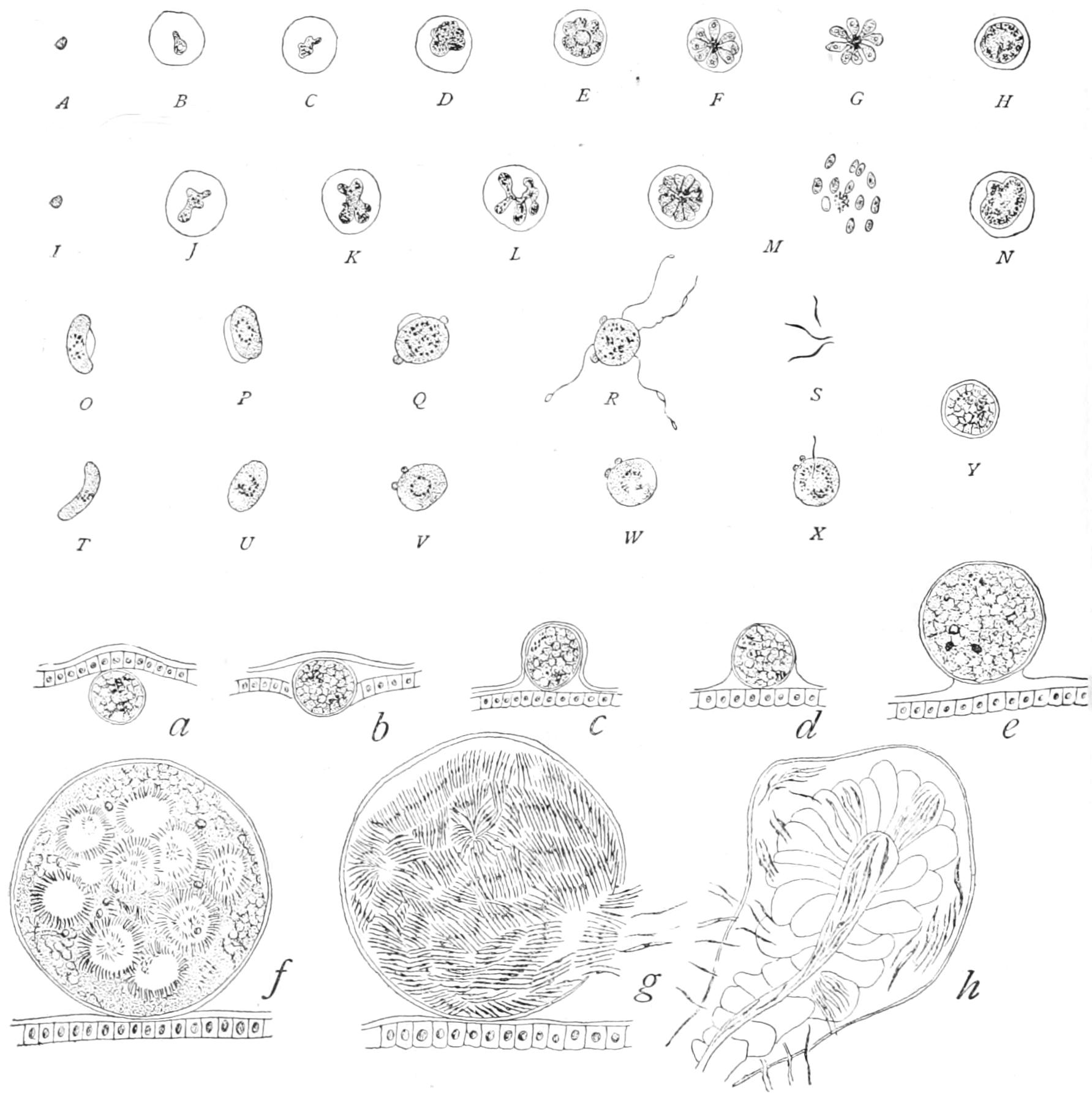
Fig. 35.—Life-history of Malarial Parasites. A-G, Amoebula of quartan parasite to sporulation; H, its gametocyte; I-M, amoebula of tertian parasite to sporulation; N, its gametocyte; O, T, "crescents" or gametocytes of Laverania; P-S, sperm-formation; U-W, maturation of oosphere; X, fertilisation; Y, zygote. a, Zygote enlarging in gut of Mosquito; b-e, passing into the coelom; f, the contents segmented into naked spores; g, the spores forming sickle-germs or sporozoites; h, sporozoites passing into the salivary glands. (From Calkins's Protozoa, after Ross and Fielding Ould.)
The nucleus of the schizont now divides repeatedly, and then the schizont segments into a flat brood of germs (merozoites), relatively few in the parasite of quartan fever (Haemamoeba malariae, Fig. 35, E-G), many in that of tertian (H. vivax, Fig. 35, M). These brood-cells escape and behave for the most part as before. But after the disease has persisted for some time we find that in the genus Haemamoeba, which induces the common malarial fevers of temperate regions, certain of the full-grown germs, instead of behaving as schizonts, pass, as it were, to rest as round cells; while in the allied genus Laverania, (Haemomenas, Ross) these resting-cells are crescentic, with blunt horns, and are usually termed half-moons (Fig. 35, O, T), characteristic of the bilious or pernicious remittent fevers of the tropics and of the warmer temperate regions in summer. These round or crescent-shaped cells are the gametocytes, which only develop further in the drawn blood, whether under the microscope, protected against evaporation, or in the stomach of the Anopheles: the crescents become round, and then they, like the already round ones of Haemamoeba, differentiate in exactly the same way as the corresponding cells of Coccidium schubergi. The female cell only exhibits certain changes in its nucleus to convert it into an oosphere: the male emits a small number of sperms, long flagellum-like bodies, each with a nucleus; and these, by their wriggling, detach themselves from the central core, no longer nucleated. The male gametogonium with its protruded sperms was termed the "Polymitus form," and was by some regarded as a degeneration-form, until MacCallum discovered that a "flagellum" regularly undergoes sexual fusion with an oosphere in Halteridium, as has since been found in the other genera. The oosperm (Y) so formed is at first motile ("ookinete"), as it is in Haemosporidae, and passes into the epithelium of the stomach of the gnat and then through the wall, acquiring a cyst-wall and finally projecting into the coelom (a-e). Here it segments into a number of spheres ("zygotomeres" of Ross) corresponding to the Coccidian spores, but which never acquire a proper wall (f). These by segmentation produce at their surface an immense quantity of elongated sporozoites (the "zygotoblasts" or "blasts" of Ross, Fig. 35, g), these are ultimately freed by the disappearance of the cyst-wall of the oosperm, pass through the coelom into the salivary gland (h), and are discharged with its secretion into the wound that the gnat inflicts in biting. In the blood the blasts follow the ordinary development of merozoites in the blood corpuscle, and the patient shows the corresponding signs of fever. This has been completely proved by rearing the insect from the egg, feeding it on the blood of a patient in whose blood there were ascertained to be the germs of a definite species of Haemamoeba, sending it to England, where it was made to bite Dr. Manson's son, who had never had fever and whose blood on repeated examination had proved free from any germs. In the usual time he had a well-defined attack of the fever corresponding to that germ, and his blood on examination revealed the Haemamoeba of the proper type. A few doses of quinine relieved him of the consequences of his mild martyrdom to science. Experiments of similar character but of less rigorous nature had been previously made in Italy with analogous results. Again, it has been shown that by mere precautions against the bites of Anopheles, and these only, all residents who adopted them during the malarious season in the most unhealthy districts of Italy escaped fever during a whole season; while those who did not adopt the precautions were badly attacked.[113]
Anopheles flourishes in shallow puddles, or small vessels such as tins, etc., the pools left by dried-up brooks and torrents, as well as larger masses of stagnant water, canals, and slow-flowing streams. Sticklebacks and minnows feed freely on the larvae and keep down the numbers of the species; where the fish are not found, the larvae may be destroyed by pouring paraffin oil on the surface of the water and by drainage. A combination of protective measures in Freetown (Sierra Leone) and other ports on the west coast of Africa, Ismailia, and elsewhere, has met with remarkable success during the short time for which it has been tried; and it seems not improbable, that as the relatively benign intermittent fevers have within the last century been banished from our own fen and marsh districts, so the Guinea coast may within the next decade lose its sad title of "The White Man's Grave."
So closely allied to this group in form, habit, and life-cycle are some species of the Flagellate genus Trypanosoma, that in their less active states they have been unhesitatingly placed here (see p. 119). Schaudinn has seen Trypanosomic characters in the "blasts" of this group, which apparently is the most primitive of the Sporozoa and a direct offshoot of the Flagellates.
The Myxosporidiaceae (Fig. 36) are parasitic in various cold-blooded animals. They are at least binucleate in the youngest free state, and become large and multinucleate apocytes, which may bud off outgrowths as well as reproduce by spores. The spores of the apocyte are not produced by simultaneous breaking up, but by successive differentiation. A single nucleus aggregates around itself a limited portion of the cytoplasm, and this again forms a membrane, becoming an archespore or a "pansporoblast," destined to produce two spores; within this, nuclear division takes place so as to form about eight nuclei, two of which are extruded as abortive, and of the other six, three are used up in the formation of each of the two spores. Of these three nuclei in each spore, two form nematocysts, like those of a Coelenterate (p. 246 f.), at the expense of the surrounding plasm; while the third nucleus divides to form the two final nuclei of the reproductive body. The whole aggregate of the reproductive body and the two nematocysts is enveloped in a bivalve shell. In what we may call germination, the nematocysts eject a thread that serves for attachment, the valves of the shell open, and the binucleate mass crawls out and grows afresh. Nosema bombycis Nägeli (the spore of which has a single nematocyst) is the organism of the "Pébrine" of the silkworm, which was estimated to have caused a total loss in France of some £40,000,000 before Pasteur investigated the malady and prescribed the effectual cure, or rather precaution against its spread. This consisted in crushing each mother in water after it had laid its eggs and seeking for pébrine germs. If the mother proved to be infected, her eggs were destroyed, as the eggs she had laid were certain to be also tainted. Balbiani completed the study of the organism from a morphological standpoint. Some Myxosporidiaceae produce destructive epidemics in fish.
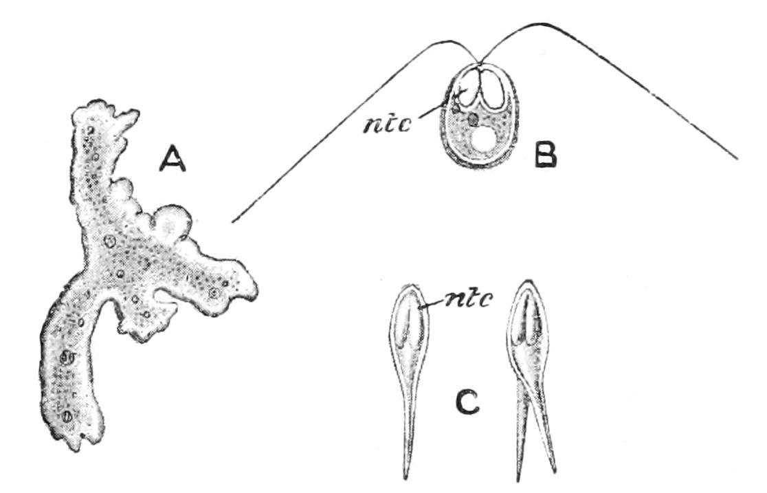
Fig. 36.—A, Myxidium lieberkühnii, amoeboid phase; B, Myxobolus mülleri, spore with discharged nematocysts (ntc); C, spores (psorosperms) of a Myxosporidian. ntc, nematocysts. (From Parker and Haswell.)
The Dolichosporidia or Sarcosporidiaceae are, in the adult state, elongated sacs, often found in the substance of the voluntary muscles, and known as "Rainey's" or "Miescher's Tubes"; they are at first uninucleate, then multinucleate, and then break up successively into uninucleate cells, the spores, in each of which, by division, are formed the sickle-shaped zoospores.[114]