Chapter XVII
Reading edition
ECHINODERMATA (CONTINUED): OPHIUROIDEA = BRITTLE STARS
CLASS II. OPHIUROIDEA
The second class of Eleutherozoa are familiarly known as "Brittle Stars," on account of their tendency, when seized, to escape by snapping off an arm, although this habit is by no means confined to them, but is shared in a marked degree by many Asteroidea, such as Luidia, for instance. Like the Asteroidea, they are "starfish," that is to say, they consist of a disc and of arms radiating from it; but the scientific name Ophiuroidea really expresses the great dominating feature of their organisation. Literally it signifies "Snake-tail" (ὄφις, snake; οὐρά, tail), and thus vividly describes the wriggling, writhing movements of the long thin arms, by means of which the Ophiuroid climbs in and out of the crevices between the stones and gravel in which it lives. This feature, viz. the effecting of movement by means of muscular jerks of the arms, instead of by the slow protrusion and retraction of the tube-feet, is the key to the understanding of most of the points wherein the Brittle Stars differ from the true Starfish.
Asteroidea and Ophiuroidea agree in the common ground-plan of their structure, that is, they both possess arms; but the most obvious difference in their outer appearance is that whereas in Asteroidea the arms merge insensibly into the disc, in Ophiuroidea the disc is circular in outline and is sharply marked off from the arms. Closer inspection shows that in the Ophiuroid the arms are continued inwards along grooves, which run on the under surface of the disc, and that they finally coalesce to form a buccal framework surrounding the mouth. In the very young Ophiuroid the arms melt into a small central disc, as in the Starfish, but the disc of the adult is made up of a series of interradial dorsal outgrowths which meet one another above the arms.
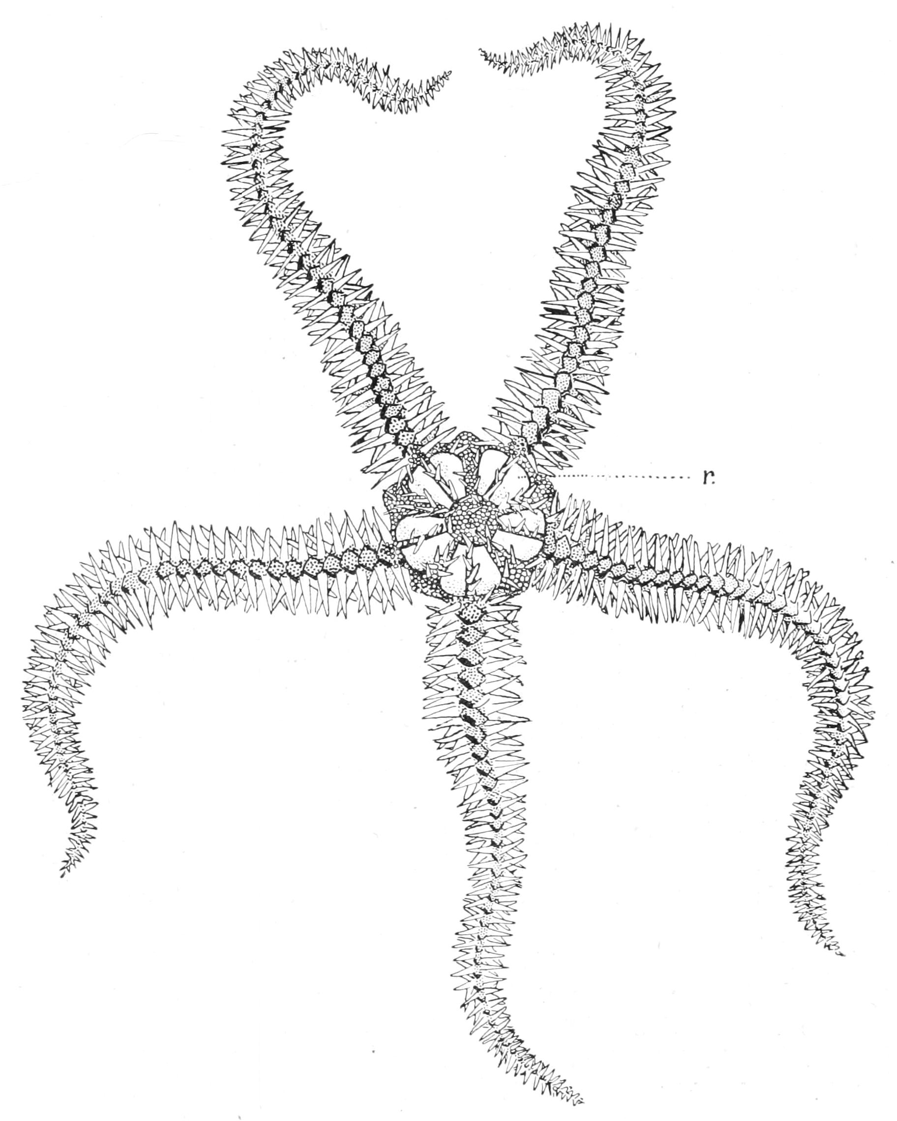
Fig. 207.—Aboral view of Ophiothrix fragilis. × 1. r, Radial plate.
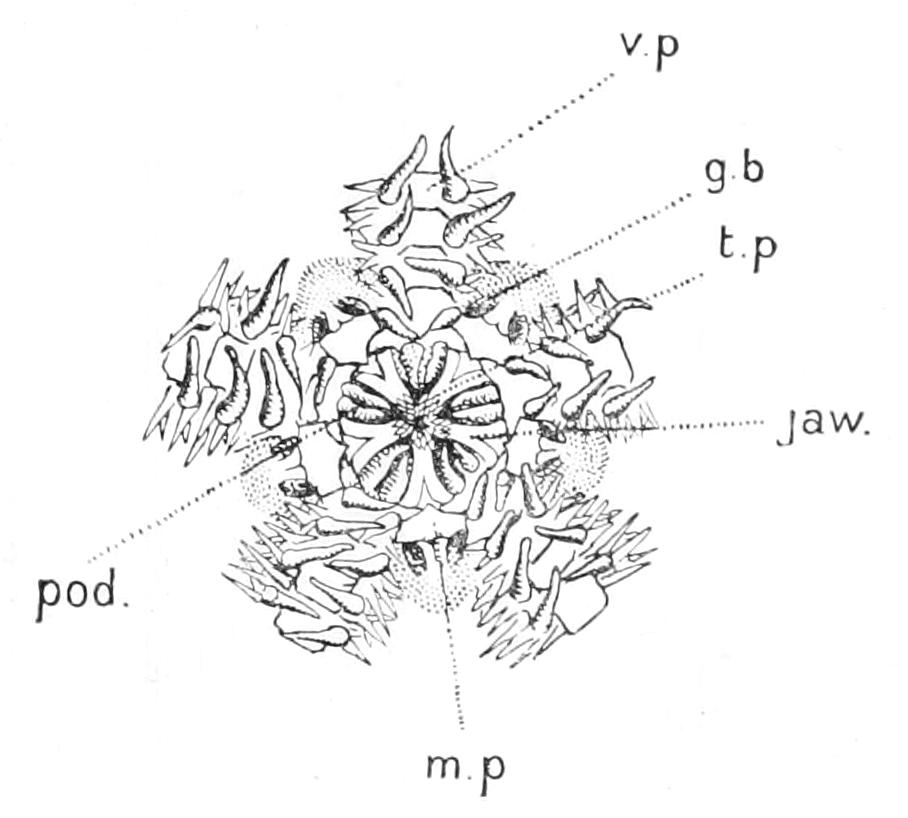
Fig. 208.—Oral view of the disc of Ophiothrix fragilis. g.b, Opening of the genital bursa; m.p, madreporite; pod, podia; t.p, tooth-papillae; v.p, ventral plates of the arms. × 1.
One of the commonest British Ophiuroids is Ophiothrix fragilis (Figs. 207, 208), which is found in swarms in shallow water off the west coast of England and Scotland. We may therefore select it as the type, and, since the arm is the most characteristic organ of an Ophiuroid, we may commence by studying it. Speaking generally, an Ophiuroid either drags itself forward by two arms and pushes itself by the other three (Fig. 207),[458] or else it drags itself by one and pushes with the other four (Fig. 217). The arms during this process are bent into characteristic curves, by the straightening of which in the posterior arms the animal is pushed onwards, whilst the intensification of these curves in the anterior arms causes the animal to be dragged forwards. The grip of the arm on the substratum is chiefly in the distal portion of the curve. The alteration of the curvature is due to the contraction of the muscles on one side of the arms. There is no ambulacral groove such as is found on the under side of the arms of all Asteroidea, for the arm is completely ensheathed by four series of plates, an upper row of dorsal plates, an under row of ventral plates, and two lateral rows of lateral plates. The last named, which in all probability correspond to the adambulacral plates of Starfish, bear each a transverse row of seven spines with roughened surfaces; these enable the animal to get a grip on the substratum over which it moves. The podia in Ophiuroidea are termed "tentacles"; they are totally devoid of suckers, being simple conical papillae used as sense-organs, and are of little, if any, service in locomotion. They issue from openings called "tentacle-pores" situated between the edges of the ventral and lateral plates, guarded each by a valve-like plate called the "tentacle-scale." In Ophiothrix they are covered with sense-organs, each consisting of a hillock-like elevation of the ectoderm, in which are cells carrying long stiff sense-hairs. In most Ophiuroids such organs are not present, though abundant scattered sense-cells occur, and the outer surface of the tube-feet and the lining of certain pockets called "genital bursae" (Fig. 208, g.b) are the only portions of the surface where the ectoderm persists. Everywhere else, although present in the young, it disappears, leaving as remnants a few nuclei here and there attached to the under side of the cuticle.[459]
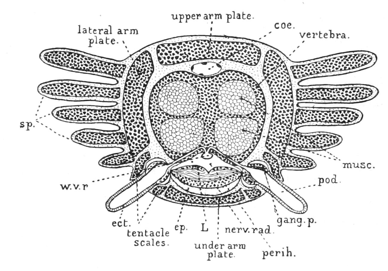
Fig. 209.—Diagrammatic transverse section of the arm of an Ophiuroid. coe, Dorsal coelomic canal; ect, ectoderm covering the tube-foot; ep, epineural canal; gang.p, pedal ganglion; L, nerve-cord; musc, longitudinal muscles attaching one vertebra to the next; nerv.rad, radial nerve-cord; perih, radial perihaemal canal; pod, podium (tube-foot); sp, lateral spines; w.v.r, radial water-vascular canal.
The greater part of the section of the arm is occupied by a disc-like ossicle called the "vertebra." Each vertebra articulates with its predecessor and successor by cup-and-ball joints, and it is connected to each of them by four powerful longitudinal muscles. Above, its outline is notched by a groove, in which lies an extension of the coelom of the disc (Fig. 209, coe), but contains no outgrowth of the alimentary canal, as is the case in Asteroidea. The vertebra is also grooved below, and in this lower groove are contained the radial water-vascular canal (Fig. 209, w.v.r), and below it perihaemal canals as in Asteroidea; below this again the radial nerve-cord (L), and beneath this again a canal called the "epineural canal" (ep), which represents the missing ambulacral groove. This canal in the very young Brittle Star is an open groove, but becomes closed by the approximation of its edges. The vertebra, which has a double origin, represents a pair of fused ambulacral ossicles. In Ophiohelus these are only slightly adherent to one another (Fig. 216).
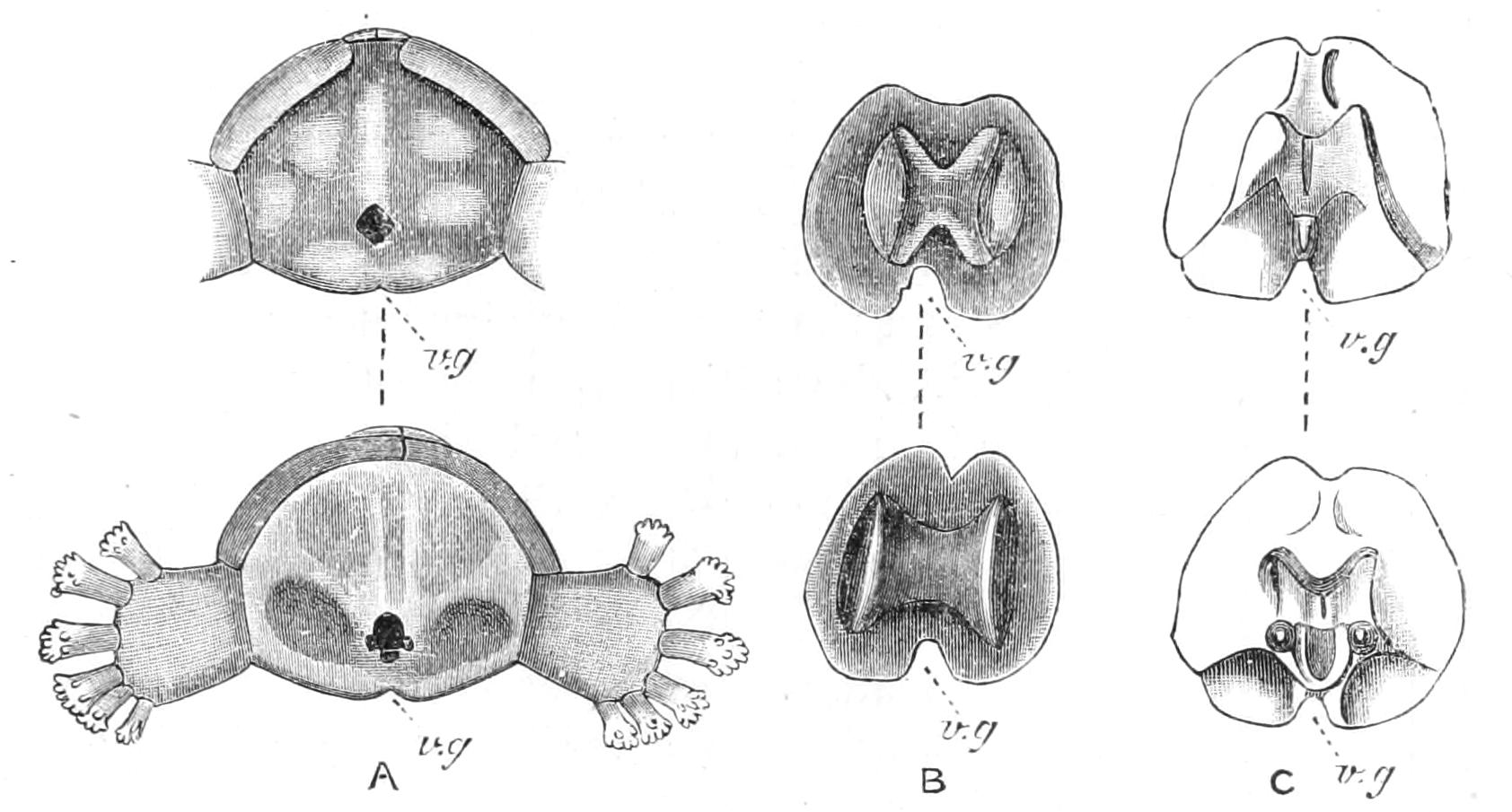
Fig. 210.—Proximal and distal views of the three types of vertebra found amongst Ophiuroidea. A, Ophioteresis, a type of the Streptophiurae (after Bell), × 24; B, Astroschema, a type of the Cladophiurae (after Lyman), × 10; C, Ophiarachna, a type of the Zygophiurae (after Ludwig), × 3. The upper figure in all cases represents the distal aspect, the lower the proximal aspect of the vertebra. v.g, Ventral groove.
When the surface of a vertebra is examined it is found that it can be divided into a thin border, to which are attached the four muscles by which it is connected to its successor and predecessor, and a central portion, on which are situated the knobs and pits, by means of which it articulates with the next vertebra.
The simultaneous contraction of the two upper muscles causes the arm to bend upwards. The contraction of the two lower bend it downwards, whilst a sideward movement is effected by the contraction of the upper and lower muscle of the same side. On the proximal surface of the central portion of the vertebra there is a central knob and two ventro-lateral knobs, a median ventral pit and two dorso-lateral pits, and on the distal surface there are pits corresponding to the knobs on the proximal side and vice versa (Fig. 210, C). These knobs and pits restrict the movement of one vertebra on the next, so that although the arms can undergo an unlimited amount of flexion from side to side, they cannot be rolled up in the vertical plane. When the under surface of the vertebra is examined there is seen on each side of the central groove two round holes, a distal and a proximal. The distal pair are for the passage of the canals connecting the radial water-vessel with the tentacles, these canals traversing the substance of the vertebra for a part of their course; the proximal pair are for nerves going to the longitudinal muscles, which likewise perforate part of the ventral border of the vertebra.
In order to understand the anomalous circumstance that the canals going to the tentacles actually perforate the vertebrae, it must be clearly borne in mind that the basis of the body-wall in all Echinoderms is a mass of jelly with amoebocytes in it, to which we must assign the power of secreting carbonate of lime, and all we have to assume in the case of Ophiuroids is that calcification spread outwards from the original ambulacral ossicles into the surrounding jelly, enclosing any organs that happened to traverse it.
When the ossicles of the arm are followed inwards towards the mouth, they are seen to undergo a profound modification, so as to form, by union with the corresponding ossicles of adjacent arms, a structure called the mouth-frame. The general character of this modification is similar to that affecting the first ambulacral and adambulacral ossicles in the arms of an Asteroid, but in the Ophiuroid the change is much more profound. The first apparent vertebra consists of two separated halves, and each is fused with the first adambulacral (lateral) plate, which in turn is firmly united with the corresponding plate in the adjoining arm. Thus is formed the "jaw," as the projection is called. The extensions of the mouth-cavity between adjacent jaws are termed "mouth-angles." To the apex of each jaw is attached a plate bearing a vertical row of seven short blunt spines called "teeth" (Fig. 212, p). The plate is called the "torus angularis" (Fig. 211, T), and on its ventral edge there is a tuft of spines which are termed "tooth-papillae" (Fig. 208, t.p). On the upper aspect of the jaw are a pair of plates termed "peristomial plates." These discs—of which there are two in each radius, one on each jaw which flanks the radius—possibly represent the separated halves of the first vertebra, the apparent first vertebra being really the second. On the flank of the jaw there is dorsally a groove for the water-vascular ring and nerve-ring (Fig. 212, n.r), and beneath this a groove for the first tentacle and a pore for the second, both of which spring directly from the ring-canal; below these, in most Ophiuroidea, but not in Ophiothrix, there is a row of blunt triangular spines called "mouth-papillae" (Fig. 212, p1).
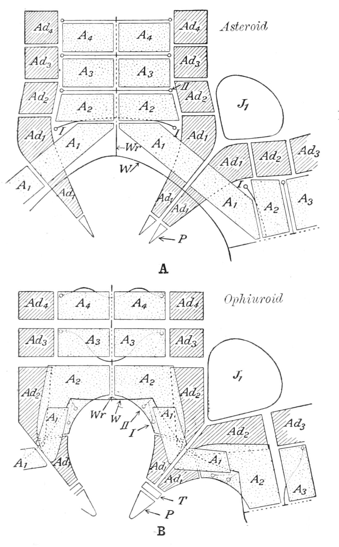
Fig. 211.—Diagrams to show the modification of the ambulacral and adambulacral ossicles to form the armature of the mouth. A, Asteroid; B, Ophiuroid. A1-A4, the first four ambulacra ossicles; Ad1-Ad4, the first four adambulacral ossicles; J1, the first plate of the interradius (in the Ophiuroid the scutum buccale); P, the spines borne by the jaw (in the Ophiuroid the teeth); T, the torus angularis; W, the water-vascular ring; Wr, the radial water-vessel; I, II, the first two pairs of tube-feet. (After Ludwig.)
The words "jaw" and "tooth" are misleading. There is no evidence that the jaws of a Brittle Star are ever used for crushing food, but by means of the muscles attaching them to the first complete vertebra in the arm they can be rotated downwards so as greatly to enlarge the mouth, and again rotated upwards and inwards, when they form an excellent strainer to prevent the entrance of coarse particles. To permit this extensive movement the articulatory facets on the proximal surface of the first vertebra have been much modified; the median knob and pit have disappeared, and the dorso-lateral pits are raised on to the surface of processes, so that there are in all four processes, two of which articulate with one half of a jaw.
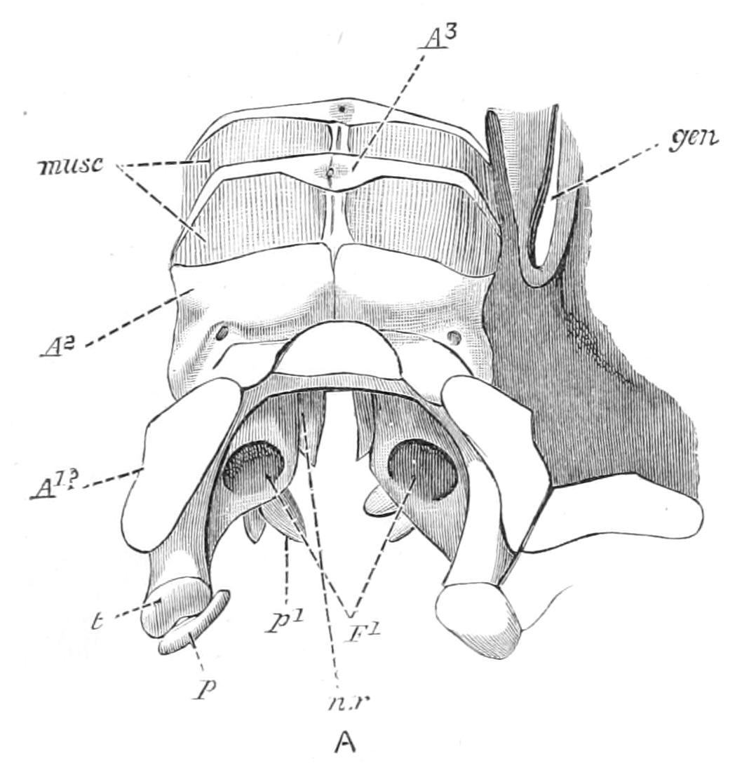
Fig. 212.—Lateral view of mouth-frame of Ophiarachna incrassata. × 4. A1?, peristomial plate, possibly the half of the first vertebra; A2, the half of the second vertebra; A3, the third vertebra; F1, pores for pair of tentacles; gen, genital scale lying beside opening of genital bursa; musc, longitudinal muscles connecting vertebrae; n.r, groove for nerve-ring; p, tooth; p1, mouth-papilla; t, torus angularis. (After Ludwig.)
The mouth can be narrowed and the jaws forced inwards towards the centre by the simultaneous contraction of five muscles (musc. tr, Fig. 213) each, which unite the two halves of a jaw.
Turning now to the skeleton of the disc, we notice that dorsally it consists of a closely-fitting mosaic of small plates, which are usually concealed from view by a covering of minute spines. Opposite the insertion of each arm there are, however, a pair of large triangular plates ("radials"), which extend outwards to the periphery and strengthen it, much as the ribs do in an umbrella. These radial plates are always exposed, in Ophiothrix, even when the rest of the dorsal plates are concealed by spines. On the under surface there is a similar plating; but adjoining the jaws are five large, more or less rhomboidal, plates termed "scuta buccalia" (Fig. 211, J1), on one of which open the few madreporic pores which the animal possesses. Attached to the sides of the scuta buccalia are the "lateral mouth shields," which are in fact the adambulacral plates belonging to the second pair of ambulacral plates which form the main mass of the jaws. Further out, on the under side of the disc, there is, on each side of each arm, a long narrow slit—the opening of the genital bursa (Fig. 208, g.b), so that there are ten genital bursae. The "genital bursa" (Fig. 214) is a sac lined by ciliated ectoderm projecting into the interior of the disc. It is called genital because the openings of the genital organs are situated on its surface; its main function, however, is respiratory, the cilia bringing about a constant inward current of fresh sea-water, the oxygen contained in which diffuses through the thin wall of the sac into the coelomic fluid. The opening of the bursa is strengthened on its radial side by a rod-like ossicle, the "genital plate," and on its interradial side by an ossicle called the "genital scale" (Fig. 212, gen), and in Ophiothrix the outer end of the radial plate articulates with the outer end of the genital plate. Muscles connect the two plates running on either side of the articulation.
Observations on Ophiothrix[460] show that in this species at any rate the radial plates can be raised or lowered. When they are raised the centre of the disc is lifted into a cone and water is sucked into the genital bursae, whereas when they are lowered the bursae are compressed and water is expelled. This forced respiration appears to come into play when the supply of oxygen is getting scanty.
The alimentary canal of Ophiothrix is a simple flattened sac (Fig. 213). It is devoid of an anus and cannot be everted through the mouth. There is a horizontal pouch given off into each interradial lobe of the disc. The sac is attached to the dorsal wall of the coelom by numerous mesenteries, fibrous cords traversing the coelomic cavity and clothed on the outer side by coelomic epithelium. To the mouth-frame it is attached by a circular membrane, which we have reason for believing is a functionless remnant of the retractor muscles of the stomach of Asteroidea. In the young Asteroid there is a similar sheet of membrane, which later becomes resolved into the ten retractor bands.
The simple structure of the alimentary canal appears to be correlated with the exceedingly simple character of the food. Ophiothrix feeds on the most superficial layer of mud at the bottom of the sea. This deposit consists partly of microscopic Algae and partly of decaying organic matter, and is much more easily disposed of than the living animals on which the Starfish preys. The food is shovelled into the mouth by the first two or "buccal" pairs of tube-feet in each ray.
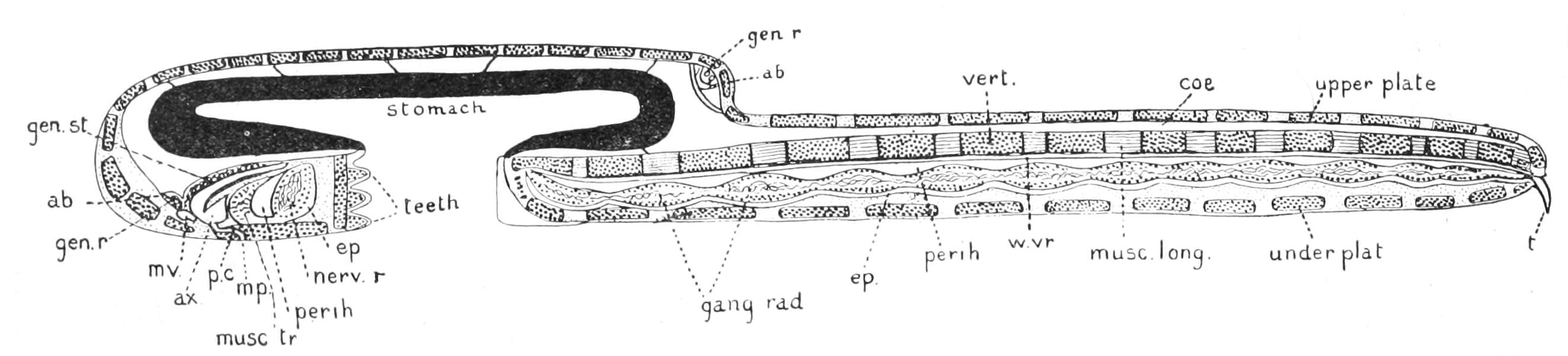
Fig. 213.—Longitudinal section through the disc of a young Ophiuroid passing along one arm and the middle of the opposite interradius. (Diagrammatised from an actual section of Amphiura squamata.) ab, Aboral sinus (dorsal in the radius, ventral in the interradius); ax, axial sinus; coe, dorsal coelomic canal of the arm; ep, epineural canal; gang.rad, ganglion of the radial nerve; gen.r, genital rachis contained in the aboral sinus; gen.st, genital stolon; mp, madreporic pore; musc.long, longitudinal muscle of the arm; musc.tr, transverse muscle uniting the two halves of each jaw; mv, madreporic vesicle; nerv.r, nerve-ring; p.c, pore-canal; perih, perihaemal canal; vert, vertebra; w.vr, radial water-vessel.
The water-vascular system has undergone a most interesting set of modifications, which can be explained by noticing the fact that the tube-feet have almost, if not quite, lost their locomotor function and are now used as tactile organs. The ampulla, or swollen inner end of the tube-foot, has disappeared, and the upper end of the organ is directly connected with the radial canal by means of a curved canal, which traverses the outermost flange of the vertebra, appearing on its surface in a groove on the outer side of the dorsal lateral knob on the distal side of the ossicle. As in Asteroidea there are valves, which regulate the entrance of fluid into the tube-foot. The stone-canal is a curved tube of simple circular section and excessively narrow bore which extends from the water-vascular ring downwards to the madreporite (Fig. 213, mp) situated on one of the scuta buccalia. The madreporite, in Ophiothrix as in most Brittle Stars, is an exceedingly rudimentary structure, consisting of one or two pores leading into as many pore-canals. From each interradius, except that in which the stone-canal lies, a large Polian vesicle hangs down from the water-vascular ring into the coelom.
We saw that in the Asteroid the ampulla was used like the bulb of a pipette to force the fluid in the tube-foot down into the tip, so as to press the sucker against the substratum. But when the tube-foot is used as a sense-organ, a few circular fibres around its upper end suffice to bring about all the extension that is needed. Since the extension is no longer a very vigorous act, the loss of fluid by transudation has probably been rendered insignificant, and hence the stone-canal and madreporite, whose function it is to repair the loss, have been reduced in size. The curious ventral curvature of the stone-canal is, however, due to another cause. In the very young Ophiuroid the madreporite is on the edge of the disc, and the stone-canal extends horizontally outwards; and in some Asteroidea there is a similar outward direction in its course. As development proceeds the dorsal interradial areas of the disc of the young Ophiuroid grow out into lobes, building up the conspicuous adult disc and forcing the madreporite, and with it the stone-canal, downwards towards the ventral surface.
The pores of the madreporite in Ophiothrix, like some of those in the Asteroid, open not directly into the stone-canal but into the axial sinus (Fig. 213, ax). This is a large ovoid sac, lined with thin epithelium, lying between the stone-canal and the mouth-frame, since of course it has shared in the ventral rotation of the stone-canal. Its open connexion with the stone-canal was easily recognised by Ludwig, who termed it, on this account, the "ampulla."[461] The name "axial sinus" was bestowed mistakenly on another cavity, which will be mentioned in connexion with the genital organs.
The radial perihaemal spaces of the arms open into a "perihaemal ring" representing the outer perihaemal ring of Asteroids; but the axial sinus does not have any such extension as constitutes the inner perihaemal ring in Starfish. So-called oral circular and radial blood strands are to be found in similar positions to the corresponding structures in Asteroidea.
The nervous system might have been expected to have become very much modified, since the activities of the Brittle Stars are so different from those of the Starfish. It is indeed a universal rule in the Animal Kingdom that, concomitantly with the increase in size and activity of a muscle, there is a corresponding increase in the number of ganglion-cells which control it. An accurate radial section of an arm shows that there is, corresponding to the interspaces between the two vertebrae, a ganglionic swelling of the nerve-cord. As in Asteroids, there are not only ectodermic ganglion-cells on the under surface of the cord abutting on the epineural canal, but also coelomic ganglion-cells derived from the floor of the radial perihaemal canal. Both these categories of cells are largely increased in number in the ganglion. From the dorsal-cells arise a pair of large nerves which pass directly up and supply the great intervertebral muscles. From the interspace between the ganglia a direct prolongation of the ventral part of the nerve-cord, the so-called pedal nerve, extends out along the side of the tentacle, as in Asteroids. In Ophiuroids it swells out into a ganglion, completely surrounding the tentacle and giving off nerves to the surfaces of the arm which terminate in the cuticle.
There is a large ganglion where the radial cord joins the nerve-ring, and, owing to the more specialised condition of the nervous system, a severed arm in an Ophiuroid is much more helpless than an arm of an Asteroid. It will not carry out "escape movements," and is for a long time rigid under the shock of section; at last it simply gives reflex movements on stimulation.
Preyer[462] endeavoured to test the "intelligence" of Ophiuroids by observing how they would adapt themselves to circumstances which it might be fairly assumed they had never encountered in their ordinary experience. To this end he passed over the arm of a specimen a piece of indiarubber tubing, which clung to it tightly. He found that the animal first tried walking off, pressing the encumbered arm against the ground, so that the piece of tubing was rubbed off. It was then replaced more tightly than before; the animal, having tried the first method without result, waved the arm to and fro in the water till the rubber floated off. In a third experiment the animal held the rubber against the ground by a neighbouring arm, and drew the encumbered arm out. When the rubber was replaced a fourth time, the animal kicked it off by alternately pressing neighbouring arms against it. Finally, when the rubber was put on so firmly that all the above-mentioned methods failed, the arm was broken off. Preyer concludes from this that Ophiuroids have a high degree of intelligence; but this may be doubted, and the reader is referred to the account of Uexküll's experiments given in the next chapter. There is, however, no doubt at all that Ophiuroidea are by far the most active of all Echinoderms, and one would naturally correlate this with higher psychic development.
The radial nerve ends in a terminal tentacle sheltered by a median plate at the end of the arm; but eyes, such as are found in Asteroids, are wanting, and the animal does not appear to be sensitive to light.
The reproductive system in Ophiuroids consists of a genital stolon giving rise at its distal end to a genital rachis, which extends in a circular course round the disc, ensheathed in an "aboral sinus" (Fig. 213, ab) and swelling out so as to form the gonads (testes or ovaries), where it passes over the inner side of the genital bursae. The genital stolon (Fig. 213, gen.st) is a compact ovoid organ, often termed on account of its shape the "ovoid gland." It is situated close to the stone-canal, and, as in Starfish, it indents the outer wall of the axial sinus; but, unlike the stolon of the Asteroid, it is separated from the general coelom by a space, of which it forms the inner wall, but whose outer wall is formed by a sheet of membrane. This cavity must be carefully distinguished from the axial sinus of Asteroidea, to which it was supposed at one time to correspond; it is really formed by a pocket-like ingrowth of the general coelom into the septum dividing it from the axial sinus. The cells forming the inner side of this pocket form the primitive germ-cells, which constitute the main mass of the ovoid gland; those of the outer side remain thin. The cavity of the ingrowth is shut off from the general coelom, but persists throughout life. In Asteroids a similar ingrowth takes place, but both walls thicken and become converted into germ cells, and the cavity disappears, and, as in Asteroidea, a considerable number of the germ-cells in the stolon degenerate.
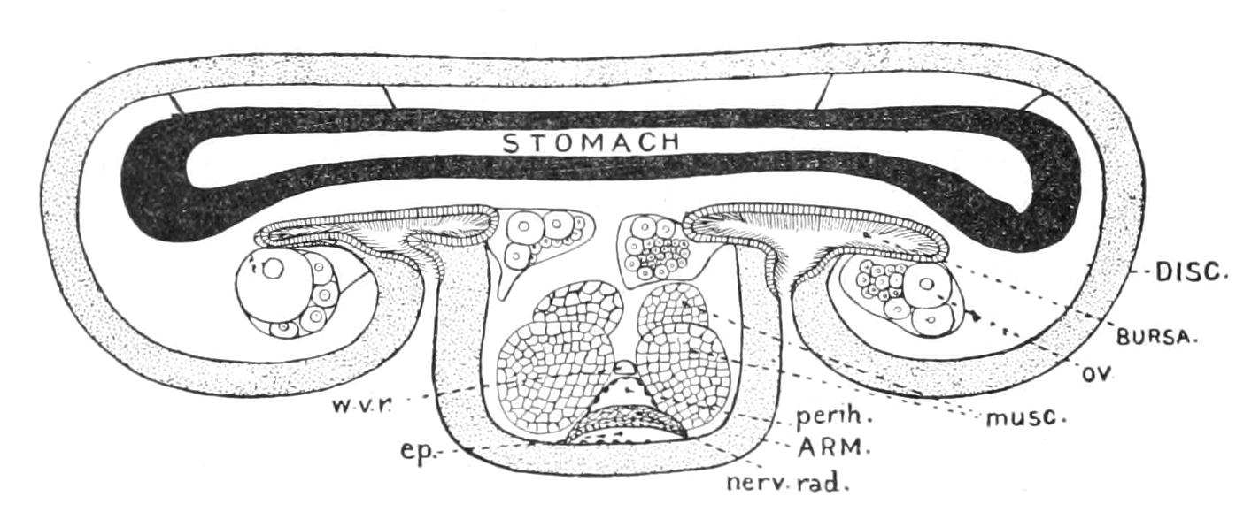
Fig. 214.—Diagram of a tangential section through the edge of the disc of an Ophiuroid to show the relations of the disc, arm, and genital bursae. ep, Epineural canal; musc, longitudinal muscle of the arm; nerv.rad, radial nerve cord; ov, ovary; perih, radial perihaemal canal; w.v.r, radial water-vessel.
The genital rachis (Fig. 213, gen.r) is an outgrowth of the distal end of the genital stolon, which extends in a complete circle round the disc. The rachis does not, however, lie everywhere in the same plane, but by its undulating course bears witness to the distortion which the disc has undergone. In the radii it is, as in the Asteroid, dorsal; but in the interradii it is ventral, this ventral portion having, like stone-canal and axial sinus, been carried down by the preponderant growth of the dorsal parts of the disc. It is everywhere ensheathed by the aboral sinus, which, as in Asteroids, is an outgrowth of the coelom. The rachis is embedded in a strand of modified connective tissue, to which we may (as in the case of Asterias) apply the name "aboral blood-ring." Both on the central and peripheral sides of this sinus are vertical muscles connecting the genital and the radial plates, which bring about the respiratory movements already referred to. Just above the madreporite, at the end of the genital stolon, is a small, completely closed space, which by its position corresponds with the madreporic vesicle of Asteroids and represents the right hydrocoel (Fig. 213, mv). As the rachis passes over the genital bursa it gives off branches, which swell up to form the genital organs. In Ophiothrix there is one such organ on each side of each bursa, but in other genera (cf. Ophiarachna) a large number of small ones. The genital products are shed into the water through the bursae.
Classification of Ophiuroidea.
Before proceeding to study the classification of Brittle Stars, it is necessary to give some account of the range of structure met with in the group.
Number of Radii.—The number of arms is rarely increased, and hardly ever exceeds six; a few species (each an isolated one in its genus) have six arms, and in one case (Ophiactis virens), at any rate, this is associated with the power of transverse fission. In many Cladophiurae the arms fork repeatedly, so that although there are only five radii, there is quite a crowd of terminal branches.
Vertebrae.—The vertebrae differ in the manner in which they articulate with one another. In Ophiothrix fragilis taken as the type, which in this respect resembles the vast majority of species (Zygophiurae), the knobs and pits on the faces of the vertebrae prevent the arms from being coiled in the vertical plane. In Ophioteresis (Fig. 210, A) and some allied genera (Streptophiurae) the knobs are almost obsolete, and the arms are free to coil in the vertical plane; whilst in Gorgonocephalus and Astrophyton (Cladophiurae) the arms are repeatedly branched and the vertebrae have saddle-shaped articulating surfaces, so that they have quite a snake-like capacity for coiling themselves round external objects. In Ophiohelus (Fig. 216) each vertebra consists of two rod-like plates placed parallel with the long axis of the arm and fused at both ends, but divergent in the middle, leaving a hole between them.
Covering Plates of the Arms.—The upper arm-plates are the most variable. They may be surrounded by small supplementary plates (Ophiopholis) or double (Ophioteresis). In all (?) Cladophiurae and most Streptophiurae they are absent, being replaced by minute calcareous granules. Under arm-plates are absent in Ophioteresis and in the distal portion of the arms in many Cladophiurae. Side arm-plates are constantly present, and in most Cladophiurae meet in the middle line below.
Arm-Spines.—The spines borne by the lateral covering plates of the arms vary greatly in character. In Ophiura and its allies they are short and smooth, and are borne by the hinder edge of the arm and directed backwards; but in the larger number of genera they are borne nearer the centre of the plate, and are directed outwards at right angles to the arm. They may be covered by small asperities, as in Ophiothrix (Fig. 215, C), when they are said to be rough; or these asperities may become secondary spines, as in Ophiacantha (Fig. 215, B), when they are said to be thorny. In Ophiopteron all the spines borne by a single plate are united by a web of skin so as to constitute a swimming organ. The small plates guarding the ends of the tentacles (tentacle-scales) may be absent, or more rarely double. In Cladophiurae there is a regular transition from tentacle-scale to arm-spine; the tentacle-scale being merely the smallest of the series of lateral spines.
True pedicellariae are unknown amongst Ophiuroidea, since there is no longer a soft ectoderm to protect, but in some cases, as for instance in Ophiohelus, small hooks movable on a basal piece attached to the arms are found which may represent the vestiges of such organs (Fig. 216). Similar hooks are found in the young Ophiothrix fragilis just after metamorphosis and in all Cladophiurae, replacing in the latter case the arm-spines in the distal portion of the arm.
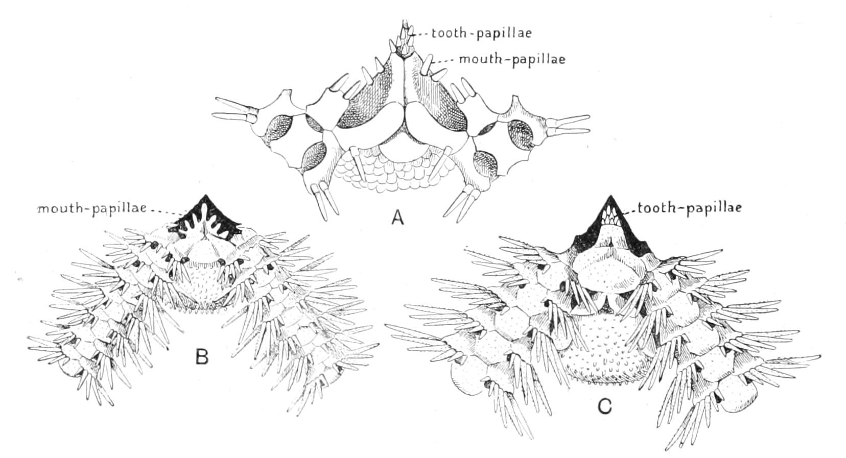
Fig. 215.—Three types of mouth-frame found in Zygophiurae. A, Ophioscolex, × 10; B, Ophiacantha, × 6; C, Ophiothrix, × 6. (After Lyman.)
Mouth-Frame.—In its broad outlines there is practically no variation in this organ throughout the group, but in respect of the spines, which are borne on the flanks of the jaws (mouth-papillae) and on their apices (teeth and tooth-papillae) there is very great variation. Teeth are always present. Mouth-papillae are very frequently present, tooth-papillae are rarer, and it is only in a restricted number of genera (Ophiocoma and its allies) that both mouth-papillae and tooth-papillae are present at the same time.
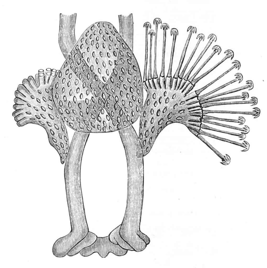
Fig. 216.—A portion of an arm of Ophiohelus umbella, near the distal extremity, treated with potash to show the skeleton, × 55. The vertebrae are seen to consist of two curved rods united at their ends. The triangular side-plates bear a row of movable hooks which articulate with basal outgrowths of the plate. (After Lyman.)
Skeleton of the Disc.—This is typically composed of a mosaic of plates of different sizes, but in some cases (Ophiomyxa, most Streptophiurae, and Cladophiurae) these, with the exception of the radials and genitals, are entirely absent, and the disc is then quite soft and covered with a columnar epithelium, the persistent ectoderm. Even the scuta buccalia may disappear. Radial shields are absent in Ophiohelus. In many cases (Ophiothrix and Ophiocoma) all the dorsal plates except the radials are concealed from view by a covering of small spines. In some genera (Ophiopyrgus) there are five large plates in the centre of the upper part of the disc, which have been termed "calycinals" from a mistaken comparison with the plates forming the cup or calyx of the Pelmatozoa, but there is no connexion between the two sets of structures.
The madreporite is usually quite rudimentary, but in Cladophiurae there may be five madreporites, each with about 200 pores, and, of course, five stone-canals.
The number of genital organs varies very much. In the small Amphiura squamata there are two gonads, an ovary and a testis, attached to each bursa, but in the larger species there may be very many more.
We follow Bell's classification,[463] according to which the Ophiuroidea are divided, according to the manner in which the vertebrae move on one another (cf. Fig. 210), into three main orders, since these movements are of prime importance in their lives.
(1) Streptophiurae, in which the faces of the vertebrae have rudimentary knobs and corresponding depressions, so that the arms can be coiled in the vertical plane. These are regarded as the most primitive of Ophiuroidea.
(2) Zygophiurae, in which the vertebral faces have knobs and pits which prevent their coiling in a vertical plane.
(3) Cladophiurae, in which the arms can be coiled as in (1) and are in most cases forked. No teeth; the arm-spines are papillae, the covering plates of the arms are reduced to granules.
Order I. Streptophiurae.
This is not a very well defined order; it includes a few genera intermediate in character between the Cladophiurae and the Zygophiurae, and believed to be the most primitive Ophiuroids living. It is not divided into families. The vertebrae have rudimentary articulating surfaces, there being two low bosses and corresponding hollows on each side, and so they are capable of being moved in a vertical plane, as in the Cladophiurae; the arms never branch, and further, they always bear arm-spines and lateral arm-plates at least. No species of this order are found on the British coast, but Ophiomyxa pentagona, in which the dorsal part of the disc is represented only by soft skin, is common in the Mediterranean.
Ophioteresis is devoid of ventral plates on the arms, and appears to possess an open ambulacral groove, though this point has not been tested in sections. Ophiohelus and Ophiogeron have vertebrae in which traces of the double origin persist (see p. 491).
Order II. Zygophiurae.
This group includes all the common and better-known British forms. They are divided into five families, all of which are represented in British waters.
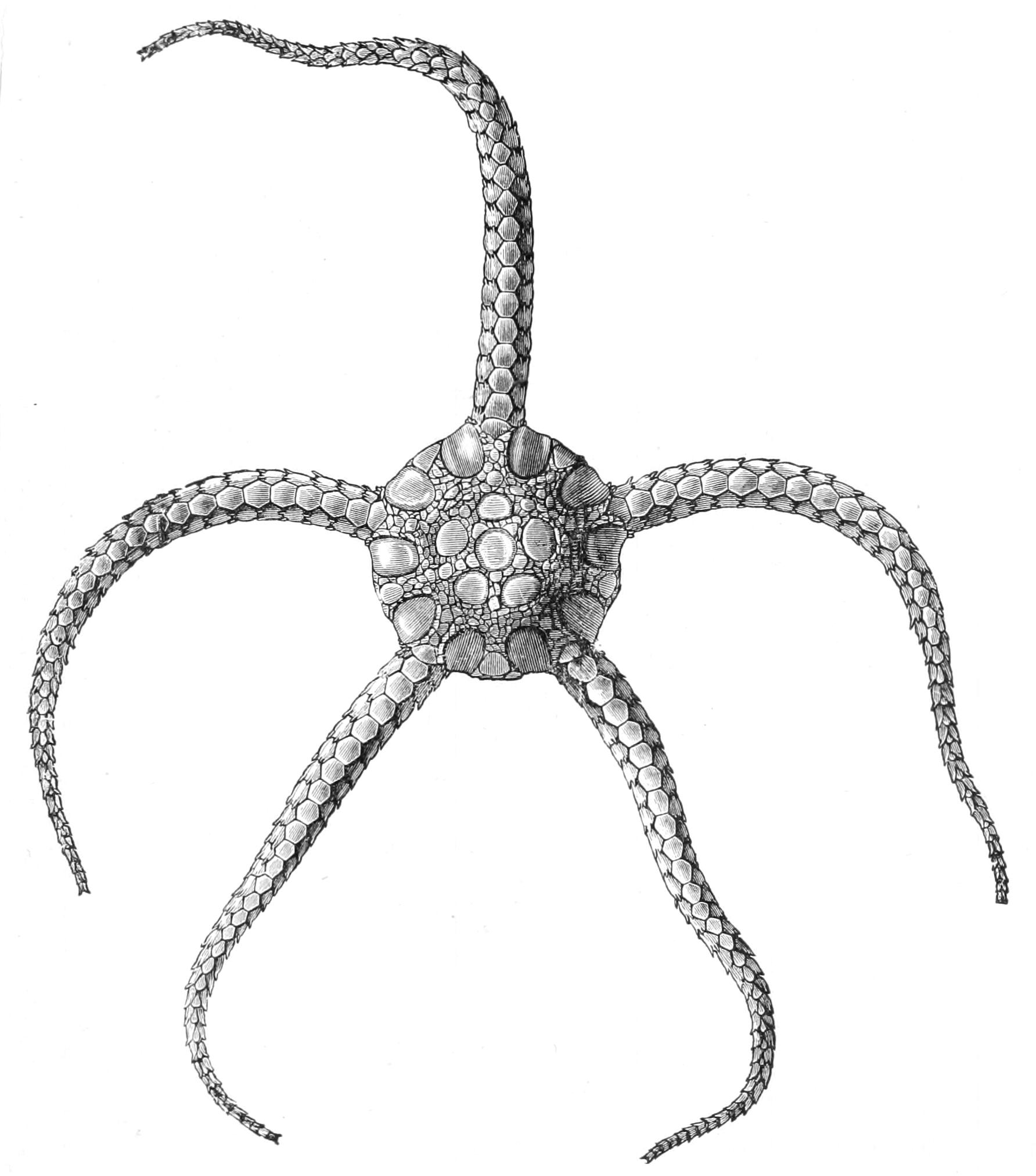
Fig. 217.—Aboral view of Ophioglypha (Ophiura) bullata. × 3. (From Wyville Thomson.)
Fam. 1. Ophiolepididae.[464]—Arm inserted in a definite cleft in the disc, or (expressing the same fact in another way) the interradial lobes out of which the disc is composed are not completely united. Radial shields and dorsal plates naked. Arm-spines smooth and inserted on the posterior border of the lateral arm-plates.
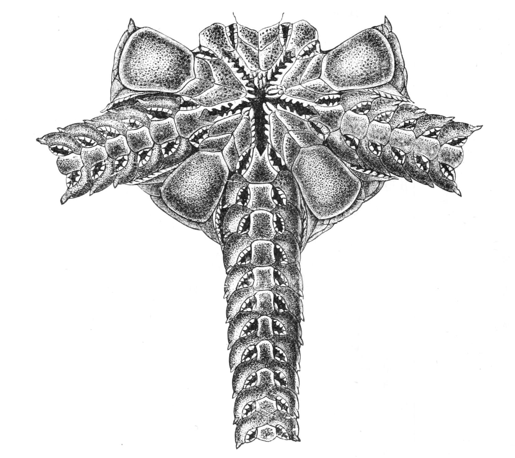
Fig. 218.—Oral view of Ophioglypha (Ophiura) bullata. × 5. (From Wyville Thomson.)
This family includes all the Brittle Stars of smooth porcelanous aspect and provided with only short spines. Forbes[465] called them Sand-stars, since their short spines render these animals incapable of burrowing or of climbing well, and hence they appear to move comparatively rapidly over firm ground, sand, gravel, or muddy sand, and they are active enough to be able to capture small worms and Crustacea. The prey is seized by coiling one of the arms around it.
One genus, Ophiura, is fairly common round the British coast, being represented by O. ciliaris and O. albida; the former is the commoner. An allied species dredged by H.M.S. "Challenger" is represented in Figs. 217 and 218.
Ophiomusium (Fig. 219) is a very peculiar genus. The mouth-papillae on each side of each mouth-angle are confluent, forming a razor-like projection on each side of each mouth-angle (Fig. 220). The arms are short, and the podia are only developed at the bases of the arms. Ophiopyrgus has the dorsal surface raised into a conical elevation protected by a central plate surrounded by five large plates.
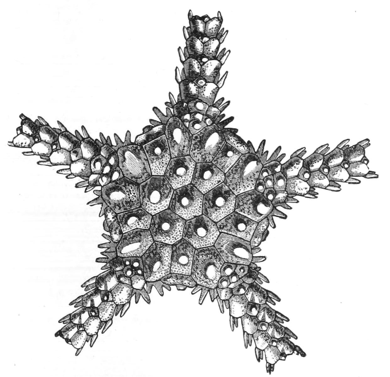
Fig. 219.—Aboral view of Ophiomusium pulchellum. × 7. (From Wyville Thomson.)
In the remaining four families the arms are inserted on the under surface of the disc; in other words, the interradial lobes which make up the disc have completely coalesced dorsally; and the spines stand out at right angles to the arm.
Fam. 2. Amphiuridae.—Mouth-papillae present, but no tooth-papillae; radial shields naked; small scuta buccalia.
The most interesting Brittle Star belonging to this family is Amphiura squamata (elegans), a small form, with a disc about ¼ inch in diameter covered with naked plates. It is hermaphrodite and viviparous, the young completing their development inside the bursae of the mother. Occasionally the whole disc, with the exception of the mouth-frame, is thrown off and regenerated. This appears to be a device to enable the young to escape. Three other species of Amphiura are found in British waters.
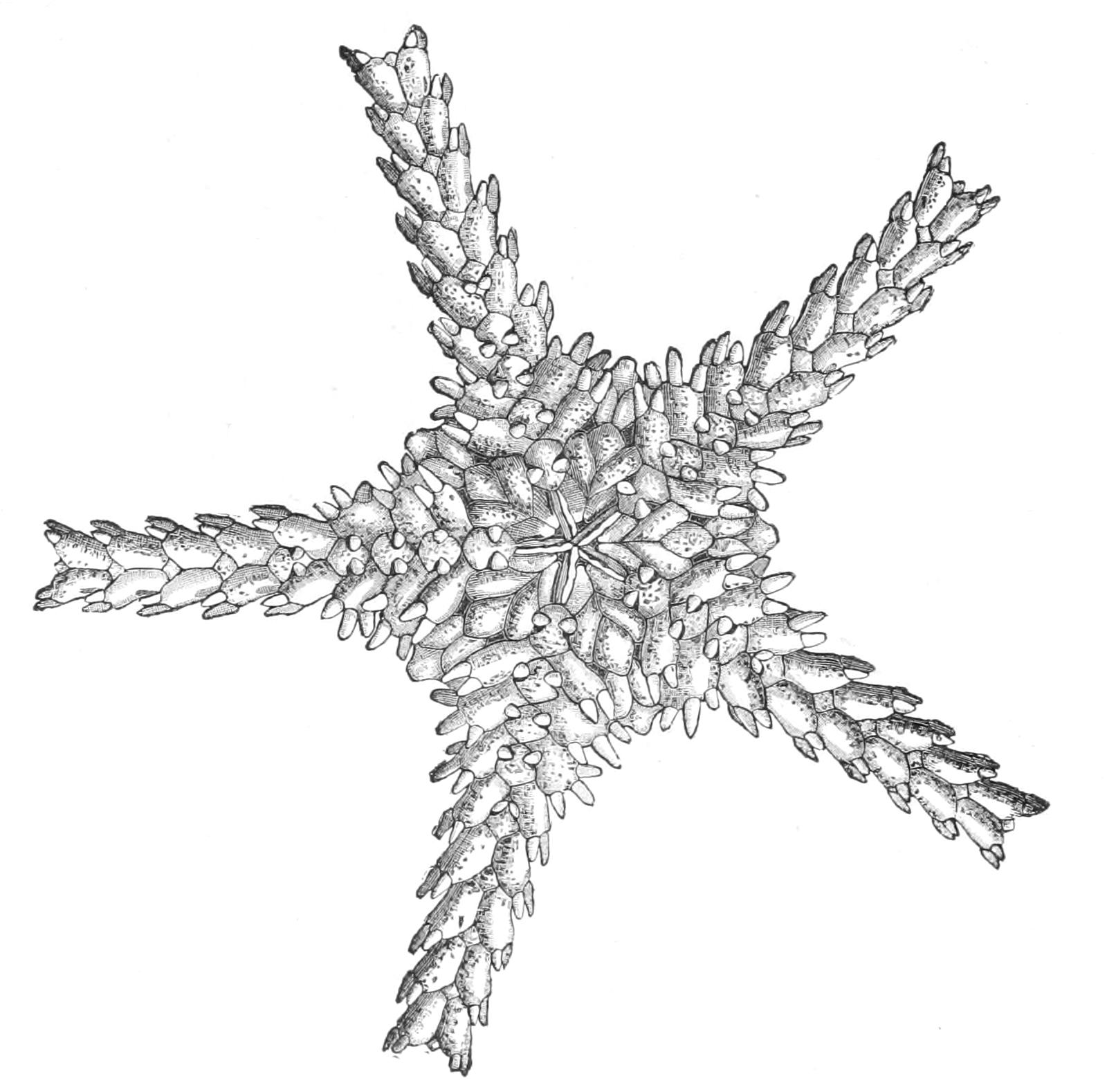
Fig. 220.—Oral view of Ophiomusium pulchellum. × 7. (From Wyville Thomson.)
Ophiactis is another genus belonging to this family, distinguished from Amphiura by its shorter arms and smoother arm-spines. It lives in the interstices of hard gravel. The British species, O. balli, presents no special features of interest, but the Neapolitan O. virens is an extraordinary form. It has six arms, three of which are usually larger than the other three, for it is always undergoing a process of transverse division, each half regenerating the missing part. It has from 1 to 5 stone-canals, the number increasing with age; numerous long-stalked Polian-vesicles in each interradius, and in addition a number of long tubular canals which spring from the ring-canal, and entwine themselves amongst the viscera.[466] All the canals of the water-vascular system, except the stone-canals, contain non-nucleated corpuscles, carrying haemoglobin,[467] the respiratory value of which compensates for the loss of the genital bursae, which have entirely disappeared.
Ophiopholis is distinguished from the foregoing genera by the granular covering of its dorsal plates; whilst in Ophiacantha these granules develop into prominent spinelets, and the arm-spines are also thorny. Ophiopholis aculeata occurs in swarms in the branches of the Firth of Clyde, and presents a most remarkable series of variations in colour. Ophiopsila is a closely allied form, distinguished by its large peristomial plates.
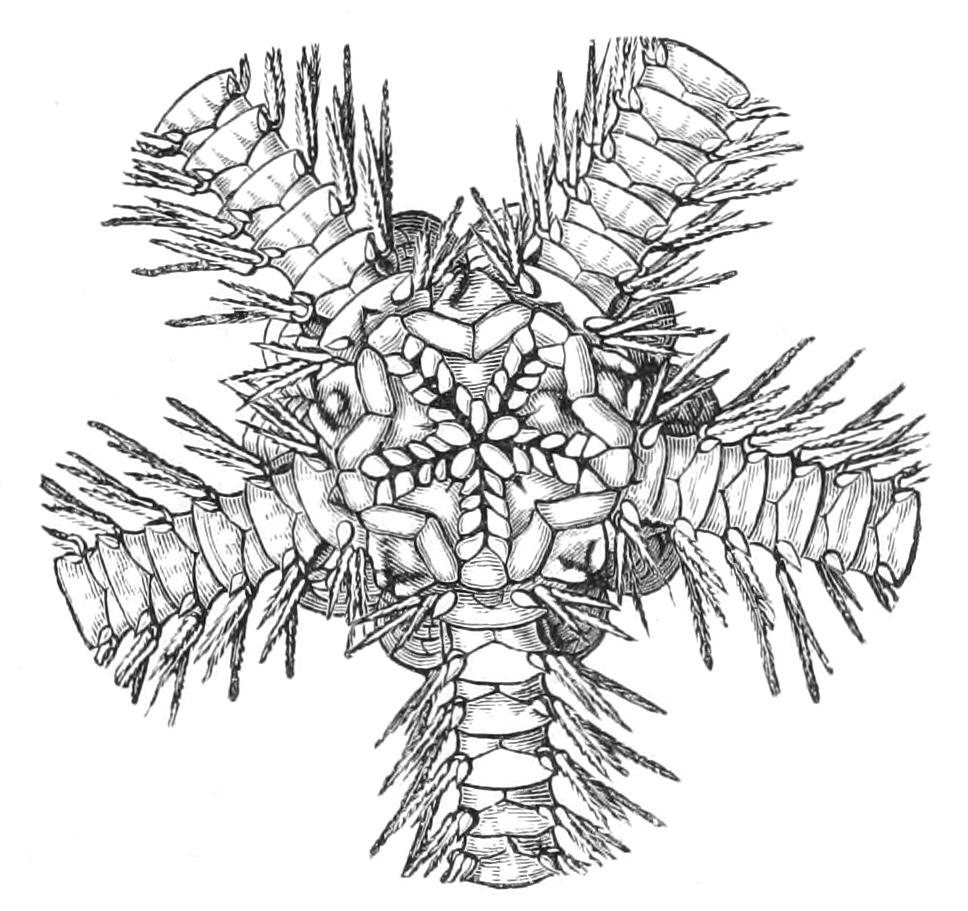
Fig. 221.—Oral view of Ophiacantha chelys. × 4. (From Wyville Thomson.)
Fam. 3. Ophiocomidae.—Both mouth-papillae and tooth-papillae are present;[468] the arm-spines are smooth, and the disc is covered with granules.
Ophiocoma nigra is the only common British representative of this family. In this species the plates of the dorsal surface are completely hidden from view by a covering of granules. Ophiarachna.
Fam. 4. Ophiothricidae.—Tooth-papillae alone present, mouth-papillae absent; arm-spines roughened or thorny.
This family is represented only by Ophiothrix fragilis, which is perhaps the most abundant of all British Ophiuroids, and has been selected as the type for special description.
The back is covered with spinules, having, however, the triangular radial plates bare. This produces a contrast-effect, which suggested the name pentaphyllum, formerly used by some naturalists for the species. It occurs in swarms, and presents variations in colour nearly as marked as those of Ophiopholis. Ophiopteron is probably a swimming Ophiuroid, as the lateral spines of each segment of the arm are connected by a web of skin.
Order III. Cladophiurae.
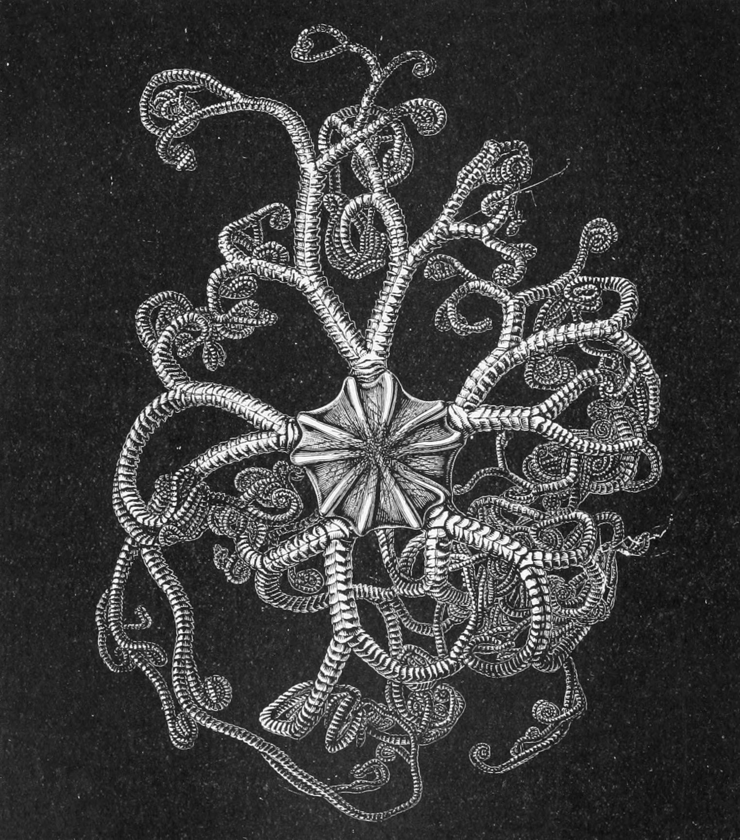
Fig. 222.—Aboral view of young Astrophyton linckii, slightly enlarged. (From Wyville Thomson.)
These, like the Streptophiurae, have the power of rolling the arms in a vertical plane, but the articulating surfaces of the vertebrae are well-developed and saddle-shaped. The dorsal surface of the disc and arms is covered with a thick skin with minute calcifications. Upper-arm plates wanting. Radial plates always present, though occasionally represented by lines of scales. The order is divided into three families, two of which are represented in British waters.
Fam. 1. Astroschemidae.—Arms unbranched. Astronyx is comparatively common in the sea-lochs of Scotland. There are a series of pad-like ridges on the arms, representing the side-plates and bearing the spines. Astroschema.
Fam. 2. Trichasteridae.—Arms forked only at the distal ends. Trichaster, Astrocnida.
Fam. 3. Euryalidae.—Arms forked to their bases. Gorgonocephalus is occasionally taken in deep water off the north coast of Scotland. In it the arms repeatedly fork, so that a regular crown of interlacing arms is formed. The animal obviously clings to external objects with these, for it is often taken in fishermen's nets with its arms coiled around the meshes. The genital bursae are said to be represented by slits which open directly into the coelom. (Lyman describes the coelom as divided into ten compartments by radiating septa; it is possible—even probable—that these are really the bursae.) An allied species is common in the Bay of Fundy, being found in comparatively shallow water. Astrophyton (Fig. 222) is closely allied to Gorgonocephalus, differing only in trifling points. It is doubtful whether the separation of these two genera is justified.
Fossil Ophiuroidea.—The Ophiuroidea are rather sparsely represented among fossils, but in the Silurian and Devonian a series of very interesting forms occur which are intermediate in character between Starfish and Brittle Stars, and which were therefore in all probability closely allied to the common ancestors of modern Ophiuroids and Asteroids. Jaekel[469] has recently added largely to our knowledge of these primitive forms, and has described a number of new genera. Thus Eophiura from the Lower Silurian has an open ambulacral groove, and the vertebrae are represented by an alternating series of quadrate ossicles, each deeply grooved on its under surface for the reception of the tentacle, which was not yet (as in modern forms) enclosed in the vertebra. The lateral or adambulacral plates extended horizontally outwards, and each bore a series of spines at its outer edge.
A remarkable fact is that where the halves of the vertebrae (i.e. the ambulacral ossicles) diverge in order to form the mouth-angles, no less than five or six vertebrae are thus affected, instead of only two as in modern forms. The actual "jaw," however, seems, as in modern forms, to consist only of the first adambulacral fused to the second ambulacral, so that instead of concluding with Jaekel that the "jaws" of modern forms result from the fusion of five or six vertebrae, a conclusion which would require that a number of tentacles had disappeared, we may suppose that the gaping "angles" of these old forms have, so to speak, healed up, except at their innermost portions.
In Bohemura, which belongs to a somewhat younger stratum, the structure is much the same, but the groove in the ambulacral ossicle for the tentacle has become converted into a canal, and the ambulacral groove itself has begun to be closed at the tip of the arm by the meeting of the adambulacrals.
In Sympterura, a Devonian form described by Bather,[470] the two ambulacral plates of each pair have thoroughly coalesced to form a vertebra, but there is still an open ventral groove, and no ventral plates.
In the Trias occurs the remarkable form Aspidura, which had short triangular arms, in which the tentacle pores were enormous and the ventral plates very small. The radial plates formed a continuous ring round the edge of the disc. Geocoma from the Jurassic is a still more typical Ophiuroid; it has long whip-like arms, and the dorsal skeleton of the disc is made of fifteen plates, ten radials, and five interradials. In the Jurassic the living genus Ophioglypha, appears.
The Cladophiurae are represented already in the Upper Silurian by Eucladia, in which, however, the arms branch not dichotomously, as they do in modern forms, but monopodially. There is a large single madreporite.
Onychaster, with unbranched arms, which occurs in the Carboniferous, is a representative of the Streptophiurae.
It will therefore be seen that the evolution of Ophiuroidea must have begun in the Lower Silurian epoch. The Streptophiurae are a few slightly modified survivors of the first Ophiuroids. By the time the Devonian period had commenced, the division of the group into Zygophiurae and Cladophiurae had been accomplished.