Chapter XI
Reading edition
HYDROZOA (CONTINUED): TRACHOMEDUSAE—NARCOMEDUSAE—SIPHONOPHORA
Order VII. Trachomedusae.
The orders Trachomedusae and Narcomedusae are probably closely related to one another and to some of the families of Medusae at present included in the order Calyptoblastea, and it seems probable that when the life-histories of a few more genera are made known the three orders will be united into one. Very little is known of the hydrosome stage of the Trachomedusae, but Brooks[322] has shown that in Liriope, and Murbach[323] that in Gonionema, the fertilised ovum gives rise to a Hydra-like form, and in the latter this exhibits a process of reproduction by gemmation before it gives rise to Medusae. Any general statement, therefore, to the effect that the development of the Trachomedusae is direct would be incorrect. The fact that the hydrosomes already known are epizoic or free-swimming does not afford a character of importance for distinction from the Leptomedusae, for it is quite possible that in this order of Medusae the hydrosomes of many genera may be similar in form and habits to those of Liriope and Gonionema.
The free border of the umbrella of the Trachomedusae is entire; that is to say, it is not lobed or fringed as it is in the Narcomedusae. The sense-organs are statocysts, each consisting of a vesicle formed by a more or less complete fold of the surrounding wall of the margin of the umbrella, containing a reduced clapper-like tentacle loaded at its extremity with a statolith.
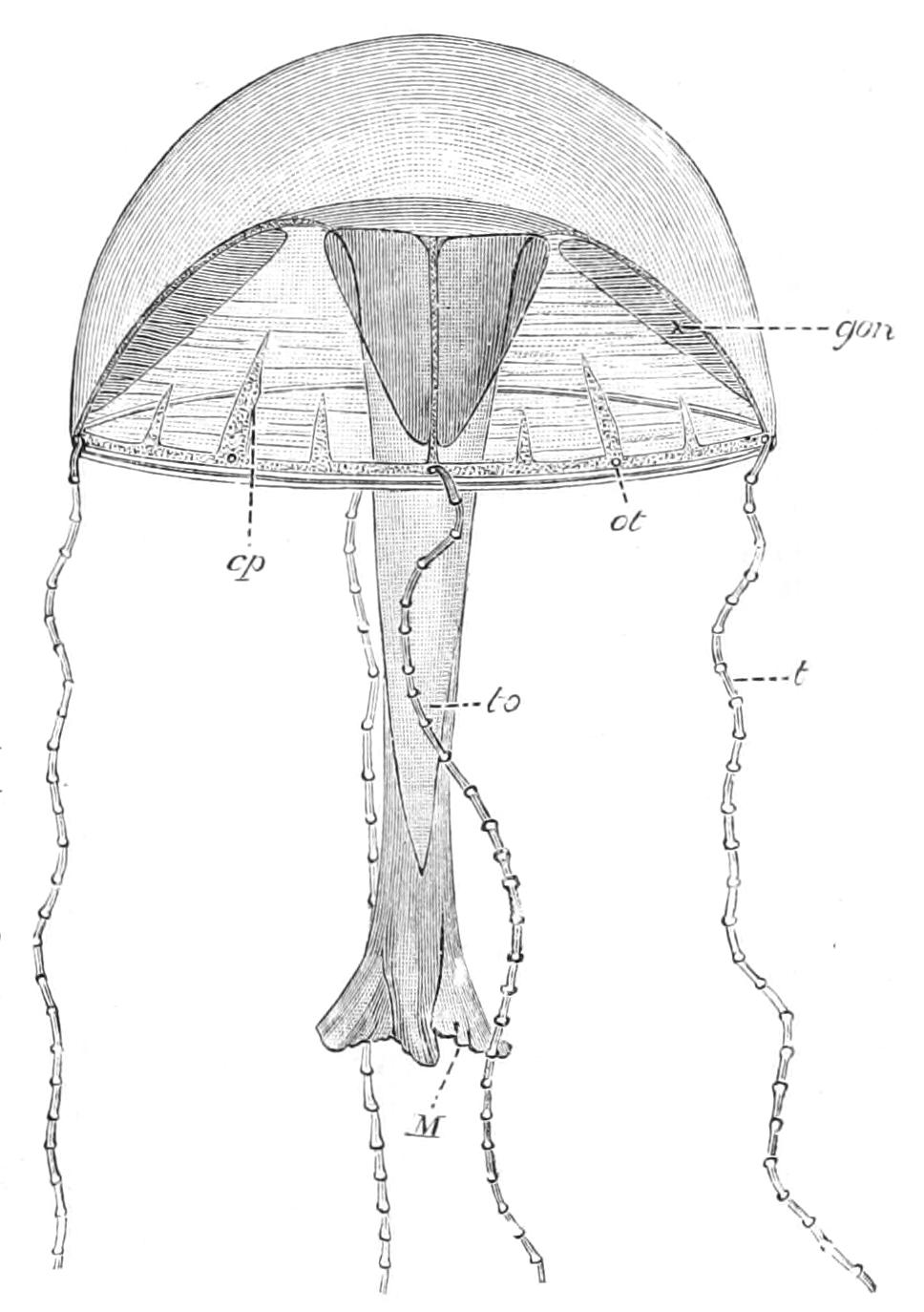
Fig. 137.—Liriope rosacea, one of the Geryoniidae, from the west side of North and Central America. Size, 15-20 mm. Colour, rose. cp, Centripetal canal; gon, gonad; M, mouth at the end of a long manubrium; ot, statocyst; t, tentacle; to, tongue. (After Maas.)
This statocyst is innervated by the outer nerve ring. There appears to be a very marked difference between these marginal sense-organs in some of the best-known examples of Trachomedusae and the corresponding organs of the Leptomedusae. The absence of a stalk supporting the statolith and the innervation of the otocyst by the inner instead of by the outer nerve ring in the Leptomedusae form characters that may be of supplementary value, but cannot be regarded as absolutely distinguishing the two orders. The statorhab of the Trachomedusae is probably the more primitive of the two types, and represents a marginal tentacle of the umbrella reduced in size, loaded with a statolith and enclosed by the mesogloea. Intermediate stages between this type and an ordinary tentacle have already been discovered and described. In the type that is usually found in the Leptomedusae the modified tentacle is still further reduced, and all that can be recognised of it is the statolith attached to the wall of the statocyst, but intermediate stages between the two types are seen in the family Olindiidae, in which the stalk supporting the statolith passes gradually into the tissue surrounding the statolith on the one hand and the vesicle wall on the other. The radial canals are four or eight in number or more numerous. They communicate at the margin of the umbrella with a ring canal from which a number of short blind tubes run in the umbrella-wall towards the centre of the Medusa (Fig. 137, cp). These "centripetal canals" are subject to considerable variation, but are useful characters in distinguishing the Trachomedusae from the Leptomedusae. The tentacles are situated on the margin of the umbrella, and are four or eight in number or, in some cases, more numerous. The gonads are situated as in Leptomedusae on the sub-umbrella aspect of the radial canals.
In Gonionema murbachii the fertilised eggs give rise to a free-swimming ciliated larva of an oval shape with one pole longer and narrower than the other. The mouth appears subsequently at the narrower pole. The larva settles down upon the broader pole, the mouth appears at the free extremity, and in a few days two, and later two more, tentacles are formed (Fig. 138).
At this stage the larva may be said to be Hydra-like in character, and as shown in Fig. 138 it feeds and lives an independent existence. From its body-wall buds arise which separate from the parent and give rise to similar Hydra-like individuals. An asexual generation thus gives rise to new individuals by gemmation as in the hydrosome of the Calyptoblastea. The origin of the Medusae from this Hydra-like stage has not been satisfactorily determined, but it seems probable that by a process of metamorphosis the hydriform persons are directly changed into the Medusae.[324]
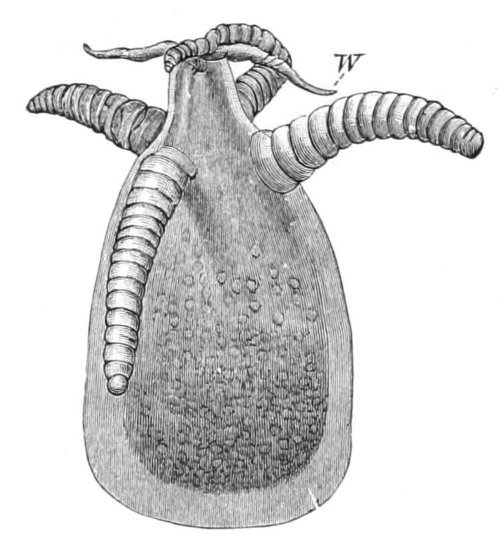
Fig. 138.—Hydra-like stage in the development of Gonionema murbachii. One of the tentacles is carrying a worm (W) to the mouth. The tentacles are shown very much contracted, but they are capable of extending to a length of 2 mm. Height of zooid about 1 mm. (After Perkins.)
In the development of Liriope the free-swimming larva develops into a hydriform person with four tentacles and an enormously elongated hypostome or manubrium; and, according to Brooks, it undergoes a metamorphosis which directly converts it into a Medusa.
There can be very little doubt that in a large number of Trachomedusae the development is direct, the fertilised ovum giving rise to a medusome without the intervention of a hydrosome stage. In some cases, however (Geryonia, etc.), the tentacles appear in development before there is any trace of a sub-umbrella cavity, and this has been interpreted to be a transitory but definite Hydroid stage. It may be supposed that the elimination of the hydrosome stage in these Coelenterates may be associated with their adaptation to a life in the ocean far from the coast.
During the growth of the Medusa from the younger to the adult stages several changes probably occur of a not unimportant character, and it may prove that several genera now placed in the same or even different families are stages in the development, of the same species. In the development of Liriantha appendiculata,[325] for example, four interradial tentacles appear in the first stage which disappear and are replaced by four radial tentacles in the second stage.
As with many other groups of free-swimming marine animals the Trachomedusae have a very wide geographical distribution, and some genera may prove to be almost cosmopolitan, but the majority of the species appear to be characteristic of the warmer regions of the high seas. Sometimes they are found at the surface, but more usually they swim at a depth of a few fathoms to a hundred or more from the surface. The Pectyllidae appear to be confined to the bottom of the sea at great depths.
The principal families of the Trachomedusae are:—
Fam. Olindiidae.—This family appears to be structurally and in development most closely related to the Leptomedusae, and is indeed regarded by Goto[326] as closely related to the Eucopidae in that order. They have two sets of tentacles, velar and exumbrellar; the statocysts are numerous, two on each side of the exumbrellar tentacles. Radial canals four or six. Manubrium well developed and quadrate, with distinct lips. There is an adhesive disc on each exumbrellar tentacle.
Genera: Olindias, Olindioides, Gonionema (Fig. 139), and Halicalyx.
As in other families of Medusae the distribution of the genera is very wide. Olindias mülleri occurs in the Mediterranean, Olindioides formosa off the coast of Japan, Gonionema murbachii is found in abundance in the eel pond at Wood's Holl, United States of America, and Halicalyx off Florida.
Two genera may be referred to in this place, although their systematic position in relation to each other and to other Medusae has not been satisfactorily determined.
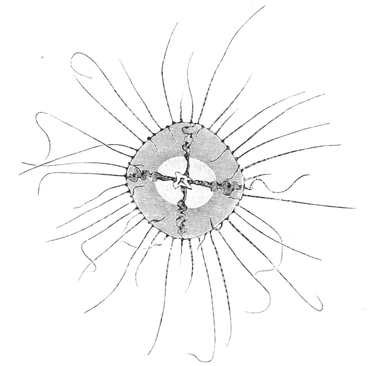
Fig. 139.—Gonionema murbachii. Adult Medusa, shown inverted, and clinging to the bottom. Nat. size. (After Perkins.)
Limnocodium sowerbyi is a small Medusa that was first discovered in the Victoria regia tanks in the Botanic Gardens, Regent's Park, London, in the year 1880. It has lately made its appearance in the Victoria regia tank in the Parc de la Bête d'Or at Lyons.[327] As it was, at the time of its discovery, the only fresh-water jelly-fish known, it excited considerable interest, and this interest was not diminished when the peculiarities of its structure were described by Lankester and others. It has a rather flattened umbrella, with entire margin and numerous marginal tentacles, the manubrium is long, quadrate, and has four distinct lips. There are four radial canals, and the male gonads (all the specimens discovered were of the male sex) are sac-like bodies on the sub-umbrellar aspect of the middle points of the four radial canals. In these characters the genus shows general affinities with the Olindiidae. The difficult question of the origin of the statoliths from the primary germ layers of the embryo and some other points in the minute anatomy of the Medusa have suggested the view that Limnocodium is not properly placed in any of the other orders. Goto,[328] however, in a recent paper, confirms the view of the affinities of Limnocodium with the Olindiidae.
The life-history of Limnocodium is not known, but a curious Hydroid form attached to Pontederia roots was found in the same tank as the Medusae, and this in all probability represents the hydrosome stage of its development. The Medusae are formed apparently by a process of transverse fission of the Hydroid stock[329] similar in some respects to that observed in the production of certain Acraspedote Medusae. This is quite unlike the asexual mode of formation of Medusae in any other Craspedote form. The structure of this hydrosome is, moreover, very different to that of any other Hydroid, and consequently the relations of the genus with the Trachomedusae cannot be regarded as very close.
Limnocodium has only been found in the somewhat artificial conditions of the tanks in botanical gardens, and its native locality is not known, but its association with the Victoria regia water-lily seems to indicate that its home is in tropical South America.
Limnocnida tanganyicae is another remarkable fresh-water Medusa, about seven-eights of an inch in diameter, found in the lakes Tanganyika and Victoria Nyanza of Central Africa.[330] It differs from Limnocodium in having a short collar-like manubrium with a large round mouth two-thirds the diameter of the umbrella, and in several other not unimportant particulars. It produces in May and June a large number of Medusa-buds by gemmation on the manubrium, and in August and September the sexual organs are formed in the same situation.
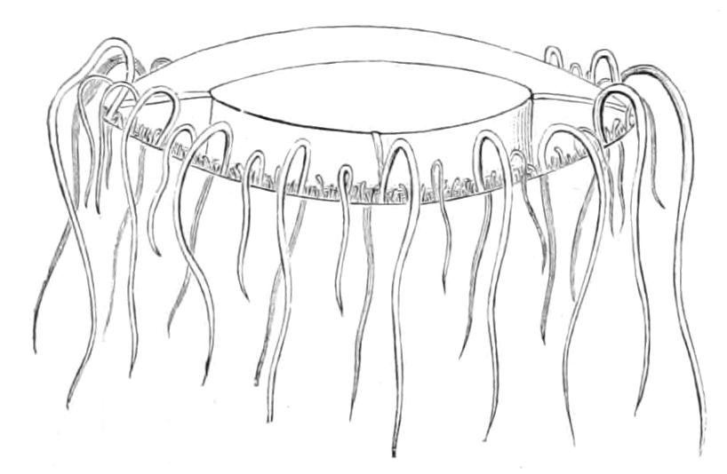
Fig. 140.—Limnocnida tanganyicae. × 2. (After Günther.)
The fixed hydrosome stage, if such a stage occurs in the life-history, has not been discovered; but Mr. Moore[331] believes that the development is direct from ciliated planulae to the Medusae. The occurrence of Limnocnida in Lake Tanganyika is supposed by the same authority to afford a strong support to the view that this lake represents the remnants of a sea which in Jurassic times spread over part of the African continent. This theory has, however, been adversely criticised from several sides.[332]
The character of the manubrium and the position of the sexual cells suggest that Limnocnida has affinities with the Narcomedusae or Anthomedusae, but the marginal sense-organs and the number and position of the tentacles, showing considerable similarity with those of Limnocodium, justify the more convenient plan of placing the two genera in the same family.
Fam. Petasidae.—The genus Petasus is a small Medusa with four radial canals, four gonads, four tentacles, and four free marginal statorhabs. A few other genera associated with Petasus show simple characters as regards the canals and the marginal organs, but as very little is known of any of the genera the family may be regarded as provisional only. Petasus is found in the Mediterranean and off the Canaries.
Fam. Trachynemidae.—In this family there are eight radial canals, and the statorhabs are sunk into a marginal vesicle. Trachynema, characterised by its very long manubrium, is a not uncommon Medusa of the Mediterranean and the eastern Atlantic Ocean. Many of the species are small, but T. funerarium has sometimes a disc two inches in diameter. Homoconema and Pentachogon have numerous very short tentacles.
Fam. Pectyllidae.—This family contains a few deep-sea species with characters similar to those of the preceding family, but the tentacles are provided with terminal suckers. Pectyllis is found in the Atlantic Ocean at depths of over 1000 fathoms.
Fam. Aglauridae.—The radial canals are eight in number and the statorhabs are usually free. In the manubrium there is a rod-like projection of the mesogloea from the aboral wall of the gastric cavity, covered by a thin epithelium of endoderm, which occupies a considerable portion of the lumen of the manubrium. This organ may be called the tongue. Aglaura has an octagonal umbrella, and a manubrium which does not project beyond the velum. It occurs in the Atlantic Ocean and Mediterranean Sea.
Fam. Geryoniidae.—In this family there are four or six radial canals, the statorhabs are sunk in the mesogloea, and a tongue is present in the manubrium. Liriope (Fig. 137) is sometimes as much as three inches in diameter. It has a very long manubrium, and the tongue sometimes projects beyond the mouth. There are four very long radial tentacles. It is found in the Atlantic Ocean, the Mediterranean Sea, and the Pacific and Indian Oceans. Geryonia has a wider geographical distribution than Liriope, and is sometimes four inches in diameter. It differs from Liriope in having six, or a multiple of six, radial canals. Carmarina of the Mediterranean and other seas becomes larger even than Geryonia, from which it differs in the arrangement of the centripetal canals.
Liriantha appendiculata sometimes occurs on the south coast of England during September, October, or at other times.
Order VIII. Narcomedusae.
The Narcomedusae differ from the Trachomedusae in having the margin of the umbrella divided into a number of lobes, and in bearing the gonads on the sub-umbrellar wall of the gastral cavity instead of upon the radial canals. The tentacles are situated at some little distance from the margin of the umbrella at points on the aboral surface corresponding with the angles between the umbrella lobes. Between the base of the tentacle and the marginal angle there is a tract of modified epithelium called the "peronium." The manubrium is usually short, and the mouth leads into an expanded gastral chamber which is provided with lobular diverticula reaching as far as the bases of the tentacles. The marginal sense-organs are in the form of unprotected statorhabs. Very little is known concerning the life-history of any of the Narcomedusae. In Cunoctantha octonaria the peculiar ciliated larva with two tentacles and a very long proboscis soon develops two more tentacles and creeps into the bell of the Anthomedusan Turritopsis, where, attached by its tentacles, it lives a parasitic life. Before being converted into a Medusa it gives rise by gemmation to a number of similar individuals, all of which become, in time, Medusae. The parasitic stage is often regarded as the representative of the hydrosome stage reduced and adapted to the oceanic habit of the adult.
In Cunina proboscidea, and in some other species, a very remarkable method of reproduction has been described by Metschnikoff, called by him "sporogony." In these cases young sexual cells (male or female) wander from the gonad of the parent into the mesogloea of the umbrella, where they develop parthenogenetically into ciliated morulae. These escape by the radial canals into the gastric cavity, and there form a stolon from which young Medusae are formed by gemmation. In C. proboscidea these young Medusae are like the genus Solmaris, but in C. rhododactyla they have the form of the parent. In some cases the ciliated larvae leave the parent altogether and become attached to a Geryonia or some other Medusa, where they form the stolon.
This very interesting method of reproduction cannot be regarded as a primitive one, and throws no light on the origin of the order. It might be regarded as a further stage in the degeneration of the hydrosome stage in its adaptation to a parasitic existence.
The Narcomedusae have a wide geographical distribution. Species of Aeginopsis occur in the White Sea and Bering Strait, but the genera are more characteristic of warmer waters. Some species occur in moderately deep water, and Cunarcha was found in 1675 fathoms off the Canaries, but they are more usually found at or near the surface of the sea.
Fam. Cunanthidae.—Narcomedusae with large gastral diverticula corresponding in position with the bases of the tentacles. Cunina and Cunoctantha, occurring in the Mediterranean and in the Atlantic and Pacific Oceans, belong to this family. In Cunina the tentacles may be eight in number, or some multiple of four between eight and twenty-four. In Cunoctantha the number of tentacles appears to be constantly eight.
Fam. Peganthidae.—There appear to be no gastral pouches in this family. The species of Pegantha are found at depths of about 80 fathoms in the Indian and Pacific Oceans.
Fam. Aeginidae.—The large gastral pouches of this family alternate with the bases of the tentacles. Aegina occurs in the Atlantic and Pacific Oceans. Aeginopsis.
Fam. Solmaridae.—In this family the gastral pouches are variable, sometimes corresponding with, sometimes alternating with, the bases of the tentacles. The circular canal is represented in some genera by solid cords of endoderm. Solmaris sometimes appears in the English Channel, but it is probably a wanderer from the warmer regions of the Atlantic Ocean. It is found in abundance during November on the west coast of Ireland.
Order IX. Siphonophora.
In this order the naturalist finds collected together a number of very beautiful, delicate transparent organisms to which the general term "jelly-fish" may be applied, although their organisation is far more complicated and difficult to describe than that of any of the Medusae. In several of the Hydrozoa the phenomenon of dimorphism has already been noticed. In these cases one set of individuals in a colony performs functions of stinging and catching food and another the functions of devouring and digesting it. In many of the Siphonophora there appears to be a colony of individuals in which the division of labour is carried to a much further extent than it is in the dimorphic Hydrozoa referred to above. Not only are there specialised gastrozooids and dactylozooids, but also gonozooids, zooids for propelling the colony through the water ("nectocalyces"), protective zooids ("hydrophyllia"), and in some cases a specialised zooid for hydrostatic functions; the whole forming a swimming or floating polymorphic colony. But this conception of the construction of the Siphonophora is not the only one that has met with support. By some zoologists the Siphonophoran body is regarded not as a colony of individuals, but as a single individual in which the various organs have become multiplied and dislocated.
The multiplication or repetition of organs that are usually single in each individual is not unknown in other Hydrozoa. In the Medusa of the Gymnoblast Syncoryne, usually known as Sarsia, for example, there is sometimes a remarkable proliferation of the manubrium, and specimens have been found with three or four long manubria attached by a tubular stalk to the centre of the umbrella. Moreover, this complex of manubria may become detached from the umbrella and live for a considerable time an independent existence.[333]
If we regard the manubrium of a Medusa as an organ of the animal's body, it might be thought obvious that the phenomenon observed in the Medusae of Syncoryne is a case of a simple repetition of the parts of an individual; but the power that the group of manubria possesses of leading an independent existence renders its interpretation as a group of organs a matter of some inconvenience. If we can conceive the idea that an organ may become detached and lead an independent existence, there is no reason why we should not regard the Medusa itself of Syncoryne as an organ, and we should be driven to the paradoxical conclusion that, as regards several genera and families of Hydrozoa, we know nothing at present of the individuals, but only of their free-swimming organs, and that in others the individual has degenerated, although one of its organs remains.
There is, however, no convincing argument to support either the conception that the Siphonophoran body is a colony of individuals, or that it is an individual with disjointed organs. These two conceptions are sometimes called the "Poly-person" and "Poly-organ" theories respectively. The difficulty is caused by the impossibility of giving any satisfactory definition in the case of the Hydrozoa of the biological terms "organ" and "individual." In the higher animals, where the correlation of parts is far more complex and essential than it is in Coelenterata, a defined limit to the scope of these terms can be laid down, but in the lower animals the conception of what is termed an organ merges into that which is called an individual, and no definite boundary line between the two exists in Nature. The difficulty is therefore a permanent one, and, in using the expression "colony" for the Siphonophoran body, it must be understood that it is used for convenience' sake rather than because it represents the only correct conception of the organisation of these remarkable Coelenterates.
Regarding the Siphonophora as polymorphic colonies, then, the following forms of zooids may be found.
Nectocalyces.—The nectocalyces are in the form of the umbrella of a medusa attached to the stolon of the colony by the aboral pole. They are provided with a velum and, usually, four radial canals and a circular canal. There is no manubrium, and the marginal tentacles and sense-organs are rudimentary or absent. There may be one or more nectocalyces in each colony, and their function is, by rhythmic contractions, to propel the colony through the water (Fig. 142, N).
Gastrozooids.—These are tubular or saccular zooids provided with a mouth and attached by their aboral extremity to the stolon (Fig. 142, G). In some cases the aboral region of the zooid is differentiated as a stomach. It is dilated and bears the digestive cells, the oral extremity or hypostome being narrower and more transparent. In some cases the mouth is a simple round aperture at the extremity of the hypostome, but in others it is dilated to form a trumpet-like lip.
Dactylozooids.—In Velella and Porpita the dactylozooids are similar in general characters to the tentacles of many Medusae. They are arranged as a frill round the margin of the colony, and each consists of a simple tube of ectoderm and endoderm terminating in a knobbed extremity richly provided with nematocysts.
In many other Siphonophora, however, the dactylozooids are very long and elaborate filaments, which extend for a great distance from the colony into the sea. They reach their most elaborate condition in the Calycophorae.
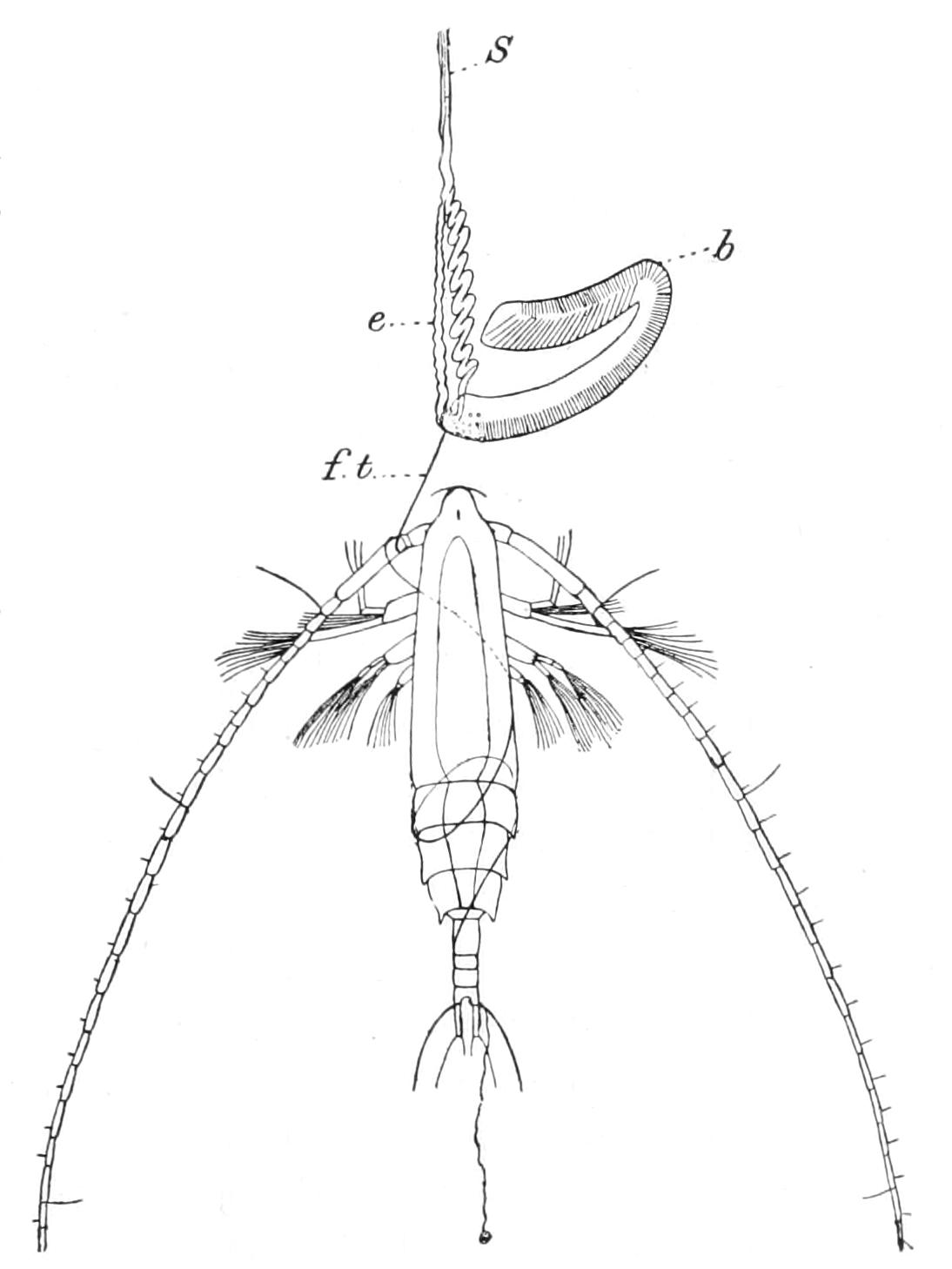
Fig. 141.—A small Crustacean (Rhinocalanus) caught by a terminal filament (f.t) of a battery of Stephanophyes. b, The proximal end of the battery with the most powerful nematocysts; e, elastic band; S, stalk supporting the battery on the dactylozooid. (After Chun.)
The dactylozooid in these forms has a hollow axis, and the lumen is continuous with the cavity of the neighbouring gastrozooid. Arranged at regular intervals on the axis is a series of tentacles ("tentilla"), and each of these supports a kidney-shaped swelling, the "cnidosac," or battery, which is sometimes protected by a hood. Each battery contains an enormous number of nematocysts. In Stephanophyes, for example, there are about 1700 nematocysts of four different kinds in each battery. At the extremity of the battery there is a delicate terminal filament. The action of the battery in Stephanophyes is, according to Chun,[334] a very complicated one. The terminal filament lassos the prey and discharges its somewhat feeble nematocysts at it (Fig. 141). If this kills it, the dactylozooid contracts and passes the prey to a gastrozooid. If the animal continues its struggles, it is drawn up to the distal end of the battery and receives the discharge of a large number of nematocysts; and if this also fails to put an end to its life, a membrane covering the largest and most powerful nematocysts at the proximal end of the whole battery is ruptured, and a final broadside of stinging threads is shot at it.
The larger nematocysts of these batteries in the Siphonophora are among the largest found in Coelenterata, being from 0.5 to 0.1 mm. in length, and they are frequently capable of inflicting painful stings on the human skin. The species of Physalia, commonly called "Portuguese Men-of-War," have perhaps the worst reputation in this respect, the pain being not only intense but lasting a long time.
Hydrophyllia.—In many Siphonophora a number of short, mouthless, non-sexual zooids occur, which appear to have no other function than that of shielding or protecting other and more vital parts of the colony. They consist of an axis of firm mesogloea, covered by a layer of flattened ectoderm, and they may be finger-shaped or triangular in form. In Agalma and Praya an endoderm canal perforates the mesogloea and terminates in a little mouth at the free extremity. In Athoria and Rhodophysa the hydrophyllium terminates in a little nectocalyx.
Pneumatophore.—In all the Siphonophora, with the exception of the Calycophorae, there is found on one side or at one extremity of the colony a vesicle or bladder containing a gas,[335] which serves as a float to support the colony in the water. This bladder or pneumatophore is probably in all cases a much modified nectocalyx. It shows great variations in size and structure in the group. It is sometimes relatively very large, as in Physalia and Velella, sometimes very small, as in Physophora. It is provided with an apical pore in some genera (Rhizophysa), or a basal pore in others (Auronectidae), but it is generally closed. In the many chambered pneumatophore of the Chondrophoridae there are several pores.
In many forms two distinct parts of the pneumatophore can be recognised—a distal region lined by chitin,[336] probably representing the sub-umbrellar cavity of the nectocalyx, and a small funnel-shaped region lined by an epithelium, the homology of which is a matter of dispute. It is believed that the gas is secreted by this epithelium. In the Auronectidae the region with secretory epithelium is relatively large and of a more complicated histological character. It is remarkable also that in this family the pore communicates, not with the chitin-lined region, but directly with the epithelium-lined region.
There is no pneumatophore in the Calycophorae, but in this sub-order a diverticulum of an endoderm canal secretes a globule of oil which may serve the same hydrostatic function.
The stolon is the common stem which supports the different zooids of the colony. In the Calycophorae the stolon is a long, delicate, and extremely contractile thread attached at one end to a nectocalyx, and bearing the zooids in discontinuous groups. These groups of zooids arranged at intervals on the stolon are called the "cormidia." The stolon is a tube with very thick walls. Its lumen is lined by a ciliated endoderm with circular muscular processes, and the surface is covered with an ectoderm, also provided with circular muscular processes. Between these two layers there is a relatively thick mesogloea showing on the outer side deep and compound folds and grooves supporting an elaborate system of longitudinal muscular fibres. In many Physonectidae the stolon is long and filamentous, but not so contractile as it is in Calycophorae, but in others it is much reduced in length and relatively stouter. The reduction in length of the stolon is accompanied by a complication of structure, the simple tubular condition being replaced by a spongy complex of tubes covered by a common sheath of ectoderm. In the Auronectidae the stolon is represented by a conical or hemispherical spongy mass bearing the zooids, and in the Rhizophysaliidae and Chondrophoridae it becomes a disc or ribbon-shaped pad spreading over the under side of the pneumatophore.
Gonozooids.—The gonozooids are simple tubular processes attached to the stolon which bear the Medusae or the degenerate medusiform gonophores. In the Chondrophoridae the gonozooids possess a mouth, but in most Siphonophora they have neither mouth nor tentacles. In some cases, such as Anthophysa, the colonies are bisexual—the male and female gonophores being borne by separate gonozooids—but in others (e.g. Physalia) the colonies appear to be unisexual.
As a general rule the gonophores of Siphonophora do not escape from the parent colony as free-swimming Medusae, but an exception occurs in Velella, which produces a number of small free-swimming Medusae formerly described by Gegenbaur under the generic name Chrysomitra. This Medusa has a velum, a single tentacle, eight to sixteen radial canals, and it bears the gonads on the short manubrium. The Medusa of Velella has, in fact, the essential characters of the Anthomedusae.
Our knowledge of the life-history of the Siphonophora is very incomplete, but there are indications, from scattered observations, that in some genera, at least, it may be very complicated.
The fertilised ovum of Velella gives rise to a planula which sinks to the bottom of the sea, and changes into a remarkable larva known as the Conaria larva. This larva was discovered by Woltereck[337] at depths of 600-1000 metres in great numbers. It is very delicate and transparent, but the endoderm is red (the colour so characteristic of animals inhabiting deep water), and it may be regarded as essentially a deep-sea larva. The larva rises to the surface and changes into the form known as the Ratarula larva, which has a simple one-chambered pneumatophore containing a gas, and a rudiment of the sail. In contrast to the Conaria, the Ratarula is blue in colour. With the development of the zooids on the under side of this larva (i.e. the side opposite to the pneumatophore), a definite octoradial symmetry is shown, there being for some time eight dactylozooids and eight definite folds in the wall of the pneumatophore. This octoradial symmetry, however, is soon lost as the number of folds in the pneumatophore and the number of tentacles increase.
It is probable that in the Siphonophora, as in many other Coelenterata, the production of sexual cells by an individual is no sign that its life-history is completed. There may possibly be two or more phases of life in which sexual maturity is reached.
An example of a complicated life-history is found in the Calycophoran species Muggiaea kochii. The embryo gives rise to a form with a single nectocalyx which is like a Monophyes, and this by the budding of a second nectocalyx produces a form that has a remarkable resemblance to a Diphyes, but the primary nectocalyx degenerates and is cast off, while the secondary one assumes the characters of the single Muggiaea nectocalyx. The stolon of the Muggiaea produces a series of cormidia, and as the sexual cells of the cormidia develop, a special nectocalyx is formed at the base of each one of them, and the group of zooids is detached as an independent colony, formerly known as Eudoxia eschscholtzii. In a similar manner the cormidia of Doramasia picta give rise to the sexual free-swimming monogastric forms, known by the name Ersaea picta (Fig. 142). In these cases it seems possible that the production of ripe sexual cells is confined to the Eudoxia and Ersaea stages respectively, but it is probable that in other species the cormidia do not break off from the stolon, or may escape only from the older colonies.
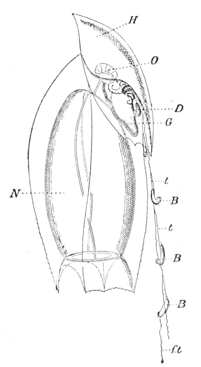
Fig. 142.—Free-swimming Ersaea group of Doramasia picta. B, B, batteries of nematocysts borne by the tentilla; D, dactylozooid; G, gastrozooid; H, hydrophyllium; N, nectocalyx; O, oleocyst; f.t, terminal filament of a battery; t, t, tentilla. The gonozooid is hidden by the gastrozooid. × 10. (After Chun.)
The Siphonophora are essentially free-swimming pelagic organisms. Some of them (Auronectidae) appear to have become adapted to a deep-sea habit, others are usually found in intermediate waters, but the majority occur with the pelagic plankton at or very near the surface of the open sea. Although the order may be said to be cosmopolitan in its distribution, the Siphonophora are only found in great numbers and variety in the sub-tropical and tropical zones. In the temperate and arctic zones they are relatively rare, but Galeolaria biloba and Physophora borealis appear to be true northern forms. The only British species are Muggiaea atlantica and Cupulita sarsii. Velella spirans occasionally drifts from the Atlantic on to our western shores, and sometimes great numbers of the pneumatophores of this species may be found cast up on the beach. Diphyes sp., Physalia sp., and Physophora borealis are also occasionally brought to the British shores by the Gulf Stream.
The Calycophorae are usually perfectly colourless and transparent, with the exception of the oil-globule in the oleocyst, which is yellow or orange in colour. Many of the other Siphonophora, however, are of a transparent, deep indigo blue colour, similar to that of many other components of the plankton.
Most of the Siphonophora, although, strictly speaking, surface animals, are habitually submerged; the large pneumatophores of Velella and Physalia, however, project above the surface, and these animals are therefore frequently drifted by the prevailing wind into large shoals, or blown ashore. At Mentone, on the Mediterranean, Velella is sometimes drifted into the harbour in countless numbers. Agassiz mentions the lines of deep blue Velellas drifted ashore on the coast of Florida; and a small species of blue Physalia may often be seen in long lines on the shore of some of the islands of the Malay Archipelago.
The food of most of the Siphonophora consists of small Crustacea and other minute organisms, but some of the larger forms are capable of catching and devouring fish. It is stated by Bigelow[338] that a big Physalia will capture and devour a full-grown Mackerel. The manner in which it feeds is described as follows:—"It floats on the sea, quietly waiting for some heedless individual to bump its head against one of the tentacles. The fish, on striking, is stung by the nettle-cells, and fastened probably by them to the tentacle. Trying to run away the fish pulls on the tentacle. The tension on its peduncle thus produced acts as a stimulus on apparently some centre there which causes it to contract. The fish in this way is drawn up so that it touches the sticky mouths of the squirming siphons [i.e. gastrozooids]. As soon as the mouths, covered as they are with a gluey substance and provided with nettle-cells, touch the fish they stick fast, a few at first, and gradually more. The mouths open, and their lips are spread out over the fish until they touch, so that by the time he is dead the fish is enclosed in a tight bag composed of the lips of a dozen or more siphon mouths. Here the fish is digested. As it begins to disintegrate partially digested fragments are taken into the stomachs of the attached siphons (gastrozooids). When they have become gorged they detach themselves from the remains of the fish, the process of digestion is completed in the stomachs, and the nutrient fluid is distributed...."
In consequence of the very unsatisfactory state of our knowledge of the life-history of the Siphonophora the classification of the order is a matter of unusual difficulty.
Sub-Order I. Calycophorae.
The character which distinguishes this sub-order is the absence of a pneumatophore.
The colony usually consists of a long, slender, contractile stolon, provided at one end with one, two, or several nectocalyces. Upon the stolon are arranged several groups ("cormidia") of polymorphic zooids.
The nectocalyces have a well-developed velum, four radial canals, and a muscular umbrella-wall. A special peculiarity of the nectocalyx of this sub-order is a diverticulum (oleocyst) from one of the radial canals, containing a coloured globule of oil. The function of this oil-globule is probably similar to that of the pneumatophore, and assists the muscular efforts of the nectocalyces in keeping the colony afloat. One of the nectocalyces of each colony exhibits on one side a deep ectodermic fold, which is frequently converted into a pit. At the bottom of this pit is attached the end of the stolon, the whole of which with its numerous cormidia can be withdrawn into the shelter of the pit when danger threatens. The cormidia consist of at least four kinds of zooids: a gastrozooid with a trumpet-shaped mouth armed with nematocysts, a long dactylozooid provided with a series of tentilla, and a rudimentary gonozooid bearing numbers of male or female medusiform gonophores. These three kinds of zooids are partially covered and protected by a bent shield-shaped phyllozooid or hydrophyllium.
Each of the cormidia is unisexual, but the colony as a whole is usually hermaphrodite, the male and female cormidia regularly alternating, or the male cormidia being arranged on the nectocalycine half and the female cormidia on the opposite half of the stolon.
The families of the Calycophorae are:—
Fam. 1. Monophyidae.—In this family there is a single conical or mitre-shaped nectocalyx. The cormidia become detached as free-swimming Eudoxia or Ersaea forms.
Sub-Fam. 1. Sphaeronectinae.—The primary nectocalyx persists throughout life—Monophyes and Sphaeronectes.
Sub-Fam. 2. Cymbonectinae.—The primary nectocalyx is thrown off, and is replaced by a secondary and permanent nectocalyx—Cymbonectes, Muggiaea, and Doramasia.
Fam. 2. Diphyidae.—The primary mitre-shaped nectocalyx is thrown off and replaced by two secondary rounded, prismatic, or pyramidal, heteromorphic nectocalyces.
This family contains several sub-families, which are arranged in two groups: the Diphyidae Oppositae, in which the two secondary bells are opposite one another, and do not exhibit pronounced ridges; and the Diphyidae Superpositae, in which one of the two secondary nectocalyces is situated in front of the other, and each nectocalyx is provided externally with very definite and often wing-like ridges. In all the Diphyidae Oppositae the cormidia remain attached, whereas in most of the Diphyidae Superpositae they become free-swimming, as in the Monophyidae.
The sub-families of the Diphyidae Oppositae are:—
Sub-Fam. 1. Amphicaryoninae.—One of the two secondary nectocalyces becomes flattened above to form a shield, and at the same time its sub-umbrellar cavity is atrophied, and its radial canals reduced. Mitrophyes, Atlantic Ocean.
Sub-Fam. 2. Prayinae.—The colony exhibits a pair of large, obtuse nectocalyces, with a relatively small sub-umbrellar cavity. Praya, Mediterranean and Atlantic.
Sub-Fam. 3. Desmophyinae.—The colony bears a large number of reserve or tertiary nectocalyces arranged in two rows. Desmophyes, Indian Ocean.
Sub-Fam. 4. Stephanophyinae.—There are four nectocalyces arranged in a horizontal plane. Each one of the cormidia bears a nectocalyx, which is periodically replaced. This sub-family is constituted for Stephanophyes superba from the Canary Islands. It attains a length of 25 cm., and is probably the largest and most beautiful of all the Calycophoridae.[339]
The group Diphyidae Superpositae contains the following:—
Sub-Fam. 1. Galeolarinae.—Galeolaria.
Sub-Fam. 2. Diphyopsinae.—Diphyes.
Sub-Fam. 3. Abylinae.—Abyla.
These sub-families differ from one another in the character and shape of the nectocalyces and in other characters. They have a world-wide distribution, Diphyes and Galeolaria extending north into the Arctic Seas. Diphyes is British.
Fam. 3. Polyphyidae.—The nectocalyces are numerous, and superposed in two rows. The cormidia remain attached.
The family contains the genera Polyphyes and Hippopodius, both probably cosmopolitan in warm waters.
Sub-Order II. Physophorae.
In this sub-order the primary nectocalyx gives rise to a definite pneumatophore. There are four families.
Fam. 1. Physonectidae.—In this, the largest family of the sub-order, there is a monothalamic pneumatophore supporting a stolon, which in some forms is of great length, but in others is reduced to a stump or pad, on which there are usually found several nectocalyces, hydrophyllia, gastrozooids, gonozooids, and tentilla.
The principal sub-families are:—
Agalminae.—With a long stolon, bearing at the upper end (i.e. the end next to the pneumatophore) two rows of nectocalyces. The other zooids are arranged in cormidia on the stolon, each covered by a hydrophyllium. Dactylozooids with tentilla. Agalma and Cupulita, Mediterranean Sea.
Apoleminae.—Similar to the above, but without tentilla. Apolemia—this genus attains a length of two or three metres. Mediterranean Sea. Dicymba, Indian Ocean.
Physophorinae.—The pneumatophore larger in proportion than it is in the preceding families. The stolon is short, and bears rows of nectocalyces at the upper end. The gastrozooids, dactylozooids, and gonozooids are arranged in verticils on the lower expanded part of the stolon. Hydrophyllia absent. Physophora, cosmopolitan in the areas of warm sea water.
Fam. 2. Auronectidae.—The pneumatophore is large. The stolon is reduced to a spongy mass of tissue on the under side of the pneumatophore, and this bears numerous cormidia arranged in a helicoid spiral. Projecting from the base of the pneumatophore there is a peculiar organ called the "aurophore," provided with an apical pore. This organ has been described as a specially modified nectocalyx, but it is probably a specialised development of the epithelium-lined portion of the pneumatophore of other Physophorae. The Auronectidae are found only at considerable depths, 300 to 1400 fathoms, and are probably specially adapted to that habitat. Rhodalia, Stephalia, Atlantic Ocean.
Fam. 3. Rhizophysaliidae.—The pneumatophore is large, or very large, in this family. The zooids are arranged in horizontal rows on the under side of the pneumatophore (Physalia), or in a helicoid spiral on a short stolon (Epibulia). There are no nectocalyces nor hydrophyllia.
The genus Physalia is the notorious "Portuguese Man-of-War." The pneumatophore is a large bladder-like vesicle, sometimes attaining a length of 12 cm. One species described by Haeckel under the generic name Caravella has a pneumatophore 30 cm. and more in length, and dactylozooids attaining a length of 20 metres. It is a curious fact that only the male colonies of Physalia are known, and it is suggested that the female may have quite a different form.[340] Epibulia has a much smaller bladder than Physalia. Both genera have a cosmopolitan distribution at the surface of the warm seas.
Fam. 4. Chondrophoridae.—This family stands quite by itself in the sub-order Physophorae, and is placed in a separate division of the sub-order by Chun, who gives it the name Tracheophysa. The essential distinguishing characters of the family are the large polythalamic pneumatophore and the single large central gastrozooid.
The colony is disc-shaped, and has a superficial resemblance to a Medusa. On the upper side is the flattened pneumatophore, covered by a fold of tissue continuous with that at the edge of the disc. In Velella a vertical triangular sail or crest rises from the upper side, but this is absent in Porpita.
The mouth of the gastrozooid opens into a large digestive cavity, and between this and the under surface of the pneumatophore there is a glandular spongy tissue called the liver. The liver extends over the whole of the under side of the pneumatophore, and sends processes round the edge of the disc into the tissues of its upper surface. Intimately associated with the liver, and penetrating its interstices, is an organ which appears to be entirely composed of nematocysts, derived from the ectoderm, and called the central organ. At the margin of the disc there is a fringe of simple digitiform dactylozooids, and between the dactylozooids and the centrally placed gastrozooid are numerous gonozooids. Each of the gonozooids is provided with a distinct mouth, and bears the gonophores, which escape before the ripening of the gonads as the free-swimming Medusae called Chrysomitra. The pneumatophore consists of a number of annular chambers arranged in a concentric manner round the central original chamber formed from a modified zooid. These annular chambers are in communication with one another, and have each two pores (pneumatopyles) opening above to the exterior. The most remarkable feature, however, of the system is a series of fine branching tubes ("tracheae"), which pass from the annular chambers of the pneumatophore downwards into the hepatic mass and ramify there.
There are two well-known genera: Velella with a sail, and Porpita without a sail. They are both found at the surface of the warmer regions of the great oceans and in the Mediterranean. Velella sometimes drifts on to British coasts from the Atlantic.
The genus Discalia has a much more simple octoradial structure. It was found at depths of 2600 and 2750 fathoms in the Pacific Ocean.