Chapter XX
Reading edition
ECHINODERMATA (CONTINUED): PELMATOZOA—CRINOIDEA = SEA-LILIES—THECOIDEA—CARPOIDEA—CYSTOIDEA—BLASTOIDEA
SUB-PHYLUM II. PELMATOZOA
The Pelmatozoa differ from the Eleutherozoa in several important respects. They are fixed (at any rate in the young stage) by the centre of the aboral surface, and this portion of the body usually takes on the form of a stem supported by a definite series of ossicles, so that we can discriminate a "calyx"—the main part of the body—from the "stem." Further, the podia and the ambulacral grooves seem to be always covered with powerful cilia, which are employed in producing a current which sweeps small organisms to the mouth. The podia are never locomotor in function; their use is similar to that of the tentacles on the lophophore of Polyzoa and Brachiopoda.
The living Pelmatozoa are very few in number compared with the extinct forms. It may with justice be said that the group is nearly extinct; indeed, out of its five classes one alone, and that the most highly specialised class, survives till the present day. Now we have already seen that, in the case of the Eleutherozoa, if the annectant fossil types were taken into consideration, the definition of the classes would be difficult, so that it is not to be wondered at if the classes of the Pelmatozoa are also somewhat difficult to define; and it must be added that this difficulty is not only due to the fact that intermediate types occasionally occur, but also to our ignorance of the functions of many structures found in fossil types, speculations regarding which are to be received with caution. Bearing in mind, then, the provisional nature of the classification, we may give the diagnoses of the principal divisions as follows:—
Class I. Crinoidea.—Pelmatozoa provided typically with a well-marked stem; calyx consisting of an aboral "patina" of two or three circles of plates, and a flexible "tegmen" or oral surface with small plates or none; radial canals supported by long branched arms, which are developed as direct prolongations of the uppermost circle of plates in the patina.
Class II. Thecoidea (Jaekel) = Edrioasteroidea (Bather).—Pelmatozoa without a stalk, fixed to the substratum by the whole aboral surface. The radial canals run out over the oral surface in grooves, which are closed by specially modified plates; but there are no arms of any kind.
Class III. Carpoidea (Jaekel).—Pelmatozoa with a well-developed stalk. The radial canals and their branches are devoid of a skeleton, and either produce no modifications at all on the skeleton of the calyx, or at most are supported by short horn-like outgrowths of some of its plates.
Class IV. Cystoidea.—Pelmatozoa which typically possess a well-developed stalk, a sac-like calyx contracted at the mouth and covered with plates, some of which are pierced with pores or slits; the radial canals, though they may for part of their course run over the surface in grooves, have their terminal portions supported by free unbranched arms ("fingers").
Class V. Blastoidea.—Pelmatozoa provided with a well-developed stalk and ovoid bud-like calyx. From the mouth the radial canals run backwards over the calyx, as in Echinoidea, but they give rise to numerous lateral branches, which are supported by free unbranched arms ("fingers"). Special respiratory organs occur on the interradial areas in the form of parallel folds called "hydrospires."
CLASS I. CRINOIDEA
This is the only class which has living representatives. There are twelve recent genera, of which eight retain the stalk throughout life; the remaining four lose it when adult, retaining only a stump, termed the "centro-dorsal," covered with fixing organs ("cirri"). The stalked forms are confined to considerable depths, and can only be obtained by deep dredging, whereas many of the stalkless forms are comparatively common. We shall select as type for special description the common Feather-star, Antedon rosacea (bifida), which can be dredged in depths of ten fathoms off the south-west coast of England.
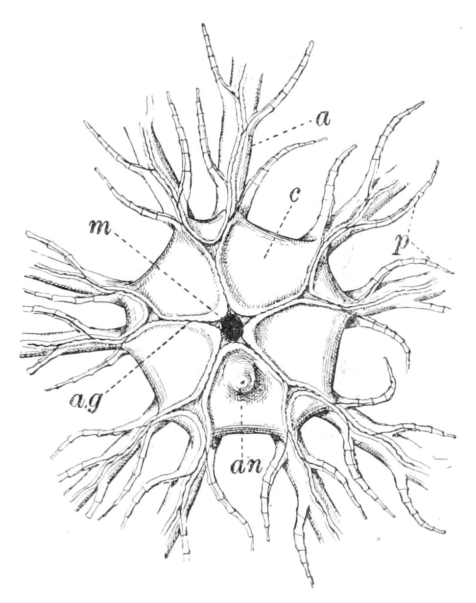
Fig. 264.—Oral view of Antedon rosacea. × 3. a, Arm; a.g, ambulacral groove; an, anus; c, calyx; m, mouth; p, pinnules.
The animal consists of a small flattened calyx, from which radiate out ten long delicate arms, each fringed with a double series of short branches called "pinnules." In the centre of the aboral surface can be seen the centre-dorsal plate (Fig. 265, c), a knob-like stump of the broken-off stem, covered with small whip-like outgrowths called "cirri," by means of which the animal is anchored to the substratum (Fig. 265, cir). When Antedon is disturbed it relaxes its hold, and swims by graceful muscular movements of the arms. These are arranged in five pairs, and the corresponding members (right and left) of all the pairs are bent and relaxed together. On coming to rest the animal reattaches itself by means of the cirri. These are composed of cylindrical ossicles joined to one another by muscles, and they can thus act as efficient grasping organs. In the centre of the oral surface, which is termed the "tegmen," and is soft, flexible, and without visible calcifications, is situated the mouth, surrounded by five short triangular flaps called "oral valves." In the intervals between these valves, grooves radiate from the mouth which bifurcate at the points of origin of each pair of arms, and are continued over their surfaces. These grooves correspond to the ambulacral grooves of Asteroidea, and to the epineural canals of the other classes of Eleutherozoa. At each side of each groove are to be found a series of podia in the form of delicate finger-like processes, which serve only for respiration and for producing a current of water, their surfaces, like that of the grooves between them, being covered with powerful cilia. The anus is at the extremity of a little knob called the anal papilla, situated in one of the interradii (Fig. 264, an).
As in Ophiuroidea, the ectoderm cells have disappeared over the whole surface of the body, except the grooves and the podia, the only trace of their former existence being a cuticle with adherent nuclei. Pedicellariae are unknown in all Pelmatozoa; and spines have only been described from one fossil species of Crinoid. Beneath the cuticle is the dermis, having the composition described in the case of Asterias rubens; this on the aboral side of the calyx gives rise to the "patina," consisting of plates, in part movable on one another, in part immovably fused together. Those visible from the outside are (1) the centro-dorsal ossicle, from which the cirri spring; (2) five columns of ossicles termed radials (Fig. 266, R1, R2, R3); each column consists of three radials, extending from the centro-dorsal to the origin of a pair of arms. The uppermost radial in each column bears two facets for the articulation of these arms. Each arm is supported by a series of "brachial ossicles" (Br).
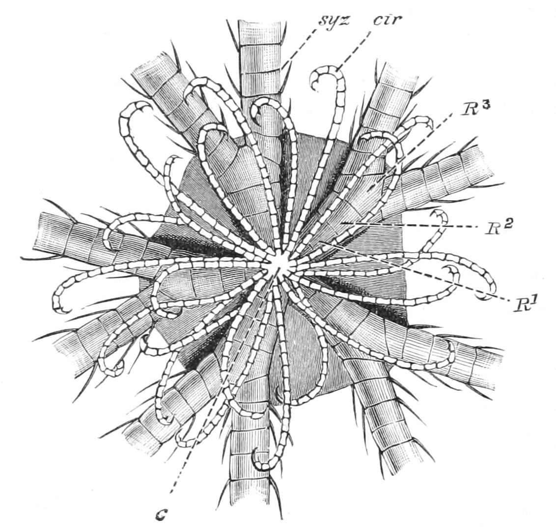
Fig. 265.—View of Antedon rosacea from aboral surface, × 4. c, Centro-dorsal; cir, cirrus; R1, R2, R3, the three radial plates of one column; syz, syzygy.
It is evident, both from the number of ambulacral grooves and of the columns of radials, that Antedon has only five radii, and each pair of arms must be regarded as having arisen by the bifurcation of a primitive arm. This is proved to be true by a study of the development, and it can further be shown that the arms fork repeatedly; but in these further bifurcations one fork remains short, and forms a pinnule, whilst the other continues the arm. Thus the arm, instead of being a single axis, is really a series of axes—in a word, it is a "sympodium."
If in the case of any bifurcation the two forks were to develop equally, the number of arms in that ray would be doubled, and this actually happens in the case of other species of Antedon.
Digestive System.—The mouth leads through a short vertical oesophagus into an enlarged stomach, which lies horizontally curved around the axis of the calyx. The stomach is succeeded by a short intestine, which leads into the anal papilla. Both oesophagus and stomach are ciliated, and the food consists of minute organisms, swept into the mouth by the current produced by the cilia covering the ambulacral grooves and podia; the ten arms may indeed be compared to a net spread out in the water to catch swimming prey.
The water-vascular system consists of a ring closely surrounding the mouth, from which radial canals are given off which underlie the ambulacral grooves and bifurcate with them. The podia have no ampullae, but muscular strands traverse the cavities of the radial canals, and that of the ring-canal, and by their action water can be forced into the podia, which are thus extended. Numerous stone-canals hang down from the ring-canal, and open freely into the coelom; they do not, as in Holothuroidea (where the same arrangement occurs), end in sieve-like madreporites. The tegmen, i.e. the ventral surface of the calyx, is pierced by a number of isolated pores lined by ciliated cells, which suck in water. In the oldest Pelmatozoa there seems to have been a regular madreporite. In the larva of Antedon there is but one pore-canal, which, as in most Eleutherozoa, leads into a special section of the coelom, the "axial sinus," embedded in the body-wall, with which also the single stone-canal communicates; but later the division between the axial sinus and the rest of the coelom breaks down, and then the pore-canals and stone-canals become multiplied independently of each other (Fig. 266, m.p, p.c, and st.c).
Nervous System.—In the young stalked form the nervous system, as in other Echinoderms, consists of a ring round the mouth, from which radial cords are given off which run under the ambulacral grooves (Fig. 266, nerv.rad.v).
The fibres of this nervous system are, as in Asteroidea, immediately beneath the bases of the ectoderm cells. A large band of fibres is given off to each podium, which is covered with minute elevations, each with pointed sense-hairs in the centre. As the animal grows, another nervous system makes its appearance, which is developed from the coelomic wall, the cells of certain tracts of which multiply and bud off ganglion cells from which the fibres grow out.
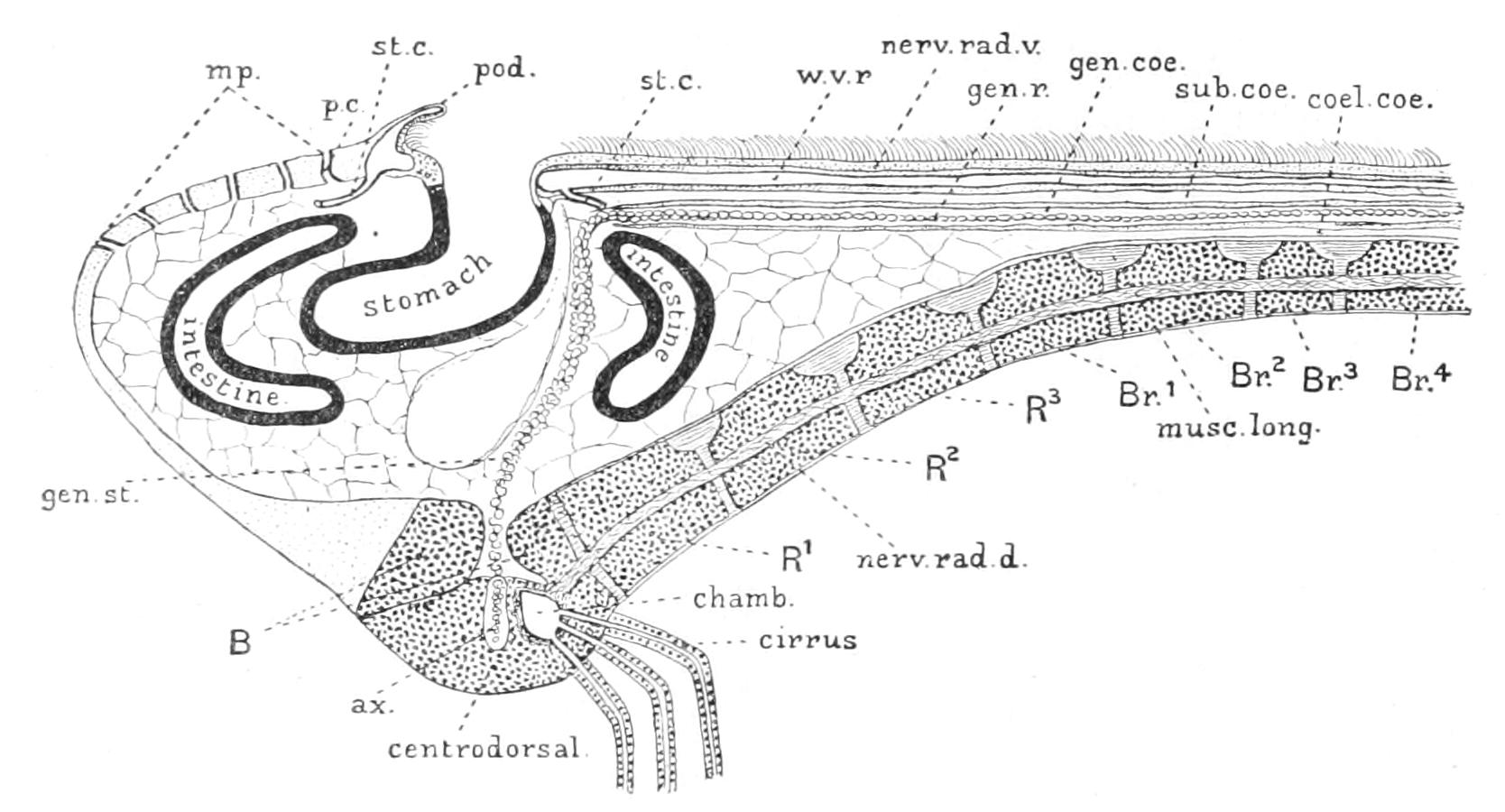
Fig. 266.—Diagrammatic longitudinal section through one arm and the opposite interradius of Antedon. ax, Central canal of centro-dorsal, with prolongation of genital stolon; B, rosette, consisting of coalesced basals; Br1, Br2, Br3, Br4, the first four brachial ossicles; chamb, chambered organ; coel.coe, coelomic canal of arm; gen.coe, genital canal of arm; gen.r, genital rachis; gen.st, genital stolon; m.p, madreporic pores; musc.long, longitudinal muscle; nerv.rad.d, dorsal radial nerve; nerv.rad.v, ventral radial nerve; p.c, pore-canal; pod, podium; R1-R3, 1st to 3rd radials; st.c, stone-canal; sub.coe, sub-tentacular canal of arm; w.v.r, radial water-vessel.
This "aboral nervous system," as it is called, has its centre in the "chambered organ" (Fig. 266, chamb), which is embedded in the centro-dorsal ossicle, and is roofed over by a plate called the "rosette." This represents the five coalesced "basals," a ring of plates which in other forms alternate with the lowest radials, and it intervenes between these and the centro-dorsal. The chambered organ consists of a ring of five vesicles, which have originated as pouches of the aboral coelom (Fig. 266, chamb). The walls of these vesicles develop nervous matter; from them radiate out five great cords, deeply embedded in the plates of the patina. These cords rapidly fork, and one division of each of two adjacent cords enters the lowest radial. In the third radial all the cords are connected by a commissure which runs completely round the calyx. Each of the cords in the third radial forks again, and one branch of each cord enters each of the two arms connected with it, and the two branches entering an arm coalesce to form a single cord. In the arms, as in the calyx, the cords are deeply embedded in the ossicles, but branches extend to the ventral surface of the arms and here unite to form two longitudinal cords, one on each side of the groove. In the tegmen these cords are connected by an outer nerve-ring, branches from which join the ectodermal nerve-ring already described.
The researches first of W. B. Carpenter[512] and then of Marshall[513] have proved that it is the aboral nervous system which really controls the movements of the animal. If the chambered organ is destroyed by cautery, the whole movements of the animal are paralysed; but it will carry out its characteristic swimming movements just as well if the whole tegmen with the ambulacral nerve-ring and the whole of the alimentary canal are torn away. The commissure in the third radials co-ordinates the movements of the arms. If it is cut they move independently of one another. The position of the radial cords inside the ossicles is gradually acquired. At first they are gutter-like evaginations of the coelom; by upgrowth of their sides the gutters become canals, and are then surrounded by calcified tissue. The cirri have each a cord traversing them which originates from the chambered organ.
Coelom.—In the young stalked form the coelom consists of the water-vascular system ("hydrocoel"), and underlying it an oral coelom, separated from an aboral coelom by a horizontal mesentery. As the animal grows, this horizontal mesentery becomes largely absorbed, and the coelom becomes everywhere traversed by cellular cords (trabeculae), which are later calcified.
Both oral and aboral coelom become, like the hydrocoel, bent into hoops, and along the axis of the aboral coelom a cord of germ-cells is developed, which constitutes the "genital stolon." The chambered organ is developed from the aboral coelom, and in the centre of its five chambers a median pocket grows down into the centro-dorsal, along the side of which is an extension of the genital stolon. In the arms the mesentery separating the extensions of the oral and aboral coelom persists; the oral extension consists of two parallel canals called "subtentacular" (Fig. 267, s.c), whilst the aboral space is termed the "coeliac" canal (Fig. 267, c.c). In the tip of the pinnule, that is to say at the extremity of a ramification of the arm, the coeliac and subtentacular canals communicate. As portions of the lining of both canals are ciliated, a circulation of the coelomic fluid is thus kept up. The genital stolon gives rise at the level of the remnant of the horizontal mesentery in the disc to a circular genital rachis, whence cords pass down the arms in the tissue separating subtentacular and coeliac canals (Fig. 267, g.r). Each cord is contained in a special tube, the "genital canal," which is probably developed in the same way as the aboral sinus of the Eleutherozoa, i.e. as a special sheltering outgrowth of the coelom (Fig. 267, g.c). In the pinnule the rachis swells out into a genital organ, from which a short duct is developed when the organ is mature. The eggs are large (3 mm. in diameter), and adhere for a considerable period of their development to the pinnules.
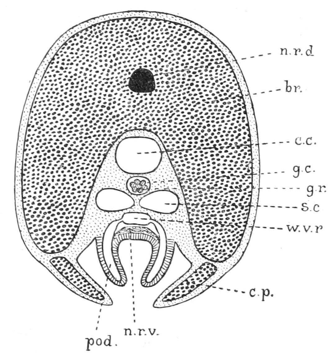
Fig. 267.—Diagrammatic transverse section of arm of Antedon. To compare it with the section of an Ophiuroid arm it is inverted from its natural position. br, Brachial ossicle; c.c, coeliac canal; c.p, covering plate; g.c, genital canal; g.r, genital rachis; n.r.d, dorsal nerve-cord; n.r.v, ventral nerve-cord; pod, podium; s.c, subtentacular canal; w.v.r, radial water-vessel.
The muscles of Antedon are of two kinds. Those of the water-vascular system are, as in Eleutherozoa, basal outgrowths of the cells forming the walls of the system. The muscles moving the joints of the arms appear to be modifications of connective-tissue cells. When the brachials are isolated their terminal faces, strikingly long, recall those of Ophiuroid vertebrae. There is a ventral groove for the coelomic canal. Above this groove the face is divided by ridges into four areas for attachment of the muscles. Dorsal to this is the pit for the strong ligament which binds the ossicles together; then comes the canal for the aboral nerve-cord, whilst dorsal to this is the pit for what is called the "dorsal elastic ligament." The theory underlying this name is that the muscles bend the arms ventrally, and the ligament by its elasticity restores them to their places; but there seems reason to believe that the "ligament" is really a dorsal muscle. It is particularly to be noted that similar muscles occur between the first and second radials, proving that the primary arm really begins with the first radial. The second and third radials, as also the first two ossicles and certain others of each arm, are closely united by calcified fibres, and this kind of union is called a "syzygy" (Fig. 265, syz). The cirri have all their ossicles united by muscular attachment, and can move rapidly.
The blood system (see pp. 449-451) forms a ring consisting of a network of strings round the oesophagus. This is termed the "labial plexus." From this cords can be traced to the wall of the stomach and to the surface of the genital stolon. The assertion that radial strands intervene between the ectodermic nerve-cord and the radial water-vascular canal, though usually made, does not appear to be justified, since what is termed the vessel appears to be a crevice formed by shrinkage in preservation.
The process of respiration is doubtless largely carried out by the podia, but it must be assisted by the constant instreaming of fresh sea-water through the pore-canals. The process of excretion has not been directly observed in Antedon, but structures called "sacculi" may be connected with this function. These are spherical masses of amoebocytes embedded in the tegmen. During life they are colourless, but after death they become coloured, showing that they secrete a peculiar compound. These sacculi abound in the disc, and a row of them is to be found at each side of the ambulacral groove in the arms. When, as in tropical species, the groove is supported by side-plates, these are notched for the reception of the sacculi.
Turning now to survey the group Crinoidea as a whole, lack of space forces us to confine our attention mainly to the living forms. These differ amongst themselves chiefly in the following points: (1) the condition of the stem; (2) the structure of the calyx; (3) what is intimately connected with this, the method of branching of the arms; and (4) the length of the alimentary canal.
Condition of the Stem.—This is represented by a centro-dorsal stump in Antedon and most of its allies, but in Actinometra it becomes a flat plate, and in some species in old age all the cirri drop off. In Uintacrinus and Marsupites (fossil genera) there is no trace of cirri. In Pentacrinidae there is a long stem, pentagonal in cross-section, in which alternate ossicles carry whorls of cirri; in Rhizocrinidae the stem consists of compressed ossicles, elliptical in section, bearing cirri only at the rooting tip, whilst in Hyocrinus the stem is made up of cylindrical ossicles, cirri being apparently absent. Finally, in Holopus the stem is represented by an uncalcified leathery outgrowth from the calyx.
Skeleton of Calyx and Arm.—In living Crinoidea, with the doubtful exceptions of Holopus and Hyocrinus, the calyx is supposed to be built up originally of four whorls of plates, viz. "infra-basals," "basals," "radials," and "orals," the last named forming the skeleton of the oral valves round the mouth. In the two exceptions named there is no certain evidence of the existence of infra-basals. In living forms the infra-basals coalesce with the uppermost joint of the stem; the basals remain large and conspicuous, though they are fused into a ring in Rhizocrinidae, Atelecrinus, and Thaumatocrinus, whilst in Pentacrinidae this ring is nearly, and in Antedon and its allies completely, hidden when the calyx is viewed from the outside. In Hyocrinus the basals are represented by three ossicles. The lowest radials are an important element in the patina in every case, but the upper radials, the incipient portions of the arms, may be incorporated in the calyx (Pentacrinus, Antedon) or may be free (Rhizocrinidae and Hyocrinus); in Metacrinus there are five to eight radials in each column, all incorporated.
The oral plates are very large in Hyocrinus, Holopus, and Thaumatocrinus, small in Rhizocrinus, vestigial or absent in Bathycrinus, and completely absorbed in Antedon. In addition to these main elements, in many species small accessory plates are developed (a) at the sides of the ambulacral groove, over which they can close down (many species of Antedon, Hyocrinus, Holopus, Rhizocrinidae, some species of Pentacrinidae); these are "covering plates," and correspond in function to the adambulacrals of Asteroidea; (b) supporting the sides of the groove and corresponding to the ambulacrals of Asteroidea;[514] these are "side-plates," and the covering plates articulate with them; (c) on the surface of the tegmen; these are the interradial plates, which in Thaumatocrinus alone among recent forms, but in many fossil forms, are continued into the patina, where they separate the radial plates.
Mode of Branching of the Arms.—All modern Crinoids have pinnules, and this, as has already been explained, is due to a suppressed dichotomy. The extent of the suppression determines the number of arms, which varies within the same genus.
Alimentary Canal.—In Hyocrinus there is no dilatation which could be called a stomach; in Actinometra the mouth is excentric, and the anal papilla occupies the centre of the tegmen. The intestine is elongated, and describes several turns round the papilla before ending in the anus.
The classification of Crinoidea cannot properly be considered without taking account of fossil forms, but to do so at all adequately is impossible on account of limitations of space. Less regret may be felt because the three specialists in this branch, viz. Bather in England, Springer in America, and Jaekel in Germany, come to fundamentally different conclusions on the subject. If we confine our attention to living forms we may, with P. H. Carpenter,[515] select the stem as the basis of classification. As the method of gaining food is the same in all cases, the Crinoidea have probably split on the method of attachment to the substratum. These families—it is impossible, in view of the greater range of variety in fossils, to dignify them with the name of orders—are as follow:—
Fam. 1. Hyocrinidae.—Stem long and persistent; cirri absent; stem ossicles cylindrical—ligaments uniting them not specialised. Arms (five) short, but with extremely long pinnules. Patina composed of long exposed basals and a ring of five spade-shaped radials. Five large persistent orals. Interradials and covering plates present. One species (Hyocrinus bethellianus, Figs. 268, 269) dredged up in the Southern Pacific Ocean.
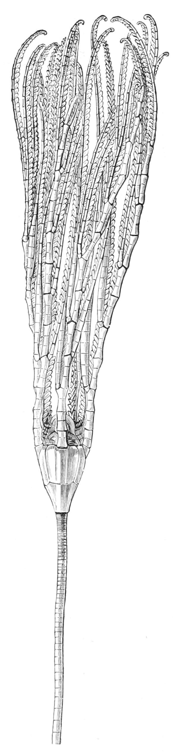
Fig. 268.—Hyocrinus bethellianus. × 2. (From Wyville Thomson.)
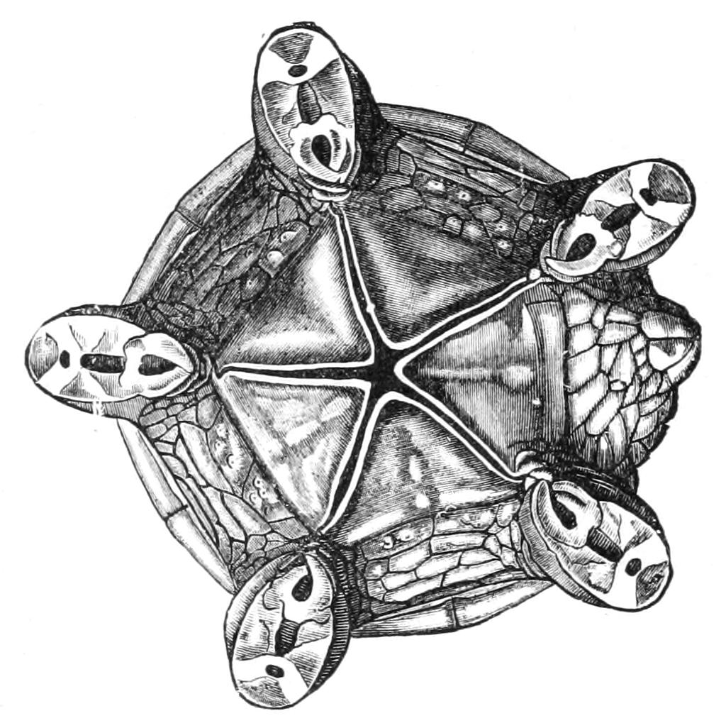
Fig. 269.—Tegmen of Hyocrinus, viewed from above after removal of the arms, × 8. (From Wyville Thomson.)
Fam. 2. Rhizocrinidae.—Stem long and persistent; cirri confined to a few near the root or replaced by rooting branches of the stem. Stem-ossicles thin and pentagonal at the summit, but lower down compressed, elliptical in section, and united by two ligaments separated by a transverse ridge for articulation. Patina composed of exposed basals and a ring of five short radials. Orals large or vestigial. Covering plates and interradials present. Two genera, both from great depths in the Atlantic—Rhizocrinus (Fig. 270), with five arms and well developed orals (attachment by branching root-cirri); and Bathycrinus, with ten arms and vestigial orals; attachment by root-like branches of stem (this is essentially the same as root-cirri).
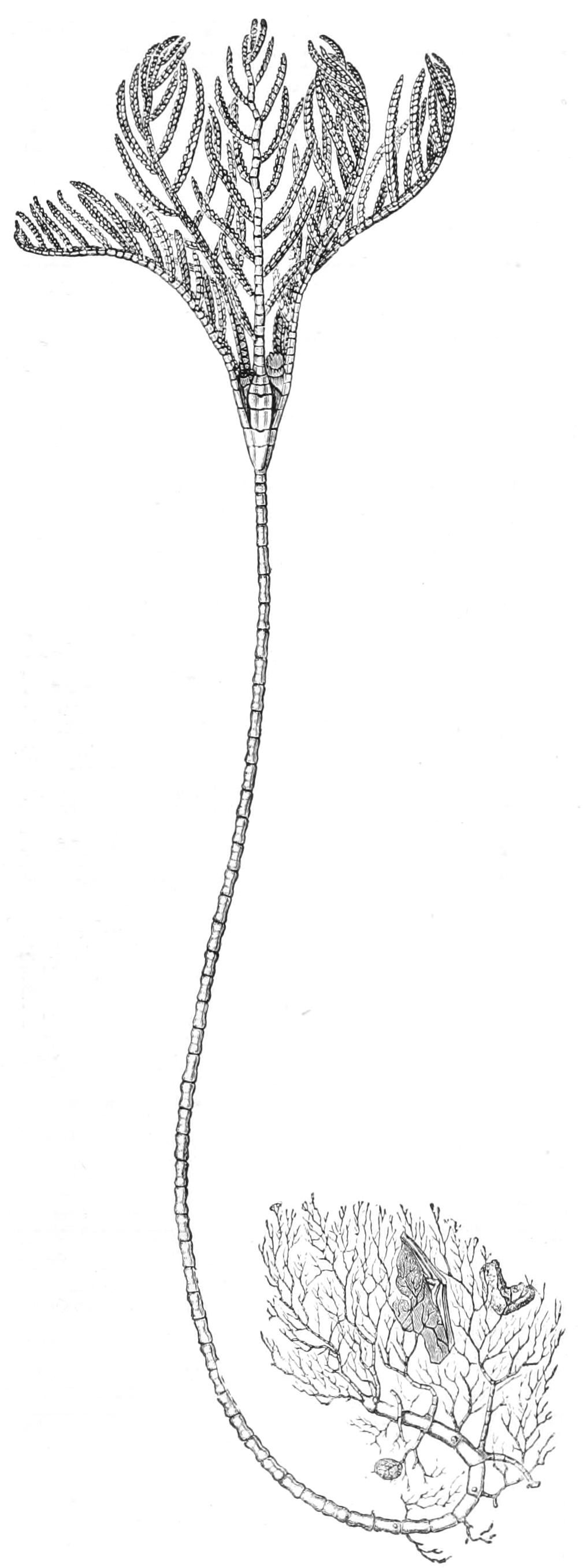
Fig. 270.—Rhizocrinus lofotensis. × 1½. (From Wyville Thomson.)
Fam. 3. Pentacrinidae.—Stem consisting of ossicles which are pentagonal in section, united in pairs by syzygy, the upper one of each pair bearing a whorl of cirri and united by five bundles of fibres of petal-like section with the lower one of the pair above it. No rooting processes. Patina consists almost entirely of columns of radials, the basals being almost or completely hidden. Orals absent, but side-plates in the ambulacral grooves. Two recent genera, Pentacrinus (Isocrinus), with three radials in each column; Metacrinus, with five to eight radials in a column, but the third radial bears a pinnule. Pentacrinus is found in both the Caribbean Sea and the Pacific Ocean; Metacrinus in the Pacific. It appears that the Pentacrinidae when young are attached by a foot-plate at the apex of the stem; but when adult, the stem is broken in two and the animals, like Antedon, swim by movements of the arms, dragging a large part of the stem after them, by which they effect temporary attachment. As in other stalked forms, the cavities of the chambered organ are prolonged into canals which traverse the stalk; but in this family there is the peculiarity that a repetition of the chambered organ is found opposite every whorl of cirri.
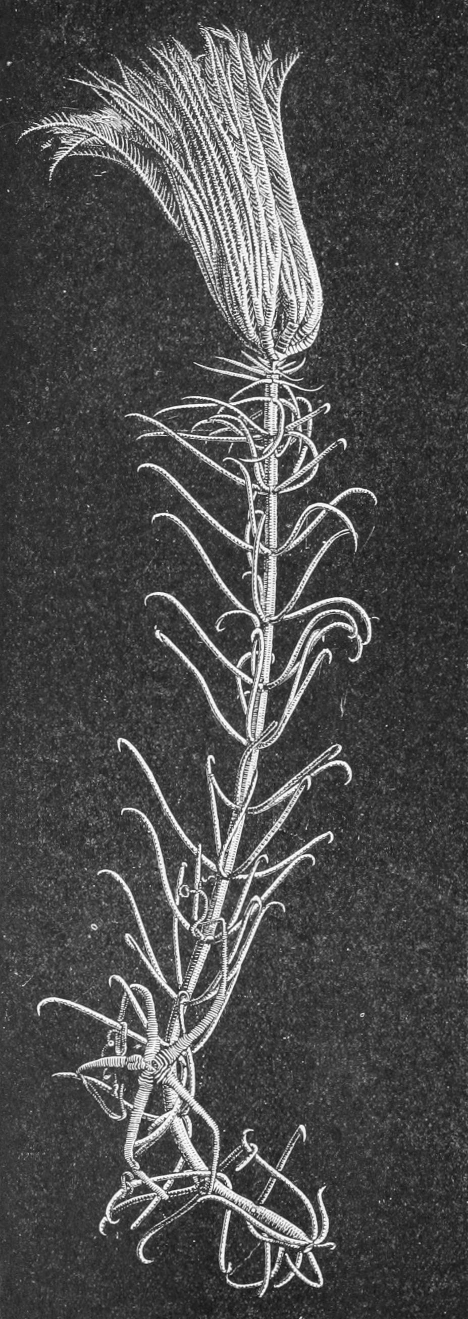
Fig. 271.—Pentacrinus asteria. × ¼. (From Wyville Thomson.)
Fam. 4. Holopodidae.—Stem represented by a leathery noncalcified outgrowth from the base of the calyx; one circle of radials indistinguishably fused with the basals and with each other to form the walls of the calyx. Large oral plates, ten short arms. One genus, Holopus, in shallow water in the Caribbean Sea.
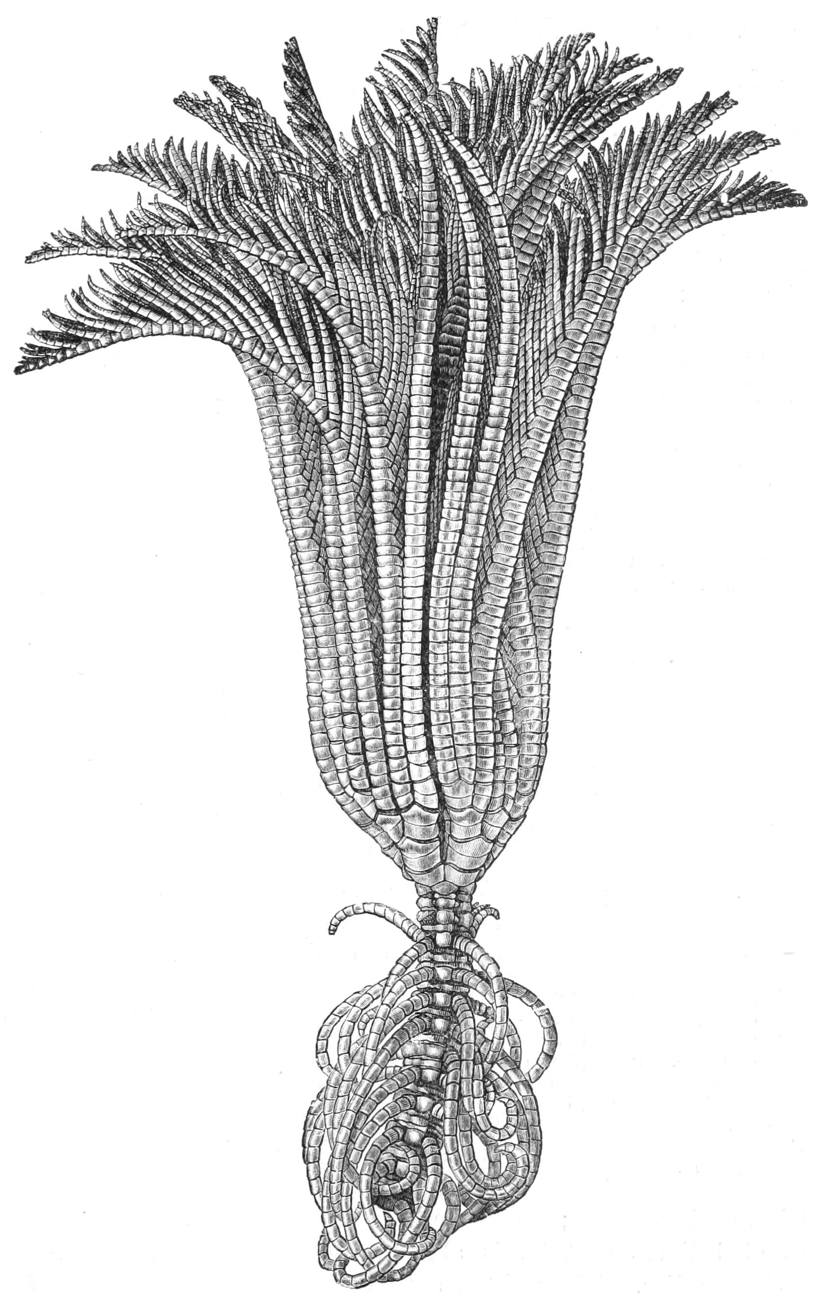
Fig. 272.—Arms and portion of stem of Pentacrinus maclearanus, slightly enlarged. In this species the basals can be seen. (From Wyville Thomson.)
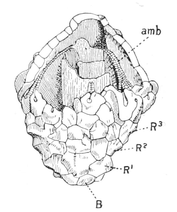
Fig. 273.—Calyx of Actinocrinus, one of the Camerata, broken open to show structure. amb, Ambulacral groove enclosed in covering plates; B, basal; R1, R2, R3, the three radials of a column. (After Zittel.).
Fam. 5. Comatulidae.—Stem in the adult broken off, leaving only a stump, the centro-dorsal, covered with cirri. Six genera. Antedon (= Comatula) has already been described; many tropical species have numerous arms and often side-plates and covering plates. Actinometra is distinguished by its excentric mouth, and by the fact that the centro-dorsal is flat and has cirri only round its edges; Atelecrinus has an acorn-shaped centro-dorsal, and the basals are externally visible; Eudiocrinus differs from Antedon only in having five arms; Promachocrinus is a remarkable form, having ten radii (this is a unique feature in Crinoidea); finally, Thaumatocrinus has basals externally visible, large persistent orals and interradial plates, and in addition a short free appendage of several plates on the anal interradius. Antedon and Actinometra are almost world-wide. Six species of the first have been recorded from British waters, of which the commonest is Antedon rosacea; four others are distinguished by having longer cirri, and do not seem to be well defined; but A. eschrichtii, a northern form, is larger, and is distinguished by having long proximal pinnules. The other genera are rare, and occur in deep water.
When we turn to survey fossil Crinoidea, we are met with a bewildering variety of forms ranging from the Lower Cambrian to the present day. As already mentioned, there is no agreement amongst experts as to how they should be classified. Bather makes the fundamental cleavage depend on the possession of two whorls of plates in the base (Fig. 274), or of only one whorl. These two divisions he calls Dicyclica and Monocyclica respectively. He admits that in many forms allied to Dicyclica the infra-basals have disappeared; these he terms "pseudomonocyclic" forms, and believes that he is able to discriminate them from true Monocyclica.
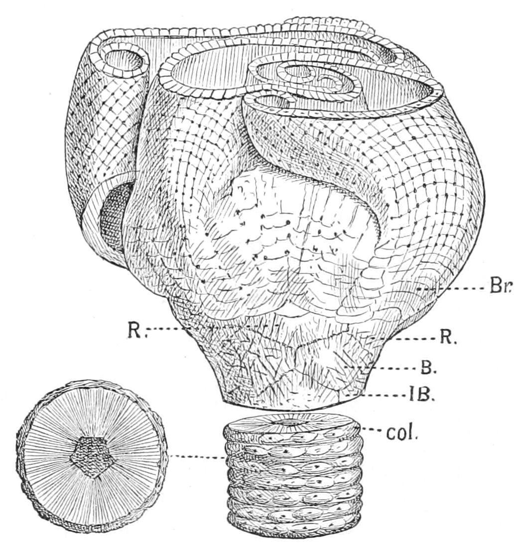
Fig. 274.—Crotalocrinus pulcher. × 1. B, basal; Br, arm-fan of adhering branches; col, ossicle of stem; IB, infra-basal; R, radial. (After Zittel.)
The present author is utterly unable to believe that the Crinoidea diverged into two groups on what is a trifling point of meristic variation comparable to the varying number of rows of plates in the interradial areas of the older Echinoidea; and he is equally sceptical as to the validity of Jaekel's division of the group into Cladocrinoidea and Pentacrinoidea, leading to the view that organs like pinnules represent totally different structures in different groups. Wachsmuth and Springer adopt as bases of classification the extent to which the arms and their branches are incorporated in the disc, and they recognise three main divisions: Inadunata, in which the arms are completely free from the calyx; Articulata, in which the arms are partly incorporated but the tegmen remains flexible; and finally Camerata, in which the arms and their first branches are largely incorporated in the cup; the tegmen is converted into a rigid dome and the ambulacral grooves on it become closed, as does the mouth, by the meeting of overarching folds; the grooves remaining, of course, open in the distal portions of the arms (Fig. 273). This classification, founded as it is on physiological factors, seems to the present author more satisfactory. Speaking generally, the points in which fossil Crinoids may differ from living genera are: (1) the total absence or irregular nature of the branching in the arms, so that pinnules may be said to be absent; (2) the closure of the ambulacral grooves and mouth already alluded to, and (3) the adhesion of the arms in the same ray to produce net-like structures (Crotalocrinus, Fig. 274), or a fan-shaped structure (Petalocrinus); (4) the frequent presence of two rows of brachials in one arm (biserial structure); (5) the development of an enormous anal tube, so large that in extreme cases (Eucalyptocrinus) the arms may be lodged in grooves of it.
CLASS II. THECOIDEA (EDRIOASTEROIDEA, Bather)
These remarkable Pelmatozoa are the most primitive known. They have sac-like or sometimes cushion-shaped or even disc-shaped bodies, covered with numerous irregular plates without any symmetry in their arrangement. There is no stem, but when they are fixed this is effected by an adhesion of the aboral pole. There are no arms, but on the upper surface is to be seen the impression of five ambulacral grooves radiating from a central mouth. These grooves are bordered by covering plates, which in the earliest form (Stromacystis) are seen to be slight modifications of the plates covering the upper surface of the body, but in the later genera (Fig. 275, Thecocystis) become specialised. The anus is situated on the side, as is also the madreporite. It has been suggested that Eleutherozoa were derived from this group; that individuals were occasionally overturned by the waves or currents, and in this way compelled to use their podia for locomotion. When Eleutherozoa, however, have a fixed stage in their development, they are fixed by the oral, not the aboral, surface, and hence can have no close affinity to Thecoidea. Thecoidea begin in the Middle Cambrian, but according to Jaekel impressions in the Lower Cambrian, referred to Medusae, may be casts of this group.
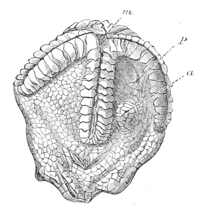
Fig. 275.—Thecocystis saeculus. × 6. a. Anus; m, mouth; p, madreporite(?) (After Jaekel.)
CLASS III. CARPOIDEA
Pelmatozoa with a well-developed stem; body bilaterally compressed; only two rays apparently developed. These are indicated only by grooves radiating from the mouth; but in some cases slight horn-like outgrowths of some of the plates of the calyx may support prolongations of the grooves.
This group, which, like the foregoing, commences in the Cambrian, is perhaps more primitive than the Thecoidea in showing less influence of the water-vascular system on the skeleton; but in the presence of a differentiated stem and the development of only two rays, it is more differentiated. The anus is on one of the flat sides, covered with a flat plate acting as a valve. The members of this group were formerly confounded with Cystoidea, from which they differ in the absence of the characteristic pores. Trochocystis, the genus figured, is devoid of any horn-like outgrowths of the calyx.
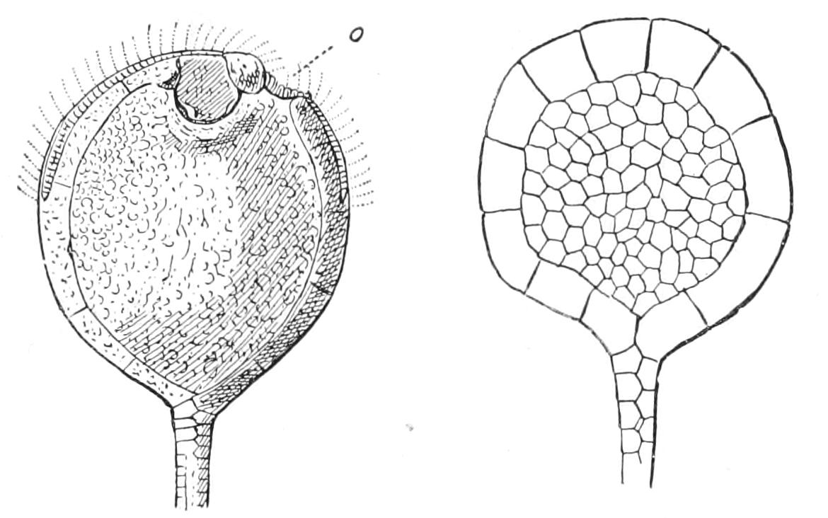
Fig. 276.—Trochocystis bohemicus, viewed from two sides. o, Mouth. (After Jaekel.)
CLASS IV. CYSTOIDEA
Pelmatozoa with respiratory organs in the form of "diplopores" or "pore-rhombs." In a great many cases there is a stalk, but in other cases this is atrophied, and the animal is attached by the base of the calyx. The radial canals run for a shorter or longer distance over the calyx, but the plates of the calyx themselves are not modified for them. Either they run in simple grooves, or they are protected by a special series of plates lying above the plates of the calyx. The terminal portions of the radial canals are in all cases free, supported by unbranched arms consisting usually of a double row of ossicles. These arms are termed "fingers."
It will be gathered from the description just given that the fingers and the respiratory organs distinguish Cystoidea from the two foregoing classes. Formerly this class was a lumber-room in which were placed all primitive irregular Pelmatozoa. The labours of Jaekel[516] have, however, dispelled the mist which enveloped this group, and in his monograph all that can be extracted both from superficial examination and dissection of these fossils is contained. It seems possible to the present author that the class may eventually require to be divided into two, corresponding to the two main divisions which Jaekel recognises, viz. Dichoporita, with pectinated rhombs, and Diploporita, with diplopores.
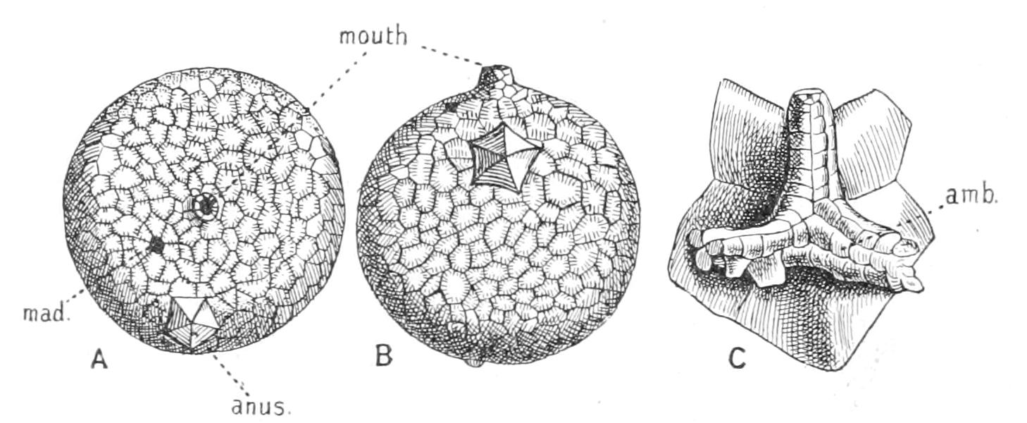
Fig. 277.—Echinosphaerites aurantium. A, from above; B, from the side; C, neighbourhood of mouth, enlarged. amb, Ambulacral groove with side-plates and covering plate; mad, madreporite. The short parallel lines across the sutures are the "pore-rhombs." (After Zittel.)
The pore-rhombs of the Dichoporita (indicated in Fig. 277 by the small parallel lines crossing the boundaries of the plates) were, according to Jaekel, nothing but a series of folds of thin integument projecting into the interior, the outer opening of which in most cases adhered in the middle, leaving two pores connected by a groove. The inner boundaries of the folds are sometimes preserved, but in many cases they were entirely devoid of calcification, and so were lost. The radial vessels either branched a great deal, giving rise to a multitude of fingers, or, as in Echinosphaerites (Fig. 277), there were a few long fingers supporting a reduced number of radial canals. In some cases the calyx can be analysed into a regular series of cycles of plates, consisting of basals, orals, and three intervening whorls, thus including one more ring than the calyx of Crinoidea. Jaekel regards this as a primitive arrangement, believing that the irregularity seen in Echinosphaerites secondary. This is a doubtful hypothesis.
The diplopores of the Diploporita appear to consist of two canals traversing the body-wall, opening close together into a common pit externally, but diverging internally. Since in some cases, as in Aristocystis (Fig. 278), this common pit is proved to have been closed externally by a very delicate layer of calcification, it is probable that the pores represent in other cases the points of origin of finger-like gills similar to those of Asteroidea. Where they were closed by calcification this was so thin and porous that the diffusion through it sufficed for respiration. Jaekel regards the Diploporita as a group derived from Dichoporita, but this seems to be extremely doubtful.
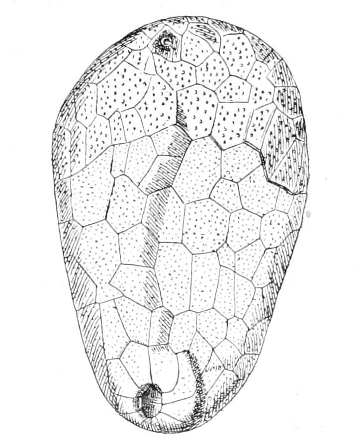
Fig. 278.—Aristocystis. In the upper part of the calyx the heavy dots are "diplopores," seen owing to removal of the superficial layer. (After Zittel.)
CLASS V. BLASTOIDEA
Pelmatozoa with respiratory organs in the form of longitudinal calcified folds, termed "hydrospires," radiating from the mouth. Stem well developed; calyx regular, consisting of a whorl of basals surmounted by a whorl of forked radiais, in the clefts of which lay the recumbent radial water-vascular vessels, supported each on a special plate ("lancet plate"), and giving off two rows of branches supported by short fingers (Fig. 279). Side-plates and covering plates were also developed; five orals ("deltoids") completed the calyx. The anus was at the side, just beneath one of the orals.
The hydrospires, which are the great characteristic of the class, are seen in section in Fig. 279, B (hyd). They consist of a varying number of parallel folds on each side of each "pseudambulacrum," as the lancet plate with its adhering side-plates and covering plates has been termed. In the most primitive genus, Codaster, they appear to have opened directly to the exterior, and to have been placed at right angles to the lines of union of the radial and oral plates, just like the grooves of a pectinated rhomb. In more modified forms, such as Pentremites and Granatocrinus (Fig. 279), the outer openings were overarched by the extension of the side-plates of the radial vessel, and the whole group of folds has a common opening near the mouth; indeed, in the highest form there is one common "spiracle" for the two groups of folds in an interradius, which in one interradius is confluent with the anus. The hydrospires, when they reach this form, irresistibly recall the genital bursae of Ophiuroidea (Fig. 214, p. 490), and very possibly served the same purpose.
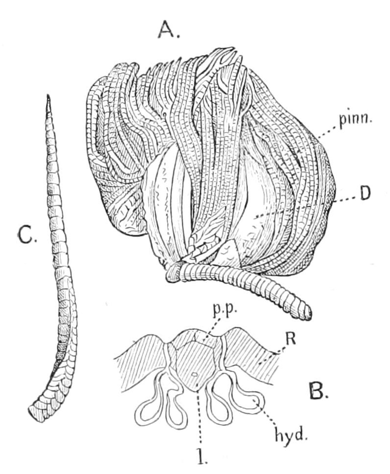
Fig. 279.—Granatocrinus norwoodi. A, view of whole animal; B, section of radius; C, an isolated finger. hyd, Hydrospire; l, lancet plate; pinn, finger; p.p, covering plate; R and D both signify radial plate. (After Zittel.)
Reviewing the whole group of the Pelmatozoa, we see that in the Cambrian they begin with the extremely primitive Thecoidea and Carpoidea, together with some obscure forms which, combining a stem with pentamerous symmetry in the calyx, are supposed to be the forerunners of the Crinoidea. In the Lower Silurian or Ordovician the two groups of the Cystoidea make their appearance, possibly independently developed from either Carpoidea or primitive Crinoidea, which in this period are present in unmistakable form. In the Upper Silurian the Blastoidea appear, distinguishable from the most regular Cystoidea only by their hydrospires. It seems practically certain that they were developed from Cystoidea, and we follow Jaekel in believing that they arose from Dichoporita. The Carpoidea do not extend beyond the Ordovician, and by the end of the Carboniferous period Cystoidea and Blastoidea die out, leaving only the Crinoidea, which at that period were at their maximum development. From the Carboniferous to the present day the Crinoidea have continually decreased, leaving in recent seas, as sole representatives of the Pelmatozoa, only the few forms described at the beginning of this chapter.