Chapter XVI
Reading edition
ECHINODERMATA—INTRODUCTION—CLASSIFICATION—ANATOMY OF A STARFISH—SYSTEMATIC ACCOUNT OF ASTEROIDEA
The name Echinodermata[436] means literally "spiny-skinned," and thus brings into prominence one very conspicuous feature of most of the animals belonging to this phylum. All, it is true, do not possess spines; but with one or two doubtful exceptions, all have calcareous plates embedded in the skin, and these plates, in many cases, push out projections which raise the skin into corresponding elevations, which are called the spines. The spines are, like the other plates, inside the skin, and to speak of an Echinoderm living in its shell, as we speak of a Snail, is a serious error. The shell of a Mollusc is fundamentally a secretion poured forth from the skin, and is thus entirely external to the real living parts; but the plates and spines of an Echinoderm may be compared to our own bones, which are embedded deeply in the flesh. Hence the name ossicle (little bone) is used to designate these organs.
Besides the possession of these spines, Echinoderms are characterised by having their organisation pervaded by a fundamental radial symmetry. The principal organs of the body are repeated and are arranged like the spokes of a wheel round a central axis instead of being, as, for example, in Chaetopoda, arranged behind one another in longitudinal series.
In addition to these striking peculiarities, Echinoderms possess a most interesting internal organisation, being in this respect almost exactly intermediate between the Coelenterata and the higher Invertebrata. Like so many of the latter, the Echinodermata have an anus, that is, a second opening to the alimentary canal through which indigestible material is rejected; like them also, they have a body-cavity or coelom surrounding the alimentary canal—from the lining of which the genital cells are developed. On the other hand, there is no definite circulatory system, nor any specialised excretory organ, and the nervous system exhibits no concentration which could be called a brain, and is, moreover, in close connexion with the skin. In all these points the Echinodermata resemble the Coelenterata.
One of the most characteristic features of the internal anatomy of Echinodermata is the presence of a peculiar series of organs, known collectively as the water-vascular system or hydrocoel. This is really a special division of the coelom or body-cavity which takes on the form of a ring-shaped canal embracing the mouth, from which are given off long radial canals, usually five in number, running to the more peripheral parts of the body.[437] Each radial canal carries a double series of lateral branches, which push out the skin so as to appear as appendages of the body. These appendages are known as tentacles or tube-feet; they are both sensory and respiratory in function, and often in addition, as the name tube-foot indicates, assist in locomotion. As a general term for these appendages, to be applied in all cases without reference to their function, the name podium has been suggested and will be employed here. A system of canals, in many ways resembling the water-vascular system, is found in Brachiopoda, Gephyrea and Polyzoa, but the peculiarity of Echinodermata is the way in which it is kept filled with fluid. From the ring-canal in the interval (or interradius) between two radial canals, a vertical canal, termed the stone-canal, is given off, which communicates with the exterior by means of a sieve-like plate, the madreporite, pierced by fine canals. These canals and the stone-canal itself are lined with powerful cilia, which produce a strong inward current, and keep the water-vascular system tensely filled with sea water.
The phylum includes the familiar Starfish and Sea-urchins, which in sheltered spots are found between tide-marks; the Brittle Stars and Sea-cucumbers, which can be dredged up from below low-water mark, and lastly the beautiful Feather-stars, of which there are comparatively few species still living, although huge beds of limestone are composed of the remains of fossil Feather-stars.
One species of Sea-cucumber (Synapta similis)[438] is said to enter brackish water in the mangrove swamps of the tropics; but, with this exception, the whole phylum is marine. A few species can endure partial exposure to the air when left bare by the receding tide, but the overwhelming majority are only found beneath low-water mark, and a considerable number live in the deepest recesses of the ocean.
Their distribution is, no doubt, partly determined by food, a number of species being strictly confined to the neighbourhood of the shore. On the other hand, since a very large number of species live on the layer of mud impregnated with animal remains which forms the superficial layer of the deposit covering the sea-floor, it is not surprising to learn that many have an exceedingly wide range, since this deposit is very widely distributed. Another equally important factor in determining distribution is wave-disturbance, and it is surprising to learn to what a depth this extends. Off the west coast of Ireland a large wave literally breaks on a submerged rock 15 fathoms beneath the surface. Speaking generally, it is useless to look for Echinoderms on an exposed coast, and the same species, which in the sheltered waters of the Clyde are exposed at low water, must be dredged up from 20 to 30 fathoms outside Plymouth Sound.
The ordinary collector is attracted to the group chiefly by the regularity and beauty of the patterns produced by the radial symmetry, but to the scientific zoologist they are interesting from many other points of view. Differing widely nevertheless from the higher Invertebrata in their symmetry when adult, they have as larvae a marked bilateral symmetry, and the secondary development of the radial symmetry constitutes one of the most remarkable life-histories known in the animal kingdom.
Then again, owing to the possession of ossicles, the Echinodermata are one of the few groups of Invertebrata of which abundant remains occur fossilised. In attempting, therefore, to decipher the past history of life from the fossil record, it is necessary to have an exact and detailed knowledge of Echinoderm skeletons and their relation to the soft parts. Lastly, the internal organisation of Echinoderms throws valuable light on the origin of the complicated systems of organs found in the higher animals.
Echinodermata are divided into two great sub-phyla, which must have very early diverged from one another. These are:—
(1) Eleutherozoa, (2) Pelmatozoa.[439]
The sub-phylum Pelmatozoa, to which the living Feather-stars (Crinoidea) and the majority of the known fossil species belong, is characterised by the possession of a fixing organ placed in the centre of the surface opposite the mouth—the aboral surface as it is called. Ordinarily this organ takes on the form of a jointed stalk, but in most modern species it is a little knob with a tuft of rooting processes, termed cirri. In the other sub-phylum, the Eleutherozoa, no such organ is found, and the animals wander about freely during their adult life, though for a brief period of their larval existence they may be fixed by a stalk-like protuberance arising from the oral surface.
SUB-PHYLUM I. ELEUTHEROZOA
The Eleutherozoa are divided into four main classes, between which no intermediate forms are found amongst the living species, though intermediate types have been found fossil.
The four classes into which the Eleutherozoa are divided are defined as follows:—
(1) Asteroidea (Starfish).—"Star"-shaped or pentagonal Eleutherozoa with five or more triangular arms, not sharply marked off from the central disc. The mouth is in the centre of one surface, called from this circumstance the "oral"; the anus is in the centre of the opposite surface, termed the "aboral." From the mouth a groove runs out on the under surface of each arm towards its tip, termed the "ambulacral" groove. Projecting from the ambulacral groove are found the podia or tube-feet, the organs of movement and sensation of the animal.
(2) Ophiuroidea (Brittle Stars).—Eleutherozoa, in which the body consists of a round disc with long worm-like arms inserted in grooves on its under surface. No anus is present, and the ambulacral grooves are represented by closed canals. The podia are merely sensory and respiratory, locomotion being effected by muscular jerks of the arms.
(3) Echinoidea (Sea-urchins).—Globular or disc-shaped Eleutherozoa, in which the skeleton forms a compact cuirass except for a short distance round the mouth (peristome) and round the anus (periproct). The ambulacral grooves are represented by canals which, like meridians of longitude on a school-globe, run from the neighbourhood of the mouth to near the aboral pole of the body. The spines are large and movably articulated with the plates. The animals move by means of podia and spines, or by means of the latter only. The anus is usually situated at the aboral pole, but is sometimes displaced towards the side, or even on to the ventral surface.
(4) Holothuroidea (Sea-cucumbers).—Sausage-shaped Eleutherozoa, in which the skeleton is represented only by isolated nodules of calcium carbonate, and in which the body-wall is highly muscular. The mouth and anus are situated at opposite ends of the body, and the ambulacral grooves (represented by closed canals) run from near the mouth to the proximity of the anus. Movement is accomplished by means of the podia, aided by worm-like contractions of the body.
CLASS I. ASTEROIDEA[440] (Starfish)
The Starfish derive their name from their resemblance in shape to the conventional image of a star. The body consists of broad triangular arms (generally five in number) which coalesce in the centre to form a disc. The skin is soft and semi-transparent, permitting the skeleton to be easily detected; this consists of a mesh-work of rods or plates, leaving between them intervals of soft skin. In a living Starfish it can be seen that many of these soft places are raised up into finger-like outgrowths, which are termed "papulae" or "dermal gills," through the thin walls of which an active interchange of gases with the surrounding water takes place, and the animal obtains in this way the oxygen necessary for its respiration.
Very few and feeble muscle-fibres exist in the body-wall, and the movements of the arms, as a whole, are very slow and limited in range. There is a membranous lip surrounding the mouth, from which five broad grooves run outwards, one on the underside of each arm. These are termed the "ambulacral grooves." Each groove is Λ-shaped, and its sides are stiffened by a series of rod-like ossicles called the "ambulacral ossicles."
The animal progresses by the aid of a large number of translucent tentacles, termed "tube-feet" or "podia," which are attached to the walls of the ambulacral grooves.
Anatomy of a Starfish.—As an introduction to the study of the anatomy not only of Starfish but of Echinodermata as a whole, we select Asterias rubens, the common Starfish of the British coasts, which in many places may be found on the beach near low-water mark.
External Features.—In this species (Fig. 185) the skeleton is a net-work of rod-like plates, leaving wide meshes between them, through which protrude a perfect forest of transparent papulae. From the points of junction of the rods arise short blunt spines surrounded by thick cushions of skin. The surfaces of these cushions are covered with a multitude of whitish specks, which, on closer inspection, are seen to have the form of minute pincers, each consisting of two movable blades crossing each other below and articulated to a basal piece. These peculiar organs are termed "pedicellariae" (Fig. 186), and their function is to keep the animal clean by seizing hold of any minute organisms which would attempt to settle on the soft and delicate skin. When irritated the blades open and then snap together violently, and remain closed for a long time.[441] These actions are brought about by appropriate muscles attaching the blades to the basal piece.
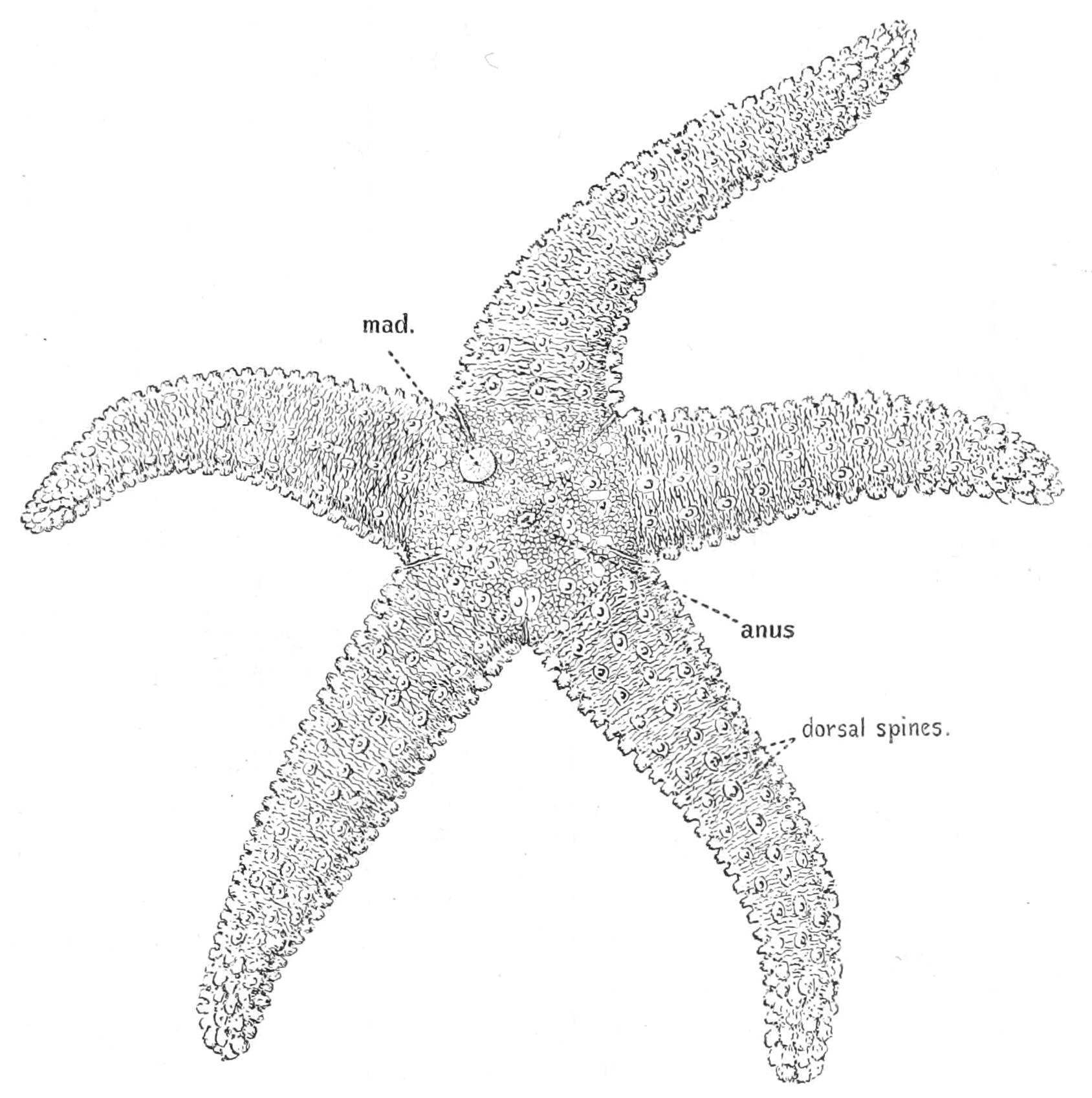
Fig. 185.—Asterias rubens, seen from the aboral surface, × 1. mad, Madreporite.
The last-named ossicle increases the certainty of the grip by fixing the lower parts of each blade in the same vertical plane, and preventing lateral slipping, so that it serves the same purpose as the pivot in a pair of scissors. Each blade, in fact, fits into a groove on the side of this piece. The muscles which close the blades arise from the lower ends (handles) of the blades, and are united below to form a common muscular string which attaches the whole organ to one of the plates of the skeleton. An attempt of the victim to tear the pedicellaria out is resisted by the contraction of this string, which thus brings about a closer grip of the blades. In order that the blades may open they must first be lifted out of the grooves on the basal piece—this is effected by special lifting muscles. The opening is brought about by muscles extending from the "handle" of one blade to the upper part of the other.
Scattered about amongst the papulae between the cushions are other pedicellariae of a larger size in which the blades do not cross one another (Fig. 186, B).
In the space or "interradius" between two arms, on the aboral surface, there is found a button-shaped ossicle. This is covered with fine grooves, and from a fancied resemblance between it and some forms of coral it has received the name "madreporite" (Fig. 185, mad). The bottoms of the grooves are perforated by capillary canals lined by flagella, through the action of which water is constantly being introduced into the water-vascular system.
The anus is situated near the centre of the upper surface of the disc, but it is so minute as to require careful inspection in order to discover its position (Fig. 185).
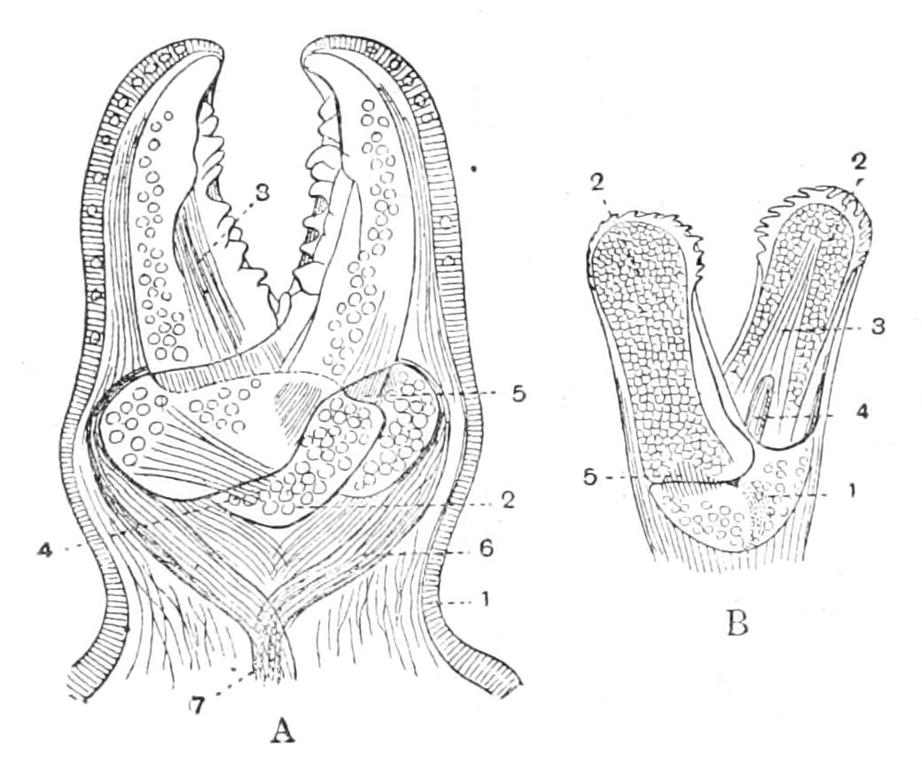
Fig. 186.—View of pedicellariae of A. glacialis. A, Crossed form, × 100. 1, Ectoderm covering the whole organ; 2, basal piece; 3, auxiliary muscle closing the blades; 4, muscle lifting right blade out of the groove; 5, handle of left blade; 6, muscles closing the blades, and uniting to form 7, the muscular string attaching the pedicellaria to the skeleton. B, straight form, × 10. 1, Basal piece; 2, blades; 3 and 4, muscles closing the blades; 5, muscle opening the blades. (From Cuénot.)
On the under side of the animal the most conspicuous features are the five ambulacral grooves which radiate out from the "peristome," a thin membranous area surrounding the central mouth. The grooves are filled with the tube-feet, which are closely crowded together and apparently arranged in four rows.
Skeleton.—The sides of the ambulacral grooves are stiffened by the rod-like "ambulacral ossicles." To the outer ends of these are articulated a set of shorter rods termed the "adambulacral ossicles" which carry each two or three rod-like spines, the "adambulacral spines," the skin covering which bears numerous pedicellariae (Fig. 187, B). When the animal is irritated the edges of the groove are brought together, and these spines then form a trellis-work covering and protecting the delicate tube-feet; the numerous pedicellariae are then in a position to make it unpleasant for any intruder. The closure of the groove is effected by means of powerful muscles connecting each ambulacral ossicle with its fellow. There are also feebler muscles connecting these plates with their successors and predecessors, which enable the arm to be bent downwards in a vertical plane. It is raised by a muscular band running along the dorsal wall of the coelom to the point of the arm.
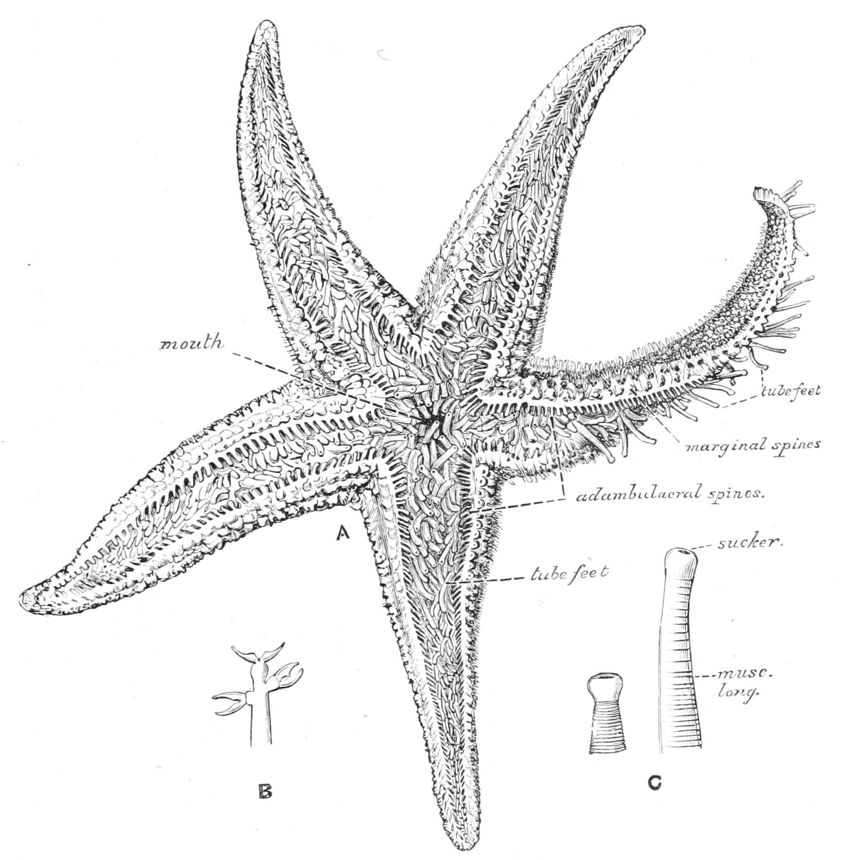
Fig. 187.—A, Asterias rubens, seen from the oral surface, drawn from a living specimen, × 1. B, an adambulacral spine, showing three straight pedicellariae; C, a tube-foot expanded and contracted.
When the series of ambulacral and adambulacral ossicles is followed inwards towards the mouth it is seen that the first ambulacral ossicle is closely fixed to the second, but is widely separated from its fellow, remaining, however, connected with the latter by a powerful adductor muscle. In consequence of the separation of this pair of ossicles each is brought into closer contact with the corresponding ossicle in the adjacent radius, to which it is connected by a muscle called the abductor. The first adambulacrals in adjacent radii are also brought into closer contact and carry long spines which, when the ambulacral grooves are contracted, project like a grating over the mouth. In the order of Asteroidea to which Asterias belongs, the adambulacrals themselves do not project much, but in all other cases they form prominent mouth-angles, so that the opening of the mouth becomes star-shaped (Fig. 211, p. 483).
Except in the case of the ambulacral and adambulacral plates little regular arrangement is to be detected in the ossicles of the skeleton which, as has already been mentioned, form a mesh-work. If, however, the arm be cut open and viewed from the inside it will be seen that the edge is strengthened above and below by very thick, powerful, rod-like plates. These are called the "supero-marginal" and "infero-marginal" ossicles; they are not visible from the outside, since they are covered by a thick layer of the body-wall containing other smaller plates (Fig. 190, marg). In many genera, however, they are exposed, and form a conspicuous edging to the arm above and below. In many genera, also, there are three conspicuous series of plates on the back of each arm, viz. a median row, called "carinals" (car., Fig. 191), and two lateral rows, termed "dorso-laterals" (d.lat., Fig. 191). These three rows, with the two rows of marginals, one of ambulacrals, and one of adambulacrals on each side (11 rows in all), constitute the primitive skeleton of the arm, and appear first in development.
The structure of all these elements of the skeleton is the same. They may be described as scaffoldings of carbonate of lime, interpenetrated by a mesh-work of cells fused with one another, by which the carbonate of lime has been deposited. The matrix in which the ossicles lie is a jelly-like substance traversed by a few bands of fibres which connect the various rods with one another. This jelly is almost fluid in the fresh state, but when heated forms a hard compound, possibly allied to mucin, which will turn the edge of a razor.
When the covering of the back is dissected off the coelom is opened. This is a spacious cavity which apparently surrounds the alimentary canal and extends into the arms. It has, however, its own proper wall, which is called the "peritoneum," both on the outer side, where it abuts on the skin, and on the inner side, where it comes in contact with the wall of the alimentary canal. The outer wall is called the "somatic peritoneum," and it is possible to dissect off the rest of the body-wall and leave it intact; the inner wall, from its close association with the alimentary canal, is termed the "splanchnic peritoneum." This wall can only be distinguished in microscopic sections from that of the alimentary canal, to which it is closely applied.
The coelom is filled with a fluid, which is practically sea water with a little albuminous matter in solution. Through the thin walls of the papulae oxygen passes into this fluid, whence it easily reaches the inner organs, since they are all in contact with some part of the coelomic wall. Similarly CO2 is absorbed by the coelomic fluid from all parts of the body, and diffuses through the papulae to the surrounding water.
The Starfish possesses no definite kidney for getting rid of nitrogenous waste. In most of the higher animals with a well-developed coelom it has been proved that the kidney is simply a specialised portion of the coelom, and in many cases some parts of the coelomic wall still retain their excretory functions, which apparently the whole originally possessed. In the Starfish and in Echinodermata generally this primitive state of affairs is still retained. From the cells forming the coelomic wall, cells are budded off into the fluid, where they swim about. These cells from their movements are called amoebocytes. If a substance such as indigo-carmine, which when introduced into the tissues of the higher animals is eliminated by the kidney, is injected into the Starfish, it is found soon after to be vigorously absorbed by the amoebocytes. These later accumulate in the dermal branchiae, through the thin walls of which they make their way[442] to the outside, where they degenerate.
The coelom is indented by five folds, which project inwards from the interradii. These folds are called the "interradial septa"; they are stiffened by a calcareous deposit, which is not, however, sufficiently dense to constitute a plate. In one of the septa the axial sinus and stone-canal (see below) are embedded. These septa are to be regarded as areas of lateral adhesion between the arms.
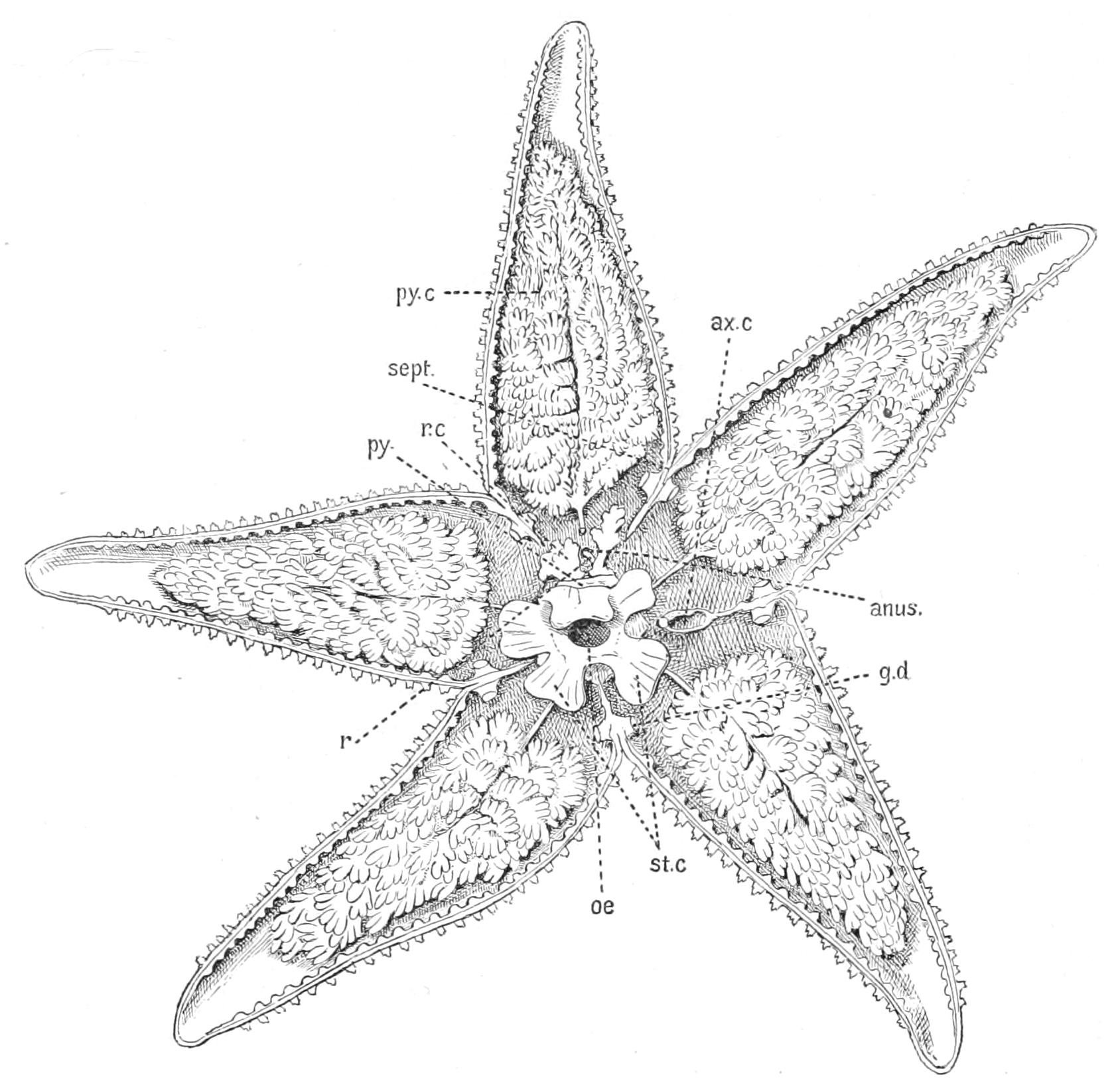
Fig. 188.—View of upper half of a specimen of Asterias rubens, which has been split horizontally into two halves. ax.c, Axial sinus; g.d, genital duct; oe, cut end of the oesophagus, the narrow neck of the stomach; py, pyloric sac; py.c, pyloric caeca; r, rectum; r.c, rectal caeca; sept, interradial septum; st.c, stomach lobe.
The alimentary canal consists of several distinct portions. The mouth leads by a narrow neck called the "oesophagus" into a voluminous baggy sac termed the "stomach," which is produced into ten short pouches, two projecting into each arm. The stomach leads in turn by a wide opening into a pentagonal flattened sac, the "pyloric sac," which lies above it. Each angle of the pyloric sac is prolonged into a tube—the so-called "pyloric duct"—running out into the arm, where it immediately bifurcates into two forks, each beset by a large number of small pouches and attached to the dorsal wall of the coelom by suspensory bands of membrane called mesenteries. These ten forks are called "pyloric caeca"; they are of a deep green colour owing to the pigment in their wall. Beyond the pyloric sac the alimentary canal is continued as the slender "rectum" to the anus. The rectum gives off two small branched pouches of a brown colour called "rectal caeca." This comparatively complicated form of alimentary canal is related to the nature of the food of the animal and the method it employs to capture its prey.
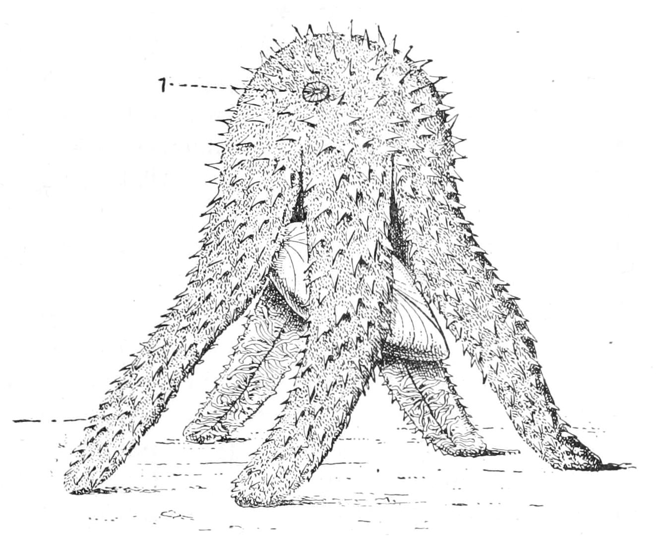
Fig. 189.—View of a Starfish (Echinaster) devouring a Mussel. 1. The madreporite.
The favourite food[443] of Asterias consists of the common bivalves of the coast, notably of the Mussel (Mytilus edulis). There is, however, no animal which it will not attack if it is fortunate enough to be able to catch it. The Starfish seizes its prey by the tube-feet, and places it directly under its mouth, folding its arms down over it in umbrella fashion. The muscles which run around the arms and disc in the body-wall contract, and the pressure thus brought to bear on the incompressible fluid contained in the coelom, forces out the thin membranous peristome and partially turns the stomach inside out. The everted edge of the stomach is wrapped round the prey.
Soon the bivalve is forced to relax its muscles and allow the valves to gape. The edge of the stomach is then inserted between the valves and applied directly to the soft parts of the prey which is thus completely digested. When the Starfish moves away nothing but the cleaned shell is left behind. If the bivalve is small it may be completely taken into the stomach, and the empty shell later rejected through the mouth.
It was for a long time a puzzle in what way the bivalve was forced to open. Schiemenz[444] has, however, shown that when the Starfish folds itself in umbrella-like form over the prey it holds on to the substratum by means of the tube-feet of the distal portions of the arms, whilst, by means of the tube-feet belonging to the central portions, it drags apart the valves by main force. He has shown experimentally: (1) that whilst a bivalve may be able to resist a sudden pull of 4000 grammes it will yield to a pull of 900 grammes long continued; (2) that a Starfish can exert a pull of 1350 grammes; (3) that a Starfish is unable to open a bivalve unless it be allowed to raise itself into a hump, so that the pull of the central tube-feet is at right angles to the prey. A Starfish confined between two glass plates walked about all day carrying with it a bivalve which it was unable to open.
The lining of the stomach is found to consist very largely of mucus-forming cells, which are swollen with large drops of mucus or some similar substance. It used to be supposed that this substance had some poisonous action on the prey and paralysed it, but the researches of Schiemenz show that this is incorrect. If when an Asterias is devouring a bivalve another be offered to it, it will open it, but will not digest it, and the victim shows no sign of injury but soon recovers. The cells forming the walls of the pyloric sac and its appendages are tall narrow cylindrical cells crowded with granules which appear to be of the nature of digestive ferment. This substance flows into the stomach and digests the captured prey.
A very small amount of matter passes into the rectum and escapes by the anus, as the digestive powers of the Starfish are very complete. The rectal caeca are lined by cells which secrete from the coelomic fluid a brown material, in all probability an excretion, which is got rid of by the anus.
When the meal is finished the stomach is restored to its former place by the action of five pairs of retractor muscles, one pair of which originates from the upper surface of the ambulacral ossicles in each arm and extends to the wall of the stomach, where they are inserted (Fig. 190, ret).
The tube-feet, which are at once the locomotor and the principal sensory organs of the Starfish, are appendages of that peculiar system of tubes known as the water-vascular system, which is derived from a part of the coelom cut off from the rest during the development of the animal. This system, as already mentioned, consists of (1) a narrow "ring-canal," encircling the mouth and lying on the inner surface of the membranous peristome; (2) a radial canal leaving the ring-canal and running along the under surface of each arm just above the ambulacral groove; (3) a vertical stone-canal running from the madreporite downwards to open into the ring-canal in the interspace between two arms. The madreporite is covered externally by grooves lined with long cilia, and is pierced with narrow canals of excessively fine calibre, the walls of which are also lined by powerful cilia. Most of these narrow canals open below into a main collecting canal, the stone-canal, but some open into a division of the coelom termed the axial sinus, with which also the stone-canal communicates by a lateral opening. The cavity of the stone-canal is reduced by the outgrowth from its walls of a peculiar Y-shaped projection, the ends being rolled on themselves in a complicated way (Fig. 190, B). The walls of the canal consist of a layer of very long narrow cells, which carry powerful flagella, and outside this of a crust of calcareous deposit, which gives rigidity to the walls and has suggested the name stone-canal.
The tube-feet are covered externally by ectoderm, inside which is a tube in connexion with the radial water-vascular canal. This latter is lined by flattened cells, which in the very young Starfish are prolonged into muscular tails; in the older animal these tails are separated off as a distinct muscular layer lying between the ectoderm and the cells lining the cavity of the tube. The tube-foot is prolonged inwards into a bulb termed the "ampulla," which projects into the coelom of the arm and in consequence is covered outside by somatic peritoneum. Just where the ampulla passes into the tube-foot proper the organ passes downwards between two of the powerful ambulacral ossicles which support the ambulacral groove, and a little below this spot a short transverse canal connects the tube-foot with the radial canal which lies beneath these ossicles (Fig. 191).
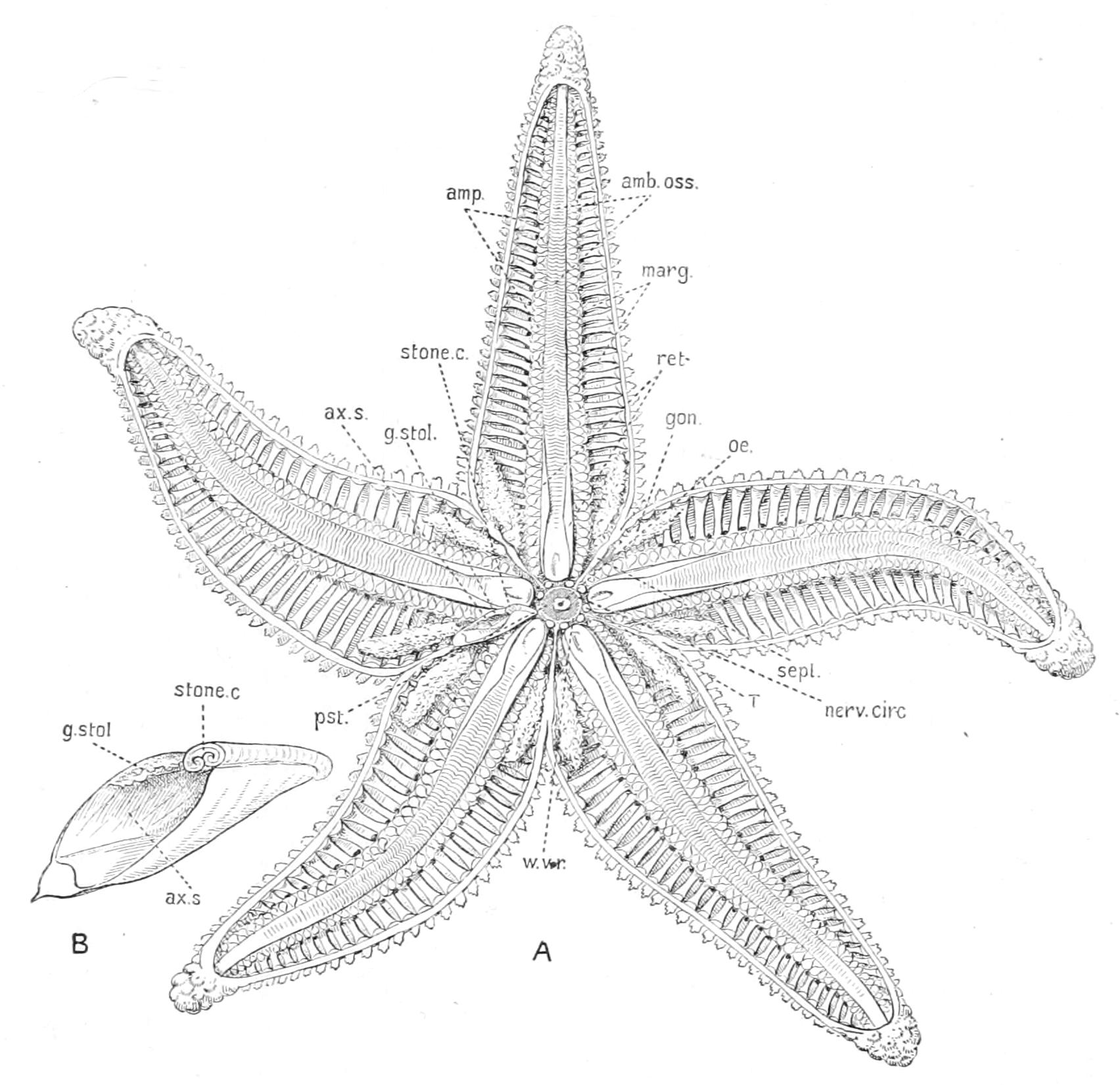
Fig. 190.—A, view of the under half of a specimen of Asterias rubens, which has been horizontally divided into two halves. B, enlarged view of the axial sinus, stone-canal and genital stolon cut across. amb.oss, Ambulacral ossicle; amp. ampullae of the tube-feet; ax.s, axial sinus; gon, gonad; g.stol, genital stolon; marg, marginal ossicle; nerv.circ, nerve ring; oe, cut end of oesophagus; pst, peristome; ret, retractor muscle of the stomach; sept, interradial septum; stone c, stone-canal; T, Tiedemann's body; w.v.r, water-vascular ring-canal.
The tube-feet are, therefore, really a double row of lateral branches of the radial canal. The appearance of being arranged in four rows is due to the fact that the transverse canals connecting them with the radial canal are alternately longer and shorter so as to give room for more tube-feet in a given length of the arm. Each tube-foot ends in a round disc with a slightly thickened edge. The radial canal terminates in a finger-shaped appendage, called the median tentacle, at the base of which is the eye.
The manner in which this complicated system acts is as follows:—When the tube-foot is to be stretched out the ampulla contracts and drives the fluid downwards. The contraction of the ampulla is brought about by muscles running circularly around it. The tube-foot is thus distended and its broad flattened end is brought in contact with the surface of the stone over which it is moving and is pressed close against it. The muscles of the tube-foot itself, which are arranged longitudinally, now commence to act, and the pressure of the water preventing the tearing away of the sucker from the object to which it adheres, the Starfish is slowly drawn forward, whilst the fluid in the tube-foot flows back into the ampulla.
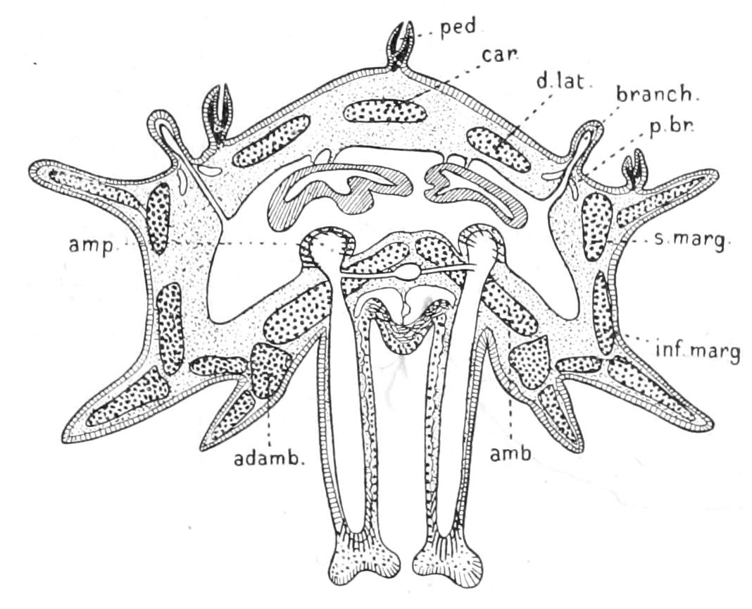
Fig. 191.—Diagrammatic cross-section of the arm of a Starfish. adamb, Adambulacral ossicle; amb, ambulacral ossicle; amp, ampulla of tube-foot; branch, papula; car, carinal plate; d.lat, dorso-lateral plate; inf.marg, infero-marginal plate; p.br, peribranchial space; ped, pedicellaria; s.marg, supero-marginal plate. The nervous ridge between the bases of the tube-feet and the two perihaemal canals above this ridge are shown in the figure but not lettered.
If each tube-foot were practically water-tight, then each would be entirely independent of all the rest, and it would not be easy to suggest a reason for the presence of the complicated system of radial canals and stone-canal. Just at the spot, however, where the transverse canal leading from the radial canal enters the tube-foot there is a pair of valves which open inwards and allow fluid to pass from the radial canal into the tube-foot but prevent any passing outwards in the reverse direction. The presence of these valves renders it probable that the tube-foot is not quite water-tight; that when it is distended under the pressure produced by the contraction of the muscles of the ampulla, some fluid escapes through the permeable walls; and that the loss thus suffered is made up by the entry of fresh fluid from the radial canal. The radial canal in turn draws from the ring-canal, and this last is supplied by the stone-canal, the cilia of which keep up a constant inward current.
In the fluid contained in the water-vascular system, as in the coelomic fluid, there are amoebocytes floating about. These are produced in short pouches of the ring-canal, nine in number, which are called after their discoverer "Tiedemann's bodies" (Fig. 190, T). From the cells lining these the amoebocytes are budded off.
The nervous system of the Starfish is in a very interesting condition. The essential characteristic of all nervous systems is the presence of the "neuron," a cell primitively belonging to an epithelium but which generally has sunk below the level of the others and lies amongst their bases. This type of cell possesses a round body produced in one direction into a long straight process, the "axon," whilst in the other it may have several root-like processes, or "dendrites," which may spring from a common stem, in which case the neuron is said to be "bipolar." The axon is often distinguished as a "nerve-fibre" from the round body which is termed the "nerve-cell." This is due to the fact that for a long time it was not recognised that these two structures are parts of a whole.
Now at the base of the ectoderm all over the body of the Starfish there is to be found a very fine tangle of fibrils; these are to be found partly in connexion with small bipolar neurons lying amongst them and partly with isolated sense-cells scattered amongst the ordinary ectoderm cells. This nervous layer is, however, very much thickened in certain places, so as to cause the ectoderm to project as a ridge. One such ridge is found at the summit of each ambulacral groove running along the whole under surface of the arm and terminating in a cushion at the base of the median tentacle of the water-vascular system. This ridge is called the radial nerve-cord. The five radial nerve-cords are united by a circular cord, the nerve-ring, which appears as a thickening on the peristome surrounding the mouth.
The sense-organs of the Starfish are chiefly the discs of the tube-feet. Round the edges of these there is a special aggregation of sense-cells; elsewhere, as in the skin of the back, only isolated sense-cells are found, and it becomes impossible to speak of a sense-organ.
A prolongation of the radial nerve-cord extends outwards along one side of each tube-foot. This is often spoken of as the "pedal nerve," but the term nerve is properly retained for a mere bundle of axons such as we find in the higher animals, whereas the structure referred to contains the bodies of nerve-cells as well as their outgrowths or cell-fibres and is therefore a prolongation of the nerve-cord.
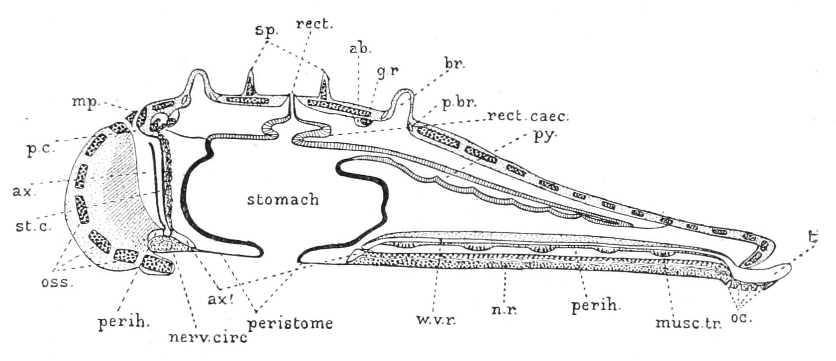
Fig. 192.—Diagrammatic longitudinal section through a young Asteroid passing through the tip of one arm and the middle of the opposite interradius. This diagram is generalised from a section of Asterina gibbosa. ab, Aboral sinus; ax, axial sinus; ax1, basal extension of axial sinus forming the inner perihaemal ring-canal; br, branchia = gill = papula; g.r, genital rachis; mp, madreporite; musc.tr, muscle uniting a pair of ambulacral ossicles; nerv.circ, nerve-ring; n.r, radial nerve-cord; oc, eye-pit; oss, ossicles in skin; p.br, peribranchial sinus; p.c, pore canal; perih (on the right), perihaemal radial canal, (on the left), outer perihaemal ring-canal; py, pyloric caecum; rect, rectum; rect.caec, rectal caeca; sp, spines; st.c, stone-canal; t, median tentacle terminating radial canal; w.v.r, water-vascular radial canal. The genital stolon (not marked by a reference line) is seen as an irregular band accompanying the stone-canal, its upper end projects into a small closed sac, also unmarked, which is the right hydrocoele or madreporic vesicle.
At the base of the terminal tentacle the radial nerve-cord ends in a cushion. This cushion is called the "eye," for it is beset with a large number of cup-shaped pockets of the ectoderm. Each pocket is lined partly by cells containing a bright orange pigment and partly by visual cells each of which ends in a small clear rod projecting into the cavity of the pit (Fig. 193, A, vis.r). The pit is apparently closed by a thin sheet of cuticle secreted by the most superficial cells.
An exposed nervous system and simple sense-organs such as the Starfish possesses lend themselves admirably to the purposes of physiological experiment, and so Starfish have been favourite "corpora vilia" with many physiologists.
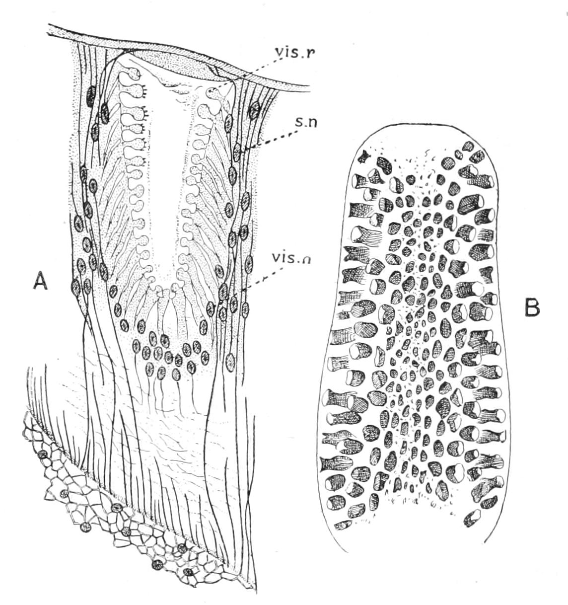
Fig. 193.—A, longitudinal section of a single eye-pit of Asterias. s.n, Nucleus of supporting cell; vis.n, nucleus of visual cell; vis.r, visual rod. B, view of the terminal tentacle showing the eye-pits scattered over it. (After Pfeffer.)
The light-perceiving function of the eye is easily demonstrated. If a number of Starfish be put into a dark tank which is illuminated only by a narrow beam of light they will be found after an interval to have collected in the space reached by the beam of light.[445] If all the median tentacles but one be removed this will still be the case; if, however, they are all removed the Starfish will exhibit indifference to the light.
If the under surface of a Starfish be irritated by an electric shock or a hot needle, or a drop of acid, the tube-feet of the affected area will be strongly retracted, and this irritation will be carried by the pedal nerves to the radial nerve-cord, with the result that finally all the tube-feet in the groove will be retracted and the groove closed by the action of the transverse muscle connecting each ambulacral ossicle with its fellow. If, on the other hand, the back of a Starfish be irritated this may produce a contraction of the tube-feet if the irritation be strong, but this will be followed by active alternate expansions and contractions, in a word, by endeavours to move. Preyer[446] by suspending a Starfish ventral surface upward, by means of a small zinc plate to which a string was attached which passed through a hole bored in the back and through the mouth, caused movements of this description which lasted for hours. Irritation of the back causes also activity of the local pedicellariae, which open their valves widely and then close them with a snap in the endeavour to seize the aggressor.
The uninjured Starfish in moving pursues a definite direction, one arm being generally directed forwards, but this may be any one of the five. The tube-feet of this arm are directed forwards when they are stretched out, by the slightly unequal contraction of the longitudinal muscles of opposite sides of the foot, which persists even when the circular muscles of the ampulla are contracting. They thus may be said to swing parallel to the long axis of the arm. The tube-feet of the other arms assist in the movement, and hence swing obliquely with reference to the long axis of the arm to which they belong, although they move parallel to the general direction in which the Starfish is moving. A change in the direction of the swing of the tube-feet will bring about a change in the direction of the movement of the animal as a whole. If now the connexion of each radial nerve-cord with the nerve-ring be cut through, each arm will act as a separate Starfish and will move its tube-feet without reference to the movement of those in the other arms, so that the animal is pulled first one way and then another according as the influence first of one arm and then of another predominates. Similarly, when a Starfish is placed on its back, it rights itself by the combined action of the tube-feet of all the arms, extending them all as widely as possible, those which first catch hold being used as the pivot for the turning movement. If, however, the radial nerve-cords are cut through, each arm tries to right itself and it is only by chance that the efforts of one so predominate as to turn the whole animal over. From these experiments it is clear that the nerve-ring acts as co-ordinator of the movements of the Starfish, that is to say as its brain.
If a section be taken across the arm of a Starfish (Fig. 191), it will be seen that between the V-shaped ridge constituting the radial nerve-cord and the radial water-vascular canal there are two canals lying side by side and separated from one another by a vertical septum. These canals are not mere splits in the substance of the body-wall, but have a well-defined wall of flattened cells. They are termed, for reasons which will be explained subsequently, perihaemal canals, and they open into a circular canal called the "outer perihaemal ring," situated just beneath the water-vascular ring-canal (Fig. 192, perih). These canals originate as outgrowths from the coelom. From their upper walls are developed the muscles which connect the pairs of ambulacral ossicles and close the groove, and also those which connect each ossicle with its successor and predecessor and help to elevate or depress the tip of the arm.
In most of the higher animals the processes of many of the ganglion-cells are connected together in bundles called "motor nerves," which can be traced into contact with the muscles, and thus the path along which the stimulus travels in order to evoke movement can clearly be seen. No such well-defined nerves can be made out in the case of the Starfish, and it is therefore interesting when exceptionally the paths along which stimuli travel to the muscles can be traced. This can be done in the case of the muscles mentioned above. Whereas they originate from the dorsal walls of the perihaemal canals, ganglion-cells develop from the ventral walls of these canals, which are in close contact with the nerve-cord, so that the nervous system of the Starfish is partly ectodermic and partly coelomic in origin. Stimuli reaching the ectodermic ganglion-cells are transmitted by them to the nervous part of the wall of the perihaemal canal and from that to the muscular portion of the same layer of cells.
Besides the radial perihaemal canals and their connecting outer perihaemal ring there are several other tubular extensions of the coelom found in the body-wall. These are:—
(1) The "inner perihaemal canal," a circular canal in close contact with the inner side of the outer perihaemal canal (Fig. 192, ax1).
(2) The "axial sinus" (ax) a wide vertical canal embedded in the body-wall outside the stone-canal. This canal opens into the inner perihaemal canal below; above it opens into several of the pore-canals and into the stone-canal. The separation of the axial sinus from the rest of the coelom is the remains of a feebly marked metamerism in the larva.
(3) The "madreporic vesicle," a closed sac embedded in the dorsal body-wall just under the madreporite. This sac by its history in the larva appears to be a rudimentary counterpart of the water-vascular system, since this organ in correspondence with the general bilateral symmetry of the larva is at first paired. Into this a special process of the genital stolon projects.
(4) The "aboral sinus" (Fig. 192, ab), a tube embedded in the dorsal body-wall running horizontally round the disc. The aboral sinus surrounds the genital rachis (see p. 452) and gives off into each arm two branches, the ends of which swell so as to surround the genital organs. It has no connexion with the axial sinus though the contrary has often been stated by Ludwig.[447]
(5) The "peribranchial spaces," circular spaces which surround the basal parts of the papulae (Fig. 192, p.br).
Besides these, large irregular spaces have been described as existing in the body-wall by Hamann[448] and other authors, but for various reasons and especially because they possess no definite wall they appear to be nothing more than rents caused by the escape of CO2 gas during the process of decalcifying, to which the tissues of the Starfish must be subjected before it is easy to cut sections of them.
The question as to whether or not there is a blood system in the Starfish has an interesting history. It must be remembered that the examination of the structure of Echinodermata was first undertaken by human anatomists, who approached the subject imbued with the idea that representatives of all the systems of organs found in the human subject would be found in the lower animals also. So the perihaemal canals were originally described as blood-vessels. Later, Ludwig[449] discovered a strand of strongly staining material running in each septum which separates the two perihaemal canals of the arm. Each of these radial strands could be traced into connexion with a circular strand interposed between the outer and the inner perihaemal ring-canals. This circular strand again came into connexion with a brown, lobed organ, lying in the wall of the axial sinus, and this in turn joined at its upper end a circular cord of pigmented material adhering to the dorsal wall of the coelom (lying in fact within the aboral sinus), from which branches could be traced to the generative organs. Ludwig concluded that he had at last discovered the true blood-vessels, though the facts that the radial strands and the oral circular strand absorbed neutral carmine strongly and that the vertical and aboral strands were pigmented, constituted a very slender basis on which to found such a conclusion. The colour apparently appealed to the imagination, and it is undoubtedly true that the "plasma" or blood-fluid of other animals often absorbs stain strongly.
The strands were accordingly named "radial blood-vessels," "oral blood-ring," "aboral blood-ring"; and the brown vertical strand was called the "heart," although no circulation or pulsations had ever been observed. When later investigations revealed the fact that the so-called heart was practically solid, the term "central blood-plexus" was substituted for heart, although it was still regarded as the central organ of the system. The name "perihaemal" was given to the spaces so called because they surrounded the supposed blood-vessels.
In order to come to a satisfactory conclusion on the matter some general idea as to the fundamental nature and function of the blood-vessels in general must be arrived at. Investigations made on various groups of animals, such as Annelida, Mollusca, Crustacea, Vertebrata, show that at an early period of development a considerable space intervenes between the alimentary canal and the ectoderm, which is filled with a more or less fluid jelly. Into this cavity, the so-called "primary body-cavity" or "archicoel," amoebocytes, budded from the ectoderm or endoderm or both, penetrate. In this jelly with its contained amoebocytes is to be found the common rudiment both of the connective tissue and of the blood system. The resemblance of the archicoele and its contents to the jelly of a Medusa is too obvious to require special insistence on, and therefore in the Coelenterata it may be stated that there is to be found a tissue which is neither blood system nor connective tissue but is the forerunner of both.
In the higher animals as development proceeds the jelly undergoes differentiation, for some of the amoebocytes become stationary and connected with their pseudopodia so as to form a protoplasmic network. A portion of this network becomes altered into tough fibres, but a portion of each strand remains living, and in this way the connective tissue is formed. In the interstices of the network of fibres a semi-fluid substance (the unaltered jelly) is found, and this is traversed by free, wandering amoebocytes. In other places the jelly becomes more fluid and forms the plasma, or liquid of the blood, whilst the amoebocytes form the blood corpuscles. The blood system thus arises from regions of the archicoel where fibres are not precipitated.
Now in the Starfish the whole substance of the body-wall intervening between the ectoderm and the coelomic epithelium really represents the archicoel. The formation of fibres has, it is true, proceeded to a certain extent, since there are interlacing bundles of these, but there are left wide meshes in which amoebocytes can still move freely. Apart from the skeleton, therefore, the tissues of the body-wall of the Starfish do not exhibit much advance on those of a Jellyfish. If anything is to be compared to the blood system of the higher animals it must be these meshes in the connective tissue. From observations made on other Echinoderms it appears probable that the colour of the skin is due to amoebocytes loaded with pigment wandering outwards through the jelly of the body-wall and disintegrating there. The strands regarded as blood-vessels by Ludwig are specially modified tracts of connective tissue in which fibres are sparse, and in which there are large quantities of amoebocytes and in which the "jelly" stains easily. Cuénot[450] suggests that they are placed where new amoebocytes are formed; this is quite possible, and in this case they ought to be compared to the spleen and other lymphatic organs of Vertebrates, and not to the blood-vessels.[451]
The organ regarded as the heart, however, belongs to a different category: it is really the original seat of the genital cells and should be termed the "genital stolon." Careful sections show that at its upper end it is continuous with a strand of primitive germ-cells which lies inside the so-called aboral blood-vessel, and is termed the "genital rachis" (Fig. 192, g.r). The germ-cells are distinguished by their large nuclei and their granular protoplasm. The genital organs are only local swellings of the genital rachis, and from the shape of some of the germ-cells it is regarded as highly probable that the primitive germ-cells wander along the rachis and accumulate in the genital organs. The genital rachis itself is an outgrowth from the genital stolon, and this latter originates as a pocket-like ingrowth of the coelom into the wall separating it from the axial sinus; when fully formed it projects into and is apparently contained in this latter space.
Not all the cells forming the genital stolon become sexual cells. Many degenerate and become pigment-cells, a circumstance to which the organ owes its brown colour. In very many species of Starfish many of the cells of the genital rachis undergo a similar degeneration, and hence is produced the apparent aboral blood-vessel. Further, the rachis is embedded in connective tissue which has undergone what we may call the "lymphatic" modification, and this for want of a better name we call the "aboral" blood-ring.
The size of the genital organs varies with the season of the year; they are feather-shaped, and attached to the genital rachis by their bases, but project freely into the coelom of the arm. From their great variation in size and also from the shape of some of the cells in the genital rachis, Hamann concludes that as each period of maturity approaches fresh germ-cells are formed in the rachis and wander into the genital organ and grow there in size. It is probable that the aboral end of the genital stolon is the seat of the formation of new germ-cells.
In the Starfish, therefore, as in other animals with a well-defined coelom, the genital cells ultimately originate from the coelomic wall.
The genital ducts are formed by the burrowing outwards of the germ-cells. When it is remembered that the fundamental substance of the body-wall is semi-fluid jelly, this process will be better understood.
When the ova and spermatozoa are ripe, they are simply shed out into the sea and fertilisation occurs there. The development is described in Chapter XXI. The free-swimming larval period lasts about six weeks.
Having described a single species with some degree of fulness, we must now give some account of the range of variation of structure met with in the group.
Number of Arms.—In the overwhelming majority of Starfish the number of arms is 5, but deviations from this rule are met with not only as individual variations, but as the characteristics of species, genera, and even families.
The number 5 is rarely diminished, but amongst a large collection of specimens of Asterina gibbosa, belonging to the author, some 4-rayed individuals are met with. One species of Culcita, C. tetragona, is normally 4-rayed.
On the other hand the number 5 is often exceeded. The families Heliasteridae and Brisingidae are characterised by possessing numerous (19-25) arms. In the normally 5-rayed family Asteriidae Pycnopodia has 22 arms; and in the Solasteridae the genera Rhipidaster and Solaster are characterised by possessing 8 and 11-15 arms respectively; whilst Korethraster and Peribolaster have only 5. The common Starfish of the Gulf of St. Lawrence, Asterias polaris, is 6-rayed, whilst most of the other species of the same genus are 5-rayed, though 6 rays are often met with as a variation.
In some species the fact that the number of arms exceeds 5 seems to be connected with the power of multiplication by transverse fission. Thus Ludwig[452] has shown that in Asterias tenuispina the number of arms is usually 7, but sometimes 5, 6, or 8, and that in most cases the arms are arranged in two groups—one consisting of small arms, the other of large.
Shape.—Apart from the varying number of arms, differences in the shape of the Starfish are due to two circumstances:—
(1) The proportion of breadth to length of arm; and
(2) The amount of adhesion between adjacent arms.
The adhesion can go so far that the animal acquires the shape of a pentagonal disc. This is the case for instance in Culcita. The fact that the body of this animal is really composed of adherent arms is at once made clear when the coelom is opened. This space is found to be divided up by inwardly projecting folds called interradial septa, which are stiffened by calcareous deposits and represent the conjoined adjacent walls of two arms.
In the family Heliasteridae the mutual adhesion between the arms has gone on merely to a slight extent, for the interradial septa are still double.
Skeleton.—Most of the schemes of classification have been founded on the skeleton, largely because the greater number of species have only been examined in the dried condition, and little is known of their internal anatomy or habits. There is, however, this justification for this procedure, that the habits and food of the species (with the exception of the Paxillosa) which have been observed in the living condition appear to be very uniform, and that it is with regard to the skeleton that Asteroidea seem to have split into divergent groups through adopting different means of protecting themselves from their foes.
The description of the various elements of the skeleton will be arranged under the following heads:—(a) Main framework; (b) Spines; (c) Pedicellariae; (d) Ambulacral skeleton.
(a) Main Framework.—The type of skeleton which supports the body-wall of Asterias is called reticulate. As already indicated it consists of a series of rods bound together by bundles of connective-tissue fibres so as to form a mesh-work. This is a very common type of aboral skeleton, but in a large number of Starfish a different type occurs, consisting of a series of plates which may fit edge to edge, leaving between them only narrow interstices, as in the Zoroasteridae, or which may be placed obliquely (as in Asterina) so that they imbricate or overlap one another. In a very large number of Asteroidea the supero- and infero-marginal ossicles are represented by squarish plates even when the rest of the skeleton is reticulate; this is the so-called "phanerozonate" structure, the term "cryptozonate" being used when the marginals are rod-like and inconspicuous. In other cases (Ganeriidae) the whole skeleton of the ventral surface is made of tightly fitting plates, whilst the aboral skeleton is either reticulate or made of imbricating plates. Lastly, the skeleton may be represented only by nodules forming the bases of paxillae (see p. 455), as in the Astropectinidae, or may be entirely absent over wide areas (Brisingidae).
(b) Spines.—The spines vary more than any other part of the skeleton. They may be close set and small, or few and large, and often bear spines of the second order, or spinelets, attached to them. In Asterias and its allies they are comparatively short, blunt tubercles, covered with thick skin. In the Echinasteridae and Asterinidae they are short and blunt, but they are very numerous and thick set. In the Solasteridae they are long, and arranged in bundles diverging from a common base. Such bundles may be termed sheaves, and starting from an arrangement like this, two distinct lines of modification may be traced. Thus (1) the members of a sheaf become connected by a web of skin, so that the sheaf becomes an umbrella, and successive umbrellas may adhere, so that a supra-dorsal tent is formed (a structure characteristic of the Pterasteridae), or (2) the members of a sheaf may become arranged in a circle round a central vertical axis so that a structure like a capstan is produced, which is called a "paxilla" (characteristic of Astropectinidae, Porcellanasteridae, and Archasteridae). The axis,[453] as shown by its development, represents the plate which bore the bundle of spines. Again, the skeleton may consist of plates with a close covering of granules (Pentagonasteridae, etc.). Lastly, in Porania spines are absent, the plates being deeply embedded in a thick leathery skin.
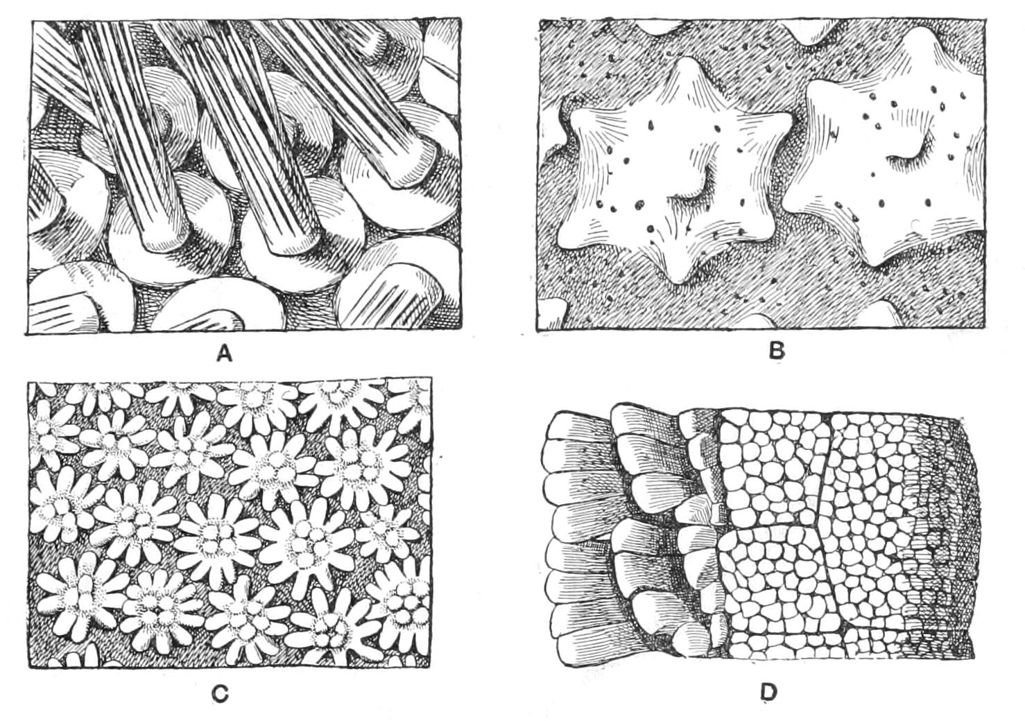
Fig. 194.—Views of portions of the aboral surface of different genera of Asteroidea in order to show the main varieties of skeleton. A, Solaster, showing spines arranged in sheaves; B, Pteraster, showing webs forming supra-dorsal membrane supported by diverging spines; C, Astropecten, showing paxillae; D, Nardoa, showing uniform plating of granules. × 8. (After Sladen.)
(c) Pedicellariae.—These are to be looked on as spines of the second order. In Asterina and its allies they are not present, but groups of little spines arranged in twos and threes, each group being attached to a special small plate, are scattered over the aboral surface; and these on irritation approach one another, and represent the rudiment out of which pedicellariae have been developed. The most perfect form, termed "forcipulate," in which there is a basal ossicle, is found in Asteriidae, Brisingidae, Heliasteridae, Pedicellasteridae, Zoroasteridae, Stichasteridae. There are two varieties of forcipulate pedicellariae, the "crossed" and the "straight," which have been described on p. 432. In all other cases the pedicellariae are devoid of the basal ossicle, and the two or more spinelets forming the jaws are directly attached to one of the main plates of the skeleton.
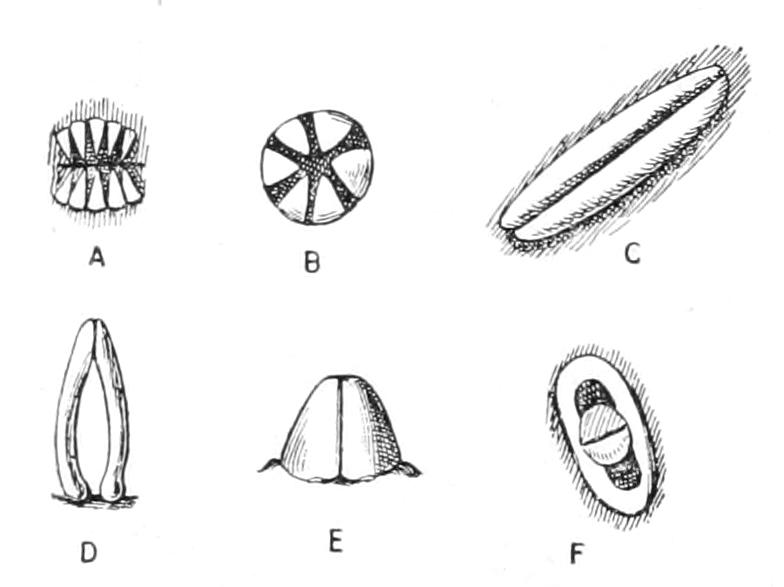
Fig. 195.—Different forms of pedicellariae (excluding the forcipulate form, for which see Fig. 186). A, pectinate; B, pectinate; C, valvate; D, pincer-shaped; E, alveolate, from the side; F, alveolate, from above. × 10. (After Sladen.)
The simplest variety is termed "pectinate"; these pedicellariae are composed of two parallel rows of small spines opposed to each other. They are found in the Archasteridae, and are hardly more advanced in structure than the groups of spines found in Asterina. In Leptogonaster and its allies there are pincer-shaped pedicellariae composed of two curved rods articulating with one of the plates of the skeleton, and also "alveolate" pedicellariae, composed of two short prongs which are implanted on a concave tubercle borne on one of the plates of the skeleton. In the Antheneidae every plate of the ventral surface bears a large "valvate" pedicellaria consisting of two horizontally elongated ridges, which can meet one another. It is possible that valvate pedicellariae have been derived from a pectinate form in which successive spinules of one row have become adherent.
(d) Ambulacral Skeleton.—In every case, whether spines are developed elsewhere or not, the adambulacral plates bear spines. Where the spines are elsewhere represented by granules (Nardoa and its allies) (Fig. 194, D) the adambulacral spines are short and blunt. The terms "monacanthid" and "diplacanthid" are used to express the occurrence of one or two rows of spines respectively on each adambulacral plate.
In the Zoroasteridae the adambulacral plates are curved, and are alternately convex and concave towards the ambulacral groove, so that this groove presents a wavy outline.
In the description of Asterias it was pointed out that the first adambulacral plates in adjacent radii are closely approximated to one another, and bear spines which can to some extent form a trellis-work over the mouth. In very many species not only is this the case, but the plates themselves project inwards over the mouth so as to form prominent "mouth-angles." This is not the case in the Asteriidae or the allied families.
Papulae.—In Asteriidae and many allied families these organs are found both on the upper and under surface of the disc, but in another large group consisting of Astropectinidae, Pentacerotidae, and allied families, papulae are only borne on the dorsal surface, and, in some cases, are restricted to a few groups at the base of the arms. In most Asteroidea the papulae are arranged singly, that is to say, each occupies one of the interspaces between the plates of the skeleton, but in Asterias and some other genera they are arranged in tufts of two or three.
Water-vascular System.—In its general structure this system of organs is very constant, the two most important variations being found, one, in Asteriidae and a few allied families, and the other, in the Astropectinidae and the families allied to them.
The first of the variations alluded to concerns the number of the tube-feet in a radius. In Asterias and its allies these are so numerous that there is not room for them one behind the other, but they follow one another in a zigzag line, the transverse canals connecting them with the radial canals being alternately longer and shorter. In this way the appearance of four rows of tube-feet is produced, and the advantage of this increase in number can be recognised by any one who has compared the quick movements of Asterias and the slow ones of a Cribrella, for instance.
The second important variation referred to is the complete loss of the sucker of the tube-foot, and, concomitantly, the loss of the power of climbing. Starfish which have undergone this change live on sandy bottoms and run over the surface of the sand. They are also incapable of forcing asunder the valves of Molluscs, and hence are compelled to swallow their prey whole.
"Polian vesicles," or stalked sac-like outgrowths of the water-vascular ring, are absent from the Asteriidae, but are found in many families—the Asterinidae, Solasteridae, Astropectinidae, for example. They project outwards from the water-vascular ring in the interradii; when there are several present in one interradius they often arise from a common stalk. Cuénot believes that their sole function, like that of Tiedemann's bodies, is to produce amoebocytes, but this appears unlikely. It is more probable that they act as store-houses of fluid for the water-vascular ring.
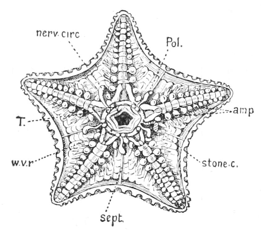
Fig. 196.—Dissection of Ctenodiscus to show the Polian vesicles. amp, Ampullae of the tube-feet; nerv.circ, nerve-ring; Pol, Polian vesicle; sept, interradial septum; stone c, stone-canal; T, Tiedemann's body; w.v.r, water-vascular ring. × 1.
The stone-canal is rarely repeated, but this occurs in the aberrant genus Acanthaster, where there may even be several in one interradius, and each stone-canal has an axial sinus, genital stolon, and madreporite annexed to it. According to Cuénot, in Asterias, when 6-rayed specimens occur in a species normally 5-rayed, there are two stone-canals, suggesting that the repetition of stone-canals is a suppressed effort at multiplication by division. This is also true of Echinaster, but in Ophidiaster two madreporites may occur in an individual with five arms. In the Asterinidae the Y-shaped fold which projects into the cavity of the stone-canal is feebly developed, whereas in the Pentacerotidae it meets the opposite side of the stone-canal, and in Culcita gives out branches which reduce the cavity of the canal to a series of channels. In Echinasteridae and some Asterinidae, and in Astropectinidae and Pentacerotidae the ampullae become so deeply indented as to be almost divided into two, so that each tube-foot has virtually two ampullae.
The alimentary canal has a remarkably constant structure. The only important variation from the type, as described in Asterias, is found amongst the Astropectinidae and Porcellanasteridae, where the anus is wanting. In Astropecten the rectum and the rectal caeca still persist, but in Luidia even these have disappeared. The rectal caeca are remarkably variable structures. In Asterias there are two, but in Pentacerotidae there are five forked caeca, in Asterina five simple caeca, and in the Echinasteridae and Astropectinidae one large flat slightly 5-lobed caecum. In the Asterinidae the pyloric caeca are remarkable for the size of the enlarged basal portion in each radius, which serves as a reservoir for the juices secreted by the branched forks of the caecum. In Porcellanaster pacificus the pyloric caeca are vestigial, and in Hyphalaster moseri they are absent.[454]
The genital organs are, as we have seen, outgrowths from radial branches of the genital rachis. In most species, as in Asterias, they are limited to a single cluster of tubes on each branch of the rachis, but in the Astropectinidae and Pentacerotidae each branch gives rise to a large number of clusters, arranged in longitudinal series, each cluster having its independent opening to the exterior.
Asexual reproduction, as a regular occurrence, is not common amongst Asteroidea. If, however, a Starfish loses some of its arms, it has the power of regenerating the missing members. Even a single arm will regenerate the whole Starfish. Now in some cases (Astropectinidae, Linckiidae) Starfish will readily snap off their arms on irritation. In Linckia this occurs at regular intervals and the separated arm forms a new individual. In one of the Asterinidae, Asterina wega, a small Starfish with seven arms, transverse fission regularly occurs, a portion with three arms separating from one with four. The same is believed to occur in two species of Asterias, and as has already been pointed out, the repetition of the madreporite and stone-canal is, in many cases, possibly connected with this tendency to transverse fission.
Classification of Asteroidea.
Whilst there is considerable agreement amongst the authorities as to the number of families, or minor divisions of unequivocal relationship, to be found in the class Asteroidea, there has been great uncertainty both as to the number and limits of the orders into which the class should be divided, and also as to the limits of the various species. The difficulty about the species is by no means confined to the group Echinodermata; in all cases where the attempt is made to determine species by an examination of a few specimens of unknown age there is bound to be uncertainty; the more so, as it becomes increasingly evident that there is no sharp line to be drawn between local varieties and species. In Echinodermata, however, there is the additional difficulty that the acquisition of ripe genital cells does not necessarily mark the termination of growth; the animals can continue to grow and at the same time slightly alter their characters. For this reason many of the species described may be merely immature forms. In proportion, however, as the collections from various localities increase in number and size, difficulties connected with species will tend to disappear.
The disputes, however, as to the number of orders included in the Asteroidea proceed from a different cause. The attempt to construct detailed phylogenies involves the assumption that one set of structures, which we take as the mark of the class, has remained constant, whilst others which are regarded as adaptive, may have been developed twice or thrice. As the two sets of structures are often of about equal importance it will be seen to what an enormous extent the personal equation enters in the determination of these questions.
Where, as in Asteroidea, the internal organisation is very uniform, the best method of classification is to take as our basis the different methods in which the demands of the environment have been met. It is in this way, we hold, that divergence of character has been produced, for whilst species may differ in trifling details, families and orders differ in points of functional importance. The fact that one of the orders may have sprung from several allied species instead of one may be admitted, and at the same time the hopelessness of trying to push phylogenetic inference into details asserted.
Sladen, in his Monograph of the Asteroidea collected by the "Challenger" expedition, took for the basis of his system the presence or absence of distinct pavement-like marginal plates along the edges of the arms and the restriction of the papulae to the aboral surface, or their distribution over the whole surface of the body. What connexion, if any, the presence of these pavement-like plates has with the habits it is impossible to say, but it is unlikely to be of the high importance with which it was regarded by Sladen, for in the same family we have genera with inconspicuous marginals (Asterina) and others with conspicuous marginals (Palmipes). The restriction of the papulae to the back also varies within the same family (Linckiidae), and whilst, on the whole, it is perhaps a primitive arrangement, it is in many cases connected with burrowing habits, which can scarcely be deemed to have been the original mode of life of the class.
A far better basis is supplied by the system of Perrier,[455] who divides the Asteroidea into five orders according to the character of the dorsal skeleton; and this classification really corresponds with the different habits assumed by groups of Asteroidea in order to meet what must be regarded as one of their chief dangers, viz. assaults by other animals, especially parasites, on their soft and delicate skins. Since the food (so far as is known) of all Asteroidea is more or less similar, the great differentiating factor in their development must have been the means they adopt to shelter themselves from their enemies. Perrier's classification, which we shall adopt, is as follows:—
Order 1. Spinulosa.—Asteroidea in which the plates of the dorsal skeleton bear spines arranged singly or in groups. The tube-feet have suckers and there are no pedicellariae. Marginals sometimes conspicuous, sometimes rod-like.
Order 2. Velata.—Asteroidea in which the dorsal surface of the animal is concealed from view by a false membrane composed of the webs of skin stretched between diverging groups of spines united at the base with one another. No pedicellariae. Tube-feet with suckers.
Order 3. Paxillosa.—Asteroidea in which the dorsal surface is beset with paxillae (upright spines bearing two or three circles of horizontal spinelets). Pedicellariae, when present, few, and never of the forcipulate variety; often absent. Marginals large. Papulae only on dorsal surface. Tube-feet mostly devoid of suckers.
Order 4. Valvata.—Asteroidea in which the dorsal surface is protected by plates covered with a mail of minute granules. Pedicellariae of the valvate or alveolate type. Marginals large.
Order 5. Forcipulata.—Asteroidea in which the dorsal surface is beset with small spines surrounded by numerous forcipulate pedicellariae. Tube-feet with suckers and arranged in four rows. Marginals rod-like and inconspicuous.
Order I. Spinulosa.
This is by far the most primitive order of Asteroidea. The tube-feet are arranged in two rows only, and there is no special means of protecting the back, other than the small close-set plates bearing spines, with which it is covered. In some cases, as Asterina, these spines have a tendency to converge when irritated, and thus act somewhat like pedicellariae. This circumstance suggests strongly the manner in which pedicellariae have been developed from small groups of spines. The order is divided into six families, of which four have common representatives on the British coast.
Fam. 1. Echinasteridae.—Spinulosa in which the aboral skeleton is composed of close set plates bearing comparatively small spines. This family is represented on the British coasts by the beautiful scarlet Starfish Cribrella (Henricia) sanguinolenta. It is also found on the Norwegian coast and on the east coast of North America. On the Pacific coast it is replaced by a larger species, C. laeviuscula. The narrow ambulacral grooves and sluggish movements at once distinguish it from the Starfish described as the type. Indeed, all the Spinulosa seem to be slow in their movements in contrast to the comparatively active Asterias and its allies. Cribrella is remarkable for its large eggs, which have a rapid development. The larva never swims at the surface but glides only for a short time over the bottom. Echinaster is an allied genus in which each plate bears a single somewhat enlarged spine. It possesses on the skin of the aboral surface numerous pits lined by glandular walls, which probably secrete a poisonous fluid which defends it. Acanthaster has thorny spines, more than ten arms, and several stone-canals and madreporites.
Fam. 2. Solasteridae.—Spinulosa in which the aboral skeleton is a network of rods. Spines arranged in diverging bundles (sheaves) attached to a basal button. This family includes the well-known "Sun-stars," with numerous arms and a wide peristome. There are two species found on both sides of the Atlantic. Solaster papposus, with thirteen or fourteen arms and long bundles of spines on the dorsal surface, which is of an orange colour variegated with yellow, and S. endeca with eleven rays and shorter spines and of a reddish violet colour. Rhipidaster has eight arms. Some genera have, however, only five arms, as, for instance, Peribolaster and Korethraster (Fig. 197). In this family there are conspicuous "Polian vesicles" attached to the water-vascular ring.
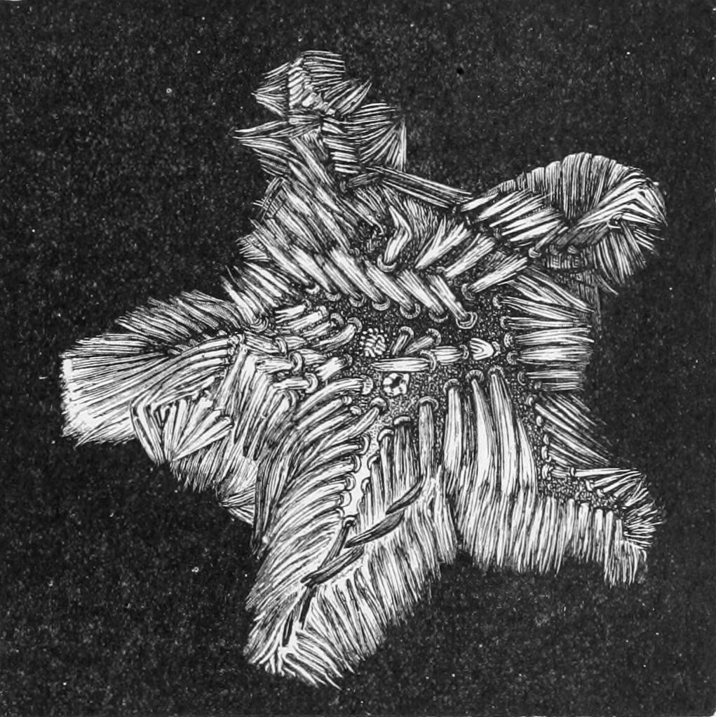
Fig. 197.—Korethraster hispidus. × 2. (From Wyville Thomson.)
Fam. 3. Asterinidae.—Spinulosa in which the aboral skeleton consists of overlapping plates, each bearing a few small spines. The common British representative of this family is the small Asterina gibbosa, in which the arms are short and stout and of somewhat unequal length. This Starfish differs from most of its allies in being littoral in its habit. At low tide on the south and west coasts of England it can be found on the underside of stones feeding on the Sponges and Ascidians with which they are covered. Like Cribrella sanguinolenta this species has a modified development. The larva resembles that of Cribrella, and the larval stage only lasts about a week. Owing to the fact that Asterina lays its eggs in accessible localities, its development has been more thoroughly worked out than that of any other species. Palmipes membranaceus, an animal of extraordinary thinness and flatness, is sometimes dredged up off the coast of Britain in deeper water. Its arms are so short that the general form is pentagonal. The infero-marginal plates are long and rod-like, and form a conspicuous border to the body when viewed from below.
Fam. 4. Poraniidae.—Spinulosa allied to the Asterinidae but possessing a thick gelatinous body-wall in which the plates and spines are buried, the marginals forming a conspicuous border to the body. This family is represented in British waters only by Porania pulvillus, a cushion-shaped Starfish with very short arms and of a magnificent reddish-purple colour. It is occasionally, but rarely, exposed at low tide.
Fam. 5. Ganeriidae.—Spinulosa allied to the Asterinidae but distinguished by the large marginals and by the fact that the skeleton of the oral surface consists of plates each bearing a few large spines. Ganeria, Marginaster.
Fam. 6. Mithrodiidae.—Spinulosa with a reticulate aboral skeleton. The spines are large and blunt, covered with minute spinules. Mithrodia, sole genus.
These last two families are not represented in British waters.
Order II. Velata.
This is a very extraordinary group of Starfish, about the habits of which nothing is known, since they all live at very considerable depths. Their nearest allies amongst the Spinulosa must be looked for amongst the Solasteridae. If the sheaves of spines with which the latter family are provided were to become adherent at their bases, and connected with webs of skin so as to form umbrella-like structures, and if then these umbrellas were to become united at their edges, we should have a supra-dorsal membrane formed such as is characteristic of the order.
Fam. 1. Pythonasteridae.—Velata in which each sheaf of spines is enveloped in a globular expansion of the skin and is not united with the neighbouring sheaves. Pythonaster, sole genus.
Fam. 2. Myxasteridae.—Velata with numerous arms in which the sheaves of spines are long and form with their connecting "umbrellas" web-like expansions which do not fuse with one another. Myxaster, sole genus.
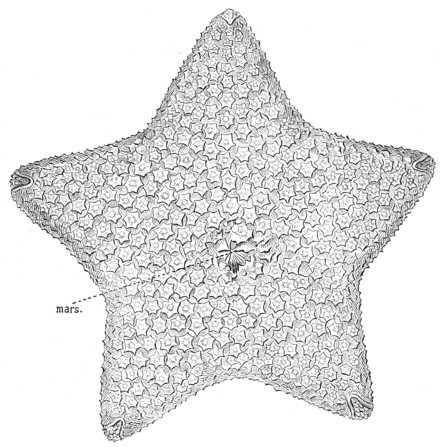
Fig. 198.—Aboral view of Pteraster stellifer. mars, Dorsal brood-pouch, × 1½. (From Sladen.)
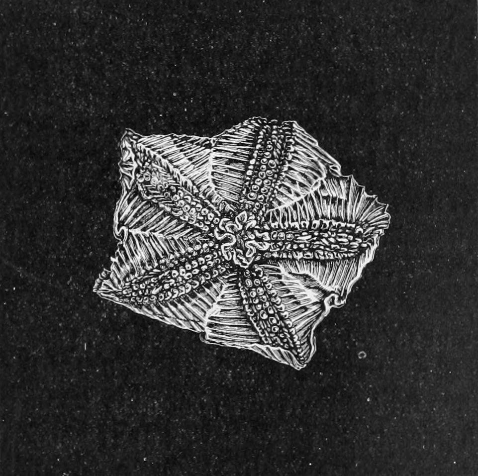
Fig. 199.—Oral view of Hymenaster pellucidus. × 1. (From Wyville Thomson.)
Fam. 3. Pterasteridae.—Velata in which the membranes supported by the sheaves of spines are united so as to form a continuous supra-dorsal tent. The Pterasteridae are represented in British waters by a single species, Pteraster militaris, which is occasionally dredged in deep water off the British coast, and is found also in the Norwegian fjords and off the east coast of Canada. This interesting Starfish has five short, blunt arms, and its general appearance at first sight recalls that of Asterina. Closer inspection reveals the "false back." The anus is surrounded by five fan-like valves, supported by spines (Fig. 198), underneath which is a space in which the young complete their development, Pteraster being one of the genera in which the normal larval form is not developed. The tendency towards the union of adjacent spines by webs is deeply rooted in the organisation of the animal. It is seen on the under side where the spines borne by the ventral plates are united so as to form transverse combs. In Hymenaster (Fig. 199) the spines borne by the ventral plates are long and free.
Order III. Paxillosa.
This is an exceedingly well-marked order. The armature of the upper surface consists of paxillae. These organs as already mentioned are probably to be traced back to sheaves of spines like those of the Solasteridae. The same end as that striven after in the case of the Velata has been attained, but in a different way. The horizontal spinelets of the paxillae meet one another and form a close-fitting mail which is almost as efficient a protection as the webs and umbrellas of the Velata. Pedicellariae are occasionally present, but they are always of the pectinate or pincer variety, never forcipulate.
Fam. 1. Archasteridae.—Paxillosa in which the anus is still retained and in which the tube-feet have suckers.
The Archasteridae are a most interesting family. Thus Pararchaster has no true paxillae, but only small isolated groups of spines. The pectinate pedicellariae are composed each of two parallel rows of somewhat smaller spines. The members of this family are to some extent intermediate in structure between the Spinulosa, such as Echinasteridae, and the other families of the Paxillosa—some genera, indeed, might almost be classed as Spinulosa. At the same time they are apparently closely allied with the more primitive Valvata such as Astrogonium and its allies, some of which have paxillae on the upper surface; although the retention of the anus and of the suckers on the tube-feet (in which characters they agree with the Archasteridae) distinguishes them from the more typical Paxillosa, in which both anus and suckers are lost. Archaster (Figs. 200, 201). Leptogonaster.
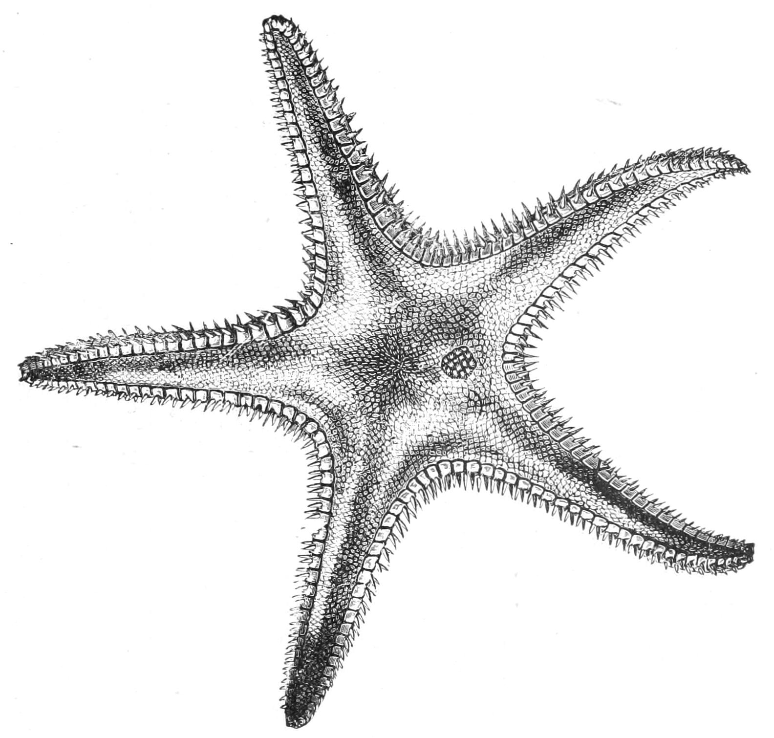
Fig. 200.—Aboral view of Archaster bifrons. × ¾. (From Wyville Thomson.)
Fam. 2. Astropectinidae.—Paxillosa which have lost the anus, but which possess neither aboral protuberance nor interradial grooves. The marginal plates are thick, covered with spinules and placed horizontally. The tube-feet have no suckers.
This family is the only one of the order which occurs in British waters, where it is represented by two genera, Astropecten and Luidia. In Astropecten the inferior marginal plate is in immediate contact with the adambulacral, whilst in Luidia it is separated from it by a small intermediate plate.
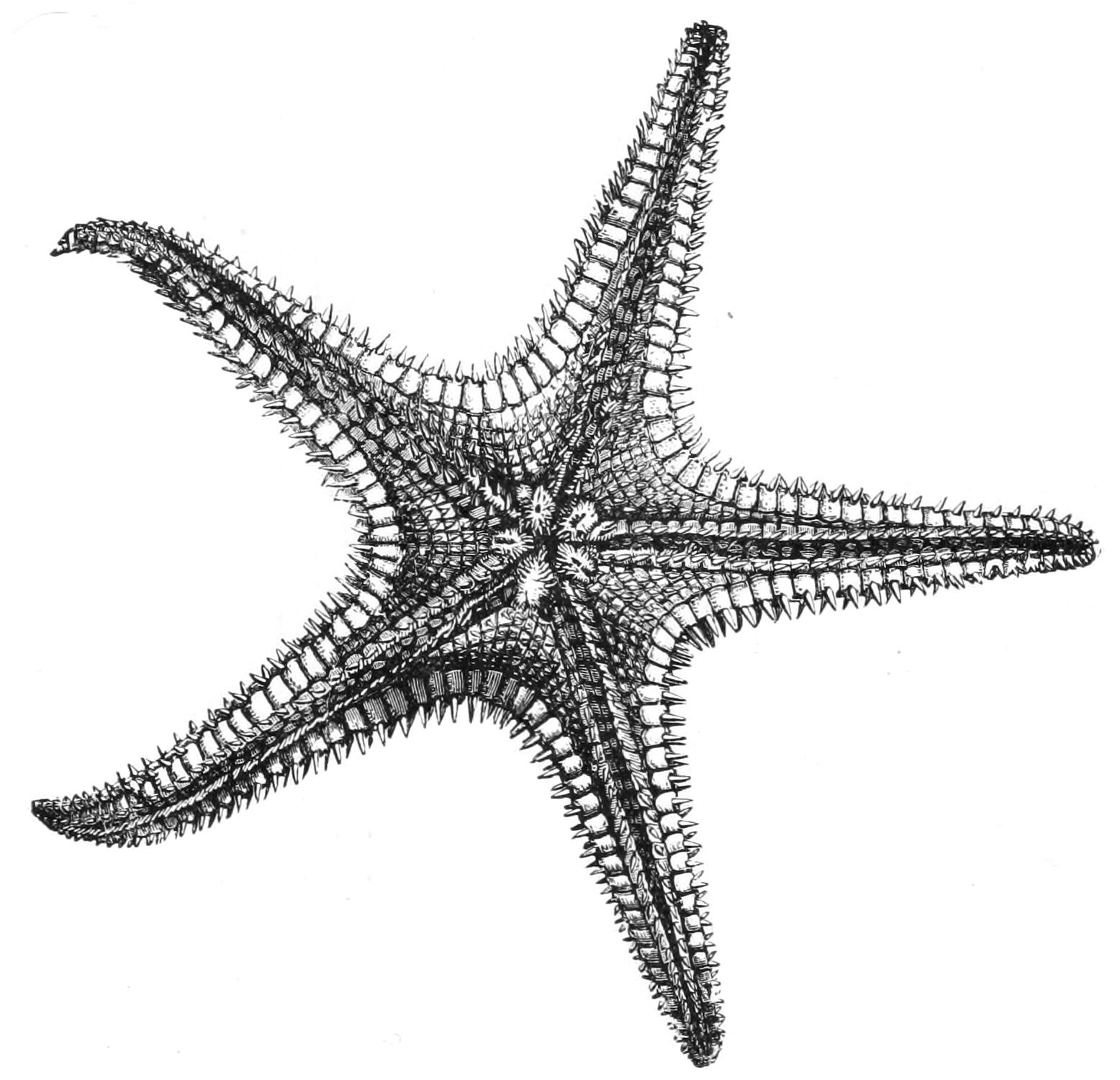
Fig. 201.—Oral view of Archaster bifrons. × ¾. (From Wyville Thomson.)
Astropecten irregularis is a very common species on the coast of Britain, and a study of its habits when in captivity has thrown a great deal of light on many obscure points in the anatomy of the Paxillosa. Owing to the loss of suckers it is unable to climb over rocks and stones like the ordinary species, but it runs over the surface of the hard sand in which it lives by means of its pointed tube-feet. The arms are highly muscular, and the animal when laid on its back rights itself by throwing the arms upwards and gradually overbalancing itself. The loss of suckers has also rendered Astropecten and its allies incapable of feeding in the manner described in the case of Asterias rubens. They are unable forcibly to open the valves of shell-fish, and the only resource left to them is to swallow their prey whole. The mouth is consequently wide, and the unfortunate victims, once inside the stomach, are compelled by suffocation to open sooner or later, when they are digested.[456]
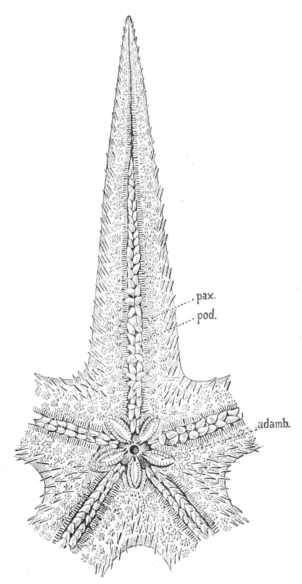
Fig. 202.—Oral view of Psilaster acuminatus. × 4⁄3. adamb, Adambulacral spines; pax, paxillae; pod, pointed tube-feet devoid of sucker. (After Sladen.)
Many interesting experiments have been made on Astropecten by Preyer and other investigators, but one important fact[457] has escaped their notice, that Astropecten, when at rest, lies buried in the sand, whilst the centre of the aboral surface is raised into a cone which projects above the surface. On the sides of this cone the few papulae which this species possesses are distributed. This raising of the aboral surface is obviously an expedient to facilitate respiration. It loosens the sand over the region of the papulae, and thus allows the water to have access to them. We can thus understand how the restriction of the papulae to the dorsal surface, so characteristic of the Paxillosa, is not always as Sladen imagined, a primitive characteristic, but often an adaptation to the burrowing habits which in all probability are characteristic of the whole order. In both Luidia and Astropecten Cuénot has described short spines covered with cilia in the interspaces between the marginal plates, these also subserve respiration by drawing a current of water over the gills. Psilaster (Fig. 202).
Fam. 3. Porcellanasteridae.—Paxillosa which have lost the anus. There is a conical prominence in the centre of the dorsal surface termed the epiproctal cone, and in the interradial angles there are vertical grooves bordered by folds of membrane produced into papillae, the so-called "cribriform organs." The marginal plates are thin and form the vertical border of the thick disc. The tube-feet have no suckers.
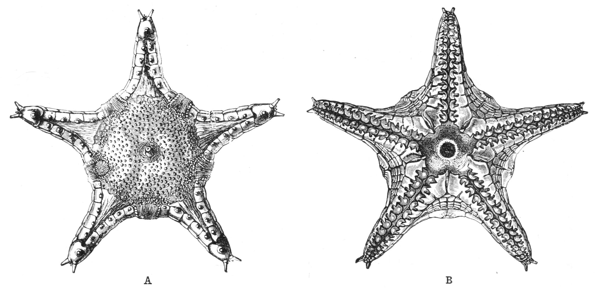
Fig. 203.—Porcellanaster caeruleus. A, aboral view; B, oral view, × 1. (From Wyville Thomson.)
Comparing the Porcellanasteridae with the Astropectinidae we see at once that the "epiproctal cone" is a permanent representative of the temporary aboral elevation in Astropecten, and we are inclined to suspect that the cribriform organs are grooves lined with cilia which keep up a respiratory current like the ciliated spines of Luidia. In all probability the Porcellanasteridae are more habitual burrowers than even the Astropectinidae.
Ctenodiscus (Fig. 196), a genus in which there is a short epiproctal cone and numerous feeble cribriform organs in each interradius, is found in deep water north of the Shetland Islands. Porcellanaster (Fig. 203) is a more typical genus, with one large cribriform organ in each interradius. Hyphalaster has long arms, on which the supero-marginal plates meet above.
Order IV. Valvata.
The Starfish included in this order are characterised by the absence of prominent spines and by the superficial covering of minute granules. The skeleton consists, in most cases, of plates, and these plates with their covering of granules probably represent the first stage in the evolution of paxillae.
The tube-feet possess well-developed suckers. No members of this order can properly be said to be British.
Fam. 1. Linckiidae.—Valvata with long arms, the marginals being developed equally throughout the whole length. These Starfish are distinguished by their long narrow arms and small disc. It is possible that these forms, so different in many respects from the other families of the order, have been directly derived from the long-armed Echinasteridae. Ophidiaster, Nardoa, Linckia.
Fam. 2. Pentagonasteridae.—Valvata with short arms, the marginals being especially developed at the base and in the interradial angles. The aboral skeleton consists of close-fitting plates. Pentagonaster (Fig. 204), Astrogonium.
Fam. 3. Gymnasteridae.—Valvata allied to the foregoing but distinguished by possessing a very thick skin in which the plates are completely buried. Dermasterias, Asteropsis.
Fam. 4. Antheneidae.—Valvata with short arms. The dorsal skeleton is reticulate and each ventral plate bears one or several large valvular pedicellariae (Fig. 195, C). Hippasterias, Goniaster.
Fam. 5. Pentacerotidae.—Valvata with arms of moderate length. The dorsal skeleton is reticulate but the ventral plates bear only small pedicellariae or none. The upper marginals are smaller than the ventral ones.
The Pentacerotidae include both short-armed and long-armed forms. Amongst the former is Culcita, in which the body is a pentagonal disc, all outer trace of the arms being lost; Pentaceros is a long-armed form.
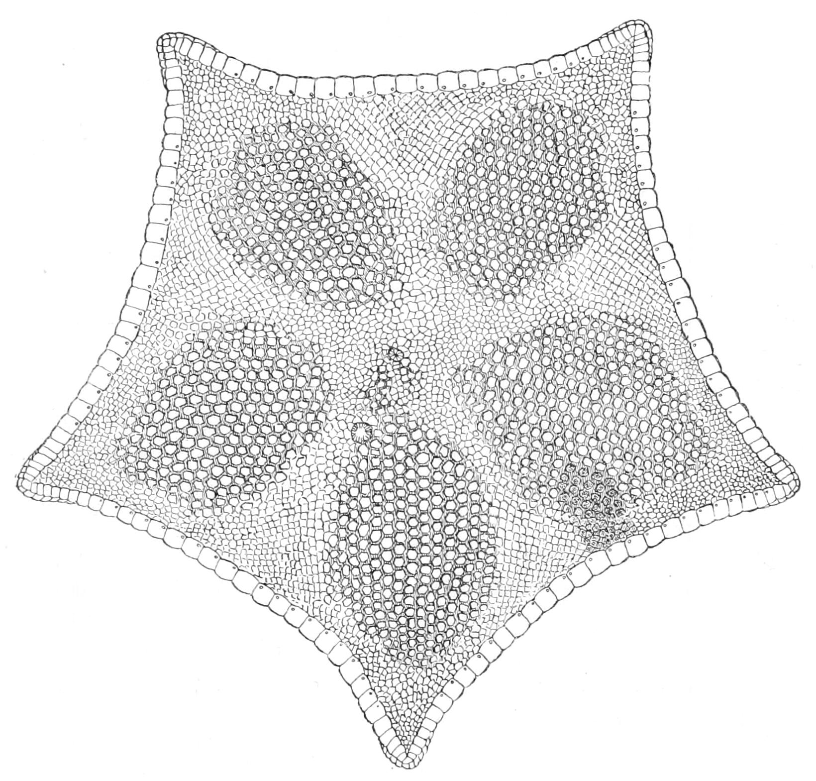
Fig. 204.—Pentagonaster japonicus. × ⅔. (After Sladen.)
The family Pentagonasteridae furnishes the key to the understanding of most of the forms contained in this order. It contains genera such as Astrogonium which possess on the back unmistakable paxillae, whilst on the under surface they have the characteristic covering of granules; these genera seem to be closely allied to the short-armed species of the Archasteridae, from which they are distinguished chiefly by the granular covering of the marginals. From a study of these cases it seems clear that the plates of the dorsal skeleton of the Valvata correspond to the supporting knobs of the paxillae much broadened out, and the granules correspond to the spinelets of the paxillae increased in number and diminished in size.
As mentioned above, Ludwig has proved that the paxillae develop in the life-history of the individual out of ordinary plates, the axis of the paxilla representing the plate.
Order V. Forcipulata.
This order, which includes the most highly developed members of the class Asteroidea, is at once distinguished by the possession of forcipulate pedicellariae which, as we have seen, possess a well-marked basal piece with which the two plates articulate. The pedicellariae are consequently sharply marked off from the spinelets, and no intermediate forms occur. The first conjoined adambulacrals, which in other orders form the "teeth" or mouth-angles, do not here project beyond the first pairs of ambulacral plates.
Fam. 1. Asteriidae.—Forcipulata in which the tube-feet are apparently arranged in four rows. Aboral skeleton a loose reticulum.
The general features of the family Asteriidae have been explained in the description of Asterias rubens (p. 432). There are five well-marked species of the genus found on the British coasts. Of these A. glacialis is found chiefly in the south-western parts of the English Channel. It is a large Starfish of a purplish-grey colour, with large spines surrounded by cushions of pedicellariae arranged in one or two rows down each arm. A. muelleri resembles the foregoing species, but is of much smaller size, and is further distinguished by having straight pedicellariae in the neighbourhood of the ambulacral groove only. It is found on the east coast of Scotland, and carries its comparatively large eggs about with it until development is completed. A. rubens is the commonest species, and is found on both east and west coasts. Its colour is a bright orange, but varies to almost a straw colour. It is at once distinguished from the foregoing species by the spines of the dorsal surface, which are small and numerous, an irregular line of somewhat larger ones being sometimes seen down the centre of each arm. A. murrayi is a peculiar species restricted to the west coast of Scotland and Ireland. It has flattened arms, with vertical sides, and only three rows of small spines on the dorsal surface. It is of a violet colour. A. hispida is also a western species. It is a small Starfish with short stout arms; there are no straight pedicellariae, and only a few sharp spines on the dorsal surface.
On the eastern coast of North America there are several species of Asterias, of which the most noteworthy is the 6-rayed A. polaris of the Gulf of St. Lawrence. This species exhibits a marvellous range of colour-variation, ranging from bluish-violet through purple to red and straw-coloured. This variation seems to show that colour, as such, is of no importance to the animal, but probably depends on some compound of slightly varying composition which is being carried by the amoebocytes towards the exterior. On the Pacific coast there is a rich fauna of Starfish, among which we may mention as members of this family Asterias ochracea, a large violet species, so strong that it requires a severe wrench to detach it from the rock, and Pycnopodia with twenty-two arms.
Fam. 2. Heliasteridae.—Forcipulata allied to the Asteriidae, but with very numerous arms and double interradial septa. Heliaster.
Fam. 3. Zoroasteridae.—Forcipulata with the tube-feet in four rows at the base of the arm, in two rows at the tip. Aboral skeleton of almost contiguous plates bearing small spines or flattened scales. Zoroaster, Pholidaster.
Fam. 4. Stichasteridae.—Forcipulata with the tube-feet in four rows. Aboral skeleton of almost contiguous plates covered with granules. Stichaster, Tarsaster.
The Stichasteridae and Zoroasteridae have acquired a superficial resemblance to some of the long-armed Valvata, from which they are at once distinguished by their pedicellariae. It would be exceedingly interesting if more could be found out concerning the normal environment of these animals; it might then be possible to discover what is the cause of the assumption of this uniform mail of plates.
Fam. 5. Pedicellasteridae.—Forcipulata with two rows of tube-feet. The aboral skeleton bears projecting spines surrounded by cushions of straight pedicellariae. Pedicellaster, Coronaster.
Fam. 6. Brisingidae.—Forcipulata with numerous arms and only two rows of tube-feet. Aboral skeleton largely rudimentary and confined to the base of the arms. The small blunt spines are contained in sacs of skin covered with pedicellariae.
The Brisingidae, including Brisinga and Odinia, are a very remarkable family, chiefly on account of the smallness of the disc and of the extraordinary length of the arms. The arms have what we must consider to have been the primitive arrangement, since there is no lateral adhesion between them, and interbrachial septa are consequently entirely absent. The reduction of the skeleton is a very marked peculiarity and, like the tendency to the reduction of the skeleton of deep-sea fish, may stand in some relation to the great pressure under which the animals live.
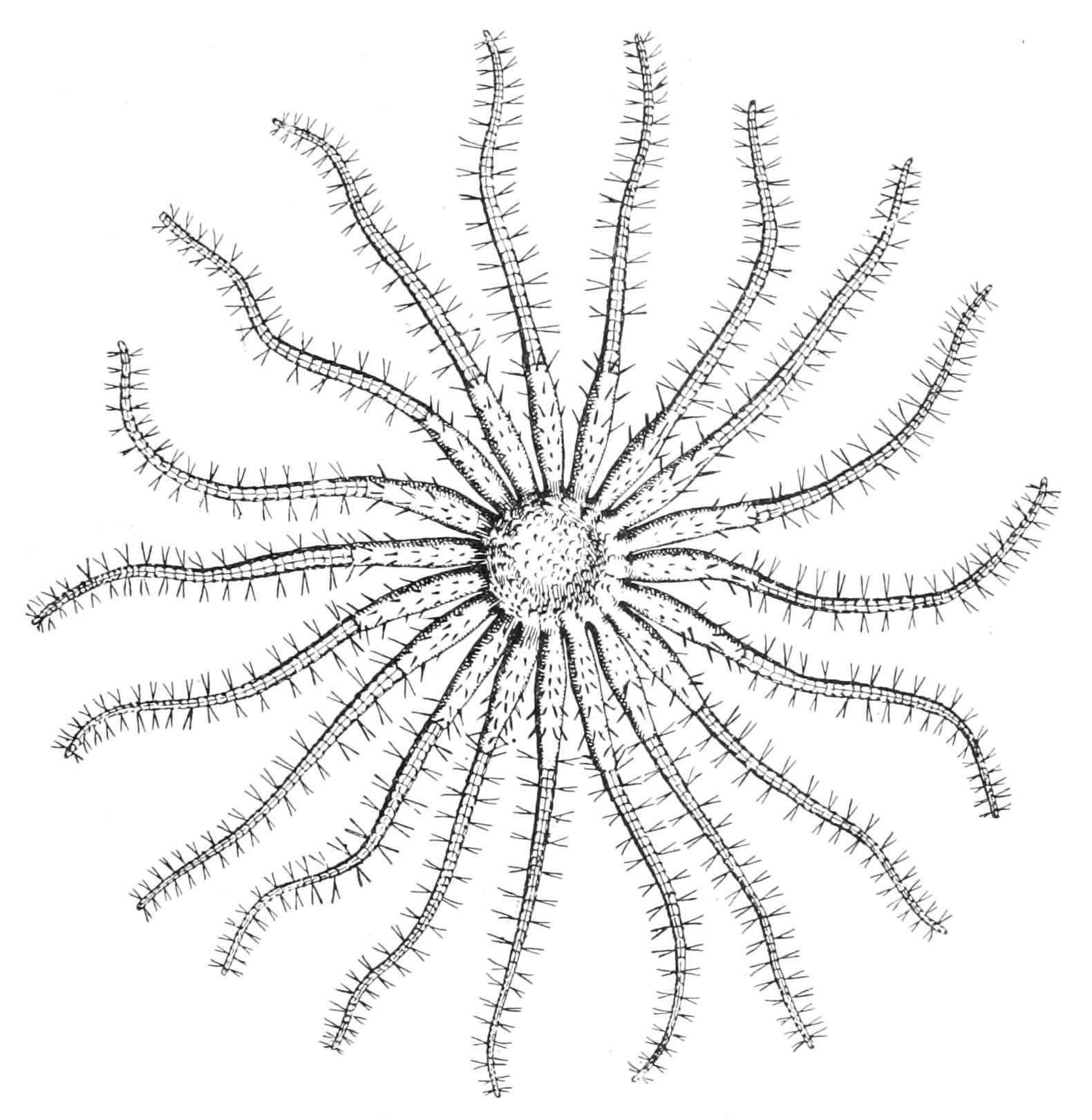
Fig. 205.—Aboral view of Odinia. × ⅔. (After Perrier.)
Fossil Asteroidea.
The Asteroidea occur somewhat plentifully as fossils. In the Lower Jurassic Asterias, Astropecten, Luidia, Solaster, and Goniaster have already made their appearance. In the Cretaceous Pentaceros appears. In the older rocks occur a number of forms of different character from any now existing. Of these Aspidosoma (Fig. 206), with short lancet-shaped arms sharply distinguished from the disc and continued along its under surface, seems to be intermediate between Asteroidea and Ophiuroidea. The skeleton of the arm is composed of alternating ambulacral ossicles bordered by adambulacral ossicles, which are at the same time marginals and sharply distinguished from the marginals forming the edge of the disc. Palaeaster, on the other hand, is a true Asteroid; there are marginals distinct from the adambulacrals, but the disc is reduced to its smallest dimensions, there being only one plate on the ventral side of each interradius. There are a number of genera (Palaeocoma, for instance) with a large disc and very short arms and very shallow ambulacral grooves; all have alternating ambulacral plates. Some genera appear to have had the madreporite on the ventral surface of an interradius. On the other hand, in the Devonian occurs Xenaster, which was a fairly normal Asteroid, with pavement-like marginals, deep ambulacral grooves, and broad arms.
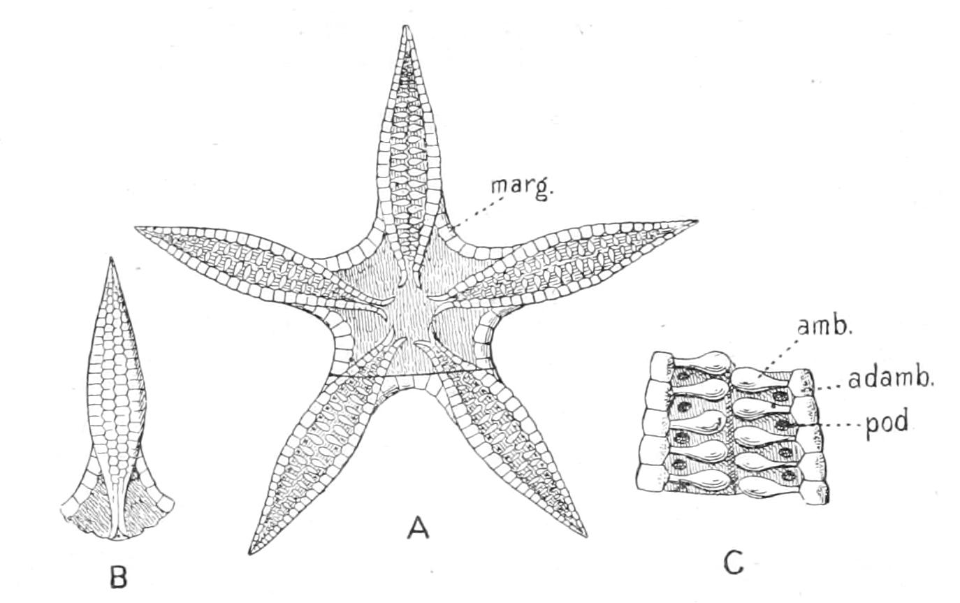
Fig. 206.—Three views of Aspidosoma, a fossil Asteroid. A, oral view; B, aboral view of one arm; C, enlarged view of a portion of the ambulacral groove. adamb, Adambulacral plate; amb, ambulacral plate; marg, marginal plate; pod, aperture for extension of tube-foot.
Thus it will be seen that already in Jurassic times the three orders, Forcipulata, Paxillosa, and Spinulosa were differentiated from each other, but how these are related to the older Palaeozoic forms it is at present impossible to say.