Chapter XVIII
Reading edition
ECHINODERMATA (CONTINUED): ECHINOIDEA = SEA-URCHINS
CLASS III. ECHINOIDEA
The Sea-urchins or Echinoidea (Gr. ἐχῖνος, Hedgehog or Sea-urchin), which constitute the third class of the Eleutherozoa, have derived both their popular and scientific names from the covering of long spines with which they are provided. At first sight but little resemblance is to be discerned between them and the Starfish and Brittle Stars. They are devoid of any outgrowths that could be called arms; their outline is generally either circular or that of an equilateral pentagon, but as their height is almost always smaller than their diameter, they are never quite spherical; sometimes it is so small that the animals have the form of flattened discs.
All doubt as to the relationship of the Echinoidea to the Starfish is at once dispelled in the mind of any one who sees one of the common species alive. The surface is beset with delicate translucent tube-feet, terminated by suckers resembling those of Starfish, although capable of much more extension. The animal throws out these organs, which attach themselves by their suckers to the substratum and so pull the body along, whilst the spines are used to steady it and prevent it from overturning under the unbalanced pull of the tube-feet. When moving quickly the animal walks on its spines, the tube-feet being little used. The tube-feet are distributed over five bands, which run like meridians from one pole of the animal to the other. These bands are termed "radii," and they extend from the mouth, which is situated in the centre of the lower surface, up to the neighbourhood of the aboral pole. The radii must be compared to the ambulacral grooves on the oral surface of the arms of Starfish, and hence in Urchins the aboral surfaces of the arms have, so to speak, been absorbed into the disc, so that the oral surfaces have become bent in the form of a semicircle. The radii are separated from one another by meridional bands called "interradii," which correspond to the interradial angles of the disc of a Starfish and to the sides of its arms. The small area enclosed between the upper terminations of the radii is called the "periproct," and this corresponds to the entire dorsal surface of the Starfish, including that of the arms.
One of the commonest species of British Sea-urchin is Echinus esculentus. In sheltered inlets, such as the Clyde, it is often left exposed by the receding tide, whilst everywhere on the coast in suitable localities it may be obtained by dredging at moderate depths on suitable ground. In the Clyde it is easy to observe the habits of the animal through the clear still water. It is then seen to frequent chiefly rocky ground, and to exhibit a liking for hiding itself in crevices. Often specimens will be seen clinging to the rock by some of their tube-feet, and, as it were, pawing the under surface of the water with the others. In the Clyde it feeds chiefly on the brown fronds of Laminaria, with which the rocks are covered. In more exposed situations, such as Plymouth Sound, it does not occur in shallower water than 18 to 20 fathoms. At this depth it occurs on a rocky ridge; but in 1899, after a south-west gale, all the specimens had disappeared from this ridge, showing at what a depth wave disturbance is felt.
A full-grown specimen is as large as a very large orange; its under surface is flattened, and it tapers somewhat towards the aboral pole. The outline is that of a pentagon with rounded angles. The spines in Echinus esculentus are short in comparison to the diameter of the body, and this is one of the characteristics of the species.
The animal is provided with a well-developed skeleton, consisting of a mail of plates fitting closely edge to edge, and carrying the spines. This cuirass bears the name "corona" (Fig. 227). It has two openings, an upper and a lower, which are both covered with flexible skin. The upper area is known as the "periproct" (Fig. 227, 2); it has in it small isolated plates, and the anus, situated at the end of a small papilla, projects from it on one side of the centre. The lower area of flexible skin surrounds the mouth, and is called the "peristome" (Fig. 229), though it corresponds to considerably more than the peristome of Asteroidea. In the mouth the tips of the five white chisel-like teeth can be seen.
The plates forming the corona are, like all the elements of the skeleton of Echinodermata, products of the connective tissue which underlies the ectoderm, which in Echinoidea remains in a fully developed condition covering the plates, and does not, as in Ophiuroidea, dry up so as to form a mere cuticle. The ectoderm consists of the same elements as that of Asteroidea, viz. delicate tapering sense-cells with short sense-hairs, somewhat stouter supporting cells and glandular cells. It is everywhere underlaid by a plexus of nerve fibrils, which, in part, are to be regarded as the basal outgrowths of the sense-cells and partly as the outgrowths of a number of small bipolar ganglion-cells, found intermixed with the fibres.
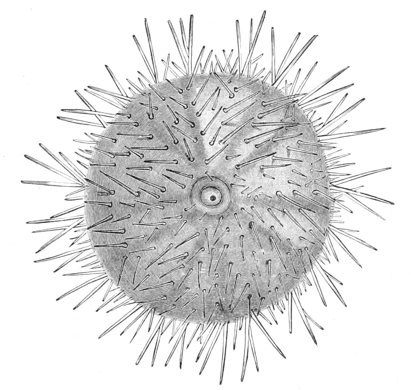
Fig. 223.—Aboral view of Echinus esculentus. × ½. (After Mortensen.)
Just as the muscular arm has been the determining factor in the structure of the Ophiuroidea, so the movable spine has been the leading factor in the evolution of Echinoidea. The spines have cup-shaped basal ends, which are inserted on special projections of the plates of the skeleton called tubercles. The tubercle is much larger than the cup, and hence the spine has a great range of possible motion. The spines differ from those of Starfish and Brittle Stars in being connected with their tubercles by means of cylindrical sheaths of muscle fibres, by the contraction of which they can be moved in any direction. The muscles composing the sheath consist of an outer translucent and an inner white layer. The former are easily stimulated and soon relax; they cause the movements of the spines. The latter require stronger stimulation, but when aroused respond with a prolonged tetanus-like contraction, which causes the spines to stand up stiffly in one position; these muscles can be torn across sooner than forced to relax. Uexküll[471] has appropriately named them "block musculature." These sheaths, like everything else, are covered with ectoderm, which is, however, specially nervous, so that we may say that the muscular ring is covered by a nerve-ring from which stimuli are given off to the muscles.
The spines are, speaking generally, of two sizes, the larger being known as "primary spines" and the smaller as "secondary." In many Echinoidea these two varieties are very sharply contrasted, but in Echinus esculentus there is not such a great difference in length, and intermediate kinds occur. The forest of spines has an undergrowth of pedicellariae. All Echinoidea possess pedicellariae, which are much more highly developed than those of any Asteroid. With few exceptions all the pedicellariae of Echinoidea possess three jaws and a basal piece. This latter is, however, drawn out so as to form a slender rod, which articulates with a minute boss on a plate of the skeleton.
Of these pedicellariae there are in E. esculentus four varieties, viz. (1) "tridactyle" (Fig. 225, C; Fig. 226, B): large conspicuous pedicellariae with three pointed jaws, each armed with two rows of teeth on the edges. There is a flexible stalk, the basal rod reaching only half way up. These are scattered over the whole surface of the animal.
(2) "Gemmiform" (Fig. 225, A, B; Fig. 226, A), so called from the translucent, almost globular head. The appearance of the head is due to the fact that there is on the outer surface of each jaw a sac-like gland developed as a pouch of the ectoderm. From it are given off two ducts which cross to the inner side of the blades and, uniting into one, run in a groove to near the tip. The gland secretes a poisonous fluid. The basal rod reaches up to the jaws, so that this form of pedicellaria has a stiff stalk. On the inner side of each blade, near the base, there is a slight elevation (Fig. 225, B, s), consisting of cells bearing long cilia; this is a sense-organ for perceiving mechanical stimuli. The gemmiform pedicellariae are particularly abundant on the upper surface of the animal.
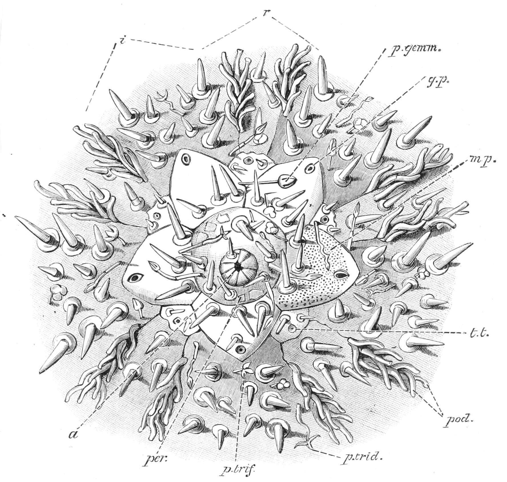
Fig. 224.—View of the apical region of Echinus esculentus, showing spines and pedicellariae; drawn from the living specimen, × 3. a, Anus; g.p, genital pore; i, interradius; mp, madreporite; per, periproct; p.gemm, gemmiform pedicellaria; pod, podia; p.trid, tridactyle pedicellaria; p.trif, trifoliate pedicellaria; r, radius; t.t, pore for terminal tentacle of the radial water-vascular canal.
(3) "Trifoliate" (Fig. 225, E; Fig. 226, D): these are very small pedicellariae, in which the jaws are shaped like leaves with the broad end projecting outwards. They are scattered over the whole surface of the body.
(4) "Ophicephalous" (Fig. 225, D; Fig. 226, C): pedicellariae in which the jaws have broad rounded distal ends fringed with teeth; these ends bear a resemblance to a snake's head, whence the name. The bases are also broad and thin, with a strong median rib and a peculiar semicircular hoop beneath the spot where they articulate with one another. The three hoops of the three jaws work inside each other in such a way as to cause the jaws to have a strong grip and to be very difficult to dislocate from their mutual articulation.
The ophicephalous pedicellariae are in Echinus the most abundant of all; and they alone extend on to the peristome, where a special small variety of them is found.
A thorough investigation of the functions and reactions of the pedicellariae has quite recently been made by von Uexküll.[472] He showed, first of all, that there is a nervous centre in the stalk of each pedicellaria (see below), which causes the organ to incline towards a weak stimulus, but to bend away from a stronger stimulus. In the head there is an independent nervous centre, which regulates the opening and closing of the valves, and causes these to open on slight stimulus and close when a stronger one is applied. The amount of stimulus necessary to cause the pedicellariae to retreat varies with the kind of pedicellariae, being least with the tridactyle and most with the gemmiform, so that when a chemical stimulus, such as a drop of dilute ammonia, is applied to the skin, the tridactyle pedicellariae may be seen to flee from and the gemmiform to approach the point of stimulation. In a living Sea-urchin, if the attempt is made to seize the tridactyle pedicellariae they will evade the forceps, but the ophiocephalous are easy to catch.
The tridactyle pedicellariae open with the very slightest mechanical stimulus and close with rather greater mechanical stimuli or with exceedingly slight chemical ones. Uexküll calls them "Snap-pedicellariae," and their function is to seize and destroy the minute swimming larvae of various sessile parasitic animals, which would otherwise settle on the delicate exposed ectoderm of the Sea-urchin.
The gemmiform pedicellariae are brought into action when a more serious danger threatens the Sea-urchin, such as an attack of a Starfish. The corrosive chemical influence, which it can be proved exudes not only from the stomach but even from the tube-feet of the Starfish, causes the gemmiform pedicellariae to approach and open widely. When the foe approaches so closely as to touch the sense-organs (Fig. 225, B, s) situated on the inner side of the valves of these pedicellariae, the blades close violently, wounding the aggressor and causing its juice to exude, thus producing a renewed and severe chemical stimulation which irritates the poison glands and causes the poison to exude. The virulence of the poison may be gauged from the fact that the bite of a single gemmiform pedicellaria caused a frog's heart to stop beating.
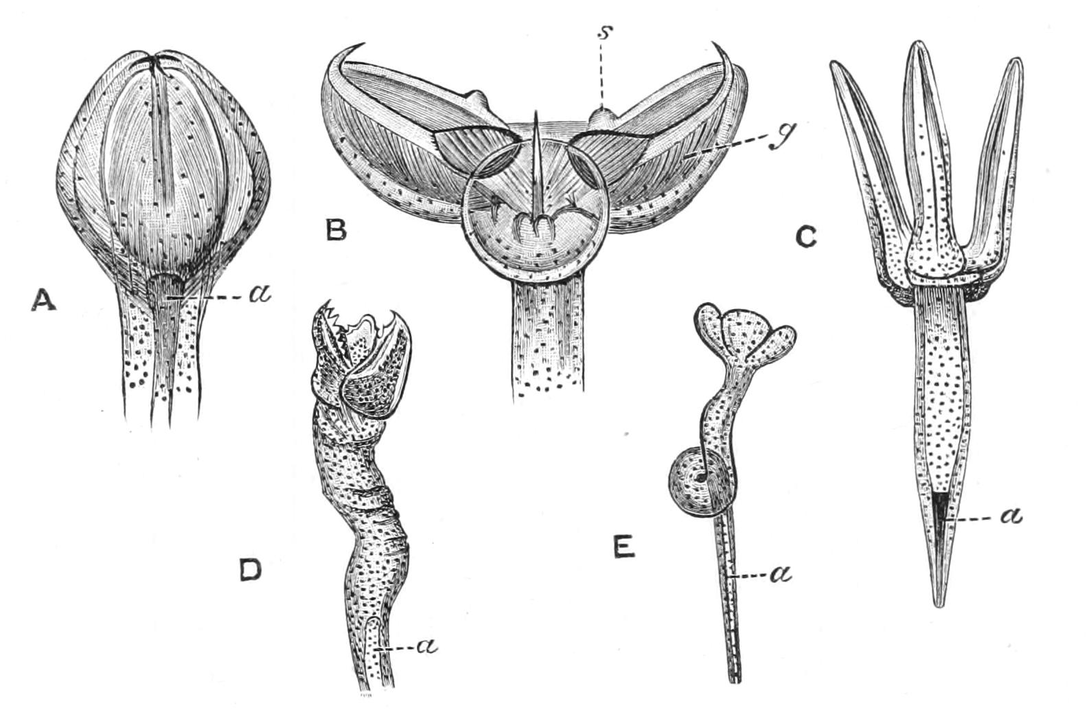
Fig. 225.—The pedicellariae of Echinus acutus, drawn from a living specimen. A, gemmiform pedicellaria, closed. B, gemmiform pedicellaria, open; g, poison gland; s, sense-organ, × 3. C, tridactyle pedicellaria, × 6. D, ophicephalous pedicellaria, × 9. E, trifoliate pedicellaria, × 12; a (in all figures), axial rod of the stalk. (After Uexküll.)
Prouho[473] has described a combat between a Sea-urchin and a Starfish. When the latter approached, the spines of the Sea-urchin diverged widely (strong form of reaction to chemical stimulus), exposing the gemmiform pedicellariae. These at once seized the tube-feet of the enemy and the Starfish retreated, wrenching off the heads of these pedicellariae; then the Starfish returned to the attack and the same result followed, and this was repeated till all the pedicellariae were wrenched off, when the Starfish enwrapped its helpless victim with its stomach.
The minute trifoliate pedicellariae are brought into play by any prolonged general irritation of the skin, such as bright light or a rain of particles of grit or mud. They have the peculiarity that not all the blades close at once, so that an object may be held by two blades and smashed by the third. They may be seen in action if a shower of powdered chalk is poured on the animal, when they seize the particles and by breaking up any incipient lumps reduce the whole to an impalpable powder, which the cilia covering the skin speedily remove. In thus assisting in the removal of mechanical "dirt" they earn the name which Uexküll has bestowed on them, of "cleaning pedicellariae."
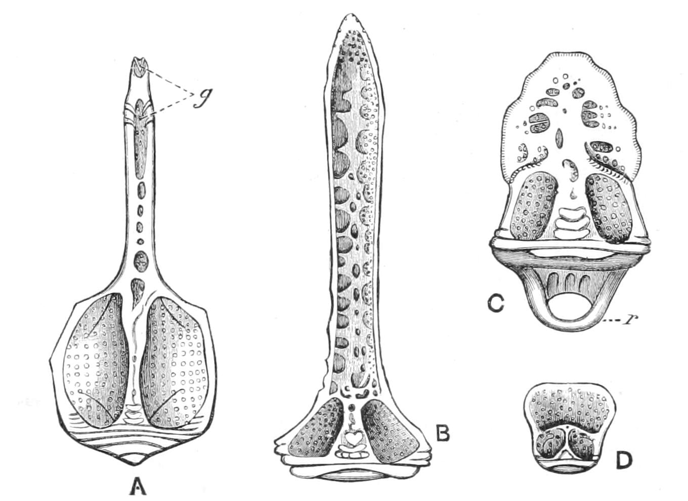
Fig. 226.—Views of a single blade of each kind of pedicellaria. A, blade of gemmiform pedicellaria of Echinus elegans; g, groove for duct of poison gland; B, blade of tridactyle pedicellaria of the same species; C, blade of ophicephalous pedicellaria of the same species; r, ring for clamping this blade to the other blades; D, blade of trifoliate pedicellaria of E. alexandri. (After Mortensen.)
The ophicephalous pedicellariae, with their powerful bull-dog grip, assist in holding small animals, such as Crustacea, till the tube-feet can reach them and convey them to the mouth.
The number and variety of the pedicellariae, then, is an eloquent testimony to the dangers to which the soft sensitive skins of the Sea-urchin and other Echinodermata are exposed, and afford confirmatory evidence in support of the view expressed above, that the method adopted to defend the skin was one of the great determining features which led to the division of the Asteroidea into different races.
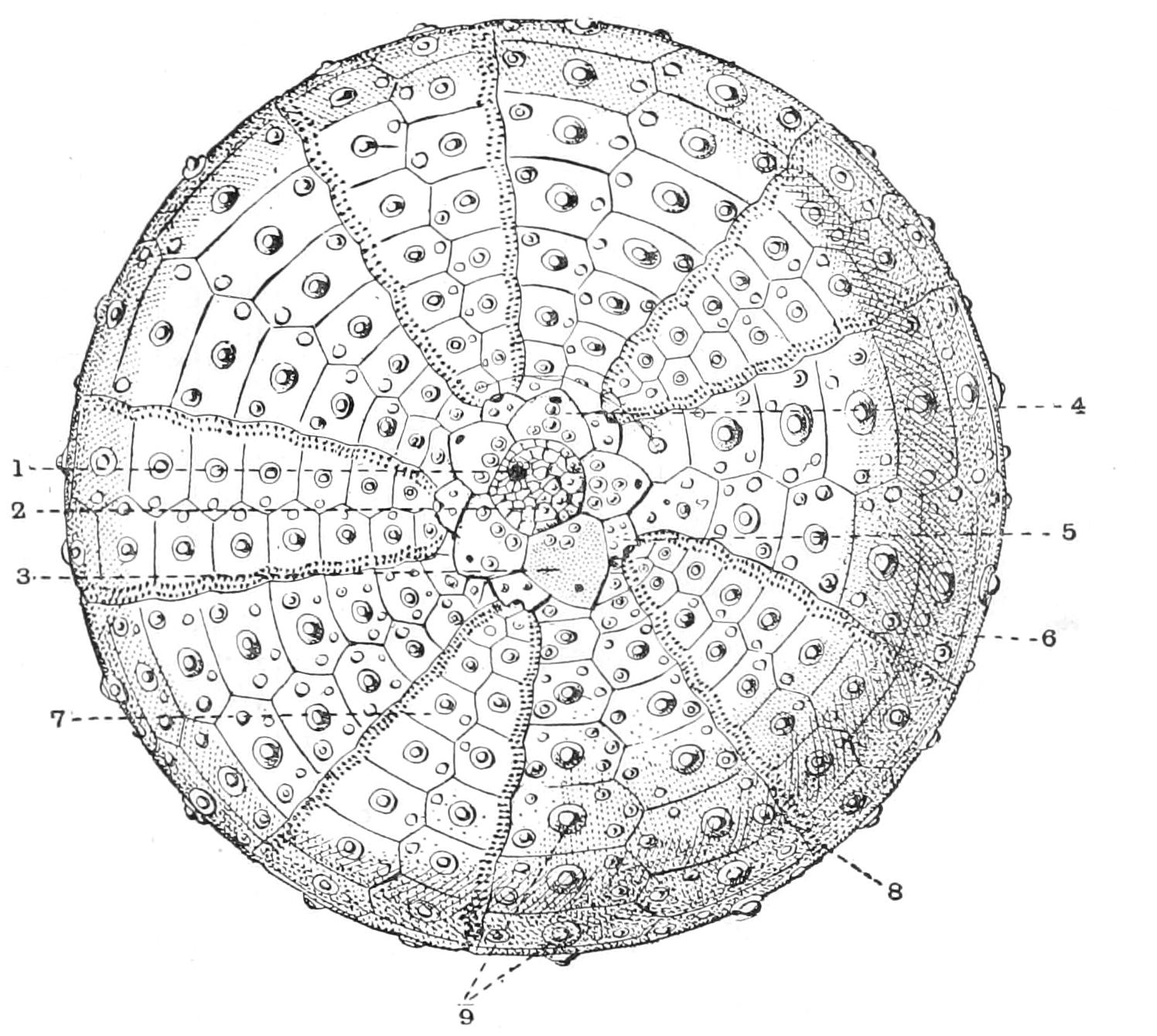
Fig. 227.—Dried shell of Echinus esculentus, showing the arrangement of the plates of the corona. × 1. 1, The anus; 2, periproct, with irregular plates; 3, the madreporite; 4, one of the other genital plates; 5, an ocular plate; 6, an interambulacral plate; 7, an ambulacral plate; 8, pores for protrusion of the tube-feet; 9, tubercles of the primary spines, i.e. primary tubercles.
The corona consists of five radial or "ambulacral" bands of plates and five interradial, or as they are usually termed, "interambulacral" bands of plates—ten in all. Each of the ten consists of two vertical rows of plates throughout most of its extent, and each plate is studded with large bosses, or "primary tubercles" for the primary spines, smaller bosses called "secondary tubercles" for the secondary spines, and finally, minute elevations called "miliary tubercles" for the pedicellariae.
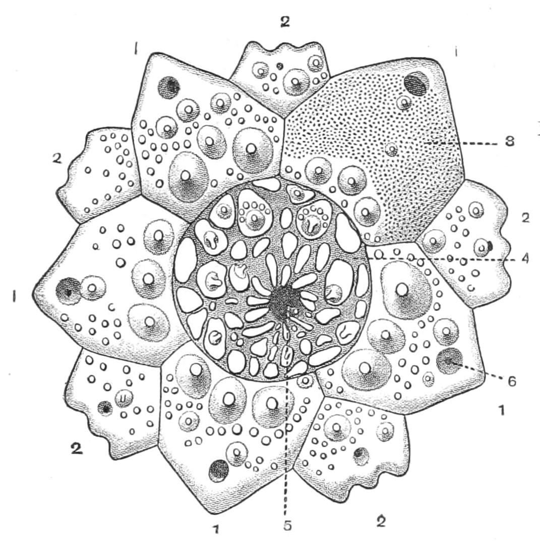
Fig. 228.—The so-called calyx and the periproct of Echinus esculentus. × 4. 1, Genital plates with genital pores; 2, ocular plates with pores for terminal tentacles of the radial water-vascular canals; 3, madreporite; 4, periproct with irregular plates; 5, anus. (After Chadwick.)
Even in the dried skeleton, however, the ambulacral plates can be discriminated from the interambulacral by the presence of pores to permit the passage of the tube-feet. These pores are arranged in pairs, and each pair corresponds to a single tube-foot, since the canal connecting the ampulla with the external portion of the tube-foot is double in the Echinoidea. In Echinus esculentus there are three pairs of such pores in each plate, in Strongylocentrotus droëbachiensis four pairs. The ambulacral plate is really made up of a series of "pore-plates," each carrying a single pair of pores, and these become united in threes in Echinus and fours in Strongylocentrotus, while in primitive forms like the Cidaridae they remain separate. Each ambulacral and interambulacral area ends at the edge of the periproct with a single plate. The plate terminating the ambulacral band is pierced by a single pore for the exit of the median tentacle, which, as in Asteroids, terminates the radial water-vascular canal. Thus the aboral end of the radius in an Echinoid corresponds to the tip of the arm in an Asteroid. The plate is termed "ocular," because the terminal tentacle has a mass of pigmented cells at its base; but no eye-cups can be seen, and there is no evidence that this spot is specially sensitive to light. Species which show special sensitiveness to light have often a large number of what we may perhaps term secondary eyes. The plate terminating the interambulacral series is termed the "genital plate," because it is pierced by the duct of one of the five genital organs. One of the genital plates is also pierced by the madreporic pores. Some zoologists have separated the ocular and the genital plates under the name of "calyx" from the rest of the corona, under a mistaken idea that they are homologous with the plates of the body or calyx of a Crinoid.
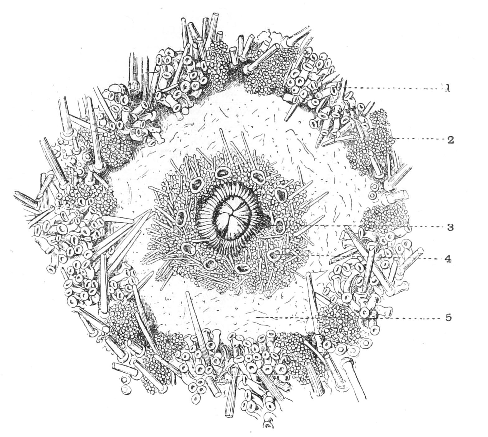
Fig. 229.—The peristome of Echinus esculentus. × 2. 1, Tube-feet of the lower ends of the radii; 2, gill; 3, teeth; 4, buccal tube-foot; 5, smooth peristomial membrane. (After Kükenthal.)
The periproct (Fig. 228, 4) is covered with small plates and bears a few pedicellariae. The peristome (Fig. 229) is covered by flexible skin with abundant pedicellariae; it terminates in a thick lip surrounding the mouth, from which the tips of five white teeth are just seen projecting. There are ten short tube-feet projecting from the peristome—one pair in each radius—and each tube-foot terminates in an oval disc and is capable of little extension, and each has around its base a little plate. The presence of these tube-feet shows that in Echinus the peristome extends outwards beyond the water-vascular ring, whereas in Asteroidea it is contained entirely within the ring. In the primitive Cidaridae (Fig. 235) the whole peristome down to the lip surrounding the mouth is covered with a series of ambulacral and interambulacral plates similar to those forming the corona, though smaller and not immovably united, and the series of tube-feet is continued on to it. It is thus evident that the peristome is merely part of the corona, which has become movable so as to permit of the extension of the teeth. In Echinus the peristome is continued in each interradius into two branched outgrowths called gills, the relation of which to the respiratory function will be described later. These gills (Fig. 229, 2) are situated in indentations of the edge of the corona called "gill-clefts" (Fig. 230, g).
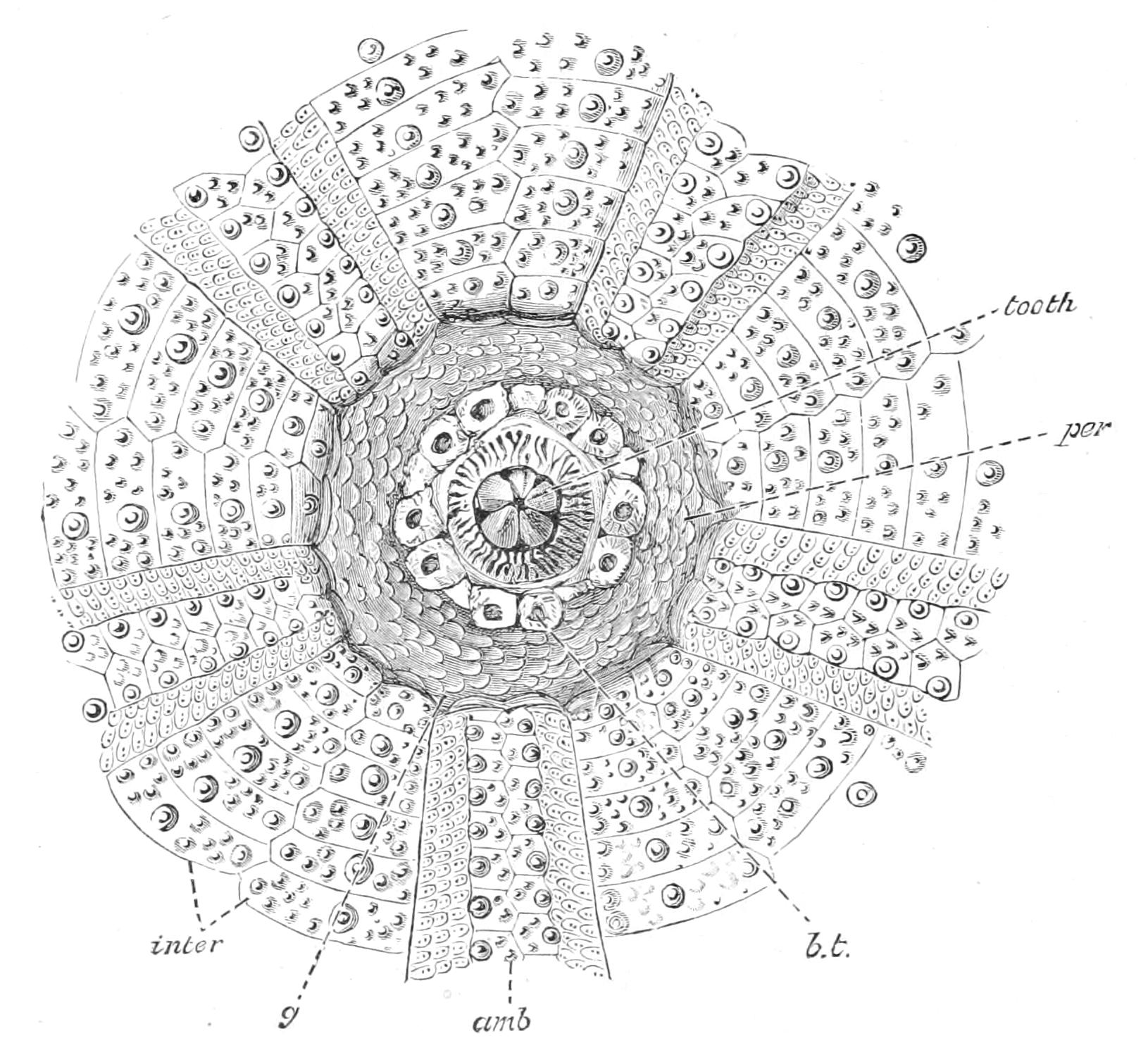
Fig. 230.—The dried peristome of Echinus esculentus and the surrounding portions of the corona. × 1. amb, Ambulacral plate; b.t, buccal tube-foot; g, gill-cleft; inter, interambulacrum; per, peristome.
The most conspicuous plates in the peristome are those surrounding the buccal tube-feet; besides these, however, there are in Echinus esculentus, and probably in most species, a large number of thinner irregularly-scattered plates (Fig. 230).
The term ambulacral plate, applied to the plate pierced by the pores for the tube-feet, conveys a misleading comparison with the ambulacral plate of an Asteroid. In Echinoids the ambulacral groove has become converted into a canal called the "epineural canal," and the ambulacral plates form the floor, not the roof, of this canal; they may perhaps correspond with the adambulacral plates of the Starfish, which one may imagine to have become continually approximated as the groove became narrower until they met.
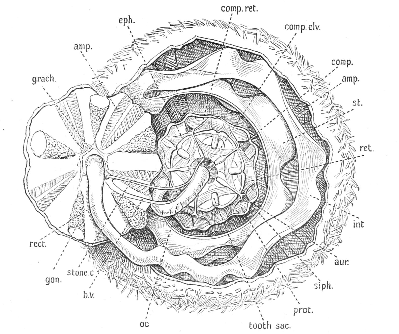
Fig. 231.—Dissection of Echinus esculentus. × 1. The animal has been opened by a circumferential cut separating a small piece of the skeleton at the aboral end, which is turned outwards exposing the viscera on its inner surface. The other viscera are seen through the hole thus made. amp, Ampullae of the tube-feet; aur, auricle; b.v, so-called "dorsal blood-vessel"; comp, "compasses" of Aristotle's lantern, often termed "radii" by English authors; comp.elv, elevator muscles of the compasses; comp.ret, retractor muscles of the compasses; eph, epiphyses of the jaws in Aristotle's lantern; gon, gonad; g.rach, genital rachis; int, intestine; oe, oesophagus; prot, protractor of Aristotle's lantern; rect, rectum; ret, retractor of Aristotle's lantern; siph, siphon; st, stomach; stone.c, stone-canal.
The internal organs of the Urchin can best be examined by making a horizontal incision about one-third the distance from the mouth and pulling the two parts gently asunder. A large amount of fluid escapes from the exceedingly spacious coelomic cavity, the alimentary canal being comparatively narrow.
The alimentary canal commences with a short vertical tube which has been shown to be a stomodaeum; this is surrounded by the upper ends of the teeth and their supporting ossicles, which are collectively termed "Aristotle's lantern." The oesophagus leads into a baggy, flattened tube, the stomach, which runs horizontally round the animal, supported by strings of tissue from the coelomic wall, so that it hangs down in a series of festoons. Having encircled the animal, it bends directly back on itself and immediately opens into the intestine, which is also a flattened tube, which runs round the circumference of the animal, but in the opposite direction, the festoons of the second circle alternating with those of the first. The intestine opens into a short rectum which ascends vertically to open by the anus. The stomach is accompanied by a small cylindrical tube called the "siphon" (Fig. 231, siph), which opens into it at both ends; this represents merely a gutter which has been completely grooved off from the main intestine; it is lined by cilia, and its function is believed to be that of keeping a stream of fresh water flowing through the gut, so as to subserve respiration.
Echinus esculentus seems to feed chiefly on the brown fronds of Laminaria and the small animals found thereon, which it chews up with its teeth, but it may regale itself on the same diet as Brittle Stars, as Allen[474] has shown to be the case in Plymouth Sound. Dohrn[475] has described the Neapolitan Sphaerechinus granularis attacking and capturing Crustacea such as Squilla.
The water-vascular system presents several features of great interest. The ring-canal is situated at a considerable distance above the nerve-ring, and is separated from it by the whole of the jaws and teeth. It has five small interradial pouches on it, which apparently correspond to Tiedemann's bodies in an Asteroid. The stone-canal (Fig. 231) opens as usual into the ring-canal, and is accompanied by the axial sinus and genital stolon. The name "stone-canal" is very unsuitable in this order, for there are no calcifications in its walls; it is a simple membranous tube of circular section. On reaching the upper wall of the test it expands into an ampulla, into which the numerous ciliated pore-canals traversing the madreporite open. The radial canals, starting from the ring-canal, pursue a downward course till they come into contact with the radial nerve-cords, and they then bend upwards and run along the centre of the ambulacral region, finally terminating in the small terminal tentacles. In the just metamorphosed Echinoid these are well-developed tube-feet, each with a well-developed sucker, in the centre of which is a conical sensory prominence, but as development proceeds they become enclosed in a circular outgrowth of the test, so that only the tip projects in the adult.
The long extensible tube-feet are connected by transverse canals with the radial canal. Instead of the pair of valves which in Asteroids prevent the reflux of liquid into the canal, there is a perforated diaphragm[476] with circular muscles, which by contraction close the opening in the diaphragm, while when they are relaxed fluid can return from the tube-foot. The ampulla is flattened, and is contracted by muscular fibres called "trabeculae" stretching across its cavity. These muscular strands are developed by the cells lining the ampulla. The external portion of the tube-foot, as in Asteroids, is provided with powerful longitudinal muscles, and there is the same alternate filling and emptying of the ampulla as the tube-foot is contracted and expanded. The tube-foot is connected by a double canal with the ampulla, the object of which is to assist in respiration. The cells lining it are ciliated, and produce a current up one side of the tube-foot and down the other, and the double canal leading to the ampulla separates these two currents and prevents them interfering with one another. Thus water is continually transported from the ampulla to the tube-foot, through the thin walls of which it absorbs oxygen, and it is then carried back to the ampulla, and transfers its oxygen to the fluid of the general body-cavity through the walls of the ampulla. The disc of the tube-foot is supported by a calcareous plate (Fig. 232, oss), a circumstance which enabled Johannes Müller to recognise the Echinoid larva when the form of the adult was as yet unrecognisable. Below the edge of the disc there is a well-marked nerve-ring, from which two bundles of nerve-fibres go to the disc itself, in the edge of which there is an abundance of sense-cells.
The buccal tube-feet (Fig. 229, 4) are much shorter than the rest, and are provided with oval discs which are highly sensory. These feet are not used for seizing, but for tasting food; when a piece of food is placed near them they are thrown into the most violent agitation.
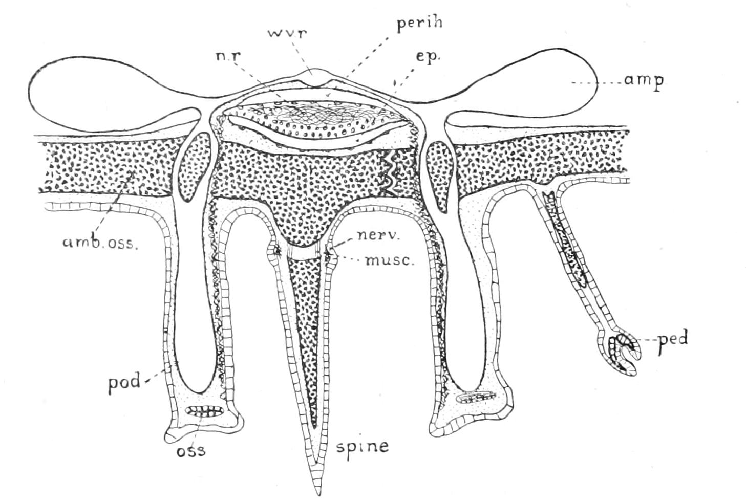
Fig. 232.—Diagrammatic transverse section of the radius of an Echinoid. amb.oss, Ambulacral ossicle; amp, ampulla of the tube-foot; ep, epineural canal; musc, muscles attaching spine to its boss; nerv, nervous ring in base of spine; n.r, radial nerve-cord; oss, ossicle in sucker of tube-foot; ped, tridactyle pedicellaria; perih, radial perihaemal canal; pod, tube-foot; wv.r, radial water-vascular canal.
The nervous system has the same form as in an Asteroid, viz. that of a ring surrounding the mouth and giving off radial nerve-cords (Fig. 232, n.r), one of which accompanies each water-vascular canal to the terminal tentacle, where it forms a nervous cushion in which pigmented cells are embedded.
A large band-like nerve is given off from the radial nerve-cord to each tube-foot. This pedal nerve, as it is called, contains bipolar neurons, and is really an extension of the nerve-cord itself. Beneath the sucker it branches out to form a sensory ring. From the base of the pedal nerve, branches are given off which run to the ectoderm and enter into connexion with the plexus there. Romanes[477] scraped away the radial cords and found that the spines still converged when a point on the ectoderm was stimulated, but that, on the other hand, if definite locomotor movements were to be carried out, the presence of these cords was a necessity; hence he concluded that the superficial plexus sufficed for ordinary reflexes, but that for purposeful movements the central nervous system was necessary.
Von Uexküll[478] has made an exhaustive study of the physiology of the nervous system in the Echinoidea. He points out that all the organs controlled by the nervous system, spines, pedicellariae, tube-feet, and (see below) Aristotle's lantern, give two opposite reactions in response to the same stimulus according as it is strong or weak, bending away from the point of stimulation when it is strong and towards it when it is weak. This reversal of reaction can only be due to the action of the neuron in altering the effect of the stimulus on the muscles, and this Uexküll regards as its fundamental property. Thus in Preyer's[479] experiments with Starfish the strong form of stimulation is obtained by directly applying the stimulus to the radial cord or to the tube-feet, the weak form by stimulating the back, when of course the stimulus has to traverse a longer path before affecting the tube-feet, and is consequently weakened. Von Uexküll also introduces the conception of "tone" with regard to the nervous system. This term has been used to denote the amount of chronic contraction in a muscle, and it is to be distinguished from the fleeting contractions which cause movement. The more tone there is in a muscle the less responsive it is to stimuli tending to bring about movement. As applied to the nervous system "tone" denotes a condition when it is not receptive to small stimuli, but when it is maintaining a condition of tone in a muscle by which of course its own tone is measured. Tone in a neuron can therefore be measured by the produced tone in the muscle, and the one is to be discriminated from the other only by using stimulants, such as caffeine, which have no direct action on muscle. Tone can also be measured by the amount of stimulus necessary to irritate the neuron. When muscles are stretched the tone is lowered, and this loss of tone extends to the neuron controlling the muscle, and vice versa. When the spines on being gently stimulated bend towards the point of stimulation, this is due to the contraction of the muscles on the side towards the point of stimulus, for if the superficial plexus of nerve-fibres be cut through so that the stimulus has to pursue a round-about course the spine will bend towards the direction from which the stimulus comes. The bending of the spines away from the stronger stimulus is likewise due to the muscles on the side towards the stimulus. It is caused by a sudden fall of tone in these muscles, which causes them to yield to the tone of the muscles on the opposite side, and this fall of tone is due to a fall of tone in the neurons, for it can be produced by chemicals, and the direct action of all chemicals applied to muscle is to raise tone.
In Arbacia this form of reaction cannot be produced; the spines respond to stimuli of all degrees of intensity by convergence towards the point of stimulation.
When a general skin-irritant like dilute acetic acid, or even strong light, is applied to the skin of a Sea-urchin the spines bend alternately to all points of the compass, or, in a word, rotate. This is due to the fact that the weight of the inclined spine stretches the muscles of one side and so renders them more open to the general stimulus; these muscles in consequence, contract, and so move the spine to a new position in which other muscles are stretched, and a similar result follows. A continuation of this process brings about rotation.
When a piece of glass rod or other light object is laid on the spines of a Sea-urchin, it naturally, by its weight, presses asunder the spines and stretches their muscles on one side, thus lowering the tone. If now the skin be stimulated at any point the piece of rod will be rolled by the spines towards the point of stimulation. This is caused by the fact that the muscles of the spines holding the rod are made more receptive by being stretched, and therefore they contract more than do the others in response to the stimulation, and so the rod is rolled onwards on to the next spines, which then act in the same manner. This passage of stimulus is entirely independent of direct nervous connexion between the bases of the spines, for it will traverse at right angles a crack going clean through the shell; it is merely the result of the mechanical weight of the object and of the juxtaposition of the spines.
If the stimulation be too violent the first spines affected diverge wildly and strike their neighbours with vehemence, so arousing into activity the block musculature of these. This causes them to stand rigidly up, and so the path of the stimulus is barred.
Now the escape movements of the animal under strong stimulation which Romanes[480] alludes to are just an example of this handing on of stimulation from spine to spine, not by nervous connexion but by mechanical touch only; the object in this case is the substratum on which the animal lies, which is, so to speak, rolled towards the point of stimulation, or putting it otherwise, the animal is rolled away from it. Righting when upset is another example of the same phenomenon; the aboral spines are stretched by the weight of the animal, and the animal acts as if it were stimulated in the region of the periproct. When a Sea-urchin is in its normal position and is stimulated in the periproct (as for instance by a strong light), it would, according to this rule, tend to move downwards, which is of course impossible; but as the stimulus never affects all sides quite alike the result is that the Urchin rotates, turning itself ever away from the point of strongest stimulation. In the case of Strongylocentrotus lividus when living on limestone, as on the west coast of Ireland, this results in the animal excavating for itself holes in the rock, where it is safe from the action of the breakers.[481]
But it may be objected that no account is taken in the above description of the action of the "central nervous system," i.e. of the ring and the radial cords, and yet Romanes found that when they were removed the escape movements could not be carried out. The answer is that the central nervous system is a store-house of tone, not, as in higher animals, a controlling centre for co-ordinating the movements of the spines. When it is removed at first the escape movements can be carried out, but in a day or two all tone in the spine-muscles is lost, and then, since the tone of all is equally low, there is no tendency in those that are stretched to be more responsive than others, and hence the escape movements cannot be carried out. Sea-urchins kept in the tanks of an aquarium are apt to lose the tone of their spines owing to the poisoning of the nervous system.
The central nervous system is, however, the system which controls the movements of the tube-feet. As we have seen, extensions of the radial nerves run to the tip of each podium. Tube-feet are chiefly used in ordinary progression; when this is quickened the spines come into play exclusively. The extent to which these two organs of locomotion are used varies from genus to genus. Thus Centrostephanus uses its spines a good deal, Echinus and Strongylocentrotus very little. The last-named genus sometimes walks on its tube-feet entirely without touching the ground with its spines.
The faculty of vision in its simplest form may be defined as sensitiveness to light and shade. Now strong light acts on all Sea-urchins as a general skin irritant. They fly from it towards the darkest corner, and then if it continues the spines rotate. A number of little violet spines on the aboral pole of Centrostephanus longispinosus are especially sensitive to light, and hence are almost constantly in rotation. This is due, according to Uexküll,[482] to a pigment of a purple colour, which can be extracted by means of alcohol and which is decomposed by light, the products of decomposition being supposed to irritate the nerves. Centrostephanus when exposed to light becomes darker in colour. This is due to the migration outwards of amoebocytes, which carry a pigment which acts as a screen in order to prevent the valuable visual purple being too rapidly decomposed. Not all Sea-urchins, in fact very few of those living in northern waters, give a reaction to shadow. C. longispinosus is one of the few; it reacts to a shadow by converging its spines towards it. A much larger number of species inhabiting tropical waters show this reaction. It is entirely stopped if the radial nerve-cords be removed, whereas the reaction to strong light continues. The reaction to shade is strongest after a long previous exposure to light, hence Uexküll has given the following explanation of it. The continued irritation due to light, having spread to all the spines, eventually reaches the radial cords and is there stored in the bipolar nerve-cells as tone. When the light-stimulus is interrupted some of the stored tone spreads upwards to the spines, causing the weak form of spine reaction, and the spines converge.
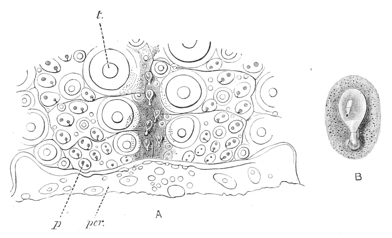
Fig. 233.—To show character and distribution of the sphaeridia in Strongylocentrotus droëbachiensis. A, a portion of a radius, with sphaeridia, and the adjoining edge of the peristome. p, Pair of pores for a tube-foot; per, peristome; t, primary tubercle. B, an isolated sphaeridium. (After Lovén.)
It will be seen therefore that the so-called central nervous system of Echinus does not act in any sense as a brain, as indeed might have been guessed from the absence of any differentiation in it. As Uexküll points out, when an animal is covered all over with similar organs, such as spines and pedicellariae, capable of acting automatically, a brain is not needed. The object of a brain is to direct organs which are in a certain place to a danger which may come from any quarter, but in the Sea-urchin any spine is as good as any other spine, and such orientation is not needed. "In a dog the animal moves its legs, in a Sea-urchin the legs move the animal." What the Sea-urchin does need is a means to prevent its pedicellariae attacking its own organs with which they may come into contact. Thus it possesses an "autodermin," a chemical contained in the ectoderm which paralyses the muscles of the pedicellariae, as may be seen by offering to them a spine of the same animal. If, however, the spine be treated with boiling water, and then offered, it is viciously seized, showing that this substance can be dissolved out.
Just as in the case of the Starfish, when the nerve-ring is cut through, the tube-feet in the various radii are no longer co-ordinated with one another.
Besides the tips of the tube-feet the Urchin possesses another kind of sense-organ, the sphaeridia (Fig. 233). These are minute glassy spheres of calcareous matter attached by connective tissue to equally minute bosses on the plates of the ambulacra, generally near the middle line. They are in fact diminutive spines, and like the latter are covered with a thick layer of ectoderm, beneath which is a particularly well-developed cushion of nerve-fibrils. Only the layer of muscles which connects a normal spine with its boss is wanting. Although definite experimental proof is lacking, the whole structure of the sphaeridia shows that they belong to the category of "balancing organs." As the animal sways from side to side climbing over uneven ground, the heavier head of the sphaeridia will incline more to one side or to another, and thus exercise a strain on different parts of the sheath, and in this way the animal learns its position with regard to the vertical.
Intervening between the radial nerve-cord and the radial vessel is a single radial perihaemal canal (Fig. 232, perih), representing the two parallel canals found in the same position in the Asteroid. The five perihaemal canals lead downwards to a space called the lantern-coelom, surrounding the oesophagus.[483] Since the skeleton of the corona is composed of plates immovably connected together, muscles corresponding to the ambulacral muscles of the Asteroids would be useless, and so the wall of the perihaemal canal remains thin and the side of it turned towards the general coelom develops no muscles, and that turned towards the nerve-cord no nerve-cells. Where, however, the radial nerve enters the nerve-ring, and on the ring itself, an inner layer of nerve-cells is developed from the lantern-coelom which represents the lower or oral portions of the radial perihaemal canals. These cells control the muscles moving the teeth. These canals are originally parts of the lantern-coelom, but in the adult they become closed off from it.
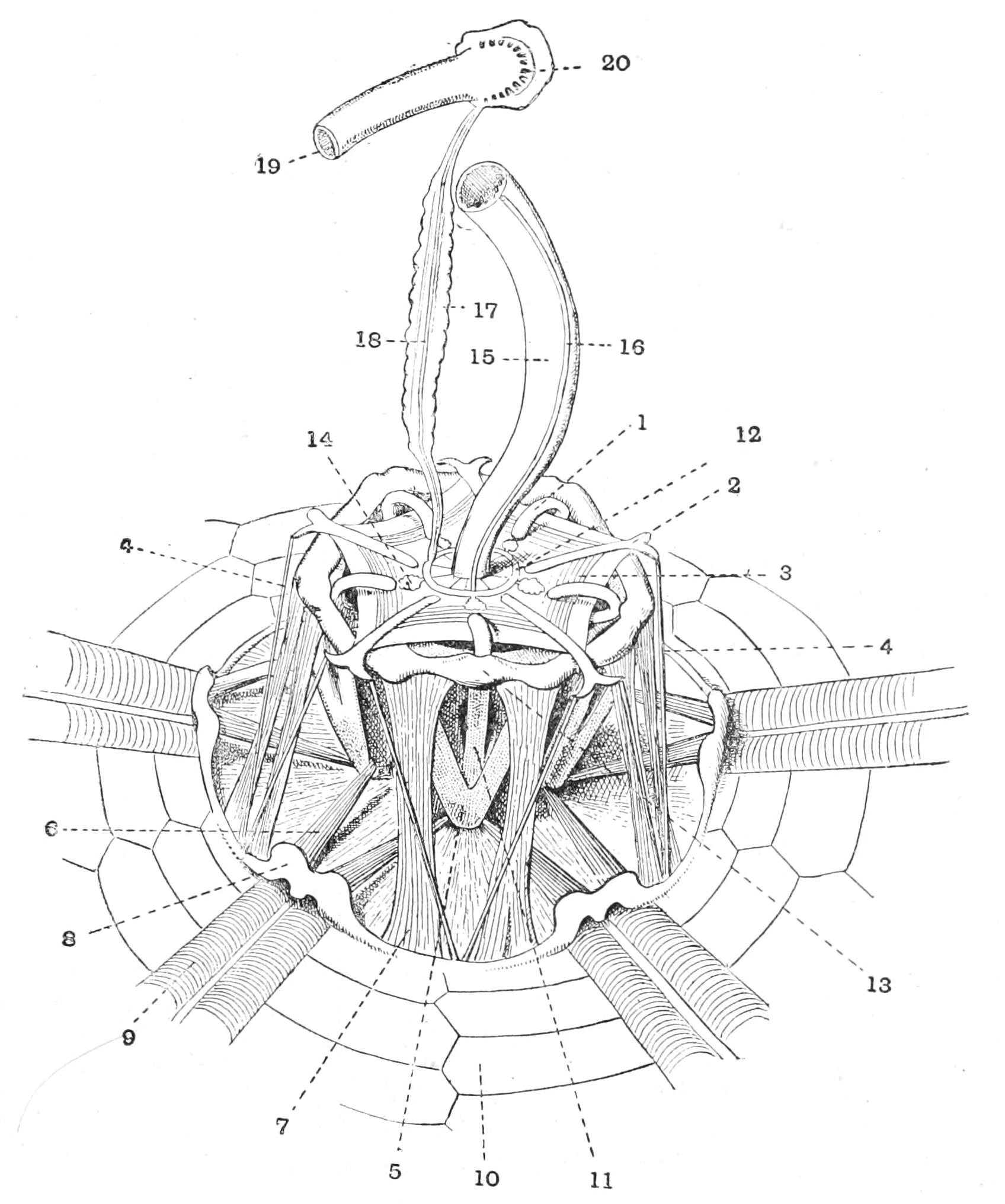
Fig. 234.—Echinus esculentus dissected in order to display Aristotle's lantern, × 2. The whole upper part of the shell has been cut away. 1, Upper growing end of tooth; 2, outer forked end of one "compass"; 3, muscle joining adjacent compasses and acting as elevator of these ossicles; 4, depressor of the compasses; 5, lower end of jaw; 6, retractor of the whole lantern; 7, protractor of the whole lantern; 8, auricle; 9, ampullae of the tube-feet; 10, interambulacral plate; 11, lower part of tooth; 12, water-vascular ring; 13, meeting-point of a pair of epiphyses; 14, so-called Polian vesicle, really equivalent to Tiedemann's body in an Asteroid; 15, oesophagus; 16, so-called ventral blood-vessel; 17, genital stolon; 18, stone-canal; 19, rectum; 20, aboral sinus. (Partly after Chadwick.)
In the outer wall of this space are developed the calcareous rods forming Aristotle's lantern. These are first: five teeth (Fig. 234, 11), chisel-shaped ossicles of peculiarly hard and close-set calcareous matter, the upper ends (1) pushing out projections of the upper wall of the lantern-coelom. These projections are the growing points of the teeth, whose lower ends pierce the ectoderm and project into the lower end of the oesophagus. Each tooth is firmly fixed by a pair of ossicles inclined towards one another like the limbs of a V and meeting below. Each ossicle is called an "alveolus," and taken together they form a "jaw." Their upper ends are connected by a pair of ossicles called "epiphyses" (13). These two epiphyses meet in an arch above. The jaws and their contained teeth are situated interradially. Intervening between successive alveoli are radial pieces called "rotulae," which extend directly inwards towards the oesophagus. Above the rotulae are pieces termed "radii" or "compasses" (2), which are not firmly attached to the other pieces but lie loosely in the flexible roof of the lantern-coelom.
The uses of the various components of this structure can be made out from an inspection of the muscles which connect them together.
Overarching each radial perihaemal canal where it leaves the lantern is a bridge of calcareous matter called the "auricula" (Fig. 234, 8). This arises as two rods which meet each other in a pent-house over the canal. It is the only part of the skeleton which can be compared to the ambulacral ossicles of the Asteroidea, and like them it serves as the point of insertion for important muscles. Thus we find (1) protractor (Fig. 234, 7) muscles which arise from the upper ends of the alveoli and are inserted in the auricula; when these contract they tend to push the whole "lantern" outwards so as to expose the tips of the teeth. (2) The retractor muscles (Fig. 234, 6) extend from the auriculae to the lower ends of the jaws and restore the lantern when it has been extruded to its original position. (3) The comminator muscles connect adjacent jaws with one another: these on contraction approximate the pair of jaws into which they are inserted, and it will easily be seen that by the successive contraction of the five comminator muscles a rotating movement of the teeth would be produced which would cause them to exert an action something like that of an auger; by their simultaneous contraction the teeth are brought to a point. (4) The internal and external rotula muscles: these are small muscles which connect the outer side of the epiphysis with the rotula. There are two facets on the epiphysis, which permit it to rock to and fro on the rotula under the action of these muscles. This rocking action must greatly increase the cutting power of the tooth. These muscles are controlled by the nerve-ring and the incipient portions of the radial nerves, which, as we have seen, have an inner layer of nerve-cells. If the nerve-ring be gently stimulated on one side the upper end of the lantern bends away from the spot, causing the lower end, i.e., the teeth, to move towards it; but a stronger stimulation produces the opposite effect, just as is the case with spines. But besides these masticatory muscles there are others which have nothing to do with moving the teeth. These muscles are attached to the rods called radii or compasses (Fig. 234, 2),[484] which lie in the upper wall of the lantern-coelom, and may be termed the compass muscles. There are two sets:—(1) The elevator muscles (Fig. 234, 3), which connect the inner ends of the compasses with one another. When these contract, the radii tend to bend upwards at the inner ends and thus raise the roof of the coelom. (2) The depressor muscles (Fig. 234, 4), which run downwards from the forked outer ends of the compasses to the auriculae. Uexküll[485] has shown that the function of these muscles and of the rods to which they are attached is respiratory. These muscles are also controlled by the nerve-ring. If this be stimulated by passing a pin-head into the oesophagus, the roof of the lantern cavity is raised by the contraction of the elevator muscles. This is followed by contraction of the depressor muscles lowering it; the same result may be brought about by placing the animal in water with excess of carbonic acid. The ten branched gills described on p. 514 are outgrowths of the lantern-coelom. When the roof of this cavity is depressed the fluid contents are driven out into the gills, which are thus expanded and then absorb oxygen from the surrounding sea water. When, on the other hand, the roof is raised the aerated water is sucked back into the lantern cavity, and the oxygen passes easily through the thin walls of the lantern into the fluid filling the main coelomic cavity. There are thus two independent respiratory mechanisms in the Sea-urchin, the one being the compass muscles, the other the cilia lining the interior of the tube-feet.
The function of excretion is performed, as in Asteroidea, by the amoebocytes floating in the general coelomic cavity. These in part escape through the thin bases of the gills. In other parts of the body they seem not to succeed in reaching the exterior at all, but to degenerate and to form masses of pigment; the colour of the animal is largely due to these excrementitious substances.
The reproductive system, as in the two preceding orders, consists of a vertical pillar, the "genital stolon," and a circular "genital rachis" giving off interradial branches from which the genital organs bud. The genital stolon is developed from the wall of the general coelom near the upper end of the axial sinus; it attains a great development and ultimately completely surrounds the axial sinus, which then appears like the cavity of a glandular tube, the walls of which are constituted by the genital stolon. The compound structure consisting of stolon and axial sinus was actually described as a nephridium by the Sarasins[486] in the case of Asthenosoma. Its true nature, however, is shown when the upper end is examined; it is then seen to open into the stone-canal and to be in communication with the ampulla, into which the pore-canals open. Lying alongside the upper end of the axial sinus is the somewhat elongated "madreporic vesicle," or right hydrocoele, which was described by Sarasin as the accessory kidney (Nebenniere), since like the axial sinus it is partly enveloped by the genital stolon. Leipoldt,[487] however, showed clearly that it is a completely closed space.
The genital rachis springs from the upper end of the stolon, and as in Asteroids, it lies in the outer wall of a space called the "aboral sinus" (Fig. 234, 20) intervening between it and the test. In adult specimens it seems to degenerate. The genital organs are situated at the ends of five interradial branches of the rachis (Fig. 231, gon). Each is an immense tree-like structure consisting of branching tubes, which are lined by the sexual cells. So enormous do they become in the breeding season that they form an article of food among fishermen. The term esculentus is derived from this circumstance. Other species are regularly sold for food as Frutta di Mare (Fruit of the Sea) at Naples, and as "sea eggs" in the West Indian Islands. One female Echinus esculentus will produce 20,000,000 eggs in a season.
The so-called blood system is more distinctly developed in Echinoidea than in Asteroidea and Ophiuroidea. There is an oral ring of lymphoid tissue surrounding the oesophagus below the water-vascular ring. From this are given off two strands, the so-called "dorsal" (Fig. 231, b.v), and "ventral" vessels (Fig. 234, 16), which run along the two opposite sides of the stomach or first coil of the alimentary canal. The position of these strands suggests that like the lacteals of the human intestine they are channels along which the products of digestion exude from the stomach. The dorsal strand is situated on the same side as the genital stolon, and from it branches are given off which ramify on the surface of the stolon, on account of which this organ, as in Asteroidea, was at one time regarded as a "heart," but the distinction of the stolon from the strands is easily made out. An aboral ring enclosing the genital rachis lies embedded in the septum dividing the aboral sinus (Fig. 234, 20) from the general coelom.
Classification of Echinoidea.
The Echinoidea are sharply divided into three main orders, which differ from each other profoundly in their habits and structure. These are: (1) The Endocyclica or Regular Urchins, of which the species just described may be taken as the type. (2) The Clypeastroidea or Cake-urchins, which are of extremely flattened form, and in which the periproct is shifted from the apical pole so that it is no longer surrounded by the genital plates, while some of the tube-feet of the dorsal surface are flattened so as to serve as gills. (3) The Spatangoidea or Heart-urchins, in which the outline is oval: the periproct is shifted, as in the Cake-urchins, and the dorsal tube-feet are similarly modified; but the Heart-urchins have totally lost Aristotle's lantern, whilst the Cake-urchins have retained it. This strongly-marked cleavage of the group was primarily due, as in all such cases, to the adoption of different habits by different members of the same group. Were we to term the three orders Rock-urchins, Sand-urchins, and Burrowing-urchins, it would not be entirely true, for secondary invasions of the other's territory on the part of each order have undoubtedly taken place; but still the statement would remain roughly true, and would give a fair idea of the differences in habitat which have led to the differentiation of the group.
Order I. Endocyclica (Regular Urchins).
The principal variations concern (1) the peristome, (2) the periproct, (3) the corona, (4) Aristotle's lantern and its appendages, (5) the spines, (6) the pedicellariae, and lastly, (7) the tube-feet. We shall consider these points in order.
Peristome.—In the vast majority of species this region is covered only with flexible skin in which ten small plates are embedded, pierced by pores for the buccal tube-feet; besides these there are irregularly arranged thin plates. In the Cidaridae both the ambulacral and the interambulacral series of plates are continued on it; these plates differ from those of the corona in being movable on one another. In Echinothuriidae only the ambulacral series of plates is continued on to the peristome. In the case of both these families there are a considerable number of tube-feet within the region of the peristome which may be classed as buccal.
Periproct.—This area, which represents the whole dorsal surface of Asteroidea, is very large in the Cidaridae, where, as in Echinus, it is covered with leathery skin in which small plates are embedded. In the Saleniidae it is covered with a single large sur-anal plate, in the edge of which the anus is excavated; in the Arbaciidae it is covered with four valve-like plates; whilst in the remaining species its condition is similar to that described in the case of Echinus esculentus.
Corona.—In Echinothuriidae all the plates are separated by slips of membranous skin, so that the test is flexible. In all other families it is an unyielding cuirass. In the Cidaridae the pore-plates remain separate throughout life, and are therefore identical with the ambulacral plates. These are small and placed in two vertical rows, and so the ambulacra are exceedingly narrow. In Echinothuriidae there is some tendency to adhesion amongst the pore-plates; these are of different sizes, and usually one larger and one smaller adhere to one another. In all other species regular ambulacral plates are formed at least in the lower part of the radii near the peristome by the adhesion of the pore-plates in groups of two, three, or more. Sometimes as many as nine pore-plates may thus adhere.
When adhesion takes place between the pore-plates it is of course preceded by crowding, and this interferes with their equal development. Some which extend so far horizontally as to meet their fellows of the opposite side of the radius are called primary plates; others which are small and wedged in between the larger ones are called demi-plates. Systems of classification have been built up (chiefly by palaeontologists) in which great stress has been laid on how the primaries and secondaries enter into the constitution of the compound plate, but it does not seem to the present author as if this were at all a satisfactory basis for classification. All the pore-plates are primarily equivalent, and the question as to which are interfered with in their growth so as to become secondary is trivial. The so-called Arbacioid type consists of one primary with a secondary on each side; the Diadematoid type of three primaries, with occasionally a secondary between the aboral and the middle primary; and finally the Triplechinoid type of two primaries, with one or more secondaries between them.
Aristotle's Lantern.—Under this head we may consider the auriculae and gills as well as the jaws and teeth. In Cidaridae external gills appear to be absent, but from the lantern coelom large radial pouches project upwards into the general coelom cavity. These pouches are supposed to be respiratory, and are termed internal gills or Stewart's organs.[488] They co-exist with external gills in Echinothuriidae and in Diadematidae, though in the last family they are present only in a vestigial form, two being found in each radius. The auricular arch both in Cidaridae and in Arbaciidae is composed of two pillars which do not meet, but in the last-named family they are based, as in Echinidae, generally on the ambulacral plates, whereas in Cidaridae they arise from the interambulacral plates (the ambulacral plates being here very narrow). The epiphyses are absent in Cidaridae and Arbaciidae, and are imperfect in Diadematidae.
Spines.—These organs are extraordinarily variable, and usually differ very much in species of the same genus. In the vast majority of species there is a limited number of long spines called "primaries," amongst the bases of which a large number of much shorter "secondaries" are distributed. In Cidaridae the primaries are very long and thick and blunt at the ends, and the secondaries form small circles around their bases. The primaries in Cidaridae and the tips of the primaries in Arbaciidae and Echinothuriidae are covered with a special investment of extremely close, hard, calcareous matter very different from the loosely fenestrated material out of which the bodies of the spines of all species are composed. In Colobocentrotus and Heterocentrotus the primaries are very thick and triangular in section, whilst the secondaries on the aboral surface have expanded outer ends, which form a close-set pavement protecting the ectoderm from the shocks of the breakers. In Echinothuriidae the primaries are short and so delicate as to be termed silky.
Pedicellariae.—In Cidaridae only gemmiform and tridactyle pedicellariae are found. In the gemmiform the glands lie inside the grooved blades instead of outside as normally, and they are covered internally by ingrowths of calcareous matter from the edges. In Echinothuriidae only tridactyle and trifoliate are found in most species, but rudimentary gemmiform are found in one species and well-developed ophicephalous in another. In some species (Centrostephanus longispinosus) there are found gemmiform pedicellariae which have lost the jaws but retained the glands. These are termed "globiferae." Mortensen[489] uses minute details in the structure of the pedicellariae to discriminate species and even genera, but in this the present author is not prepared to follow him.
Tube-feet.—The tube-feet belonging to the aboral surface are pointed and devoid of a sucker in Diadematidae, Echinothuriidae, Arbaciidae, and Cidaridae;[490] in the last-named family those belonging to the oral surface have suckers, in the centre of which a pointed (sensory) prominence is to be noted.
The classification of the Endocyclica is by no means in a satisfactory condition, and different authorities have arrived at widely different results. Agassiz,[491] for instance, places the genera Echinus (the common British form) and Strongylocentrotus (the commonest American form) in different families. Bell,[492] on the other hand, considers them to be closely allied. Bell's system, based as it is on the development of the peristome, seems to the present author the most justifiable, for the peristome is undoubtedly a differentiation of the corona, which has been brought about by the manner in which the animal breathes and masticates, two functions of prime importance. The periproct is also of importance, representing as it does the whole aboral surface of the Starfish, and so are to a less extent the arrangements of the spines and of the tube-feet. Proceeding in this way, living Endocyclica can be divided into six families, which are briefly described below.
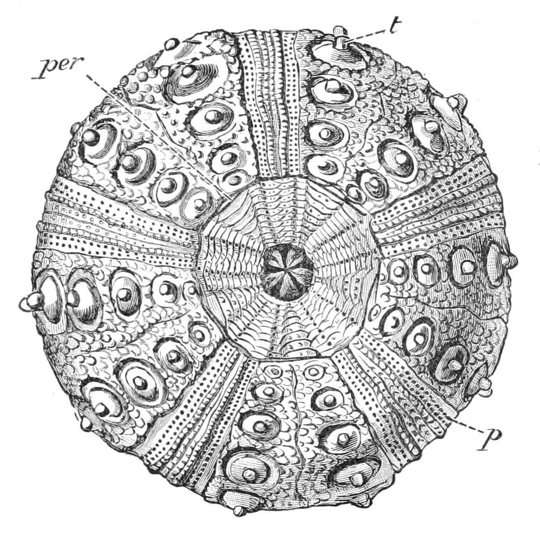
Fig. 235.—Oral view of dried and cleaned test of Cidaris. p, Pores for tube-feet arranged in single series; per, peristome with both ambulacral and interambulacral plates; t, tubercle of a large interambulacral spine.
Fam. 1. Cidaridae.—Endocyclica with a large peristome and a large periproct. The peristome is covered with a regular series of both ambulacral and interambulacral plates, the former pierced by tube-feet. No special buccal tube-feet and no external gills. The periproct is large, and is covered with irregular plates (Fig. 236, A). The lantern coelom is provided with large Stewart's organs.
The auriculae are incomplete and consist only of pillars arising from the interambulacral plates. The ambulacral pore-plates remain disunited, and the pores are arranged in a single vertical series; hence the ambulacra are very narrow. The interambulacral plates each bear one large primary spine surrounded by several circles of secondaries. No ophicephalous or trifoliate pedicellariae are to be found, and the gland of the gemmiform pedicellaria is placed inside the concavity of the blade.
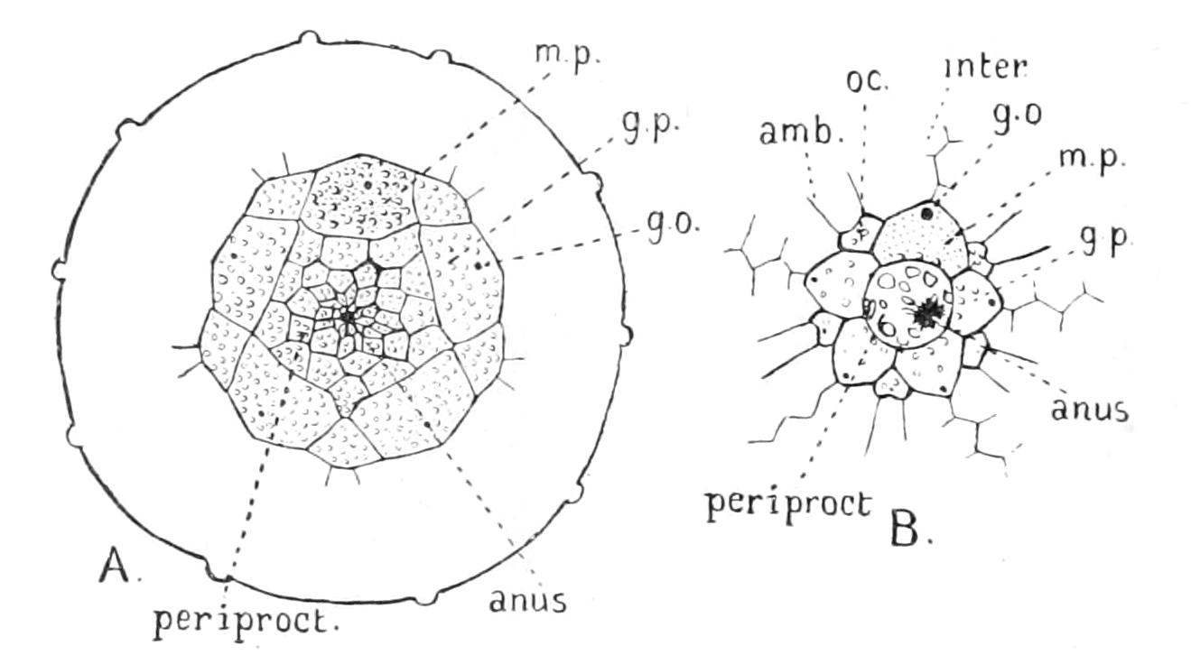
Fig. 236.—Figure showing periprocts of A, Cidaris; B, Echinus, × 1. amb, Ambulacral plate; g.o, genital opening; g.p, genital plate; inter, interambulacral plate; m.p, madreporite; oc, ocular pore.
The Cidaridae are in many respects the most primitive of the six families living. They are distributed all over the world, and chiefly inhabit deep water. No two naturalists agree as to how they are to be divided into genera. Mortensen,[493] who takes the structure of the pedicellariae as his principal guide, recognises fourteen genera. Others (as for instance Bell) have been inclined to attribute nearly all the living species to one polymorphic genus, Cidaris, finding all attempts to divide the genera from one another frustrated by the discovery of transitional forms. Goniocidaris (Fig. 237), however, distinguished by its comparatively broad poriferous zones, by bare places in the middle line of both radii and interradii, and by deep pits on the lines of suture of the plates, is by general consent distinct. This genus is confined to the Eastern Pacific, but from British waters three species of Cidaris have been recorded, only one of which, C. (Dorocidaris) papillata, is at all common. It is found in water from 100 to 500 fathoms in depth off the western coast of Ireland and Scotland. It also occurs in the Mediterranean, and has been carefully examined and described when living by Prouho.[494] From his description it appears that locomotion is effected almost entirely by spines, and that the tube-feet of the lower parts of the radii have each in the centre of the disc a pointed sense-organ like those in the centre of the first tube-feet of the just metamorphosed Echinus, whilst those of the aboral surface have no suckers.
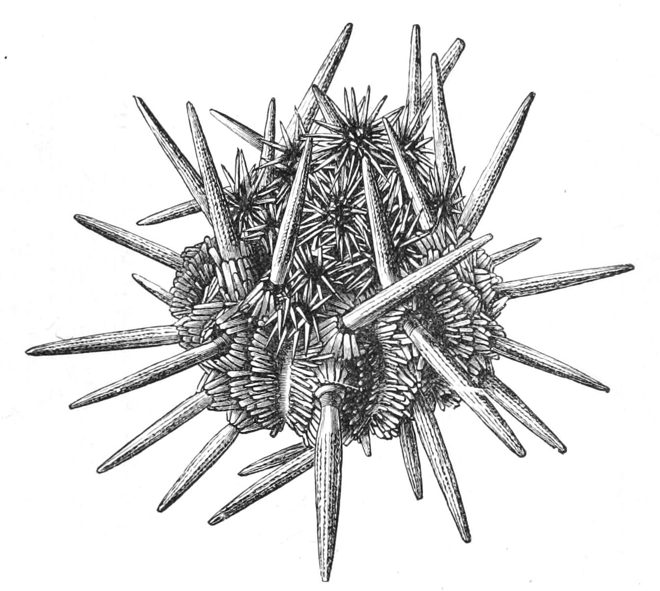
Fig. 237.—Goniocidaris canaliculata. × 2. (From Wyville Thomson.)
Fam. 2. Echinothuriidae.—Endocyclica with a large peristome and comparatively small periproct. The peristome has a regular series of ambulacral plates bearing pores for tube-feet, but no interambulacral plates. No specially modified buccal tube-feet, but external gills are present, and internal gills (Stewart's organs) also occur. The periproct is covered with numerous small plates. All the plates of the corona are separated by thin slips of flexible body wall. Numerous comparatively short primary spines on both ambulacral and interambulacral plates; these spines are covered on the tips with a layer of hard dense material.
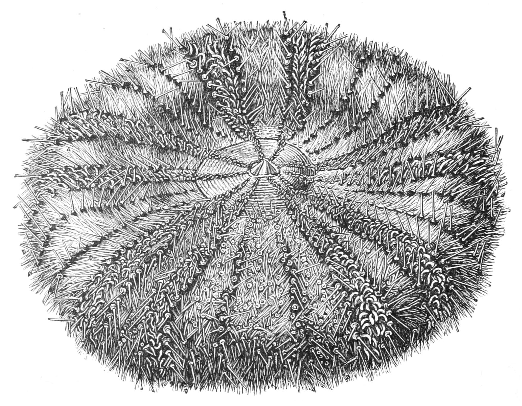
Fig. 238.—Oral view of Asthenosoma hystrix. × ⅔. (From Wyville Thomson.)
This remarkable family is divided by Mortensen into ten genera, based as usual on the pedicellariae, but taking into account also the shape of the tip of hard material on the spines. Most authors refer the majority of the species to two genera, Phormosoma and Asthenosoma (Fig. 238), recognising also a genus Sperosoma for one or two aberrant species. Asthenosoma is distinguished by having wide interspaces of membrane between the plates, and by having ten longitudinal folds of the body-wall, two in each radius, in which powerful longitudinal muscles are developed projecting inwards in the radii. The organs of Stewart are very large. In Phormosoma, on the contrary, the interspaces of membrane are very narrow, and the longitudinal folds are thin and membranous and the organs of Stewart are vestigial. Asthenosoma hystrix and Phormosoma placenta have both been dredged in deep water off the Irish coast. A. urens, in which there are ectodermic poison-sacs at the bases of the spines, inhabits the Indian Ocean near Ceylon, and was thoroughly described by the Sarasins,[495] who regarded its structure as a proof that Echinoidea were derived from Holothuroidea. Both palaeontology and embryology have, however, yielded strong evidence that Echinoidea were derived from Asteroidea, and hence there is ground for believing that Holothuroidea are descended from primitive Echinoidea and not vice versa. The Echinothuriidae may perhaps be regarded as showing the first steps in the change, and though possibly not closely related to the actual ancestors of the Holothuroidea, they at any rate show parallel modifications.
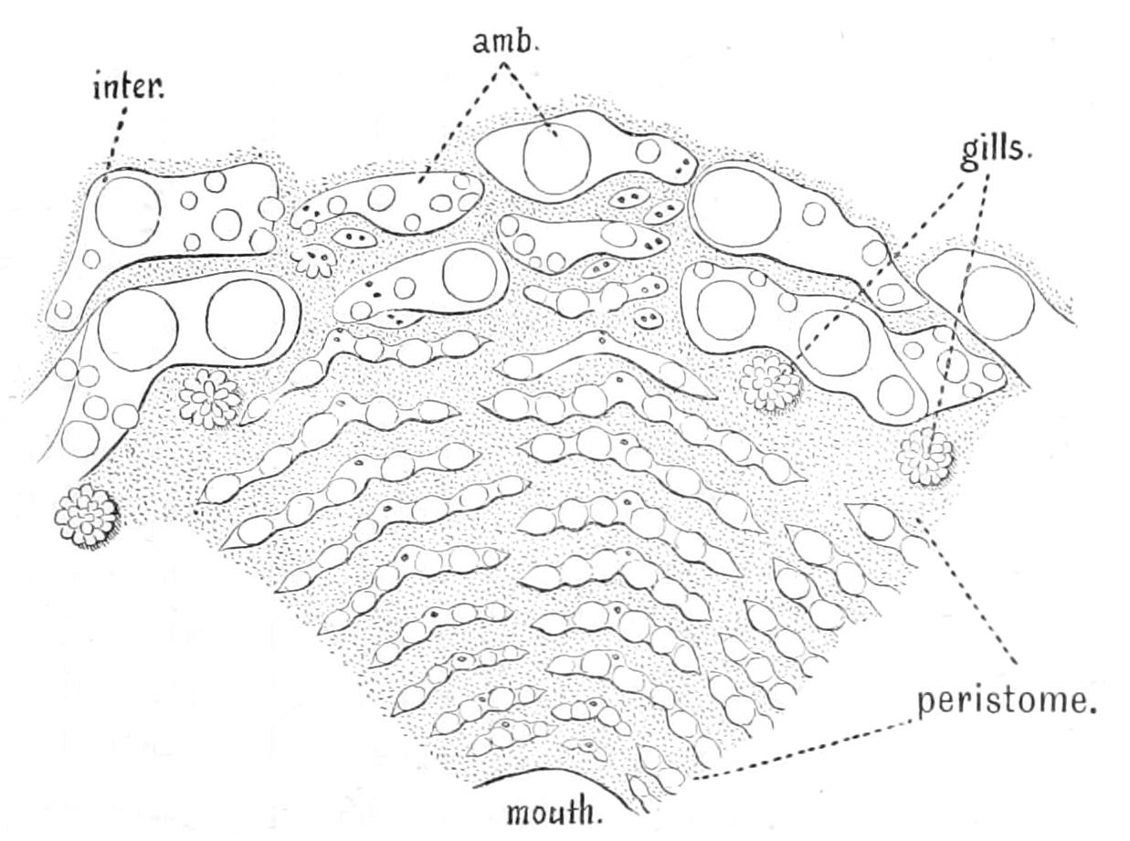
Fig. 239.—View of peristome of Asthenosoma hystrix. amb, Ambulacral plates on the lower edge of the corona; inter, lower plates of the interambulacral area. (From Wyville Thomson.)
Fam. 3. Saleniidae.—Endocyclica with a large peristome and periproct. The peristome is covered with thin, scattered, irregular plates. There are five pairs of special buccal tube-feet, each supported by a special plate, and there are external gills. The periproct is excavated in the side of a large central pentagonal plate. It is covered with fifteen or twenty plates.[496] The ambulacral plates are separate as in the Cidaridae, but occasionally adhere in pairs near the peristome. The interambulacral plates also, as in Cidaridae, each bear one large primary spine surrounded by a circle of secondaries. A few deep-water forms belong to this family, the type genus Salenia (Fig. 240) being the best known. None occur in the British area. Superficially they resemble the Cidaridae, but in reality they are widely separated by the essentially modern character of the peristome.
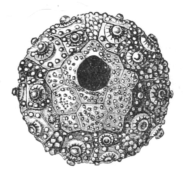
Fig. 240.—Dried and cleaned shell of Salenia varispina, showing periproct covered by one large plate. × 4. (From Wyville Thomson.)
Fam. 4. Arbaciidae.—Endocyclica with a peristome on which, as in Saleniidae, there are only ten prominent plates perforated by the buccal tube-feet, and besides these thin irregular plates; external gills are present, and the auricles consist of incomplete arches springing from the ambulacral plates. The periproct is covered by four valve-like plates. The ambulacral pore-plates are separate near the periproct, but near the peristome unite on the "Arbacioid" pattern (v. p. 531) to form secondary plates. The interambulacral plates each carry several spines. No representatives of this remarkable family are known in British waters, but Arbacia is found both on the east coast of North America and in the Mediterranean. It is distinguished by its conical test. All the upper tube-feet are devoid of a sucker; only those on the oral surface are used for locomotion.
Uexküll has studied the Mediterranean species, and has shown that the spines converge no matter how strong the stimulus may be, and so are incapable of aiding in locomotion; also that the ectoderm is devoid of ciliation, and hence the faecal matter which falls on the surface of the animal is not, as in other genera, allowed to fall off by the divergence of the spines nor swept off by the action of the cilia. In its natural habitat the wash of the ripples on the shore cleanses the animal. In captivity it is liable to suffocate itself.
Fam. 5. Diadematidae.—Endocyclica with a peristome similar to that of the Arbaciidae and the Saleniidae. External gills present and ten buccal tube-feet. Periproct small, covered with numerous small plates. The auricles form complete arches arising from the ambulacral region. Aristotle's lantern is provided with rudimentary Stewart's organs. The ambulacral pore-plates are separated at the apex, but unite orally in "Diadematoid" fashion (p. 531) to form compound plates. The interambulacral plates bear numerous primaries. The aboral tube-feet are pointed, having lost their suckers.
This family is represented (according to Agassiz) at the present day by seven genera, none of which are found in British waters, though one (Centrostephanus) enters the Mediterranean. C. longispinus[497] was investigated by Uexküll and found to be distinguished by its sensitiveness to light and shade, and by the quickness of its movements, which were mainly carried out by its long spines. The family resembles the Arbaciidae in the pointed aboral tube-feet, but in the complete auriculae it resembles the next family.
Fam. 6. Echinidae.—Endocyclica with peristome and periproct as in the preceding family. External gills and buccal tube-feet present, but Stewart's organs totally absent. Ambulacral plates combined on the "Triplechinoid" plan (p. 531) to form secondary plates. Interambulacral plates with numerous tubercles. All the tube-feet have suckers.
This family contains by far the larger number of living genera. It is divided into two sub-families, viz.:—
(a) Temnopleurinae.—Echinidae in which the plates of the corona dovetail into each other by means of pits and knobs along the line of suture. This sub-family does not occur in British waters; almost all the species are confined to the Indian and Pacific Oceans, but on the east coast of America it is represented by several genera, which however inhabit deep water, e.g. Trigonocidaris arbacina.
(b) Echininae.—Echinidae in which the plates meet each other in straight, simple sutures.
This sub-family is represented in British waters by three genera, viz. Echinus, Sphaerechinus, and Strongylocentrotus. Echinus is distinguished by having its pores arranged in arcs of three, owing to the fact that its pore-plates are united in threes to form secondary plates, whilst in the other two genera the ambulacral plates are composed of four or more pore-plates. Six species of Echinus have been recorded from British waters, viz. E. esculentus, E. acutus, E. miliaris, E. norvegicus, E. microstoma, and E. elegans. The validity of the last three is very doubtful. Mortensen[498] regards E. norvegicus and E. microstoma as mere variations of E. acutus, and this is probably correct. E. esculentus has already been described; its most marked character is the forest of comparatively short, close-packed, reddish or white primary spines with which it is covered, between the bases of which the delicate secondaries are hard to detect. It is essentially a shallow-water species. E. acutus is distinguished by having much fewer and longer primaries and numerous delicate secondaries. It is an inhabitant of deeper water, being abundant at 100 fathoms, though stragglers are found in shallower water. At the depths at which it lives wave-disturbance can scarcely be felt, and hence the long primaries are not irritated.
E. elegans has spines intermediate in character between those of E. esculentus and those of E. acutus. Like the latter it is an inhabitant of the deeper water. It seems to the present author not at all improbable that further research might show that E. acutus, E. elegans, and E. esculentus are all members of continuous series of forms; certainly the larvae and early development of E. acutus and E. esculentus, the extreme members of the series, are strikingly similar.
E. miliaris differs somewhat widely from the other species and is closely allied to E. microtuberculatus of the Mediterranean, from which it is distinguished mainly by the greater thickness of the scattered plates on the peristome of the latter species. From the other British species it differs in its much smaller size and in the greenish hue of its primary spines, which are short and thick and possess purple tips. Its larva is markedly distinct from the larva of E. esculentus. E. miliaris is a littoral species, and is found in great numbers in some of the Scottish sea-lochs; when the tide recedes, under every stone of the gravelly beach several specimens will be found. It has a curious habit of "dressing" itself, i.e. of covering itself with fragments of dead shell, sea-weed, etc., which are held in position by the aboral tube-feet. This habit aids in concealing the animal, and has probably been developed on account of the dangers to which E. miliaris is exposed owing to its littoral habit of life.
Sphaerechinus differs from Echinus in the structure of the ambulacral plates, in which it agrees with Strongylocentrotus, but it is distinguished from this genus by the very deep gill-clefts, or indentations of the edge of the corona from which the gills are extruded. Its most marked peculiarity, however, as shown by both Mortensen and Uexküll, consists in the highly developed character of its gemmiform pedicellariae, on the stalks of which are situated glands. When the head with its poison-glands is torn off, the secretion of these stalk-glands can envelop an enemy with a glutinous secretion, which impedes its movements. The blades on a slight mechanical stimulus divaricate very widely and become locked in this position, so that the enemy's body gets in well within their reach. The muscles of the poison-glands contract, but their ducts are bent by the act of opening, so that the secretion cannot escape. The sense-organs have stiff hairs, which penetrate the surface of the enemy and cause its juices to exude and so stimulate the blades to close, and at the same time permit the poison to be expelled. It will be remembered that the gemmiform pedicellariae of Echinus open in response to a chemical stimulus and close on a mechanical one being superadded; so that their responses are the direct opposite of what occurs in Sphaerechinus. S. granularis, a Mediterranean species with short red spines, just reaches the Channel Islands.
Strongylocentrotus has shallow gill-clefts and gemmiform pedicellariae, like those of Echinus, except that they have a muscular stalk. In the British area it is represented by two species, S. lividus, in which the primary spines are markedly longer than the secondaries and are of a brownish purple colour, and S. droëbachiensis, in which the primaries are little longer than the secondaries and are of a greenish brown colour. S. lividus occurs abundantly in the Mediterranean, and reaches the English Channel and the west coast of Ireland. In the last-named locality, where it is exposed to the full sweep of the Atlantic, it is said to excavate holes for itself in the limestone rocks, about ten inches in depth.[499] S. droëbachiensis, which has been recorded in the British area, chiefly from the west coast of Scotland, is one of the most abundant members of the fauna of the east coast of America. In the Gulf of St. Lawrence and in the branches of the Bay of Fundy it is found in thousands, and is frequently left bare at low tide. It thus takes the place of E. miliaris in the British fauna. An allied if not identical species, S. purpuratus, is found in Puget Sound on the Pacific coast.
Other interesting genera of the Echininae are Echinometra, Colobocentrotus, and Heterocentrotus. All possess large, thick primaries, and all are elliptical in outline. In Echinometra the primaries are pointed, and the long axis of the body makes an oblique angle with the axis passing through mouth and madreporite. In Colobocentrotus and Heterocentrotus the axis passing through mouth and madreporite is the short axis of the ellipse, and the primary spines are very thick and triangular in section, whilst the expanded ends of the secondaries form a closely set armour between the bases of these. In Colobocentrotus the test is markedly flattened on the under side, and this flattened area is fringed with a circle of primaries; but in Heterocentrotus there are a few rows of primaries all over the test. These are tropical genera and are found on the outer side of coral reefs, and they require the cuirass of expanded secondaries to protect them against the waves.
Order II. Clypeastroidea (Cake-urchins).
The "Cake-urchins" have only one representative in the British area, and this is unsuitable for dissection on account of its small size. We shall therefore select as type the "Sand-dollar" Echinarachnius parma (Figs. 241, 242), which occurs abundantly in shallow water on the east coast of North America. As its popular name implies, this is an extremely flattened Sea-urchin of nearly circular outline, so as to suggest a resemblance to the silver dollar of North American currency. The peristome is exceedingly small, and is placed in the centre of the lower surface (Fig. 241), whilst the periproct is placed on one edge. The outline is not quite circular, for the periproct lies in a slight indentation of the edge; and this side is broader and of a lesser degree of curvature than the opposite one, so that a secondary bilateral symmetry is superimposed on the fundamental radial symmetry common to all Echinoderms. A line drawn so as to pass through the anus and the centre of the disc will divide the animal into two similar halves; the periproct of course lies in an interradius and the axis of symmetry passes through the centre of one radius. We can thus distinguish an anterior group of three radii, or "trivium," from a posterior pair or "bivium." The madreporite lies in the left anterior interradius. The five genitals and five oculars surround a dorso-central plate, which covers the spot which in Endocyclica is occupied by the periproct.
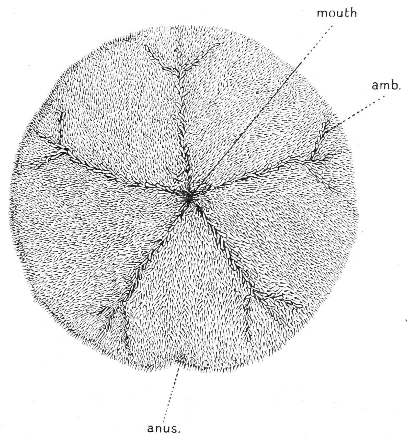
Fig. 241.—Oral view of "Sand-dollar" (Echinarachnius parma), with spines. amb, Ambulacral furrow, × 1.
The whole test is covered with extremely short delicate spines, which form a velvety felt-work, and are all of approximately the same length; they are of a brownish purple colour. The spines on the dorsal surface are all ciliated, and these cilia cause a current of fresh sea-water to flow continually over the modified tube-feet. Pedicellariae are scattered amongst the bases of the spines; they are of the tridactyle, the gemmiform, and the ophicephalous types, but they have only two jaws.
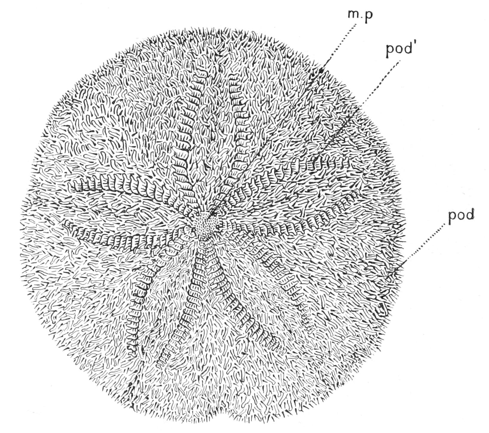
Fig. 242.—Aboral view of the "Sand-dollar" (Echinarachnius parma), with its spines. m.p, Madreporite; pod, small tube-foot with sucker; pod', flattened respiratory tube-foot. × 1.
The ambulacral areas on the upper surface of the test can be distinguished only by the flattened respiratory tube-feet (Fig. 242, pod'), which can be seen protruding from between the spines. Below these areas are clearly marked, for in the centre of each is a well-marked groove proceeding inwards to the peristome. This groove receives lateral branches on its course which traverse the adjacent interambulacral regions. The purpose of these grooves will be explained later. The interambulacral regions do not reach the peristome, which is entirely surrounded by the ambulacral areas. The ambulacral and interambulacral areas both consist of somewhat large hexagonal plates, except in the region of the respiratory tube-feet. Here the pore-plates are not united with one another. This region in each radius is termed a "petal" (Fig. 243, A, p), for the respiratory tube-feet are arranged in two rows which diverge from their commencement at the "calyx" and slightly converge again towards the outer margin of the disc, and thus in a dried specimen the two rows of double pores outline an area having some resemblance in shape to the petal of a flower. Besides these double pores for the larger tube-feet there are numerous small single pores for the smaller tube-feet; these are found in all the plates, ambulacral and interambulacral, of the dorsal surface, but in the neighbourhood of the grooves only on the ventral side.
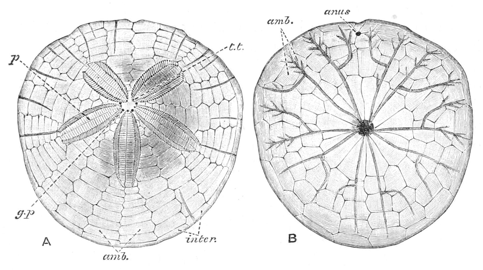
Fig. 243.—A, aboral, B, oral view of Echinarachnius parma after spines have been removed, amb (in A), Ambulacral plates, (in B), ambulacral furrows; g.p, genital pore; inter, interambulacral plate; p, petal; t.t, pore for terminal tentacle, × ½.
The sphaeridia are only present to the number of one in each radius. Each sphaeridium is enclosed in a pit situated near the edge of the peristome.
A remarkable feature in the skeleton of Echinarachnius which is characteristic in greater or lesser degree of all Clypeastroidea is the presence of vertical partitions of calcareous matter traversing the coelom and stretching from the upper to the lower surface of the test. These are found principally in the peripheral region of the animal; and there can be no doubt that they have originated as cellular bands traversing the coelom, for the formation of similar structures can be followed step by step in the Crinoidea. In the axis of these trabeculae, or folds of the coelomic wall, jelly is secreted, and into this the lime-producing amoebocytes wander. In Echinarachnius these partitions are arranged in groups, each group radiating from a common centre.
The main peculiarities in the structure of Echinarachnius are comprehensible when the species is viewed from above in its normal environment. It is found in comparatively shallow water on a sandy bottom, and normally is nearly but not quite buried in the sand. It might thus be overturned by the force of the waves and currents, and it is protected against this fate by its flattened shape. This shape, however, necessitates some kind of support for the upper part of the test, and this is provided by the internal partitions.
In order to view the internal anatomy of the "Sand-dollar," it is necessary carefully to pick away the dorsal surface of the shell piece by piece. In this way the whole course of the alimentary canal is exposed; as in Echinus esculentus it can be seen to issue from the upper surface of Aristotle's lantern. It then bends sharply to the left, and makes a complete circle round the edge of the disc; this portion is the stomach, and is considerably inflated and accompanied by a "siphon." It then bends sharply back on itself, but only goes half way round; when it reaches the posterior interradius it ends in the anus (Fig. 244).
Aristotle's lantern is greatly simplified as compared with its condition in the Regular Urchins. Both rotulae and compasses are absent; the jaws are sharply bent on themselves, and their appearance gives one the impression that they have shared in the process of compression which the test as a whole has undergone, and have thus become bent. The teeth are nearly horizontal, and they actually articulate with the auriculae, which, as in Cidaridae, consist of disconnected pillars and spring from the plates of the interradius. Each pillar is fused with the adjacent one belonging to the next radius, so that the system which in Echinus consists of five radial arches here consists of five interradial pillars. Aristotle's lantern has lost its respiratory function and apparently its masticatory function as well, for the teeth are used as spades to shovel into the mouth the sand mixed with organic detritus and small organisms on which the animal lives.
The water-vascular system is highly modified. There are two sharply marked kinds of tube-feet—(a) the respiratory tube-feet, (b) the locomotor tube-feet. Both kinds are terminated by suckers, but the first variety are much larger than the second; they possess a flattened lobed base, and are connected with the ampulla by a double canal. They issue only from the double pores which form the petal. The locomotor tube-feet are small and cylindrical; they are, as already mentioned, scattered over the whole upper surface of the test, penetrating both ambulacral and interambulacral plates, but all are connected by transverse canals with the radial canals of the water-vascular system. On the under surface they are confined to the neighbourhood of the ambulacral grooves, which have nothing to do with the ambulacral grooves of an Asteroid, but are due to secondary localisations of the tube-feet, which are here also connected in each radius with a single radial canal. The appearance of a living Echinarachnius covered with a veritable forest of short brown tube-feet is very striking.[500]
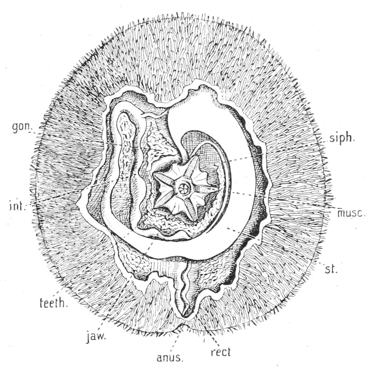
Fig. 244.—Dissection of Echinarachnius parma. × 1. The oesophagus has been cut through and moved to one side so as to expose Aristotle's lantern. The aboral part of the test has been removed. gon, Genital organ; int, intestine; musc, transverse muscle connecting jaws of adjacent interradii; rect, rectum; siph, siphon; st, stomach.
The condition of the water-vascular system is to be explained entirely by the peculiar environment of the animal. The demand for specialised respiratory organs is brought about by the habit of living half buried in the sand. Under these circumstances the strain of supplying the needful oxygen is thrown on the dorsal tube-feet, and they become modified in order to fit them for this function. The locomotor tube-feet are very small and feeble compared with those of Echinus esculentus, but this is comprehensible when it is recollected how little resistance the yielding sand would offer to the pull of a powerful tube-foot like that of the Regular Urchins, for in order to move the creature through the sand a multitude of feeble pulls distributed all over its surface is necessary, and the locomotor tube-feet are exactly fitted, both as to size and number, for this object.
The principal points in which Clypeastroidea vary amongst themselves are (1) the nature of the internal skeleton, (2) the shape, and (3) the spines.
Internal Skeleton.—In Echinocyamus and its allies this consists in each interradius of two simple partitions radiating out towards the edge of the disc; in Laganum it consists of walls parallel to the edge of the disc; in Clypeaster, of isolated pillars.
Shape.—In Echinocyamus the outline is oval and the test comparatively high. In Clypeaster and its allies the outline is pentagonal, and the test is swollen up into a blunt elevation in the centre. In a large number of genera, however, the test is, as in Echinarachnius, extremely thin and flat, and the outline may be variously indented. A first indication of this process is seen in Echinarachnius itself, but in Rotula the edge is drawn out into finger-like processes which are all interradial. In Mellita these processes unite with one another distally so as to surround spaces called "lunules," which appear as perforations of the test.
The Classification of the Clypeastroidea adopted by Agassiz is based chiefly on the degree of development of the internal skeleton, and as this is of great physiological importance to the animals we shall follow it here; but since it was published the remarkable discovery has been made of Pygastrides, a type previously known only from fossils. We must therefore recognise two sub-orders:—
Sub-Order I. Protoclypeastroidea.
Anus on dorsal surface near apical pole. One species, Pygastrides relictus,[501] with no "petals," from deep water in the Caribbean Sea.
Sub-Order II. Euclypeastroidea.
Anus on under surface.
Fam. 1. Fibularidae.—The "petals" are short and imperfect, and the internal skeleton consists of two short outwardly-directed septa in each interradius. To this family the only British Clypeastroid, Echinocyamus pusillus, belongs. This animal never exceeds an inch in length, and has an oval outline. It inhabits shallow water, and is often found in the same ground as Echinus miliaris, but like all Clypeastroids it prefers a sandy bottom.
Fam. 2. Echinanthidae or Clypeastridae.—"Petals" well marked, internal skeleton consisting of isolated pillars. The largest Cake-urchins belong to this family, which is found chiefly in tropical waters. Clypeaster, the great Cake-urchin, with a deeply sunken peristome, belongs to this family.
Fam. 3. Laganidae.—Closely allied to the foregoing, but distinguished by the fact that the internal skeleton consists of walls parallel to the edge of the test. (Laganum, Arachnoides, Peronella.)
Fam. 4. Scutellidae.—This family includes about half the genera, and is sharply distinguished from all the rest by (1) the extremely flattened shape, (2) the indentation of the outline in the anal interradius and often elsewhere, (3) the branching of ambulacral furrows on the under surface. Echinarachnius, taken as the type in describing the anatomy of the Cake-urchins, is the best-known genus. Others are Mellita, with five perforations in the edge of the test; and Rotula, with the edge produced into a number of finger-like processes.
Order III. Spatangoidea (Heart-urchins).
As the type we may select Echinocardium cordatum, which occurs abundantly in the Clyde and on the west coast of Ireland. The animal is found buried in sand at a depth of about 8-10 inches from the surface. At this depth it lies in a burrow, the walls of which are kept from collapsing by the somewhat broadened tips to the spines. This burrow communicates with the surface by a narrow cylindrical opening similar to the opening of the burrows made by the Clams and other bivalves. A little practice, however, enables one to distinguish the burrow of the Heart-urchin from these.
The animal is about the size of a small potato, and is of light straw colour. Its outline is oval, and the test is about two-thirds as high as the shorter diameter. It is thus higher in proportion to its width than is the case with any living Cake-urchin. The highest point is behind the centre. The narrower end of the animal terminates in a vertical edge, in the upper part of which is a large periproct covered with a number of thin movable plates. The mouth is situated on the under surface, considerably nearer the front end of the test than the hinder end. It is entirely devoid of jaws or of teeth, and also of gills or of a movable peristome.
Aristotle's lantern has entirely disappeared, leaving as the only trace of its former presence a canal with membranous walls encircling the mouth, which has the form of a transverse slit, the posterior lip projecting considerably forward.
The ambulacral areas are easily distinguishable from the interambulacral areas by being comparatively bare of spines. On the upper surface they are distinctly grooved, the groove being especially deep in the case of the anterior one. On the lower surface they coalesce round the mouth, shutting out the interambulacral regions, and are here perforated by the large pores of the buccal tube-feet. Between the two posterior radii on the oral surface there is a space with specially arranged spines called the plastron or sternum. The interambulacral plates composing this region are very much lengthened, and interdigitate with one another at the sutures. To this lengthening is due the apparent forward shift of the mouth. The spines are very characteristic, and are very different from any which have as yet been described. They are the sole organs of locomotion. The primaries are long and curved, with flattened tips, admirably adapted to plough through the sand in which the animal lives. On the upper surface, mingled with the tube-feet, are a large number of small secondary spines. Between the two posterior petals there is a hoop-shaped band of very small black spines. These spines are ciliated, and draw a current of fresh sea-water over the respiratory tube-feet. Beneath the periproct there is a similar band called the "sub-anal fasciole"; this probably produces a current of water which sweeps away the material ejected from the anus.
The pedicellariae are of the trifoliate and gemmiform varieties. The sphaeridia are situated in open pits, one or two in each, situated at the bases of the tube-feet nearest the mouth.
When the upper part of the test is picked away, the course of the alimentary canal is exposed (Fig. 247). It is very similar to the alimentary canal of Echinarachnius, except that from the first coil a large blind pouch, called the caecum, is given off.
The water-vascular system shows many characteristic features. The tube-feet are confined to two rows in each ambulacrum, the scattered smaller feet found in such abundance in Echinarachnius being entirely absent. There are four distinct varieties of tube-feet in Echinocardium, which are as follows:—(a) The respiratory tube-feet of the petals. These have, as in Echinarachnius, broad flat bases, but they have lost the sucker. (b) The prehensile tube-feet of the anterior ambulacrum. These are enormously long structures, measuring when expanded several times the length of the body. They end in discs, which are frayed out into fingers, so as to look like miniature sea-anemones. These tube-feet are comparatively few in number and are confined to the apical portion of the anterior ambulacrum. (c) The buccal tube-feet. These are short, thick, and pointed, and covered with a multitude of club-shaped processes. They are found on all the ambulacra in the neighbourhood of the mouth. (d) The degenerate tube-feet found in the portions of the ambulacra between the "floscelle" (see p. 553) and the petals. These are single and pointed, few in number, and issue from single pores in the test.
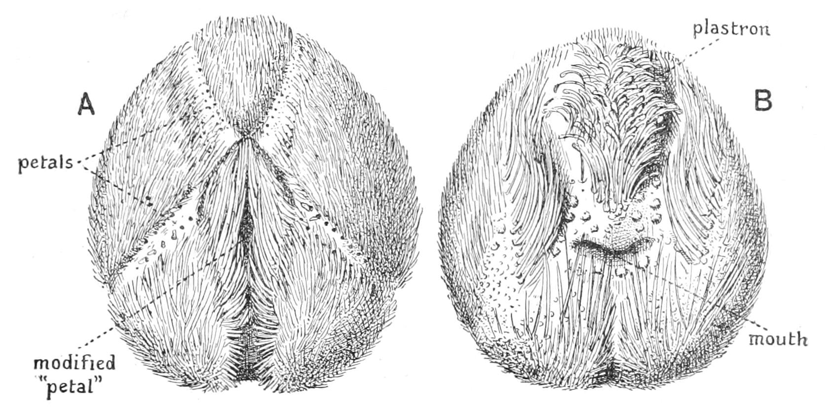
Fig. 245.—Echinocardium cordatum. A, aboral view; B, oral view, × 1.
This extraordinary diversity in the tube-feet is fully explained when the habits of the animal are known. The function of the respiratory tube-feet requires, of course, no special elucidation, but the peculiar anterior ambulacrum was a mystery till the feeding habits of the animal were observed by the late Dr. Robertson[502] of Cumbrae. He found that the animal protruded the long prehensile tube-feet through the opening of the burrow up to the surface of the sand. With their finger-like processes they then collected the surface film of the sand, which was impregnated with Diatoms and other small organisms. When a "handful," so to speak, of this nutritive material has been collected, the long tube-foot is withdrawn down the burrow and passed over the deeply grooved part of the ambulacrum to the buccal tube-feet, to which the food is given up. These last then push it into the mouth. Only one prehensile tube-foot is extended at a time.
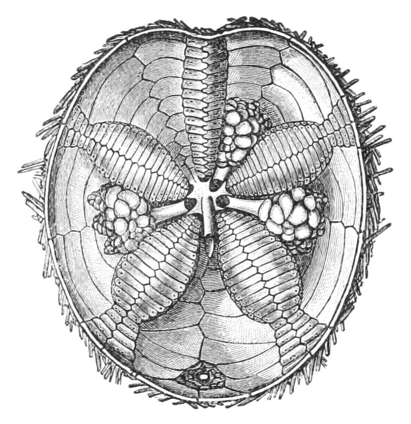
Fig. 246.—Interior of test of Hemiaster philippi, showing the genital organs and their ducts (only three are developed in this species). Slightly enlarged. (From Wyville Thomson.)
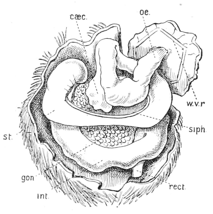
Fig. 247.—Dissection of Echinocardium cordatum. × 1. The oral part of the test has been removed. caec, Blind pouch of the stomach; gon, genital organ; int, intestine; oe, oesophagus; rect, rectum; siph, siphon; st, stomach; w.v.r, water-vascular ring.
The stone-canal is very short and soon opens into the axial sinus; it is widely separated from the pore-canals which traverse the madreporite. Communication between the two is effected by the long axial sinus. There are only four genital organs.
Heart-urchins vary amongst themselves chiefly in the following points, viz.:—(1) the shape and position of the peristome, (2) the characters of the "petals," and (3) the number and position of the fascioles.
Peristome.—In many genera this is pentagonal and central, and in these cases the interradii commence at the peristome with a single plate, which is often covered with a thick crowd of small spines and is termed a "bourrelet." The oral ends of the radii also often consist of a crowded series of narrow plates, looking something like a "petal," and termed a "phyllode." The five bourrelets and five phyllodes constitute a flower-like figure termed a "floscelle."
Ambulacra.—In Echinoneus all five are alike and are provided with similar tube-feet, which are respiratory but possess suckers. The ambulacra are not grooved, and the petaloid arrangement of the pores is hardly marked; but in Cassidulus, Pourtalesia, and many other genera the five petals are well marked, though they are all similar to one another.
Fascioles.—These structures are often entirely absent; the sub-anal one alone is present in Spatangus. In Eupatagus a peripetalous one is added. This surrounds all the "petals," and has obviously the function of sweeping fresh water over the respiratory tube-feet. In Echinocardium, as we have seen, there is an "internal fasciole" between the two anterior petals which has a similar function. In addition, this genus possesses an anal fasciole which surrounds the anus and sweeps away the faeces.
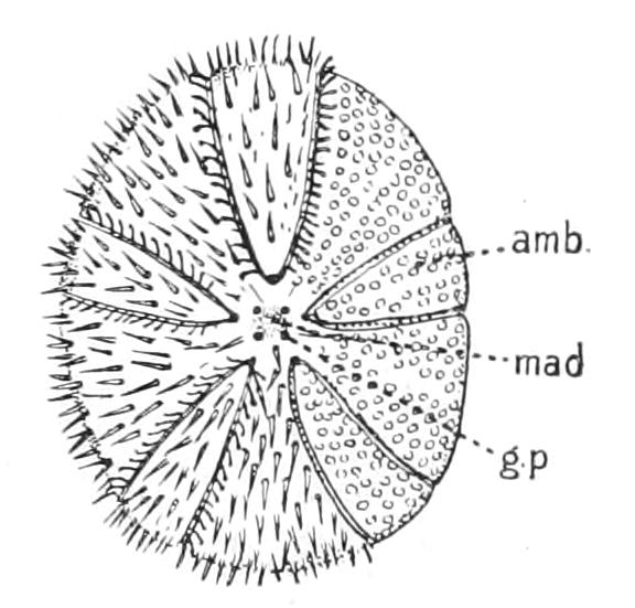
Fig. 248.—Young Echinoneus to show five equal radii scarcely petaloid. amb, Ambulacral area; g.p, genital pore; mad, madreporite. (After Agassiz.)
The Classification of the Spatangoidea is based mainly on the degree of development of the petals, that is to say, on the extent to which the burrowing habit has been developed. But weight is also laid on the shape of the peristome, the pentagonal form being more primitive. Seven families are recognised, which are as follows:—
Fam. 1. Echinonidae.—"Petals" hardly marked at all; peristome in the centre of the lower surface and pentagonal. Floscelle not developed.
One genus, Echinoneus (Fig. 248).
Fam. 2. Nucleolidae.—"Petals" distinct; peristome as in the foregoing family. No floscelle. Nucleolites, with the anus in a furrow. Anochanus, with a concave apical system serving as brood-pouch.
Fam. 3. Cassidulidae.—"Petals" usually distinct; peristome eccentric, but provided with a well-marked floscelle.
Echinolampas, with the anus on the under surface.
Neolampas, with the anus on a projecting papilla. One specimen of this genus has been dredged in the British area.
The three foregoing families probably use their tube-feet to walk with, and bury themselves only to a slight extent. They are often united as a sub-order, the Asternata, and distinguished from all the rest which possess an eccentric mouth and well-marked plastron. These families are then grouped together as Sternata. They are as follows:—
Fam. 4. Ananchytidae.[503]—Spatangoidea with elongated apical system, ambulacra all similar and not grooved. Petals feebly marked. Pourtalesia, with bottle-shaped posterior prolongation of the test. Platybrissus, with flattened test.
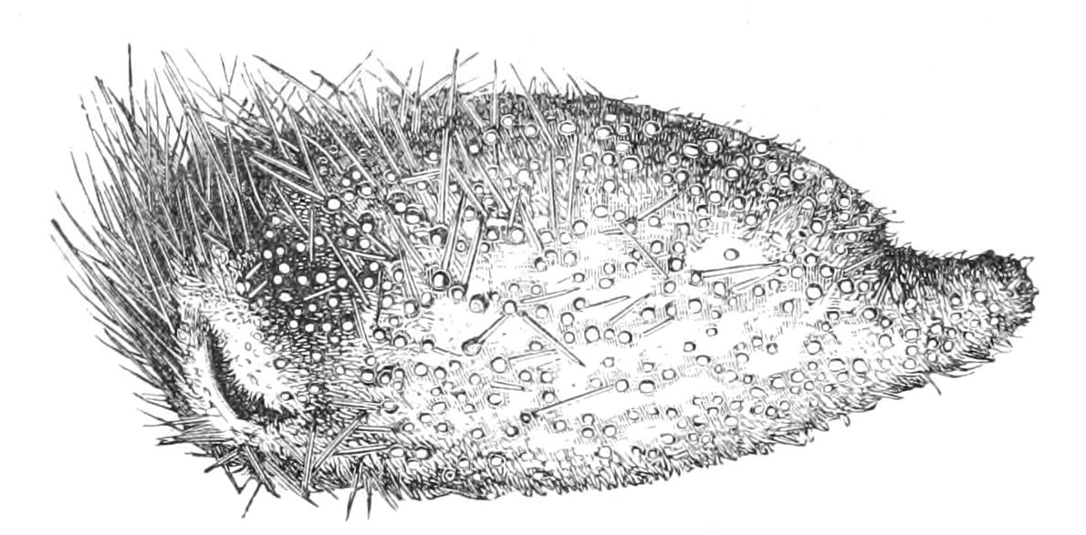
Fig. 249.—Pourtalesia jeffreysi, slightly enlarged. (From Wyville Thomson.)
Fam. 5. Palaeostomatidae.—An aberrant family consisting of one genus, Palaeostoma. Petals grooved, with a peripetalous fasciole, but peristome central and pentagonal.
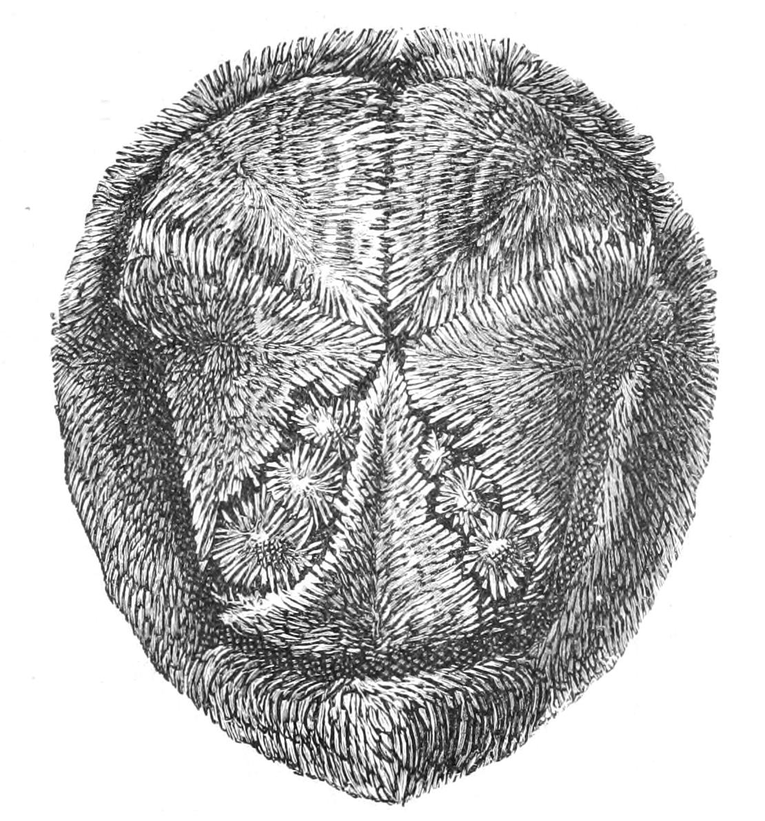
Fig. 250.—Hemiaster philippi. × 2. (From Wyville Thomson.)
Fam. 6. Spatangidae.—Spatangoidea of more or less flattened shape, with well-marked petals and a sub-anal plastron as well as the ventral one. One fascicle at least, but a peripetalous one never present. The anterior ambulacrum grooved and different from the rest. This family is represented in British waters by two genera, Spatangus and Echinocardium. The former possesses only a sub-anal fasciole, and has specially long curved spines on the ventral plastron. It is represented by two species, S. purpureus and S. raschi, the latter being distinguished by a pointed lower lip. It is a deep-water species, found in 100 fathoms and over on the west coast. S. purpureus is fairly common in rather shallow water. From observations made on specimens kept in confinement it appears to burrow only so far as to leave the petals uncovered; hence there is no need of a peripetalous fasciole. Echinocardium is devoid of the thicker spines on the plastron, and has an internal fasciole and a perianal one as well as the sub-anal. As already mentioned, it is a deep burrower. It is represented by three species, E. cordatum, E. pennatifidum, and E. flavescens. The first, described as the type of the Spatangoidea, has a deeply grooved anterior ambulacrum. In the remaining two species this ambulacrum is not grooved. E. flavescens has only six or seven pairs of pores in the posterior petals, E. pennatifidum twelve to fourteen. Both come from deeper water than E. cordatum.