Chapter III
Reading edition
PROTOZOA (CONTINUED): SARCODINA
I. Sarcodina.
Protozoa performing most of their life-processes by pseudopodia; nucleus frequently giving off fragments (chromidia) which may play a part in nuclear reconstitution on division; sometimes with brood-cells, which may be at first flagellate; but never reproducing in the flagellate state.[64]
1. Rhizopoda
Sarcodina of simple form, whose pseudopodia never coalesce into networks (1),[65] nor contain an axial filament (2), which commonly multiply by binary fission (3), though a brood-formation may occur; which may temporarily aggregate, or undergo temporary or permanent plastogamic union, but never to form large plasmodia or complex fructifications as a prelude to spore-formation (4); test when present gelatinous, chitinous, sandy, or siliceous, simple and 1-chambered (5).
Classification.[66]
I. Ectoplasm distinct, clear; pseudopodia blunt or tapering, but not branching at the apex Lobosa
Amoeba, Auctt.; Pelomyxa, Greeff; Trichosphaerium, A. Schneid.; Dinamoeba, Leidy; Amphizonella, Greeff; Centropyxis, Stein; Arcella, Ehr.; Difflugia, Leclercq; Lecqueureusia, Schlumberger; Hyalosphenia, Stein; Quadrula, F. E. Sch.; Heleopera, Leidy; Podostoma, Cl. and L.; Arcuothrix, Hallez.
II. Ectoplasm undifferentiated, containing moving granules; pseudopodia branching freely towards the tips Filosa
Euglypha, Duj.; Paulinella, Lauterb.; Cyphoderia, Schlumb.; Campascus, Leidy; Chlamydophrys, Cienk.; Gromia, Duj. = Hyalopus, M. Sch.
We have defined this group mainly by negative characters, as such are the only means for their differentiation from the remaining Sarcodina; and indeed from Flagellata, since in this group zoospores are sometimes formed which possess flagella. Moreover, indeed, in a few of this group (Podostoma, Arcuothrix), as in some Heliozoa, the flagellum or flagella may persist or be reproduced side by side with the pseudopodia. The subdivision of the Rhizopoda is again a matter of great difficulty, the characters presented being so mixed up that it is hard to choose: however, the character of the outer layer of the cytoplasm is perhaps the most obvious to select. In Lobosa there is a clear layer of ectosarc, which appears to be of a greasy nature at its surface film, so that it is not wetted. In the Filosa, as in most other Sarcodina, this film is absent, and the ectoplasm is not marked off from the endoplasm, and may have a granular surface. Corresponding to this, the pseudopodia of the Lobosa are usually blunt, never branching and fraying out, as it were, at the tip, as in the Filosa; nay, in the normal movements of Amoeba limax (Fig. 1, p. 5) the front of the cell forms one gigantic pseudopodium, which constantly glides forward. Apart from this distinction the two groups are parallel in almost every respect.
There may be a single contractile vacuole, or a plurality; or none, especially in marine and endoparasitic species. The nucleus may remain single or multiply without inducing fission, thus leading to apocytial forms. It often gives off "chromidial" fragments, which may play an important part in reproduction.[67] In Amoeba binucleata there are constantly two nuclei, both of which divide as an antecedent to fission, each giving a separate nucleus to either daughter-cell. Pelomyxa palustris, the giant of the group, attaining a diameter of 1‴ (2 mm.), has very blunt pseudopodia, an enormous number of nuclei, and no contractile vacuole, though it is a fresh-water dweller, living in the bottom ooze of ponds, etc., richly charged with organic débris. It is remarkable also for containing symbiotic bacteria, and brilliant vesicles with a distinct membranous wall, containing a solution of glycogen.[68] Few, if any, of the Filosa are recorded as plurinuclear.
The simplest Lobosa have no investment, nor indeed any distinction of front or back. In some forms of Amoeba, however, the hinder part is more adhesive, and may assume the form of a sucker-like disc, or be drawn into a tuft of short filaments or villi, to which particles adhere. Other species of Lobosa and all Filosa have a "test," or "theca," i.e. an investment distinct from the outermost layer of the cell-body. The simplest cases are those of Amphizonella, Dinamoeba, and Trichosphaerium, where this is gelatinous, and in the two former allows the passage of food particles through it into the body by mere sinking in, like the protoplasm itself, closing again without a trace of perforation over the rupture. In Trichosphaerium (Fig. 9) the test is perforated by numerous pores of constant position for the passage of the pseudopodia, closing when these are retracted; and in the "A" form of the species (see below) it is studded with radial spicules of magnesium carbonate. Elsewhere the test is more consistent and possesses at least one aperture for the emission of pseudopodia and the reception of food; to avoid confusion we call this opening not the mouth but the "pylome": some Filosa have two symmetrically placed pylomes. When the test is a mere pellicle, it may be recognised by the limitation of the pseudopodia to the one pylomic area. But the shell is often hard. In Arcella (Fig. 10, C), a form common among Bog-mosses and Confervas, it is chitinous and shagreened, circular, with a shelf running in like that of a diving-bell around the pylome: there are two or more contractile vacuoles, and at least two nuclei. Like some other genera, it has the power of secreting carbonic acid gas in the form of minute bubbles in its cytoplasm, so as to enable it to float up to the surface of the water. The chitinous test shows minute hexagonal sculpturing, the expression of vertical partitions reaching from the inner to the outer layer.
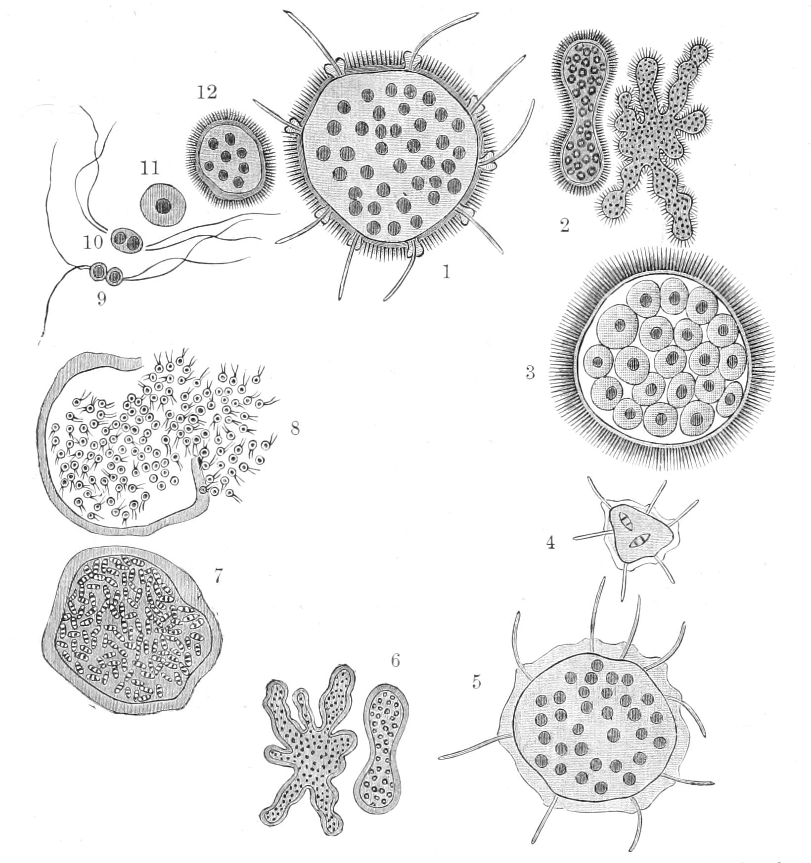
Fig. 9.—Trichosphaerium sieboldii. 1, Adult of "A" form; 2, its multiplication by fission and gemmation; 3, resolution into 1-nucleate amoeboid zoospores; 4, development (from zoospores of "A") into "B" form (5); 6, its multiplication by fission and gemmation; 7, its resolution after nuclear bipartition into minute 2-flagellate zoospores or (exogametes); 8, liberation of gametes; 9, 10, more highly magnified pairing of gametes of different origin; 11, 12, zygote developing into "A" form. (After Schaudinn.)
Several genera have tests of siliceous or chitinous plates, formed in the cytoplasm in the neighbourhood of the nucleus, and connected by chitinous cement. Among these Quadrula (Fig. 10, A) is Lobose, with square plates, Euglypha (Fig. 8, p. 29), and Paulinella[69] are Filose, with hexagonal plates. In the latter they are in five longitudinal rows, with a pentagonal oral plate, perforated by the oval pylome. In other genera again, such as Cyphoderia (Filosa), the plates are merely chitinous. Again, the shell may be encrusted with sand-grains derived directly from without, or from ingested particles, as shown in Centropyxis, Difflugia (Fig. 10, D), Heleopera, and Campascus when supplied with powdered glass instead of sand. The cement in Difflugia is a sort of organic mortar, infiltrated with ferric oxide (more probably ferric hydrate). In Lecqueureusia spiralis (formerly united with Difflugia) the test is formed of minute sausage-shaped granules, in which may be identified the partly dissolved valves of Diatoms taken as food; it is spirally twisted at the apex, as if it had enlarged after its first formation, a very rare occurrence in this group. The most frequent mode of fission in the testaceous Rhizopods (Figs. 8, 10) is what Schaudinn aptly terms "bud-fission," where half the protoplasm protrudes and accumulates at the mouth of the shell, and remains till a test has formed for it, while the other half retains the test of the original animal. The materials for the shell, whether sand-granules or plates, pass from the depths of the original shell outwards into the naked cell, and through its cytoplasm to the surface, where they become connected by cementing matter into a continuous test. The nucleus now divides into two, one of which passes into the external animal; after this the two daughter-cells separate, the one with the old shell, the other, larger, with the new one.
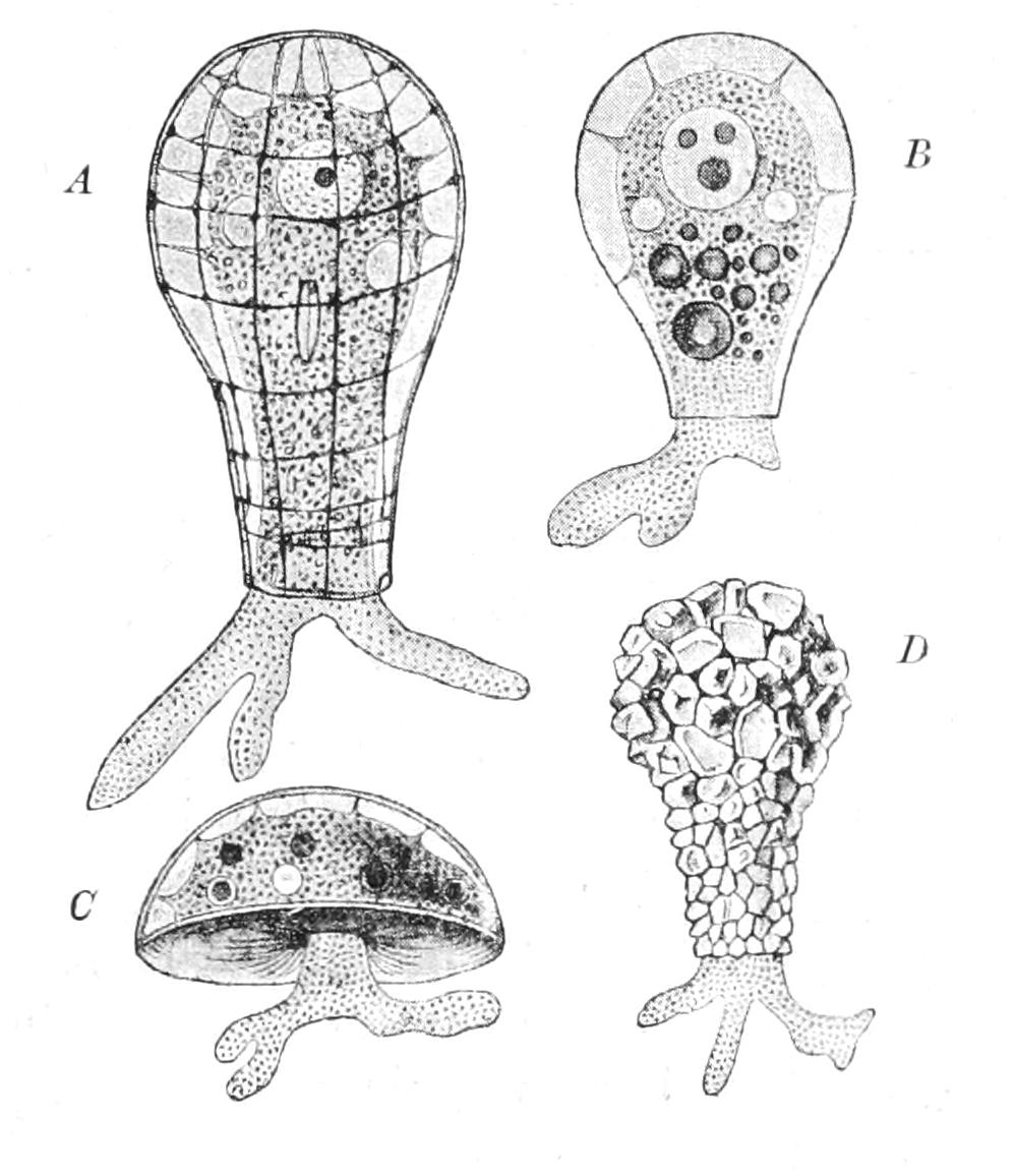
Fig. 10.—Test-bearing Rhizopods. A, Quadrula symmetrica: B, Hyalosphenia lata; C, Arcella vulgaris; D, Difflugia pyriformis. (From Lang's Comparative Anatomy.)
If two individuals of the shelled species undergo bud-fission in close proximity, the offspring may partially coalesce, so that a monstrous shell is produced having two pylomes.
Reproduction by fission has been clearly made out in most members of the group; some of the multinucleate species often abstrict a portion, sometimes at several points simultaneously, so that fission here passes into budding[70] (Fig. 9, 2, 6).
Brood-division, either by resolution in the multinucleate species, or preceded by multiple nuclear division in the habitually 1-nucleate, though presumably a necessary incident in the life-history of every species, has only been seen, or at least thoroughly worked out, in a few cases, where it is usually preceded by encystment, and mostly by the extrusion into the cyst of any undigested matter.[71]
In Trichosphaerium (Fig. 9) the cycle described by Schaudinn is very complex, and may be divided into two phases, which we may term the A and the B subcycles. The members of the A cycle are distinguished by the gelatinous investment being armed with radial spicules, which are absent from the B form. The close of the A cycle is marked by the large multinucleate body resolving itself into amoeboid zoospores (3), which escape from the gelatinous test, and develop into the large multinucleate adults of the B form. These, like the A form, may reproduce by fission or budding. At the term of growth, however, they retract their pseudopodia, expel the excreta, and multiply their nuclei by mitosis (7). Then the body is resolved into minute 2-flagellate microzoospores (8), which are exogamous gametes, i.e. they will only pair with similar zoospores from another cyst. The zygote (9-11) resulting from this conjugation is a minute amoeboid; its nucleus divides repeatedly, a gelatinous test is formed within which the spicules appear, and so the A form is reconstituted. In many of the test-bearing forms, whether Lobose or Filose, plastogamic unions occur, and the two nuclei may remain distinct, leading to plurinucleate monsters in their offspring by fission, or they may fuse and form a giant nucleus, a process which has here no relation to normal syngamy, as it is not associated with any marked change in the alternation of feeding and fission, etc. In Trichosphaerium also plastogamic unions between small individuals have for their only result the increase of size, enabling the produce to deal with larger prey. Temporary encystment in a "hypnocyst" is not infrequent in both naked and shelled species, and enables them to tide over drought and other unfavourable conditions.
Schaudinn has discovered and worked out true syngamic processes, some bisexual, some exogamous, in several other Rhizopods. In Chlamydophrys stercorea the pairing-cells are equal, and are formed by the aggregation of the chromidia into minute nuclei around which the greater part of the cytoplasm aggregates, while the old nucleus (with a little cytoplasm) is lost. These brood-cells are 2-flagellate pairing-cells, which are exogamous: the zygote is a brown cyst; if this be swallowed by a mammal, the original Chlamydophrys appears in its faeces.[72]
Centropyxis aculeata, a species very common in mud or moss, allied to Difflugia, also forms a brood by aggregation around nuclei derived from chromidia. The brood-cells are amoeboid, and secrete hemispherical shells like those of Arcella; some first divide into four smaller ones, before secreting the shell. Pairing takes place between the large and the small forms; and the zygote encysts. Weeks or months afterwards the cyst opens and its contents creep out as a minute Centropyxis. Finally, Amoeba coli produces its zygote in a way recalling that of Actinosphaerium (pp. 73-75, Fig. 21): the cell encysts; its nucleus divides, and each daughter divides again into two, which fuse reciprocally. Thus the cyst contains two zygote nuclei. After a time each of these divides twice, so that the mature cyst contains eight nuclei. Probably when swallowed by another animal they liberate a brood of eight young amoebae. Thus in different members of this group we have exogamy, both equal and bisexual, and endogamy.
Most of the Rhizopoda live among filamentous Algae in pools, ponds, and in shallow seas, etc.; some are "sapropelic" or mud-dwellers (many species of Amoeba, Pelomyxa, Difflugia, etc.), others frequent the roots of mosses. Amoeba coli is often found as a harmless denizen of the large intestine of man. Amoeba histolytica, lately distinguished therefrom by Schaudinn, is the cause of tropical dysentery. It multiplies enormously in the gut, and is found extending into the tissues, and making its way into the abscesses that so frequently supervene in the liver and other organs. Chlamydophrys stercorea is found in the faeces of several mammals. The best monograph of this group is that of Penard.[73]
2. Foraminifera[74]
Sarcodina with no central capsule or distinction of ectosarc; the pseudopodia fine, branching freely, and fusing where they meet to form protoplasmic networks, or the outermost in the pelagic forms radiating, but without a central or axial filament: sometimes dimorphic, reproducing by fission and by rhizopod or flagellate germs in the few cases thoroughly investigated: all marine (with the exception of some of the Allogromidiaceae), and usually provided with a test of carbonate of lime ("vitreous" calcite, or "porcellanous" aragonite?), or of cemented particles of sand ("arenaceous"); test-wall continuous, or with the walls perforated by minute pores or interstices for the protrusion of pseudopodia.
The classification of Carpenter (into Vitreous or Perforate, Porcellanous or Imperforate, and Arenaceous), according to the structure of the shell, had proved too artificial to be used by Brady in the great Monograph of the Foraminifera collected by the "Challenger" Expedition,[75] and has been modified by him and others since then. We reproduce Lister's account of Brady's classification.[76] We must, however, warn the tyro that its characterisations are not definitions (a feature of all other recent systems), for rigid definitions are impossible: here as in the case, for instance, of many Natural Orders of Plants, transitional forms making the establishment of absolute boundaries out of the question. In the following classification we do not think it, therefore, necessary to complete the characterisations by noting the extremes of variation within the orders:—
1. Allogromidiaceae: simple forms, often fresh-water and similar to Rhizopoda; test 0, or chitinous, gelatinous, or formed of cemented particles, whether secreted platelets or ingested granules. Biomyxa, Leidy = Gymnophrys, Cienk.; Diaphorodon, Archer; Allogromia, Rhumbl. (= Gromia, auctt.[77] nec Duj.) (Fig. 14, 1); Lieberkühnia, Cl. and Lachm. (Fig. 12); Microgromia, R. Hertw. (Fig. 11); Pamphagus, Bailey.
2. Astrorhizidaceae: test arenaceous, often large, never truly chambered, or if so, asymmetrical. Astrorhiza, Sandahl; Haliphysema, Bowerb.; Saccammina, M. Sars (Fig. 13, 1); Loftusia, Brady.
3. Lituolidaceae: test arenaceous, often symmetrical or regularly spiral, isomorphous with calcareous forms: the chambers when old often "labyrinthine" by the ingrowth of wall-material. Lituola, Lam.; Reophax, Montf.; Ammodiscus, Reuss; Trochammina, Parker and Jeffreys.
4. Miliolidaceae: test porcellanous, imperforate, spirally coiled or cyclic, often chambered except in Cornuspira: simple in Squamulina. Cornuspira, Max Sch.; Peneroplis, Montf.; Miliolina, Lam. (incl. Biloculina (Fig. 15), Triloculina, Quinqueloculina (Figs. 14, 4; 15, B), Spiroloculina (Fig. 13, 5) of d'Orb.); Alveolina, d'Orb.; Hauerina, d'Orb.; Calcituba, Roboz; Orbitolites, Lam.; Orbiculina, Lam.; Alveolina, Park. and Jeffr.; Nubecularia, Def.; Squamulina, Max Sch. (Fig. 14, 3).
5. Textulariaceae: test calcareous, hyaline, perforated; chambers increasing in size in two alternating rows, or three, or passing into a spiral. Textularia, Def.; Bulimina, d'Orb.; Cassidulina, d'Orb.
6. Cheilostomellaceae: test vitreous, delicate, finely perforated, chambered, isomorphic with the spiral forms of the Miliolidaceae. Cheilostomella, Reuss.
7. Lagenaceae: Test vitreous, very finely perforate, chambers with a distinct pylome projecting (ectosolenial), or turned in (entosolenial), often succeeding to form a necklace-like shell. Lagena, Walker and Boys (Fig. 13, 2); Nodosaria, Lam. (Fig. 13, 3); Cristellaria, Lam.; Frondicularia, Def. (Fig. 13, 4); Polymorphina, Lam.; Ramulina, Wright.
8. Globigerinidae: test vitreous, perforate; chambers few, dilated, and arranged in a flat or conical spiral, usually with a crescentic pylome to the last. Globigerina, d'Orb. (Figs. 13, 6; 16, 2); Hastigerina, Wyv. Thoms.; Orbulina, d'Orb. (Fig. 16, 1).
9. Rotaliaceae; test vitreous, perforate, usually a conical spiral (like a snail), chambers often subdivided into chamberlets, and with a proper wall, and intermediate skeleton traversed by canals. Rotalia, Lam. (Fig. 14, 2); Planorbulina, d'Orb. (Fig. 13, 9); Polytrema, Risso; Spirillina, Ehr. (non-septate); Patellina, Will.; Discorbina, P. and J. (Fig. 13, 7).
10. Nummulitaceae: test usually a complex spiral, the turns completely investing their predecessors: wall finely tubular, often with a proper wall and intermediate skeleton. Fusulina, Fisch.; Polystomella, Lam.; Nummulites, d'Orb. (Fig. 13, 11); Orbitoides, d'Orb.
The Allogromidiaceae are a well-marked and distinct order, on the whole resembling the Rhizopoda Filosa, and are often found with them in fresh water, while all other Foraminifera are marine. The type genus, Allogromia (Fig. 14, 1), has an oval chitinous shell. Microgromia socialis (Fig. 11) is often found in aggregates, the pseudopodia of neighbours fusing where they meet into a common network. This is due to the fact that one of the two daughter-cells at each fission, that does not retain the parent shell, remains in connexion with its sister that does: sometimes, however, it retracts its pseudopodia, except two which become flagella, wherewith it can swim off. The test of Pamphagus is a mere pellicle. In Lieberkühnia (Fig. 12) it is hardly that; though the body does not give off the fine pseudopodia directly, but emits a thick process or "stylopodium"[78] comparable to the protoplasm protruded through the pylome of its better protected allies; and from this, which often stretches back parallel to the elongated body, the reticulum of pseudopodia is emitted. Diaphorodon has a shell recalling that of Difflugia (Fig. 10, D, p. 55), formed of sandy fragments, but with interstices between them through which as well as through the two pylomes the pseudopodia pass. In all of these the shell is formed as in the Rhizopods once for all, and does not grow afterwards; and the fresh-water forms, which are the majority, have one or more contractile vacuoles; in Allogromia they are very numerous, scattered on the expanded protoplasmic network.
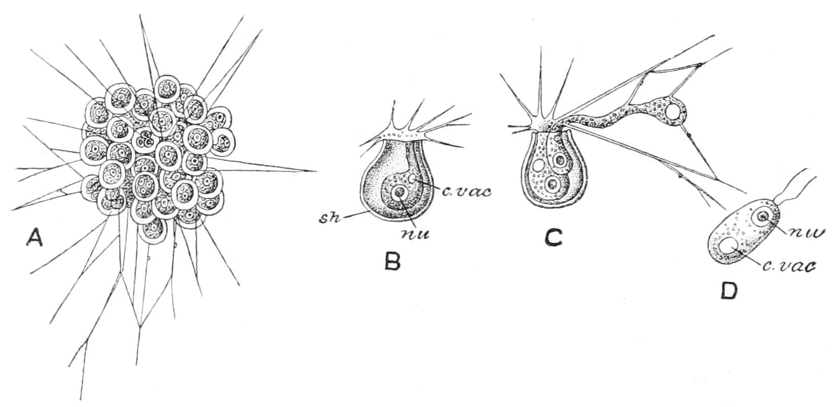
Fig. 11.—Microgromia socialis. A, entire colony; B, single zooid; C, zooid which has undergone binary fission, with one of the daughter-cells creeping out of the shell; D, flagellula. c.vac, Contractile vacuole; nu, nucleus; sh, shell. (From Parker and Haswell, after Hertwig and Lesser.)
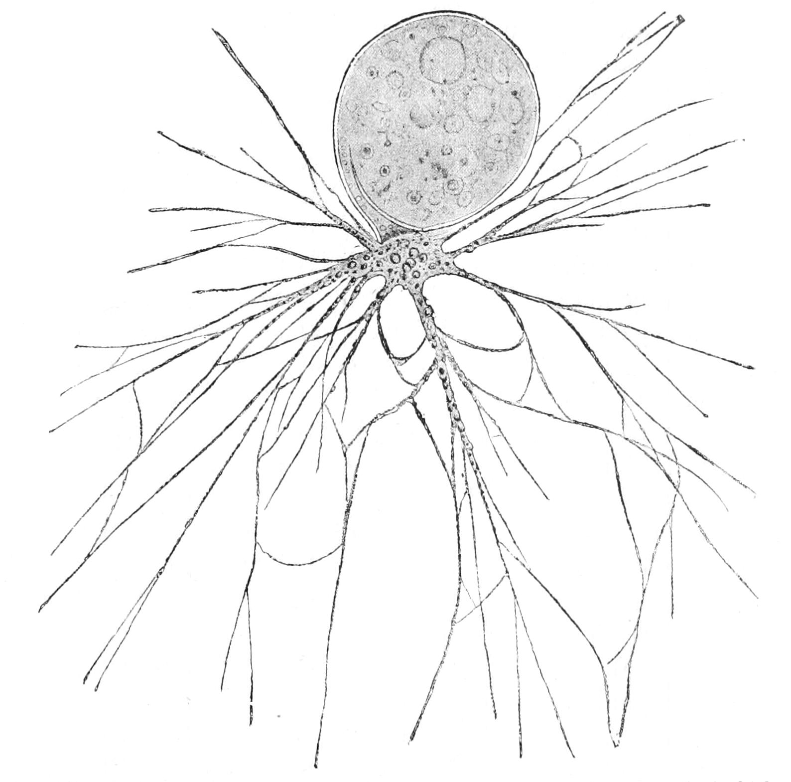
Fig. 12.—Lieberkühnia, a fresh-water Rhizopod, from the egg-shaped shell of which branched pseudopodial filaments protrude. (From Verworn.)
The remaining marine families may all be treated of generally, before noting their special characters. Their marine habitat is variable, but in most cases restricted. A few extend up the brackish water of estuaries: a large number are found between tide-marks, or on the so-called littoral shelf extending to deep water; they are for the most part adherent to seaweeds, or lie among sand or on the mud. Other forms, again, are pelagic, such as Globigerina (Figs. 13, 6, 16, 17) and its allies, and float as part of the plankton, having the surface of their shells extended by delicate spines, their pseudopodia long and radiating, and the outer part of their cytoplasm richly vacuolated ("alveolate"), and probably containing a liquid lighter than sea water, as in the Radiolaria. Even these, after their death and the decay of the protoplasm, must sink to the bottom (losing the fine spines by solution as they fall); and they accumulate there, to form a light oozy mud, the "Globigerina-ooze" of geographers, at depths where the carbonic acid under pressure is not adequate to dissolve the more solid calcareous matter. Grey Chalk is such an ooze, consolidated by the lapse of time and the pressure of superincumbent layers. Some Foraminifera live on the sea bottom even at the greatest depths, and of course their shell is not composed of calcareous matter. Foraminifera may be obtained for examination by carefully washing sand or mud, collected on the beach at different levels between tide-marks, or from dredgings, or by carefully searching the surface of seaweeds, or by washing their roots, or, again, by the surface or deep-sea tow-net. The sand used to weight sponges for sale is the ready source of a large number of forms, and may be obtained for the asking from the sponge-dealers to whom it is a useless waste product. If this sand is dried in an oven, and then poured into water, the empty shells, filled with air, will float to the surface, and may be sorted by fine silk or wire gauze.
From the resemblance of the shells of many of them to the Nautilus they were at first described as minute Cephalopods, or Cuttlefish, by d'Orbigny,[79] and their true nature was only elucidated in the last century by the labours of Williamson, Carpenter, Dujardin, and Max Schultze. At first they possess only one nucleus, but in the adult stage may become plurinucleate without dividing, and this is especially the case in the "microsphaeric" states exhibited by many of those with a complex shell; the nucleus is apt to give off fragments (chromidia) which lie scattered in the cytoplasm. At first, too, in all cases, the shell has but a single chamber, a state that persists through life in some. When the number of chambers increases, their number has no relation to that of the nuclei, which remains much smaller till brood-formation sets in.
The shell-substance, if calcareous, has one of the two types, porcellanous or vitreous, that we have already mentioned, but Polytrema, a form of very irregular shape, though freely perforated, is of a lovely pink colour. In the calcareous shells sandy particles may be intercalated, forming a transition to the Arenacea. In these the cement has an organic base associated with calcareous or ferruginous matter; in some, however, the cement is a phosphate of iron. The porcellanous shells are often deep brown by transmitted light.
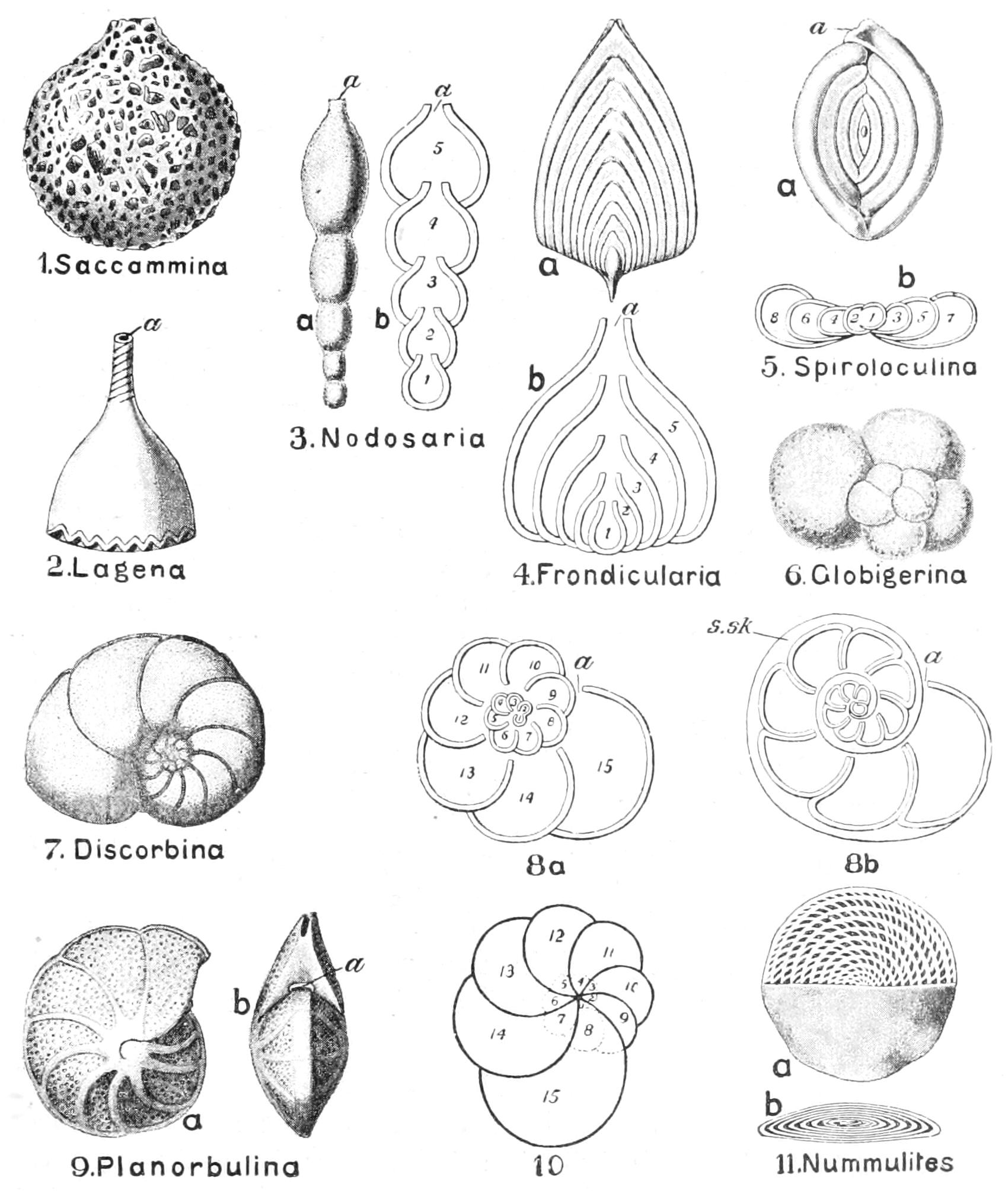
Fig. 13.—Shells of Foraminifera. In 3, 4, and 5, a shows the surface view, and b a section; 8a is a diagram of a coiled cell without supplemental skeleton; 8b of a similar form with supplemental skeleton (s.sk); and 10 of a form with overlapping whorls; in 11a half the shell is shown in horizontal section; b is a vertical section; a, aperture of the shell; 1-15, successive chambers, 1 being always the oldest or initial chamber. (From Parker and Haswell, after other authors.)
Despite the apparent uniformity of the protoplasmic body in this group, the shell is infinitely varied in form. As Carpenter writes, in reference to the Arenacea, "There is nothing more wonderful in nature than the building up of these elaborate and symmetrical structures by mere jelly-specks, presenting no traces whatever of that definite organisation which we are accustomed to regard as necessary to the manifestations of conscious life.... The tests (shells) they construct when highly magnified bear comparison with the most skilful masonry of man. From the same sandy bottom one species picks up the coarsest quartz grains, unites them together with a ferruginous cement, and thus constructs a flask-shaped test, having a short neck and a single large orifice; another picks up the finer grains and puts them together with the same cement into perfectly spherical tests of the most extraordinary finish, perforated with numerous small pores disposed at pretty regular intervals. Another species selects the minutest sand grains and the terminal portions of sponge-spicules, and works them up together—apparently with no cement at all, but by the mere laying of the spicules—into perfect white spheres like homoeopathic globules, each showing a single-fissured orifice. And another, which makes a straight, many-chambered test, the conical mouth of each chamber projecting into the cavity of the next, while forming the walls of its chambers of ordinary sand grains rather loosely held together, shapes the conical mouths of the chambers by firmly cementing together the quartz grains which border it." The structure of the shell is indeed variable. The pylome may be single or represented by a row of holes (Peneroplis, Orbitolites), or, again, there may be several pylomes (Calcituba); and, again, there are in addition numerous scattered pores for the protrusion of pseudopodia elsewhere than from the stylopodium, in the whole of the "Vitrea" and in many "Arenacea"; and, as we shall see, this may exercise a marked influence on the structure of the shell.
In some cases the shell is simple, and in Cornuspira and Spirillina increases so as to have the form of a flat coiled tube. In Calcituba the shell branches irregularly in a dichotomous way, and the older parts break away as the seaweed on which they grow is eaten away, and fall to the bottom, while the younger branches go on growing and branching. The fallen pieces, if they light on living weed, attach themselves thereto and repeat the original growth; if not, the protoplasm crawls out and finds a fresh weed and forms a new tube. In the "Polythalamia" new chambers are formed by the excess of the protoplasm emerging and surrounding itself with a shell, organically united with the existing chamber or chambers, and in a space-relation which follows definite laws characteristic of the species or of its stage of growth, so as to give rise to circular, spiral, or irregular complexes (see Fig. 13).
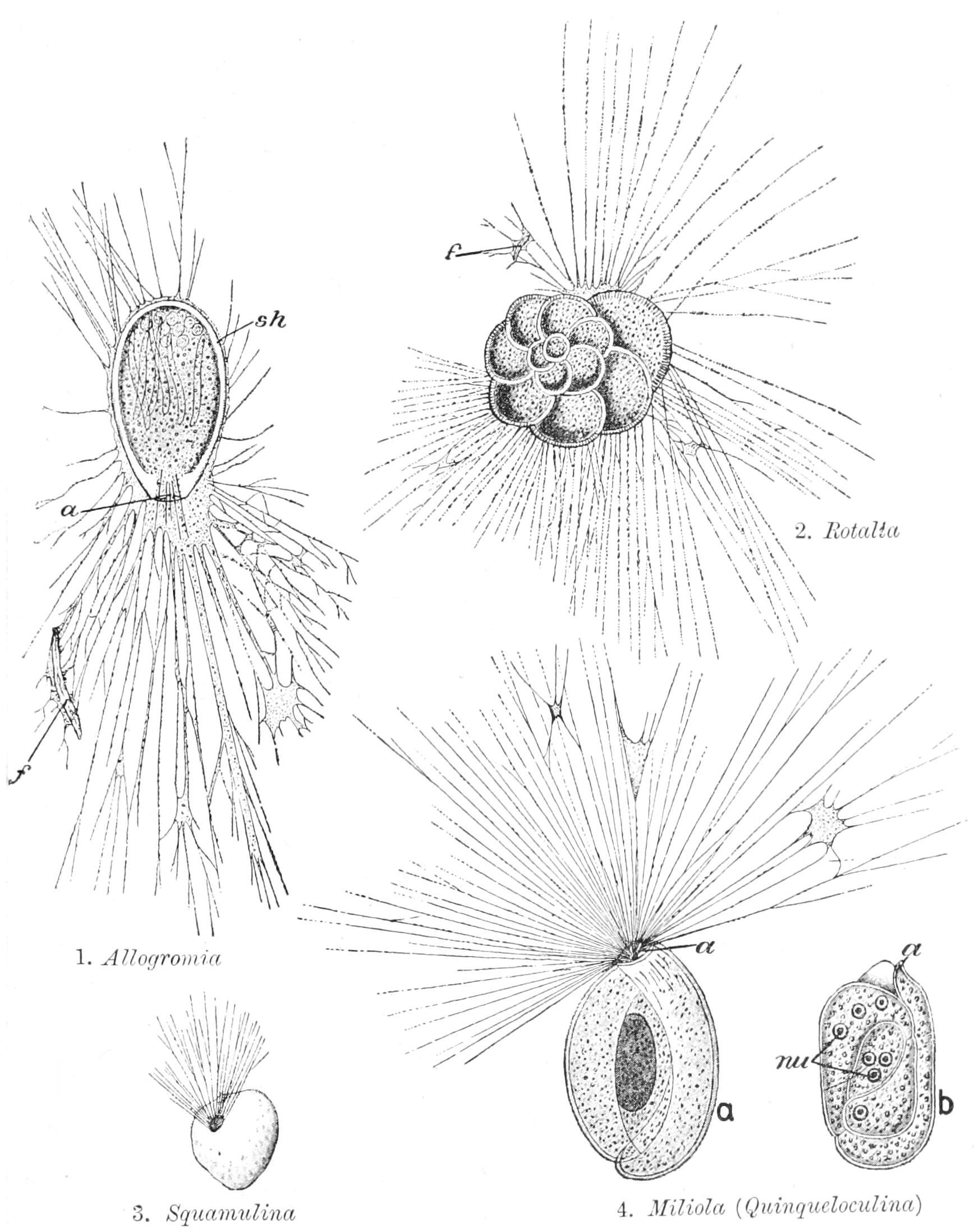
Fig. 14.—Various forms of Foraminifera. In 4, Miliola, a, shows the living animal; b, the same killed and stained; a, aperture of shell; f, food particles; nu, nucleus; sh, shell. (From Parker and Haswell, after other authors.)
In most cases the part of the previously existing chamber next the pylome serves as the hinder part of the new chamber, and the old pylome becomes the pore of communication. But in some of the "Perforata" each new chamber forms a complete wall of its own ("proper wall," Fig. 13, 8b), and the space between the two adjacent walls is filled with an intermediate layer traversed by canals communicating with the cavities of the chambers ("intermediate skeleton"), while an external layer of the same character may form a continuous covering. The shell of the Perforata may be adorned with pittings or fine spines, which serve to increase the surface of support in such floating forms as Globigerina, Hastigerina, and the like (Fig. 17). In the "Imperforata" the outer layer is often ornamented with regular patterns of pits, prominences, etc., which are probably formed by a thin reflected external layer of protoplasm. In some of the "Arenacea" a "labyrinthine" complex of laminae is formed.
A very remarkable point which has led to great confusion in the study of the Foraminifera, is the fact that the shell on which we base our characters of classification, may vary very much, even within the same individual. Thus in the genus Orbitolites the first few chambers of the shell have the character of a Milioline, in Orbiculina of a Peneroplis. The arrangements of the Milioline shell, known as Triloculine, Quinqueloculine, and Biloculine respectively, may succeed one another in the same shell (Figs. 14 4, 15). A shell may begin as a spiral and end by a straight continuation: again, the spherical Orbulina (Fig. 16 1) is formed as an investment to a shell indistinguishable from Globigerina, which is ultimately absorbed. In some cases, as Rhumbler has pointed out, the more recent and higher development shows itself in the first formed chambers, while the later, younger chambers remain at a lowlier stage, as in the case of the spiral passing into a straight succession; but the other cases we have cited show that this is not always the case. In Lagena (Fig. 13 2) the pylome is produced into a short tube, which may protrude from the shell or be turned into it, so that for the latter form the genus Entosolenia was founded. Shells identical in minute sculpture are, however, found with either form of neck, and, moreover, the polythalamial shells (Nodosaria, Fig. 13 3), formed of a nearly straight succession of Lagena-like chambers, may have these chambers with their communications on either type. Rhumbler goes so far as to suggest that all so-called Lagena shells are either the first formed chamber of a Nodosaria which has not yet become polythalamian by the formation of younger ones, or are produced by the separation of an adult Nodosaria into separate chambers.
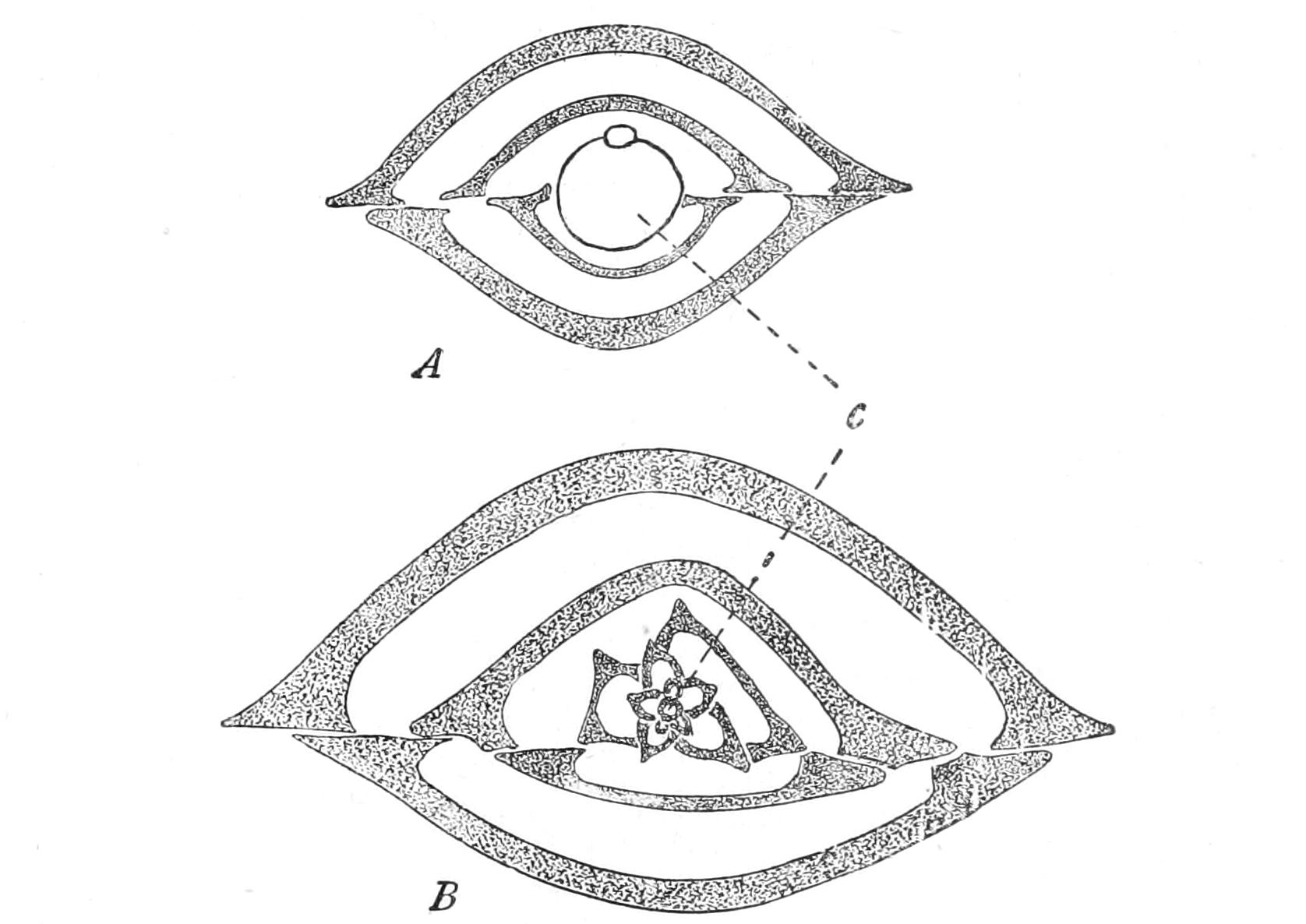
Fig. 15.—A, Megalospheric; B, microspheric shell of Biloculina. c, The initial chamber. The microspheric form begins on the Quinqueloculina type. (From Calkins' Protozoa.)
Many of the chambered species show a remarkable dimorphism, first noted by Schlumberger, and finally elucidated by J. J. Lister and Schaudinn. It reveals itself in the size of the initial chamber; accordingly, the two forms may be distinguished as "microspheric" and "megalospheric" respectively (Fig. 15), the latter being much the commoner. The microspheric form has always a plurality of nuclei, the megalospheric a single one, except at the approach of reproduction. Chromidial masses are, however, present in both forms. The life-history has been fully worked out in Polystomella by Schaudinn, and in great part in Polystomella, Orbitolites, etc., by Lister; and the same scheme appears to be general in the class, at least where the dimorphism noted occurs. The microspheric form gives birth only to the megalospheric, but the latter may reproduce megalospheric broods, or give rise to swarmers, which by their (exogamous) conjugation produce the microspheric young. The microspheric forms early become multinucleate, and have also numerous chromidia detached from the nuclei, which they ultimately replace. These collect in the outer part of the shell and aggregate into new nuclei, around which the cytoplasm concentrates, to separate into as many amoeboid young "pseudopodiospores" as there are nuclei. These escape from the shell or are liberated by its disintegration, and invest themselves with a shell to form the initial large central chamber or megalosphere.
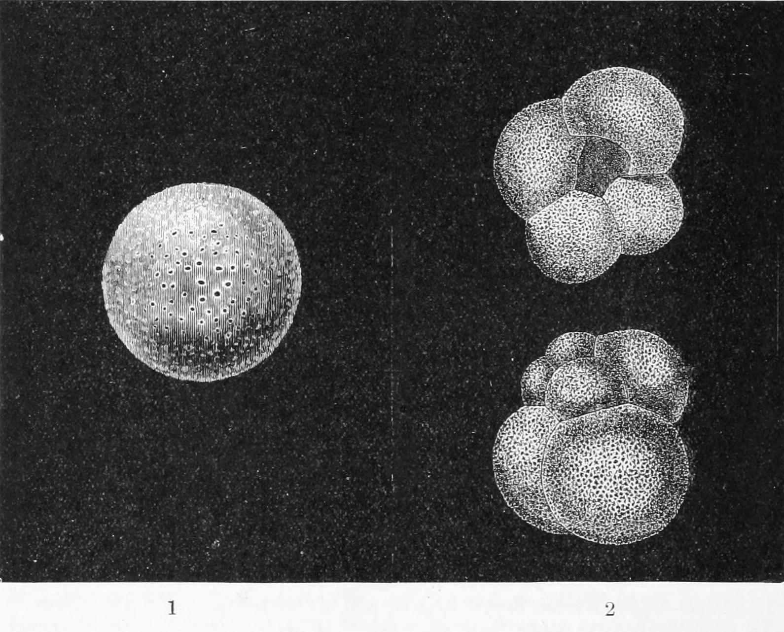
Fig. 16.—1, Orbulina universa. Highly magnified. 2, Globigerina bulloides. Highly magnified. (From Wyville Thomson, after d'Orbigny.)
In the ordinary life of the megalospheric form the greater part of the chromatic matter is aggregated into a nucleus, some still remaining diffused. At the end of growth the nucleus itself disintegrates, and the chromidia concentrate into a number of small vesicular nuclei, each of which appropriates to itself a small surrounding zone of thick plasm and then divides by mitosis twice; and the 4-nucleate cells so formed are resolved into as many 1-nucleate, 2-flagellate swarmers, which conjugate only exogamously.[80] The fusion of their nuclei takes place after some delay: ultimately the zygote nucleus divides into two, a shell is formed, and we have the microsphere, which is thus pluri-nucleate ab initio. As we have seen, the nuclei of the microsphere are ultimately replaced by chromidia, and the whole plasmic body divides into pseudopodiospores, which grow into the megalospheric form.
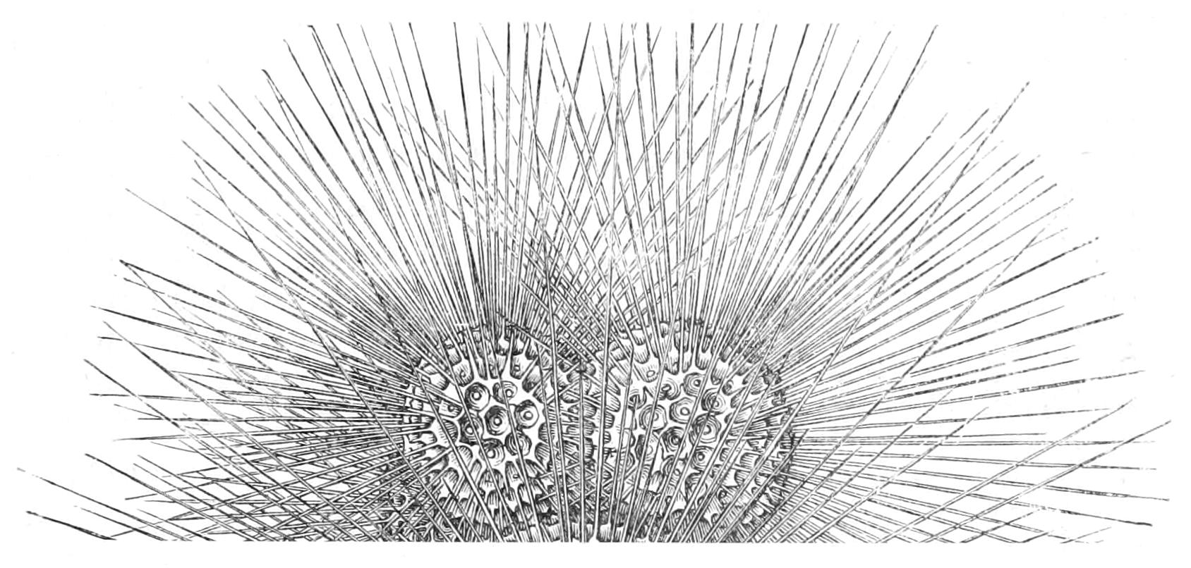
Fig. 17.—Shell of Globigerina bulloides, from tow-net, showing investment of spines. (From Wyville Thomson.)
In the Perforate genera, Patellina and Discorbina, plastogamy precedes brood formation, the cytoplasms of the 2-5 pairing individuals contracting a close union; and then the nuclei proceed to break up without fusion, while the cytoplasm aggregates around the young nuclei to form amoebulae, which acquire a shell and separate. In both cases it is the forms with a single nucleus, corresponding to megalospheric forms that so pair, and the brood-formation is, mutatis mutandis, the same as in these forms. Similar individuals may reproduce in the same way, in both genera, without this plastogamic pairing, which is therefore, though probably advantageous, not essential. If pseudopodiospores form their shells while near one another, they may coalesce to form monsters, as often happens in Orbitolites.[81]
The direct economic uses of the Foraminifera are perhaps greater than those of any other group of Protozoa. The Chalk is composed largely of Textularia and allied forms, mixed with the skeletons of Coccolithophoridae (pp. 113-114), known as Coccoliths, etc. The Calcaire Grossier of Paris, used as a building stone, is mainly composed of the shells of Miliolines of Eocene age; the Nummulites of the same age of the Mediterranean basin are the chief constituent of the stone of which the Pyramids of Egypt are built. Our own Oolitic limestones are composed of concretions around a central nucleus, which is often found to be a minute Foraminiferous shell.
The palaeontology of the individual genera is treated of in Chapman's and Lister's recent works. They range from the Lower Cambrian characterised by perforated hyaline genera, such as Lagena, to the present day. Gigantic arenaceous forms, such as Loftusia, are among the Tertiary representatives; but the limestones formed principally of their shells commence at the Carboniferous. The so-called Greensands contain greenish granules of "glauconite," containing a ferrous silicate, deposited as a cast in the chambers of Foraminifera, and often left exposed by the solution of the calcareous shell itself. Such granules occur in deep-sea deposits of the present day.[82]
3. Heliozoa
Sarcodina with radiate non-anastomosing pseudopodia of granular protoplasm, each with a stiff axial rod passing into the body plasma; no central capsule, nor clear ectoplasm; skeleton when present siliceous; nucleus single or multiple; contractile vacuole (or vacuoles) in fresh-water species, superficial and prominent at the surface in diastole; reproduction by fission or budding in the active condition, or by brood-formation in a cyst, giving rise to resting spores; conjugation isogamous in the only two species fully studied; habitat floating or among weeds, mostly fresh water.
1. Naked or with an investment only when encysted.
Aphrothoraca.—Actinolophus F.E. Sch.; Myxastrum Haeck.; Gymnosphaera Sassaki; Dimorpha (Fig. 37, 5, p. 112) Gruber; Actinomonas Kent; Actinophrys Ehrb.; Actinosphaerium St.; Camptonema Schaud; Nuclearia Cienk.
2. Invested with a gelatinous layer, sometimes traversed by a firmer elastic network.
Chlamydophora.—Heterophrys Arch.; Mastigophrys Frenzel; Acanthocystis, Carter.
3. Ectoplasm with distinct siliceous spicules.
Chalarothoraca.—Raphidiophrys Arch.
4. Skeleton a continuous, fenestrated shell, sometimes stalked.
Desmothoraca.—Myriophrys Penard; Clathrulina Cienk.; Orbulinella Entz.
This class were at first regarded and described as fresh-water Radiolaria, but the differences were too great to escape the greatest living specialist in this latter group, Ernst Haeckel, who in 1866 created the Heliozoa for their reception. We owe our knowledge of it mainly to the labours of Cienkowsky, the late William Archer, F. E. Schulze, R. Hertwig, Lesser, and latterly to Schaudinn, who has monographed it for the "Tierreich" (1896); and Penard has published a more recent account.
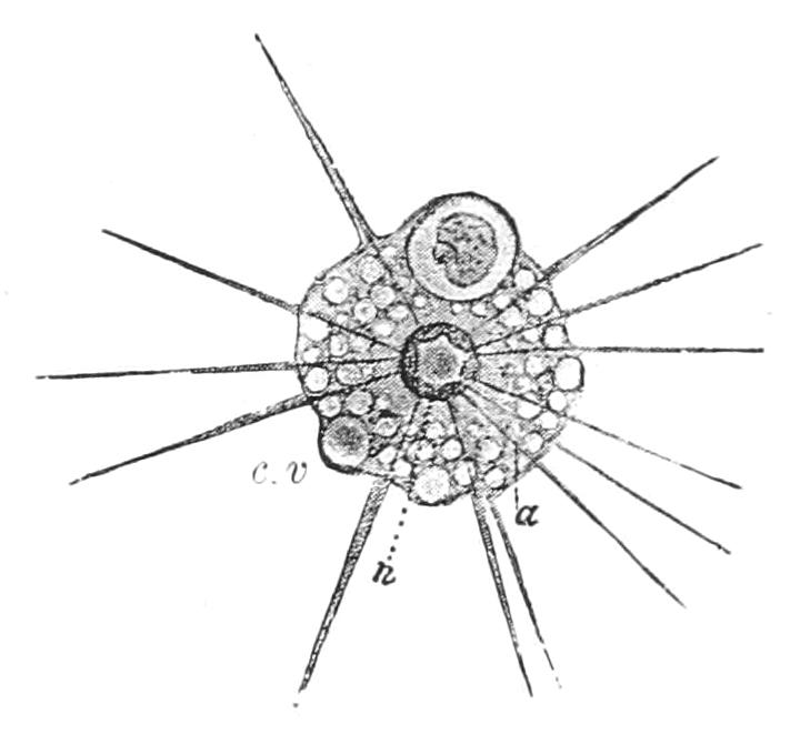
Fig. 18.—Actinophrys sol. a, Axial filament of pseudopod; c.v, contractile vacuole; n, nucleus. (From Lang's Comparative Anatomy, after Grenacher.)
Actinophrys sol Ehrb. (Fig. 18) is a good and common type. It owes its name to its resemblance to a conventional drawing of the sun, with a spherical body and numerous close-set diverging rays. The cytoplasm shows a more coarsely vacuolated outer layer, sometimes called the ectosarc, and a denser internal layer the endosarc. In the centre of the figure is the large nucleus, to which the continuations of the rays may be seen to converge; the pseudopodia contain each a stiffish axial filament,[83] which is covered by the fine granular plasm, showing currents of the granules. The axial filament disappears when the pseudopodia are retracted or bent, and is regenerated afterwards. This bending occurs when a living prey touches and adheres to a ray, all its neighbours bending in like the tentacles of a Sundew. The prey is carried down to the surface of the ectoplasm, and sinks into it with a little water, to form a nutritive vacuole. Fission is the commonest mode of reproduction, and temporary plastogamic unions are not uncommon. Arising from these true conjugations occur, two and two, as described by Schaudinn. A gelatinous cyst wall forms about the two which are scarcely more than in contact with their rays withdrawn. Then in each the nucleus divides into two, one of which passes to the surface, and is lost (as a "polar body"), while the other approaches the corresponding nucleus of the mate, and unites with it, while at the same time the cytoplasms fuse. Within the gelatinous cyst the zygote so formed divides to produce two sister resting spores, from each of which, after a few days, a young Actinophrys escapes, as may take place indeed after encystment of an ordinary form without conjugation.
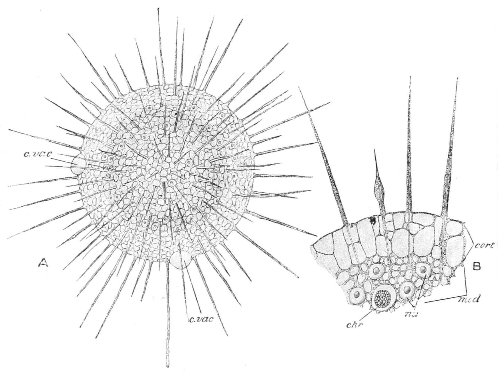
Fig. 19.—Actinosphaerium eichornii. A, entire animal with two contractile vacuoles (c.vac); B, a portion much magnified, showing alveolate cytoplasm, pseudopodia with axial rods, non-nucleate cortex (cort), multiple nuclei (nu) of endoplasm (med), and food-vacuole (chr). (From Parker and Haswell.)
The axial rods of the pseudopodia may pass either to the circumference of the nucleus or to a central granule, corresponding, it would appear, to a centrosome or blepharoplast; or again, in the plurinucleate marine genus Camptonema, each rod abuts on a separate cap on the outer side of each nucleus. The nucleus is single in all but the genera Actinosphaerium, Myxastrum, Camptonema, and Gymnosphaera. The movements of this group are very slow, and are not well understood. A slow rolling over on the points of the rays has been noted, and in Camptonema they move very decidedly to effect locomotion, the whole body also moving Amoeba-fashion; but of the distinct movements of the species when floating no explanation can be given. The richly vacuolate ectoplasm undoubtedly helps to sustain the cell, and the extended rays must subserve the same purpose by so widely extending the surface. Dimorpha (Fig. 37, 5, p. 112) has the power of swimming by protruding a pair of long flagella from the neighbourhood of the eccentric nucleus; and Myriophrys has an investment of long flagelliform cilia. Actinomonas has a stalk and a single flagellum in addition to the pseudopodia; these genera form a transition to the Flagellata.
Several species habitually contain green bodies, which multiply by bipartition, and are probably Zoochlorellae, Chlamydomonadidae of the same nature as we shall find in certain Ciliata (pp. 154, 158) in fresh-water Sponges (see p. 175), in Hydra viridis (p. 256), and the marine Turbellarian Convoluta (Vol. II. p. 43).
Reproduction by fission is not rare, and in some cases (Acanthocystis) the cell becomes multinuclear, and buds off 1-nucleate cells. In such cases the buds at first lack a centrosome, and a new one is formed first in the nucleus, and passes out into the cytoplasm. These buds become 2-flagellate before settling down. In Clathrulina the formation of 2-flagellate zoospores has long been known (Fig. 20, 3). In Actinosphaerium (Figs. 19, 21), a large species, differing from Actinophrys only in the presence of numerous nuclei in its endoplasm, a peculiar process, which we have characterised as endogamy, results in the formation of resting spores. The animal retracts its rays and encysts; and the number of nuclei is much reduced by their mutual fusion, or by the solution of many of them, or by a combination of the two processes. The body then breaks up into cells with a single nucleus, and each of these surrounds itself with a wall to form a cyst of the second order.
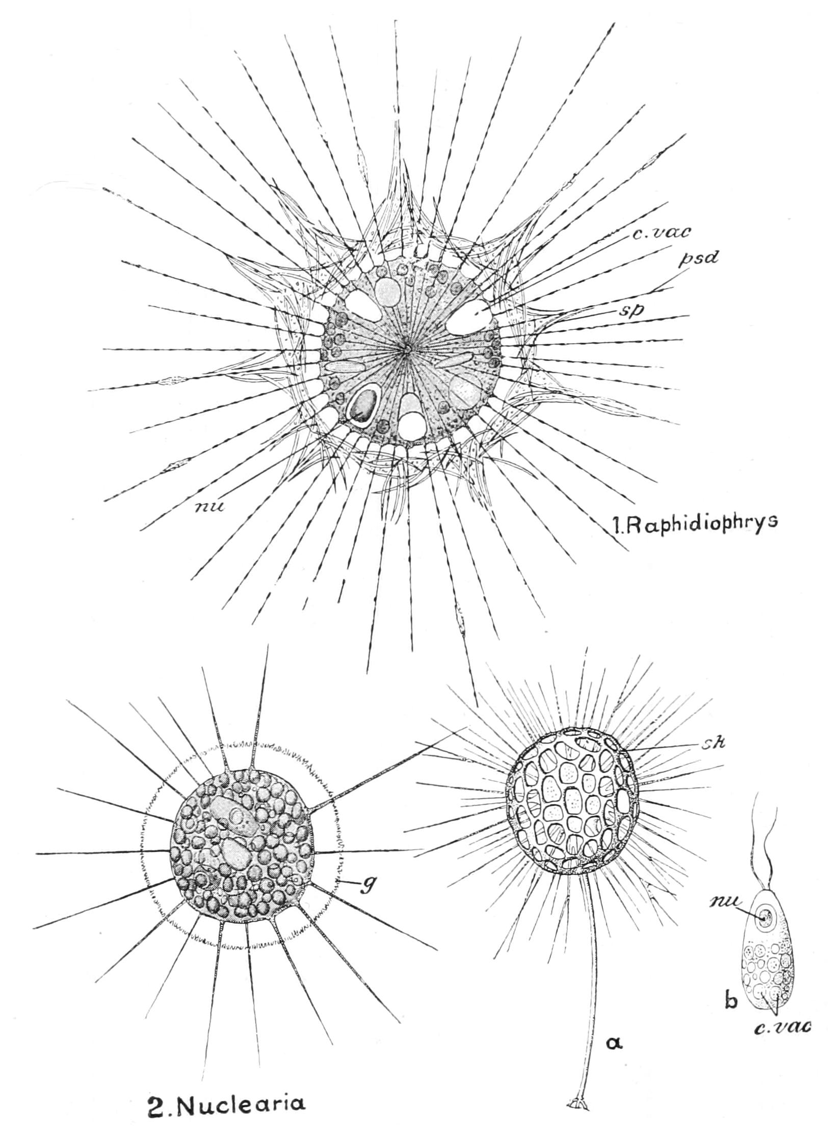
Fig. 20.—Various forms of Heliozoa. In 3, a is the entire animal and b the flagellula; c.vac, contractile vacuole; g, gelatinous investment; nu, nucleus; psd, pseudopodia; sk, siliceous skeleton; sp, spicules. (From Parker and Haswell, after other authors.)
Each of these divides, and the two sister cells then conjugate after the same fashion as in Actinophrys, but the nuclear divisions to form the coupling nucleus are two in number, i.e. the nucleus divides into two, one of which goes to the surface as the first polar body, and the sister of this again divides to form a second polar body (which also passes to the surface) and a pairing nucleus.[84] The two cells then fuse completely, and surround themselves with a second gelatinous cyst wall, separated from the outer one by a layer of siliceous spicules. The nucleus appears to divide at least twice before the young creep out, to divide immediately into as many Actinophrys-like cells as there were nuclei; then each of these multiplies its nuclei, to become apocytial like the adult form.

Fig. 21.—Diagram illustrating the conjugation of Actinosphaerium. 1, Original cell; 2, nucleus divides to form two, N2N2; 3, each nucleus again divides to form two, N3 and n3, the latter passing out with a little cytoplasm as an abortive cell; 4, repetition of the same process as in 3; 5, the two nuclei N4 have fused in syngamy to form the zygote nucleus Nz.
Schaudinn admits 24 genera (and 7 doubtful) and 41 species (and 18 doubtful). None are known fossil. Their geographical distribution is cosmopolitan, as is the case with most of the minute fresh-water Protista; 8 genera are exclusively marine, and Orbulinella has only been found in a salt-pond; Actinophrys sol is both fresh-water and marine, and Actinolophus has 1 species fresh-water, the other marine. One of the 14 species of Acanthocystis is marine; the remaining genera and species are all inhabitants of fresh water.[85]
4. Radiolaria
Sarcodina with the protoplasm divided by a perforated chitinous central capsule into a central mass surrounding the nucleus, and an outer layer; the pseudopodia radiate, never anastomosing enough to form a marked network; skeleton either siliceous, of spicules, or perforated; or of definitely arranged spicules of proteid matter (acanthin), sometimes also coalescing into a latticed shell; reproduction by fission and by zoospores formed in the central capsule. Habitat marine, suspended at the surface (plankton), at varying depths (zonarial), or near the bottom (abyssal).
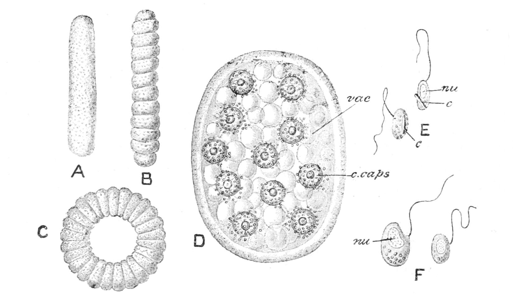
Fig. 22.—Collozoum inerme. A, B, C, three forms of colony; D, small colony with central capsules (c.caps), containing nuclei, and alveoli (vac) in ectoplasm; E, isospores, with crystals (c); F, anisospores; nu, nucleus. (From Parker and Haswell.)
The following is Haeckel's classification of the Radiolaria:—
I. Porulosa (Holotrypasta).—Homaxonic, or nearly so. Central capsule spherical in the first instance; pores numerous, minute, scattered; mostly pelagic.
A. Spumellaria (Peripylaea).—Pores evenly scattered; skeleton of solid siliceous spicules, or continuous, and reticulate or latticed, rarely absent; nucleus dividing late, as an antecedent to reproduction.
B. Acantharia (Actipylaea).—Pores aggregated into distinct areas; skeleton of usually 20 centrogenous, regularly radiating spines of acanthin, whose branches may coalesce into a latticed shell; nucleus dividing early.
II. Osculosa (Monotrypasta).—Monaxonic; pores of central capsule limited to the basal area (osculum), sometimes accompanied by two (or more) smaller oscula at apical pole, mostly zonarial or abyssal.
C. Nassellaria (Monopylaea).—Central capsule ovoid, of a single layer; pores numerous on the operculum or basal field; skeleton siliceous, usually with a principal tripod or calthrop-shaped spicule passing, by branching, into a complex ring or a latticed bell-shaped shell; nucleus eccentric, near apical pole.
D. Phaeodaria (Cannopylaea, Haeck.; Tripylaea, Hertw.).—Central capsule spheroidal, of two layers, in its outer layer an operculum, with radiate ribs and a single aperture, beyond which protrudes the outer layer; osculum basal, a dependent tube (proboscis); accessory oscula, when present, simpler, usually two placed symmetrically about the apical pole; skeleton siliceous, with a combination of organic matter, often of hollow spicules; nucleus sphaeroidal, eccentric; extracapsular protoplasm containing an accumulation of dusky pigment granules ("phaeodium").
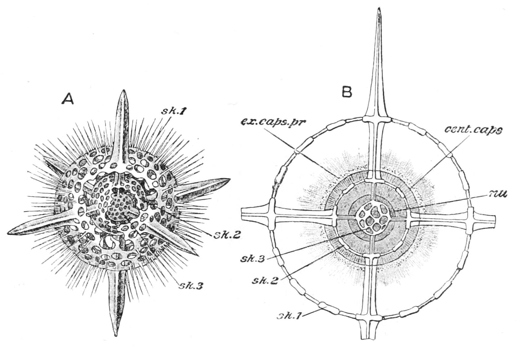
Fig. 23.—Actinomma asteracanthion. A, the shell with portions of the two outer spheres broken away; B, section showing the relations of the skeleton to the animal, cent.caps, Central capsule; ex.caps.pr, extra-capsular protoplasm: nu, nucleus; sk.1, outer, sk.2, middle, sk.3, inner sphere of skeleton. (From Parker and Haswell, after Haeckel and Hertwig.)
A. Spumellaria.
Sublegion (1). Collodaria.[86]—Skeleton absent or of detached spicules; colonial or simple.
Order i. Colloidea.—Skeleton absent. (Families 1, 2.) Thalassicolla Huxl.; Thalassophysa Haeck.; Collozoum Haeck.; Collosphaera J. Müll.; Actissa Haeck.
Order ii. Beloidea.—Skeleton spicular. (Families 3, 4.)
Sublegion (2). Sphaerellaria.—Skeleton continuous, latticed or spongy, reticulate.
Order iii. Sphaeroidea.—Skeleton of one or several concentric spherical shells; sometimes colonial. (Families 5-10.) Haliomma Ehrb.; Actinomma Haeck. (Fig. 23).
Order iv. Prunoidea.—Skeleton a prolate sphaeroid or cylinder, sometimes constricted towards the middle, single or concentric. (Families 11-17.)
Order v. Discoidea.—Shell flattened, of circular plan, simple or concentric, rarely spiral. (Families 18-23.)
Order vi. Larcoidea.—Shell ellipsoidal, with all three axes unequal or irregular, sometimes becoming spiral. (Families 24-32.)[87]
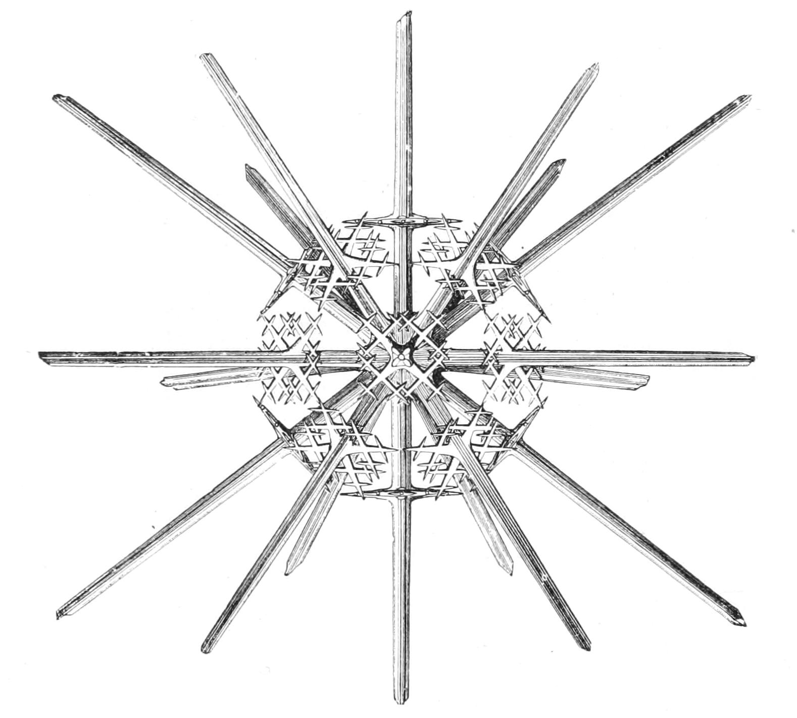
Fig. 24.—Xiphacantha (Acantharia). From the surface. The skeleton only, × 100, (From Wyville Thomson.)
B. Acantharia.
Order vii. Actinelida.—Radial spines numerous, more than 20, usually grouped irregularly. (Families 33-35.) Xiphacantha Haeck.
Order viii. Acanthonida.—Radial spines equal. (Families 36-38.)
Order ix. Sphaerophracta.—Radial spines 20, with a latticed spherical shell, independent of, or formed from the reticulations of the spines. (Families 39-41.) Dorataspis Haeck. (Fig. 25, A).
Order x. Prunophracta.—Radial spines 20, unequal; latticed shell, ellipsoidal, lenticular, or doubly conical. (Families 42-44.)
C. Nassellaria.
Order xi. Nassoidea.—Skeleton absent. (Family 45.)
Order xii. Plectoidea.—Skeleton of a single branching spicule, the branches sometimes reticulate, but never forming a latticed shell or a sagittal ring. (Families 46-47.)
Order xiii. Stephoidea.—Skeleton with a sagittal ring continuous with the branched spicule, and sometimes other rings or branches. (Families 48-51.) Lithocercus Théel (Fig. 26, A).
Order xiv. Spyroidea.—Skeleton with a latticed shell developed around the sagittal ring (cephalis), and constricted in the sagittal plane, with a lower chamber (thorax) sometimes added. (Families 52-55.)
Order xv. Botryoidea.—As in Spyroidea, but with the cephalis 3-4 lobed; lower chambers, one or several successively formed. (Families 56-58.)
Order xvi. Cyrtoidea.—Shell as in the preceding orders, but without lobing or constrictions. (Families 59-70.) Theoconus Haeck. (Fig. 25, B).
D. Phaeodaria.
Order xvii. Phaeocystina.—Skeleton 0 or of distinct spicules; capsule centric. (Families 71-73.) Aulactinium Haeck. (Fig. 26, B).
Order xviii. Phaeosphaeria.—Skeleton a simple or latticed sphere, with no oral opening (pylome); capsule central. (Families 74-77.)
Order xix. Phaeogromia.—Skeleton a simple latticed shell with a pylome at one end of the principal axis; capsule excentric, sub-apical. (Families 78-82.) Pharyngella Haeck.; Tuscarora Murr.; Haeckeliana Murr. (Fig. 28).
Order xx. Phaeoconchia.—Shell of two valves, opening in the plane ("frontal") of the three openings of the capsule. (Families 83-85.)
We exclude Haeckel's Dictyochida, with a skeleton recalling that of the Stephoidea, but of the impure hollow substance of the Phaeodaria (p. 84). They rank now as Silicoflagellates (p. 114).
The Radiolarian is distinguished from all other Protozoa by the chitinous central capsule, so that its cytoplasm is separated into an outer layer, the extracapsular protoplasm (ectoplasm), and a central mass, the intracapsular, containing the nucleus.[88]
The extracapsular layer forms in its substance a gelatinous mass, of variable reaction, through which the plasma itself ramifies as a network of threads ("sarcodictyum"), uniting at the surface to constitute the foundation for the pseudopodia. This gelatinous matter constitutes the "calymma." It is largely vacuolated, the vacuoles ("alveoli"), of exceptional size, lying in the nodes of the plasmic network, and containing a liquid probably of lower specific gravity than seawater; and they are especially abundant towards the surface, where they touch and become polygonal. On mechanical irritation they disappear, to be formed anew after an interval, a fact that may explain the sinking from the surface in disturbed water. This layer may contain minute pigment granules, but the droplets of oil and of albuminous matter frequent in the central layer are rare here. The "yellow cells" of a symbiotic Flagellate or Alga, Zooxanthella, are embedded in the jelly of all except Phaeodaria, and the whole ectosarc has the average consistency of a firm jelly.
The pseudopodia are long and radiating, with a granular external layer, whose streaming movements are continuous with those of the inner network. In the Acantharia they contain a firm axial filament, like that of the Heliozoa, which is traceable to the central capsule; and occasionally a bundle of pseudopodia may coalesce to form a stout process like a flagellum ("sarcoflagellum"). Here, too, each spine, at its exit from the jelly, is surrounded by a little cone of contractile filaments, the myophrisks, whose action seems to be to pull up the jelly and increase the volume of the spherical body so as to diminish its density.
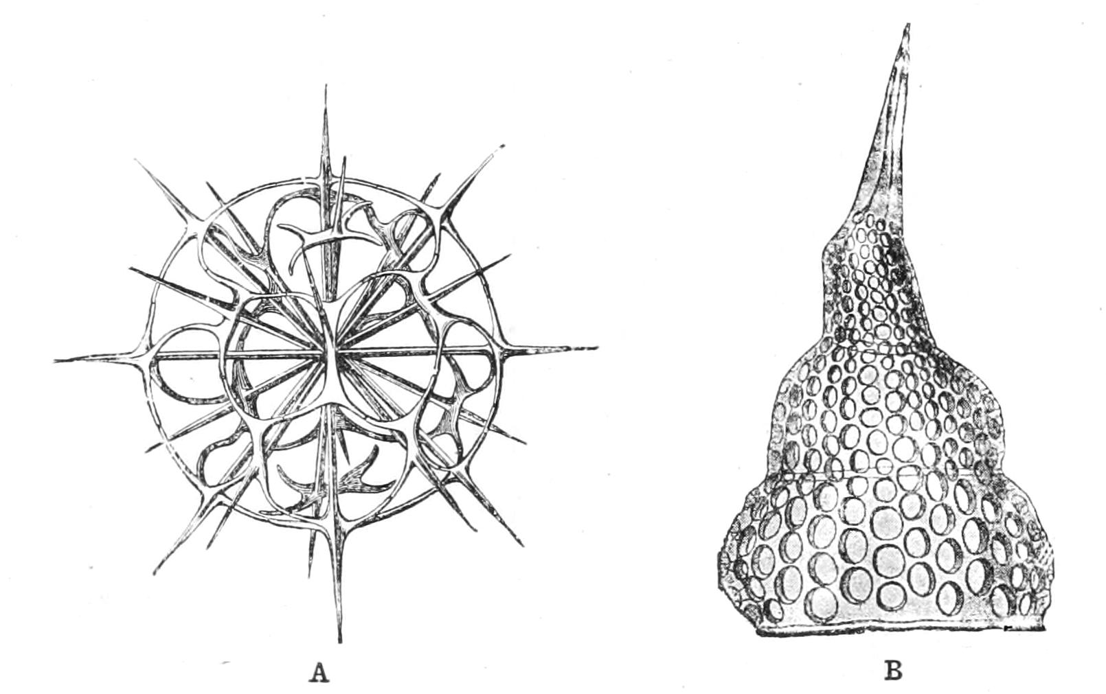
Fig. 25.—Skeletons of Radiolaria. A, Dorataspis; B, Theoconus. (After Haeckel.)
The intracapsular protoplasm is free from Zooxanthella except in the Acantharia. It is less abundantly vacuolated, and is finely granular. In the Porulosa it shows a radial arrangement, with pyramidal stretches of hyaline plasma separated by intervals rich in granules. Besides the alveoli with watery contents, others are present with albuminoid matter in solution. Oil-drops, often brilliantly coloured, occur either in the plasma or floating in either kind of vacuole; and they are often luminous at night. Added to these, the intracapsular plasm contains pigment-granules, most frequently red or orange, passing into yellow or brown, though violet, blue, and green also occur. The "phaeodium,"[89] however, that gives its name to the Phaeodaria, is an aggregate of dark grey, green, or brown granules which are probably formed in the endoplasm, but accumulate in the extracapsular plasm of the oral side of the central capsule. Inorganic concretions and crystals are also found in the contents of the central capsule, as well as aggregates of unknown composition, resembling starch-grains in structure.
In the Monopylaea, or Nassellaria (Figs. 25, B, 26, A), the endoplasm is differentiated above the perforated area of the central capsule into a cone of radiating filaments termed the "porocone," which may be channels for the communication between the exoplasm and the endoplasm, or perhaps serve, as Haeckel suggests, to raise, by their contraction, the perforated area: he compares them to the myophane striae of Infusoria. In the Phaeodaria (Fig. 26, B), a radiating laminated cone is seen in the outermost layer of the endoplasm above the principal opening ("astropyle"), and a fibrillar one around the two accessory ones ("parapyles"); and in some cases, continuous with these, the whole outer layer of the endoplasm shows a meridional striation.
The nucleus is contained in the endoplasm, and is always at first single, though it may divide again and again. The nuclear wall is a firm membrane, sometimes finely porous. If there are concentric shells it at first occupies the innermost, which it may actually come to enclose, protruding lobes which grow through the several perforations of the lattice-work, finally coalescing outside completely, so as to show no signs of the joins. In the Nassellaria a similar process usually results in the formation of a lobed nucleus, contained in an equally lobed central capsule. The chromatin of the nucleus may be concentrated into a central mass, or distributed into several "nucleoli," or it may assume the form of a twisted, gut-like filament, or, again, the nuclear plasm may be reticulated, with the chromatin deposited at the nodes of the network.
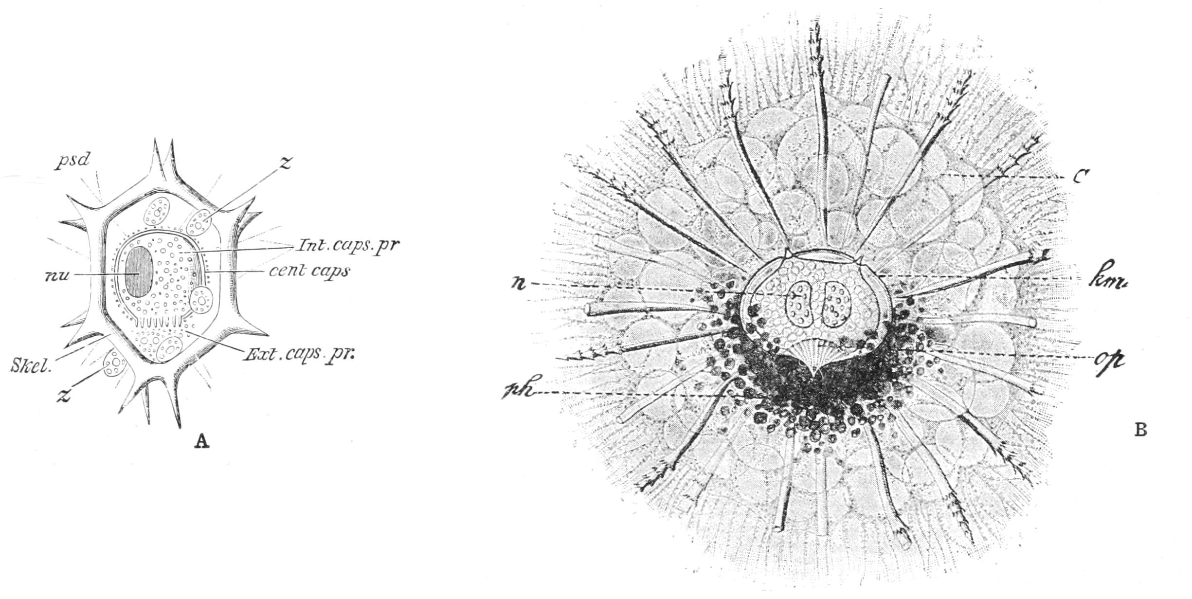
Fig. 26.—A, Lithocercus annularis, with sagittal ring (from Parker and Haswell). B, Aulactinium actinastrum. C, calymma; cent.caps., km, central capsule; Ext.caps.pr., Extracapsular, and Int.caps.pr., intracapsular protoplasm; n, nu, nucleus; op, operculum; ph, phaeodium; psd, pseudopodium; Skel., skeleton; z, Zooxanthella. (From Lang's Comparative Anatomy, after Haeckel.)
The skeleton of this group varies, as shown in our conspectus, in the several divisions.[90] The Acantharia (Figs. 24, 25, A) have a skeleton of radiating spines meeting in the centre of figure of the endoplasm, and forcing the nucleus to one side. The spines are typically 20 in number, and emerge from the surface of the regular spherical forms (from which the others may be readily derived) radially, in five sets of four in the regions corresponding to the equator and the tropics and polar circles of our world. The four rays of adjacent circles alternate, so that the "polar" and "equatorial" rays are on one set of meridians 90° apart, and the "tropical" spines are on the intermediate meridians, as shown in the figures. By tangential branching, and the meeting or coalescence of the branches, reticulate (Figs. 23, 24, 25) and latticed shells are formed in some families, with circles of openings or pylomes round the bases of the spines. In the Sphaerocapsidae the spines are absent, but their original sites are inferred from the 20 circles of pylomes.
In the Spumellaria the simplest form of the (siliceous) skeleton is that of detached spicules, simple or complex, or passing into a latticed shell, often with one or more larger openings (pylomes). Radiating spines often traverse the whole of the cavity, becoming continuous with its latticed wall, and bind firmly the successive zones when present (Fig. 23).
Calcaromma calcarea was described by Wyville Thomson as having a shell of apposed calcareous discs, and Myxobrachia, by Haeckel, as having collections of the calcareous Coccoliths and Coccospheres. In both cases we have to do with a Radiolarian not possessing a skeleton, but retaining the undigested shells of its food, in the former case (Actissa) in a continuous layer, in the latter (Thalassicolla) in accumulations that, by their weight, droop and pull out the lower hemisphere into distinct arms.
The (siliceous) skeleton of the Nassellaria is absent only in the Nassoidea, and is never represented by distinct spicules. Its simplest form is a "tripod" with the legs downward, and the central capsule resting on its apex. The addition of a fourth limb converts the tripod into a "calthrop," the central capsule in this case resting between the upturned leg and two of the lower three regarded as the "anterolateral"; the odd lower leg, like the upturned one, being "posterior." Again, the skeleton may present a "sagittal ring," often branched and spiny (Fig. 26, A), or combined with the tripod or calthrop, or complicated by the addition of one or more horizontal rings. Another type is presented by the "latticed chamber" surrounding the central capsule, with a wide mouth ("pylome") below. This is termed the "cephalis"; it may be combined in various ways with the sagittal ring and the tripod or calthrop; and, again, it may be prolonged by the addition of one, two, or three chambers below, the last one opening by a pylome (Fig. 25, B). These are termed "thorax," "abdomen," and "post-abdomen" respectively.
In the Phaeodaria the skeleton may be absent, spicular (of loose or connected spicules) or latticed, continuous or bivalve. It is composed of silica combined with organic matter, so that it chars when heated, is more readily dissolved, and is not preserved in fossilisation. The spicules or lattice-work are hollow, often with a central filament running in the centre of the gelatinous contents. The latticed structure of the shell of the Challengeridae (Fig. 28) is so fine as to recall that of the Diatomaceae. In the Phaeoconchida the shell is in two halves, parted along the "frontal" plane of the three apertures of the capsule.
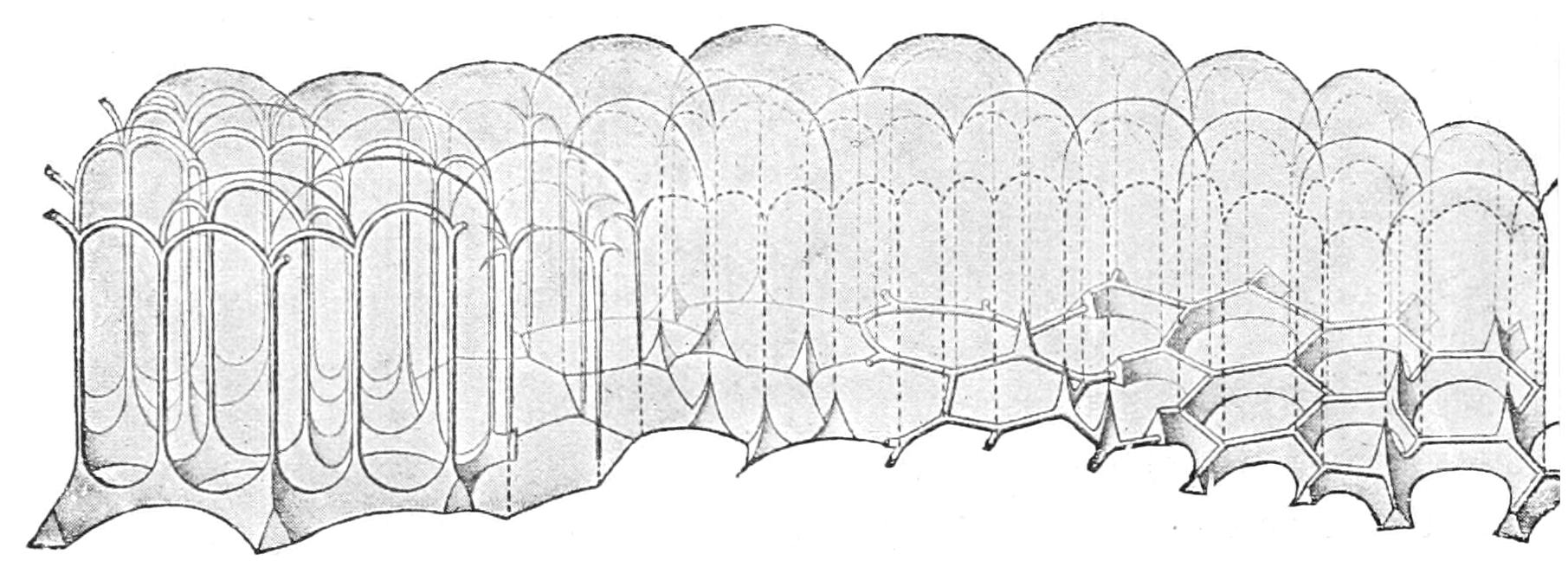
Fig. 27.—Scheme of various possible skeletal forms deposited in the meshes of an alveolar system, most of which are realised in the Radiolaria. (From Verworn, after Dreyer.)
The central capsule (rarely inconspicuous and difficult, if not impossible to demonstrate) is of a substance which resembles chitin, though its chemical reactions have not been fully studied hitherto, and indeed vary from species to species. It is composed of a single layer, except in Phaeodaria, where it is double. The operculum in this group, i.e. the area around the aperture, is composed of an outer layer, which is radially thickened, and a thin inner layer; the former is produced into the projecting tube ("proboscis").
Reproduction in the Radiolaria may be simple fission due to the binary fission of the nucleus, the capsule, and the ectoplasm in succession. If this last feature is omitted we have a colonial organism, composed of the common ectoplasm containing numerous central capsules; and the genera in which this occurs, all belonging to the Peripylaea, were formerly separated (as Polycyttaria) from the remaining Radiolaria (Monocyttaria). They may either lack a skeleton (Collozoidae, Fig. 22), or have a skeleton of detached spicules (Sphaerozoidae), or possess latticed shells (Collosphaeridae) one for each capsule, and would seem therefore to belong, as only differentiated by their colonial habit, to the several groups having these respective characters. Fission has been well studied in Aulacantha (a Phaeodarian) by Borgert.[91] He finds that in this case the skeleton is divided between the daughter-cells, and the missing part is regenerated. In cases where this is impossible one of the daughter-cells retains the old skeleton, and the other escapes as a bud to form a new skeleton.
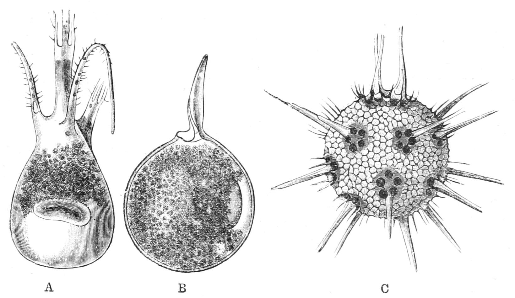
Fig. 28.—Shells of Challengeridae: A, Tuscarora; B, Pharyngella; C, Haeckeliana. (From Wyville Thomson.)
Two modes of reproduction by flagellate zoospores have been described (Fig. 22). In the one mode all the zoospores are alike—isospores—and frequently contain a crystal of proteid nature as well as oil-globules. In the Polycyttaria alone has the second mode of spore-formation been seen, and that in the same species in which the formation of isospores occurs. Here "anisospores" are formed, namely, large "mega-," and small "micro-zoospores." They probably conjugate as male and female respectively; but neither has the process been observed, nor has any product of such conjugation (zygote) been recognised. In every case the formation of the zoospores only involves the endoplasm: the nucleus first undergoes brood division, and the plasma within the capsule becomes concentrated about its offspring, and segregates into the spores; the extracapsular plasm disintegrates.[92]
The Yellow Cells (Zooxanthella), so frequently found in the Radiolaria were long thought to be constituents of their body. Cienkowsky found that when the host died from being kept in unchanged water, the yellow cells survived and multiplied freely, often escaping from the gelatinised cell-wall as biflagellate zoospores. The cell-wall is of cellulose. The cell contains two chloroplastids, or plates coloured with the vegetal pigment "diatomin." Besides ordinary transverse fission in the ordinary encysted state in the ectoplasm of the host, when free they may pass into what is known as a "Palmella-state," the cell-walls gelatinising; in this condition they multiply freely, and constitute a jelly in which the individual cells are seen as rounded bodies. They contain starch in two forms—large hollow granules, not doubly refractive, and small solid granules which polarise light. We may regard them as Chrysomonadaceae (p. 113). Similar organisms occur in many Anthozoa (see pp. 261, 339, 373 f., 396). Diatomaceae (yellow Algae with silicified cell-walls) sometimes live in the jelly of certain Collosphaera. Both these forms live in the state known as "symbiosis" with their host; i.e. they are in mutually helpful association, the Radiolarian absorbing salts from the water for the nutrition of both, and the Alga or Flagellate taking up the CO2 due to the respiration of the host, and building up organic material, the surplus of which is doubtless utilised, at least in part, for the nutrition of the host. A similar union between a Fungus and a coloured vegetal ("holophytic") organism is known as a Lichen.
The Suctorian Infusorian Amoebophrya is parasitic in the ectoplasm of certain Acantharia, and in the peculiar genus Sticholonche which appears to be intermediate between this group and Heliozoa.
The Silicoflagellate family Dictyochidae are found temporarily embedded in the ectoplasm of some of the Phaeocystina, and have a skeleton of similar nature. Their true nature was shown by Borgert.
The Amphipod crustacean Hyperia[93] may enter the jelly of the colonial forms, and feed there at will on the host.[94]
Haeckel, in his Monograph of the Radiolaria of the Challenger enumerated 739 genera, comprising 4318 species; and Dreyer has added 6 new genera, comprising 39 species, besides 7 belonging to known genera. Possibly, as we shall see, many of the species may be mere states of growth, for it is impossible to study the life-histories of this group; on the other hand, it is pretty certain that new forms are likely to be discovered and described. The Radiolaria are found living at all depths in the sea, by the superficial or deep tow-net; and some appear to live near the bottom, where the durable forms of the whole range also settle and accumulate. They thus form what is known as Radiolarian ooze, which is distinguished from other shallower deposits chiefly through the disappearance by solution of all calcareous skeletons, as they slowly fell through the waters whereon they originally floated at the same time with the siliceous remains of the Radiolaria. The greatest wealth of forms is found in tropical seas, though in some places in cold regions large numbers of individuals of a limited range of species have been found.
Radiolaria of the groups with a pure siliceous skeleton can alone be fossilised, even the impure siliceous skeleton of the Phaeodaria readily dissolving in the depths at which they live: they have been generally described by Ehrenberg's name Polycystineae. Tripolis (Kieselguhr) of Tertiary ages have been found in many parts of the globe, consisting largely or mainly of Radiolaria, and representing a Radiolarian ooze. That of the Miocene of Barbados contains at least 400 species; that of Gruppe at least 130. In Secondary and Palaeozoic rocks such oozes pass into Radiolarian quartzites (some as recent as the Jurassic). They occur also in fossilised excrement (coprolites), and in flint or chert concretions, as far down as the lowest fossiliferous rocks, the Cambrian. The older forms are simple Sphaerellaria and Nassellaria. From a synopsis of the history of the order in Haeckel's Monograph (pp. clxxxvi.-clxxxviii.) we learn that while a large number of skeletal forms had been described by Ehrenberg, Huxley in 1851 published the first account of the living animal. Since then our knowledge has been extended by the labours of Haeckel, Cienkowsky, R. Hertwig, Karl Brandt, and A. Borgert.
5. Proteomyxa
Sarcodina without a clear ectoplasm, whose active forms are amoeboid or flagellate, or pass from the latter form to the former; multiplying chiefly, if not exclusively, by brood-formation in a cyst. No complete cell-pairing (syngamy) known, though the cytoplasms may unite into plasmodia; pseudopodia of the amoeboid forms usually radiate or filose, but without axial filaments. Saprophytic or parasitic in living animals or plants.
This group is a sort of lumber-room for forms which it is hard to place under Rhizopoda or Flagellata, and which produce simple cysts for reproduction, not fructifications like the Mycetozoa. The cyst may be formed for protection under drought ("hypnocyst"), or as a preliminary to spore-formation ("sporocyst"). The latter may have a simple wall (simple sporocyst), or else two or three formed in succession ("resting cyst"), so as to enable it to resist prolonged desiccation, etc.: both differing from the hypnocyst in that their contents undergo brood formation. On encystment any indigestible food materials are extruded into the cyst, and in the "resting cysts," which are usually of at least two layers, this faecal mass lies in the space between them. The brood-cells escape, either as flagellate-cells, resembling the simpler Protomastigina, called "flagellulae," and which often become amoeboid (Fig. 29); or already furnished with pseudopodia, and called "amoebulae," though they usually recall Actinophrys rather than Amoeba. In Vampyrella and some others the amoebulae fuse, and so attain a greater size, which is most probably advantageous for feeding purposes. But usually it is as a uninucleate cell that the being encysts. They may feed either by ingestion by the pseudopodia, by the whole surface contained in a living host-cell, or by passing a pseudopodium into a host-cell (Fig. 29 5). They may be divided as follows:—
A. Myxoidea.—Flagella 1-3; zoospores separating at once.
1. Zoosporeae.—Brood-cells escaping as flagellulae, even if they become amoeboid later. Ciliophrys Cienk.; Pseudospora Cienk. (Fig. 29).
2. Azoosporeae.—Cells never flagellate. Protomyxa Haeckel; Plasmodiophora Woronin; Vampyrella Cienk.; Serumsporidium L. Pfeiffer.
B. Catallacta.—Brood-cells of cyst on liberation adhering at the centre to form a spherical colony, multiflagellate; afterwards separating, and becoming amoeboid. Magosphaera Haeckel (marine).[95]
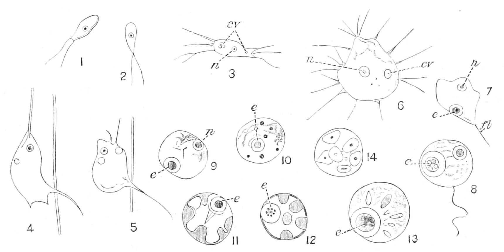
Fig. 29.—Pseudospora lindstedtii. 1, 2, Flagellate zoospores; 3, young amoebula, with two contractile vacuoles, one being reconstituted by three minute formative vacuoles; 4, 5, an amoebula migrating to a fungus hypha through the wall of which it has sent a long pseudopodium; 6, amoebula full-grown; 7, 8, mature cells rounded off, protruding a flagellum, before encysting; 9, young sporocyst; 10, the nucleus has divided into a brood of eight; 11-14, stages of formation of zoospores. cv, Contractile vacuole; e, mass of faecal granules; fl, flagellum; n, nucleus, × about 750⁄1.
Plasmodiophora infests the roots of Crucifers, causing the disease known as "Hanburies," or "fingers and toes," in turnips, etc. Serumsporidium dwells in the body cavity of small Crustacea. Many of this group were described by Cienkowsky under the name of "Monadineae" (in Arch. Mikr. Anat. i. 1865, p. 203). Zopf has added more than anyone else since then to our knowledge. He monographed them under Cienkowsky's name, as a subordinate group of the Myxomycetes, "Pilzthiere oder Schleimpilze," in Schenk's Handb. d. Bot. vol. iii. pt. ii. (1887). To Lankester (Encycl. Brit., reprint 1891) we owe the name here adopted. Zopf has successfully pursued their study in recent papers in his Beitr. Nied. Org. The Chytridieae, usually ascribed to Fungi, are so closely allied to this group that Zopf proposes to include at least the Synchytrieae herein.
This group is very closely allied to Sporozoa; for the absence of cytogamy, and of sickle-germs,[96] and of the complex spores and cysts of the Neosporidia, are the only absolute distinctions.
6. Mycetozoa (Myxomycetes, Myxogastres)
Sarcodina moving and feeding by pseudopodia, with no skeleton, aggregating more or less completely into complex "fructifications" before forming 1-nucleate resting spores; these may in the first instance liberate flagellate zoospores, which afterwards become amoeboid, or may be amoeboid from the first; zoospores capable of forming hypnocysts from which the contents escape in the original form.
1. Aggregation taking place without plastogamy, zoospores amoeboid, with a clear ectosarc Acrasieae.
Copromyxa Zopf; Dictyostelium Brefeld.
2. Aggregation remaining lax, with merely thread-like connexions, except when encystment is to take place; cytoplasm finely granular throughout; complete fusion of the cytoplasm doubtful Filoplasmodieae
Labyrinthula Cienk.; Chlamydomyxa Archer; Leydenia (?) Schaud.
3. Plasmodium formation complete, eventuating in the formation of a complex fructification often traversed by elastic, hygroscopic threads, which by their contraction scatter the spores; zoospores usually flagellate at first Myxomycetes.
Fuligo Hall.; Chondrioderma Rostaf.; Didymium Schrad. (Fig. 30).
I. The Acrasieae are a small group of saprophytes, often in the most literal sense, though in some cases it has been proved that the actual food is the bacteria of putrefaction. In them, since no cell-division takes place in the fructification, it is certain that the multiplication of the species must be due to the fissions of the amoeboid zoospores, which often have the habit of Amoeba limax (Fig. 1, p. 5).
II. Filoplasmodieae.—Chlamydomyxa[97] is a not uncommon inhabitant of the cells of bog-mosses and bog-pools, and its nutrition may be holophytic, as it contains chromoplasts; but it can also feed amoeba-fashion. Labyrinthula is marine, and in its fructification each of the component cells forms four spores. Leydenia has been found in the fluid of ascitic dropsy, associated with malignant tumour.
III. Myxomycetes.—The fructification in this group is not formed by the mere aggregation of the zoospores, but these fuse by their cytoplasm to form a multinucleate body, the "plasmodium," which, after moving and growing (with nuclear division) for some time like a great multinucleate Reticularian, passes into rest, and develops a fructification by the formation of a complex outer wall; within this the contents, after multiplication of the nuclei, resolve themselves into uninucleate spores, each with its own cyst-wall. The fructifications of this group are often conspicuous, and resemble those of the Gasteromycetous fungi (e.g., the Puffballs), whence they were at first called Myxogastres. De Bary first discovered their true nature in 1859, and ever since they have been claimed by botanist and zoologist alike.
The spore on germination liberates its contents as a minute flagellate, with a single anterior lash and a contractile vacuole (Fig. 30, C). It soon loses the lash, becomes amoeboid, and feeds on bacteria, etc. (Fig. 30, D, E). In this state it can pass into hypnocysts, from which, as from the spores, it emerges as a flagellula. After a time the amoeboids, which may multiply by fission, fuse on meeting, so as to form the plasmodium (Fig. 30, F). This contains numerous nuclei, which multiply as it grows, and numerous contractile vacuoles. When it attains full size it becomes negatively hydrotactic, crawls to a dry place, and resolves itself into the fructification. The external wall, and sometimes a basal support to the fruit, are differentiated from the outer layer of protoplasm; while the nuclei within, after undergoing a final bipartition, concentrate each around an independent portion of plasma, which again is surrounded as a spore by a cyst-wall. Often the maturing plasmodium within the wall of the fruit is traversed by a network of anastomosing tubes filled with liquid, the walls of which become differentiated into membrane like the fruit-wall, and are continuous therewith. As the fruit ripens the liquid dries, and the tubes now form a network of hollow threads, the "capillitium," often with external spiral ridges (Fig. 30, A, B). These are very hygroscopic, and by their expansion and contraction determine the rupture of the fruit-wall and the scattering of the spores.
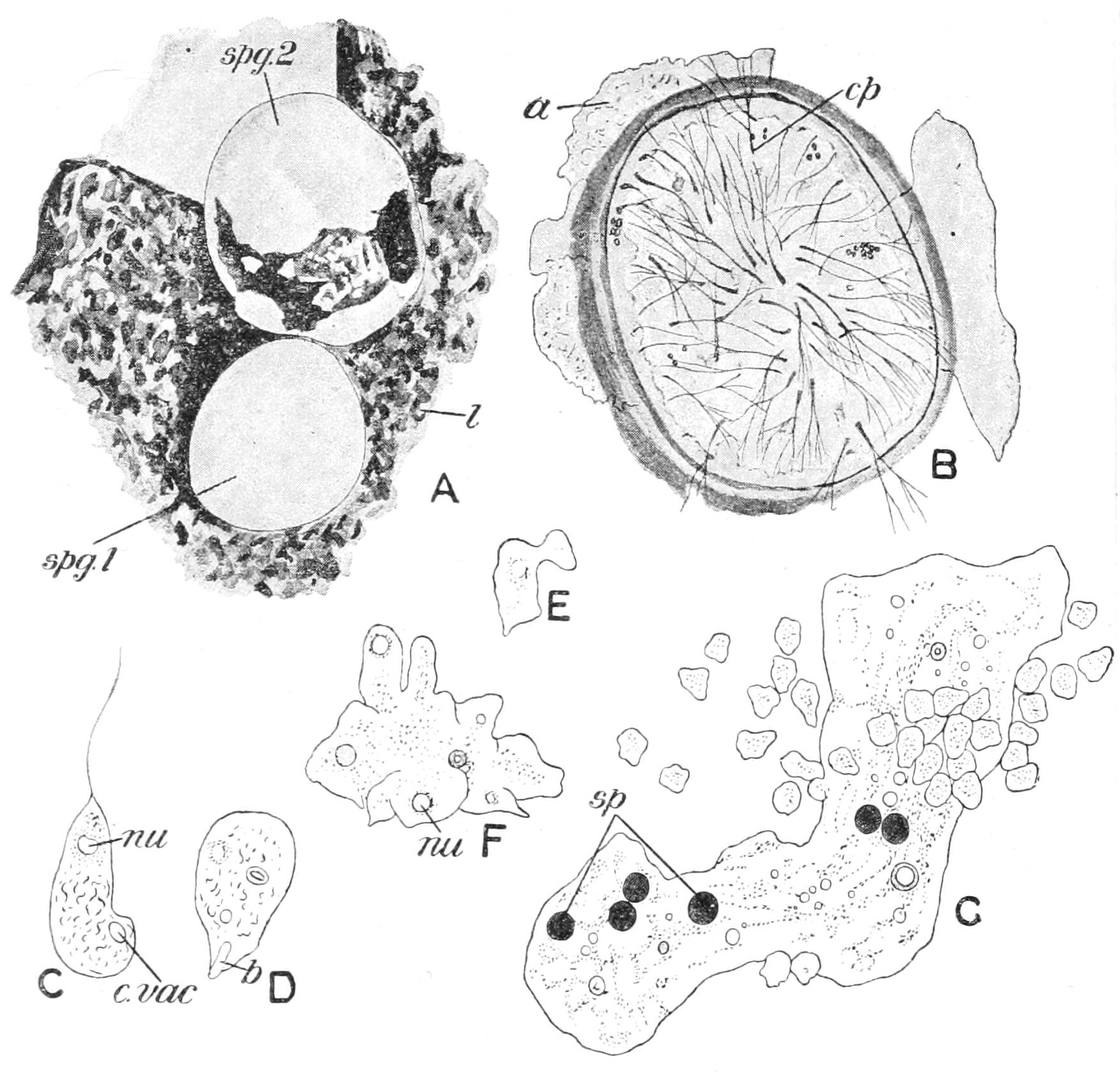
Fig. 30.—Didymium difforme. A, two sporangia (spg 1 and 2) on a fragment of leaf (l); B, section of sporangium, with ruptured outer layer (a), and threads of capillitium (cp); C, a flagellula with contractile vacuole (c.vac) and nucleus (nu); D, the same after loss of flagellum; b, an ingested bacillus; E, an amoebula; F, conjugation of amoebulae to form a small plasmodium; G, a larger plasmodium accompanied by numerous amoebulae; sp, ingested spores. (After Lister.)
Again, in some cases the plasmodia themselves aggregate in the same way as the amoeboids do in the Acrasieae, and combine to form a compound fruit termed an "aethalium,"[98] with the regions of the separate plasmodia more or less clearly marked off. The species formerly termed Aethalium septicum is now known as Fuligo varians. It is a large and conspicuous species, common on tan, and is a pest in the tanpits. Its aethalia may reach a diameter of a foot and more, and a thickness of two inches. Chondrioderma diffusum, often utilised as a convenient "laboratory type," is common on the decaying haulms of beans in the late autumn. The interest of this group is entirely biological, save for the "flowers of tan."[99]