Chapter V
Reading edition
PROTOZOA (CONTINUED): FLAGELLATA
III. Flagellata.
Protozoa moving (and feeding in holozoic forms) by long flagella: pseudopodia when developed usually transitory: nucleus single or if multiple not biform: reproduction occurring in the active state and usually by longitudinal fission, sometimes alternating with brood-formation in the cyst or more rarely in the active state: form usually definite: a firm pellicle or distinct cell-wall often present.
The Flagellates thus defined correspond to Bütschli's group of the Mastigophora. The lowest and simplest forms, often loosely called "Monads," are only distinguishable from Sarcodina (especially Proteomyxa) and Sporozoa by the above characters: their artificial nature is obvious when we remember that many of the Sarcodina have a flagellate stage, and that the sperms of bisexual Sporozoa are flagellate (as are indeed those of all Metazoa except Nematodes and most Crustacea). Even as thus limited the group is of enormous extent, and passes into the Chytridieae and Phycomycetes Zoosporeae on the one hand, and by its holophytic colonial members into the Algae, on the other.[115]
Classification.
A. Fission usually longitudinal (transverse only in a cyst), or if multiple, radial and complete: pellicle absent, thin, or if armour-like, with not more than two valves.
I. Food taken in at any part of the body by pseudopodia 1. PANTOSTOMATA
Multicilia Cienk.; Mastigamoeba F. E. Sch. (Fig. 37, 4).
II. Food taken in at a definite point or points, or by absorption, or nutrition holophytic.
1. No reticulate siliceous shell. Diameter under 500 µ (1⁄50").
* Contractile vacuole simple (one or more).
(α) Colourless: reserves usually fat: holozoic, saprophytic or parasitic 2. Protomastigaceae
(β) Plastids yellow or brown: reserves fat or proteid: nutrition variable: body naked, often amoeboid in active state (C. nudae), or with a test, sometimes containing calcareous discs ("coccoliths," "rhabdoliths") of peculiar form (C. loricatae) 3. Chrysomonadaceae
Chromulina Cienk.; Chrysamoeba Klebs; Hydrurus Ag. Dinobryon Ehrb. (Fig. 37, 11); Syncrypta Ehrb. (Fig. 37, 12); Zooxanthella Brandt; Pontosphaera Lohm.; Coccolithophora Lohm.; Rhabdosphaera Haeck.
(γ) Green, (more rarely yellow or brown) or colourless: reserves starch: fission longitudinal 4. Cryptomonadaceae
Cryptomonas Ehrb. (Fig. 37, 9); Paramoeba Greeff.
(δ) Green (rarely colourless): fission multiple, radial 5. Volvocaceae
** System of contractile vacuoles complex, with accessory formative vacuoles or reservoir, or both.
(ε) Pellicle delicate or absent: pseudopodia often emitted: excretory pore distinct from flagellar pit: reserves fat 6. Chloromonadaceae
Chloramoeba Lagerheim; Thaumatomastix, Lauterborn.
(ζ) Pellicle dense, tough or hard, often wrinkled or striate: contractile vacuole discharging by the flagellar pit. Nutrition variable 7. Euglenaceae
Euglena Ehrb.; Astasia Duj. (Fig. 37, 3); Anisonema Duj.; Eutreptia Perty (Fig. 42, p. 124); Trachelomonas Ehrb. (Fig. 37, 1); Cryptoglena Ehrb.
2. Skeleton an open network of hollow siliceous spicules. Plastids yellow. Diameter under 500 µ. 8. Silicoflagellata
Dictyocha Ehrb.
3. Diameter over 500 µ. Mouth opening into a large reticulate endoplasm: flagella 1, or 2, very unequal. 9. Cystoflagellata
Noctiluca Suriray (Fig. 48); Leptodiscus R. Hertw.
B. Fission oblique or transverse: flagella two, dissimilar, the one coiled round the base of the other or in a traverse groove; pellicle often dense, of numerous armour-like plates 10. Dinoflagellata
Ceratium Schrank; Gymnodinium Stein; Peridinium Ehrb. (Fig. 46); Pouchetia Schütt; Pyrocystis Murray (Fig. 47); Polykrikos Bütschli.
The Protomastigaceae and Volvocaceae are so extensive as to require further subdivision.
Protomastigaceae
I. Oral spots 2. Flagella distant in pairs. Distomatidae
II. Oral spot 1 or 0.
A. Flagellum 1.
(a) No anterior process: often parasitic Oikomonadidae
Oikomonas K. (Figs. 37, 2, 8); Trypanosoma Gruby (Fig. 39, a-f); Treponema Vuill. (Fig. 39, g-i).
(b) Anterior process unilateral or proboscidiform: cell often thecate Bicoecidae
Bicoeca Clark; Poteriodendron St.
(c) Anterior process a funnel, surrounding the base of the flagellum: cells often thecate.
(i.) Funnel free Craspedomonadidae
Codosiga Clark; Monosiga Cl.; Polyoeca Kent; Proterospongia Kent; Salpingoeca Cl.
(ii.) Funnel not emerging from the general gelatinous investment Phalansteridae
B. Flagella 2, unequal or dissimilar in function, the one sometimes short and thick.
(a) Both flagella directed forwards Monadidae
Monas St.; Anthophysa Bory (Fig. 37, 13).
(b) One flagellum, usually the longer, turned backwards Bodonidae
Bodo St. (Fig. 38).
C. Flagella 2, equal and similar Amphimonadidae
Amphimonas Duj.; Diplomita K. (Fig. 37, 10); Rhipidodendron St. (Fig. 37, 14).
D. Flagella 3 Trimastigidae
Dallingeria K. (Fig. 37, 6); Costia Leclercq.
E. Flagella 4 or more: mostly parasitic in Metazoa Polymastigidae
Trichomonas Donne; Tetramitus Perty (Fig. 37, 7); Hexamitus Duj.; Lamblia Blanchard.
F. Flagella numerous, sometimes constituting a complete ciliiform investment, and occasionally accompanied by an undulating membrane: parasitic in Metazoa.
(a) Flagella long: nucleus single: parasitic in insects Trichonymphidae
Dinenympha Leidy; Joenia Grassi; Pyrsonympha Leidy; Trichonympha Leidy; Lophomonas St.; Maupasia Schew.
(b) Flagella short, ciliiform, uniformly distributed: nuclei very numerous, all similar: parasitic in Amphibia Opalinidae
Opalina Purkinje and Valentin (Fig. 41).
Volvocaceae
A. Cells usually isolated, separating after fission or brood-formation. Usually green (sometimes red), more rarely colourless saprophytes Chlamydomonadidae
Chlamydomonas Ehrb.; Phacotus Perty; Polytoma Ehrb.; Sphaerella Sommerf. (Fig. 43); Zoochlorella.
B. Cells multiplying in the active state by radial divisions in the same plane and usually incurving to form a spherical colony, united in a gelatinous investment, sometimes traversed by plasmic threads Volvocidae
Gonium O.F.M.; Eudorina Ehrb.; Pandorina Bory (Fig. 45); Stephanosphaera Cohn; Volvox L. (Fig. 44).
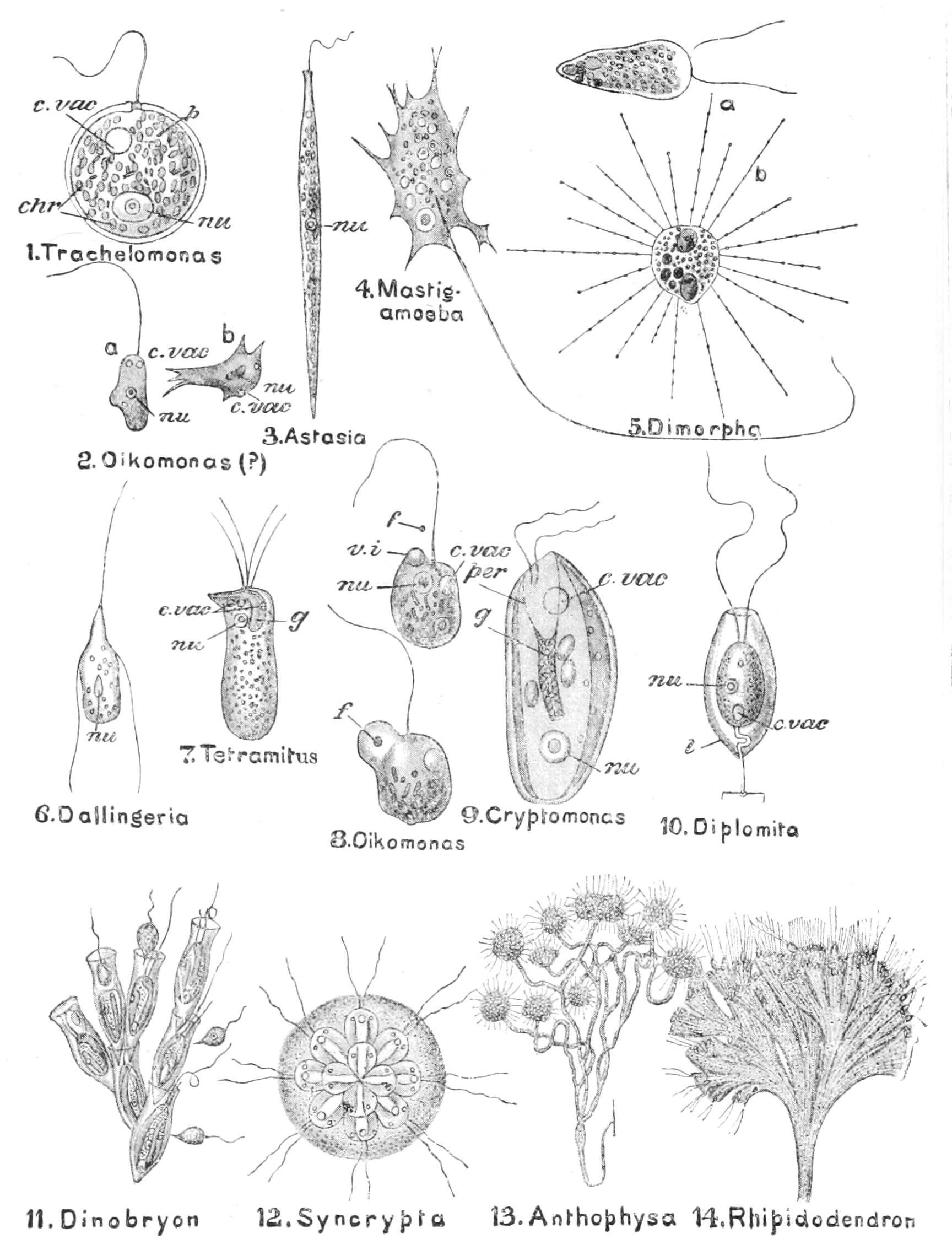
Fig. 37.—Various forms of Flagellata. 2, 6-8, 10, 13, 14, Protomastigaceae; 11, 12, Chrysomonadaceae; 9, Cryptomonadaceae; 1, 3, Euglenaceae; 4, Pantostomata: note branched stalk in 13; branched tubular theca in 14; distinct thecae in 11; stalk and theca in 10. In 2, flagellate (a) and amoeboid (b) phases are shown; in 5, flagellate (a) and Heliozoan (b) phases[116]; in 8 are shown two stages in the ingestion of a food particle (f); chr, plastoids; c.vac, contractile vacuole; f, food particle; g, gullet; l, theca; nu, nucleus; p, protoplasm; per, peristome; v.i, vacuole of ingestion. (From Parker and Haswell, mostly from Bütschli's Protozoa.)
The modes of nutrition are threefold: the simplest forms live in liquids containing decaying organic matter which they absorb through their surface ("saprophytic"): others take in food either Amoeba fashion, or into a vacuole formed for the purpose, or into a definite mouth ("holozoic"): others again have coloured plastids, green or brown or yellow ("holophytic"), having the plant's faculty of manufacturing their own food-supply. But we meet with species that show chromatophores at one time and lack them at another; or, again, the same individual (Euglena) may pass from holozoic life to saprophytic (Paramoeba, some Dinoflagellates) as conditions alter.
Many secrete a stalk at the hinder end: by "continuous" formation of this, without rupture at fission, a branching colony is formed (Polyoeca). This stalk may have a varying consistency. In Anthophysa (Fig. 37, 13) it appears to be due to the welding of excrementitious particles voided at the hinder end of the body with a gelatinous excretion; but the division of the stalk is here occasional or intermittent, so that the cells are found in tufts at the apex of the branches. A corresponding secretion, gelatinous or chitinous, around the body of the cell forms a cup or "theca," within which the cell lies quite free or sticking to it by its surface, or attached to it by a rigid or contractile thread. The theca, again, may assume the form of a mere gelatinous mass in which the cell-bodies may be completely plunged, so that only the flagella protrude, as in Volvocidae, Proterospongia (Fig. 75, p. 182), and Rhipidodendron (Fig. 37, 14). Often this jelly assumes the form of a fan (Phalansterium), the branching tubes of which it is composed lying for some way alongside, and ultimately diverging. In Hydrurus, the branching jelly assumes the form of a branching Confervoid.[117]
The cell-body may be bounded by an ill-defined plasmatic layer in Chrysomonadaceae and some Protomastigaceae,[118] or it may form a plasmatic membrane or "pellicle," sometimes very firm and tough, or striated as in Euglenaceae, or it may have a separate "cuticle" (in the holophytic species formed of cellulose), or even a bivalve or multivalve shell of distinct plates, hinged or overlapping (Cryptoglena, Phacotus, Dinoflagellates). The wall of the Coccolithophoridae, a family of Chrysomonadaceae, is strengthened by embedded calcareous spicules ("coccoliths," "cyatholiths," "rhabdoliths"), which in the most complex forms (cyatholiths) are like a shirt-stud, traversed by a tube passing through the stem and opening at both ends. These organisms[119] constitute a large proportion of the plankton; the spicules isolated, or in their original state of aggregation ("coccospheres," "rhabdospheres"), enter largely into the composition of deep-sea calcareous oozes. They occur fossil from Cambrian times (Potsdam sandstone of Michigan and Canada), and are in some strata extremely abundant, 800,000 occurring to the mm. cube in an Eocene marl.
The Silicoflagellates have siliceous skeletons resembling that of many Radiolaria, to which they were referred until the living organism was described (see pp. 79, 86 f.).
The flagellum has been shown by Fischer to have one of two forms: either it is whip-like, the stick, alone visible in the fresh specimen, being seen when stained to be continued into a long lash, hitherto invisible; or the whole length is fringed with fine ciliiform lateral outgrowths. If single it is almost always protruded as a tugging organ ("tractellum");[120] the chief exceptions are the Craspedomonads, where it is posterior and acts as a scull ("pulsellum"), and some Dinoflagellates, where it is reversible in action or posterior. In addition to the anterior flagellum there may be one or more posterior ones, which trail behind as sense organs, or may anchor the cell by their tips. Dallingeria has two of these, and Bodo saltans a single anterior anchoring lash, by which they spring up and down against the organic débris among which they live, and disintegrate it. The numerous similar long flagella of the Trichonymphidae afford a transition in the genus Pyrsonympha to the short abundant cilia of Opalina, usually referred to the Ciliate Infusoria.
An undulating membrane occurs, sometimes passing into the flagellum in certain genera, all parasitic, such as Trypanosoma (incl. Herpetomonas), Trichomonas, Hexamitus, and Dinenympha.
In some cases the flagellum (or flagella) is inserted into a definite pit, which in allied forms is the mouth-opening. The contractile vacuole is present in the fresh-water forms, but not in all the marine ones, nor in the endoparasites. It may be single or surrounded by a ring of minute "formative" vacuoles or discharge into a permanently visible "reservoir." This again may discharge directly to the surface or through the pit or canal in which the flagellum takes origin (Euglena).
The "chromatophore" may be a single or double plate, or multiple.[121] In the peculiar form Paramoeba the chromatophore may degenerate and be reproduced anew. It often encloses rounded or polygonal granules of uncoloured plasma, very refractive, known as "pyrenoids." These, like the chromatophores, multiply by direct fission. The "reserves" may be (1) fat-globules; (2) granules of a possibly proteid substance termed "leucosin"; (3) a carbohydrate termed "paramylum," differing slightly from starch (see p. 95); (4) true starch, which is usually deposited in minute granules to form an investment for the pyrenoid when such is present.
A strongly staining granule is usually present in the plasma near the base of the flagellum. This we may term a "blepharoplast" or a "centrosome" in the wider sense.
Fission is usually longitudinal in the active state; a few exceptions are recorded. Encystment is not uncommon; and in the coloured forms the cyst-wall is of cellulose. Division in the cyst is usually multiple;[122] in the coloured forms, however, vegetative growth often alternates with division, giving rise to plant-like bodies. Polytoma and other Chlamydomonadidae multiply by "brood-formation" in the active state; the blepharoplast, as Dangeard suggests, persisting to continue the motion of the flagella of the parent, while the rest of the plasm divides to form the brood. Conjugation has been observed in many species. In some species of Chlamydomonas it takes place after one or both of the two cells have come to rest, but in most cases it occurs between active cells. We find every transition between equal unions and differentiated sexual unions, as we shall see in discussing the Volvocaceae.[123] The "coupled-cell" differs in behaviour in the different groups, but almost always goes to rest and encysts at once, whatever it may do afterwards.
The life-history of many Flagellates has been successfully studied by various observers, and has shed a flood of light on many of the processes of living beings that were hitherto obscure. The first studies were carried through by the patient labours of Drysdale and Dallinger. A delicate mechanical stage enabled the observer to keep in the field of view a single Flagellate, and, when it divided into two, to follow up one of the products. A binocular eye-piece saved much fatigue, and enabled the observers to exchange places without losing sight of the special Flagellate under observation; for the one who came to relieve would put one eye to the instrument and recognise the individual Flagellate under view as he passed his hand round to the mechanism of the stage before the first watcher finally relinquished his place at the end of the spell of work. Spoon-feeding by Mrs. Dallinger enabled such shifts to be prolonged, the longest being one of nine hours by Dr. Dallinger.
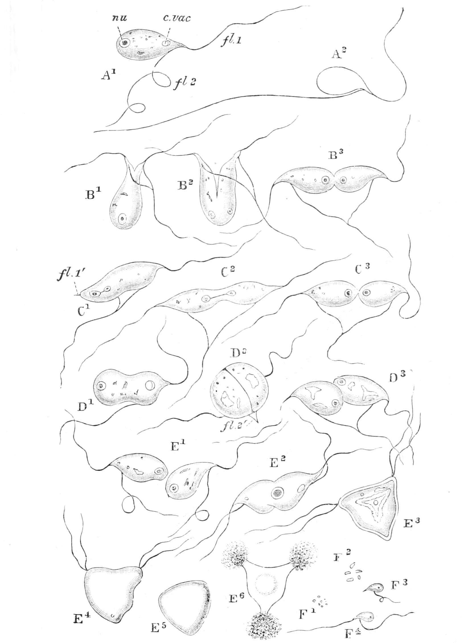
Fig. 38.—Bodo saltans. A, the positions assumed in the springing movements of the anchored form; B, longitudinal fission of anchored forms; C, transverse fission of the same; D, fission of free-swimming form; E1-E4, conjugation of free-swimming with anchored form; E5, zygote; E6, emission of spores from zygote; F, growth of spores: c.vac, contractile vacuole; fl.1, anterior; fl.2, ventral flagellum; nu, nucleus. (From Parker's Biology, after Dallinger.)
The life-cycles varied considerably in length. It was in every case found that after a series of fissions the species ultimately underwent conjugation (more or less unequal or bisexual in character);[124] the zygote encysted; and within the cyst the protoplasmic body underwent brood-formation, the outcome of which was a mass of spores discharged by the rupture of the cyst (Fig. 38). These spores grow from a size too minute for resolution by our microscopes into the ordinary flagellate form. They withstand the effects of drying, if this be effected immediately on their escape from the ruptured cyst; so that it is probable that each spore has itself a delicate cyst-wall and an aplanospore, from which a single zoospore escapes. The complex cycle, of course, comprises the whole course from spore-formation to spore-formation. Such complete and regular "life-histories," each characteristic of the species, were the final argument against those who held to the belief that spontaneous generation of living beings took place in infusions of decomposing organic matter.
Previous to the work of these observers it had been almost universally believed that the temperature of boiling water was adequate to kill all living germs, and that any life that appeared in a closed vessel after boiling must be due to spontaneous change in its contents. But they now showed that, while none of the species studied resisted exposure in the active condition to a temperature of 138°-140° F., the spores only succumbed, in liquid, to temperatures that might even reach 268° F., or when dry, even 300° F. or more. Such facts explain the constant occurrence of one or more such minute species in liquids putrefying under ordinary conditions, the spores doubtless being present in the dust of the air. Very often several species may co-exist in one infusion; but they separate themselves into different zones, according to their respective need for air, when a drop of the liquid is placed on the slide and covered for examination. Dallinger[125] has made a series of experiments on the resistance of these organisms in their successive cycles to a gradual rise of temperature. Starting with a liquid containing three distinct species, which grew and multiplied normally at 60° F., he placed it under conditions in which he could slowly raise the temperature. While all the original inmates would have perished at 142° F., he succeeded in finally producing races that throve at 158° F., a scalding heat, when an accident put an end to that series of experiments. In no instance was the temperature raised so much as to kill off the beings, so that the increased tolerance of their descendants was due not, as might have been anticipated, to selection of those that best resisted, but to the inheritance of an increased toleration and resistance from one generation or cycle to another.
As we noted above (p. 40), the study of the Flagellates has been largely in the hands of botanists. After the work of Bütschli in Bronn's Thier-Reich, Klebs[126] took up their study; and the principal monographs during the last decade have appeared in Engler and Prantl's Pflanzenfamilien, where Senn[127] treats the Flagellates generally, Wille[128] the Volvocaceae, and Schütt the "Peridiniales" or Dinoflagellata;[129] while only the Cystoflagellata, with but two genera, have been left to the undisputed sway of the zoologists.[130]
Among this group the majority are saprophytes, found in water containing putrefying matter or bacteria. The forms so carefully studied by Dallinger and Drysdale belong to the genera Bodo, Cercomonas, Tetramitus, Monas, and Dallingeria. Many others are parasites in the blood or internal cavities of higher animals, some apparently harmless, such as Trichomonas vaginalis, parasitic in man, others of singular malignity. Costia necatrix, infesting the epithelial scales of fresh-water fish, often devastates hatcheries. The genus Trypanosoma, Gruby, contributes a number of parasites, giving rise to deadly disease in man and beast.[131] T. lewisii is common in Rodents, but is relatively harmless. T. evansii is the cause of the Surra disease of Ruminants in India, and is apparently communicated by the bites of "large brown flies" (almost certainly Breeze Flies or Tabanidae, Vol. VI. p. 481). T. brucei, transferred to cattle by the Tsetse Fly, Glossina morsitans (see Vol. VI. Fig. 244, p. 513) in Equatorial Africa, is the cause of the deadly Nagana disease, which renders whole tracts of country impassable to ox or horse. Other Trypanosomic diseases of animals are, in Algeria and the Punjab, "dourine," infecting horses and dogs; in South America, Mal de Caderas (falling-sickness), an epidemic paralysis of cattle. During the printing of this book, much additional knowledge has been gained on this genus and the diseases it engenders. The Trypanosomic fever recently recognised on the West Coast has been found to be the early stage of the sleeping-sickness, that well-known and most deadly epidemic of Tropical Africa. Through the researches of Castellani, Nabarro, and especially Colonel and Mrs. Bruce, we know now that the parasite T. gambiense is transferred by an intermediate host, a kind of Tsetse Fly (Glossina palpalis). Schaudinn's full study of a parasite of the blood corpuscles of the Owl has shown that while in its intracorpuscular state it resembles closely the malarial parasites in behaviour, and in its schizogenic multiplication, so that it was considered an Acystosporidian, under the name of Halteridium, it is really a Trypanosoma;[132] for the accomplishment of successful sexual reproduction it requires transference to the gut of a gnat (Culex). The germs may infect the ovary, and give the offspring of the insect the innate power of infecting Owls. Thus a new light is shed on the origin of the Coccidiaceae, whose "blasts" in the insect host resemble Trypanosoma in their morphology.
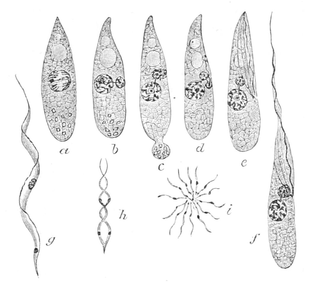
Fig. 39.—Morphology of Trypanosoma. a-f, Stages in development of Trypanosoma noctuae from the active zygote ("ookinete"); b, first division of nucleus into larger (trophic) and smaller (kineto-) nucleus; c, d, division of smaller nucleus and its transformations to form "blepharoplast" and myonemes; f, adult Trypanosoma; g, h, i, Treponema zeemannii of Owl; g, Trypanosome form; h, Spirochaeta form; i, rosette aggregate. (After Schaudinn.)
The human Tick fever of the Western United States and the epizootic Texas fever are known to be due to blood parasites of the genus Piroplasma (Babesia), of which the free state is that of a Trypanosome. It appears certain that Texas fever, though due to Tick bites, is not transferred directly from one beast to another by the same Tick; but the offspring of a female Tick that has sucked an infected ox contains Trypanosome germs, and will by their bites infect other animals. It would seem probable that the virulence of the Persian Tick (Argas persica) is due to similar causes. The Indian maladies known as "Kala Azar" and "Oriental Sore" are characterised by blood parasites, at first called after their discoverer the "Leishman bodies," which have proved to be the effects of a Piroplasma.
Trypanosoma is distinguished by the expansion of its flagellum into an undulating membrane, that runs down the edge of the body, and may project behind as a second lash. In this membrane run eight fine muscular filaments, or myonemes, four on either surface, within the undulating membrane; at their lower end they are all connected with a rounded body, the "blepharoplast," which is here in its origin, as well as in its behaviour in reproductive processes, a true modified nucleus, comparable in some respects, as was first noted by Plimmer and Rose Bradford,[133] with the micronucleus of the Infusoria. Part of the segmentation spindle persists in the form of a filament uniting the blepharoplast with the large true functional nucleus (Fig. 39, a-f).
The blood of patients suffering from relapsing fever contains a fine wriggling parasite, which was described as a Schizomycete, allied to the bacteria, and hitherto termed Spirochaeta obermeieri. Schaudinn has shown that this and other similar blood parasites are closely allied to Trypanosoma; and since the original genus was founded on organisms of putrefaction which are undoubtedly Schizomycetes, Vuillemin has suggested the name Treponema. T. pallidum is found in syphilitic patients, and appears to be responsible for their illness.[134]
The Craspedomonadidae (often called Choanoflagellates, Fig. 40) are a group whose true nature was elucidated some forty years ago by the American zoologist, H. James-Clark. They are attached either to a substratum, by a stalk produced by the base of the cell, or to other members of the same colony; they are distinguished by the protrusion of the cytoplasm around the base of the single flagellum into a pellucid funnel,[135] in which the plasma is in constant motion, though the funnel retains its shape and size, except when, as sometimes happens, it is retracted.
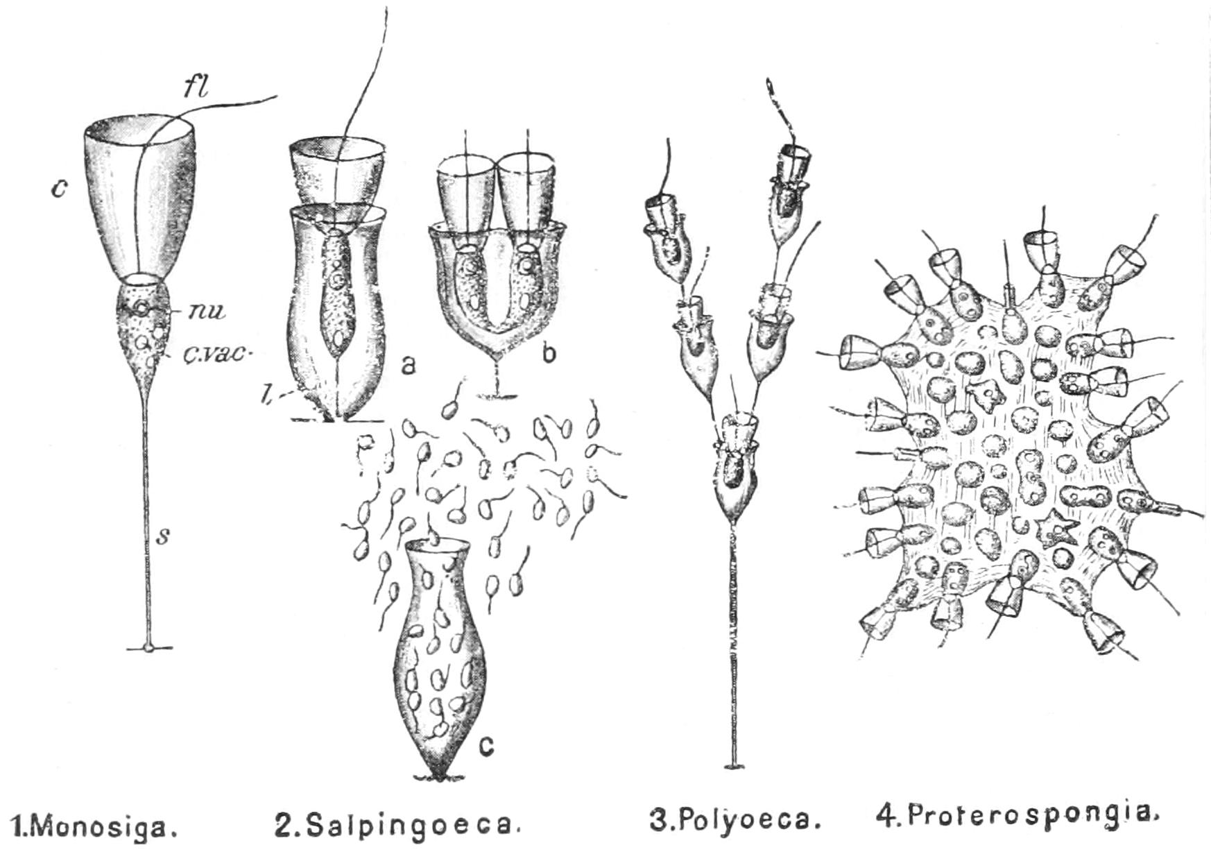
Fig. 40.—Various forms of Craspedomonadidae. 2, a, Adult cell; 2, b, longitudinal fission; 2, c, the production of flagellulae by brood-formation; c, collar; c.vac, contractile vacuole; fl, flagellum; l, theca; nu, nucleus; s, stalk. (After Saville Kent.)
The agitation of the flagellum determines a stream of water upwards along the outer walls of the funnel; and the food-particles brought along adhere to the outside of the funnel, and are carried by its streaming movement to the basal constriction, where they are swallowed by the plasma, which appears to form a swallowing vacuole at that point. Longitudinal fission is the ordinary mode of reproduction, extending up through the funnel. If the two so formed continue to produce a stalk, the result is the formation of a tree-like stem, whose twigs bear at the ends the funnelled cells, or "collar-cells" as they are usually called. In Salpingoeca, as in so many other Flagellates, each cell forms a cup or theca, often of most graceful vase-like outline, the rim being elegantly turned back. Proterospongia (Fig. 75, p. 182) secretes a gelatinous investment for the colony, which is attached to solid bodies. In this species, according to Saville Kent, the central members of the colony retract their collar, lose their flagellum, become amoeboid, and finally undergo brood-formation to produce minute zoospores. This is the form which by its differentiation recalls the Sponges, and has been regarded as a transition towards them; for the flagellate, nutritive cells of the Sponges are provided with a collar, which exists in no other group of Metazoa (see pp. 171, 181, and Fig. 70, p. 176). The most recent monographer of the family is Raoul Francé, but James-Clark and Saville Kent did the pioneering work.
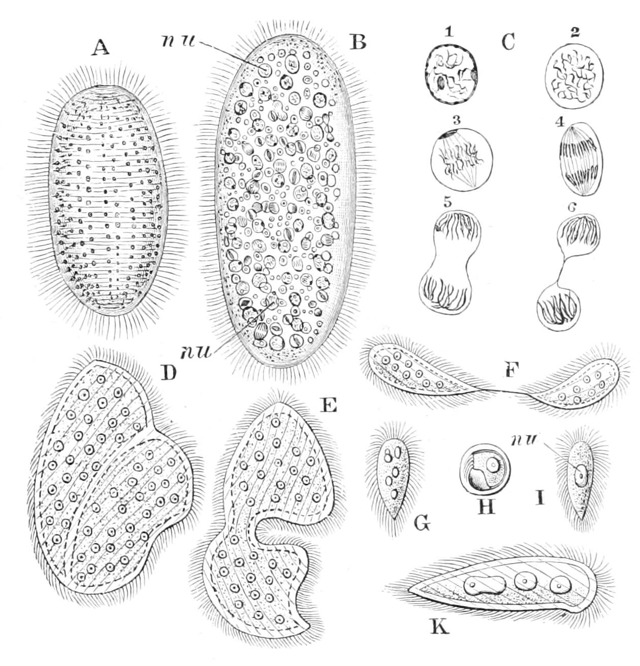
Fig. 41.—Opalina ranarum. A, living specimen; B, stained specimen showing nuclei; C, stages in nuclear division; D-F, stages in fission; G, final product of fission; H, encysted form; I, young form liberated from cyst; K, the same after multiplication of the nucleus has begun. nu, Nucleus. (From Parker's Biology, after Saville Kent and Zeller.)
Of the life-history of the Trichonymphidae,[136] all of which are parasitic in the alimentary canal of Insects, especially Termites or White Ants (Vol. V. p. 356), nothing is known. Some of them have a complete investment of motile flagella, like enormously long cilia, which in Dinenympha appear to coalesce into four longitudinal undulating membranes. Lophomonas inhabits the gut of the Cockroach and Mole-cricket. The Opalinidae have also a complete investment of cilia, which are short, and give the aspect of a Ciliate to the animal, which is common in the rectum of Amphibia, and dies when transferred to water. But despite the outward resemblance, the nuclei, of which there may be as many as 200, are all similar, and consequently this group cannot be placed among the Infusoria at all. Opalina has no mouth nor contractile vacuole. It multiplies by dividing irregularly and at intervals, resolving finally into 1-nucleate fragments, which encyst and pass into the water. When swallowed the cyst dissolves, its contents enlarge, and ultimately assume the adult form.[137]
Maupasia has a partial investment of cilia, a single long flagellum and mouth, a contractile vesicle, and a single simple nucleus. It seems to find an appropriate place near the two above groups, though it is free, and possesses a mouth.
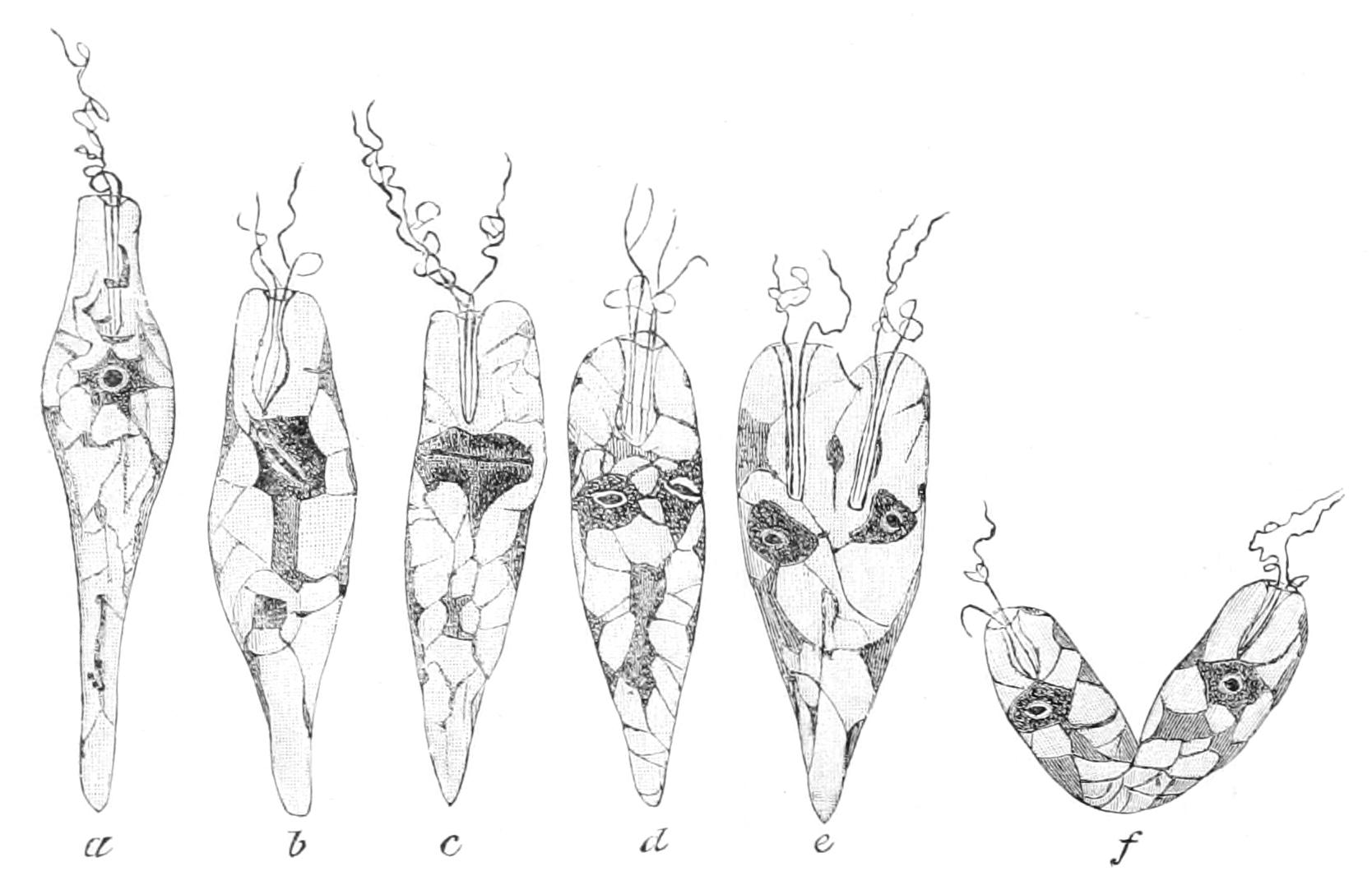
Fig. 42.—Longitudinal Fission of Eutreptia viridis (Euglenaceae), showing chloroplasts, nucleus, and flagella arising from pharynx-tube. (After Steuer.)
Among the Euglenaceae, Euglena viridis is a very common form, giving the green colour to stagnant or slow-flowing ditches and puddles in light places, especially when contaminated by a fair amount of dung, as by the overflow of a pig-sty, in company with a few hardy Rotifers, such as Hydatina senta (Vol. II. Fig. 106, p. 199) and Brachionus. Euglena is about 0.1 mm. in length when fully extended, oval, pointed behind, obliquely truncate in front, with a flagellum arising from the pharyngeal pit. It shows a peculiar wriggling motion, waves of transverse constriction passing along the body from end to end, as well as flexures in different meridians. Such motions are termed "euglenoid." The front part is colourless, but under a low power the rest of the cell is green, owing to the numerous chlorophyll bodies or chloroplasts. The outermost layer of the cytoplasm shows a somewhat spiral longitudinal striation, possibly due to muscular fibrils. The interior contains many laminated plates of paramylum, and a large single nucleus. At the front of the body at the base of the flagellum is a red "eye-spot" on the dorsal side of the pharynx-tube or pit, from which the flagellum protrudes. Wager has shown that this tube receives, also on its dorsal side, the opening of a large vacuole, sometimes called the reservoir, for into it discharges the contractile vacuole (or vacuoles). The eye-spot is composed of numerous granules, containing the vegetal colouring matter "haematochrome." It embraces the lower or posterior side of the communication between the tube and the reservoir. The flagellum has been traced by Wager through the tube into the reservoir, branching into two roots where it enters the aperture of communication, and these are inserted on the wall of the reservoir at the side opposite the eye-spot. But on one of the roots near the bifurcation is a dilatation which lies close against the eye-spot, so that it can receive the light reaction. Euglena is an extremely phototactic organism. It shows various wrigglings along the longitudinal axis, and transverse waves of contraction and expansion may pass from pole to pole.[138]
Among the Chrysomonadaceae the genus Zooxanthella, Brandt, has already been described under the Radiolaria (p. 86), in the jelly of which it is symbiotic. It also occurs in similar union in the marine Ciliates, Vorticella sertulariae and Scyphidia scorpaenae, and in Millepora (p. 261) and many Anthozoa (pp. 373 f., 396).
Of the Chlamydomonadidae, Sphaerella (Haematococcus, Ag.) pluvialis (Fig. 43), and S. nivalis, in which the green is masked by red pigment, give rise to the phenomena of "red snow" and "bloody rain." The type genus, Chlamydomonas, is remarkable for the variations from species to species in the character and behaviour of the gametes. Sometimes they are equal, at other times of two sizes. In some species they fuse immediately on approximation, in the naked active state; in others, they encyst on approaching, and unite by the emission of a fertilising tube, as in the Algal Conjugatae. Zoochlorella is symbiotic in green Ciliata (pp. 153 f., 158), Sponges (p. 175), Hydra (p. 256), and Turbellaria (Vol. II. p. 43).
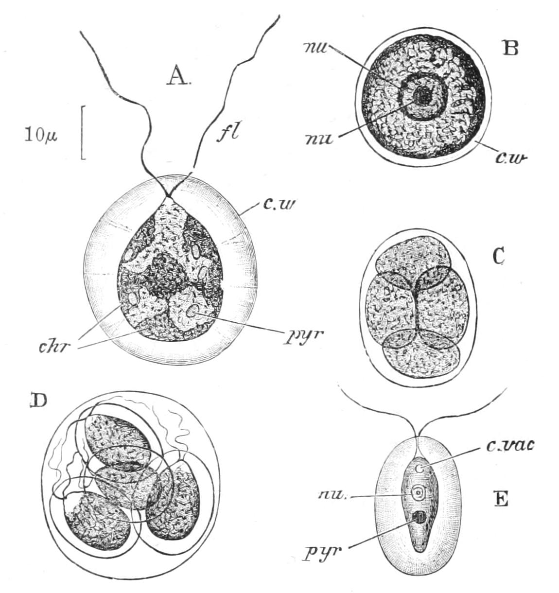
Fig. 43.—Sphaerella pluvialis. A, motile stage; B, resting stage; C, D, two modes of fission; E, Sphaerella lacustris, motile stage. chr, Chromatophores; c.vac, contractile vacuole; c.w, cell-wall; fl, flagella; nu, nucleus; nu', nucleolus; pyr, pyrenoids. (From Parker's Biology.)
Of the Volvocidae, Volvox (Fig. 44) is the largest and most conspicuous genus. Its colony forms a globe the size of a pin's head, floating on the surface of ponds, drains, or even puddles or water-barrels freely open to the light. It has what may be called a skeleton of gelatinous matter,[139] condensed towards the surface into a denser layer in which the minute cells are scattered. These have each an eye-spot, a contractile vacuole, and two flagella, by the combined action of which the colony is propelled. Delicate boundary lines in the colonial wall mark out the proper investment of each cell. The cells give off delicate plasmic threads which meet those of their neighbours, and form a bond between them. In that half of the hemisphere which is posterior in swimming, a few (five to eight) larger cells ("macrogonidia" of older writers) are evenly distributed, protruding as they increase in size into the central jelly. These as they grow segment to form a new colony.
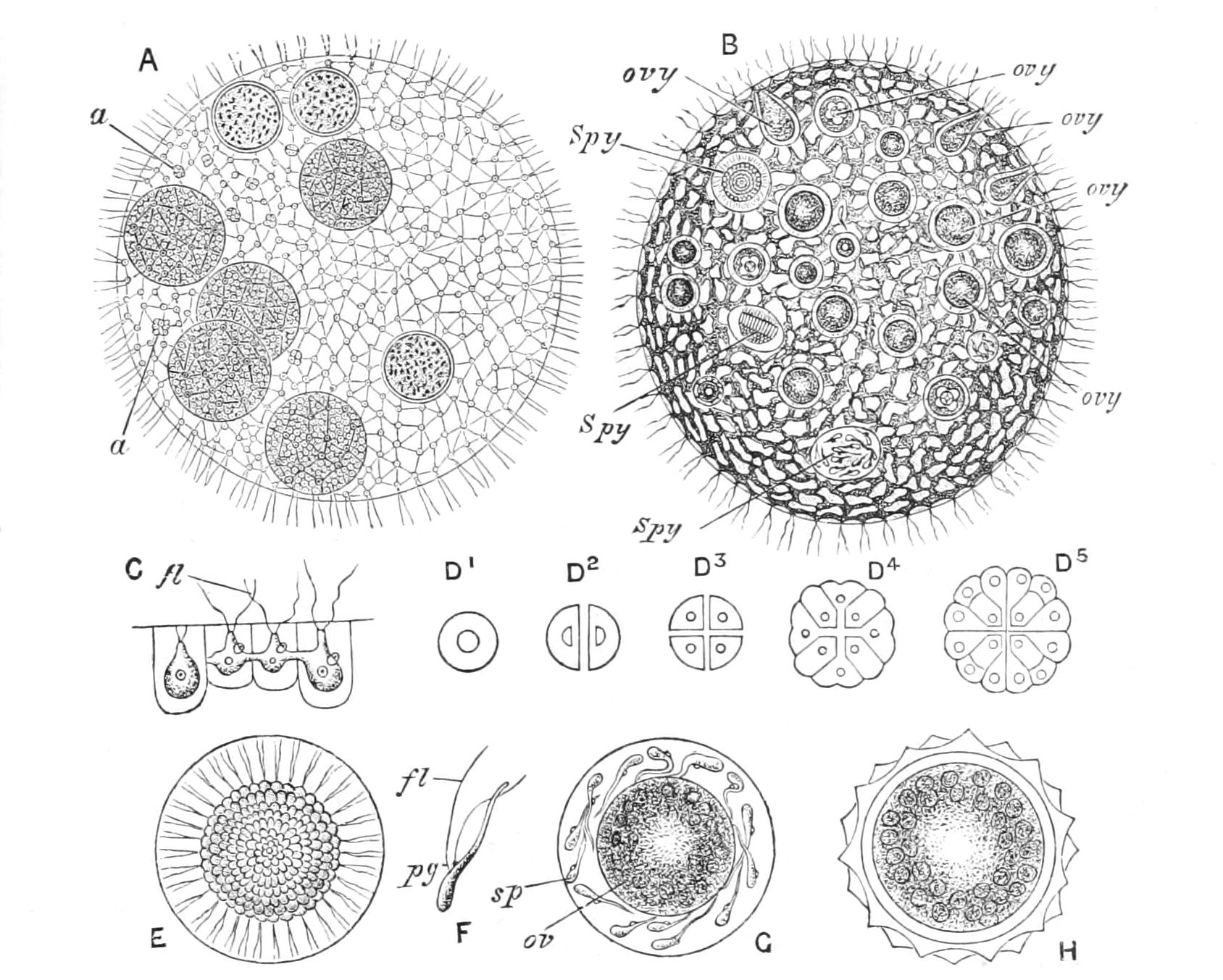
Fig. 44.—Volvox globator. A, entire colony, enclosing several daughter-colonies; B, the same during sexual maturity; C, four zooids in optical section; D1-D5, development of parthenogonidium; E, ripe spermogonium; F, sperm; G, ovum; H, oosperm. a, Parthenogonidia; fl, flagellum; ov, ovum; ovy, ovaries; pg, pigment spot; sp, sperms; Spy, spermogonia dividing to form sperms. (From Parker's Biology, after Cohn and Kirchner.)
The divisions are only in two planes at right angles, so that the young colony is at first a plate, but as the cells multiply the plate bends up (as in the gastrulation of the double cellular plate of the Nematode Cucullanus, Vol. II. p. 136), and finally forms a hollow sphere bounded by a single layer of cells: the site of the original orifice may be traced even in the adult as a blank space larger than exists elsewhere. Among the cells of the young colony some cease to divide, but continue to grow at an early period, and these are destined to become in turn the mothers ("parthenogonidia") of a new colony; they begin segmenting before the colony of which they are cells is freed. The young colonies are ultimately liberated by the rupture of the sphere as small-sized spheres, which henceforth only grow by enlargement of the sphere as a whole, and the wider separation of the vegetative cells. Thus the vegetative cells soon cease to grow; all the supply of food material due to their living activities goes to the nourishment of the parthenogonidia, or the young colonies, as the case may be. These vegetative cells have therefore surrendered the power of fission elsewhere inherent in the Protist cell. Moreover, when the sphere ruptures for the liberation of the young colonies, it sinks and is doomed to death, whether because its light-loving cells are submerged in the ooze of the bottom, or because they have no further capacity for life. When conjugation is about to take place, it is the cells that otherwise would be parthenogonidia that either act as oospheres or divide as "spermogonia" to form a flat brood of minute yellow male cells ("sperms"). These resemble vegetative cells, in the possession of an eye-spot and two contractile vacuoles, but differ in the enormously enlarged nucleus which determines a beaked process in front. After one of these has fused with the female cell ("oosphere") the product ("oosperm") encysts, passes into a stage of profound rest, and finally gives rise to a new colony. The oospheres and sperm-broods may arise in the same colony or in distinct ones, according to the species.
Before we consider the bearings of the syngamic processes of Volvox, we will study those presented by its nearer allies, which have the same habitat, but are much more minute. Three of these are well known, Stephanosphaera, Pandorina, and Eudorina, all of which have spherical colonies of from eight to thirty-two cells embedded at the surface of a sphere, and no differentiation into vegetative cells and parthenogonidia (or reproductive cells).
Stephanosphaera has its eight cells spindle shaped, and lying along equidistant meridians of its sphere; in vegetative reproduction each of these breaks up in its place to form a young colony, and the eight daughter-colonies are then freed. In conjugation, each cell of the colony breaks up into broods of 4, 8, 16, or 32 small gametes, which swim about within the general envelope, and pair and fuse two and two: this is "isogamous," "endogamous" conjugation. In Pandorina (Fig. 45) the cells are rounded, and are from 16 to 32 in each colony. The vegetative reproduction in this, as in Eudorina, is essentially the same as in Stephanosphaera. In conjugation the cells are set free, and are of three sizes in different colonies, small (S), medium (M), and large (L). The following fusions may occur: S × S, S × M, S × L, M × M, M × L. Thus the large are always female, as it were, the medium may play the part of male to the large, female to the small; the small are males to the medium and to the large. The medium and small are capable, each with its like, of equal, undifferentiated conjugation; so that we have a differentiation of sex far other than that of ordinary, binary sex. Eudorina, however, has attained to "binary sex," for the female cells are the ordinary vegetative cells, at most a little enlarged, and the male cells are formed by ordinary cells producing a large flat colony of sixty-four minute males or sperms. In some cases four cells at the apex of a colony are spermogonia, producing each a brood of sperms, while the rest are the oospheres. The transition to Volvox must have arisen through the sterilisation of the majority of cells of a colony for the better nutrition of the few that are destined alone for reproduction.
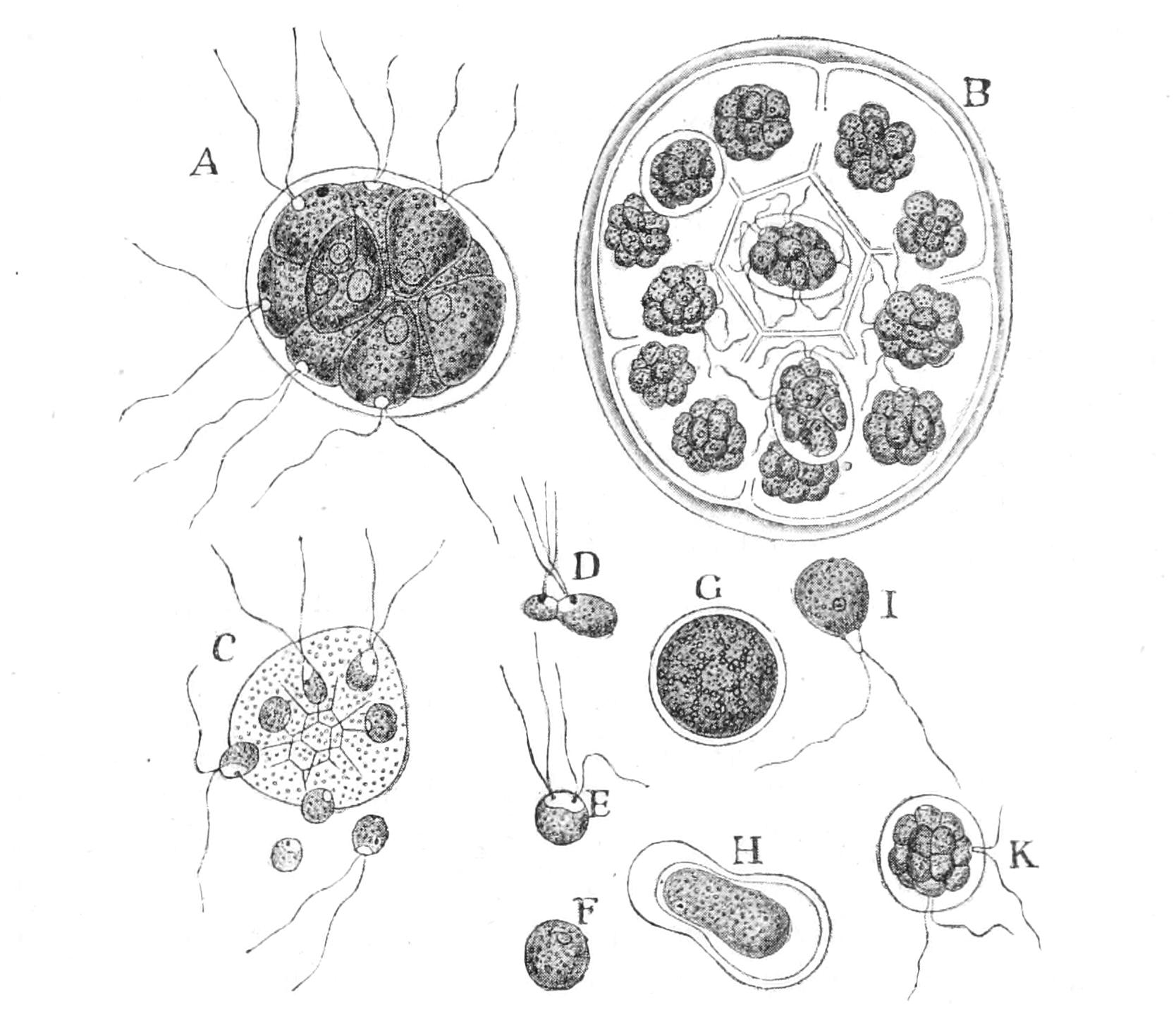
Fig. 45.—Pandorina morum. A, entire colony; B, asexual reproduction, each zooid dividing into a daughter-colony; C, liberation of gametes; D-F, three stages in conjugation of gametes; G, zygote; H-K, development of zygote into a new colony. (From Parker's Biology, after Goebel.)
Volvox, as we have seen, has attained a specialisation entirely comparable to that of a Metazoon, where the segmentation of the fertilised ovum results in two classes of cells: those destined to form tissues, and condemned to ultimate death with the body as a whole, and those that ultimately give rise to the reproductive cells, ova, and sperms. But this is a mere parallelism, not indicating any sort of relationship: the oospores of the Volvocaceae show that tendency to an encysted state, in which fission takes place, that is so characteristic of Algae, and these again show the way to Cryptogams of a higher status. Thus, Volvox, despite the fact that in its free life and cellular differentiation it is the most animal of all known Flagellates, is yet, with the rest of the Volvocaceae, inseparable from the Vegetable Kingdom, and is placed here only because of the impossibility of cleaving the Flagellates into two.
The Dinoflagellata (Figs. 46, 47) are often of exceptionally large dimensions in this class, attaining a maximum diameter of 150 µ (1⁄160") and even 375 µ (1⁄67") in Pyrocystis noctiluca. The special character of the group is the presence of two flagella; the one, filiform, arises in a longitudinal groove, and extending its whole length projects behind the animal, and is the conspicuous organ of motion: the other, band-like, arises also in the longitudinal groove, but extends along a somewhat spiral transverse groove,[140] and never protrudes from it in life, executing undulating movements that simulate those of a girdle of cilia, or a continuous undulating membrane (Fig. 46). This appearance led to the old name "Cilioflagellata," which had of course to be abandoned when Klebs discovered the true structure.[141] There is a distinct cellulose membrane, sometimes silicified, to the ectoplasm, only interrupted by a bare space in the longitudinal groove, whence the flagella take origin. This cuticle is usually hard, sculptured, and divided into plates of definite form, bevelled and overlapping at their junction; occasionally the cell has been seen to moult them.
A large vacuolar space, traversed by plasmic strings, separates the peripheral cytoplasm from the central, within which is the large nucleus. There are in most species one or more chromatophores, coloured by a yellowish or brownish pigment, which is a mixture of lipochromes, distinct from diatomin. In a few species the presence of these is not constant, and these species show variability as to their nutrition, which is sometimes holozoic. Under these conditions the cell can take in food-particles as bulky as the eggs of Rotifers and Copepods, by the protrusion of a pseudopod at the junction of the two grooves. As in most coloured forms an eye-spot is often present, a cup-shaped aggregation of pigment, with a lenticular refractive body in its hollow. A contractile vacuole, here termed a "pusule," occurs in many species, communicating with the longitudinal groove by a canal. Nematocysts (see p. 246 f.) are present in Polykrikos, trichocysts (see p. 142) in several genera.
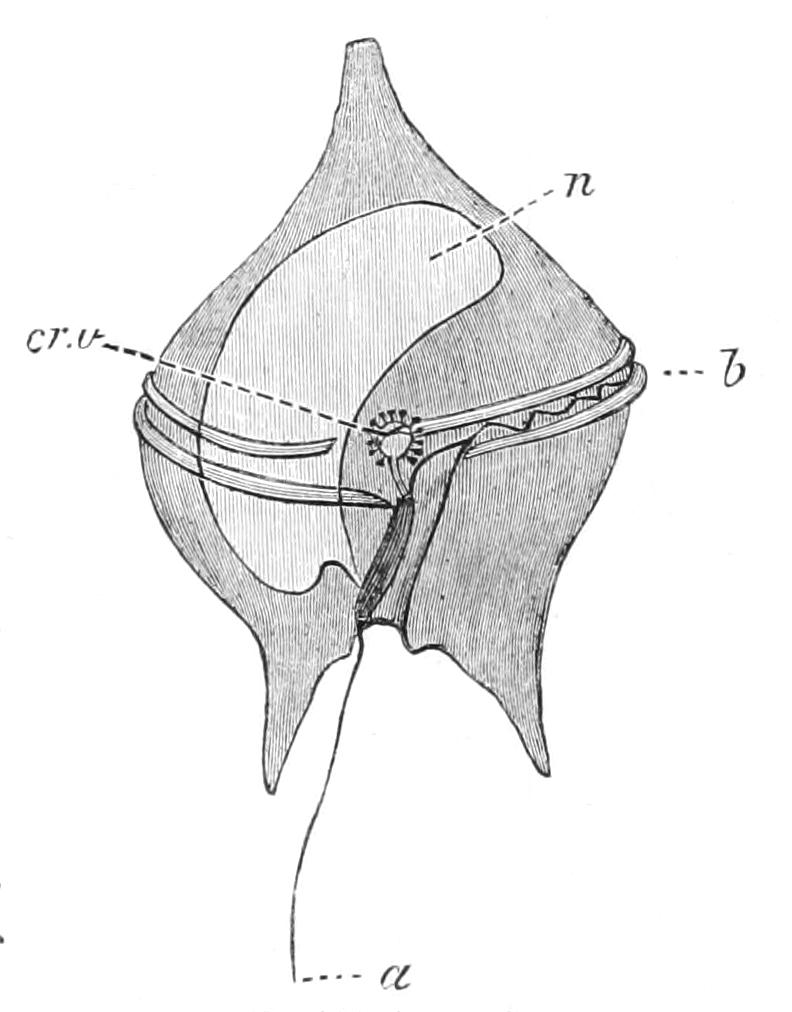
Fig. 46.—Peridinium divergens. a, Flagellum of longitudinal groove; b, flagellum of transverse groove; cr.v, contractile vacuole surrounded by formative vacuoles; n, nucleus. (After Schütt.)
Division is usually oblique, dividing the body into two dissimilar halves, each of which has to undergo a peculiar growth to reconstitute the missing portion, and complete the shell. The incomplete separation of the young cells leads to the formation of chains, notably in Ceratium and Polykrikos, the latter dividing transversely and occurring in chains of as many as eight. The process of division may take place when the cell is active, or in a cyst, as in Pyrocystis (Fig. 47). Again, encystment may precede multiple fission, resulting in the formation of a brood of minute swarmers. It has been suggested that these are capable of playing the part of gametes, and conjugating in pairs.[142]
The Dinoflagellates are for the most part pelagic in habit, floating at the surface, and when abundant tinge the water of fresh-water lakes or even ponds red or brown. Peridinium (Fig. 46) and Ceratium (the latter remarkable for the horn-like backward prolongations of the lower end) are common genera both in the sea and fresh-waters. Gymnodinium pulvisculus is sometimes parasitic in Appendicularia (Vol. VII. p. 68). Polykrikos[143] has four transverse grooves, each with its flagellum, besides the terminal one. Many of the marine species are phosphorescent, and play a large part in the luminosity of the sea, and some give it a red colour.
Several fossil forms have been described. Peridinium is certainly found fossil in the firestone of Delitzet, belonging to the Cretaceous. A full monograph of the group under the name "Peridiniales" was published by Schütt.[144]
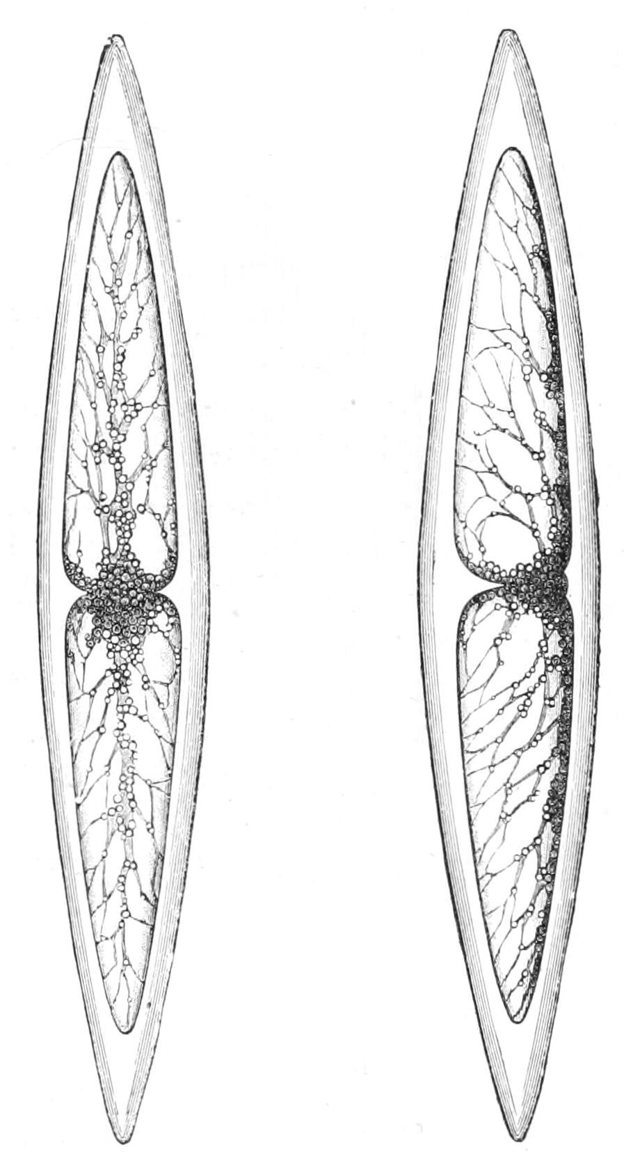
Fig. 47.—Pyrocystis fusiformis, Murray. × 100. From the surface in the Guinea Current. (From Wyville Thomson.)
The Cystoflagellates contain only two genera,[145] Noctiluca, common at the surface of tranquil seas, to which, as its name implies, it gives phosphorescence, and Leptodiscus, found by R. Hertwig in the Mediterranean. Noctiluca is enormous for a Flagellate, for with the form of a miniature melon it measures about 1 mm. (1⁄25") or more in diameter. In the depression is the "oral cleft," from one end of which rises, by a broad base, a large coarse flagellum, as long as the body or longer and transversely striated. In front of the base of the flagellum are two lip-like prominences, of which one, a little firmer than the other, and transversely ridged, is called the tooth; at the junction of the two is a second, minute, flagellum, usually called the cilium. Behind these the oral groove has an oval space, the proper mouth; behind this, again, the oral groove is continued for some way, with a distinct rod-like ridge in its furrow. The whole body, including the big flagellum, is coated by a strong cuticular pellicle, except at the oblong mouth, and the lips and rod are mere thickenings of this. The cytoplasm has a reticulate arrangement: the mouth opens into a central aggregate, from which strands diverge branching as they recede to the periphery, where they pass into a continuous lining for the cuticular wall, liquid filling the interspaces. The whole arrangement is not unlike that found in many plant-cells, but the only other Protists in which it occurs are the Ciliata Trachelius (Fig. 56, p. 153) and Loxodes. The central mass contains the large nucleus. Noctiluca is an animal feeder, and expels its excreta through the mouth. The large flagellum is remarkable for the transverse striation of its plasma, especially on the ventral side. The cuticle may be moulted as in the Dinoflagellates. As a prelude to fission the external differentiations disappear, the nucleus divides in the plane of the oral groove, and a meridional constriction parts the two halves, the new external organs being regenerated. Conjugation occurs also, the two organisms fusing by their oral region; the locomotive organs and pharynx disappear; the conjoined cytoplasms unite to form a sphere, and the nuclei fuse to form a zygote or fertilisation nucleus. This conjugation is followed by sporulation or brood-formation.[146]
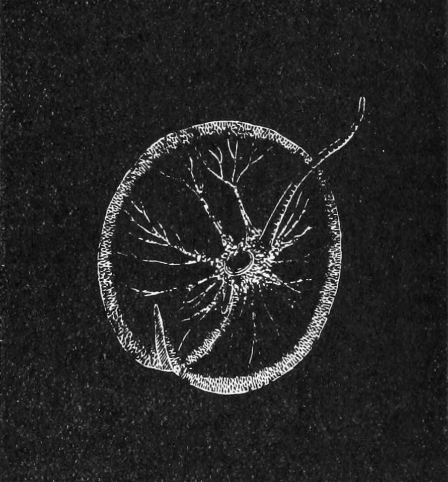
Fig. 48.—Noctiluca miliaris, a marine Cystoflagellate. (From Verworn.)
The nucleus passes towards the surface, undergoes successive fissions, and as division goes on the numerous daughter-nuclei occupy little prominences formed by the upgrowth of the cytoplasm of the upper pole. The rest of the cytoplasm atrophies, and the hillocks formed by the plasmic outgrowths around the final daughter-nuclei become separate as so many zoospores (usually 256 or 512); each of these is oblong with a dorsal cap-like swelling, from the edge of which arises a flagellum pointing backwards; parallel to this the cap is prolonged on one side into a style also extending beyond the opposite pole of the animal.[147] In this state the zoospore is, to all outward view, a naked Dinoflagellate, whence it seems that the Cystoflagellates are to be regarded as closely allied to that group. Leptodiscus is concavo-convex, circular, with the mouth central on the convex face, 1-flagellate, and attains the enormous size of 1.5 mm. (1⁄16") in diameter.
The remarkable phosphorescence of Noctiluca is not constant. It glows with a bluish or greenish light on any agitation, but rarely when undisturbed. A persistent stimulus causes a continuous, but weak, light. This light is so weak that several teaspoonsful of the organism, collected on a filter and spread out, barely enable one to read the figures on a watch a foot away. As in other marine phosphorescence, no rise of temperature can be detected. The luminosity resides in minute points, mostly crowded in the central mass, but scattered all through the cytoplasm. A slight irritation only produces luminosity at the point touched, a strong one causes the whole to flash. Any form of irritation, whether of heat, touch, or agitation, electricity or magnetism, is stated to induce the glow. By day, it is said, Noctiluca, when present in abundance, may give the sea the appearance of tomato soup.
The earliest account of Noctiluca will be read with interest. Henry Baker writes in Employment for the Microscope:[148]—"A curious Enquirer into Nature, dwelling at Wells upon the Coast of Norfolk, affirms from his own Observations that the Sparkling of Sea Water is occasioned by Insects. His Answer to a Letter wrote to him on that Subject runs thus, 'In the Glass of Sea Water I send with this are some of the Animalcules which cause the Sparkling Light in Sea Water; they may be seen by holding the Phial up against the Light, resembling very small Bladders or Air Bubbles, and are in all Places of it from Top to Bottom, but mostly towards the Top, where they assemble when the Water has stood still some Time, unless they have been killed by keeping them too long in the Phial. Placing one of these Animalcules before a good Microscope, an exceeding minute Worm may be discovered, hanging with its Tail fixed to an opake Spot in a Kind of Bladder, which it has certainly a Power of contracting or distending, and thereby of being suspended at the Surface, or at any Depth it pleases in the including Water.'"
"The above-mentioned Phial of Sea Water came safe, and some of the Animalcules were discovered in it, but they did not emit any Light, as my Friend says they do, upon the least Motion of the Phial when the Water is newly taken up. He likewise adds, that at certain Times, if a Stone be thrown into the Sea, near the Shore, the Water will become luminous as far as the Motion reacheth: this chiefly happens when the Sea hath been greatly agitated, or after a Storm." Obviously what Mr. Sparshall, Baker's correspondent, took for a worm was the large flagellum.
The chief investigators of this group have been Huxley, Cienkowski, Allman, Bütschli, and G. Pouchet, while Ischikawa and Doflein have elucidated the conjugation.