Chapter I
Reading edition
PROTOZOA—INTRODUCTION—FUNCTIONS OF PROTOPLASM—CELL-DIVISION—ANIMALS AND PLANTS
The Free Amoeboid Cell.—If we examine under the microscope a fragment of one of the higher animals or plants, we find in it a very complex structure. A careful study shows that it always consists of certain minute elements of fundamentally the same nature, which are combined or fused into "tissues." In plants, where these units of structure were first studied, and where they are easier to recognise, each tiny unit is usually enclosed in an envelope or wall of woody or papery material, so that the whole plant is honeycombed. Each separate cavity was at first called a "cell"; and this term was then applied to the bounding wall, and finally to the unit of living matter within, the envelope receiving the name of "cell-wall." In this modern sense the "cell" consists of a viscid substance, called first in animals "sarcode" by Dujardin (1835), and later in plants "protoplasm"[1] by Von Mohl (1846). On the recognition of its common nature in both kingdoms, largely due to Max Schultze, the latter term prevailed; and it has passed from the vocabulary of biology into the domain of everyday life. We shall now examine the structure and behaviour of protoplasm and of the cell as an introduction to the detailed study of the Protozoa, or better still Protista,[2] the lowest types of living beings, and of Animals at large.
It is not in detached fragments of the tissues of the higher animals that we can best carry on this study: for here the cells are in singularly close connexion with their neighbours during life; the proper appointed work of each is intimately related to that of the others; and this co-operation has so trained and specially modified each cell that the artificial severance and isolation is detrimental to its well-being, if not necessarily fatal to its very life. Again, in plants the presence of a cell-wall interferes in many ways with the free behaviour of the cell. But in the blood and lymph of higher animals there float isolated cells, the white corpuscles or "leucocytes" of human histology, which, despite their minuteness (1⁄3000 in. in diameter), are in many respects suitable objects. Further, in our waters, fresh or salt, we may find similar free-living individual cells, in many respects resembling the leucocytes, but even better suited for our study. For, in the first place, we can far more readily reproduce under the microscope the normal conditions of their life; and, moreover, these free organisms are often many times larger than the leucocyte. Such free organisms are individual Protozoa, and are called by the general term "Amoebae." A large Amoeba may measure in its most contracted state 1⁄100 in. or 250 µ in diameter,[3] and some closely allied species (Pelomyxa, see p. 52) even twelve times this amount. If we place an Amoeba or a leucocyte under the microscope (Fig. 1), we shall find that its form, at first spherical, soon begins to alter. To confine our attention to the external changes, we note that the outline, from circular, soon becomes "island-shaped" by the outgrowth of a promontory here, the indenting of a bay there. The promontory may enlarge into a peninsula, and thus grow until it becomes a new mainland, while the old mainland dwindles into a mere promontory, and is finally lost. In this way a crawling motion is effected.[4] The promontories are called "pseudopodia" (= "false-feet"), and the general character of such motion is called "amoeboid."[5]
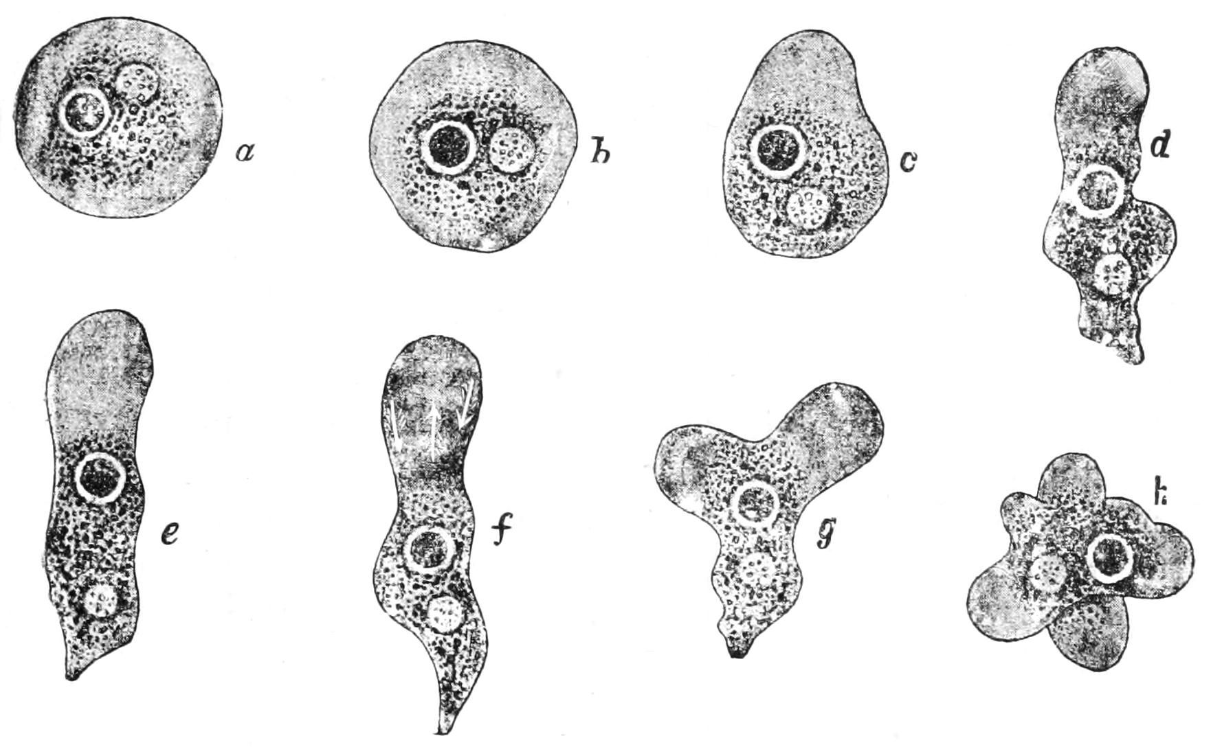
Fig. 1.—Amoeba, showing clear ectoplasm, granular endoplasm, dark nucleus, and lighter contractile vacuole. The changes of form, a-f, are of the A. limax type; g, h, of the A. proteus type. (From Verworn.)
The living substance, protoplasm,[6] has been termed a "jelly," a word, however, that is quite inapplicable to it in its living state. It is viscid, almost semi-fluid, and may well be compared to very soft dough which has already begun to rise. It resembles it in often having a number of spaces, small or large, filled with liquid (not gas). These are termed "vacuoles" or "alveoles," according to their greater or their lesser dimensions. In some cases a vacuole is traversed by strands of plasmic substance, just as we may find such strands stretching across the larger spaces of a very light loaf; but of course in the living cell these are constantly undergoing changes. If we "fix" a cell (i.e. kill it by sudden heat or certain chemical coagulants),[7] and examine it under the microscope, the intermediate substance between the vacuoles that we have already seen in life is again found either to be finely honeycombed or else resolved into a network like that of a sponge. The former structure is called a "foam" or "alveolar" structure, the latter a "reticulate" structure. The alveoles are about 1 µ in diameter, and spheroidal or polygonal by mutual contact, elongated, however, radially to any free surface, whether it be that of the cell itself or that of a larger alveole or vacuole. The inner layer of protoplasm ("endoplasm," "endosarc") contains also granules of various nature, reserve matters of various kinds, oil-globules, and particles of mineral matter[8] which are waste products, and are called "excretory." In fixed specimens these granules are seen to occupy the nodes of the network or of the alveoli, that is, the points where two or three boundaries meet.[9] The outermost layer ("ectoplasm" or "ectosarc") appears in the live Amoeba structureless and hyaline, even under conditions the most favourable for observation. The refractive index of protoplasm, when living, is always well under 1.4, that of the fixed and dehydrated substance is slightly over 1.6.
Again, within the outer protoplasm is found a body of slightly higher refractivity and of definite outline, termed the "nucleus" (Figs. 1, 2). This has a definite "wall" of plasmic nature, and a substance so closely resembling the outer protoplasm in character, that we call it the "nucleoplasm" (also "linin"), distinguishing the outer plasm as "cytoplasm"; the term "protoplasm" including both. Within the nucleoplasm are granules of a substance that stains well with the commoner dyes, especially the "basic" ones, and which has hence been called "chromatin." The linin is usually arranged in a distinct network, confluent into a "parietal layer" within the nuclear wall; the meshes traversing a cavity full of liquid, the nuclear sap, and containing in their course the granules; while in the cavity are usually found one or two droplets of a denser substance termed "nucleoles." These differ slightly in composition from the chromatin granules[10] (see p. 24 f.).
The movements of the leucocyte or Amoeba are usually most active at a temperature of about 40° C. or 100° F., the "optimum." They cease when the temperature falls to a point, the "minimum," varying with the organism, but never below freezing-point; they recommence when the temperature rises again to the same point at which they stopped. If now the temperature be raised to a certain amount above 40° they stop, but may recommence if the temperature has not exceeded a certain point, the "maximum" (45° C. is a common maximum). If it has been raised to a still higher point they will not recommence under any circumstances whatever.
Again, a slight electric shock will determine the retraction of all processes, and a period of rest in a spherical condition. A milder shock will only arrest the movements. But a stronger shock may arrest them permanently. We may often note a relation of the movements towards a surface, tending to keep the Amoeba in contact with it, whether it be the surface of a solid or that of an air-bubble in the liquid (see also p. 20).
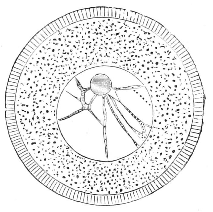
Fig. 2.—Ovum of a Sea-Urchin, showing the radially striated cell-membrane, the cytoplasm containing yolk-granules, the large nucleus (germinal vesicle), with its network of linin containing chromatin granules, and a large nucleole (germinal spot). (From Balfour's Embryology, after Hertwig.)
If a gentle current be set up in the water, we find that the movements of the Amoeba are so co-ordinated that it moves upstream; this must of course be of advantage in nature, as keeping the being in its place, against the streams set up by larger creatures, etc. (see also p. 21).
If substances soluble in water be introduced the Amoeba will, as a rule, move away from the region of greater concentration for some substances, but towards it (provided it be not excessive) for others. (See also pp. 22, 23.) We find, indeed, that there is for substances of the latter category a minimum of concentration, below which no effect is seen, and a maximum beyond which further concentration repels. The easiest way to make such observations is to take up a little strong solution in a capillary tube sealed at the far end, and to introduce its open end into the water, and let the solution diffuse out, so that this end may be regarded as surrounded by zones of continuously decreasing strength. In the process of inflammation (of a Higher Animal) it has been found that the white corpuscles are so attracted by the source of irritation that they creep out of the capillaries, and crowd towards it.
We cannot imagine a piece of dough exhibiting any of these reactions, or the like of them; it can only move passively under the action of some one or other of the recognised physical forces, and that only in direct quantitative relation to the work that such forces can effect; in other words, the dough can have work done on it, but it cannot do work. The Amoeba or leucocyte on the contrary does work. It moves under the various circumstances by the transformation of some of its internal energy from the "potential" into the "kinetic" state, the condition corresponding with this being essentially a liberation of heat or work, either by the breaking down of its internal substances, or by the combination of some of them with oxygen.[11] Such of these changes as involve the excretion of carbonic acid are termed "respiratory."
This liberation of energy is the "response" to an action of itself inadequate to produce it; and has been compared not inaptly to the discharge of a cannon, where foot-tons of energy are liberated in consequence of the pull of a few inch-grains on the trigger, or to an indefinitely small push which makes electric contact: the energy set free is that which was stored up in the charge. This capacity for liberating energy stored up within, in response to a relatively small impulse from without, is termed "irritability"; the external impulse is termed the "stimulus." The responsive act has been termed "contractility," because it so often means an obvious contraction, but is better termed "motility "; and irritability evinced by motility is characteristic of all living beings save when in the temporary condition of "rest."
Again, in the case of the cannon, the gunner after its discharge has to replenish it for future action with a fresh cartridge; the Amoeba or leucocyte can replenish itself—it "feeds." When it comes in contact with a fragment of suitable material, it enwraps it by its pseudopodia (Fig. 3), and its edges coalesce where they touch on the far side as completely as we can join up the edges of dough round the apple in a dumpling. It dissolves all that can be dissolved—i.e. it "digests" it, and then absorbs the dissolved material into its substance, both to replace what it has lost by its previous activity and to supply fuel for future liberation of energy; this process is termed "nutrition," and is another characteristic of living beings.
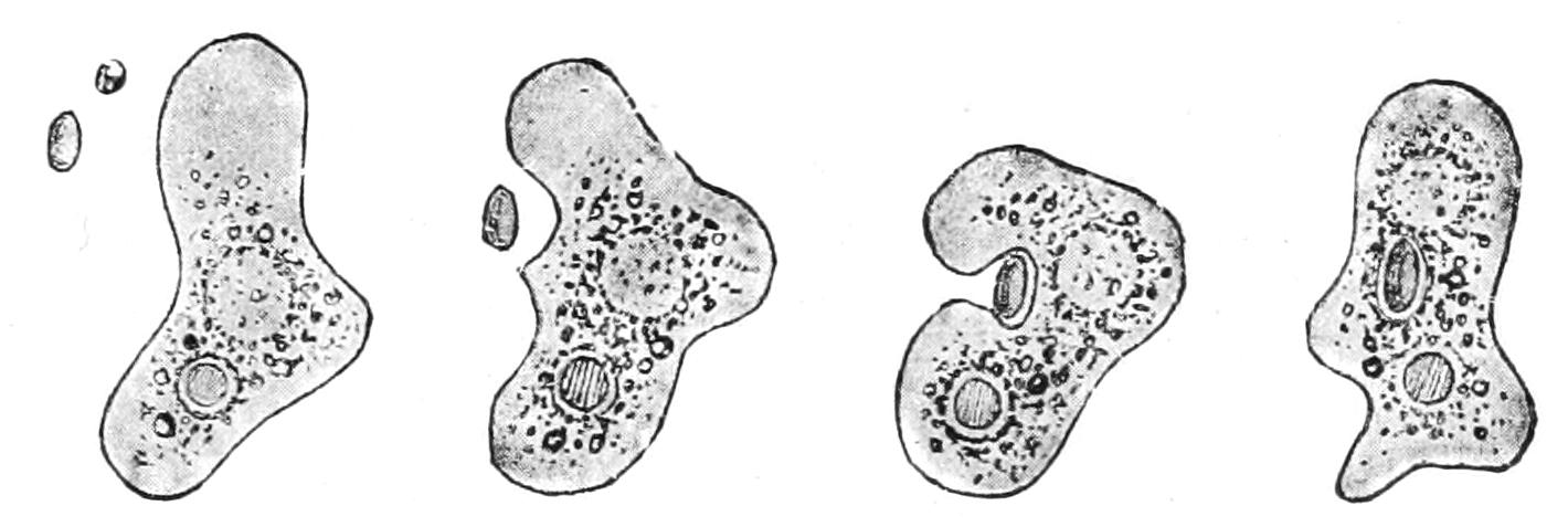
Fig. 3.—Amoeba devouring a plant cell; four successive stages of ingestion. (From Verworn.)
Again, as a second result of the nutrition, part of the food taken in goes to effect an increase of the living protoplasm, and that of every part, not merely of the surface—it is "assimilated"; while the rest of the food is transformed into reserves, or consumed and directly applied to the liberation of energy. The increase in bulk due to nutrition is thus twofold: part is the increase of the protoplasm itself—"assimilative growth," part is the storage of reserves—"accumulative growth": these reserves being available in turn by digestion, whether for future true growth or for consumption to liberate energy for the work of the cell.
We can conceive that our cannon might have an automatic feed for the supply of fresh cartridges after each shot; but not that it could make provision for an increase of its own bulk, so as to gain in calibre and strength, nor even for the restoration of its inner surface constantly worn away by the erosion of its discharges. Growth—and that growth "interstitial," operating at every point of the protoplasm, not merely at its surface—is a character of all living beings at some stage, though they may ultimately lose the capacity to grow. Nothing at all comparable to interstitial growth has been recognised in not-living matter.[12]
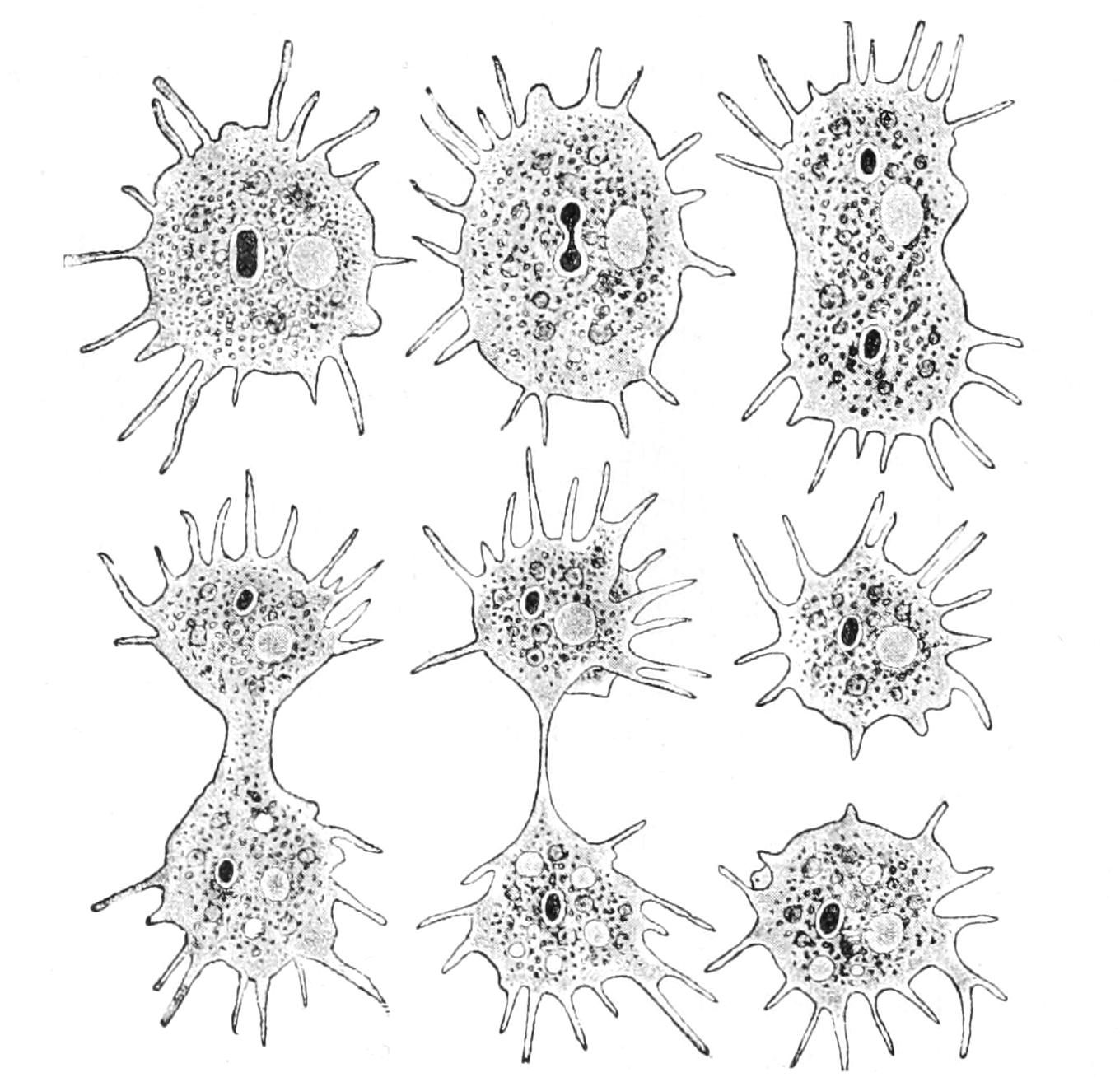
Fig. 4.—Amoeba polypodia in successive stages of equal fission; nucleus dark, contractile vacuole clear. (From Verworn, after F. E. Schulze.)
Again, when an Amoeba has grown to a certain size, its nucleus divides into two nuclei, and its cytoplasmic body, as we may term it, elongates, narrows in the middle so as to assume the shape of a dumb-bell or finger-biscuit, and the two halves, crawling in opposite directions, separate by the giving way of the connecting waist, forming two new Amoebas, each with its nucleus (Fig. 4). This is a process of "reproduction"; the special case is one of "equal fission" or "binary division." The original cell is termed the "mother," with respect to the two new ones, and these are of course with respect to it the "daughters," and "sisters" to one another. We must bear in mind that in this self-sacrificing maternity the mother is resolved into her children, and her very existence is lost in their production. The above phenomena, IRRITABILITY, MOTILITY, DIGESTION, NUTRITION, GROWTH, REPRODUCTION, are all characteristic of living beings at some stage or other, though one or more may often be temporarily or permanently absent; they are therefore called "vital processes."
If, on the other hand, we violently compress the cell, if we pass a very strong electric shock through it, or a strong continuous current, or expose it to a temperature much above 45° C., or to the action of certain chemical substances, such as strong acids or alkalies, or alcohol or corrosive sublimate, we find that all these vital processes are arrested once and for all; henceforward the cell is on a par with any not-living substance. Such a change is called "DEATH," and the "capacity for death" is one of the most marked characters of living beings. This change is associated with changes in the mechanical and optical properties of the protoplasm, which loses its viscidity and becomes opaque, having undergone a process of de-solution; for the water it contained is now held only mechanically in the interstices of a network, or in cavities of a honeycomb (as we have noted above, p. 5), while the solid forming the residuum has a refractive index of a little over 1.6. Therefore, it only regains its full transparency when the water is replaced by a liquid of high refractive index, such as an essential oil or phenol. A similar change may be effected by pouring white of egg into boiling water or absolute alcohol, and is attended with the same optical results. The study of the behaviour of coagulable colloids has been recently studied by Fischer and by Hardy, and has been of the utmost service in our interpretation of the microscopical appearances shown in biological specimens under the microscope.[13]
The death of the living being finds a certain analogy in the breaking up or the wearing out of a piece of machinery; but in no piece of machinery do we find the varied irritabilities, all conducive to the well-being of the organism (under ordinary conditions), or the so-called "automatic processes"[14] that enable the living being to go through its characteristic functions, to grow, and as we shall see, even to turn conditions unfavourable for active life and growth to the ultimate weal of the species (see p. 32). At the same time, we fully recognise that for supplies of matter and energy the organism, like the machine, depends absolutely on sources from without. The debtor and creditor sheet, in respect of matter and energy, can be proved to balance between the outside world and Higher Organisms with the utmost accuracy that our instruments can attain; and we infer that this holds for the Lower Organisms also. Many of the changes within the organism can be expressed in terms of chemistry and physics; but it is far more impossible to state them all in such terms than it would be to describe a polyphase electrical installation in terms of dynamics and hydraulics. And so far at least we are justified in speaking of "vital forces."
The living substance of protoplasm contains a large quantity of water, at least two-thirds its mass, as we have seen, in a state of physical or loose chemical combination with solids: these on death yield proteids and nucleo-proteids.[15] The living protoplasm has an alkaline reaction, while the liquid in the larger vacuoles, at least, is acid, especially in Plant-cells.[16]
Metabolism.—The chemical processes that go on in the organism are termed metabolic changes, and were roughly divided by Gaskell into (1) "anabolic," in which more complex and less stable substances are built up from less complex and more stable ones with the absorption of energy; and (2) "catabolic" changes in which the reverse takes place. Anabolic processes, in all but the cells containing plastids or chromatophores (see p. 36) under the influence of light, necessarily imply the furnishing of energy by concurrent catabolic changes in the food or reserves, or in the protoplasm itself.
Again, we have divided anabolic processes into "accumulative," where the substances formed are merely reserves for the future use of the cell, and "assimilative," where the substances go to the building of the protoplasm itself, whether for the purpose of growth or for that of repair.
Catabolic processes may involve (1) the mere breaking of complex substances into simpler ones, or (2) their combination with oxygen; in either case waste products are formed, which may either be of service to the organism as "secretions" (like the bile in Higher Animals), or of no further use (like the urine). When nitrogenous substances break down in this way they give rise to "excretions," containing urea, urates, and allied substances; other products of catabolism are carbon dioxide, water, and mineral salts, such as sulphates, phosphates, carbonates, oxalates, etc., which if not insoluble must needs be removed promptly from the organism, many of them being injurious or even poisonous. The energy liberated by the protoplasm being derived through the breakdown of another part of the same or of the food-materials or stored reserves, must give rise to waste products. The exchange of oxygen from without for carbonic acid formed within is termed "respiration," and is distinguished from the mere removal of all other waste products called "excretion." In the fresh-water Amoeba both these processes can be studied.
Respiration,[17] or the interchange of gases, must, of course, take place all over the general surface, but in addition it is combined in most fresh-water Protista with excretion in an organ termed the "contractile" or "pulsatile vacuole" (Figs. 1, 4, etc.). This particular vacuole is exceptional in its size and its constancy of position. At intervals, more or less regular, it is seen to contract, and to expel its contents through a pore; at each contraction it completely disappears, and reforms slowly, sometimes directly, sometimes by the appearance of a variable number of small "formative" vacuoles that run together, or as in Ciliata, by the discharge into it of so-called "feeding canals." As this vacuole is filled by the water that diffuses through the substance, and when distended may reach one-third the diameter of the being, in the interval between two contractions an amount of water must have soaked in equal to one-twenty-seventh the bulk of the animal, to be excreted with whatever substances it has taken up in solution, including, not only carbon dioxide, but also, it has been shown, nitrogenised waste matters allied to uric acid.[18]
That the due interchanges may take place between the cell and the surrounding medium, it is obvious that certain limits to the ratio between bulk and surface must exist, which are disturbed by growth, and which we shall study hereafter (p. 23 f.).
The Protista that live in water undergo a death by "diffluence" or "granular disintegration" on being wounded, crushed, or sometimes after an excessive electric stimulation, or contact with alkalies or with acids too weak to coagulate them. In this process the protoplasm breaks up from the surface inwards into a mass of granules, the majority of which themselves finally dissolve. If the injury be a local rupture of the external pellicle or cuticle, a vacuole forms at the point, grows and distends the overlying cytoplasm, which finally ruptures: the walls of the vacuole disintegrate; and this goes on as above described. Ciliate Infusoria are especially liable to this disintegration process, often termed "diffluence," which, repeatedly described by early observers, has recently been studied in detail by Verworn. Here we have death by "solution," while in the "fixing" of protoplasm for microscopic processes we strive to ensure death by "desolution," so as to retain as much of the late living matter as possible. It would seem not improbable that the unusual contact with water determines the formation of a zymase that acts on the living substance itself.
We have suggested[19] that one function of the contractile vacuole, in naked fresh-water Protists, is to afford a regular means of discharge of the water constantly taken up by the crystalloids in the protoplasm, and so to check the tendency to form irregular disruptive vacuoles and death by diffluence. This is supported by the fact that in the holophytic fresh-water Protista, as well as the Algae and Fungi, a contractile vacuole is present in the young naked stage (zoospore), but disappears as soon as an elastic cell-wall is formed to counterbalance by its tension the internal osmotic pressure.
Digestion is always essentially a catabolic process, both as regards the substance digested and the formation of the digesting substance by the protoplasm. The digesting substance is termed a "zymase" or "chemical ferment," and is conjectured to be produced by the partial breakdown of the protoplasm. In presence of suitable zymases, many substances are resolved into two or more new substances, often taking up the elements of water at the same time, and are said to be "dissociated" or "hydrolysed" as the case may be. Thus proteid substances are converted into the very soluble substances, "proteoses" and "peptones," often with the concurrent or ultimate formation of such relatively simple bodies as leucin, tyrosin, and other amines, etc. Starch and glycogen are converted into dextrins and sugars; fats are converted into fatty acids and glycerin. It is these products of digestion, and not the actual food-materials (save certain very simple sugars), that are really taken up by the protoplasm, whether for assimilation, for accumulation, or for the direct liberation of energy for the vital processes of the organism.
Not only food from without, but also reserves formed and stored by the protoplasm itself, must be digested by some zymase before they can be utilised by the cell. In all cases of the utilisation of reserve matter that have been investigated, it has been found that a zymase is formed by the cell itself (or sometimes, in complex organisms, by its neighbours); for, after killing the cell in which the process is going on by mechanical means or by alcohol, the process of digestion can be carried on in the laboratory.[20] The chief digestion of all the animal-feeding Protista is of the same type as in our own stomachs, known as "peptic" digestion: this involves the concurrent presence of an acid, and Le Dantec and Miss Greenwood have found the contents of food-vacuoles, in which digestion is going on, to contain acid liquid. The ferment-pepsin itself has been extracted by Krukenberg from the Myxomycete, "Flowers of tan" (Fuligo varians, p. 92), and by Professor Augustus Dixon and the author from the gigantic multinucleate Amoeba, Pelomyxa palustris (p. 52).[21] The details of the prehension of food will be treated of under the several groups.
The two modes of Anabolism—true "assimilation" in the strictest sense and "accumulation"—may sometimes go on concurrently, a certain proportion of the food material going to the protoplasm, and the rest, after allowing for waste, being converted into reserves.
Movements all demand catabolic changes, and we now proceed to consider these in more detail.
The movements of an Amoeboid[22] cell are of two kinds: "expansion," leading to the formation and enlargement of outgrowths, and "contraction," leading to their diminution and disappearance within the general surface.[23] Expansion is probably due to the lessening of the surface-tension at the point of outgrowth, contraction to the increase of surface-tension. Verworn regards these as due respectively to the combination of the oxygen in the medium with the protoplasm in diminishing surface-tension, and the effect of combination with substances from within, especially from the nucleus in increasing it. Besides these external movements, there are internal movements revealed by the contained granules, which stream freely in the more fluid interior. Those Protista that, while exhibiting amoeboid movements, have no clear external layer, such as the Radiolaria, Foraminifera, Heliozoa, etc., present this streaming even at the surface, the granules travelling up and down the pseudopodia at a rate much greater than the movements of these organs themselves. In this case the protoplasm is wetted by the medium, which it is not where there is a clear outer layer: for that behaves like a greasy film.
Motile organs.—Protoplasm often exhibits movements much more highly specialised than the simple expansion or retraction of processes, or the general change of form seen in Amoeba. If we imagine the activities of a cell concentrated on particular parts, we may well suppose that they would be at once more precise and more energetic than we see them in Amoeba or the leucocyte. In some free-swimming cells, such as the individual cells known as "Flagellata," the reproductive cells of the lower Plants, or the male cells ("spermatozoa") of Plants as high as Ferns, and even of the Highest Animals, there is an extension of the cell into one or more elongated lash-like processes, termed "flagella," which, by beating the water in a reciprocating or a spiral rhythm, cause the cell to travel through it; or, if the cell be attached, they produce currents in the water that bring food particles to the surface of the cell for ingestion. Such flagella may, indeed, be seen in some cases to be modified pseudopodia. In other cases part, or the whole, of the surface of the cell may be covered with regularly arranged short filaments of similar activity (termed "cilia," from their resemblance to a diminutive eyelash), which, however, instead of whirling round, bend sharply down to the surface and slowly recover; the movement affects the cilia successively in a definite direction in waves, and produces, like that of flagella, either locomotion of the cell or currents in the medium. We can best realise their action by recalling the waves of bending and recovery of the cornstalks in a wind-swept field; if now the haulms of the corn executed these movements of themselves, they would determine in the air above a breeze-like motion in the direction of the waves (Fig. 5).[24] Such cilia are not infrequent on those cells of even the Highest Animals that, like a mosaic, cover free surfaces ("epithelium cells"). In ourselves such cells line, for instance, the windpipe. One group of the Protozoa, the "Ciliata," are, as their name implies, ciliated cells pure and simple.

Fig. 5.—Motion of a row of cilia, in profile. (From Verworn.)
The motions of cilia and of flagella are probably also due to changes of surface tension—alternately on one side and the other in the cilium, but passing round in circular succession in the flagellum,[25] giving rise to a conical rotation like that of a weighted string that is whirled round the head. This motion is, however, strongest at the thicker basal part, which assumes a spiral form like a corkscrew of few turns, while the thin lash at the tip may seem even to be quietly extended like the point of the corkscrew. If the tip of the flagellum adhere, as it sometimes does, to any object, the motions induce a jerking motion, which in this case is reciprocating, not rotatory. When the organism is free, the flagellum is usually in advance, and the cell follows, rotating at the same time round its longitudinal axis; such an anterior flagellum, called a "tractellum," is the common form in Protista that possess a single one (Figs. 29, 7, 8; 30, C). In the spermatozoa of Higher Animals (and some Sporozoa) the flagellum is posterior, and is called a "pulsellum."
The cilium or flagellum may often be traced a certain distance into the substance of the cytoplasm to end in a dot of denser, readily-staining plasm, which corresponds to a "centrosome" or centre of plasmic forces (see below, pp. 115, 121, 141); it has been termed a "blepharoplast."[26]
Again, the cytoplasm may have differentiated in it definite streaks of specially contractile character; such streaks within its substance are called "myonemes"; they are, in fact, muscular fibrils. A "muscle-cell," in the Higher Animals, is one whose protoplasm is almost entirely so modified, with the exception of a small portion of granular cytoplasm investing the nucleus, and having mainly a nutritive function.
Definite muscular fibrils in action shorten, and at the same time become thicker. It seems probable that they contain elongated vacuoles, and that the contents of these vary, so that when they have an increased osmotic equivalent, the vacuoles absorb water, enlarge, and tend to become more spherical, i.e. shorter and thicker, and so the fibril shortens as a whole. The relaxation would be due to the diffusion outwards of the solution of the osmotically active substances which induced expansion.[27]
The Motile Reactions of the Protozoa[28] require study from another point of view: they are either (1) "spontaneous" or "arbitrary," as we may say, or (2) responsive to some stimulus. The latter kind we will take first, as they are characteristic of all free cells. The stimuli that induce movements of a responsive character are as follows:—(i.) MECHANICAL: such as agitation and contact; (ii.) force of GRAVITY, or CENTRIFUGAL FORCE; (iii.) CURRENTS in the water; (iv.) RADIANT ENERGY (LIGHT); (v.) changes in the TEMPERATURE of the medium; (vi.) ELECTRIC CURRENTS through the medium; (vii.) the presence of CHEMICAL SUBSTANCES in the medium.
These, or some of them, may induce one of three different results, or a combination thereof: (1) a single movement or an arrest of motion; (2) the assumption of a definite position; (3) movement of a definite character or direction.
(i.) Mechanical stimuli.—Any sudden touch with another body tends to arrest all motion; and if the shock be protracted or severe, the retraction of the pseudopodia follows. It is to this reaction that we must ascribe the retracted condition of the pseudopodia of most Rhizopods when first placed on the slide and covered for microscopic examination. Free-swimming Protista may, after hitting any body, either remain in contact with it, or else, after a pause, reverse their movement, turn over and swim directly away. This combination of movements is characteristic as a reaction of what we may term "repellent" stimuli in general.[29] Another mechanical reaction is that to continuous contact with a solid; and the surface film of water, either at the free surface or round an air-bubble, may play the part of a solid in exciting it; we term it "thigmotaxy" or "stereotaxy." When positive it determines a movement on to the surface, or a gliding movement along it, or merely the arrest of motion and prolongation of contact; when negative, a contact is followed by the retreat of the being. Thus Paramecium (Fig. 55, p. 151) and many other Ciliates are led to aggregate about solid particles or masses of organic débris in the water, which indeed serve to supply their food. On contact, the cell ceases to move its cilia except those of the oral groove; as these lash backwards, they hold the front end in close contact with the solid, at the same time provoking a backward stream down the groove, which may bring in minute particles from the mass.
(ii.) Most living beings are able to maintain their level in water by floating or crawling against Gravity, and they react in virtue of the same power against centrifugal force. This mode of irritability is termed (negative) "geotaxy" or "barotaxy." We can estimate the power of resisting such force by means of a whirling machine, since when the acceleration is greater than the resistance stimulated thereby in the beings, they are passively sent to the sides of the vessel. The Flagellates, Euglena and Chlamydomonas, begin to migrate towards the centre when exposed to a centrifugal force about equal to ½ G (G = 32.2 feet or 982 cm. per second); they remain at the centre until the centrifugal force is increased to 8 G; above that they yield to the force, and are driven passively to the sides. The reaction ceases or is reversed at high temperatures.
(iii.) Rheotaxy.—This is the tendency to move against the stream in flowing water. It is shown by most Protists, and can be conveniently studied in the large amoeboid plasmodia of the Myxomycetes, which crawl against the stream along wet strips of filter paper, down which water is caused to flow. Most animals, even of the highest groups, tend to react in the same way; the energetic swimming of Fishes up-stream being in marked contrast with their sluggishness the other way; and every student of pond-life knows how small Crustacea and Rotifers, no less than Ciliates, swim away from the inrush of liquid into the dipping-tube, and so evade capture. (See Vol. II. p. 216.)
(iv.) The movements of many Protozoa are affected greatly by Light. These movements have been distinguished into "photopathic," i.e. to or from the position of greatest luminosity; and "phototactic," along the direct path of the rays.[30] Those Protozoa that contain a portion of their cytoplasm, known as a "plastid" or "chromatophore" (see pp. 36, 39), coloured by a green or yellow pigment are usually "phototactic." They mostly have at the anterior end a red pigment spot, which serves as an organ of sight, and is known as an "eye-spot." In diffused light of low intensity they do not exhibit this reaction, but in bright sunlight they rise to the surface and form there a green or yellow scum.
Most of the colourless Protista are negatively phototactic or photopathic; but those which are parasitic on the coloured ones are positively phototactic, like their hosts.
Here, as in the case of other stimuli,[31] the absolute intensity of the light is of importance; for as it increases from a low degree, different organisms in turn cease to be stimulated, and then are repelled instead of being attracted. The most active part of the spectrum in determining reactions of movement are the violet and blue rays of wave-length between 40 µ/10 and 49 µ/10, while the warmer and less refractive half of the spectrum is inert save in so far as it determines changes in the temperature of the medium.
(v.) The movements of many Protozoa are rendered sluggish by cold, and active by a rise of Temperature up to what we may term the "optimum"; the species becomes sluggish again as the temperature continues to rise to a certain point when the movements are arrested, and the being is said to be in a state of "heat-rigor." Most Protozoa, again, tend to move in an unequally heated medium to the position nearest to their respective optimum temperature. This is called "thermotaxy." The temperature to which Amoeba is thermotactic is recorded as 35° C. (95° F.); that of Paramecium is 28° C. (82° F.).
(vi.) Most active Protozoa tend to take up a definite position in respect to a current of Electricity passing through the medium, and in the majority of cases, including most Ciliates, Amoeba, and Trachelomonas, they orient their long diameters in the direction of the lines of force and swim along these to assemble behind the cathode. The phenomenon is called "galvanotaxy," and this particular form is "negative." Opalina (Fig. 41, p. 123), however, and most Flagellates are "positively galvanotactic," and move towards the anode. H. H. Dale[32] has shown that the phenomenon may be possibly in reality a case of chemiotaxy, for the direction of motion varies with the nature and concentration of the medium. It would thus be a reaction to the "ion" liberated in contact with the one or other extremity of the being. Induction shocks, as we have seen, if slight, arrest the movements of Protozoa, or if a little stronger determine movements of contraction; if of sufficient intensity they kill them. No observation seems to have been made on the behaviour of Protista in an electric field. A magnetic field of the highest intensity appears to be indifferent to all Protista.
(vii.) We have already referred to the effect of dissolved Chemical Substances present in the water. If the substance is in itself not harmful, and the effect varies with the concentration, we term the reaction one of "tonotaxy," which combines with that of "chemiotaxy" for substances that in weak solution are attractive or repellent to the being. Paramecium, which feeds on bacteria, organisms of putrefaction, is positively chemiotactic to solutions of carbon dioxide, and as it gives this off in its own respiration, it is attracted to its fellows. The special case of reaction to gases in solution is termed "aerotaxy," or "pneumotaxy," according as the gas is oxygen or carbon dioxide. We find that in this respect there are degrees, so that a mixed culture of Flagellates in an organic infusion sorts itself out, under the cover of a microscopic preparation, into zones of distinct species, at different distances from the freely aerated edge, according to the demands of each species for oxygen and CO2 respectively.
Finally, we must note that the apparently "spontaneous movements" of Protists can hardly be explained as other than due either to external stimuli, such as we have just studied, or to internal stimuli, the outcome of internal changes, such as fatigue, hunger, and the like. Of the latter kind are the movements that result in REPRODUCTION.
Reproduction.—We have noted above that the growth of an organism which retains its shape alters the ratio of the surface area to the whole volume, so necessary for the changes involved in life. For the volume of an organism varies as the cube of any given diameter, whereas the surface varies with the square only. Without going into the arithmetical details, we may say that the ratio of surface to volume is lessened to roughly four-fifths of the original ratio when the cell doubles its bulk. As Herbert Spencer and others have pointed out, this must reduce the activities of the cell, and the due ratio is restored by the division of the cell into two.[33] This accounts for what we must look on as the most primitive mode of reproduction, as it is the simplest, and which we term "fission" at Spencer's "limit of growth." Other modes of reproduction will be studied later (p. 30), after a more detailed inquiry into the structure of the nucleus and of its behaviour in cell-division. All cell-division is accompanied by increased waste, and is consequently catabolic in character, though the anabolic growth of living protoplasm, at the expense of the internal reserves, may be concurrent therewith.
Cell-Division
In ordinary cases of fission of an isolated cell the cell elongates, and as it does so, like other viscid bodies, contracts in the middle, which becomes drawn out into a thread, and finally gives way. In some cases (e.g. that of the Amoeba, Fig. 4) the nucleus previously undergoes a similar division by simple constriction, which is called direct or "amitotic" division. But usually the division of the nucleus prior to cell-division is a more complex process, and involves the co-operation of the cytoplasm; and we must now study in detail the nucleus and its structure in "rest" and in fission.[34]
We have noted above (p. 6, Fig. 2) the structure of the so-called "resting nucleus,"[35] when the cell is discharging the ordinary functions of its own life, with its wall, network of linin, chromatin-granules, and nucleole or nucleoles. The chromatin-granules are most abundant at two periods in the life of the cell, (1) when it is young and fresh from division, and (2) at the term of its life, when it is itself preparing for division. In the interim they are fewer, smaller, and stain less intensely. In many Protista the whole or greater part of the chromatin is densely aggregated into a central "nuclein-mass" or karyosome suspended in the linin network (long regarded as a mere nucleole). Such a nucleus is often termed a "vesicular nucleus".[36]
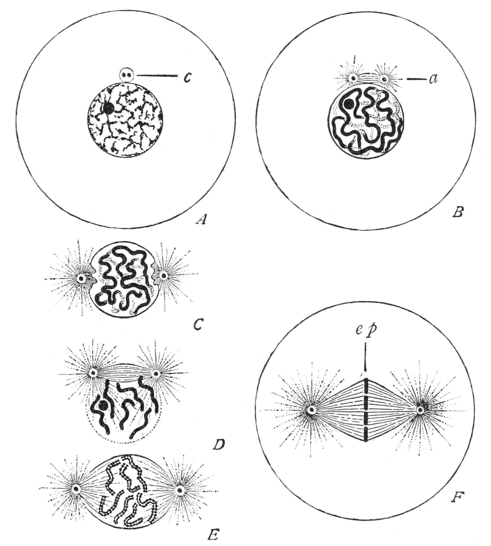
Fig. 6.—Changes in nucleus and cell in indirect (mitotic) nuclear division. A, resting nucleus with two centrioles[37] in single centrosphere (c); B, centrosphere divided, spindle and two asters (a) forming; C, centrospheres separated, nuclear wall disappearing; D, resolution of nucleus into chromosomes; E, mature plasmic spindle, with longitudinal fission of chromosomes; F, chromosomes forming equatorial plate (ep) of spindle. (From Wilson.)
When cell-division is about to take place the linin, or at least the greater part of it, assumes the character of a number of distinct threads, and the whole of the chromatin granules are distributed at even distances along these (Fig. 6, A, B, C), so as to appear like so many strings of beads. Each such thread is called a "chromosome." Then each bead divides longitudinally into two. The thread flattens into a ribbon, edged by the two lines of chromatin beads. Finally, the ribbon splits longitudinally into two single threads of beads (Fig. 6, E). During these changes the nucleole or nucleoles diminish, or even disappear, as if they had contributed their matter to the growth of the chromatin proper. In Higher Animals and Plants the nuclear wall next disappears, and certain structures become obvious, especially in the cytoplasm of Metazoa. Two minute spheres of plasm (themselves often showing a concentric structure), the "centrosomes,"[38] which hitherto lay close together at the side of the nuclear wall, now separate; but they remain connected by a spindle of clear plasmic threads (Fig. 6, B-E) which, as the centres diverge, comes to lie across the spot the nucleus occupied, and now the chromosomes lie about the equator of this spindle (Fig. 6, F). Moreover, the surrounding cytoplasm shows a radiating structure, diverging from the centrosome, so that spindle and external radiations together make up a "strain-figure," like that of the "lines of force" in relation to the poles of a magnet. Such we can demonstrate in a plane by spreading or shaking iron filings on a piece of paper above the poles of a magnet, or in space by suspending finely divided iron in a thick liquid, such as mucilage or glycerin, and bringing the vessel with the mixture into a strong magnetic field;[39] the latter mode has the advantage of enabling us to watch the changes in the distribution of the lines under changing conditions or continued strain.
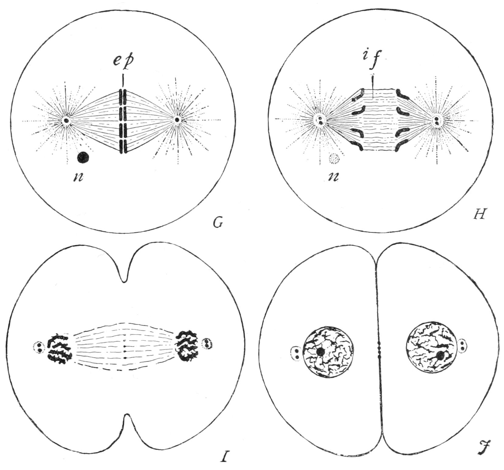
Fig. 7.—Completion of mitotic cell-division. G, splitting of equatorial plate (ep); H, recession of daughter chromosomes; I, J, reconstitution of these into new nuclei, fission of the centrioles and of the cytoplasm. if, Central fibres of spindle; n, remains of old nucleole. (From Wilson.)
The chromosomes are now completely split, each into its two daughter-segments, which glide apart (Fig. 7, G, ep), and pass each to its own pole of the spindle, stopping just short of the centrosome (I). Thus, on the inner side of either centrosome is found an aggregation of daughter-segments, each of which is sister to one at the opposite pole, while the number at either pole is identical with that of the segments into which the old nucleus had resolved itself at the outset. The daughter-segments shorten and thicken greatly as they diverge to the poles, and on their arrival crowd close together.
A distinct wall now forms around the aggregated daughter-chromosomes (J), so as to combine them into a nucleus for the daughter-cell. The reorganisation of the young nucleus certainly varies in different cases, and has been ill-studied, probably because of the rapidity of the changes that take place. The cytoplasm now divides, either tapering into a "waist" which finally ruptures, or constricting by the deepening of a narrow annular groove so as to complete the formation and isolation of the daughter-cells.
We might well compare the cell-division to the halving of a pumpkin or melon, of which the flesh as a whole is simply divided into two by a transverse cut, while the seeds and the cords that suspend them are each singly split to be divided evenly between the two halves of the fruit; the flesh would represent the cytoplasm, the cords the linin threads of the nucleus, and the seeds the chromatin granules. In this way the halving of the nucleus is much more complete and intimate than that of the cytoplasm; and this is the reason why many biologists have been led to regard the nuclear segments, and especially their chromatic granules, as the seat of the hereditary properties of the cell, properties which have to be equally transmitted on its fission to each daughter-cell.[40] But we must remember that the linin is also in great part used up in the formation of these segments, like the cords of our supposed melon; and it is open to us to regard the halving in this intimate way of the "linin" as the essence of the process, and that of the chromatin as accessory, or even as only part of the necessary machinery of the process. The halving or direct splitting lengthwise of a viscid thread is a most difficult problem from a physical point of view; and it may well be that the chromatin granules have at least for a part of their function the facilitation of this process. If such be the case, we can easily understand the increase in number, and size and staining power of these granules as cell-division approaches, and their atrophy or partial disappearance during their long intervening periods of active cell life. Hence we hesitate to accept the views so commonly maintained that the chromatin represents a "germ-plasm" or "idioplasm" of relatively great persistence, which gives the cell its own racial qualities.[41]
The process we have just examined is called "mitosis," "karyomitosis," or "karyokinesis"; and the nucleus is said to undergo "indirect" division, as compared to "direct" division by mere constriction. In an intermediate mode, common to many Protista, the nuclear wall persists throughout the whole process, though a spindle is constituted within, and chromosomes are formed and split: the division of the nucleus takes place, however, by simple constriction, as seen in the Filose Rhizopod Euglypha (Fig. 8).
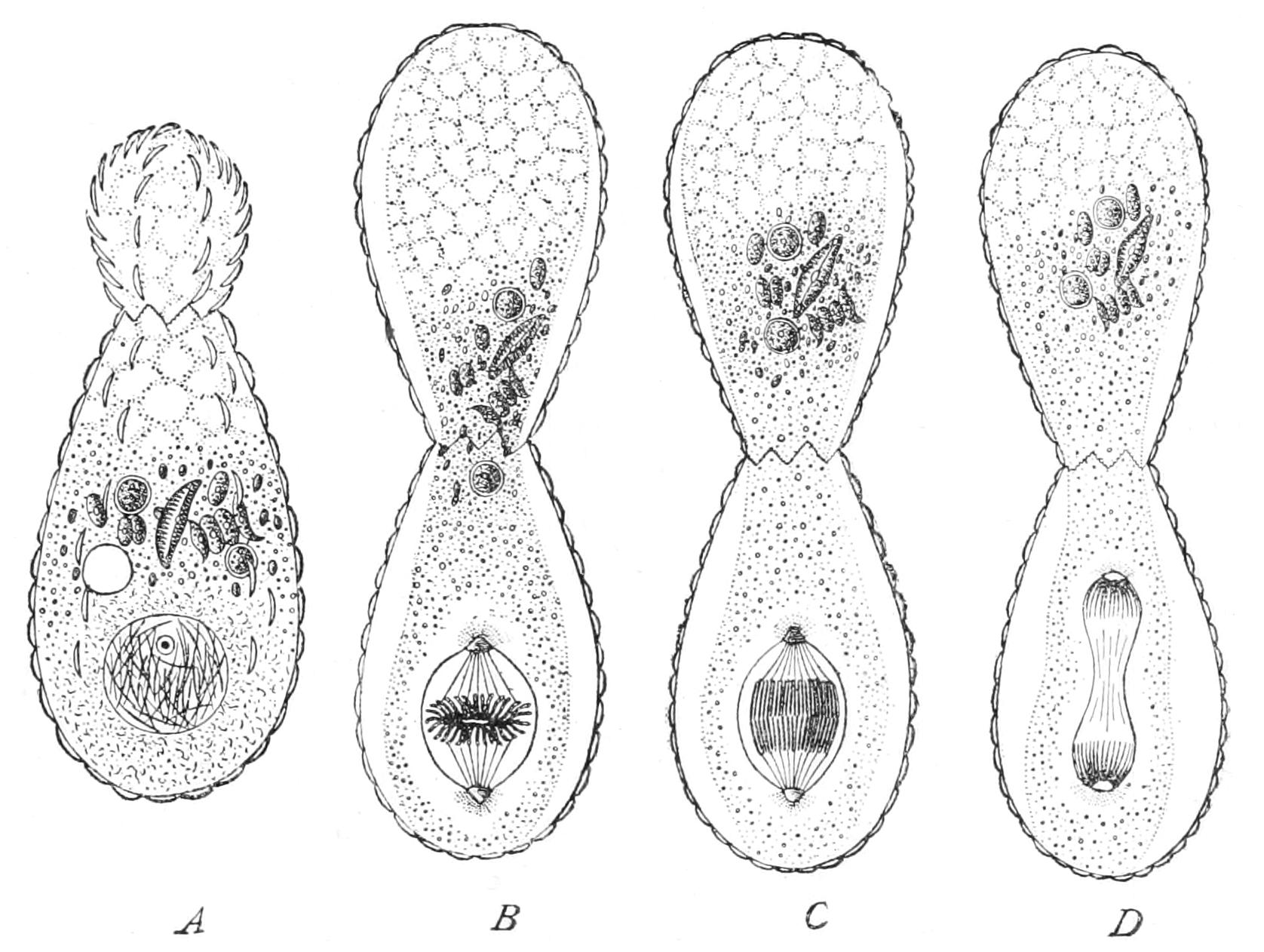
Fig. 8.—Fission with modified karyokinesis in the Filose Rhizopod Euglypha. A, outgrowth of half of the cytoplasm, passage of siliceous plates for young shell outwards; B, completion of shell of second cell, formation of intra-nuclear spindle; C, D, further stages. (From Wilson, after Schewiakoff.)
In many Sarcodina and some Sporozoa the nucleus gives off small fragments into the cytoplasm, or is resolved into them; they have been termed "chromidia" by E. Hertwig. New nuclei may be formed by their growth and coalescence, the original nucleus sometimes disappearing more or less completely.
In certain cases the division of the nucleus is not followed by that of the cytoplasm, so that a plurinucleate mass of protoplasm results: this is called an "apocyte"; and we find transitional forms between this and the uninucleate or true cell. Thus in one species of Amoeba (A. binucleata) there are always two nuclei, which divide simultaneously to provide for the outfit of the daughter-cells on fission. Again, we find in some cases that similar multinucleate masses may be formed by the union of two or more cells by their cytoplasm only: such a union is termed "permanent plastogamy," and the plurinucleate mass a "plasmodium."[42] Here again we find intermediate forms between plasmodium and apocyte, for the nuclei of the former may divide and so increase in number, without division of the still growing mass. Both kinds of plurinucleate organisms are termed "coenocytes" without reference to their mode of origin.
The rhythm of cell-life that we have just studied is called the "Spencerian" rhythm. Each cell in turn grows from half the bulk of its parent at the time it was formed to the full size of that parent, when it divides in its own turn. Rest is rare, and assumed only when the cell is exposed to such unfavourable external conditions as starvation, drought, etc.; it has no necessary relation to fission.
Multiple fission or brood-formation.—We may now turn to a new rhythm, in strong contrast to the former: a cell after having attained a size, often notably greater than its parents, divides: without any interval for growth, the daughter-cells again divide, and this may be repeated as many as ten times, or even more, so as to give rise to a number of small cells—4, 8, 16—1024,[43] etc., respectively. Such an assemblage of small cells so formed is called a brood, and well deserves this name, for they never separate until the whole series of divisions is completed. By this process the number of individuals is rapidly increased, hence it has received the name of "sporulation." The term spores is especially applied to the reproductive bodies of Cryptogams, such as Mosses, Fungi, etc.: the resulting cells are called "spores," "zoospores" if active ("amoebulae" if provided with pseudopodia, "flagellulae" if flagellate), "aplanospores," if motionless. We prefer to call them by the general term "brood-cells," the original cell the "brood-mother-cell," and the process, "multiple fission" or "brood-formation." As noted, the brood-mother-cell usually attains an exceptionally large size, and it in most cases passes into a state of rest before entering on division: thus brood-formation is frequently the ultimate term of a long series of Spencerian divisions. Two contrasting periods of brood-formation may occur in the life cycle of some beings, notably the Sporozoa.[44]
Colonial union.—In certain cases, the brood-cells instead of separating remain together to form a "colony"; and this may enlarge itself again by binary division of its individual cells at their limit of growth. Here, certain or all of the cells may (either after separation, or in their places) undergo brood-formation: such cells are often termed "reproductive cells" in contrast with the "colonial cells."
Some such colonial Protista must have been the starting-points for the Higher Animals and Plants; probably apocytial Protista were the starting-points of the Fungi. In the Higher Animals and Plants, the spermatozoa and the oospheres (the male and female pairing-cells) are alike the offspring of brood-formation: and the coupled-cell (fertilised egg) starts its new life by segmentation, which is a brood-formation in which the cells do not separate, but remain in colonial union, to differentiate in due course into the tissue-cells of the organism.
Retarded brood-formation.—The nuclear divisions may alternate with cell-divisions, as above stated, or the former may be completed before the cytoplasm divides; thus the brood-mother-cell becomes temporarily an apocyte,[45] which is then resolved simultaneously into the 1-nucleate brood-cells.
A temporary apocytial condition is often passed through in the formation of the brood of cells by repeated divisions without any interval for enlargement; for the nuclear divisions may go on more rapidly than those of the cytoplasm, or be completed before any cell-division takes place (Figs. 31, 34, 35, pp. 95, 101, 104), the nuclear process being "accelerated" or the cytoplastic being "retarded," whichever we prefer to say and to hold. Thus as many as thirty-two nuclei may have been formed by repeated binary subdivisions before any division of the cytoplasm takes place to resolve the apocyte into true 1-nucleate cells.
In many cases of brood-formation the greater part of the food-supply of the brood-mother-cell has been stored as reserve-products, which accumulate in quantity in the cell; this is notably seen in the ovum or egg of the Higher Animals. How great such an accumulation may be is indeed well seen in the enormous yolk of a bird's egg, gorged as it were to repletion. When a cell has entered on such course of "miserly" conduct, it may lose all power of drawing on its own supplies, and finally that of accumulating more, and passes into the state of "rest." To resume activity there is needed either a change in the internal conditions—demanding the lapse of time—or in the external conditions, or in both.[46] We may call this resumption "germination."
Very often in the study of a large and complex organism we are able to find processes in action on a large scale which, depending as they must do on the protoplasmic activities of its individual cells, reveal the nature of similar processes in simple unicellular beings: such a clue to the utilisation of reserves by a cell which has gorged itself with them so as to pass into a state of rest is to be found in that common multicellular organism, the Potato. This stores up reserves in its underground stems (tubers); if we plant these immediately on the completion of their growth, they will not start at once, even under what would outwardly seem to be most appropriate conditions. A certain lapse of time is an essential factor for sprouting. It would appear that in the Potato the starch can only be digested by a definite ferment, which does not exist when it is dug, but which is only formed very slowly, and not at all until a certain time has supervened; and that sprouting can only take place when soluble material has been provided in this way for the growth of the young shoots. We have also reason to believe that these ferments are only formed by the degradation of the protoplasm itself. Now obviously this degradation must be very slow in a resting organism; and any external stimulus that will tend to protoplasmic activity will thereby tend to form at the same time the digestive ferments and dissolve the stored supplies, to render them available for the life-growth and reproduction of the being. We now see why inactive "miserly" cells so often pass into a resting state before dividing, and why they go on dividing again and again when once they re-enter upon an active life, the living protoplasm growing at the expense of the reserves.[47] Resting cells of this type occur of course only at relatively rare intervals in the animal-feeding Protozoa, that have to take into their substance the food they require for their growth and life-work, and cannot therefore store up much reserves. For they are constantly producing in the narrow compass of their body those very ferments that would dissolve the reserves when formed. Internal parasites and "saprophytes," that is, beings which live on dead and decayed organic matter, on the other hand, live surrounded by a supply of dissolved food; and rarely do we find larger cells, richer in reserves, than in the parasitic Sporozoa, which owe their name to the importance of brood-formation in their life-history. In Radiolaria (p. 75 f.) a central capsule separates off an inner layer of protoplasm; the outer layer is the one in which digestion is performed, while the inner layer stores up reserves; and here brood-formation appears to be the rule. But the largest cells of all are the eggs of the Metazoa, which in reality lead a parasitic life, being nurtured by the animal as a whole, and contributing nothing to the welfare of it as an individual. Their activity is reduced to a minimum, and the consequent need for a high ratio of surface to volume is also reduced, which accounts for their inordinate size. But directly the reserve materials are rendered available by the formation of a digestive ferment, then protoplasmic growth takes place, and the need for an extended surface is felt; cell-division follows cell-division with scarcely an interval in the process of segmentation.
Syngamy.[48]—The essence of typical syngamy is, that two cells ("pairing-cells," "gametes") of the same species approach one another, and fuse, cytoplasm with cytoplasm, and nucleus with nucleus, to form a new cell ("coupled-cell," "zygote "). This process is called also "conjugation" or "cytogamy." In the simplest cases the two cells are equal and attract one another equally ("isogamy"), and have frequently the character of zoospores.
In an intermediate type, the one cell is larger and more sluggish (female), "megagamete," "oogamete," "oosphere," "egg"; the other smaller, more active (male), "microgamete," "spermogamete," "spermatozoon," "sperm"; and in the most specialised cases which prevail among the Higher Animals and Plants, the larger cell is motionless, and the smaller is active, ciliate, flagellate, or amoeboid: the coupled-cell or zygote is here termed the "oosperm."[49] It encysts immediately in most Protista except Infusoria, Acystosporidae, Haemosporidae, and Trypanosomatidae.
As the size of the two gametes is so disproportionate in most cases that the oosphere may be millions of times bigger than the sperm, and the latter at its entrance into the oosphere entirely escape unaided vision, the term "egg" is applied in lax speech, both (1) to the female cell, and (2) to the oosperm, the latter being distinguished as the "fertilised egg," a survival from the time when the union of two cells, as the essence of the process, was not understood.
We know that in many cases, and have a right to infer that in all, chemiotaxy plays an important part in attracting the pairing-cells to one another. In Mammals and Sauropsida there seems also to be a rheotactic action of the cilia lining the oviducts, which work downwards, and so induce the sperms to swim upwards to meet the ovum, a condition of things that was most puzzling until the nature of rheotaxy was understood. A remarkable fact is that equal gametes rarely appear to be attracted by members of the same brood, though they are attracted by those of any other brood of the same species.[50] It may well be that each brood has its own characteristic secretion, or "smell," as it were, slightly different from that of other broods, just as every dog has his, so easily recognisable by other dogs; and that the cells only react to different "smells" to their own. Such a secretion from the surface of the female cell would lessen its surface tension, and thereby render easier the penetration of the sperm into its substance.
As a rule, one at least of the pair-cells is fresh from division, and it would thus appear that the union of the nuclei is facilitated when one at least of them is a "young" one. Of the final mechanism of the union of the nuclei, we know nothing: they may unite in any of the earlier phases of mitosis, or even in the "resting state." A fibrillation of the cytoplasm during the process, radiating around a centrosome or two centrosomes indicates a strained condition.[51]
Regeneration.—Finally, experiments have been made by several observers as to the effects of removing parts of Protozoa, to see how far regeneration can take place. The chief results are as follows:—
1. A nucleated portion may regenerate completely, if of sufficient size. Consequently, multinucleate forms, such as Actinosphaerium (Heliozoa, Fig. 19, p. 72), may be cut into a number of fragments, and regenerate completely. In Ciliata, such as Stentor (Fig. 59, p. 156), each fragment must possess a portion of the meganucleus, and at least one micronucleus (p. 145), and, moreover, must possess a certain minimum size. A Radiolarian "central-capsule" (p. 75) with its endoplasm and nucleus may regenerate its ectoplasm, but the isolated ectoplasm being non-nucleate is doomed. A "central capsule" of one species introduced into the ectoplasm of another, closely allied, did well. All non-nucleate pieces may exhibit characteristic movements, but appear unable to digest; and they survive only a short time.[52]
"Animals" and "Plants"
Hitherto we have discussed the cell as if it were everywhere an organism that takes in food into its substance, the food being invariably "organic" material, formed by or for other cells; such nutrition is termed "holozoic." There are, however, limits to the possibilities in this direction, as there are to the fabled capacities of the Scillonians of gaining their precarious livelihood by taking in one another's washing. For part of the food material taken in in this way is applied to the supply of the energies of the cell, and is consequently split up or oxidised into simpler, more stable bodies, no longer fitted for food; and of the matter remaining to be utilised for building up the organism, a certain proportion is always wasted in by-products. Clearly, then, the supply of food under such conditions is continually lessening in the universe, and we have to seek for a manufactory of food-material from inorganic materials: this is to be found in those cells that are known as "vegetal," in the widest sense of the word. In this, sense, vegetal nutrition is the utilisation of nitrogenous substances that are more simple than proteids or peptones, together with suitable organic carbon compounds, etc., to build up proteids and protoplasm. The simplest of organisms with a vegetal nutrition are the Schizomycetes, often spoken of loosely as "bacteria" or "microbes," in which the differentiation of cytoplasm and nucleus is not clearly recognisable. Some of these can build up their proteids from the free uncombined nitrogen of the atmosphere, carbon dioxide, and inorganic salts, such as sulphates and phosphates. But the majority of vegetal feeders require the nitrogen to be combined at least in the form of a nitrate or an ammonium salt—nay, for growth in the dark, they require the carbon also to be present in some organic combination, such as a tartrate, a carbohydrate, etc. Acetates and oxalates, "aromatic" compounds[53] and nitriles are rarely capable of being utilised, and indeed are often prejudicial to life. In many vegetal feeders certain portions of the protoplasm are specialised, and have the power of forming a green, yellow, or brown pigment; these are called "plastids" or "chromatophores." They multiply by constriction within the cell, displaying thereby a certain independent individuality. These plastids have in presence of light the extraordinary power of deoxidising carbon dioxide and water to form carbohydrates (or fats, etc.) and free oxygen; and from these carbohydrates or fats, together with ammonium salts or nitrates, etc., the vegetal protoplasm at large can build up all necessary food matter. So that in presence of light of the right quality[54] and adequate intensity, such coloured vegetal beings have the capacity for building up their bodies and reserves from purely inorganic materials. Coloured vegetal nutrition, then, is a process involving the absorption of energy; the source from which this is derived in the bacteria being very obscure at present. Nutrition by means of coloured plastids is distinguished as "holophytic," and that from lower substances, which, however, contain organically combined carbon, as "saprophytic," for such are formed by the death and decomposition of living beings. The third mode of nutrition (found in some bacteria) from wholly inorganic substances, including free nitrogen, has received no technical name. All three modes are included in the term "autotrophic" (self-nourishing).
Vegetal feeders have a great tendency to accumulate reserves in insoluble forms, such as starch, paramylum, and oil-globules on the one hand, and pyrenoids, proteid crystals, aleurone granules on the other.
When an animal-feeding cell encysts or surrounds itself with a continuous membrane, this is always of nitrogenous composition, usually containing the glucosamide "chitin." The vegetal cell-wall, on the contrary, usually consists, at least primarily, of the carbohydrate "cellulose"—the vegetal cell being richly supplied with carbohydrate reserves, and drawing on them to supply the material for its garment. This substance is what we are all familiar with in cotton or tissue-paper.
Again, not only is the vegetal cell very ready to surround itself with a cell-wall, but its food-material, or rather, speaking accurately, the inorganic materials from which that food is to be manufactured, may diffuse through this wall with scarcely any difficulty. Such a cell can and does grow when encysted: it grows even more readily in this state, since none of its energies are absorbed by the necessities of locomotion, etc. Growth leads, of course, to division: there is often an economy of wall-material by the formation of a mere party-wall dividing the cavity of the old cell-wall at its limit of growth into two new cavities of equal size. Thus the division tends to form a colonial aggregate, which continues to grow in a motionless, and, so far, a "resting" state. We may call this "vegetative rest," to distinguish it from "absolute rest," when all other life-processes (as well as motion) are reduced to a minimum or absolutely suspended.
The cells of a plant colony are usually connected by very fine threads of protoplasm, passing through minute pores where the new party-wall is left incomplete after cell-division.[55] In a few plants, such as most Fungi, the cell-partitions are in abeyance for the most part, and there is formed an apocyte with a continuous investment, sometimes, however, chambered at intervals by partitions between multinucleate units of protoplasm. We started with a purely physiological consideration, and we have now arrived at a morphological distinction, very valid among higher organisms.
Higher Plants consist of cells for the most part each isolated in its own cell-cavity, save for the few slender threads of communication.
Higher Animals consist of cells that are rarely isolated in this way, but are mostly in mutual contact over the greater part of their surface.
Again, Plants take in either food or else the material for food in solution through their surface, and only by diffusion through the cell-wall. Insectivorous Plants that have the power of capturing and digesting insects have no real internal cavity. Animal-feeding Protista take in their food into the interior of their protoplasm and digest it therein, and the Metazoa have an internal cavity or stomach for the same purpose. Here again there are exceptions in the case of certain internal parasites, such as the Tapeworms and Acanthocephala (Vol. II. pp. 74, 174), which have no stomachs, living as they do in the dissolved food-supplies of their hosts, but still possessing the general tissues and organs of Metazoa.
Corresponding with the absence of mouth, and the absorption instead of the prehension of food, we find that the movements of plant-beings are limited. In the higher Plants, and many lower ones, the colonial organism is firmly fixed or attached, and the movements of its parts are confined to flexions. These are produced by inequalities of growth; or by inequalities of temporary distension of cell-masses, due to the absorption of liquid into their vacuoles, while relaxation is effected by the cytoplasm and cell-wall becoming pervious to the liquid. We find no case of a differentiation of the cytoplasm within the cell into definite muscular fibrils. In the lower Plants single naked motile cells disseminate the species; and the pairing-cells, or at least the males, have the same motile character. In higher Cryptogams, Cycads, and Ginkgo (the Maiden-hair Tree), the sperms alone are free-swimming; and as we pass to Flowering Plants, the migratory character of the male cells is restricted to the smallest limits. though never wholly absent. Intracellular movements of the protoplasm are, however, found in all Plants.
In Plants we find no distinct nervous system formed of cells and differentiated from other tissues with centres and branches and sense-organs. These are more or less obvious in all Metazoa, traces being even found in the Sponges.
We may then define Plants as beings which have the power of manufacturing true food-stuffs from lower chemical substances than proteids, often with the absorption of energy. They have the power of surrounding themselves with a cell-wall, usually of cellulose, and of growing and dividing freely in this state, in which animal-like changes of form and locomotion are impossible; their colonies are almost always fixed or floating; free locomotion is only possible in the case of their naked reproductive cells, and is transitory even in these. The movements of motile parts of complex plant-organisms are due to the changes in the osmotic powers of cells as a whole, and not to the contraction of differentiated fibrils in the cytoplasm of individual cells. Plants that can form carbohydrates with liberation of free oxygen have always definite plastids coloured with a lipochrome[56] pigment, or else (in the Phycochromaceae) the whole plasma is so coloured. Solid food is never taken into the free plant-cell nor into an internal cavity in complex Plants. If, as in insectivorous Plants, it is digested and absorbed, it is always in contact with the morphological external surface. In the complex Plants apocytes and syncytes are rare—the cells being each invested with its own wall, and, at most, only communicating by minute threads with its neighbours. No trace of a central nervous system with differentiated connexions can be made out.
Animals all require proteid food; their cyst-walls are never formed of cellulose; their cells usually divide in the naked condition only, or if encysted, no secondary party-walls are formed between the daughter-cells to unite them into a vegetative colony. Their colonies are usually locomotive, or, if fixed, their parts largely retain their powers of relative motion, and are often provided on their free surfaces with cilia or flagella; and many cells have differentiated in their cytoplasm contractile muscular fibrils. Their food (except in a few parasitic groups) is always taken into a distinct digestive cavity. A complex nervous system, of many special cells, with branched prolongations interlacing or anastomosing, and uniting superficial sense-organs with internal centres, is universally developed in Metazoa. All Metazoa fulfil the above conditions.
But when we turn to the Protozoa we find that many of the characters evade us. There are some Dinoflagellates (see p. 130) which have coloured plastids, but which differ in no other respect (even specific) from others that lack them: the former may have mouths which are functionless, the latter have functional mouths. Some colourless Flagellates are saprophytic and absorb nutritive liquids, such as decomposing infusions of organic matter, possibly free from all proteid constituents; while others, scarcely different, take in food after the fashion of Amoeba. Sporozoa in the persistence of the encysted stage are very plant-like, though they are often intracellular and are parasitic in living Animals. On the other hand, the Infusoria for the most part answer to all the physiological characters of the Animal world, but are single cells, and by the very perfection of their structure, all due to plasmic not to cellular differentiation, show that they lie quite off the possible track of the origin of Metazoa from Protozoa. Indeed, a strong natural line of demarcation lies between Metazoa and Protista. Of the Protozoa, certain groups, like the Foraminifera and Radiolaria and the Ciliate and Suctorial Infusoria are distinctly animal in their chemical activities or metabolism, their mode of nutrition, and their locomotive powers. When we turn to the Proteomyxa, Mycetozoa, and the Flagellates we find that the distinction between these and the lower Fungi is by no means easy, the former passing, indeed, into true Fungi by the Chytridieae, which it is impossible to separate sharply from those Flagellates and Proteomyxa which Cienkowsky and Zopf have so accurately studied under the name of "Monadineae." Again, many of the coloured Flagellates can only (if at all) be distinguished from Plants by the relatively greater prominence and duration of the mobile state, though classifiers are generally agreed in allotting to Plants those coloured Flagellates which in the resting state assume the form of multicellular or apocytial filaments or plates.
On these grounds we should agree with Haeckel in distinguishing the living world into the Metazoa, or Higher Animals, which are sharply marked off; the Metaphyta, or Higher Plants, which it is not so easy to characterise, but which unite at least two or more vegetal characters; and the Protista, or organisms, whose differentiation is limited to that within the cell (or apocyte), and does not involve the cells as units of tissues. These Protista, again, it is impossible to separate into animal and vegetal so sharply as to treat adequately of either group without including some of the other: thus it is that every text-book on Zoology, like the present work, treats of certain Protophyta. The most unmistakably animal group of the Protista, the Ciliata, is, as we have seen, by the complex differentiation of its protoplasm, widely removed from the Metazoa with their relatively simple cells but differentiated cell-groups and tissues. The line of probable origin of the Metazoa is to be sought, for Sponges at least, among the Choanoflagellates (pp. 121 f. 181 f.).