Chapter X
Reading edition
COELENTERATA
INTRODUCTION—CLASSIFICATION—HYDROZOA—ELEUTHEROBLASTEA—MILLEPORINA—GYMNOBLASTEA—CALYPTOBLASTEA—GRAPTOLITOIDEA—STYLASTERINA
The great division of the animal kingdom called Coelenterata was constituted in 1847 by E. Leuckart for those animals which are commonly known as polyps and jelly-fishes. Cuvier had previously included these forms in his division Radiata or Zoophyta, when they were associated with the Starfishes, Brittle-stars, and the other Echinodermata.
The splitting up of the Cuvierian division was rendered necessary by the progress of anatomical discovery, for whereas the Echinodermata possess an alimentary canal distinct from the other cavities of the body, in the polyps and jelly-fishes there is only one cavity to serve the purposes of digestion and the circulation of fluids. The name Coelenterata (κοῖλος = hollow, ἔντερον = the alimentary canal) was therefore introduced, and it may be taken to signify the important anatomical feature that the body-cavity (or coelom) and the cavity of the alimentary canal (or enteron) of these animals are not separate and distinct as they are in Echinoderms and most other animals.
Many Coelenterata have a pronounced radial symmetry, the body being star-like, with the organs arranged symmetrically on lines radiating from a common centre. In this respect they have a superficial resemblance to many of the Echinodermata, which are also radially symmetrical in the adult stage. But it cannot be insisted upon too strongly that this superficial resemblance of the Coelenterata and Echinodermata has no genetic significance. The radial symmetry has been acquired in the two divisions along different lines of descent, and has no further significance than the adaptation of different animals to somewhat similar conditions of life. It is not only in the animals formerly classed by Cuvier as Radiata, but in sedentary worms, Polyzoa, Brachiopoda, and even Cephalopoda among the Mollusca, that we find a radial arrangement of some of the organs. It is interesting in this connexion to note that the word "polyp," so frequently applied to the individual Coelenterate animal or zooid, was originally introduced on a fancied resemblance of a Hydra to a small Cuttle-fish (Fr. Poulpe, Lat. Polypus).
The body of the Coelenterate, then, consists of a body-wall enclosing a single cavity ("coelenteron"). The body-wall consists of an inner and an outer layer of cells, originally called by Allman the "endoderm" and "ectoderm" respectively. Between the two layers there is a substance chemically allied to mucin and usually of a jelly-like consistency, for which the convenient term "mesogloea," introduced by G. C. Bourne, is used (Fig. 125).
The mesogloea may be very thin and inconspicuous, as it is in Hydra and many other sedentary forms, or it may become very thick, as in the jelly-fishes and some of the sedentary Alcyonaria. When it is very thick it is penetrated by wandering isolated cells from the ectoderm or endoderm, by strings of cells or by cell-lined canals; but even when it is cellular it must not be confounded with the third germinal layer or mesoblast which characterises the higher groups of animals, from which it differs essentially in origin and other characters. The Coelenterata are two-layered animals (Diploblastica), in contrast to the Metazoa with three layers of cells (Triploblastica). The growth of the mesogloea in many Coelenterata leads to modifications of the shape of the coelenteric cavity in various directions. In the Anthozoa, for example, the growth of vertical bands of mesogloea covered by endoderm divides the peripheral parts of the cavity into a series of intermesenterial compartments in open communication with the axial part of the cavity; and in the jelly-fishes the growth of the mesogloea reduces the cavity of the outer regions of the disc to a series of vessel-like canals.
Another character, of great importance, possessed by all Coelenterata is the "nematocyst" or "thread-cell" (Fig. 124). This is an organ produced within the body of a cell called the "cnidoblast," and it consists of a vesicular wall or capsule, surrounding a cavity filled with fluid containing a long and usually spirally coiled thread continuous with the wall of the vesicle. When the nematocyst is fully developed and receives a stimulus of a certain character, the thread is shot out with great velocity and causes a sting on any part of an animal that is sufficiently delicate to be wounded by it.
The morphology and physiology of the nematocysts are subjects of very great difficulty and complication, and cannot be discussed in these pages. It may, however, be said that by some authorities the cnidoblast is supposed to be an extremely modified form of mucous or gland cell, and that the discharge of the nematocyst is subject to the control of a primitive nervous system that is continuous through the body of the zooid.
There is a considerable range of structure in the nematocysts of the Coelenterata. In Alcyonium and in many other Alcyonaria they are very small (in Alcyonium the nematocyst is 0.0075 mm. in length previous to discharge), and when discharged exhibit a simple oval capsule with a plain thread attached to it. In Hydra (Fig. 124) there are at least two kinds of nematocysts, and in the larger kind (0.02 mm. in length previous to discharge) the base of the thread is beset with a series of recurved hooks, which during the act of discharge probably assist in making a wound in the organism attacked for the injection of the irritant fluid, and possibly hold the structure in position while the thread is being discharged. In the large kind of nematocyst of Millepora and of Cerianthus there is a band of spirally arranged but very minute thorns in the middle of the thread, but none at the base. In some of the Siphonophora the undischarged nematocysts reach their maximum size, nearly 0.05 mm. in length.
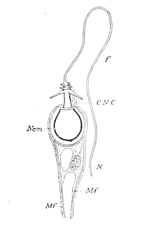
Fig. 124.—Nematocyst (Nem) of Hydra grisea, enclosed within the cnidoblast. CNC, Cnidocil; f, thread of nematocyst; Mf, myophan threads in cnidoblast; N, nucleus of cnidoblast. (After Schneider.)
When a nematocyst has once been discharged it is usually rejected from the body, and its place in the tissue is taken by a new nematocyst formed by a new cnidoblast; but in the thread of the large kind of nematocyst of Millepora there is a very delicate band, which appears to be similar to the myophan thread in the stalk of a Vorticella. Dr. Willey[283] has made the important observation that in this coral the nematocyst threads can be withdrawn after discharge, the retraction being effected with great rapidity. The "cnidoblast" is a specially modified cell. It sometimes bears at its free extremity a delicate process, the "cnidocil," which is supposed to be adapted to the reception of the special stimuli that determine the discharge of the nematocyst. In many species delicate contractile fibres (Fig. 124, Mf) can be seen in the substance of the cnidoblast, and in others its basal part is drawn out into a long and probably contractile stalk ("cnidopod"), attached to the mesogloea below.
There can be little doubt that new nematocysts are constantly formed during life to replace those that have been discharged and lost. Each nematocyst is developed within the cell-substance of a cnidoblast which is derived from the undifferentiated interstitial cell-groups. During this process the cnidoblast does not necessarily remain stationary, but may wander some considerable distance from its place of origin.[284] This habit of migration of the cnidoblast renders it difficult to determine whether the ectoderm alone, or both ectoderm and endoderm, can give rise to nematocysts. In the majority of Coelenterates the nematocysts are confined to the ectoderm, but in many Anthozoa, Scyphozoa, and Siphonophora they are found in tissues that are certainly or probably endodermic in origin. It has not been definitely proved in any case that the cnidoblast cells that form these nematocysts have originally been formed in the endoderm, and it is possible that they are always derived from ectoderm cells which migrate into the endoderm.
It is probably true that all Coelenterata have nematocysts, and that, in the few cases in which it has been stated that they are absent (e.g. Sarcophytum), they have been overlooked. It cannot, however, be definitely stated that similar structures do not occur in other animals. The nematocysts of the Mollusc Aeolis are not the product of its own tissues, but are introduced into the body with its food.[285] The nematocysts that occur in the Infusorian Epistylis umbellaria and in the Dinoflagellate Polykrikos (p. 131) require reinvestigation, but if it should prove that they are the product of the Protozoa they cannot be regarded as strictly homologous with those of Coelenterata. In many of the Turbellaria, however, and in some of the Nemertine worms, nematocysts occur in the epidermis which appear to be undoubtedly the products of these animals.
The Coelenterata are divided into three classes:—
1. Hydrozoa.—Without stomodaeum and mesenteries. Sexual cells discharged directly to the exterior.
2. Scyphozoa.—Without stomodaeum and mesenteries. Sexual cells discharged into the coelenteric cavity.
3. Anthozoa = Actinozoa.—With stomodaeum and mesenteries. Sexual cells discharged into the coelenteric cavity.
The full meaning of the brief statements concerning the structure of the three classes given above cannot be explained until the general anatomy of the classes has been described. It may be stated, however, in this place that many authors believe that structures corresponding with the stomodaeum and mesenteries of Anthozoa do occur in the Scyphozoa, which they therefore include in the class Anthozoa.
Among the more familiar animals included in the class Hydrozoa may be mentioned the fresh-water polyp Hydra, the Hydroid zoophytes, many of the smaller Medusae or jelly-fish, the Portuguese Man-of-war (Physalia), and a few of the corals.
Included in the Scyphozoa are the large jelly-fish found floating on the sea or cast up on the beach on the British shores.
The Anthozoa include the Sea-anemones, nearly all the Stony Corals, the Sea-fans, the Black Corals, the Dead-men's fingers (Alcyonium), the Sea-pens, and the Precious Coral of commerce.
CLASS I. HYDROZOA
In this Class of Coelenterata two types of body-form may be found. In such a genus as Obelia there is a fixed branching colony of zooids, and each zooid consists of a simple tubular body-wall composed of the two layers of cells, the ectoderm and the endoderm (Fig. 125), terminating distally in a conical mound—the "hypostome"—which is perforated by the mouth and surrounded by a crown of tentacles. This fixed colony, the "hydrosome," feeds and increases in size by gemmation, but does not produce sexual cells. The hydrosome produces at a certain season of the year a number of buds, which develop into small bell-like jelly-fish called the "Medusae," which swim away from the parent stock and produce the sexual cells. The Medusa (Fig. 126) consists of a delicate dome-shaped contractile bell, perforated by radial canals and fringed with tentacles; and from its centre there depends, like the clapper of a bell, a tubular process, the manubrium, which bears the mouth at its extremity. This free-swimming sexual stage in the life-history of Obelia is called the "medusome."
It is difficult to determine whether, in the evolution of the Hydrozoa, the hydrosome preceded the medusome or vice versâ. By some authors the medusome is regarded as a specially modified sexual individual of the hydrosome colony. By others the medusome is regarded as the typical adult Hydrozoon form, and the zooids of the hydrosome as nutritive individuals arrested in their development to give support to it. Whatever may be the right interpretation of the facts, however, it is found that in some forms the medusome stage is more or less degenerate and the hydrosome is predominant, whereas in others the hydrosome is degenerate or inconspicuous and the medusome is predominant. Finally, in some cases there are no traces, even in development, of a medusome stage, and the life-history is completed in the hydrosome, while in others the hydrosome stages are lost and the life-history is completed in the medusome.
If a conspicuous hydrosome stage is represented by H, a conspicuous medusome stage by M, an inconspicuous or degenerate hydrosome stage by h, an inconspicuous or degenerate medusome stage by m, and the fertilised ovum by O, the life-histories of the Hydrozoa may be represented by the following formulæ:—
1. O — H — O (Hydra)
2. O — H — m — O (Sertularia)
3. O — H — M — O (Obelia)
4. O — h — M — O (Liriope)
5. O — M — O (Geryonia)
The structure of the hydrosome is usually very simple. It consists of a branched tube opening by mouths at the ends of the branches and closed at the base. The body-wall is built up of ectoderm and endoderm. Between these layers there is a thin non-cellular lamella, the mesogloea.
In a great many Hydrozoa the ectoderm secretes a chitinous protective tube called the "perisarc." The mouth is usually a small round aperture situated on the summit of the hypostome, and at the base of the hypostome there may be one or two crowns of tentacles or an area bearing irregularly scattered tentacles. The tentacles may be hollow, containing a cavity continuous with the coelenteric cavity of the body; or solid, the endoderm cells arranged in a single row forming an axial support for the ectoderm. The ectoderm of the tentacles is provided with numerous nematocysts, usually arranged in groups or clusters on the distal two-thirds of their length, but sometimes confined to a cap-like swelling at the extremity (capitate tentacles). The hydrosome may be a single zooid producing others asexually by gemmation (or more rarely by fission), which become free from the parent, or it may be a colony of zooids in organic connexion with one another formed by the continuous gemmation of the original zooid derived from the fertilised ovum and its asexually produced offspring. When the hydrosome is a colony of zooids, specialisation of certain individuals for particular functions may occur, and the colony becomes dimorphic or polymorphic.
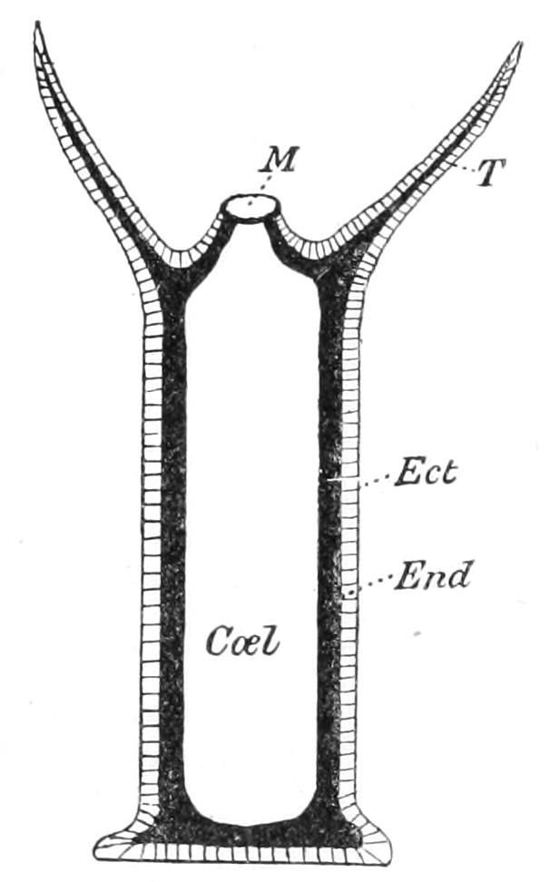
Fig. 125.—Diagram of a vertical section through a hydrosome. Coel, Coelenteron; Ect, ectoderm; End, endoderm. Between the ectoderm and the endoderm there is a thin mesogloea not represented in the diagram. M, mouth; T, tentacle.
The medusome is more complicated in structure than the hydrosome, as it is adapted to the more varied conditions of a free-swimming existence. The body is expanded to form a disc, "umbrella," or bell, which bears at the edge or margin a number of tentacles. The mouth is situated on the end of a hypostome, called the "manubrium," situated in the centre of the radially symmetrical body. The surface that bears the manubrium is called oral, and the opposite surface is called aboral. The cavity partly enclosed by the oral aspect of the body when it is cup- or bell-shaped is called the "sub-umbrellar cavity."
In the medusome of nearly all Hydrozoa there is a narrow shelf projecting inwards from the margin of the disc and guarding the opening of the sub-umbrellar cavity, called the "velum."
The mouth leads through the manubrium into a flattened part of the coelenteric cavity, which is usually called the gastric cavity, and from this a number of canals pass radially through the mesogloea to join a circular canal or ring-canal at the margin of the umbrella.
A special and important feature of the medusome is the presence of sense-organs called the "ocelli" and "statocysts," situated at the margin of the umbrella or at the base of the tentacles.
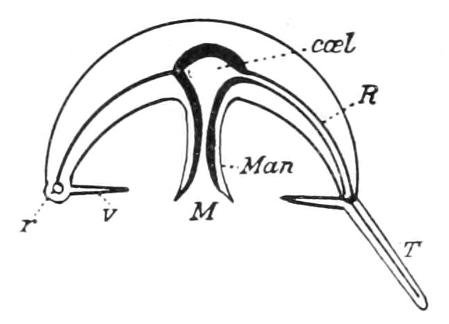
Fig. 126.—Diagram of a vertical section through a medusome. coel, Coelenteron; M, mouth; Man, manubrium; R, radial canal; r, ring or circular canal; T, tentacle; v, velum.
The ocelli may usually be recognised as opaque red or blue spots on the bases of the tentacles, in marked contrast to their transparent surroundings. The ocellus may consist simply of a cluster of pigmented cells, or may be further differentiated as a cup of pigmented cells filled with a spherical thickening of the cuticle to form a lens. The exact function of the ocelli may not be fully understood, but there can be little doubt that they are light-perceiving organs.
The function of the sense-organs known as statocysts, however, has not yet been so satisfactorily determined. They were formerly thought to be auditory organs, and were called "otocysts," but it appears now that it is impossible on physical grounds for these organs to be used for the perception of the waves of sound in water. It is more probable that they are organs of the static function, that is, the function of the perception of the position of the body in space, and they are consequently called statocysts. In the Leptomedusae each statocyst consists of a small vesicle in the mesogloea at the margin of the umbrella, containing a hard, stony body called the "statolith." In Geryonia and some other Trachomedusae the statolith is carried by a short tentacular process, the "statorhab," projecting into the vesicle; in other Trachomedusae, however, the vesicle is open, but forms a hood for the protection of the statorhab; and in others, but especially in the younger stages of development, the statorhab is not sunk into the margin of the umbrella, and resembles a short but loaded tentacle. Recent researches have shown that there is a complete series of connecting links between the vesiculate statocyst of the Leptomedusae and the free tentaculate statorhab of the Trachomedusae, and there can be little doubt of their general homology.
In the free-swimming or "Phanerocodonic" medusome the sexual cells are borne by the ectoderm of the sub-umbrellar cavity either on the walls of the manubrium or subjacent to the course of the radial canals.
Order I. Eleutheroblastea.
This order is constituted mainly for the well-known genus Hydra. By some authors Hydra is regarded as an aberrant member of the order Gymnoblastea, to which it is undoubtedly in many respects allied, but it presents so many features of special interest that it is better to keep it in a distinct group.
Hydra is one of the few examples of exclusively fresh-water Coelenterates, and like so many of the smaller fresh-water animals its distribution is almost cosmopolitan. It occurs not only in Europe and North America, but in New Zealand, Australia, tropical central Africa, and tropical central America.
Hydra is found in this country in clear, still fresh water attached to the stalks or leaves of weeds. When fully expanded it may be 25 mm. in length, but when completely retracted the same individual may be not more than 3 mm. long. The tubular body-wall is built up of ectoderm and endoderm, enclosing a simple undivided coelenteric cavity. The mouth is situated on the summit of the conical hypostome, and at the base of this there is a crown of long, delicate, but hollow tentacles. The number of tentacles is usually six in H. vulgaris and H. oligactis,[286] and eight in H. viridis, but it is variable in all species.
During the greater part of the summer the number of individuals is rapidly increased by gemmation. The young Hydras produced by gemmation are usually detached from their parents before they themselves produce buds, but in H. oligactis the buds often remain attached to the parent after they themselves have formed buds, and thus a small colony is produced. Sexual reproduction usually commences in this country in the summer and autumn, but as the statements of trustworthy authors are conflicting, it is probable that the time of appearance of the sexual organs varies according to the conditions of the environment.
Individual specimens may be male, female, or hermaphrodite. Nussbaum[287] has published the interesting observation that when the Hydras have been well fed the majority become female, when the food supply has been greatly restricted the majority become male, and when the food-supply is moderate in amount the majority become hermaphrodite. The gonads are simply clusters of sexual cells situated in the ectoderm. There is no evidence, derived from either their structure or their development, to show that they represent reduced medusiform gonophores. The testis produces a number of minute spermatozoa. In the ovary, however, only one large yolk-laden egg-cell reaches maturity by the absorption of the other eggs. The ovum is fertilised while still within the gonad, and undergoes the early stages of its development in that position. With the differentiation of an outer layer of cells a chitinous protecting membrane is formed, and the escape from the parent takes place.[288] It seems probable that at this stage, namely, that of a protected embryo, there is often a prolonged period of rest, during which it may be carried by wind and other agencies for long distances without injury.
The remarkable power that Hydra possesses of recovery from injury and of regenerating lost parts was first pointed out by Trembley in his classical memoir.[289]
A Hydra can be cut into a considerable number of pieces, and each piece, provided both ectoderm and endoderm are represented in it, will give rise by growth and regeneration to a complete zooid. There is, however, a limit of size below which fragments of Hydra will not regenerate, even if they contain cells of both layers. The statement made by Trembley, that when a Hydra is turned inside out it will continue to live in the introverted condition has not been confirmed, and it seems probable that after the experiment has been made the polyp remains in a paralysed condition for some time, and later reverts, somewhat suddenly, to the normal condition by a reversal of the process. There is certainly no substantial reason to believe that under any circumstances the ectoderm can undertake the function of the endoderm or the endoderm the functions of the ectoderm.
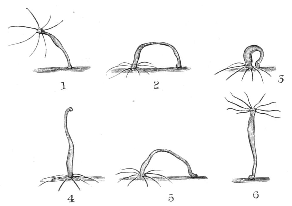
Fig. 127.—A series of drawings of Hydra, showing the attitudes it assumes during one of the more rapid movements from place to place. 1, The Hydra bending over to one side; 2, attaching itself to the support by the mouth and tentacles; 3, drawing the sucker up to the mouth; 4, inverted; 5, refixing the sucker; 6, reassuming the erect posture. (After Trembley.)
One of the characteristic features of Hydra is the slightly expanded, disc-shaped aboral extremity usually called the "foot," an unfortunate term for which the word "sucker" should be substituted. There are no root-like tendrils or processes for attachment to the support such as are found in most of the solitary Gymnoblastea. The attachment of the body to the stem or weed or surface-film by this sucker enables the animal to change its position at will. It may either progress slowly by gliding along its support without the assistance of the tentacles, in a manner similar to that observed in many Sea-anemones; or more rapidly by a series of somersaults, as originally described by Trembley. The latter mode of locomotion has been recently described as follows:—"The body, expanded and with expanded tentacles, bends over to one side. As soon as the tentacles touch the bottom they attach themselves and contract. Now one of two things happens. The foot may loosen its hold on the bottom and the body contract. In this manner the animal comes to stand on its tentacles with the foot pointing upward. The body now bends over again until the foot attaches itself close to the attached tentacles. These loosen in their turn, and so the Hydra is again in its normal position. In the other case the foot is not detached, but glides along the support until it stands close to the tentacles, which now loosen their hold."[290]
Hydra appears to be purely carnivorous. It will seize and swallow Entomostraca of relatively great size, so that the body-wall bulges to more than twice its normal diameter. But smaller Crustacea, Annelid worms, and pieces of flesh are readily seized and swallowed by a hungry Hydra. In H. viridis the chlorophyll corpuscles[291] of the endoderm may possibly assist in the nourishment of the body by the formation of starch in direct sunlight.
Three species of Hydra are usually recognised, but others which may be merely local varieties or are comparatively rare have been named.[292]
H. viridis.—Colour, grass-green. Average number of tentacles, eight. Tentacles shorter than the body. Embryonic chitinous membrane spherical and almost smooth.
H. vulgaris, Pallas (H. grisea, Linn.).—Colour, orange-brown. Tentacles rather longer than the body, average number, six. Embryonic chitinous membrane spherical, and covered with numerous pointed branched spines.
H. oligactis, Pallas (H. fusca, Linn.).—Colour, brown. Tentacles capable of great extension; sometimes, when fully expanded, several times the length of the body. Average number, six. Embryonic chitinous membrane plano-convex, its convex side only covered with spines.
The genera Microhydra (Ryder) and Protohydra (Greeff) are probably allied to Hydra, but as their sexual organs have not been observed their real affinities are not yet determined. Microhydra resembles Hydra in its general form and habits, and in its method of reproduction by gemmation, but it has no tentacles. It was found in fresh water in North America.
Protohydra[293] was found in the oyster-beds off Ostend, and resembles Microhydra in the absence of tentacles. It multiplies by transverse fission, but neither gemmation nor sexual reproduction has been observed.
Haleremita is a minute hydriform zooid which is also marine. It was found by Schaudinn[294] in the marine aquarium at Berlin in water from Rovigno, on the Adriatic. It reproduces by gemmation, but sexual organs have not been found.
Another very remarkable genus usually associated with the Eleutheroblastea is Polypodium. At one stage of its life-history it has the form of a spiral ribbon or stolon which is parasitic on the eggs of the sturgeon (Acipenser ruthenus) in the river Volga.[295] This stolon gives rise to a number of small Hydra-like zooids with twenty tentacles, of which sixteen are filamentous and eight club-shaped. These zooids multiply by longitudinal fission, and feed independently on Infusoria, Rotifers, and other minute organisms. The stages between these hydriform individuals and the parasitic stolon have not been discovered.
Order II. Milleporina.
Millepora was formerly united with the Stylasterina to form the order Hydrocorallina; but the increase of our knowledge of these Hydroid corals tends rather to emphasise than to minimise the distinction of Millepora from the Stylasterina.
Millepora resembles the Stylasterina in the production of a massive calcareous skeleton and in the dimorphism of the zooids, but in the characters of the sexual reproduction and in many minor anatomical and histological peculiarities it is distinct. As there is only one genus, Millepora, the account of its anatomy will serve as a description of the order.
The skeleton (Fig. 128) consists of large lobate, plicate, ramified, or encrusting masses of calcium carbonate, reaching a size of one or two or more feet in height and breadth. The surface is perforated by numerous pores of two distinct sizes; the larger—"gastropores"—are about 0.25 mm. in diameter, and the smaller and more numerous "dactylopores" about 0.15 mm. in diameter. In many specimens the pores are arranged in definite cycles, each gastropore being surrounded by a circle of 5-7 dactylopores; but more generally the two kinds appear to be irregularly scattered on the surface.
When a branch or lobe of a Millepore is broken across and examined in section, it is found that each pore is continued as a vertical tube divided into sections by horizontal calcareous plates (Fig. 129, Tab). These plates are the "tabulae," and constitute the character upon which Millepora was formerly placed in the now discarded group of Tabulate corals.
The coral skeleton is also perforated by a very fine reticulum of canals, by which the pore-tubes are brought into communication with one another. In the axis of the larger branches and in the centre of the larger plates a considerable quantity of the skeleton is of an irregular spongy character, caused by the disintegrating influence of a boring filamentous Alga.[296]
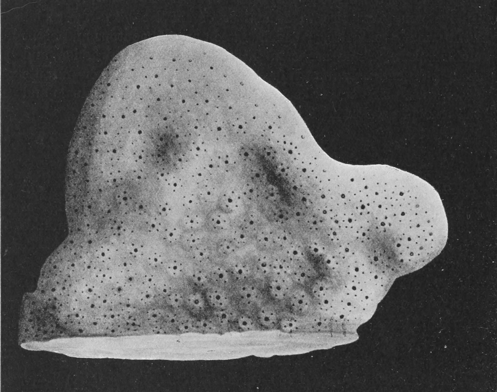
Fig. 128.—A portion of a dried colony of Millepora, showing the larger pores (gastropores) surrounded by cycles of smaller pores (dactylopores). At the edges the cycles are not well defined.
The discovery that Millepora belongs to the Hydrozoa was made by Agassiz[297] in 1859, but Moseley[298] was the first to give an adequate account of the general anatomy. The colony consists of two kinds of zooids—the short, thick gastrozooids (Fig. 129, G) provided with a mouth and digestive endoderm, and the longer and more slender mouthless dactylozooids (D)—united together by a network of canals running in the porous channels of the superficial layer of the corallum. The living tissues of the zooids extend down the pore-tubes as far as the first tabulae, and below this level the canal-system is degenerate and functionless. It is only a very thin superficial stratum of the coral, therefore, that contains living tissues.
The zooids of Millepora are very contractile, and can be withdrawn below the general surface of the coral into the shelter of the pore-tubes. When a specimen is examined in its natural position on the reef, the zooids are usually found to be thus contracted; but several observers have seen the zooids expanded in the living condition. It is probable that, as is the case with other corals, the expansion occurs principally during the night.
The colony is provided with two kinds of nematocysts—the small kind and the large. In some colonies they are powerful enough to penetrate the human skin, and Millepora has therefore received locally the name of "stinging coral." On each of the dactylozooids there are six or seven short capitate tentacles (Fig. 129, t), each head being packed with nematocysts of the small kind; similar batteries of these nematocysts are found in the four short capitate tentacles of the gastrozooids. The nematocysts of the larger kind are found in the superficial ectoderm, some distributed irregularly on the surface, others in clusters round the pores. The small nematocysts are about 0.013 mm. in length before they are exploded, and exhibit four spines at the base of the thread; the large kind are oval in outline, 0.02 × 0.025 mm. in size, and exhibit no spines at the base, but a spiral band of minute spines in the middle of the filament. There is some reason to believe that the filament of the large kind of nematocysts can be retracted.[299]
At certain seasons the colonies of Millepora produce a great number of male or female Medusae. The genus is probably dioecious, no instances of hermaphrodite colonies having yet been found. Each Medusa is formed in a cavity situated above the last-formed tabula in a pore-tube, and this cavity, the "ampulla," having a greater diameter than that of the gastrozooid tubes, can be recognised even in the dried skeleton.
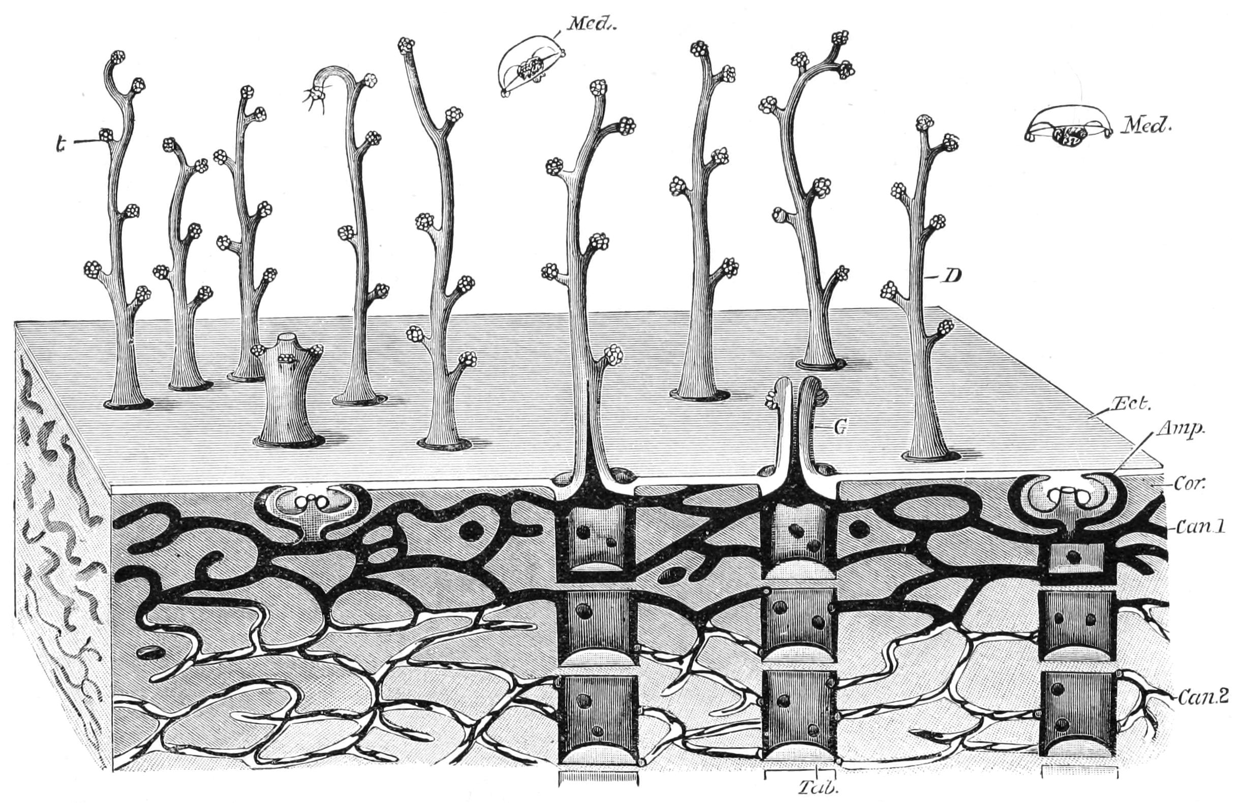
Fig. 129.—Diagrammatic sketch to show the structure of Millepora. Amp, an ampulla containing a medusa; Can.1, canal system at the surface; Can.2, canal system degenerating in the lower layers of the corallum; Cor, corallum; D, an expanded dactylozooid with its capitate tentacles; Ect, the continuous sheet of ectoderm covering the corallum (Cor); G, a gastrozooid, seen in vertical section; Med, free-swimming Medusae; t, tentacle; Tab, tabula in the pore-tubes. (Partly after Moseley.)
It is not known how frequently the sexual seasons occur, but from the rarity in the collections of our museums of Millepore skeletons which exhibit the ampullae, it may be inferred that the intervals between successive seasons are of considerable duration.
The Medusae of Millepora are extremely simple in character. There is a short mouthless manubrium bearing the sexual cells, an umbrella without radial canals, while four or five knobs at the margin, each supporting a battery of nematocysts, represent all that there is of the marginal tentacles. The male Medusae have not yet been observed to escape from the parent, but from the fact that the spermatozoa are not ripe while they are in the ampullae, it may be assumed that the Medusae are set free. Duerden, however, has observed the escape of the female Medusae, and it seems probable from his observations that their independent life is a short one, the ova being discharged very soon after liberation.
Millepora appears to be essentially a shallow-water reef coral. It may be found on the coral reefs of the Western Atlantic extending as far north as Bermuda, in the Red Sea, the Indian and Pacific Oceans. The greatest depth at which it has hitherto been found is 15 fathoms on the Macclesfield Bank, and it flourishes at a depth of 7 fathoms off Funafuti in the Pacific Ocean.
Millepora, like many other corals, bears in its canals and zooids a great number of the symbiotic unicellular "Algae" (Chrysomonadaceae, see pp. 86, 125) known as Zooxanthellae. All specimens that have been examined contain these organisms in abundance, and it has been suggested that the coral is largely dependent upon the activity of the "Algae" for its supply of nourishment. There can be no doubt that the dactylozooids do paralyse and catch living animals, which are ingested and digested by the gastrozooids, but this normal food-supply may require to be supplemented by the carbohydrates formed by the plant-cells. But as the carbohydrates can only be formed by the "Algae" in sunlight, this supplementary food-supply can only be provided in corals that live in shallow water. It must not be supposed that this is the only cause that limits the distribution of Millepora in depth, but it may be an important one.
The generic name Millepora has been applied to a great many fossils from different strata, but a critical examination of their structure fails to show any sufficient reason for including many of them in the genus or even in the order. Fossils that are undoubtedly Millepora occur in the raised coral reefs of relatively recent date, but do not extend back into Tertiary times. There seems to be no doubt, therefore, that the genus is of comparatively recent origin. Among the extinct fossils the genus that comes nearest to it is Axopora from the Eocene of France, but this genus differs from Millepora in having monomorphic, not dimorphic, pores, and in the presence of a minute spine or columella in the centre of each tube. The resemblances are to be observed in the general disposition of the canal system and of the tabulation. Whether Axopora is or is not a true Milleporine, however, cannot at present be determined, but it is the only extinct coral that merits consideration in this place.
Order III. Gymnoblastea—Anthomedusae.
This order was formerly united with the Calyptoblastea to form the order Hydromedusae, but the differences between the two are sufficiently pronounced to merit their treatment as distinct orders.
In many of the Gymnoblastea the sexual cells are borne by free Medusae, which may be recognised as the Medusae of Gymnoblastea by the possession of certain distinct characters. The name given to such Medusae, whether their hydrosome stage is known or not, is Anthomedusae. The Gymnoblastea are solitary or colonial Hydrozoa, in which the free (oral) extremity of the zooids, including the crown of tentacles, is not protected by a skeletal cup. The sexual cells may be borne by free Anthomedusae, or by more or less degenerate Anthomedusae that are never detached from the parent hydrosome. The Anthomedusae are small or minute Medusae provided with a velum, with the ovaries or sperm-sacs borne by the manubrium and with sense-organs in the form of ocelli or pigment-spots situated on the margin of the umbrella.
The solitary Gymnoblastea present so many important differences in anatomical structure that they cannot be united in a single family. They are usually fixed to some solid object by root-like processes from the aboral extremity, the "hydrorhiza," or are partly embedded in the sand (Corymorpha), into which long filamentous processes project for the support of the zooid. The remarkable species Hypolytus peregrinus[300] from Wood's Holl, however, has no aboral processes, and appears to be only temporarily attached to foreign objects by the secretion of the perisarc. Among the solitary Gymnoblastea several species reach a gigantic size. Corymorpha is 50-75 mm. in length, but Monocaulus from deep water in the Pacific and Atlantic Oceans is nearly 8 feet in length. Among the solitary forms attention must be called to the interesting pelagic Pelagohydra (see p. 274).
The method of colony formation in the Gymnoblastea is very varied. In some cases (Clava squamata) a number of zooids arise from a plexus of canals which corresponds with the system of root-like processes of the solitary forms. In Hydractinia this plexus is very dense, and the ectoderm forms a continuous sheet of tissue both above and below. The colony is increased in size in these cases by the gemmation of zooids from the hydrorhiza. In other forms, such as Tubularia larynx, new zooids arise not only from the canals of the hydrorhiza, but also from the body-walls of the upstanding zooids, and thus a bushy or shrubby colony is formed.
In another group the first-formed zooid produces a hydrorhiza of considerable proportions, which fixes the colony firmly to a stone or shell and increases in size with the growth of the colony. This zooid itself by considerable growth in length forms the axis of the colony, and by gemmation gives rise to lateral zooids, which in their turn grow to form the lateral branches and give rise to the secondary branches, and these to the tertiary branches, and so one; each branch terminating in a mouth, hypostome and crown of tentacles. Such a method of colony formation is seen in Bougainvillia (Fig. 130). A still more complicated form of colony formation is seen in Ceratella, in which not a single but a considerable number of zooids form the axis of the colony and of its branches. As each axis is covered with a continuous coat of ectoderm, and each zooid of such an axis secretes a chitinous fenestrated tube, the whole colony is far more rigid and compact than is usual in the Gymnoblastea, and has a certain superficial resemblance to a Gorgoniid Alcyonarian (Fig. 133, p. 271).
The branches of the colony and a considerable portion of the body-wall of each zooid in the Gymnoblastea are usually protected by a thin, unjointed "perisarc" of chitin secreted by the ectoderm; but this skeletal structure does not expand distally to form a cup-like receptacle in which the oral extremity of the zooid can be retracted for protection.
The zooids of the Gymnoblastea present considerable diversity of form and structure. The tentacles may be reduced to one (in Monobrachium) or two (in Lar sabellarum), but usually the number is variable in each individual colony. In many cases, such as Cordylophora, Clava, and many others, the tentacles are irregularly scattered on the sides of the zooids. In others there may be a single circlet of about ten or twelve tentacles round the base of the hypostome. In some genera the tentacles are arranged in two series (Tubularia, Corymorpha, Monocaulus), a distal series round the margin of the mouth which may be arranged in a single circlet or scattered irregularly on the hypostome, and a proximal series arranged in a single circlet some little distance from the mouth. In Branchiocerianthus imperator the number of tentacles is very great, each of the two circlets consisting of about two hundred tentacles.
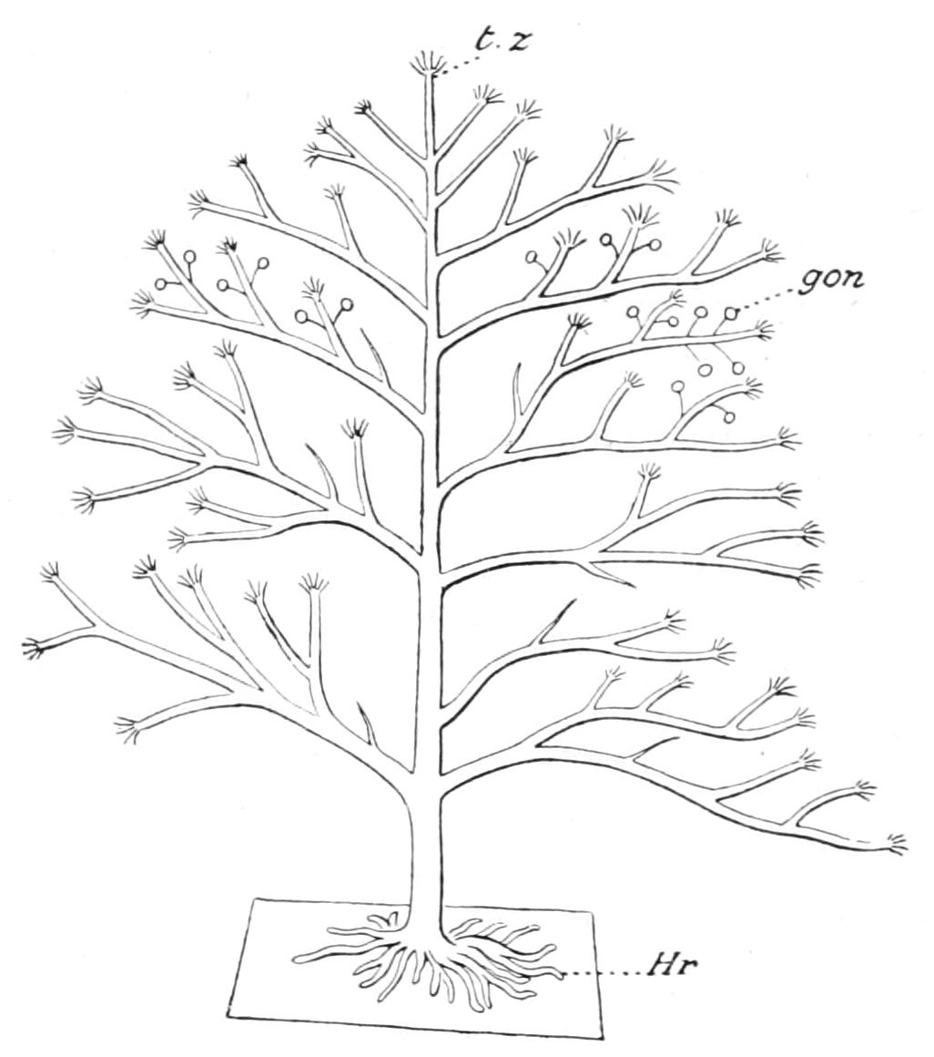
Fig. 130.—Diagrammatic sketch to show the method of branching of Bougainvillia. gon, Gonophores; Hr, hydrorhiza; t.z, terminal zooid.
The zooids of the hydrosome are usually monomorphic, but there are cases in which different forms of zooid occur in the same colony. In Hydractinia, for example, no less than four different kinds of zooids have been described. These are called gastrozooids, dactylozooids, tentaculozooids, and blastostyles respectively. The "gastrozooids" are provided with a conical hypostome bearing the mouth and two closely-set circlets of some ten to thirty tentacles. The "dactylozooids" are longer than the gastrozooids and have the habit of actively coiling and uncoiling themselves; they have a small mouth and a single circlet of rudimentary tentacles. The "tentaculozooids" are situated at the outskirts of the colony, and are very long and slender, with rudimentary tentacles and no mouth. The "blastostyles," usually shorter than the gastrozooids, have two circlets of rudimentary tentacles and a mouth. They bear on their sides the spherical or oval gonophores.
The medusome stage in the life-history of these Hydrozoa is produced by gemmation from the hydrosome, or, in some cases, by gemmation from the medusome as well as from the hydrosome. In many genera and species the medusome is set free as a minute jelly-fish or Medusa, which grows and develops as an independent organism until the time when the sexual cells are ripe, and then apparently it dies. In other Gymnoblastea the medusome either in the female or the male or in both sexes does not become detached from the parent hydrosome, but bears the ripe sexual cells, discharges them into the water, and degenerates without leading an independent life at all. In these cases the principal organs of the medusome are almost or entirely functionless, and they exhibit more or less imperfect development, or they may be so rudimentary that the medusoid characters are no longer obvious. Both the free and the undetached medusomes are gonophores, that is to say, the bearers of the sexual cells, but the former were described by Allman as the "phanerocodonic" gonophores, i.e. "with manifest bells," and the latter as the "adelocodonic" gonophores. The gonophores may arise either from an ordinary zooid of the colony (Syncoryne), from a specially modified zooid—the blastostyle—as in Hydractinia, or from the hydrorhiza as in certain species of Perigonimus. The free-swimming Medusa may itself produce Medusae by gemmation from the manubrium (Sarsia, Lizzia, Rathkea, and others), from the base of the tentacles (Sarsia, Corymorpha, Hybocodon), or from the margin of the umbrella (Eleutheria).
The free-swimming Medusae or phanerocodonic gonophores of the Gymnoblastea are usually of small size (1 or 2 mm. in diameter) when first liberated, and rarely attain a great size even when fully mature. They consist of a circular, bell-shaped or flattened disc—the umbrella—provided at its margin with a few or numerous tentacles, and a tubular manubrium bearing the mouth depending from the exact centre of the under (oral) side of the umbrella (Fig. 132, A). The mouth leads into a shallow digestive cavity, from which radial canals pass through the substance of the umbrella to join a ring-canal at the margin (Fig. 131).
The sense-organs of the Medusae of the Gymnoblastea are in the form of pigment-spots or very simple eyes (ocelli), situated at the bases of the tentacles. The orifice of the umbrella is guarded by a thin shelf or membrane, as in the Calyptoblastea, called the velum. The sexual cells are borne by the manubrium (Figs. 131 and 132, A).
There are many modifications observed in the different genera as regards the number of tentacles, the number and character of the radial canals, the minute structure of the sense-organs, and some other characters, but they agree in having a velum, ocellar sense-organs, and manubrial sexual organs. The tentacles are rudimentary in Amalthea; in Corymorpha there is only one tentacle; in Perigonimus there are two; and in Bougainvillia they are numerous; but the usual number is four or six. The radial canals are usually simple and four in number, but there are six in Lar sabellarum, which branch twice or three times before reaching the margin of the umbrella (Fig. 132, B).
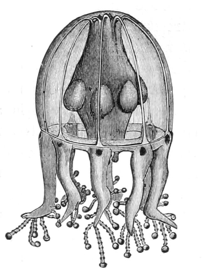
Fig. 131.—Medusa of Cladonema, from the Bahamas, showing peculiar tentacular processes on the tentacles, the ocelli at the base of the tentacles, the swellings on the manubrium that mark the position of the gonads, and the radial and ring-canals of the umbrella. (After Perkins.)
There can be no doubt that the Medusae of many Gymnoblastea undergo several important changes in their anatomical features during the period of the ripening of the sexual cells. Thus in Lar sabellarum the six radial canals are simple in the first stage of development (A); but in the second stage (B) each radial canal bifurcates before reaching the margin, and in the adult stage shows a double bifurcation. The life-history has, however, been worked out in very few of the Anthomedusae, and there can be little doubt that as our knowledge grows several forms which are now known as distinct species will be found to be different stages of growth of the same species.
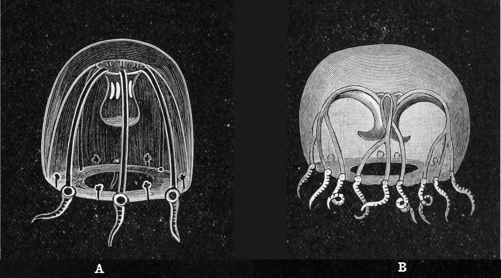
Fig. 132.—Two stages in the development of the Medusa of Lar sabellarum (Willsia stellata). A, first stage with six canals without branches; B, third stage with six canals each with two lateral branches. The developing gonads may be seen on the manubrium in A. (After Browne.)
The movements of the Medusae are well described by Allman[301] in his account of Cladonema radiatum:—"It is impossible to grow tired of watching this beautiful medusa; sometimes while dashing through the water with vigorous diastole and systole, it will all at once attach its grapples to the side of the vessel, and become suddenly arrested in its career, and then after a period of repose, during which its branched tentacles are thrown back over its umbrella and extended into long filaments which float, like some microscopic sea-weed in the water, it will once more free itself from its moorings and start off with renewed energy." The Medusa of Clavatella, "in its movements and mode of life, presents a marked contrast to the medusiform zooid of other Hydrozoa. The latter is active and mercurial, dancing gaily through the water by means of the vigorous strokes of its crystalline swimming-bell. The former strides leisurely along, or, using the adhesive discs as hands, climbs amongst the branches of the weed. In the latter stage of its existence it becomes stationary, fixing itself by means of its suckers; and thus it remains, the capitate arms standing out rigidly, like the rays of a starfish, until the embryos are ready to escape."[302]
Among the Gymnoblastea there are many examples of a curious association of the Hydroid with some other living animals. Thus Hydractinia is very often found on the shells carried by living Hermit crabs, Dicoryne on the shells of various Molluscs, Tubularia has been found on a Cephalopod, and Ectopleura (a Corymorphid) on the carapace of a crab. There is but little evidence, however, that in these cases the association is anything more than accidental. The occurrence of the curious species, Lar sabellarum, on the tubes of Sabella, of Campaniclava cleodorae on the living shells of the pelagic Mollusc Cleodora cuspidata, and of a Gorgonia on the tubes of Tubularia parasitica, appear to be cases in which there is some mutual relationship between the two comrades. The genus Stylactis, however, affords some of the most interesting examples of mutualism. Thus Stylactis vermicola is found only on the back of an Aphrodite that lives at the great depth of 2900 fathoms. S. spongicola and S. abyssicola are found associated with certain deep-sea Horny Sponges. S. minoi is spread over the skin of the little rock perch Minous inermis, which is found at depths of from 45 to 150 fathoms in the Indian seas.
In many cases it is difficult to understand what is the advantage of the Hydroid to the animal that carries it, but in this last case Alcock[303] suggests that the Stylactis assists in giving the fish a deceitful resemblance to the incrusted rocks of its environment, in order to allure, or at any rate not to scare, its prey. Whether this is the real explanation or not, the fact that in the Bay of Bengal and in the Laccadive and Malabar seas the fish is never found without this Hydroid, nor the Hydroid without this species of fish, suggests very strongly that there is a mutual advantage in the association.
Cases of undoubted parasitism are very rare in this order. The remarkable form Hydrichthys mirus,[304] supposed to be a Gymnoblastic Hydroid, but of very uncertain position in the system, appears to be somewhat modified in its structure by its parasitic habits on the fish Seriola zonata. Corydendrium parasiticum is said to be a parasite living at the expense of Eudendrium racemosum. Mnestra is a little Medusa which attaches itself by its manubrium to the Mollusc Phyllirhoe, and may possibly feed upon the skin or secretions of its host.
Nearly all the species of the order are found in shallow sea water. Stylactis vermicola and the "Challenger" specimen of Monocaulus imperator occur at a depth of 2900 fathoms, and some species of the genera Eudendrium and Myriothela descend in some localities to a depth of a few hundred fathoms. Cordylophora is the only genus known to occur in fresh water. From its habit of attaching itself to wooden piers and probably to the bottom of barges, and from its occurrence in navigable rivers and canals, it has been suggested that Cordylophora is but a recent immigrant into our fresh-water system. It has been found in England in the Victoria docks of London, in the Norfolk Broads, and in the Bridgewater Canal. It has ascended the Seine in France, and may now be found in the ponds of the Jardin des Plantes at Paris. It also occurs in the Elbe and in some of the rivers of Denmark.
The classification of the Gymnoblastea is not yet on a satisfactory basis. At present the hydrosome stage of some genera alone has been described, of others the free-swimming Medusa only is known. Until the full life-history of any one genus has been ascertained its position in the families mentioned below may be regarded as only provisional. The principal families are:—
Fam. Bougainvilliidae.—The zooids of the hydrosome have a single circlet of filiform tentacles at the base of the hypostome. In Bougainvillia belonging to this family the gonophores are liberated in the form of free-swimming Medusae formerly known by the generic name Hippocrene. In the fully grown Medusa there are numerous tentacles arranged in clusters opposite the terminations of the four radial canals. There are usually in addition tentacular processes (labial tentacles) on the lips of the manubrium. Bougainvillia is a common British zoophyte of branching habit, found in shallow water all round the coast. The medusome of Bougainvillia ramosa is said to be the common little medusa Margelis ramosa.[305] Like most of the Hydroids it has a wide geographical distribution. Other genera are Perigonimus, which has a Medusa with only two tentacles; and Dicoryne, which forms spreading colonies on Gasteropod shells and has free gonophores provided with two simple tentacles, while the other organs of the medusome are remarkably degenerate. In Garveia and Eudendrium the gonophores are adelocodonic, in the former genus arising from the body-wall of the axial zooids of the colony, and in the latter from the hydrorhiza. Stylactis is sometimes epizoic (p. 268). Among the genera that are usually placed in this family, of which the medusome stage only is known, are Lizzia (a very common British Medusa) and Rathkea. In Margelopsis the hydrosome stage consists of a single free-swimming zooid which produces Medusae by gemmation.
Fam. Podocorynidae.—The zooids have the same general features as those of the Bougainvilliidae, but the perisarc does not extend beyond the hydrorhiza.
In Podocoryne and Hydractinia belonging to this family the hydrorhiza forms an encrusting stolon which is usually found on Gasteropod shells containing a living Hermit crab. In Podocoryne the gonophores are free-swimming Medusae with a short manubrium provided with labial tentacles. Hydractinia differs from Podocoryne in having polymorphic zooids and adelocodonic gonophores.
A fossil encrusting a Nassa shell from the Pliocene deposit of Italy has been placed in the genus Hydractinia, and four species of the same genus have been described from the Miocene and Upper Greensand deposits of this country.[306] These are the only fossils known at present that can be regarded as Gymnoblastic Hydroids.
The Medusa Thamnostylus, which has only two marginal tentacles and four very long and profusely ramified labial tentacles, is placed in this family. Its hydrosome stage is not known.
Fam. Clavatellidae.—This family contains the genus Clavatella, in which the zooids of the hydrosome have a single circlet of capitate tentacles. The gonophore is a free Medusa provided with six bifurcated capitate tentacles.
Fam. Cladonemidae.—This family contains the genus Cladonema, in which the zooids have two circlets of four tentacles, the labial tentacles being capitate and the aboral filiform. The gonophore is a free Medusa with eight tentacles, each provided with a number of curious capitate tentacular processes (Fig. 131).
Fam. Tubulariidae.—This important and cosmopolitan family is represented in the British seas by several common species. The zooids of the hydrosome of Tubularia have two circlets of numerous filiform tentacles. The gonophores are adelocodonic, and are situated on long peduncles attached to the zooid on the upper side of the aboral circlet of tentacles. The larva escapes from the gonophore and acquires two tentacles, with which it beats the water and, assisted by the cilia, keeps itself afloat for some time. In this stage it is known as an "Actinula."[307]
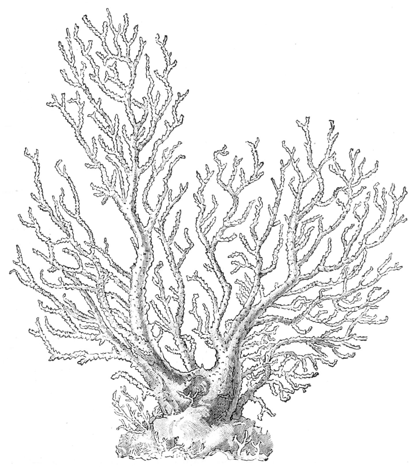
Fig. 133.—Ceratella fusca. About nat. size. (After Baldwin Spencer.[308])
Fam. Ceratellidae.—The colony of Ceratella may be five inches in height. The stem and main branches are substantial, and consist of a network of branching anastomosing tubes supported by a thick and fenestrated chitinous perisarc. The whole branch is enclosed in a common layer of ectoderm. The zooids have scattered capitate tentacles. The Ceratellidae occur in shallow water off the coast of New South Wales, extend up the coast of East Africa as far as Zanzibar, and have also been described from Japan.
Fam. Pennariidae.—In the hydrosome stage the zooids have numerous oral capitate tentacles scattered on the hypostome, and a single circlet of basilar filiform tentacles. The medusa of Pennaria, a common genus of wide distribution, is known under the name Globiceps.
Fam. Corynidae.—In the hydrosome stage the zooids of this family possess numerous capitate tentacles arranged in several circlets or scattered.
In Cladocoryne the tentacles are branched. Syncoryne is a common and widely distributed genus with numerous unbranched capitate tentacles irregularly distributed over a considerable length of the body-wall of the zooid. In many of the species the gonophores are liberated as Medusae, known by the name Sarsia, provided with four filiform tentacles and a very long manubrium. In some species (S. prolifera and S. siphonophora) the Medusae are reproduced asexually by gemmation from the long manubrium. A common British Anthomedusa of this family is Dipurena, but its hydrosome stage is not known. In the closely related genus Coryne the gonophores are adelocodonic, and exhibit very rudimentary medusoid characters.
Fam. Clavidae.—This is a large family containing many genera, some with free-swimming Medusae, others with adelocodonic gonophores. In the former group are included a number of oceanic Medusae of which the hydrosome stage has not yet been discovered. The zooids of the hydrosome have numerous scattered filiform tentacles. The free-swimming Medusae have hollow tentacles.
Clava contains a common British species with a creeping hydrorhiza frequently attached to shells, and with adelocodonic gonophores. Cordylophora is the genus which has migrated into fresh water in certain European localities (see p. 269). It forms well-developed branching colonies attached to wooden gates and piers or to the brickwork banks of canals. Several Anthomedusae, of which the hydrosome stage is not known, appear to be related to the Medusae of this family, but are sometimes separated as the family Tiaridae. Of these Tiara, a very brightly coloured jelly-fish sometimes attaining a height of 40 mm., is found on the British coasts, and Amphinema is found in considerable numbers at Plymouth in September. Turritopsis is a Medusa with a hydrosome stage like Dendroclava. For Stomatoca, see p. 415.
Fam. Corymorphidae.—This family contains the interesting British species Corymorpha nutans. The hydrosome stage consists of a solitary zooid of great size, 50-75 mm. in length, provided with two circlets of numerous long filiform tentacles. The free-swimming Medusae are produced in great numbers on the region between the two circlets of tentacles. These Medusae were formerly known by the name Steenstrupia, and are noteworthy in having only one long moniliform tentacle, opposite to one of the radial canals.
The gigantic Monocaulus imperator of Allman was obtained by the "Challenger" at the great depth of 2900 fathoms off the coast of Japan. It was nearly eight feet in length. More recently Miyajima[309] has described a specimen from 250 fathoms in the same seas which was 700 mm. (27.5 in.) in length. Miyajima's specimen resembles those described by Mark from 300 fathoms off the Pacific coast of North America as Branchiocerianthus urceolus in the remarkable feature of a distinct bilateral arrangement of the circlets of tentacles. Owing to the imperfect state of preservation of the only specimen of Allman's species it is difficult to determine whether it is also bilaterally symmetrical and belongs to the same species as the specimens described by Mark and Miyajima. These deep-sea giant species, however, appear to differ from Corymorpha in having adelocodonic gonophores.
Fam. Hydrolaridae.—This family contains the remarkable genus Lar, which was discovered by Gosse attached to the margin of the tubes of the marine Polychaete worm Sabella. The zooids have only two tentacles, and exhibit during life curious bowing and bending movements which have been compared with the exercises of a gymnast. The Medusae (Fig. 132, A and B) have been known for a long time by the name Willsia, but their life-history has only recently been worked out by Browne.[310]
Fam. Monobrachiidae.—Monobrachium, found in the White Sea by Mereschkowsky, forms a creeping stolon on the shells of Tellina. The zooids of the hydrosome have only one tentacle.
Fam. Myriothelidae.—This family contains the single genus Myriothela. The zooid of the hydrosome stage is solitary and is provided, as in the Corynidae, with numerous scattered capitate tentacles. The gonophores are borne by blastostyles situated above the region of the tentacles. In addition to these blastostyles producing gonophores there are, in M. phrygia, supplementary blastostyles which capture the eggs as they escape from the gonophores and hold them until the time when the larva is ready to escape. They were called "claspers" by Allman. In some of the Arctic species Frl. Bonnevie[311] has shown that they are absent. Each zooid of M. phrygia is hermaphrodite.
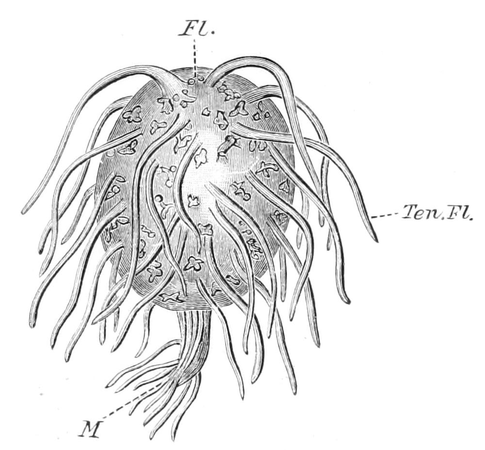
Fig. 134.—Pelagohydra mirabilis. Fl, The float; M, position of the mouth; Ten.Fl, filamentous tentacles of the float. (After Dendy.)
Fam. Pelagohydridae.—This family was constituted by Dendy[312] for the reception of Pelagohydra mirabilis, a remarkable new species discovered by him on the east coast of the South Island of New Zealand. The hydrosome is solitary and free-swimming, the proximal portion of the body being modified to form a float, the distal portion forming a flexible proboscis terminated by the mouth and a group of scattered manubrial tentacles. The tentacles are filiform and scattered over the surface of the float. Medusae are developed on stolons between the tentacles of the float. They have tentacles arranged in four radial groups of five each, at the margin of the umbrella.
As pointed out by Hartlaub,[313] Pelagohydra is not the only genus in which the hydrosome floats. Three species of the genus Margelopsis have been found that have pelagic habits, and two of them have been shown to produce numerous free-swimming Medusae by gemmation; but at present there is no reason to suppose that in these forms there is any extensive modification of the aboral extremity of the zooid to form such a highly specialised organ as the float of Pelagohydra.
The affinities of Pelagohydra are not clear, as our knowledge of the characters of the Medusa is imperfect; but according to Dendy it is most closely related to the Corymorphidae. Margelopsis belongs to the Bougainvilliidae.
Order IV. Calyptoblastea—Leptomedusae.
The hydrosome stage is characterised by the perisarc, which not only envelops the stem and branches, as in many of the Gymnoblastea, but is continued into a trumpet-shaped or tubular cup or collar called the "hydrotheca," that usually affords an efficient protection for the zooids when retracted. No solitary Calyptoblastea have been discovered. In the simpler forms the colony consists of a creeping hydrorhiza, from which the zooids arise singly (Clytia johnstoni), but these zooids may give rise to a lateral bud which grows longer than the parent zooid.
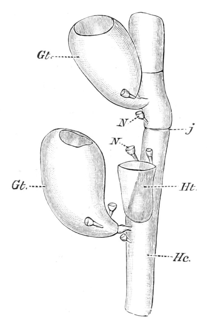
Fig. 135.—Part of a hydrocladium of a dried specimen of Plumularia profunda. Gt, Gonotheca; Hc, the stem of the hydrocladium with joints (j); Ht, a single hydrotheca; N, nematophores. Greatly enlarged. (After Nutting.)
The larger colonies are usually formed by alternate right and left budding from the last-formed zooid, so that in contrast to the Gymnoblast colony the apical zooid of the stem is the youngest, and not the oldest, zooid of the colony. In the branching colonies the axis is frequently composed of a single tube of perisarc, which may be lined internally by the ectoderm and endoderm tissues formed by the succession of zooids that have given rise to the branches by gemmation. Such a stem is said to be monosiphonic.
In some of the more complicated colonies, however, the stem is composed of several tubes, which may or may not be surrounded by a common sheath of ectoderm and perisarc, as they are in Ceratella among the Gymnoblastea. Such stems are said to be "polysiphonic" or "fascicled." The polysiphonic stem may arise in more than one way, and in some cases it is not quite clear in what manner it has arisen.[314]
In many colonies the zooids are only borne by the terminal monosiphonic branches, which receive the special name "hydrocladia." The gonophores of the Calyptoblastea are usually borne by rudimentary zooids, devoid of mouth and tentacles (the "blastostyles"), protected by a specially dilated cup of perisarc known as the "gonotheca" or "gonangium." The shape and size of the gonothecae vary a good deal in the order. They may be simply oval in shape, or globular (Schizotricha dichotoma), or greatly elongated, with the distal ends produced into slender necks (Plumularia setacea). They are spinulose in P. echinulata, and annulated in P. halecioides, Clytia, etc.
In some genera there are special modifications of the branches and hydrocladia, for the protection of the gonothecae. The name "Phylactocarp" is used to designate structures that are obviously intended to serve this purpose. The phylactocarp of the genera Aglaophenia and Thecocarpus is the largest and most remarkable of this group of structures, and has received the special name "corbula." The corbula consists of an axial stem or rachis, and of a number of corbula-leaves arising alternately from the rachis, bending upwards and then inwards to meet those of the other side above, the whole forming a pod-shaped receptacle. The gonangia are borne at the base of each of the corbula-leaves. There is some difference of opinion as to the homologies of the parts of the corbula, but the rachis seems to be that of a modified hydrocladium, as it usually bears at its base one or more hydrothecae of the normal type. The corbula-leaves are usually described as modified nematophores (vide infra), but according to Nutting[315] there is no more reason to regard them as modified nematophores than as modified hydrothecae, and he regards them as "simply the modification of a structure originally intended to protect an indefinite person, an individual that may become either a sarcostyle[316] or a hydranth."
The other forms of phylactocarps are modified branches as in Lytocarpus, and those which are morphologically appendages to branches as in Cladocarpus, Aglaophenopsis, and Streptocaulus.
The structures known as "nematophores" in the Calyptoblastea are the thecae of modified zooids, comparable with the dactylozooids of Millepora. They form a well-marked character of the very large family Plumulariidae, but they are also found in species of the genera Ophiodes, Lafoëina, Oplorhiza, Perisiphonia, Diplocyathus, Halecium, and Clathrozoon among the other Calyptoblastea. The dactylozooids are usually capitate or filiform zooids, without tentacles or a mouth, and with a solid or occasionally a perforated core of endoderm. They bear either a battery of nematocysts (Plumularia, etc.), or of peculiar adhesive cells (Aglaophenia and some species of Plumularia). The functions of the dactylozooids are to capture the prey and to serve as a defence to the colony. In the growth of the corbula of Aglaophenia the dactylozooids appear to serve another purpose, and that is, as a temporary attachment to hold the leaves together while the edges themselves are being connected by trabeculae of coenosarc.
In a very large number of Calyptoblastea the gonophore is a reduced Medusa which never escapes from the gonotheca, but in the family Eucopidae the gonophores escape as free-swimming Medusae, exhibiting certain very definite characters. The gonads are situated not on the manubrium, as in the Anthomedusae, but on the sub-umbrellar aspect of the radial canals. The marginal sense-organs may be ocelli or vesiculate statocysts. The bell is usually more flattened, and the velum smaller than it is in the Anthomedusae, and the manubrium short and quadrangular. Such Medusae are called Leptomedusae.
Leptomedusae of many specific forms are found abundantly at the surface of the sea in nearly all parts of the world, but with the exception of some genera of the Eucopidae and a few others, their connexion with a definite Calyptoblastic hydrosome has not been definitely ascertained. It may be an assumption that time will prove to be unwarranted that all the Leptomedusae pass through a Calyptoblastic hydrosome stage.
Fam. Aequoreidae.—In this family the hydrosome stage is not known except in the genus Polycanna, in which it resembles a Campanulariid. The sense-organs of the Medusae are statocysts. The radial canals are very numerous, and the genital glands are in the form of ropes of cells extending along the whole of their oral surfaces. Aequorea is a fairly common genus, with a flattened umbrella and a very rudimentary manubrium, which may attain a size of 40 mm. in diameter.
Fam. Thaumantiidae.—The Medusae of this family are distinguished from the Aequoreidae by having marginal ocelli in place of statocysts. The hydrosome of Thaumantias alone is known, and this is very similar to an Obelia.
Fam. Cannotidae.—The hydrosome is quite unknown. The Medusae are ocellate, but the radial canals, instead of being undivided, as in the Thaumantiidae, are four in number, and very much ramified before reaching the ring canal. The tentacles are very numerous. In the genus Polyorchis, from the Pacific coast of North America, the four radial canals give rise to numerous lateral short blind branches, and have therefore a remarkable pinnate appearance.
Fam. Sertulariidae.—In this family the hydrothecae are sessile, and arranged bilaterally on the stem and branches. The general form of the colony is pinnate, the branches being usually on opposite sides of the main stem. The gonophores are adelocodonic. Sertularia forms more or less arborescent colonies, springing from a creeping stolon attached to stones and shells. There are many species, several of which are very common upon the British coast. Many specimens are torn from their attachments by storms or by the trawls of fishermen and cast up on the sand or beach with other zoophytes. The popular name for one of the commonest species (S. abietina) is the "sea-fir." The genus has a wide geographical and bathymetrical range. Another common British species frequently thrown up by the tide in great quantities is Hydrallmania falcata. It has slender spirally-twisted stems and branches, and the hydrothecae are arranged unilaterally.
The genus Grammaria, sometimes placed in a separate family, is distinguished from Sertularia by several characters. The stem and branches are composed of a number of tubes which are considerably compressed. The genus is confined to the southern seas.
Fam. Plumulariidae.—The hydrothecae are sessile, and arranged in a single row on the stem and branches. Nematophores are always present. Gonophores adelocodonic. This family is the largest and most widely distributed of all the families of the Hydrozoa. Nutting calculates that it contains more than one-fourth of all the Hydroids of the world. Over 300 species have been described, and more than half of these are found in the West Indian and Australian regions. Representatives of the family occur in abundance in depths down to 300 fathoms, and not unfrequently to 500 fathoms. Only a few species have occasionally been found in depths of over 1000 fathoms.
The presence of nematophores may be taken as the most characteristic feature of the family, but similar structures are also found in some species belonging to other families (p. 277).
The family is divided into two groups of genera, the Eleutheroplea and the Statoplea. In the former the nematophores are mounted on a slender pedicel, which admits of more or less movement, and in the latter the nematophores are sessile. The genera Plumularia and Antennularia belong to the Eleutheroplea. The former is a very large genus, with several common British species, distinguished by the terminal branches being pinnately disposed, and the latter, represented by A. antennina and A. ramosa on the British coast, is distinguished by the terminal branches being arranged in verticils.
The two most important genera of the Statoplea are Aglaophenia and Cladocarpus. The former is represented by a few species in European waters, the latter is only found in American waters.
Fam. Hydroceratinidae.—The colony consists of a mass of entwined hydrorhiza, with a skeleton in the form of anastomosing chitinous tubes. Hydrothecae scattered, tubular, and sessile. Nematophores present. Gonophores probably adelocodonic.
This family was constituted for a remarkable hydroid, Clathrozoon wilsoni, described by W. B. Spencer from Victoria.[317] The zooids are sessile, and spring from more than one of the numerous anastomosing tubes of the stem and branches. The whole of the surface is studded with an enormous number of small and very simple dactylozooids, protected by tubular nematophores. Only a few specimens have hitherto been obtained, the largest being 10 inches in height by 4 inches in width. In general appearance it has some resemblance to a dark coloured fan-shaped Gorgonia.
Fam. Campanulariidae.—The hydrothecae in this family are pedunculate, and the gonophores adelocodonic.
In the cosmopolitan genus Campanularia the stem is monosiphonic, and the hydrothecae bell-shaped. Several species of this genus are very common in the rock pools of our coast between tide marks. Halecium is characterised by the rudimentary character of its hydrothecae, which are incapable of receiving the zooids even in their maximum condition of retraction. The genus Lafoea is remarkable for the development of a large number of tightly packed gonothecae on the hydrorhiza, each of which contains a blastostyle, bearing a single gonophore and, in the female, a single ovum. This group of gonothecae was regarded as a distinct genus of Hydroids, and was named Coppinia.[318] Lafoea dumosa with gonothecae of the type described as Coppinia arcta occurs on the British coast.
Perisiphonia is an interesting genus from deep water off the Azores, Australia, and New Zealand, with a stem composed of many distinct tubes.
The genus Zygophylax, from 500 fathoms off the Cape Verde, is of considerable interest in having a nematophore on each side of the hydrotheca. According to Quelch it should be placed in a distinct family.
Ophiodes has long and very active defensive zooids, protected by nematophores. It is found in the Laminarian zone on the English coast.
Fam. Eucopidae.—The hydrosome stage of this family is very similar to that of the Campanulariidae, but the gonophores are free-swimming Medusae of the Leptomedusan type.
One of the best-known genera is Obelia, of which several species are among the commonest Hydroids of the British coast.
Clytia johnstoni is also a very common Hydroid, growing on red algae or leaves of the weed Zostera. It consists of a number of upright, simple, or slightly branched stems springing from a creeping hydrorhiza. When liberated the Medusae are globular in form, with four radial canals and four marginal tentacles, but this Medusa, like many others of the order, undergoes considerable changes in form before it reaches the sexually mature stage.
Phialidium temporarium is one of the commonest Medusae of our coast, and sometimes occurs in shoals. It seems probable that it is the Medusa of Clytia johnstoni.[319] By some authors the jelly-fish known as Epenthesis is also believed to be the Medusa of a Clytia.
Fam. Dendrograptidae.—This family includes a number of fossils which have certain distinct affinities with the Calyptoblastea. In Dictyonema, common in the Ordovician rocks of Norway, but also found in the Palaeozoic rocks of North America and elsewhere, the fossil forms fan-shaped colonies of delicate filaments, united by many transverse commissures, and in well-preserved specimens the terminal branches bear well-marked uniserial hydrothecae. In some species thecae of a different character, which have been interpreted to be gonothecae and nematophores respectively, are found.
Other genera are Dendrograptus, Thamnograptus, and several others from Silurian strata.
Order V. Graptolitoidea.
A large number of fossils, usually called Graptolites, occurring in Palaeozoic strata, are generally regarded as the skeletal remains of an ancient group of Hydrozoa.
In the simpler forms the fossil consists of a delicate straight rod bearing on one side a series of small cups. It is suggested that the cups contained hydroid zooids, and should therefore be regarded as the equivalent of the hydrothecae, and that the axis represents the axis of the colony or of a branch of the Calyptoblastea. In some of the forms with two rows of cups on the axis (Diplograptus), however, it has been shown that the cups are absent from a considerable portion of one end of the axis, and that the axes of several radially arranged individuals are fused together and united to a central circular plate. Moreover, there is found in many specimens a series of vesicles, a little larger in size than the cups, attached to the plate and arranged in a circle at the base of the axes. These vesicles are called the gonothecae.
The discovery of the central plate and of the so-called gonothecae suggests that the usual comparison of a Graptolite with a Sertularian Hydroid is erroneous, and that the colony or individual, when alive, was a more or less radially symmetrical floating form, like a Medusa, of which only the distal appendages (possibly tentacles) are commonly preserved as fossils.
The evidence that the Graptolites were Hydrozoa is in reality very slight, but the proof of their relationship to any other phylum of the animal kingdom does not exist.[320] It is therefore convenient to consider them in this place, and to regard them, provisionally, as related to the Calyptoblastea.
The order is divided into three families.
Fam. 1. Monoprionidae.—Cups arranged uniserially on one side of the axis.
The principal genera are Monograptus, with the axis straight, curved, or helicoid, from many horizons in the Silurian strata; Rastrites, with a spirally coiled axis, Silurian; Didymograptus, Ordovician; and Coenograptus, Ordovician.
Fam. 2. Diprionidae.—Cups arranged in two or four vertical rows on the axis.
Diplograptus, Ordovician and Silurian; Climacograptus, Ordovician and Silurian; and Phyllograptus, in which the axis and cups are arranged in such a manner that they resemble an ovate leaf.
Fam. 3. Retiolitidae.—Cups arranged biserially on a reticulate axis.
Retiolites, Ordovician and Silurian; Stomatograptus, Retiograptus, and Glossograptus, Ordovician.
Fossil Corals possibly allied to Hydrozoa.
Among the many fossil corals that are usually classified with the Hydrozoa the genus Porosphaera is of interest as it is often supposed to be related to Millepora. It consists of globular masses about 10-20 mm. in diameter occurring in the Upper Cretaceous strata. In the centre there is usually a foreign body around which the coral was formed by concentric encrusting growth. Running radially from pores on the surface to the centre, there are numerous tubules which have a certain general resemblance to the pore-tubes of Millepora. The monomorphic character of these tubes, their very minute size, the absence of ampullae, and the general texture of the corallum, are characters which separate this fossil very distinctly from any recent Hydroid corals. Porosphaera, therefore, was probably not a Hydrozoon, and certainly not related to the recent Millepora.
Closely related to Porosphaera apparently are other globular, ellipsoidal, or fusiform corals from various strata, such as Loftusia from the Eocene of Persia, Parkeria from the Cambridge Greensand, and Heterastridium from the Alpine Trias. In the last named there is apparently a dimorphism of the radial tubes.
Allied to these genera, again, but occurring in the form of thick, concentric, calcareous lamellae, are the genera Ellipsactinia and Sphaeractinia from the Upper Jurassic.
Another important series of fossil corals is that of the family Stromatoporidae. These fossils are found in great beds of immense extent in many of the Palaeozoic rocks, and must have played an important part in the geological processes of that period. They consist of a series of calcareous lamellae, separated by considerable intervals, encrusting foreign bodies of various kinds. Sometimes they are flat and plate-like, sometimes globular or nodular in form. The lamellae are in some cases perforated by tabulate, vertical, or radial pores, but in many others these pores are absent. The zoological position of the Stromatoporidae is very uncertain, but there is not at present any very conclusive evidence that they are Hydrozoa.
Stromatopora is common in Devonian and also occurs in Silurian strata. Cannopora from the Devonian has well-marked tabulate pores, and is often found associated commensally with another coral (Aulopora or Syringopora).
Order VI. Stylasterina.
The genera included in this order resemble Millepora in producing a massive calcareous skeleton, and in showing a consistent dimorphism of the zooids, but in many respects they exhibit great divergence from the characters of the Milleporina.
The colony is arborescent in growth, the branches arising frequently only in one plane, forming a flabellum. The calcareous skeleton is perforated to a considerable depth by the gastrozooids, dactylozooids, and nutritive canals, and the gastropores and dactylopores are not provided with tabulae except in the genera Pliobothrus and Sporadopora. The character which gives the order its name is a conical, sometimes torch-like projection at the base of the gastropore, called the "style," which carries a fold of the ectoderm and endoderm layers of the body-wall, and may serve to increase the absorptive surface of the digestive cavity. In some genera a style is also present in the dactylopore, in which case it serves as an additional surface for the attachment of the retractor muscles. The pores are scattered on all aspects of the coral in the genera Sporadopora, Errina, and Pliobothrus; in Spinipora and Steganopora the scattered dactylopores are situated at the extremities of tubular spines which project from the general surface of the coral, the gastropores being situated irregularly between the spines. In Phalangopora the pores are arranged in regular longitudinal lines, and in Distichopora they are mainly in rows on the edges of the flattened branches, a single row of gastropores being flanked by a single row of dactylopores on each side. In the remaining genera the pores are arranged in definite cycles, which are frequently separated from one another by considerable intervals, and have, particularly in the dried skeleton, a certain resemblance to the calices of some of the Zoantharian corals.
In Cryptohelia the cycles are covered by a lid-like projection from the neighbouring coenenchym (Fig. 136, l 1, l 2). The gastrozooids are short, and are usually provided with a variable number of small capitate tentacles. The dactylozooids are filiform and devoid of tentacles, the endoderm of their axes being solid and scalariform.
The gonophores of the Stylasterina are situated in large oval or spherical cavities called the ampullae, and their presence can generally be detected by the dome-shaped projections they form on the surface of the coral. The female gonophore consists of a saucer-shaped pad of folded endoderm called the "trophodisc," which serves the purpose of nourishing the single large yolk-laden egg it bears; and a thin enveloping membrane composed of at least two layers of cells. The egg is fertilised while it is still within the ampulla, and does not escape to the exterior until it has reached the stage of a solid ciliated larva. All the Stylasterina are therefore viviparous. The male gonophore has a very much smaller trophodisc, which is sometimes (Allopora) prolonged into a columnar process or spadix, penetrating the greater part of the gonad. The spermatozoa escape through a peculiar spout-like duct which perforates the superficial wall of the ampulla. In some genera (Distichopora) there are several male gonophores in each ampulla.
The gonophores of the Stylasterina have been regarded as much altered medusiform gonophores, and this view may possibly prove to be correct. At present, however, the evidence of their derivation from Medusae is not conclusive, and it is possible that they may have had a totally independent origin.
Distichopora and some species of Stylaster are found in shallow water in the tropics, but most of the genera are confined to deep or very deep water, and have a wide geographical distribution. No species have been found hitherto within the British area.
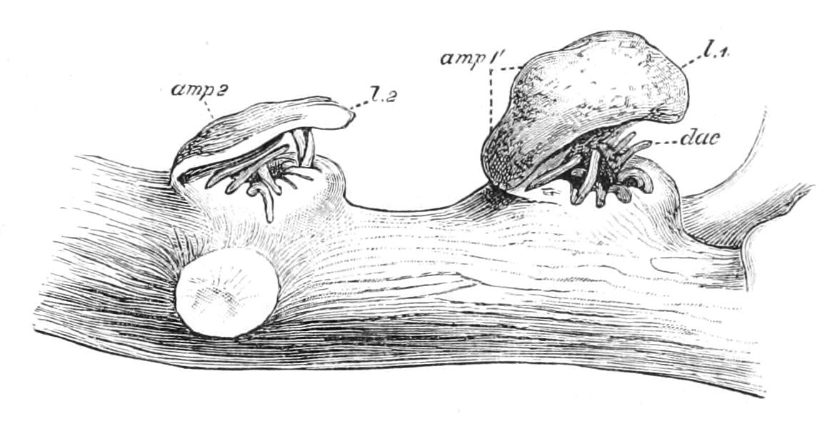
Fig. 136.—A portion of a branch of Cryptohelia ramosa, showing the lids l 1 and l 2 covering the cyclosystems, the swellings produced by the ampullae in the lids amp1, amp2, and the dactylozooids, dac. × 22. (After Hickson and England.)
A few specimens of a species of Stylaster have been found in Tertiary deposits and in some raised beaches of more recent origin, but the order is not represented in the older strata.
Fam. Stylasteridae.—All the genera at present known are included in this family.
Sporadopora is the only genus that presents a superficial general resemblance to Millepora. It forms massive, branching white coralla, with the pores scattered irregularly on the surface, and, like many varieties of Millepora, not arranged in cyclosystems. It may, however, be distinguished at once by the presence of a long, brush-like style in each of the gastropores. The ampullae are large, but are usually so deep-seated in the coenenchym that their presence cannot be detected from the surface. It was found off the Rio de la Plata in 600 fathoms of water by the "Challenger."
In Errina the pores are sometimes irregularly scattered, but in E. glabra they are arranged in rows on the sides of the branches, while in E. ramosa the gastropores occur at the angles of the branches only. The dactylopores are situated on nariform projections of the corallum. The ampullae are prominent. There are several gonophores in each ampulla of the male, but only one in each ampulla of the female. This genus is very widely distributed in water from 100 to 500 fathoms in depth.
Phalangopora differs from Errina in the absence of a style in the gastropore; Mauritius.—Pliobothrus has also no style in the gastropore, and is found in 100-600 fathoms of water off the American Atlantic shores.
Distichopora is an important genus, which is found in nearly all the shallow seas of the tropical and semi-tropical parts of the world, and may even flourish in rock pools between tide marks. It is nearly always brightly coloured—purple, violet, pale brown, or rose red. The colony usually forms a small flabellum, with anastomosing branches, and the pores are arranged in three rows, a middle row of gastropores and two lateral rows of dactylopores on the sides of the branches. There is a long style in each gastropore. The ampullae are numerous and prominent, situated on the anterior and posterior faces of the branches. Each ampulla contains a single gonophore in the female colony and two or three gonophores in the male colony.
Spinipora is a rare genus from off the Rio de la Plata in 600 fathoms. The branches are covered with blunt spines. These spines have a short gutter-like groove at the apex, which leads into a dactylopore. The gastropores are provided with a style and are situated between the spines.
Steganopora[321] from the Djilolo Passage, in about 600 fathoms, is very similar to Spinipora as regards external features, but differs from it in the absence of styles in the gastropores, and in the wide communications between the gastropores and dactylopores.
Stylaster is the largest and most widely distributed genus of the family, and exhibits a considerable range of structure in the many species it contains. It is found in all the warmer seas of the world, living between tide marks at a few fathoms, and extending to depths of 600 fathoms. Many specimens, but especially those from very shallow water, are of a beautiful rose or pink colour. The corallum is arborescent and usually flabelliform. The pores are distributed in regular cyclosystems, sometimes on one face of the corallum only, sometimes on the sides of the branches, and sometimes evenly distributed. There are styles in both gastropores and dactylopores.
Allopora is difficult to separate from Stylaster, but the species are usually more robust in habit, and the ampullae are not so prominent as they are on the more delicate branches of Stylaster. It occurs at depths of 100 fathoms in the Norwegian fjords. A very large red species (A. nobilis) occurs in False Bay, Cape of Good Hope, in 30 fathoms of water. In this locality the coral occurs in great submarine beds or forests, and the trawl that is passed over them is torn to pieces by the hard, thick branches, some of which are an inch or more in diameter.
Astylus is a genus found in the southern Philippine sea in 500 fathoms of water. It is distinguished from Stylaster by the absence of a style in the gastropore.
Cryptohelia is an interesting genus found both in the Atlantic and Pacific Oceans at depths of from 270 to about 600 fathoms. The cyclosystems are covered by a projecting lid or operculum (Fig. 136, l 1, l 2). There are no styles in either the gastropores or the dactylopores. The ampullae are prominent, and are sometimes situated in the lids. There are several gonophores in each ampulla of the female colony, and a great many in the ampulla of the male colony.