Chapter XXI
Reading edition
ECHINODERMATA (CONTINUED): DEVELOPMENT AND PHYLOGENY
In Chapter XVI. it was stated that whilst a more or less perfectly developed radial symmetry was one of the characteristic features of the phylum Echinodermata when in the adult condition, yet in the immature or larval condition the members of the group have a strongly marked bilateral symmetry. In this feature larval Echinodermata resemble the other Phyla of the animal kingdom which have a well-developed coelom, such as Annelida, Mollusca, Vertebrata, etc. Since, then, the peculiar radial symmetry is gradually acquired during the growth of the Echinoderm, we may possibly discover by a close scrutiny of the life-history what is the nature and meaning of this departure from the ordinary type of structure among coelomate animals.
There are two kinds of development met with amongst Echinodermata, which may be roughly characterised as the "embryonic" and the "larval" type respectively, although neither description is exact. In developmental histories of the first type so much reserve material is laid up in the egg in the form of food-yolk that the young animal whilst in the bilateral stage requires little or no food. In some cases, however, as in Amphiura squamata, the mother pours out a nourishing exudation; but whether this is so or not, the parent in nearly every case carries the young about with her until they have reached the adult condition. In some Asteroidea, as for instance in the Antarctic species Asterias spirabilis (Fig. 280), the young become fixed to the everted lips of the mother; in Amphiura squamata, and some other Ophiuroidea the eggs remain in the genital bursae, which serve as nurseries; in some Spatangoidea, as for instance in Hemiaster philippi (Figs. 250, 281), the eggs are carried in some of the deeply grooved petaloid ambulacra; whilst in Holothuroidea they may develop in the body-cavity (Phyllophorus urna), or they may adhere to the back of the mother (Cucumaria crocea, Fig. 259, p. 573), or they may be protected in special brood-pouches either on the ventral side of the parent (Cucumaria laevigata) or on the dorsal surface (Psolus ephippifer, Fig. 261).
The majority of these cases of embryonic development have been recorded from Arctic or Antarctic waters; it appears as if conditions there were not favourable to the larval type of development. In Pelmatozoa the development of Antedon rosacea alone is known, and that is of the embryonic type.
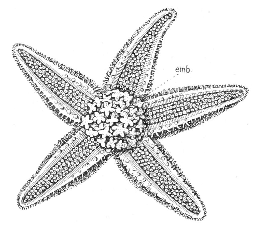
Fig. 280.—Oral view of Asterias spirabilis, slightly enlarged, showing embryos attached to the everted lips. emb, Embryos. (After Perrier.)
So far, however, as their mode of propagation is known, it may confidently be affirmed that the development of the majority of the species of Eleutherozoa is of the second or larval type. In this type there is little food-yolk in the egg, and the young animal or larva is forced from a very early period of development to seek its own living, and hence it is usually a considerable time (from a fortnight to two months) before the adult form is attained. When the embryos of different groups of Eleutherozoa are compared, there is no obvious agreement in structure between them; but the larvae of the four classes of Eleutherozoa exhibit with differences in detail a most remarkable fundamental similarity in type, and we are accordingly justified in regarding the larval development as primitive, and the embryonic type as derived from it and differently modified in each case.
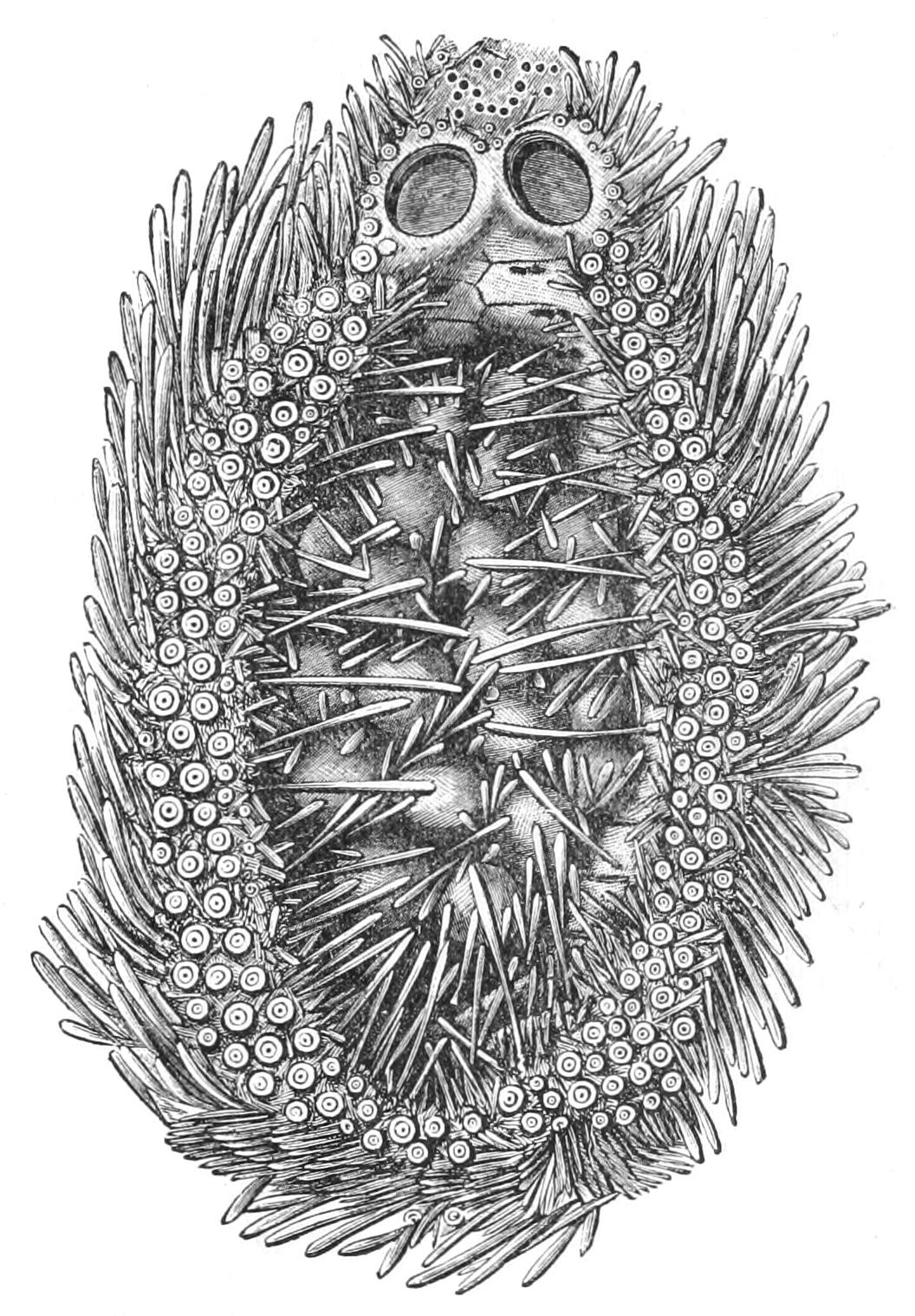
Fig. 281.—Hemiaster philippi. Enlarged view of a single petal, showing the embryos in situ. (From Wyville Thomson.) The whole animal is shown in Fig. 250, p. 555.
In the typical larval development the eggs are fertilised after being laid, and they then undergo segmentation into a number of equal, or nearly equal, segments or "blastomeres." These arrange themselves in the form of a hollow sphere or "blastula," the cavity of which is called the "blastocoel" and afterwards becomes the primary body-cavity of the larva. This cavity contains an albuminous fluid, at the expense of which development appears to be carried on (Fig. 282, B). The cells forming the blastula acquire cilia, and the embryo begins to rotate within the egg-membrane, which it soon bursts, and, rising to the surface of the sea, begins its larval life. The blastula is therefore the first well-marked larval stage, and it is found in a more or less recognisable form in life-histories of members of every large group in the animal kingdom. Only in the case of Echinodermata and of forms still lower in the scale, however, does it appear as a larval stage. The free-swimming blastula stage is reached in from twelve to twenty-four hours. Soon the spherical form of the blastula is lost; one side becomes flattened and thickened, owing to a multiplication of cells, so that they become taller and narrower in shape. Shortly afterwards this thickened plate becomes buckled inwards, encroaching on the cavity of the blastocoel. The larva has now reached the second stage of its development; it has become a "gastrula" (Fig. 282, C). The plate of thickened cells has become converted into a tube called the "archenteron" (Fig. 282, C, arch), which is the rudiment of both the alimentary canal and the coelom of the adult. This tube communicates with the exterior, in virtue of its mode of formation, by a single opening which is called the "blastopore," which becomes the anus of the later larva and adult. Whilst the gastrula stage is being acquired, the blastocoel or primary body-cavity is invaded by wandering cells budded from the wall of the archenteron (Fig. 282, A, B, C, mes). These cells, which are called "mesenchyme," are the formative cells of the skeleton, connective tissue, and wandering cells of the adult. When the larva has a skeleton they are formed very early, arising in the young blastula stage (Ophiuroidea) or in the stage immediately before the formation of the archenteron (Echinoidea, Fig. 282, A, B) and secreting the skeleton. When the larva is devoid of a skeleton (Asteroidea and Holothuroidea), the mesenchyme usually does not appear till the gastrula is fully formed.
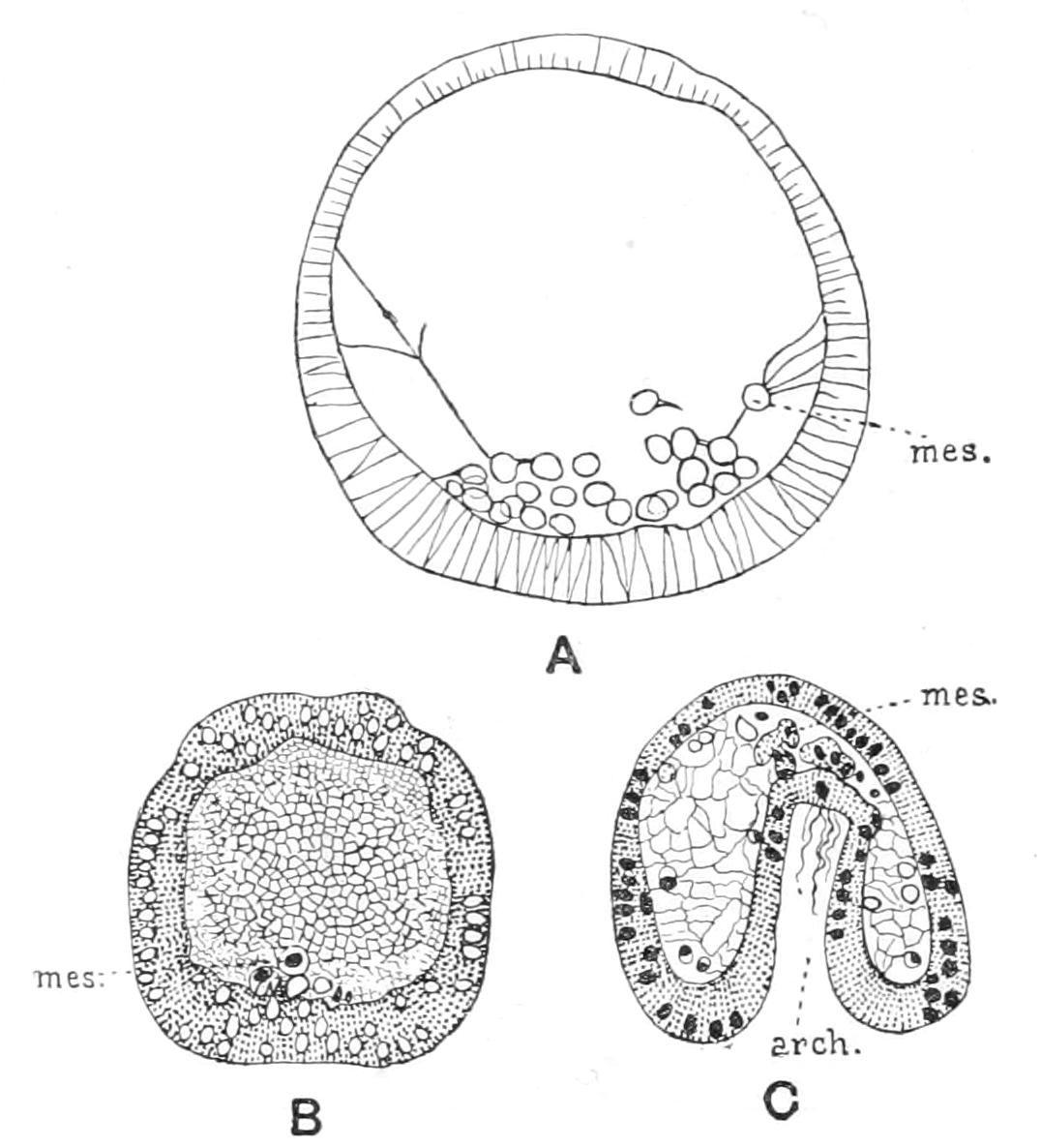
Fig. 282.—Echinus esculentus. A, optical section of living blastula. B, section of preserved blastula. The network of strings in the interior is the result of the coagulation of the albuminous fluid. C, section of gastrula. arch, Archenteron; mes, mesenchyme cells, attached by protoplasmic strands to the wall of the embryo. × 150.
The gastrula stage is reached in twenty to thirty-six hours. Then one side of the larva becomes concave, and the cilia become restricted to a thick band surrounding this area. In this way is formed the rudiment of the longitudinal band of cilia, which is the organ of locomotion throughout the larval life. At the apex of the archenteron a thin-walled vesicle is formed, which soon becomes divided off from the rest. This vesicle, which almost immediately divides into two sacs, right and left, is the rudiment of the "coelom" or secondary body-cavity of the larva; the remainder of the archenteron forms the definitive gut, and becomes divided by constriction into an oesophagus, a stomach, and an intestine, and at the same time bent into a shallower or deeper V-shape, the concavity of which is towards the concave side of the body. Within this area of the surface a new funnel-shaped depression makes its appearance. This is the "stomodaeum," the rudiment of the mouth of the larva, and it soon joins the apex of the larval oesophagus; the conjoined tubes henceforth bearing the name oesophagus since the ectodermal and endodermal parts become indistinguishably fused. Along the sides and floor of the oesophagus is formed a V-shaped ridge bearing strong cilia; this is the "adoral band of cilia" which sweeps the food (consisting of Diatoms, Infusoria, etc.) into the mouth. The larva is now known as a Dipleurula and appears in four modifications, each characteristic of a Class of Eleutherozoa. These differ from one another principally in the following points:—(a) The folding of the ciliated band; (b) the divisions of the coelomic sacs; (c) the development and fate of the praeoral lobe (i.e. the part of the body in front of the mouth); (d) the fate of the larval mouth. The types of Dipleurula are as follows:—
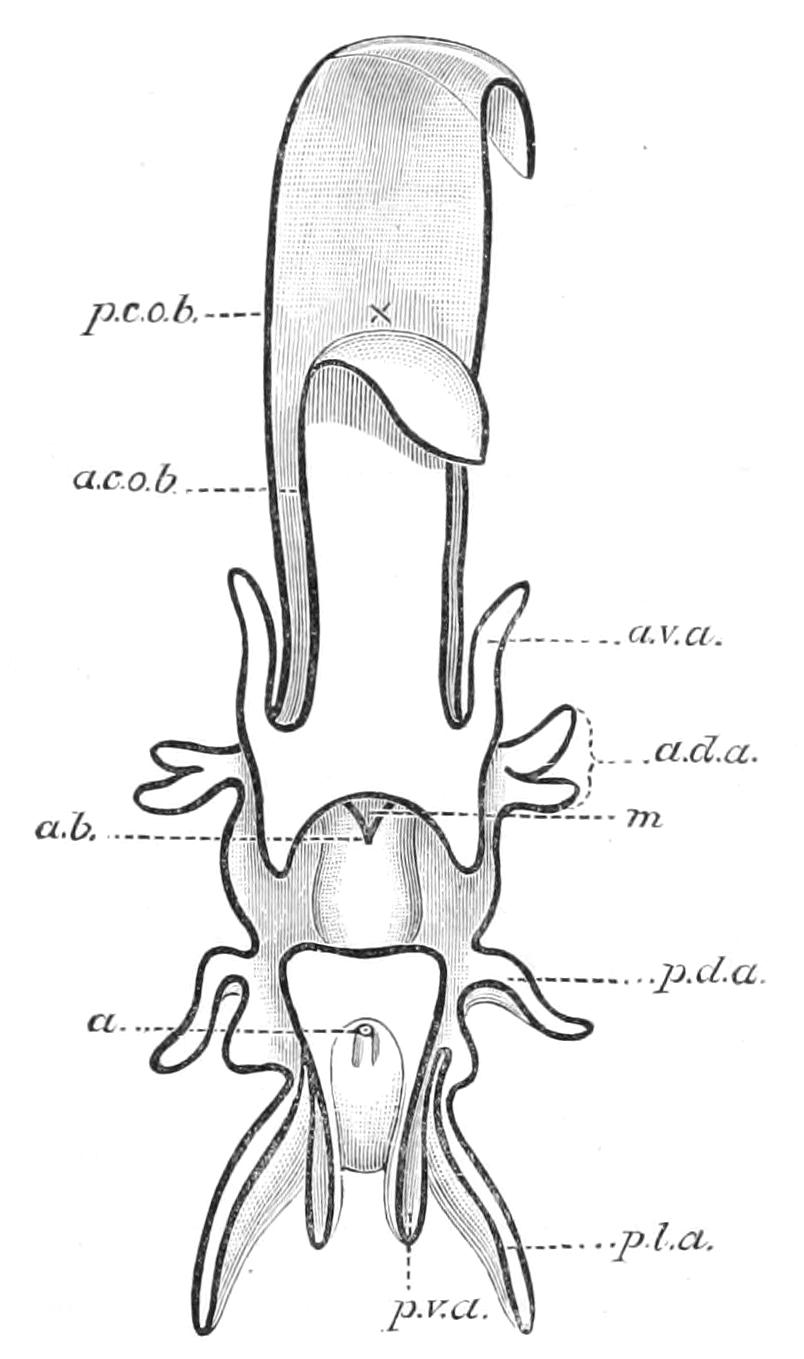
Fig. 283.—Bipinnaria of Luidia. a, Anus; a.b, adoral ciliated band; a.c.o.b, anterior median process; a.d.a, anterior dorsal process; a.v.a, prae-oral process; m, mouth; p.c.o.b, median dorsal process; p.d.a, posterior dorsal process; p.l.a, posterior lateral process; p.v.a, post-oral process. (After Garstang.)
(1) The Bipinnaria, the larva of Asteroidea. In this type there is a very long prae-oral lobe. The ciliated band runs along its edges, and is produced into a backwardly directed loop on its under surface. This loop soon becomes separated from the rest of the band as a distinct prae-oral loop, the rest forming a post-oral loop. Both loops are drawn out into short tag-like processes, in which we may distinguish (following Mortensen's[517] notation) in the prae-oral loop an anterior median process (Fig. 283, a.c.o.b), and a pair of prae-oral processes (a.v.a). In the post-oral loop there is a median dorsal process (p.c.o.b) and paired anterior dorsal (a.d.a), posterior dorsal (p.d.a), posterior lateral (p.l.a), and post-oral (p.v.a) processes. At the apex of the prae-oral lobe between prae-oral and post-oral ciliated rings there is an ectodermic thickening, recalling the so-called apical plate of Annelid larvae.
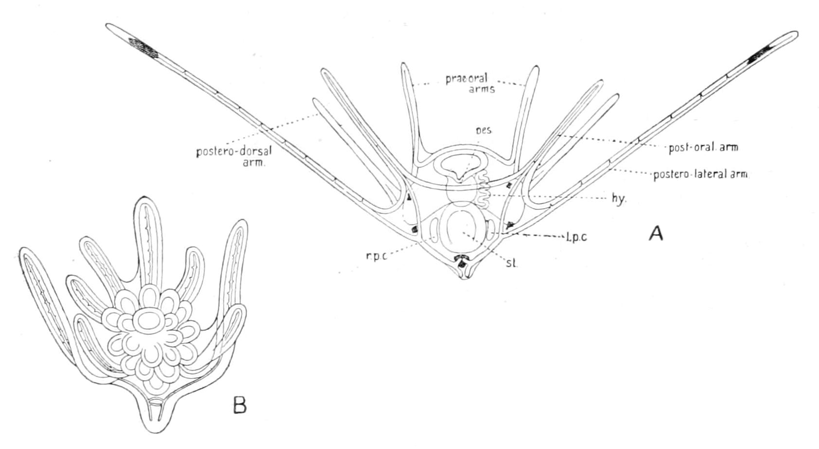
Fig. 284.—A, Ophiopluteus of Ophiothrix fragilis. hy, Hydrocoel; l.p.c, left posterior coelom; oes, oesophagus; r.p.c, right posterior coelom; st, stomach. B, metamorphosis of Ophiopluteus of Ophiura sp. (After Johannes Müller.)
(2) The Ophiopluteus, the larva of the Ophiuroidea. In this type the prae-oral lobe remains small, and the primitive ciliated band is undivided. The processes into which it is drawn out are very long, and are supported by calcareous rods. Of these processes we may distinguish prae-oral, postero-dorsal, postero-lateral, and post-oral. The postero-lateral are always much longer than the rest, so that the larva when swimming appears to the naked eye as a tiny V. In the case of Ophiothrix fragilis (Fig. 284, A) the postero-lateral processes are many times longer than the rest of the body. The Ophiopluteus was the first Echinoderm larva to be recognised. It was discovered by Johannes Müller,[518] who also discovered the other three types of Dipleurula. He named this one Pluteus (easel), from a fancied resemblance, when turned upside down, to a painter's easel. The same name was bestowed on the next type, to which it presents a superficial resemblance, and hence the distinguishing prefix "Ophio-" was added to the original name by Mortensen.
(3) The Echinopluteus, the larva of the Echinoidea. This type is strikingly like the preceding one in possessing a very small prae-oral lobe and in having the processes of the ciliated ring supported by calcareous rods, but a close inspection of these shows that they do not exactly correspond to those of the Ophiopluteus. Thus we have prae-oral, postero-dorsal, and post-oral processes (Fig. 285), but usually no postero-lateral process, and when it does occur it remains short. On the other hand, an antero-lateral process unrepresented in the Ophiopluteus is constantly present, and in its later stage the Echinopluteus develops, out of parts of the ciliated ring, horizontally-placed crescentic ridges of cilia, which are termed ciliated epaulettes (Fig. 285, a.cil.ep). There may even be, as in the larva of Echinus esculentus, a second posterior set of these (Fig. 285, p.cil.ep). In the older larva at the apex of the prae-oral lobe there is an ectodermic thickening, at the base of which are developed nerve-cells and nerve fibres constituting a larval brain (Fig. 285, ap).
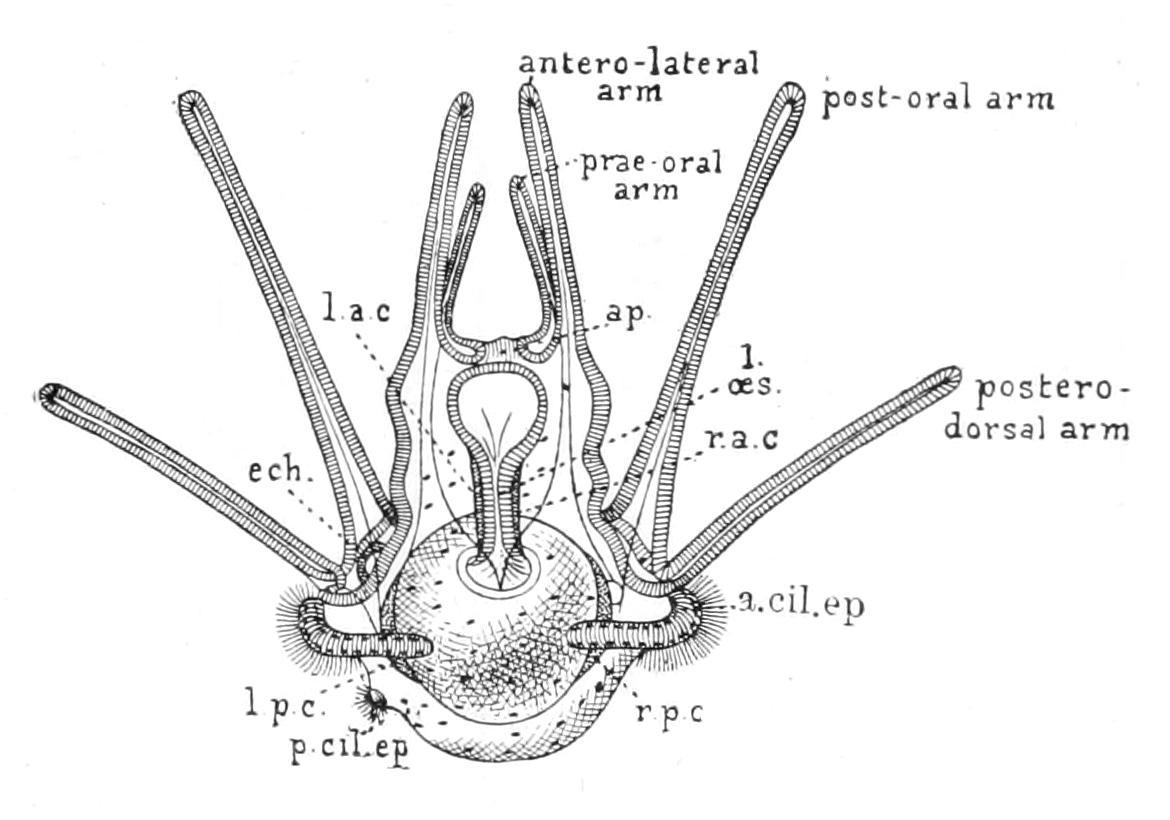
Fig. 285.—Dorsal view of larva of Echinus esculentus. × 45. a.cil.ep, Anterior ciliated epaulette; ap, apical plate or larval brain; ech, rudiment of Sea-urchin; l.a.c, left anterior coelom; l.oes, larval oesophagus; l.p.c, r.p.c, as in Fig. 284; p.cil.ep, posterior ciliated epaulette; r.a.c, right anterior coelom.
(4) The Auricularia, the larva of the Holothuroidea. This type strikingly resembles the Bipinnaria in its external features. The prae-oral lobe is well developed, and has on its under surface a backwardly projecting loop of the ciliated band, which is not, however, as in the Bipinnaria, separated from the rest of the band. The processes of the band are much more faintly marked than in the Bipinnaria, the anterior median, prae-oral, and median dorsal processes being absent; but a pair of intermediate dorsal processes are developed in the interspace between anterior and posterior dorsal.
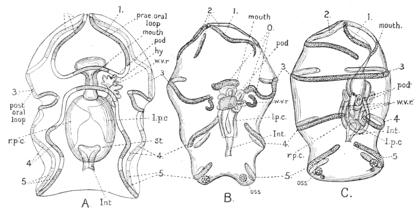
Fig. 286.—Three views of metamorphosis of Auricularia of Synapta digitata. A, fully grown Auricularia; B and C, stages in the metamorphosis. hy, Hydrocoel; Int, intestine; l.p.c, left posterior coelom; O, fragments of ciliated band which are invaginated into the stomodaeum, and coalesce to form a ring round the mouth; oss, ossicle; pod, rudiment of feelers which here spring directly from the hydrocoel; r.p.c, right posterior coelom; st, stomach; w.v.r, rudiment of water-vascular radial canals; 1-5, corresponding pieces in the three figures of the longitudinal ciliated band. (After Bury.) × 40.
In the Bipinnaria, Ophiopluteus, and Echinopluteus the coelomic vesicle, after separation from the archenteron, divides into right and left halves. The left then sends out a short dorsal process, which, fusing with the ectoderm, acquires an opening to the exterior. This opening is the primary madreporic pore, and the process of the left coelomic sac, which is ciliated, is the pore-canal. In the Auricularia the pore and pore-canal are formed before the division of the coelom. In the Bipinnaria the right and left sacs subsequently fuse in the front part of the prae-oral lobe. In the first three types of larva the coelomic sac on each side then undergoes a segmentation into anterior and posterior portions. At the hinder end of the anterior sac on each side a swelling occurs. That on the left side is the "hydrocoel," or rudiment of the water-vascular system (Fig. 287, A3, l.hy); it quickly assumes a crescentic form, and gives off five blunt outgrowths, which are the rudiments of the radial canals, and the terminal tentacles. It remains in connexion with the anterior coelom by a narrow neck, which later becomes the stone-canal. That on the right side separates completely from the right anterior coelom; it remains small, and forms the madreporic vesicle (Fig. 287, A3, r.hy) of the adult. In the Ophiopluteus and in the larva of Asterina gibbosa (v. infra) it occasionally takes on a form similar to that of the hydrocoel; from which circumstance, as well as from the similarity in its mode of origin, it is here regarded as a right hydrocoel, i.e. a rudimentary fellow of the organ which develops into the water-vascular system.
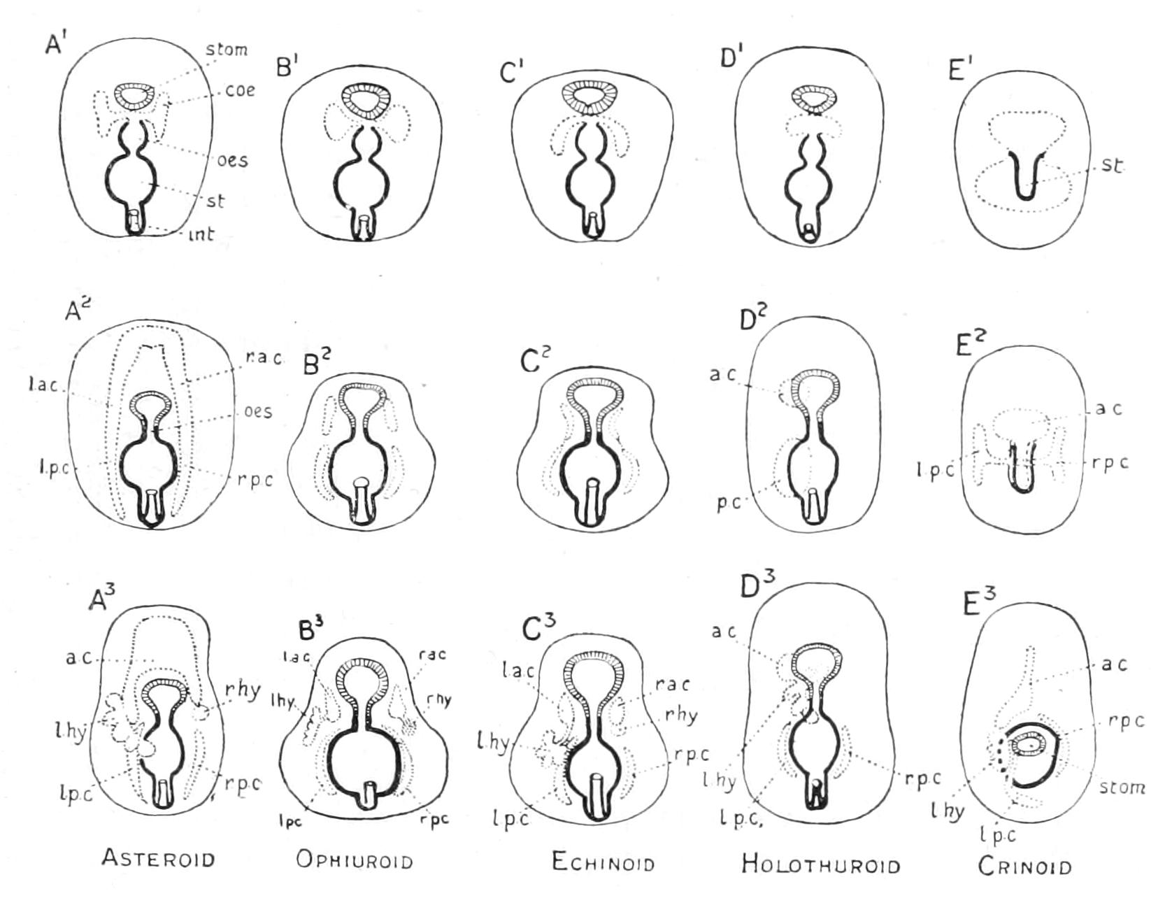
Fig. 287.—Diagrams of the mode of formation and division of the coelom in Echinodermata. a.c, Anterior coelom; coe, primitive coelomic rudiment; int, intestine; l.a.c, left anterior coelom; l.hy, left hydrocoel; l.p.c, left posterior coelom; oes, oesophagus; p.c, posterior coelom; r.a.c, right anterior coelom; r.hy, right hydrocoel; r.p.c, right posterior coelom; st, stomach; stom, stomodaeum.
In Auricularia (Fig. 287, D) the coelomic vesicle, after the pore-canal is formed, divides into an anterior and a posterior half. The posterior part then divides into right and left halves, whilst the anterior sac divides into dorsal and ventral halves, connected by a narrow neck. The ventral half soon assumes the familiar feature of the hydrocoel (Fig. 287, D3, l.hy), whilst the dorsal half forms an insignificant swelling on the course of the conjoined stone- and pore-canals, which represents the left anterior coelom of the other types; neither right anterior coelom nor right hydrocoel being developed. The neck of communication between dorsal and ventral halves is, of course, the stone-canal.
The Dipleurula larva leads a free-swimming life for a period varying from two weeks to two months, and then undergoes metamorphosis into the adult form. The details of this process have been worked out in comparatively few cases; and the species in which they are most thoroughly known is the Asteroid Asterina gibbosa. The development of this species is intermediate in character between the embryonic and larval types. The eggs are larger than is usual among Asteroidea, and are filled with a bright orange yolk. The larva differs from the Bipinnaria in the absence of the characteristic ciliated bands and in the very early occlusion of the anus. There is, however, a band of cilia round the edge of the prae-oral lobe, which corresponds to portions of the prae-oral and post-oral bands combined of the Bipinnaria.[519]
The larva has a form which may be described as boot-shaped (Figs. 288, 289). The sole of the boot is the great prae-oral lobe, behind which is the mouth. The larva takes little or no food, and completes its metamorphosis in ten to twelve days. It does not swim at the surface, but creeps slowly over the bottom by the aid of the ciliated band mentioned above, while it can also attach itself, using the edges of the prae-oral lobe as a sucker.
After leading an existence of this kind for seven or eight days it fixes itself permanently by a disc-like prominence, which appears on the anterior surface of the prae-oral lobe within the area surrounded by the thickened rim which, as explained above, forms a margin to the prae-oral lobe. The larva then becomes divided by a constriction into a disc and a stalk, and the former is gradually converted into the body of the young Starfish, whilst the latter continually diminishes in size, and eventually entirely disappears, when the young Starfish commences to walk about on its podia. The disc becomes bent downwards and to the left, so as to make nearly a right angle with the stalk, and the last vestige of the latter springs from the peristome of the Starfish inside the water-vascular ring (Figs. 289, B, C).
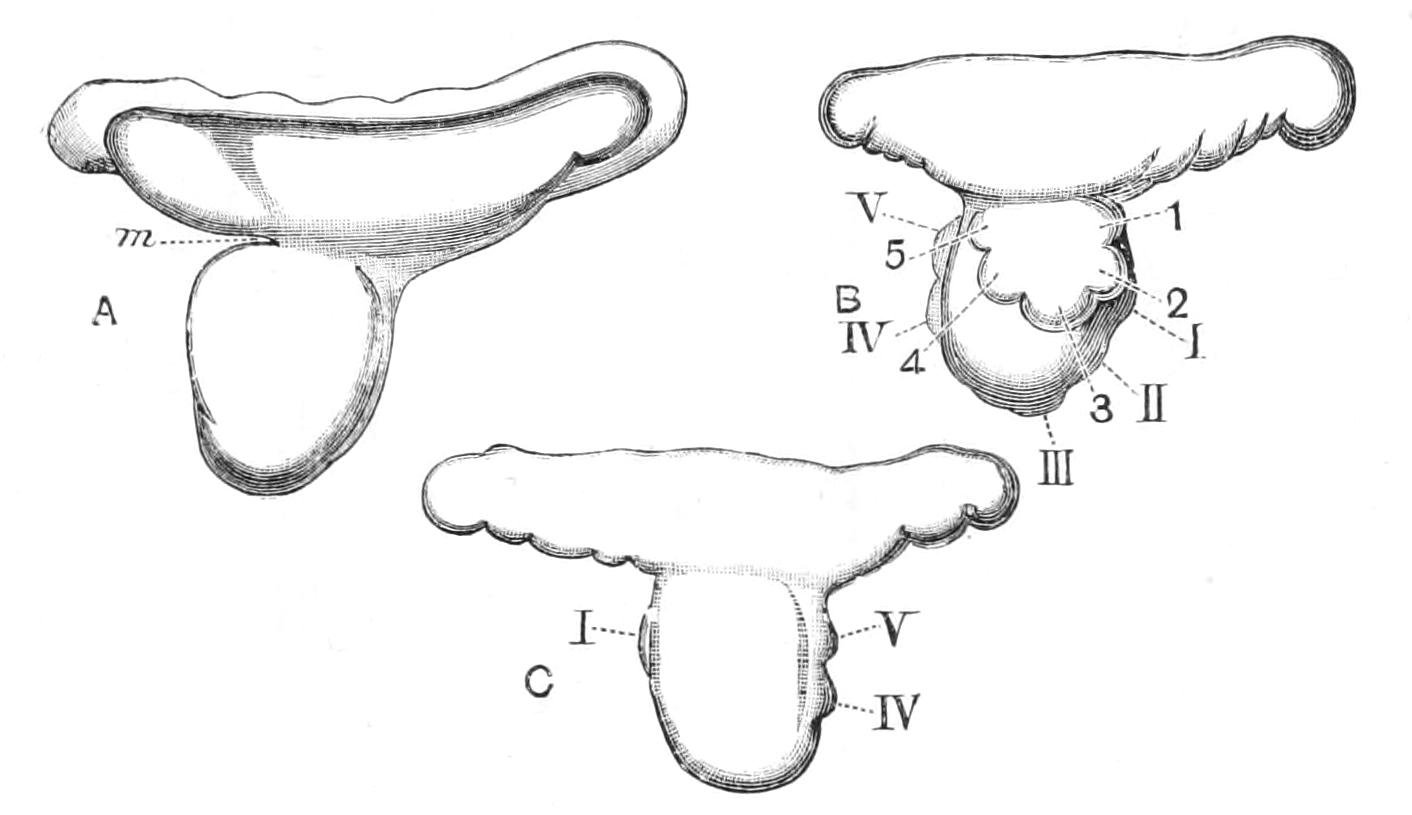
Fig. 288.—Fully grown larval stages of Asterina gibbosa. A, fully grown larva; B, left, and C, right view of a larva seven days old in the beginning of the metamorphosis. m, Mouth; 1-5, the five lobes of the hydrocoel; I.-V., the rudiments of the arms. (After Ludwig.) × 45.
The form of the Starfish is attained principally by the preponderant growth of the left hydrocoel and of the left posterior coelom. Both these sacs take on the form of hoops, which, by the meeting of their ends, are converted into rings. The hydrocoel has already grown out into five lobes, which are the rudiments of the radial water-vascular canals, and the tips of which become the terminal sensory tentacles (Figs. 288, 289, 1-5); but now the left posterior coelom grows out into five lobes also, forming a parallel but outer ring. These lobes (Figs. 288, 289, I.-V.) are the rudiments of the arms, which are at first quite independent of those of the radial canals, but gradually, when the larva has attained the age of nine days (Fig. 289, B, C), they become applied to the outgrowths of the hydrocoel. These by this time have developed each two pairs of branches, the rudiments of the first two pairs of tube-feet in each radius. The larval mouth vanishes, and a new mouth is formed on the left side in the centre of the hydrocoel ring, when the metamorphosis is complete. The adult anus is formed about the same time. The primary pore-canal in Asterina as in all Dipleurulae opens into the anterior coelom; the stone-canal is formed from a ciliated groove running along the neck of communication between this and the hydrocoel. The constriction dividing the body into disc and stalk divides the anterior coelom (single in Asterina as in the older Bipinnaria) into two parts; the portion included in the disc forms the axial sinus of the adult. The lower end of the axial sinus expands and surrounds the adult mouth, forming the inner perihaemal ring; the outer perihaemal ring is formed by the juxtaposition of four wedge-shaped outgrowths from the left posterior coelom and one from the anterior coelom. From these the radial perihaemal canals subsequently grow out into the arms. The metamorphosis of Bipinnaria has been well worked out by Goto,[520] and it agrees in essential features with that of Asterina gibbosa; in fact, the differences which Goto maintains between the two types may be reasonably explained on the supposition of some stages having escaped the notice of this observer. The larva develops on the apex of the prae-oral lobe three papillae for occasional attachment,[521] and in the centre of these a cup-shaped disc for permanent fixation when the prae-oral lobe is converted into a stalk. When these papillae (Fig. 290, fix) have been developed the larva is known as a Brachiolaria.
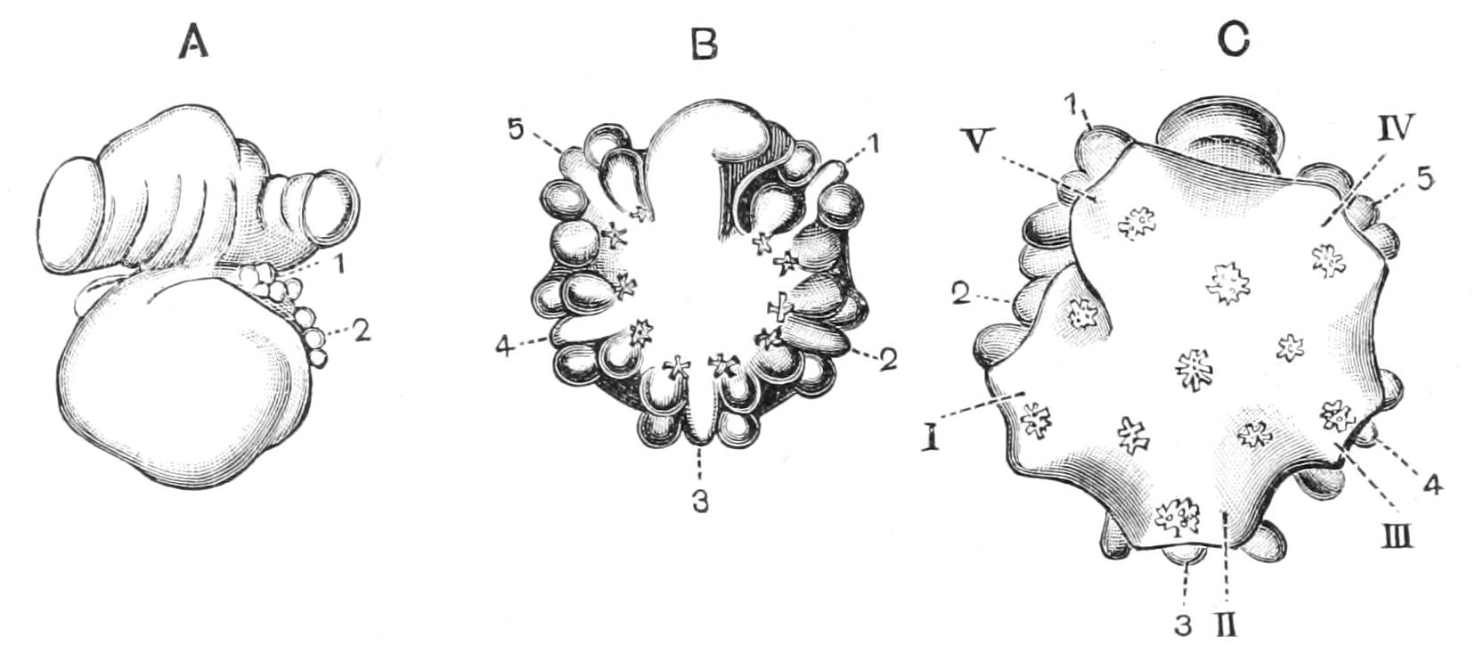
Fig. 289.—Views of larvae of Asterina gibbosa in the course of metamorphosis. A, larva of eight days, from the right; B, left, and C, right view of larva of nine days; 1-5, lobes of hydrocoel; I-V, rudiments of arms. (After Ludwig.) × 45.
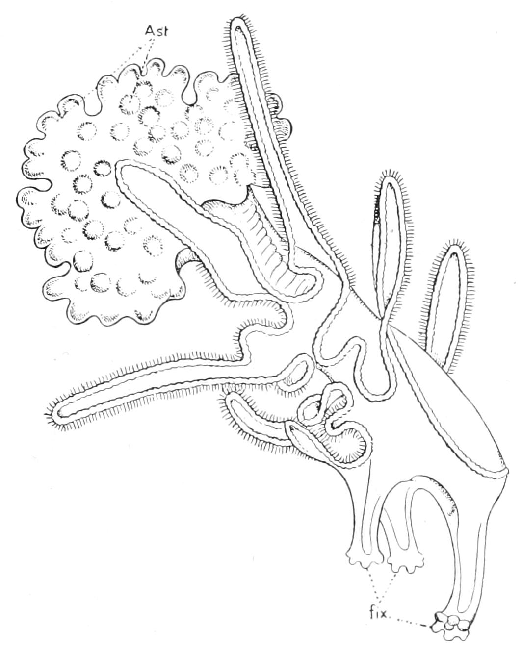
Fig. 290.—Brachiolaria fixing itself, × 60. Ast, rudiment of the body of the Starfish; fix, fixing processes. (After Johannes Müller.)
The metamorphoses of the other types of Dipleurula contain no fixed stage. They are what might be called "cataclysmal metamorphoses." That is to say, the outer form and habits of the larva are preserved till the last moment, whilst the organs of the adult are being gradually perfected; then in an hour or two all trace of larval structures disappears. The Ophiopluteus preserves the larval mouth, round which the hydrocoel grows; the long lateral ciliated processes are preserved till the animal has attained all the adult characters. Before this, however, it passes through what may be called an "Asteroid" stage in development, in which the ambulacral grooves are open. The Echinopluteus loses both larval mouth and anus. It develops the adult organs on the floor of a sac-like invagination of the ectoderm, situated on the left side within a loop of the ciliated band (Fig. 291, B, C). This invagination becomes completely closed. It is termed the "amniotic cavity," and its roof is termed the "amnion." On its floor are developed the primary tentacles, terminating the radial canals, as well as a number of spines. After taking on a creeping life and losing its larval appendages, the young Sea-urchin passes through an "Asteroid" condition, in which the arched dorsal surface, the future periproct, is greater in extent than the ventral, and the radial canals run horizontally out from the water-vascular ring and terminate in free movable podia (Fig. 291, C and D, pod), ending in suckers, in the centre of which are pointed sense-organs. These podia become later enclosed in grooves in the corona, and are reduced to vestiges in the adult.
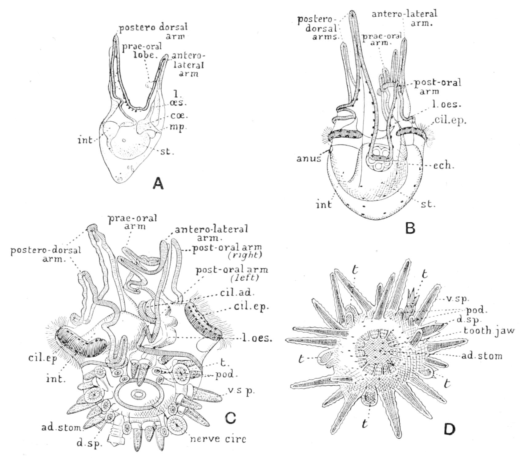
Fig. 291.—Four views of Echinopluteus from the left side, to show the metamorphosis. A, B, and C are taken from the development of Echinus miliaris. D is a young Echinus esculentus. The rudiment of the oral disc of the Echinus is seen beginning in B and larger in C. ad.stom, Adult stomodaeum; cil.ad, adoral ciliated band; cil.ep, ciliated epaulette; coe, coelom; d.sp, prismatic spine of dorsal surface (periproct) of adult; ech, rudiment of Echinus; int, intestine; l.oes, larval oesophagus; mp, madreporic pore; nerve circ, nerve-ring of adult; pod, first paired tube-feet; st, stomach; t, terminal tentacle of the radial band; v.sp, pointed ventral spine of adult. A, B, and D magnified 45 diameters; C, 60 diameters.
The Auricularia is the only type of Dipleurula in which larval mouth and anus are retained. For this reason it has been supposed that its median plane of symmetry remains the median plane of the adult. The researches of Bury[522] have shown that this is not so. As in other types of Dipleurula (with the possible exception of the Ophiopluteus) the adult position of the mouth is on the left side of the larva, and in the commencement of the metamorphosis the mouth migrates into this position (Fig. 286, C). Then the rudimentary prae-oral lobe is rapidly absorbed, so that the mouth again acquires a terminal position. The hydrocoel (Fig. 286, A, hy) has by this time completely encircled the oesophagus, and from it grow out the radial canals which bud off the feelers[523] (buccal tentacles) into the larval stomodaeum. This, although it later flattens out to form the adult peristome, forms in these stages an almost closed sac, reminding us of the amniotic cavity in the Echinopluteus. The ciliated band breaks up into a number of pieces, which rearrange themselves so as to form a series of transverse rings of cilia; so that the free-swimming life can be carried on somewhat longer. The animal in this stage is called a "pupa" (Fig. 292); it eventually loses the rings, drops to the bottom, and develops tube-feet. From specimens which the author has seen, he has little doubt that in some cases the young animal passes through an "Echinoid" stage, for it possesses, besides the feelers, only median tube-feet, terminating the radial canals, and it is covered by a cuirass of plates, which recalls the Echinoid corona.[524]
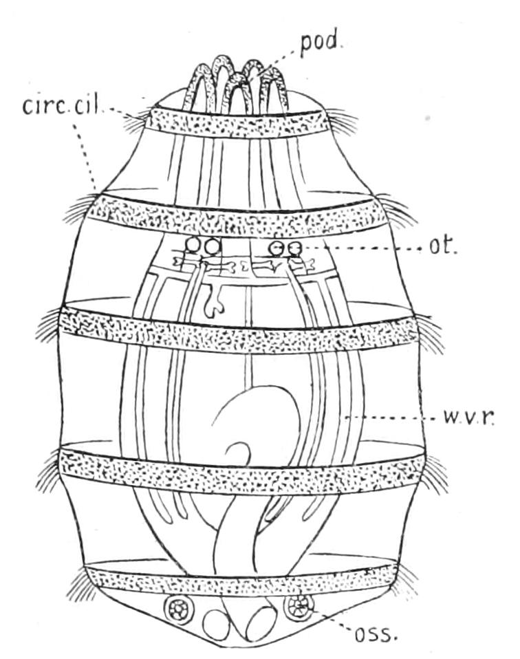
Fig. 292.—"Pupa" of Synapta digitata. × 50. circ.cil, Ciliated rings; oss, calcareous ossicle; ot, otocysts; pod, feelers; w.v.r, radial water-vessel. (After Semon.)
Reviewing the development of the Eleutherozoa in the light of the facts so far presented, and using the same method of reasoning which is employed in the case of other groups of animals, we seem to be justified in concluding that the Echinodermata are descended from a simple free-swimming ancestor possessing the fundamental characters of the Dipleurula. These would include a longitudinal folded band of cilia as the principal organ of locomotion; a thickened plate of nervous epithelium at the anterior end serving as combined sense-organ and brain; a V-shaped band of cilia projecting into the oesophagus as the organ of nutrition; a wide, shallow stomodaeum and an alimentary canal consisting of three well-marked divisions, viz. oesophagus, stomach, and intestine; and finally a secondary body-cavity or coelom, consisting of three divisions on each side, though possibly the most anterior pair were confluent in the prae-oral lobe. On the left side the anterior coelom opened to the exterior by a short ciliated canal. To the hypothetical group so defined which were certainly not Echinodermata the name Protocoelomata may be given.
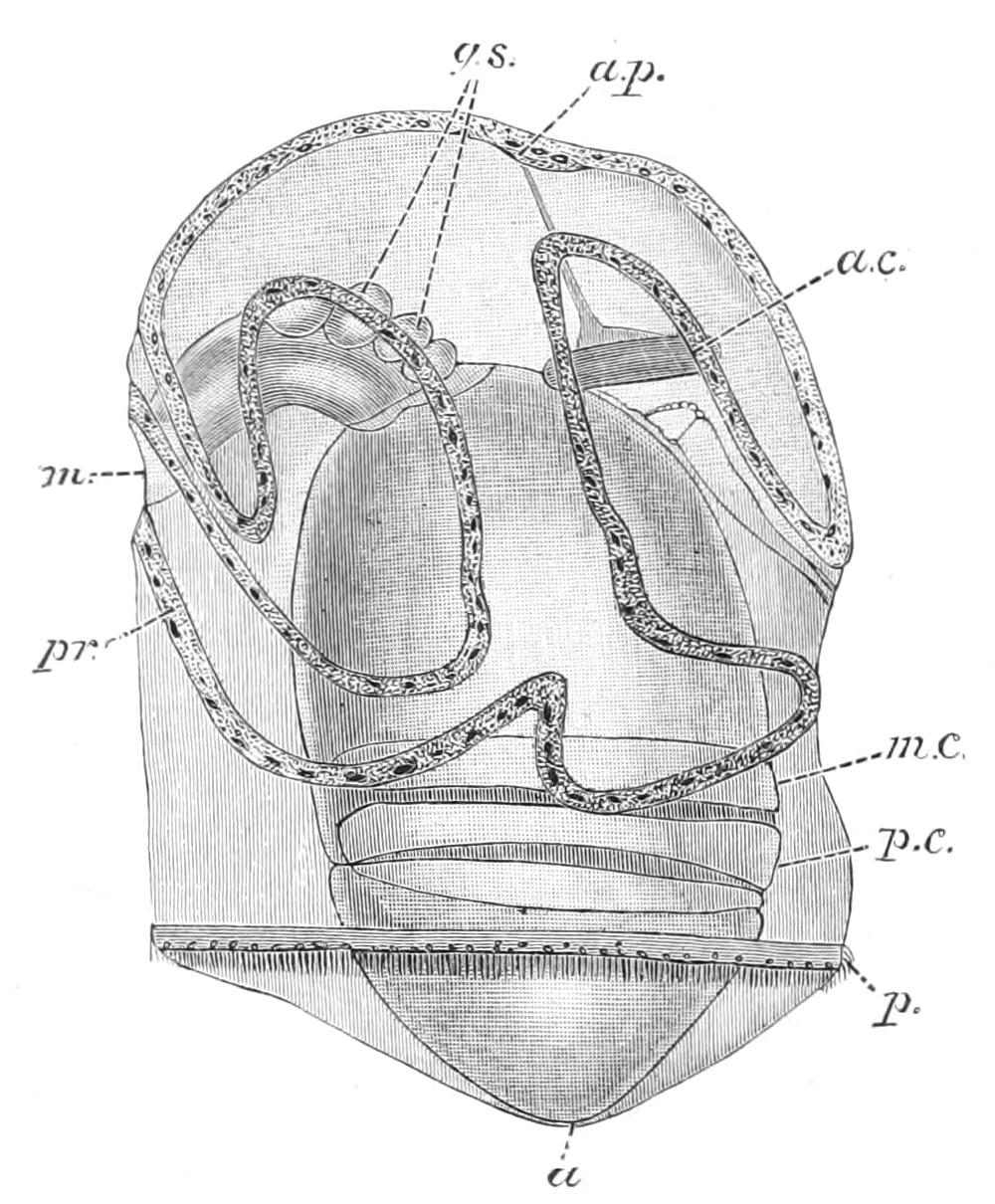
Fig. 293.—Tornaria larva. a, Anus; a.c, anterior coelom; a.p, apical plate; g.s, rudiments of gill-sacs; m, mouth; m.c, middle or "collar" coelom; p, posterior ciliated band; p.c, posterior coelom; pr, longitudinal ciliated band. (After Morgan.)
Now amongst the lowest types of animal in which traces of Vertebrate structure can be detected, there is one group, the Hemichordata (Vol. VII. p. 3), in which there is a larva which strikingly recalls the Dipleurula. This larval form belongs to Balanoglossus and is called the Tornaria. It possesses a well-marked prae-oral lobe and a folded longitudinal ciliated band, which resembles that of Auricularia. Its peculiarity is that in addition there is a posterior ring of cilia (Fig. 293, p). The coelom is in five divisions:—a median anterior sac (a.c) opening to the exterior by a short ciliated canal on the left side; and paired middle divisions (m.c) and posterior divisions (p.c). At the apex of the prae-oral lobe there is a plate consisting of sensory epithelium, with nerve-fibres at its base, which acts as a brain. Tornaria undergoes metamorphosis, assumes a worm-like form, and takes on a burrowing life. The five divisions of the coelom are retained, and it can be proved that the pore-canal, like the madreporite of Echinodermata, is used for taking in water. Further, there are two aberrant sessile members of the group (Cephalodiscus and Rhabdopleura), in which the middle divisions of the coelom which would correspond to the hydrocoels are produced into long arms, each with a double row of ciliated tentacles, which strikingly recall the radial canals and podia of the Pelmatozoa. Taking all these facts into consideration, it seems probable that Vertebrata and Echinodermata both arose from Protocoelomata.
When we turn to the developmental history of Echinodermata for light on the question as to how the bilaterally symmetrical ancestor became converted into the radially symmetrical Echinoderm, it seems probable that only in the development of the Asteroidea can we hope to find the solution of the problem. The abrupt changes of habits shown in the metamorphoses of the other types are clearly secondary phenomena. No species of animal could suddenly change its habits from swimming by means of cilia to walking with tube-feet. In the development, however, of Asterina gibbosa we get a hint of the way in which a free-swimming life could alternate with periods of temporary fixation, gradually passing into a condition in which the fixation was permanent. This period in the history of the race when ancestral Echinodermata were sessile would mark the point at which Eleutherozoa diverged from Pelmatozoa, and the former existence of a fixed ancestor explains the tendency first to asymmetry and later to radial symmetry. Bilateral symmetry is characteristic of most free-swimming animals which have to pursue a straight course through the water, but in fixed forms no disadvantage arises from want of symmetry. A radial disposition of organs is, however, valuable to them, since food must be sought and danger avoided from all points of the compass; and hence we can understand, when fixation became permanent, how one hydrocoel could grow larger than the other, and finally assume the form of a ring.
The last question which arises is the vexed one of the mutual relationships of the various Classes constituting the Phylum. Before attempting to seek for light on this problem from development, it will be necessary to sketch the life-history of Antedon rosacea, the only Pelmatozoon whose development is known.
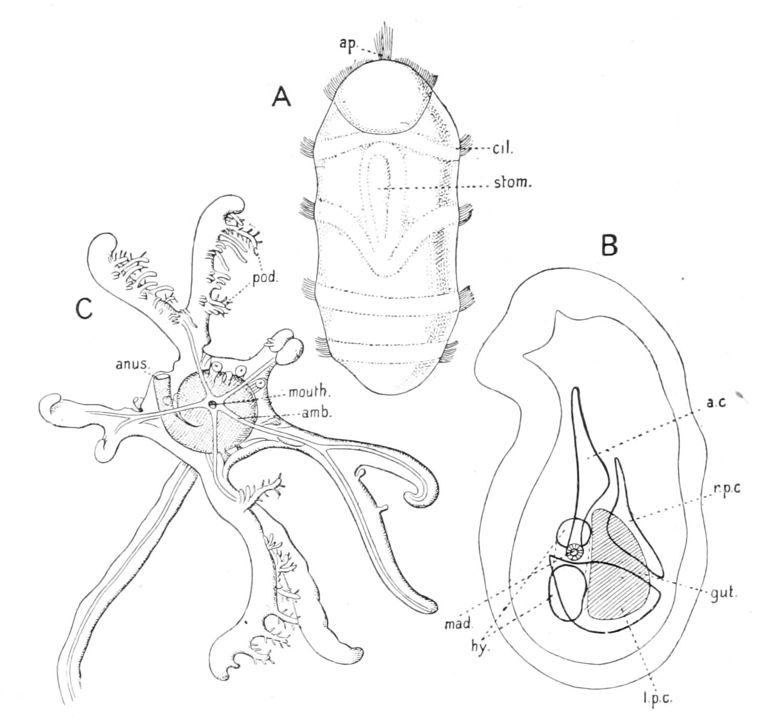
Fig. 294.—Three views of the development of Antedon rosacea. A, free-swimming larva; B, longitudinal section of free-swimming larva; C, oral view of young fixed form. a.c, Anterior coelom; amb, ambulacral groove; ap, apical plate of sensory and nervous tissue; cil, ciliated ring; hy, hydrocoel; l.p.c, left posterior coelom; mad, primary pore-canal; pod, podia; r.p.c, right posterior coelom; stom, larval stomodaeum. (A and B after Bury; C after Perrier.)
The eggs are comparatively large and full of food-yolk, and they adhere for a considerable period to the pinnules. They pass through a large portion of the development within the egg-membrane. The blastula and gastrula are formed in the usual way, but the formation of the coelom is most remarkable (Fig. 287, E1, E2). The archenteron divides into anterior and posterior divisions. The posterior divides into right and left, posterior coelomic sacs, but before the division is complete a dorsal and a ventral tongue grow out from the anterior division and unite posteriorly, encircling the band of connexion between right and left posterior coelomic sacs like a ring. This band of connexion becomes solid and is absorbed, and pari passu the ring becomes converted, by the disappearance of its central opening, into a sac, which is the definitive gut (Fig. 287, E). The rest of the anterior division divides into a thick-walled sac, the hydrocoel, on the left, and a median thin-walled anterior coelom, which sends a long extension into the anterior portion of the larva, which we may compare to the prae-oral lobe of the Bipinnaria. The anterior coelom communicates with the exterior by a short pore-canal, and later forms a connexion, the stone-canal, with the hydrocoel. At the apex of the prae-oral lobe there is formed a thickened patch of ectoderm, bearing stiff sensory hairs, and having at their bases nerve-fibres and ganglion cells. This larval brain corresponds to that of the Tornaria and Echinopluteus. Behind the brain there is a glandular pit, which is used for fixation, and recalls the similar organ in the Bipinnaria. A series of ciliated rings is then formed, and between the second and third of them an oval depression appears. This is the stomodaeum; but as the larva takes no food it does not communicate with the gut (Fig. 295, A, stom).
The larva next escapes from the egg-membrane and swims freely for a day or two, and then, like the Bipinnaria, fixes itself by the apex of the prae-oral lobe, which is converted into a stalk. The larval stomodaeum closes, and the oesophagus of the adult appears as a solid peg of cells abutting against it; round this peg the hydrocoel grows like a ring.
The closed stomodaeum and the underlying hydrocoel are now rotated backwards until they come to be at the end of the animal opposite the stalk (Fig. 295, C). The left posterior coelom, which has also, as in the Asteroid larva, assumed a hoop-like form, is carried along with them; but the right posterior coelom becomes shifted forwards and sends out five outgrowths into the stalk, which form the rudiments of the chambered organ, and a central one as a continuation of the genital stolon (Fig. 295, D, gen.st), the extension of the anterior coelom (Fig. 294, B) having disappeared.
Then the outer wall of the stomodaeum splits into five valves—the future oral valves. The radial canals appear as freely projecting tentacles, which issue in the intervals of these valves and soon acquire two pairs of lateral branches. The skeleton consists of five oral plates in the oral valves, of a ring of five basals, of three small under-basals, and of a series of "columnals," i.e. stem-ossicles, as rings embracing the stalk. The area of attachment is supported by a "foot-plate." The radial plates next appear as a ring of small ossicles between the orals and basals, and simultaneously the arms make their appearance as five outgrowths supported by the first radials, and by the other radials when these appear. The free radial canals now become adherent to the arms, but these canals soon give off paired branches of unlimited growth, which are supported by bifurcations of the primitive arms, and in this way the ten arms of the adult are established. So far, then, as the water-vascular system is concerned, the apparent forking is not a true dichotomy, but results from the production of two opposite branches, whilst the main axis ceases to grow. The appearance of cirri marks the fusion of the uppermost stem-ossicles to form a centro-dorsal, and shortly afterwards the young Antedon snaps off its stem and swims away.
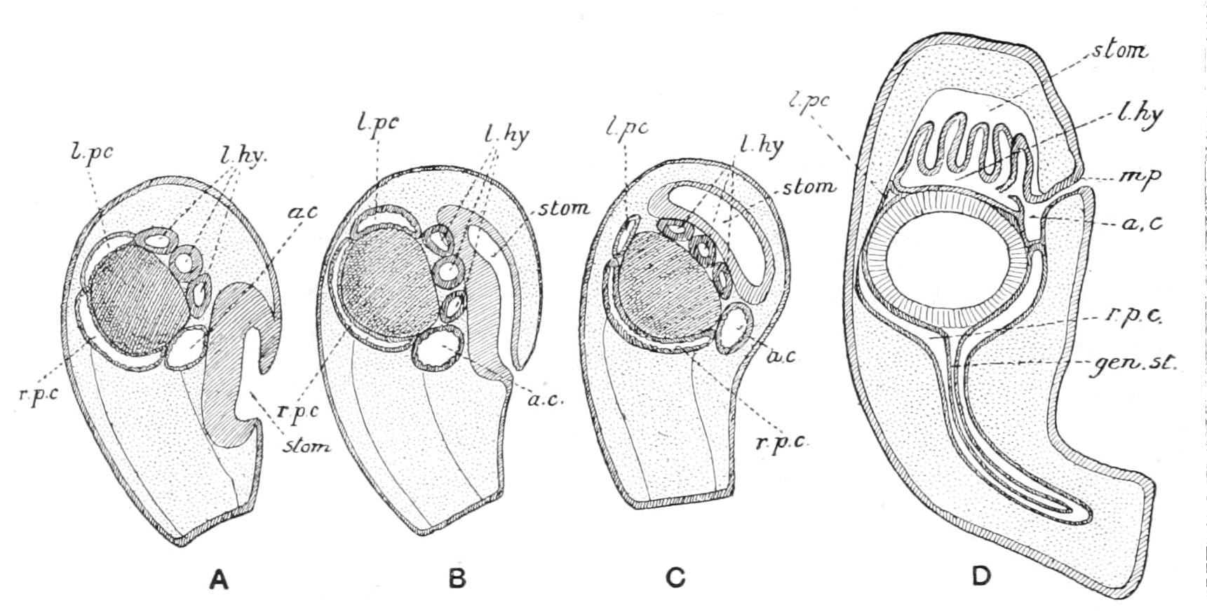
Fig. 295.—Four diagrams to explain the metamorphosis of the larva of Antedon rosacea. a.c, Anterior coelom; gen.st, genital stolon; l.hy, left hydrocoel; l.pc, left posterior coelom; mp, madreporic pore; r.p.c. right posterior coelom; stom, stomodaeum. (A, B, and C after Korschelt and Heider; D after Perrier.)
Now in reviewing this life-history we cannot fail to be struck with resemblances to the development of Asteroidea, and especially to that of Asterina gibbosa. The absence of a connexion between stomodaeum and gut is due to the embryonic mode of life. On the other hand, the presence of a long prae-oral lobe, containing an extension of the anterior coelom and having a fixing organ at its apex, can only be paralleled among Asteroidea. In broad outlines, then, up to the period of fixation the two developments are parallel, but after this point a divergence takes place, which points clearly to the splitting of the Echinoderm stem into two main branches, corresponding with two different sets of habits. In the Eleutherozoan stock, represented by the development of the Asteroidea, the disc became flexed ventrally on the stalk, so that the mouth and podia were brought within reach of material drifting along the bottom, which the podia were employed to seize. As a consequence the base of the stalk was brought near the mouth, and so it came about that the hydrocoel, when it became a ring, encircled both. In the Pelmatozoan stock, on the other hand, the podia and mouth are rotated upwards and backwards from the stalk, which thus came to have an aboral position (Fig. 296, B). The podia are thus placed in a favourable position for capturing free-swimming organisms, which their cilia sweep toward them. It is worthy of note that a similar change of position of the mouth occurs in other groups of animals which have similar habits (Polyzoa Entoprocta, Tunicata).
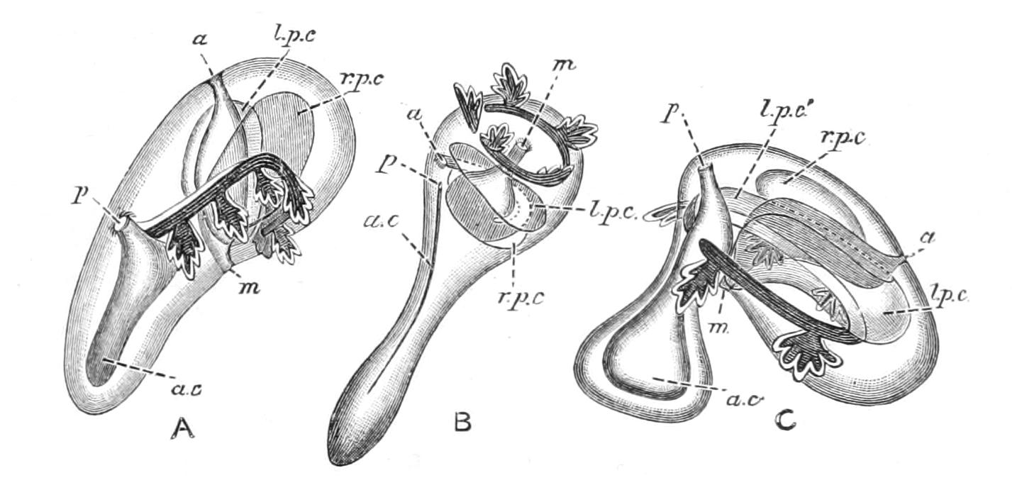
Fig. 296—Figures to show the supposed connexion of Eleutherozoa and Pelmatozoa. A, common fixed ancestor of the two stocks, still bilaterally symmetrical; B, primitive Pelmatozoon; C, primitive Eleutherozoon. a, Anus; a.c, anterior coelom; l.p.c, l.p.c1, left posterior coelom; m, mouth; p, primary pore-canal; r.p.c, right posterior coelom.
The division therefore of the phylum must have occurred at an extremely remote epoch, before the hydrocoel was a closed ring, and before, therefore, radial symmetry was completely attained.
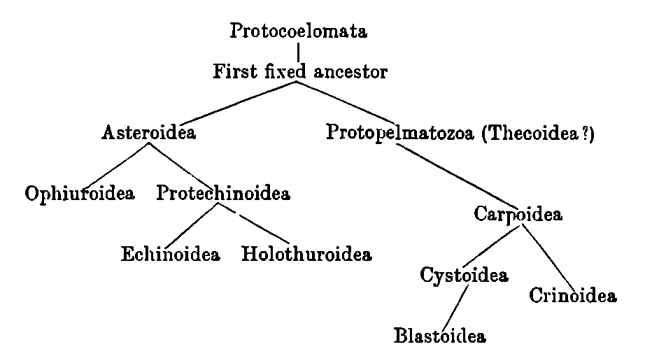
Turning now to the question of the origin of the classes of Eleutherozoa, we find that the study of development strongly reinforces the views gained from the study of adult anatomy. The Asteroidea are the most primitive group; only in their case is the fixed stage retained, and both Ophiuroidea and Echinoidea pass through an Asteroid stage in development. The only serious competitors for the position are the Holothuroidea, which many have imagined to have been directly derived from Cystoidea (in the old sense; better Thecoidea). This view, though adopted by Semon,[525] Haeckel,[526] and Bather,[527] is open to many objections. The type of Holothuroid development referred to in these discussions is that of the extremely aberrant Synapta digitata, in which the radial canals are vestigial structures which disappear in the adult. In this species, where the feelers are multiplied, some originate in the larva directly from the water-vascular ring, and thus alternate with the canals. From this circumstance Semon drew the conclusion that the radial canals of Holothuroidea are not homologous with those of other Echinoderms, but this conclusion is contradicted by the development of more normal species, in which all the feelers spring from the radial canals. The meridional course of these canals, the closure of the ambulacral grooves, coupled with the retention of a nervous ectoderm, are all features found in Echinoidea. So is also a reduction in the number of the genital organs, on which Bell[528] laid such stress that he separated Holothuroidea from all other Echinoderms. But if in Spatangoidea a reduction to four and even three can take place (Fig. 246, p. 552), why should a reduction to two or one excite surprise? The primitive outer appearance of the Auricularia is counterbalanced by the development of the coelom, which is much modified, so that the primitive bilateral arrangement is obscured. If, then, Asteroidea are the most primitive Eleutherozoa, we may imagine that primitive Echinoidea were derived from Asteroidea through adaptation to life in crevices, where an upward bending of the radii was of advantage, in order to enable the animal to attach its podia above as well as below itself; and that Holothuroidea arose from primitive Echinoidea in which the plates of the corona were still movable, through a further adaptation to narrow crevices, where worm-like wriggling would be the most successful method of adapting themselves to their environment.
The final result, then, of all our inquiries leads us to a view of the mutual affinities of the classes of Echinoderms, which may be indicated in the following table:—
We shall hazard the prophecy that if ever pre-Cambrian Echinoderms are found, there will be amongst them small stalked forms which may be superficially classed with "Cystids," but which are in reality the fixed ancestors of Asteroidea. They should have an irregular skeleton, and be devoid of arms, which are secondary formations; but they should indicate, by the proximity of the mouth to the stalk and by the relation to the stalk of the grooves for the podia, that they have diverged from the Pelmatozoan stock, and are the ancestors of Eleutherozoa.