Chapter XXV · X-RAYS IN PODIATRY
Reading edition
Roentgenology is the science that deals with the use of the Roentgen, or X-rays, in all their applications. The subject is divided into three parts, each distinct from the other, and with special required study for each part. The first branch of roentgenology is known as roentgenotherapy or actinotherapy, and comprehends the treatment of disease by the use of the X-rays or by other radiant energy.
Roentgenotherapy, to be intelligently applied by the practitioner, requires a thorough knowledge of pathology and is strictly within the province of the licensed doctor of medicine. The second branch variously styled fluoroscopy, skiascopy, radioscopy or roentgenoscopy, is utilized for examining the various parts of the body by projecting the X-rays through the body and fixating the shadows cast on the fluoroscope. This branch of the science of light is of great assistance in diagnosis and is used extensively in surgery for the location of foreign bodies in the tissues. The third division of roentgenology is called radiography, skiagraphy or roentgenography, and consists of the making of X-ray photographs by passing the ray through the body in front of a photographic plate. This branch is of great importance to the podiatrist because of the value it possesses in the diagnosis of foot lesions, involving the bony structures; it often clears up a doubt as to the true state of a lesion when other means have failed. The soft tissues through which the ray passes readily appear in light shadow, while the bones, because they are solid, cast a darker shadow on the plate.
Experiments with electricity and with the modifications of the various currents of electricity, have been responsible for all of the new discoveries and inventions along these lines, and the same may be said of the X-rays.
The two divisions of electric current are the direct and the alternating, named after the direction in which they flow. The direct current moves in one direction and may be likened to the flow of water from a faucet, while the alternating current does not maintain a steady pressure nor does it flow in the same direction continuously. The alternating current, which is commonly used for lighting purposes, reverses its direction of flow and pressure 120 times per second. It is therefore called 60 cycle current, in that it makes 60 complete cycles per second. The alternating current may be likened to the tide, which rises and falls every twelve hours. Instead of passing from ebb to flood and back again each twelve hours, the alternating current, used for illuminating purposes, does so in one-sixtieth of a second.
The alternating current, is of most value to the sciences, because its voltage or pressure may be easily changed. Thus it can be raised or lowered by passing it through special apparatus. One particular form of alternating current has a voltage of from 30,000 to 120,000 and can be used for the production of Roentgen rays, if it be transformed into a so-called pulsative or unidirectional high tension current. This transformation is easily brought about by modern Roentgen ray apparatus.
The discovery of the X-rays was preceded by an improvement in the knowledge of alternating currents. Many scientists were experimenting and looking for new rays and currents, and after the invention of the air pump and the production of glass globes, from which the air was removed by means of the pump, Geissler invented the process of sealing platinum into glass and produced the Geissler air pump, which is used to the present day. This made it possible to seal electric conductors into vacuum tubes.
Following this, Faraday and Maxwell developed the electro-magnetic theory of light and laid the foundation for a theory of the Roentgen ray, which, although later considerably changed, has proven one of the most useful adjuvants to the science of physics. The period between 1840 and the discovery of the X-rays was an active one in electric experimenting. Many men were interested in this work, and their observations and discoveries were important in the final discovery of the rays. In fact, Roentgen’s discovery was fully expected, inasmuch as several investigators, including Sir William Crookes and Roentgen himself, were convinced that rays existed of which they knew nothing. The earlier of the two workers, Crookes, had probably produced X-rays, but overlooked them and it fell to Roentgen to become aware of the conditions under which they were produced. He called them X-rays or unknown rays, but after a time, they were named after him, and are known in science as Roentgen rays. Roentgen wrote and published three papers on the subject and these have become classics on this topic. All of the facts announced by him at that time continue to be accepted, because, notwithstanding all the work done along these lines, they have never been disproven.
Experiments have been continuous, and gradually the types of generators and tubes have changed, always improving. Where at first only simple work could be accomplished, the most wonderful things in this art are now being done, with probably many more wonderful things still to come. The dangers to which the earlier workers were subjected have been gradually eliminated, and today with proper care, the danger from the X-rays has been reduced to a minor factor. This danger came about from lack of knowledge of what occurred in the tube when the ray was being generated. There are three rays produced, one of which causes destruction of the human tissues, when exposed for a prolonged period of time. By preventing the passage of this ray, by the use of lead and lead glass, as protective mediums, the danger has been practically controlled.
X-RAY APPARATUS
The apparatus used in the generation of the X-rays consists of a generator, a vacuum tube and an appliance for holding the tube in a fixed position, called the tube stand.
The Generator. The generators used in X-ray work are of three types, the motor generator, the interrupterless type and the coil generator with a chemical interrupter.
The function of the Roentgen ray apparatus is to produce high potential electric discharges in one and the same direction, or what is known as a unidirectional current. Dependent upon the kind of current used to start with, distinction can be made between the types of apparatus previously mentioned. These types are operated by either direct or alternating current.
Direct current apparatus requires an interrupting device for the purpose of producing the necessary change of flux of magnetic lines in the inductive part of the apparatus. Interrupting devices, which are used in connection with such apparatus, are of three kinds: first, the hammer interrupter; second, the mercury interrupter; third, the electrolytic interrupter. The function of any one of these interrupters is to break up the continuous flow of direct current into small fragments. Each one of these fragments then produces one impulse of high potentiality, which is then directed through the tube.
The alternating current, as its name implies, is already interrupted, but the impulses are alternately changing direction, passing first in one direction and then in the opposite direction. Hence, if the alternating current is used as a primary source of electric energy, then the secondary or high potential impulses will also be changing their direction alternately. Two methods are used in employing the alternating current as a primary source of energy. The first consists of the suppression of one phase (that which would pass in the negative direction) by means of the so-called rectifier cell. The second consists of producing high potential impulses which flow in alternate directions, and to then redirect these impulses so that all of those passing through the tube are in the one and the same direction.
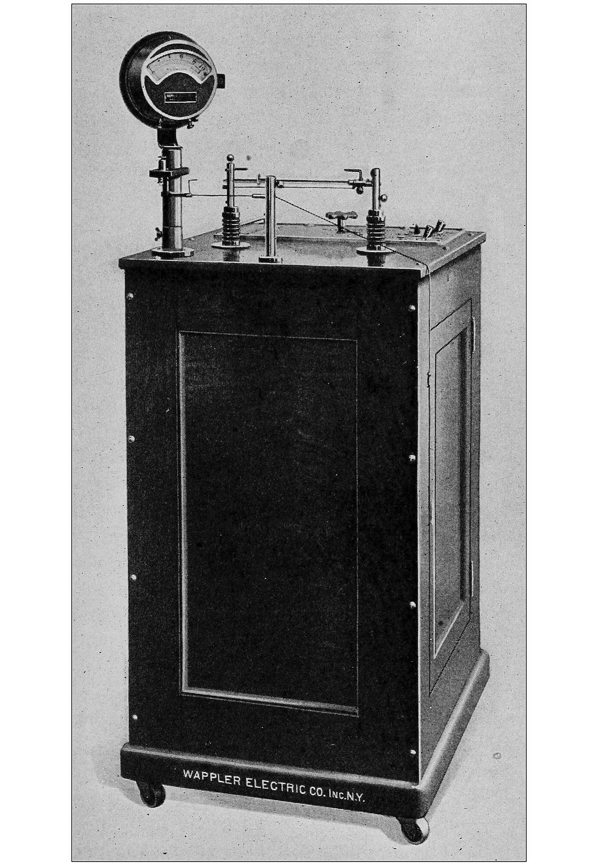
INTERRUPTERLESS TYPE GENERATOR
Accordingly, X-ray apparatus can be divided into the three classes previously mentioned. The coil, consisting of an induction coil activated by direct current, which is broken into small sections by the interrupter; or the induction coil which is energized by the alternating current of which one phase is suppressed through a liquid rectifier cell, and in which the proper interruptions are again produced by some interrupting device. The most extensively used type of apparatus is known as the “interrupterless machine.” This machine operates on either the direct or the alternating current, but, if the direct current is used as a primary source, it is transformed into an alternating current by means of a rotary converter or motor generator set. The alternating current, therefore, either produced or already available, passes into the primary coil of a transformer which changes it into alternating impulses of high potentiality. In order to transform them into impulses, all of which shall pass in the same direction, a so-called rectifying system is employed. This consists of either a disk or of cross-sticks, which are so arranged that they produce contact with both terminals of the transformer in such a way that current of the same polarity, the unidirectional current, is always delivered to the tube. The name “interrupterless machine” merely implies that the devices used for interrupting the primary current have been eliminated.
There is one other type of apparatus which is built on the principle of an electrically oscillating system. Here again a start is made with the alternating current, obtained either from the line or by changing the direct current by means of an interrupter. The current is transformed through a so-called step-up transformer into one of high potentiality which is then still alternating. This current oscillates over a spark gap. Parallel to this spark gap a resonator is connected in which impulses are produced that correspond to the discharges over the gap and which, if applied to a specially built, so-called high frequency Roentgen ray tube, will produce X-rays. Since the principle of electric oscillations of a high frequency is used in this type of apparatus, it is generally called the high frequency Roentgen ray apparatus.
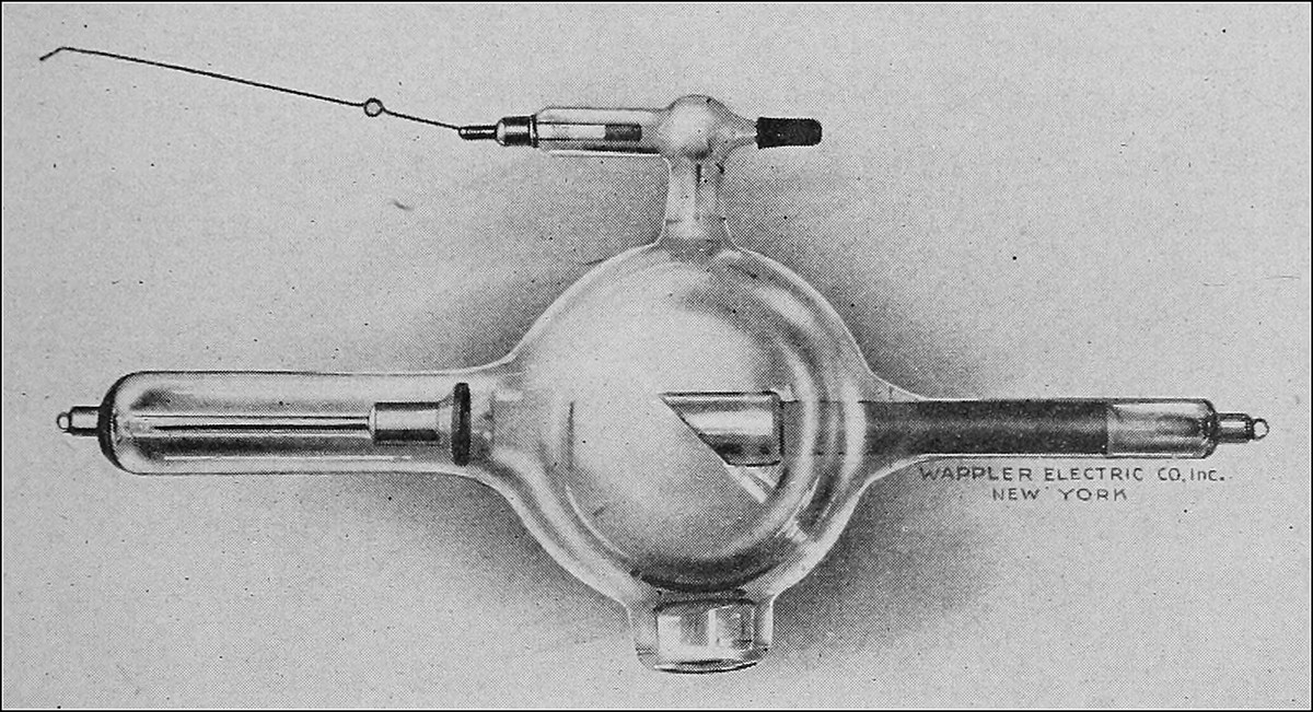
TUNGSTEN TARGET TUBE
Roentgen Ray Tubes. The various types of apparatus previously described require special forms of Roentgen ray tubes. For the coils by virtue of their relatively small capacity, platinum target tubes are generally employed. The capacity of the interrupterless type being considerably higher, the use of a material of greater resistance as a target is necessitated. For this purpose tungsten is generally employed. For the high frequency, or Tesla coils, a tube which embodies a rectifying or valve action device is necessary.
The mechanism of the production of Roentgen rays can be described in the following way: the cathode, or negative terminal of the tube, consists of a hollow spherical surface of aluminum. When this electrode is attached to a negative potential, a stream of negative ions or cathode rays is projected perpendicular to the surface of the electrode. By reason of its curvature, the electrode is therefore focused to produce a converging beam of cathode rays, the area of which is smallest where, in the centre of the tube, the tungsten block or anticathode is placed.
According to whether the area selected for the location of the anticathode is small or large, the possibility to distinguish between fine, medium or broad focused tubes arises. The discharge from the negative electrode takes place, dependent upon conditions which give to the discharge either a high or a low velocity. If the velocity is high, the change from kinetic energy to Roentgen ray energy will be greater than if the velocity of the cathode ray stream is decreased. In the latter case, the radiations will have less penetrating power. The velocity of the discharge must therefore depend upon the magnitude of the charge on the negative electrode, and also upon the number of gas particles present in the tube at that time. If there is a large quantity of gas present, there will be a reduction in the speed of the ray, due to collision and impact with the gas. The penetration of the Roentgen rays depends therefore directly upon the potential produced by the apparatus and inversely on the gas pressure (or directly on the vacuum) of the tube.
For the purpose of controlling the state of vacuum in the tube, a regulating system has been improvised which, when an electric current is passed through it, liberates a certain amount of gas which passes into the tube proper. This is a convenient device for reducing the vacuum of the tube. Up to the present time, no one has succeeded in inventing an efficient method or device for removing gas from the tube, or a means which would help to increase the vacuum. It is for that reason that operators should guard against reducing the tube unduly. It is a simple matter to reduce the vacuum but difficult to increase it. When a tube becomes low (when the gas content is high), resting the tube by allowing it to remain in its bracket for a period of time, will usually suffice to increase the vacuum.
The proper care of the tube is essential for good radiograms. It should be kept free from dust, and before being used the degree of vacuum should be determined by the testing apparatus on the generator. If the tube is dusty or dirty, the passage of the rays through the glass will be hampered, as these particles tend to deflect the rays from their straight course. If the vacuum of the tube is too high, it will affect the quality of the plate, and if it is too low there will be no penetration and therefore a blank plate.
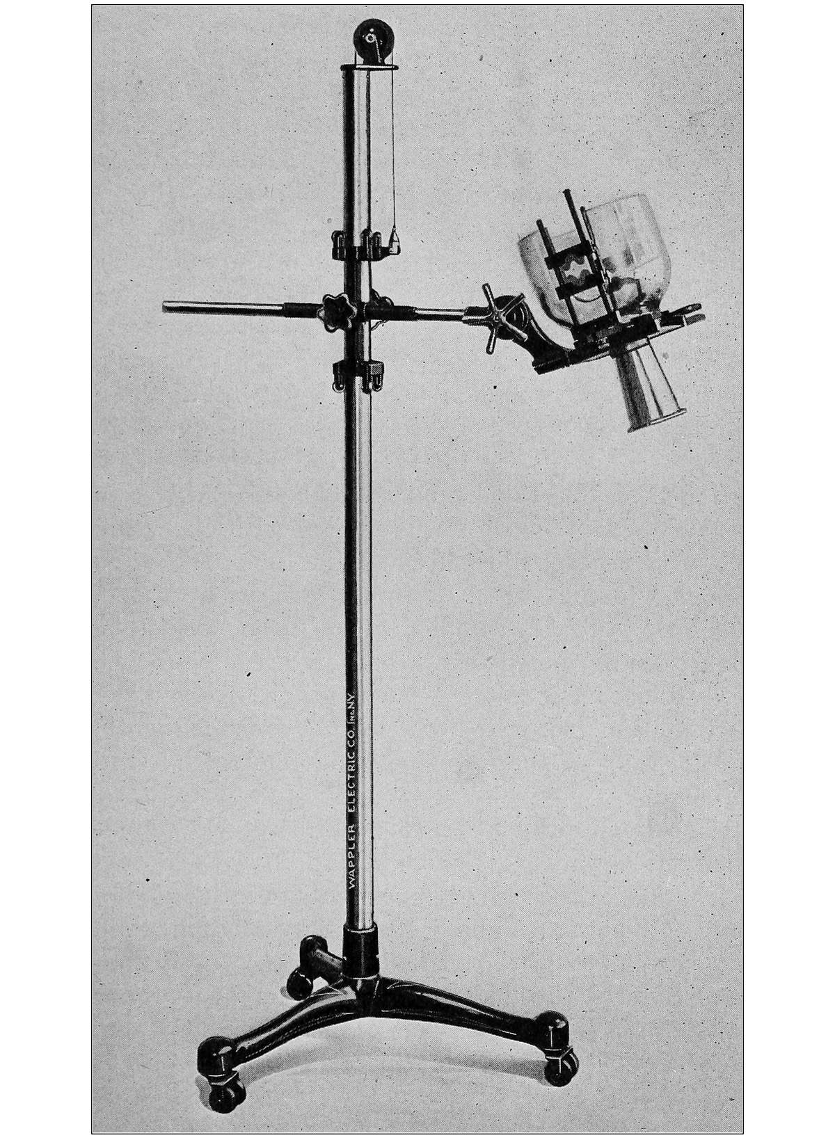
TUBE STAND
The Tube Stand. The tubes used in Roentgenology are delicate structures and great care must be exercised so as to prevent breakage. To guard against such damage it is necessary to securely hold the tube in a proper device while it is being used. The tube stand should be so arranged that it is possible to raise or lower the tube, with its connections, without being compelled to touch the tube itself, to swing it from side to side, to tilt it forward or backward, or in or out. This is best accomplished by the modern tube stand which is so arranged that after having set the tube in its proper place, it is no longer necessary to touch the apparatus in order to secure all of the above named motions. A careful study of the accompanying photograph will make this clear.
The Roentgen or X-rays. The rays generated in the apparatus heretofore described, and named after their discoverer, are of a peculiar character, and although they have proven a boon to mankind, serious trouble and even death has come to those who in their ignorance used the X-rays promiscuously. It has been discovered that there are three distinct rays generated when the high potential current is passed through the vacuum tube and they have been named after the first three letters of the Greek alphabet, namely: the alpha, the beta and the gamma rays. The alpha ray is the ray that is seen in the tube and is of no consequence. The beta and gamma rays are invisible and it is these rays that penetrate the tissues of the body. To the gamma ray is attributed the harmful effects of Roentgen’s discovery. Although the X-ray is used as a therapeutic measure it should only be applied by those who have a thorough knowledge of its properties. Prolonged exposure will cause severe burns, and in some cases continued contact with the ray has caused cancer. Their action in this respect is due to the actinic quality of the gamma rays. The symptoms of a burn do not manifest themselves until a minimum of a week or ten days after exposure and, when they do appear, they are usually severe. Sloughing of the tissues takes place, and the wounds produced do not heal readily and, in consequence, ugly disfiguring scars remain.
Radiography. This branch of Roentgenology is of most interest to the podiatrist. By means of photographic plates, properly exposed and well developed, it is often possible to make diagnoses where other means have failed. It is essential for one who would be correct in diagnosing X-ray plates, to have a thorough knowledge of the structure of the bones of the foot and to understand the meaning of the various shadows cast upon the plate.
When the X-rays penetrate the foot they pass through the tissues, and when they strike a tissue of great density they cannot penetrate it as readily as the rays passing through a tissue of less density. The result is that as the rays pass through the foot they cast a series of shadows on the sensitized photographic plate beneath, the density of which depends upon the tissues through which the rays have passed. So, in reality, the plate when finished is not a photograph, as most laymen imagine, but a shadowgraph or, as it is variously termed, a radiogram or skiagram.
For the purposes of producing radiographs that are clear and easily interpreted, it is necessary to have good materials and to follow the technic of developing and fixing the plate in every detail. The photographic plates used in general photography do not, as a rule, give satisfactory results. Plates, with specially prepared emulsions that are adapted for radiography, are preferred. These plates are sensitized with a gelatinous substance containing bromide of silver. When this substance is exposed to the action of the rays it undergoes chemical decomposition, the degree of which varies with the amount of exposure. It is upon this decomposition of the salt that the art of photography depends and so, too, the science of radiography. The plate is then developed.
Developing of a photographic plate consists of making visible the metallic silver which is produced by the decomposition of the silver bromide. This is accomplished by the use of a mixture of chemicals called the developer. The plate is put into a tray about two inches deep and large enough to allow for its easy removal, and the developing solution is poured over the emulsion. Gradually the exposed portions of the negative will appear and developing must continue until every such exposed part has been brought out in its fullest detail. The unexposed portions are then dissolved by placing the plate in a solution of sodium hyposulphite, sometimes called the fixer or the fixing bath. As its name implies, this solution permanently fixes the exposed silver, and if the plate were not treated in this manner the entire mass would become blackened upon exposure to light.
The preparation of radiographic plates is carried on in a room that is protected from light with the exception of a dull, red glow from a “ruby lamp” which has no effect upon the plate. The plate is placed into an envelope made of black paper, and this in turn is placed, flap down, into a second envelope of red paper. This insures protection from light rays when the plate is brought out of the dark room. After exposure, the negative must be taken back to the dark room before it is removed from the envelope. After it has been developed and fixed it may be brought into the light with safety.
The length of time required in making exposures varies with the different types of apparatus. When the flow of rays is great, it will require a proportionately shorter exposure. So, too, the length of exposure with a certain type of generator varies with the part being exposed. The bones of the fingers would require less time than those of the leg, while the bones of the head would require more time than either of the above for the production of a clear radiograph. It is essential in all branches of radiography to remember that the part to be skiagraphed be parallel to and in close proximity to the plate. This will prevent elongations and foreshortenings of the shadows cast.
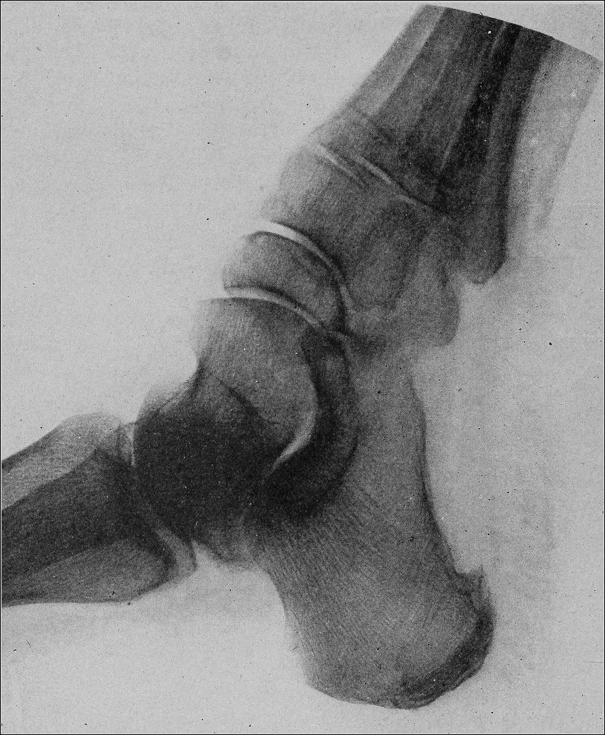
SPUR ON THE UNDER SURFACE OF THE HEEL
DIAGNOSIS OF RADIOGRAPHS
There are several lesions of the foot commonly arising in the practice of the podiatrist, which are easily recognized by radiographic examination but which otherwise are difficult of diagnosis. Some of these cases are shown in the accompanying pictures, which were selected from a large collection at The First Institute of Podiatry of New York City. They are characteristic of the lesions they depict, and it is needless to emphasize their value as an aid to diagnosis. If a condition involves the bony structure of the foot, clinical symptoms are never so certain that an absolutely positive diagnosis can be made, but the X-ray plate readily reveals such disturbances so that there is no doubt left in the minds of the practitioner as to the exact nature of the trouble. Periostitis, exostosis, fractures, arthritis, bone abscesses, bone ulcers, etc., are thus easily distinguished.
Periostitis. Periostitis is an inflammation of the periosteum, the outer covering of the bone. There are two types, the acute and the chronic, both of which are not really diseases themselves, but are indications of the reaction of the periosteum to some irritant. In acute periostitis the X-ray plate shows a slight destruction of the outer portion of the bone, and a slight thickening of the periosteum, and if suppuration is present, the lesion is a mild osteomyelitis rather than a periostitis.
Chronic periostitis causes an increase in the osteogenetic cells of the periosteum and is common in a great many lesions. Trauma, blows or contusions cause a chronic thickening of the bone covering with additional bone formation, as do syphilis and superficial abscesses in the soft tissues, in the immediate vicinity of long bones. Thus chronic ulcer of the leg over the shaft of the tibia will produce this condition. The picture shows that the even line of the bone surface is lost, and there is a rough, uneven edge, with or without an increase in the bone cells. The entire shaft of the bone is often thicker than normal, especially in the metatarsals, and it is quite common to find one of these bones greatly increased in size. The fourth metatarsal is the one most usually affected.
Exostosis. This lesion is common in the foot, and is a source of great pain and annoyance. It is usually the result of a chronic bursitis which has affected the periosteum over a localized area. Due to the destructive changes brought about by the inflammatory processes, the periosteum is absorbed and the bone cells beneath protrude in the form of a spur which shows on the plate. The most common site of exostosis of the foot is on the inferior surface of the os calcis, under the calcaneo bursa. The part is somewhat swollen and is extremely painful when pressure is brought to bear directly over the growth. The heads of the metatarsal bones on their lateral surfaces are occasionally affected, especially the outer side of the head of the first metatarsal. Lateral pressure gives rise to pain in this type of exostosis. The treatment for all exostoses is purely surgical.
Fractures. There are several kinds of fracture, and they are classified variously, but for the purposes of the podiatrist, the following types, with the description of each, will prove sufficient:
Incomplete Fractures, among which are the greenstick and the fissured fractures, are those in which there is not a complete separation of the fragments. The greenstick is really a bending rather than a breaking of the bone, and is found mostly in children under fifteen and then only rarely affects the bones of the leg. Fissured fractures are splits or cracks in the bone which do not separate it into two parts and occur occasionally in the fibula or in the metatarsals. They are easily seen in the radiograph by the dark shadow they produce in the region that would ordinarily appear light. This shadow extends over the entire length of the break.
Complete Fractures are the most common type found in the lower extremity and are divided according to the line and the seat of the breach of continuity. Thus we have transverse, longitudinal, oblique and spiral fractures. The radiograph will reveal the nature of the break, and a dark shadow will be cast between the fragments of bone. The most common of these found in the leg are the oblique and the spiral fractures.
Comminuted Fractures are those in which there is extensive splintering of the bone adjoining the fracture or one of the fragments. This class of fracture does not occur in the foot.
Impacted Fractures are those in which the fragments are driven into each other, forming a wedge, thus preventing abnormal motion, so common in other types. This occurs mostly in the neck of the femur.
Crushing or Compression Fractures are those in which the bones are crushed. The spongy portion and the cortical layer are both involved and in some cases the bones may be pulpified. The tarsal bones are subject to this type of fractures as the result of falls from heights, upon the soles of the feet. (See accompanying picture of fracture of the os calcis.)
A simple fracture is one in which a wound of the skin is absent, or if present, there is no connection between it and the broken bone.
If the bone is broken in two or more places or if two or more non-adjacent bones are simultaneously broken, the condition is called a multiple fracture.
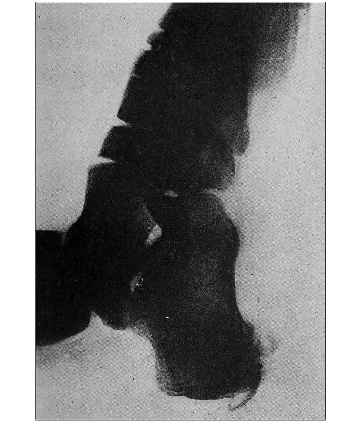
FRACTURE OF THE OS CALCIS
A compound fracture is one in which the fragments of bone pierce the soft tissues and protrude beyond the skin.
It is essential in examining a patient who has suffered from a recent fracture, to obtain a history of the case as well as a description of the accident. An examination of the part should be made, and the various symptoms such as deformity, abnormal mobility and crepitus should be noted. The X-ray picture is then taken and if a fracture is found, the case should be put in the hands of a competent surgeon for immediate and proper treatment.
Sesamoid Bones. The sesamoid bones which are found in the flexor tendons under the head of the first metatarsal bones, are subjected to injury in those who follow vocations in which the ball of the foot is put to great strains. Among such may be mentioned dancers and acrobats. The normal position of these bones is directly beneath the head of the first metatarsal bone, and when this part is put to a great strain, the bones may become fractured, or they may become displaced. The outer sesamoid is usually forced outward and the X-ray picture shows it in the first interosseous space. The inner sesamoid is not affected, but may also be forced outward, and is then found under the outer side of the first metatarsal, in the normal position of the outer sesamoid bone.
Fracture of these bones is not unusual and is a result of a severe injury. The line of division is shown by a dark shadow, much the same as in other fractures. One or both of the bones may be involved. It is essential to remember that if the bone is broken, the opposite sides will fit into each other perfectly, whereas, in cases of a freakish nature, in which there are four sesamoid bones, this will not be the case.
Arthritis. Arthritis, or inflammation of the joints, may involve any one or all of the structures which make up joints, viz., bone, cartilage, ligaments, synovial membrane and fibro-cartilage. The acute forms of inflammation are not detected by the radiogram, but the chronic type leaves its characteristic marks which, when present, are easily seen. The synovial covering of the ends of the bones is destroyed, and shows a rough, uneven surface. In cases of long standing, there is complete bony ankylosis, and the shadows of the joint line are completely obliterated. This is particularly true in the tarsal joints, in which the joint lines between the bones can hardly be seen. There is no motion in such joints, and in milder cases, in which no union has occurred, the sensation of crepitus is conveyed to the hand if the joints are moved passively. Tubercular arthritis shows a rarification of the bone with a thickening of the periosteum.
Arteriosclerosis, or hardening of the arteries, is often detected by means of the X-ray picture. Light shadows cast in the normal dark shade produced by the soft tissues, which appear over the course of the arteries, are indications of this condition.
This disease of the arteries causes a change in the vessels whereby there are calcareous salts deposited in the middle coat. These salts, containing the metal calcium, are not readily penetrated by the ray, and thus a difference between the artery and other soft tissues is established.
There are many other conditions in which the X-rays are a valuable aid in making diagnosis, but these are of no interest to the podiatrist, being within the exclusive province of the physician and surgeon. The reader is referred to books which deal with the subject of Roentgenology exclusively, for further information upon the subject.
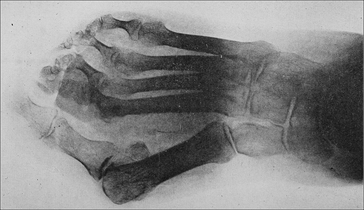
HALLUX VALGUS