Chapter XXIV · MISCELLANEOUS FOOT LESIONS
Reading edition
TRENCH FOOT
A foot lesion has arisen during the present war, which, because of the fact that it appears on the feet of those who have been subjected to long sieges of service in the trenches, has been called “Trench Foot.”
The condition has been and is being investigated by many medical men of note, and although the literature available has been rather meagre, several facts have been established and some of the data has been classified.
This chapter has been compiled from various papers upon the subject written by those who are now serving their respective countries in France, and who have had experience in dealing with the lesion. Articles by the following writers have been used, and all of the statements contained therein have been verified: B. Sherwood Dunn, M.D., of Paris, in The Medical Record; “Anonymous,” in the same publication; H. Oswald Smith, in The Lennox, a journal devoted to dentistry, and several articles in the Journal of the American Medical Association.
Trench foot is a lesion found in the lower extremity and is a result of exposure to cold and dampness in the trenches. It has been likened to frost bite, but cases reported during the summer months show that the parallel is not justified. There are several stages to the disease, and they are classified by Smith into four groups: (1) Neuritic—producing acute pain and preventing the patient from walking or sleeping. There is no swelling or discoloration of the foot. (2) Edematous—without discoloration, but acute pain is present, produced by the pressure on the nerve endings. (3) Edematous—with blisters and varying discoloration of the skin, short of gangrene. (4) Gangrenous—partial or circumscribed, with edema and blisters and reddening of the skin involving the lower leg.
Etiology. The lesion is found chiefly among men of from twenty to thirty years of age who have been in continuous service in the trenches for a minimum of three days. That it is truly the result of trench life is proven by the fact that men in the artillery, who do not see trench service, are not thus afflicted.
The constriction of the foot in ill-fitting shoes and stockings with lack of cleanliness are also etiologic factors. Cold is not accepted as the cause of trench foot, as the trouble occurs in weather above freezing and some cases have developed in the summer time. Neuritis, produced by humidity, is the cause of the lancinating pain.
Raymond and Parisot have stated that the disease is caused by bacterial invasion. They have isolated the microbe, from the mud of the trenches, and have reproduced in animals the various symptoms manifested in trench foot. The microbe was found in the purulent layer of the vesicles and the injection of these germs into the epidermis of the rabbit and guinea pig caused the same lesion as is found in man. They have reached the conclusion that the disease is similar to mycetoma, the fungus foot of Madura and misnamed by English surgeons “tuberculosis foot.”
The disease is prevalent among those standing in the soft, slushy mud or in the muddy water of the trench. The skin of the feet becomes soft and macerated, and while in such condition offers easy ingress for the microorganism, especially along the nail grooves or through abrasions caused by shoe friction.
A committee of United States army surgeons in France, headed by Major R. P. Strong, are making extensive tests in order to ascertain the cause of trench fever. Their unfinished report (they are still investigating) shows as follows: (a) the organism causing trench fever is present in the plasma of the blood; (b) the organism is not filtrable; (c) the disease is transmitted naturally by the body louse (pediculis corporis); (d) this method is apparently the important and common means of the transmission of the disease[7].
Symptomatology. The symptoms of trench foot are always the same. The ball of the great and second toe are swollen and edematous, the skin is distended and glossy and there are occasional blisters or vesicles. The edema may extend to the remaining toes and to the ball of the foot, and, from being white, may become rose-colored or even red and violet. In grave cases the liquid in the vesicles changes from citron color to a hemorrhagic hue, the skin becomes blue-black, then livid and gangrenous. Some of these vesicles may dry and the scab fall off, leaving no scar; the base of the vesicle may change in color from brown to black, and this change may extend beyond the borders and cover the entire area affected by the edema. This change is the forerunner of gangrene; the vesicle becomes a crust; when it falls off it leaves a putrid base which may gradually eat into the tendons and articulations and periosteum. It is not infrequent to have the first crust followed by a second and third, and when the disease has progressed to this stage, no medication seems to avail and the member has to be amputated at a healthy point beyond the parts affected.
There is little if any fever accompanying the lighter forms, but a temperature of 104 degrees F. may attend the graver forms, with general disturbances of the nerve trunks. Albuminuria is not infrequent.
The patient complains of lancinating pains which interrupt sleep and cause difficult locomotion. Walking is accomplished on the heels, with the toes elevated. The pain is excited at several points, chiefly by pressure on the heads of the metatarsals. In the lighter forms, the patient complains of numbness (but only in the affected members) which at times extends to a condition of anesthesia. The mildest form is characterized simply by pain and paresthesia.
The trouble appears, as a rule, after the patient has spent his assigned period in the trenches and manifests itself when he has retired to the sector for repose and removes his shoes for the first time in several days.
In the very worst cases there is gangrene accompanied by all of the dangers of infection, by destruction of tissue and loss of limbs. There is a tendency to contracture of the foot, usually described as a turning under of the foot.
Prognosis. Trench foot has a duration of from two to three weeks, in the lighter forms, and from six weeks to three months in the severe cases. The simpler lesions respond well to treatment, and in the type in which vesicles have formed, these latter dry and fall off, leaving no scar. A cure is effected in about one month. In more advanced types, in which extensive ulceration is present, the toes alone are lost and the rest of the tissues are saved under proper treatment. When general septicemia develops, and according to statistics of two thousand cases treated from October, 1916, to January, 1917, there have been only two such cases, the patients have immediately succumbed.
Complications are frequent accompaniments of the malady—abscess, neuritis, lymphangitis, etc. Tetanus has been observed with sufficient frequency to cause all patients to be injected with antitetanic serum upon admission for treatment.
Treatment. The prophylactic measures that have been adopted since trench foot has been more thoroughly understood, have eliminated it to a great extent, and the disability that it caused at that time is gradually being controlled. The perfection of general sanitary methods and the construction of better trenches have helped to diminish the incidence of the condition as well as to reduce disease and disability in general. The smaller sectors and the greater reserve of men, allowing for more frequent change, have of themselves, without the actual preventive methods instituted, reduced both the severity and the occurrence of trench foot. Where previously the men had to stay in the trenches for long periods, at present there is a change every few days.
As a general preventive measure against the lesion, it was found that the wearing of looser boots, perhaps a size too large, was of great help. This measure prevents the restriction of the circulation, which is so large a factor in the causation of trench foot. The general circulation of the body can be maintained by warmth and by appropriate movements and exercises. Socks must be changed very frequently and must be kept dry. Torn socks are especially liable to constrict the blood vessels and to produce injury to the foot. Foot rags, well paraffined, have been tried, with more or less success, to obviate the difficulty of keeping socks whole. If these foot rags are well applied, they are found to be far more comfortable than socks. Puttees must be loosely applied. Boots must be thoroughly greased before being put on. Rubber boots have been extensively used and are looked upon with favor. Thorough greasing of the feet was tried at the beginning, but was found to be offensive to the men, and consequently was not conscientiously carried out.
The newest and apparently most successful method of preventing trench foot is by means of the so-called “trench-foot washhouses.” Immediately before returning to the trenches the men go to these washhouses for treatment. Here they have their feet soaked in warm, not hot, water and washed with a special soap composed of soft potash soap 1000 parts, powdered camphor 25 parts and borate of soda 100 parts. The feet are then carefully dried and treated by the regimental surgeon or podiatrist, and are finally dusted with a mixture of powdered camphor, talc and borate of soda. In the trenches, the soldiers must be served with hot food in order to maintain warmth and keep up the general circulation. No amount of attention to the feet of the soldier can be too painstaking as regards cleanliness, shoeing and physical condition. To escape disability as the result of actual war violence and to acquire it as the result of preventable sanitary conditions, would be indefensible.
The treatment of the lesion itself, depends upon the severity and the extent of the condition.
1. Simple Edematous Form. In the beginning, the erroneous diagnosis, and the subsequent treatment directed along the lines of frost bite, and the taking of potassium iodide, salversan-methylene blue, and the application of tincture of iodine and copper sulphate, did more harm than good in some cases, and the results, at best, were irregular. The statistics of Raymond and Parisot, who have treated more than 2000 cases in a short time, are the best compiled, and as their results have been excellent they are here given.
At the earliest moment possible the feet of the patient should be thoroughly soaked in warm water and washed with liberal quantities of the soap, previously mentioned, and composed of soft soap, camphor and sodium borate. The cleansing should be thorough, but with care not to break the skin or to open any existing vesicles. The skin must be dried carefully with a soft towel and the following applied: cover a layer of absorbent cotton of the thickness of the hand with gauze; soak this in a solution of camphor, 1.1; borate of soda, 15, boiled water, 1000, and apply while still thoroughly wet (being careful to cover the ends of the toes) to all the edematous surface as far up the leg as it extends. Cover the whole with oiled silk or rubber sheeting, and fit by adhesive strips. If a roller bandage is used, it must be applied loosely. The dressing should be damp when changed the following day.
Renew this dressing daily, until the edema disappears, which should be from the second to the sixth day; after this, continue the same dressing for two or three days without the impermeable covering; following this, camphorated oil is to be applied without rubbing.
When the disease proves obstinate and is accompanied by persistent pain, some relief is afforded by bathing the feet at the time of the daily dressing in warm water, using the same liquid soap as at first.
2. Vesicular Form. The small vesicles, not larger than a ten cent piece, should be left undisturbed and will dry up without scar with the above-described treatment. When they are extensive, and especially when the contents are hemorrhagic, they should be completely denuded and the gelatinous base should be carefully removed by aid of a sterile tampon, and then these denuded surfaces should be covered with compresses soaked in a solution of camphor, 30, ether, 1000. These compresses and the whole affected surface are covered by the fomentation previously described, and should be changed daily.
When the edema subsides, omit the fomentations and continue the application of camphorated ether solution, and as the denuded surfaces show improvement, dust them with camphor powder or with boric acid powder.
3. The Formation of Crusts. The foregoing treatment will ordinarily prevent the formation of crusts or scabs; when these do form, steps must be at once taken for their removal without undue force or surgical aid, as they cover the most virulent germ collections, which are liable to produce a general infection if permitted to contaminate freshly cut surfaces. On the other hand, if permitted to remain, the germs thus protected have a tendency to burrow profoundly, involving the tendons and articulations.
The crusts must be softened and carefully removed from their borders daily, little by little, by aid of the forceps, the operator being careful to cause no bleeding. To soften the crusts, the following are recommended: (1) liberal dressings soaked in camphorated ether and covered with oiled silk or rubber sheeting; (2) collodial silver dressings; (3) camphorated oil and borated vaseline. As the crusts are gradually removed each day, the uncovered surfaces should be treated with the camphorated ether.
Not infrequently the crusts will re-form, in which event it is well to treat them with pomade of Reclus, made as follows: vaseline, 200; boric acid, 3; iodoform, 1; antipyrine, 5; salol, 3; carbolic acid (crystals), 1; bichloride of mercury, 0.1.
4. Extensive Invasion. Where the processes have progressed and involve a considerable amount of tissue, evidenced by a congested, violet-red surface, but with little or no erosion or vesicles, we recognize the presence of the malady in its worst form, and prompt measures must be taken to save the member. In these cases the germs have penetrated beyond the surfaces, and in order that they can be subjected to contact treatment and destroyed, we must reach the hidden centres of their activity.
After surgical preparation of the surfaces, they are incised by a sharp pointed bistoury in the long axis of the foot and down to the level of the infection, indicated by a purulent layer of fetid edema. If a collection of pus is found, a free and extensive incision is made and the tissues thus exposed are thoroughly washed out with an antiseptic solution, by aid of a syringe. The thermocautery should not be applied, as the results of its use in such cases have been very bad.
When the infected parts are opened they are treated with the previously mentioned camphorated ether, or with collodial silver dressings or, in the presence of purulent infiltration, with collodial gold, which can be varied by the following: eucalyptol, balsam of Peru, guaiacol, gomenol, camphor, of each 10, in ether, 1,000. Under the influence of these dressings, the hardened infiltrated surfaces soften and can be removed little by little by the aid of the forceps, in measure as the sloughing continues, until the destroyed and contaminated tissues are eliminated.
In so far as possible, surgical intervention should be avoided; where the bone is attacked, it is better to permit it to be eliminated by the progressive destructive action of the disease. Tentative or conservative operations upon the bone have been followed by extensive destructive osteitis. If an amputation becomes necessary, it should be made at a healthy point beyond the infected area.
5. Reparation Stage. When the necrosis has been arrested and the granulations show that the affected parts are disinfected, the usual emollient applications can be employed to aid the repair; but here, a treatment brought into use by Rathery and Bauzil is recommended. This forms an impermeable covering easily applied and changed, and possesses self-evident advantages. It is composed of naptholate of soda, 2; essence of thyme, essence of origanum, essence of geranium, of each 3; vaseline 1,000; paraffin (45°-50°), 5,000. Melt and thoroughly mix by aid of heat and put aside in pots holding 125 grams each; sterilize for twenty minutes at 120°.
At the time of the daily change of dressings, place the number of pots necessary in a bath of boiling water to melt the wax, cover the affected parts with a single layer of sterile gauze and paint thoroughly with the melted wax. This dressing hardens as fast as it is applied. Apply a second and third layer in the same manner, to form a perfectly occlusive dressing. Cover with cotton and bandage. Dressing comes off easily and completely without pain, and gives remarkable results in healing and repair and renewal of the skin to the parts.
6. General Treatment. In the cases complicated by septicemia, subcutaneous injections of camphorated oil in doses of from 5 to 15 c.c. per day, or intramuscular injections of collodial camphor in doses of 2 c.c. per day, are advisable.
7. Complications. The most frequent is tetanus. Every patient should at once be injected with antitetanic serum, and this should be repeated every eight days during the period of ulceration and sloughing. It is even prudent to augment the second injection in those gravely afflicted (20 c.c. of the Pasteur Institute serum, or 3000 units of the American).
Gaseous gangrene is infrequent with the treatment here indicated. If present, it can be treated in the usual way.
Smith has employed subcutaneous injections of oxygen in cases of this condition which have been neuritic, or edematous without discoloration, edematous with blisters, gangrenous, partial or circumscribed. The technic of the oxygen treatment is as follows: a Woulfe bottle is required, with two glass tubes bent at an angle and inserted into a tightly-fitting cork. To the end of one glass tube is fitted three yards of red rubber tubing connected with the usual oxygen cylinder; to the end of the other glass tube is fitted two yards of a finer rubber tube, and the distal end of the tube has fitted on it a salvarsan needle. The bottle contains a saturated solution of sodium carbonate. The needle should be sterilized in boiling oil for each individual case. The operator, having sterilized his hands, uses iodine on the proposed sites of the puncture. The needle is inserted into the subcutaneous tissue at a point midway between the heel and the external malleolus. The oxygen is allowed to enter slowly until the foot is filled up. The needle is then withdrawn and inserted midway between the internal malleolus and the heel. If the toes are black and cold the needle is inserted in the mid line at the base of the toes. It is important that the oxygen should be injected slowly. If the part is almost gangrenous, injection into the deeper tissues is of advantage.
The treatment is based on the conclusion that trench foot is akin to Raynaud’s disease. The edema produces stasis in the veins of the foot, impeding or stopping circulation, and the venous blood, if oxygenated, will help to keep the tissues alive until the serum can be drained away, while the oxygen helps to drive out the serum, slowly but steadily, through the puncture holes. Mere puncturing has been found useless in relieving the edema. The oxygen balloons the subcutaneous tissue, thus relieving pressure on the blood vessels and lymphatics. When the oxygen is absorbed and the serum drained away, circulation is quickly restored to the limb.
There is marked relief to pain at once, and under the eye of the observer certain changes can be seen taking place. The reddish blue appearance changes to pink; the redness which often extends up the leg disappears, and where blackened areas exist, a white line makes its appearance and any living tissue is noticeable by its pink color. The white line indicates what would ultimately be the line of demarcation of gangrene, and it is noted that healthy granulations start from this white line after injections of oxygen. The destruction of parts is greatly lessened, and is often confined to the tips of the toes. Areas of black blood on the dorsum of the foot rapidly undergo change, becoming grayish white and pink, and more nearly resembling healthy tissue. In many of the severe cases, recovery of the whole foot has taken place, and in some the loss of but a single digit has had to be recorded.
A minor but important point is the treatment of all blisters. They should be drained by sterilized thread passed through by means of a straight surgical needle and the ends cut short. The dead skin should be left in situ as a protective, unless pus is present.
The oxygen causes an increase in the pulse of the posterior tibial artery. There is no rise in temperature, pain is relieved, and sleep promoted. Lint, wrung out in 1 per cent. solution of picric acid, is applied to the parts and renewed every day. No cotton wool should be employed or disastrous results will ensue. The lint and bedclothes next day will be found saturated with serum, sometimes clear, and sometimes blood-stained. The skin will have a peculiar wrinkled appearance, pointing to the previous state of edema. The foot is warm even at the toes, and movement will have returned. At the end of the second day tingling sensations arise. In cases which are semi-gangrenous and where the edema still persists, a second injection may be given. One injection is usually found to be sufficient, but picric acid should be used once or twice a day to keep the part dry and sweet. The effect of the oxygen on the deep layers of the true skin is notable; a rich red velvet color persists for several days.
Smith advocates conservative treatment in all cases of trench foot unless gas gangrene or grave toxemia be present. Repair is slow but sure and many hopeless looking cases have recovered with useful limbs and minor loss of structure. The cost of the oxygen treatment is small, and oxygen is usually available. There is no risk to the patient if this treatment be intelligently used, but healthy tissue, as far as possible, should be utilized for injection purposes. The length of time for repair and recovery of the part is long in the semi-gangrenous and in the gangrenous cases. The granulation tissue is often indolent with the epithelial margin heaped up, and here, again, oxygen has been found to stimulate epithelial growth, a hypodermic needle being utilized in place of the salvarsan needle.
The conclusion drawn is, that if the edema can be relieved by withdrawing the serum, the circulation can be re-established, and during this withdrawal the tissues are supplied with oxygen to keep them nourished.
GROUND ITCH OR UNCINARIAL DERMATITIS
Prof. Paul Luttinger in addressing the students of his class at The First Institute of Podiatry gave the following outline of this disease: “this condition is known as water itch, water pox, water sores, sore feet of coolies, panighao, mazamorro (Porto Rico), tunnel-digger’s sore foot. It is an erythematous or papulo-vesicular irritation of the feet, due to infection with the larvae of the hookworm. These latter gain entrance through the skin of the lower extremities and cause the very grave tropical disease known as uncinariasis (hookworm), or tropical chlorosis.
“The eruption, which is very itchy, appears first as reddish spots between the toes and on the dorsum of the foot. These spots become macules and later vesiculate. In some cases blebs may be formed which, upon rupturing become raw, oozing surfaces accompanied by considerable local swelling. Pustulation and ulceration may result if these surfaces become infected with pus producing microorganisms.”
Treatment. Under proper treatment, this condition will subside within a few weeks; otherwise it may take months or until the systemic disease develops. Sometimes gangrene of the part may result. Cleanliness, combined with mild antiseptic lotions, is all that is necessary to cure the disease in its early phase. A three per cent. alcoholic solution of salicylic acid has been highly recommended by Barlow, who opens the blebs and applies pledgets of cotton dipped in this solution twice daily for five minutes.
The proper disposal of excreta in rural communities and the avoidance of going barefoot in the warm and rainy season, are the best methods of prevention.
GAS INFECTION AND GAS GANGRENE
Gas infection is a very common condition found in the wounded in the present war. It is controlled successfully if it is seen and treated early in its course, but when it has progressively developed it causes a most dangerous condition called gas gangrene.
Etiology. The infecting agent is called the bacillus of Welch, the bacillus aërogenes capsulatus or the bacillus perfringens. The bacillus is of the anærobic type, and being lodged in deep wounds with ragged tissues, recesses and pockets, and completely obstructed by fragments of broken-down tissues, all of which prevents the air from reaching them, they multiply rapidly with the formation of gas followed by a great destruction of tissue.
The bacillus of Welch grows rapidly and it is on that account that immediate and radical action is essential if loss of life is to be prevented. Kenneth Taylor has obtained a pure culture in a very short time as follows: a series of six or more culture tubes are inoculated, each tube from its predecessor, at intervals of only half an hour. Even in this short period bubbles of gas became evident in the successive tubes. By the time the sixth or the seventh tube is reached, one may obtain a pure culture, so far has the gas bacillus outgrown the other germs.
Symptoms. The rapidity of the development of the bacteria is responsible for early symptoms of the infection, and Bowlby has observed well-marked infection with the formation of gas within five hours, and death of an entire limb has occurred from gas gangrene in sixteen hours.
The course of the disease varies somewhat, being at first, a local condition; bubbles of gas appear in the discharges from the wound, and crackling from gas may be felt in the tissues immediately surrounding. Prompt and radical treatment will prevent further development and will restrict the lesion to a limited area. Certain muscles will be lost and with them the corresponding motions, leaving the patient crippled to that extent, but beyond this, recovery will follow.
A more serious and fatal form, is the diffuse or rapidly spreading process. The skin is discolored, the limb is extremely swollen and edematous, gas penetrates the cellular tissues and advances rapidly, so pressing upon the muscles as to render them useless, and obstructing vessels and nerves; the pulse is small and rapid, the extremities are cold; vomiting and hiccough may occur. The patient is profoundly toxic but he may not feel very ill. If improvement cannot be secured by thorough exposure of all pockets and excision, death ensues, the gas spreading rapidly upward to the abdomen, chest, and then to the neck, causing distressing pressure and finally loss of life.
In other cases the whole limb is involved suddenly, beginning within a few days of the initial injury. A wound which is doing fairly well at night may reveal a condition of gangrene—swollen, tense, and discolored in the morning; the patient, already in collapse, succumbs before the gas itself has apparently spread far enough to cause death.
Treatment. The paramount importance of the earliest possible treatment during the first stage of rapidly spreading infection, before the production of gas in any serious quantity has occurred, is self-evident. Every hour counts against the patient.
Taylor points out clearly what is to be done:
1. Destruction of the bacillus.
2. Removal of the tissues especially favoring its growth, which are the necrotic muscles.
3. Measures to prevent the destruction of the muscles as a result of mechanical pressure.
For the destruction of the bacilli, Taylor recommends a one per cent. solution of chlorhydrate of quinine. Others have found Dakin’s fluid effective.
All foreign bodies (clothing, etc.) in the wound, must be removed as they will keep up the anærobic infection; all dead tissue must be removed, the wounds kept open, and frequent antiseptic dressings used. Dakin’s fluid or Taylor’s quinine chlorhydrate, when properly used in connection with the above absolutely necessary means, will enable the surgeon to conquer the infection at the start if he sees the patient as early as possible—certainly within the first twenty-four hours.
The muscles should be opened by numerous longitudinal incisions, incisions of the muscular sheaths, and the excision of all necrosed tissue. Sometimes single muscles or a group of muscles may need to be excised. The dead muscle can be distinguished from the living by its dirty brick-red color, in contrast to the normal purple-brown. The dead muscles also lose their contractility. The focus of infection, if known, should be excised. The wound should be dressed with the chosen antiseptic solution. The incisions should be kept open by light gauze compresses, wet with this solution. No circular bandages which can exert the least compression, and so hinder the escape of the gas, are allowable. Nothing should obstruct the free escape of the gas. Everything should be done to promote it.
If gas gangrene occurs or has already set in, the same free incision should be made, unless this has already been done.
Bacteriologic diagnosis in the early stage is most important. Soon the discoloration of the skin, blebs, and crepitation make the diagnosis positive, but crepitation often appears late rather than early. The X-rays may disclose the bubbles of gas in the tissues. On incision, if the muscular tissue is bloodless, pale, dry, of a brick-red color, gangrene already exists. The best judgment then will be required to decide whether free excision of this gangrenous tissue, with suitable subsequent dressing, or immediate amputation should be done. If the limb is amputated, it should be by the so-called “guillotine” method, i.e., without flaps. The wound should be dressed with the end of the stump entirely uncovered until the infection has been conquered. Then the skin may be drawn down by lacing or by weights, and sutured as soon as feasible. The bone may have to be shortened.
Antitoxin Treatment. One of the most important contributions to surgery as a result of the war has recently appeared in the form of a paper by Carrol G. Bull and Miss Ida Pritchett, of the Rockefeller Institute. This paper describes a lengthy series of experiments with the bacilli Welchii, which were cultured from wounds caused by the war. A very powerful, soluble toxin, produced by the bacilli has been found, which has killed animals almost instantly. The most important result of the experiments is that they give promise of an antitoxin which may be as potent in the prevention of gas gangrene and gas infection as other antitoxins have been effective in their respective spheres. Among these are antitetanic, antityphoid and other antitoxins.
(The above material has largely been taken from “The Treatment of War Wounds,” by Dr. W. W. Keen, and from “Surgical Nursing in War,” by Dr. Elizabeth R. Bundy).
SYPHILIS
Although syphilis is a disease that is usually considered as of a systemic nature and manifests itself in every part of the body, the skin is a very common place for its appearance (syphiloderma), and the foot is often involved. It then becomes the function of the podiatrist to diagnose the lesion, and as this necessitates a knowledge of the general characteristics of the disease, the following should prove of interest:
Derivation. From the Greek sus and philos, “a companion of swine.”
Synonyms. Syphilis cutanea, lues, syphilis of the skin.
Definition. Syphilis is a chronic, specific, contagious, sometimes hereditary disease, caused by a germ, the spirochaeta pallida, involving the skin and nervous system, but capable of affecting any organ or tissue. Inoculation occurs usually at some part of the genitalia, the first evidence of the disease being the initial lesion or “chancre,” but inoculation may occur at any other part of the body forming the so-called “extra-genital chancre.”
Etiology. Syphilis may be hereditary, i.e., transmitted by the parent. In the majority of adults it is acquired directly from an existing chancre or other lesion, the spirochaeta pallida being conveyed directly from one to another. Extra-genital chancres are caused by kissing, by towels, by drinking cups, or by infected instruments, bites, etc. The extra-genital chancre may occur on the mucous membrane or at the site of any skin abrasion. It is essential that infected persons be informed of the danger of transmitting it to others. The contagious period exists to the end of the second year and any secretion, from the possible presence of mucous patches, may be infective.
Pathology. Syphilitic deposits are new growths and consist of round cell infiltration, especially about the vessels, generally endothelial proliferation, and in the tubercular and some other lesions, a variable number of giant cells. The initial changes are noted in the upper part of the corium. The rete, the corium, and, in the deeper lesions, the subcutaneous tissues, are involved, the retrogressive steps being by involution, through fatty degeneration and absorption, or by necrosis and ulceration.
All the various syphilitic lesions are structurally the same, amounting to an endarteritis of a special kind, slowly obliterating and tending to the production of hypertrophy of the tissues about it. The coppery ham-color in syphilitic lesions is due to blood coloring matter from the extravasated red blood corpuscles and to the sluggish nature of the inflammation. These exudation processes are found for months after the process has healed clinically.
Diagnosis. Syphilis usually runs a mild course, but occasionally it is malignant. In some instances its cutaneous symptoms resemble other skin lesions, and it is on that account that diagnosis is often difficult. The general points to be observed are the distribution, color, form, course and duration.
Distribution. The secondary lesions are more or less general and symmetric in distribution. The lesions vary in duration and may show a preference for certain locations, such as the upper part of the forehead, anus, palms and soles. The late secondary lesions, and particularly those of the tertiary period, are usually confined to one region, are grouped, and are not bilateral in distribution.
Color. Syphilodermata are dull coppery-red or ham-color. Exposure to cold air often makes the eruptions on the body more distinct. Color alone cannot be depended upon to make certain a diagnosis.
Form. The earliest lesions are round or oval, showing no tendency to grouping. In the late secondary, and in the tertiary stages, grouping occurs and the lesions may be serpiginous and circinate. This is an important point in diagnosis.
Early ulcers are superficial and the scars are insignificant. Later ulcers are deeper, and the scars, often pigmented, are sometimes diagnostic.
There are many and various forms of syphilis, all of which have characteristic symptoms, but for the podiatrist, who usually sees the later lesions, and who cannot make an examination of the entire body, it will be necessary to recognize those that appear on the foot only.
The nails are occasionally involved in syphilis. Onychia and paronychia are found in the active secondary stage of the disease. The inflammation starts in the matrix or in the nail folds, followed by nutritive disturbances, and subsequent thickening, friability and opacity, furrows and depressions. The nail is lifted up, as a rule, and if there is much ulceration, the nail falls off. Such lost nails are generally replaced by new nails which are ill-formed at first, or they may remain deformed permanently. The skin surrounding the nail is swollen and infiltrated. In infants, the ends of the toes become club-like (syphilitic dactylitis). This condition is rarely seen in adults. The pain is not severe unless accompanied by further pyogenic infection or by mechanical disturbance.
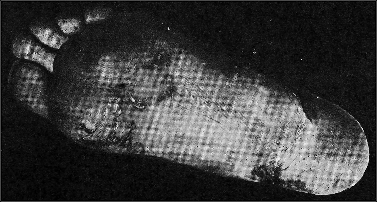
PLANTAR SYPHILID
Plantar Syphilid. The plantar surface of the foot is a common site for dry syphilids. The palm of the hand, and the sole of the foot are the most common locations for the papulosquamous lesions of the late secondary stages of the disease. The lesions are not so elevated and their edges are not so well defined, papules looking more like macules; infiltration, however, is distinct. The shape may be irregular and the usual ham-color does not appear on the surface on account of the scaliness or dry heaped-up epidermis. There is usually a central brownish, gray, callous-like thickening, surrounded by a partly visible band of brownish-red, underlying, papular infiltration. The color is disclosed by removal of the scale. Surrounding the lesion is an encircling edge of partially detached epidermis with its loose, ragged edge directed toward the centre. This semi-detached edge is of extreme value in the diagnosis of plantar syphilids.
The plantar lesions come on slowly and spread gradually. They are usually limited to one sole, but may be found on both feet. There is no itching, but they may be painful if fissures are present. If the plantar lesions are a part of a generalized eruption of the second stage of the disease, they yield readily to treatment, but if they are a recurrence of a generalized eruption, they are more obstinate. Occurring as a late manifestation, they may be extremely rebellious.
Papulosquamous syphilis is the most common lesion of syphilis found on the foot, and is most apt to be confused with eczema. The latter often occurs on the soles of the feet; the following table will show the differential diagnostic points:
PAPULOSQUAMOUS SYPHILODERM ECZEMA
1. History of syphilis. 1. History of previous outbreaks.
2. Concomitant signs present. 2. No associated signs.
3. Favorite seats, palms and soles. 3. Favorite seats, flexor surfaces.
4. Itching usually absent. 4. Itching present.
5. Edge of lesion surrounded by detached scales. 5. Scales completely detached.
6. Scales scanty and dirty-grayish. 6. Scales abundant, and granular.
7. Infiltrated, dull red papules beneath the scales. 7. Flat, reddish patches beneath the scales.
Eczema can also be differentiated by the more inflammatory aspect, the involvement of the toes and toe-ends, by its appearance on the dorsum of the foot as well as on the plantar surface, by the itching, and by the presence of inflammatory exudation. There is no attempt at configuration in eczema, while this is characteristic of syphilis. In addition, some of the elemental lesions of eczema will usually be found around one of a doubtful nature.
Blood Tests. Testing the blood is practised to a great extent for determining the presence of syphilis, but this method of diagnosis is not absolute. The Wassermann complement fixation test and the later modification by Noguchi are of value when they are positive. A negative reaction is no absolute proof of the absence of the disease. They are positive during the active secondary stages, but in the first week or two they are usually negative. In the late stages, when the bacteria may be encapsulated in a lesion, the reaction may be negative. This may be made positive by the administration of potassium iodide, which causes the germs to enter the blood or lymph stream. One should be able to make a diagnosis without the aid of the blood tests in cases of syphilis in which there are skin lesions.
The luetin test, a vaccine test, is of most value in the late stage of the disease.
Course and Duration. Secondary lesions appear rapidly and are fully developed at the end of two weeks, and in time disappear spontaneously. Palmar and plantar lesions are apt to be more persistent. There is little tendency to spontaneous disappearance of the tertiary eruption.
Treatment. The treatment of syphilis is entirely out of the domain of the podiatrist, and when such a case is discovered, it should be turned over to the dermatologist or to the general medical practitioner at once. Plantar syphilids should be treated with soap and warm water so as to remove the scales and thickened epidermis, and this may be followed by an application of salicylic acid ointment, 5 to 10%. Thereafter ointment containing mercury, the white precipitate (ammoniated mercury) preferred, should be used twice a day.
Constitutional treatment is absolutely essential if the disease is to be cured, and this should be vigorous during the first few months after inoculation. The general health must be maintained and nutritious, plain diet, rest, moderate exercise and abstinence from alcohol and tobacco must be urged upon the patient. With all of the above precautions, accompanied by the proper use of remedies, recovery is usually prompt and the symptoms are limited.
Mercury is the one drug that is used almost exclusively. It is very dependable, and should be employed freely, up to the limit of tolerance, during the active stages of the disease. In the tertiary stage, potassium or sodium iodide, in addition to the mercury, is administered. These drugs are used in various forms and in various doses, all of which are of no interest to the practitioner of podiatry.
Recently many practitioners of medicine have been using a complex compound of arsenic, viz., arsphenamine, (salvarsan). This drug is injected into the muscles or the veins. Although many have taken up the use of salvarsan and like chemicals, most practitioners still rely upon mercury. The newer preparations are used more as adjuvants than as specifics. Lesions of the mucous membranes disappear rapidly under treatments with these drugs.
Formerly it was difficult to tell when a case of syphilis had been cured; however, with the advent of the various blood tests, this has become a less doubtful matter. When the various reactions of these tests are negative in uncomplicated cases, toward the end of the second year, it is safe to say that the disease is cured.
(The above is largely compiled from the lectures of Dr. Andrew H. Montgomery, Prof. of Dermatology at The First Institute of Podiatry).
FOCAL INFECTION
Focal infection is the name given to a pathologic condition in the human body, which manifests itself in a part remote from the original focus of infection, or from the original lesion.
Up to a few years ago, focal infections were not recognized by even the most advanced members of the medical profession, and many thousands of people have suffered and died, due to the fact that lesions of this type remained undiagnosed.
Thanks to the efforts of Dr. M. L. Rhein, of New York City, and Prof. Gies, of Columbia University, who drew the attention of the scientific world to the intimate relationship between tooth infections and indefinite ailments in the body, including those of the feet, a large quantity of material has been accumulated on this subject, which throws an interesting light upon it.
It has now been firmly established that the teeth, the tonsils, the adenoids, the male and female urethra, the uterus and the gall bladder, in fact, any part of the body which serves as a portal of entrance to any infection, may become the reservoir of a chronic pathologic condition, and cause disease in any other part of the body. So the teeth, harboring certain microorganisms, have been shown to be directly responsible for heart, stomach and kidney lesions, as well as arthritis and other joint and bone diseases.
Mode of Infection. The bacteria responsible for focal infections thrive best upon dead or necrotic tissue. These bacteria, when they locate in a place where such necrotic tissue is available, such as a tooth in which some of the dead pulp remains, or in a diseased tonsil or urethra, develop so that they throw off toxins or poisons without causing any visible signs of inflammation. These toxins circulate in the blood stream, and locate in various parts of the body, especially in those which offer the least resistance to the invasion.
Relationship Between the Foot and Focal Infection. The most common lesion of the foot due to focal infection, is arthritis or inflammation of the joints. This is due to infection of the teeth, tonsils, nose or adenoids with the common microorganisms such as the staphylococci, streptococci, influenza bacilli, etc., that infest the cavity of the mouth. It may also be due to an infection of the valves of the heart with the streptococcus viridans, or of the gall bladder with typhoid bacilli, or of the genital organs with the gonococcus. In some instances, arthritis of the foot may be traced to an auto-intoxication of the intestinal tract or of the bladder by the colon bacillus.
Osteomyelitis, or inflammation of the bone marrow, periostitis or inflammation of the bone covering, and less often, osteitis or inflammation of the bone proper, may be caused by the typhoid bacillus or its toxin, which originally manifests itself in the intestinal tract. These diseases may develop during an acute attack of typhoid, but usually appear after the acute symptoms have subsided.
Gonorrheal Heel is a well defined type of focal infection due to original invasion of the urethral tract by the gonococcus. The microorganism, or its toxin, reaches the inferior surface of the os calcis, at the point where the flexor brevis digitorum muscle arises, lodges there and causes a chronic inflammation of the periosteum and the bursa. The process is a mild and slow one, and gradually the periosteum is absorbed. With their covering removed, the bone cells increase in number, causing the formation of a spur or exostosis. Walking upon this growth causes the characteristic pain referred to as “painful heel.”
Painful Feet in Women. Dr. Henry Frauenthal, of New York City, has recorded a large number of cases of painful heel in women, in which the foot manifests no signs that would warrant such pains. Investigation has shown that these women were suffering with leucorrhea. Such cases often come to the podiatrist’s office, where they may be mistreated for flat and weak foot by means of mechanical appliances which do no good and often cause additional pain. Discreet questioning will reveal the fact that the patient is a sufferer from leucorrhea, and she should be sent to a physician for treatment.
Treatment. Focal infections of all types require treatment at the initial source of infection and therefore are out of the domain of the podiatrist. These cases should be referred to the physician or dentist, as the circumstances warrant. Attempts at local treatment will prove futile, and bring the podiatrist into bad repute. His duty ceases when he has recognized such an infection and sends his patient to the proper person for professional care.
It is a well known fact that the removal of an infected tooth or of a diseased tonsil has often given immediate relief to one suffering from foot pains, due to focal infection. It is equally well known that heel pains, due to gonorrheal causes, have disappeared contemporaneously with the cure of an old venereal lesion. So fully satisfied are the authorities in charge of the Clinics of The First Institute of Podiatry of the utility of such a procedure, that means are now being devised whereby there shall be in nightly attendance, physicians and dentists, to whom are to be referred all cases of foot pains, in which, by exclusion, a diagnosis has been reached that focal infection may be the cause of the same.
MORTON’S TOE AND METATARSALGIA
Morton’s toe and metatarsalgia are conditions, so common in the practice of the podiatrist that it is deemed best to discuss them briefly in this volume, although they will be treated most exhaustively in “Podiatry Orthopedics,” the next volume of this series.
Morton’s Toe or Morton’s Neuralgia. When the foot is kept in a narrow shoe, the strain placed upon the forefoot is so great, that the fifth metatarsal bone is forced upward and the fourth one downward, and the latter bone is made to act as the pillar of the arch. In consequence, a severe pain is produced, caused by pressure upon one of the plantar nerves, between the head of the bone and the skin. This condition is called Morton’s toe, or Morton’s neuralgia, after Morton, of Philadelphia, who was the first to call attention to it.
Treatment. The pain induced by Morton’s toe, as stated, is caused by direct pinching of a branch of the external plantar nerve, and it is therefore necessary to relieve the pressure on the head of the bone which causes this condition. This is accomplished by the application of a felt pad about one inch long, three-fourths of an inch wide and three-eighths of an inch thick, properly skived and fastened by some adhesive substance, reinforced with adhesive plaster strips, well behind the head of the affected bone. This will raise the head of the bone so that pressure on the nerve will cease, thus relieving the pain. It is essential to remember that the pad must be placed behind the head of the bone, for if it is put too far forward, increased suffering will result. If the case is one in which it becomes necessary to use a pressure of this kind for any length of time, the felt should be discarded, and a plate of metal or some other stable substance should be substituted, with an elevation at the point at which the pressure is desired.
In addition to the device for raising the arch to its normal position, exercises and massage should be prescribed. The exercises should be those which will strengthen the flexor muscles of the toes. Attempting to pick up a pencil or other cylindrical object with the toes, if practised daily for ten or fifteen minutes will ultimately prove effective. Massage, to help develop these muscles and to stretch the shortened extensors, will also be of benefit. The patient should be instructed to wear shoes that are wide enough to allow for the normal spreading of the anterior part of the foot in walking. The waist of the shoe, the portion behind the ball of the foot, should be snug.
Metatarsalgia. The name applied to this lesion is, literally, a pain in the metatarsal region, but is particularly relevant to the condition in which there is a painful depression of the heads of the second, third and fourth metatarsal bones, the bones that make up the anterior arch. This condition is readily recognized by the fact that the heads of the bones are in a straight line instead of forming a concave arc, when the foot is at rest. Upon weight bearing, the normal anterior arch is obliterated.
Treatment. Metatarsalgia is treated much the same as Morton’s toe, except that the pad is made large enough to support the three middle metatarsal bones. The pad should be so shaped that it conforms to the contour of the normal arch. Metal or other devices may be worn, if properly fitted, in cases where the pressure is desired for a period of time. Exercises and massage should be used in these cases as well as in Morton’s toe, especially the former, for stretching the extensor muscles.
A tight bandage around the foot just behind the heads of the metatarsal bones will often give relief in metatarsalgia and Morton’s toe; it acts beneficially by preventing the heads of the bones from being forced below their normal level. Adhesive plaster and rubber bands may also be used for this purpose.
The shoe should have a very low heel and a broad toe, so that only a little weight is borne at the metatarsophalangeal joints, thus affording the extensor muscles of the toes a chance to stretch. This type of shoe will cause the patient to experience a feeling of falling backwards, particularly in the case of a woman who has been accustomed to wearing high heels; but this feeling and the strain that is caused on the calf muscles, will soon pass off.