Chapter II · THE SKIN
Reading edition
Podiatry deals largely with ailments involving the skin or its appendages and it is deemed advisable to describe briefly the anatomy and physiology of that organ, so as to refresh the memories of those who study this work.
It is not the intention of the authors to enter deeply into this subject and the reader is referred to the works in this series which deal exclusively with anatomy and physiology, for a more intimate knowledge of the skin. It is an accepted fact that no one can intelligently comprehend pathology without knowing the normal structure and functions of the tissues of the body to be considered, and it is for that reason that the pages to follow have been written.
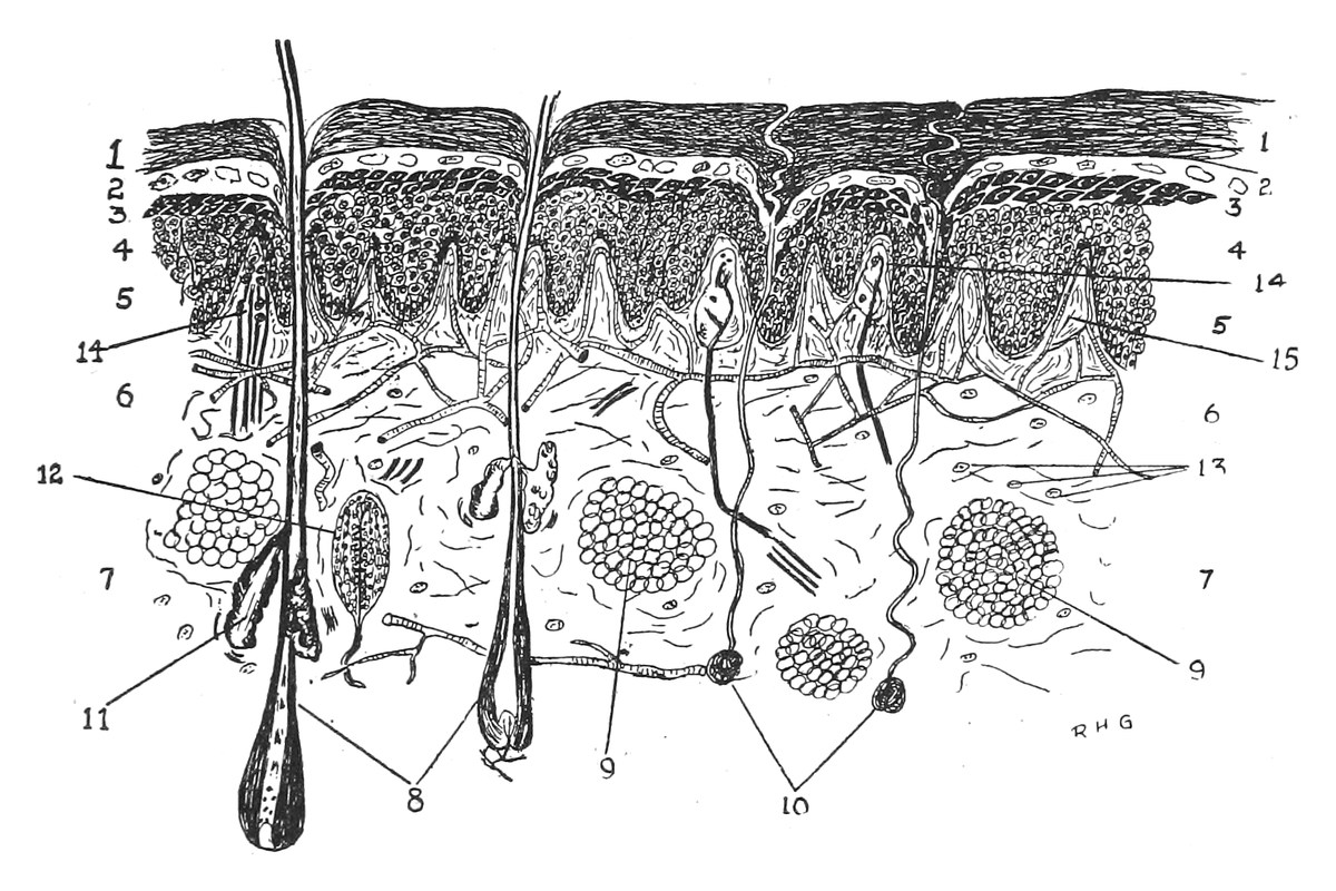
1. Stratum Corneum; 2. Stratum Lucidum; 3. Stratum Granulosum; 4. Stratum Mucosum; 5. Papillary Layer; 6. Reticular Layer; 7. Subcutaneous Tissue; 8. Hairs; 9. Fat Cells; 10. Sudoriferous Glands; 11. Sebaceous Glands; 12. Pacinian Body or Touch Corpuscle; 13. Connective Tissue Cells; 14. Nerve Endings 15. Vascular Papillæ and Capillary Loops
ANATOMY OF THE SKIN
The skin as a whole is composed of two distinct layers resting upon a third structure, the subcutaneous tissue. The outer portion is called the epidermis, cuticle or scarf skin, and is without blood and nerve supply, while the inner portion is called the corium, derma or cutis vera, and contains the capillary loops and nerve endings.
THE EPIDERMIS
The Epidermis is divided into four layers, named from without inward, the stratum corneum, the stratum lucidum, the stratum granulosum and the stratum mucosum or rete Malpighii.
The Stratum Corneum, or horny layer of the skin, is composed of many layers of horny, non-nucleated scales which are being continuously displaced by exposure to weather, water, etc., and are being as continuously renewed by the deeper layers. This layer of the skin is involved in the simpler foot lesions such as heloma and callositas.
The Stratum Lucidum, or clear layer of the skin, is composed of a few rows of transparent cells, without distinct boundary, and, except on the palms and soles, is considered a part of the stratum corneum. It is composed of from two to four layers of cells, which are like the cells of the horny layer except that they are brighter and more homogeneous. This layer is not often clearly defined and is of no importance.
The Stratum Granulosum, or granular layer of the skin is composed of several rows of polygonal shaped cells which are well marked on the soles of the feet. The nuclei of the cells are not well defined and the cell itself refracts light. The granules found in this layer are varied in shape and contain a fluid called eleidin and a peculiar solid substance called keratohyalin. This substance is derived from the cytoplasm of the cells and represents the first process in the cornification of the cells in the outer layers of the epidermis.
The Stratum Mucosum, or mucous layer of the skin, usually called the rete, or rete Malpighii, is the deepest and most important layer of the epidermis. The basal layers of cells are separated from the corium by a basement membrane or membrana propria, and these layers, which are made up of columnar cells, contain the pigment of the skin. The next few layers show elongated, oval or rounded shaped cells, the form varying with the locality, the tendency being to a rounded shape, owing to the more even pressure on the cells from above and below. The cells are irregularly formed and are made up of a soft substance with large oval or rounded nuclei. In the intercellular spaces is found a fluid which is nutrient in character. In the stratum mucosum are found the prickle cells. These cells have hairlike processes on them which serve to hold the cells together.
THE DERMA
The Derma, or Corium, is divided into two layers, the outer, called the papillary layer, or pars papillaris, and the inner, called the reticular layer, or pars reticularis. It is composed of bundles of fibrous tissue, yellow elastic tissue and connective tissue cells, the reticular layer being more compact than the papillary layer. The derma contains blood vessels, nerves, lymphatics, touch corpuscles, hairs, sweat glands and sebaceous glands.
The Papillary Layer of the skin is composed of small conical elevations called papillæ, which blend with the prolongations of the rete above. The best developed papillæ are found on the under or flexor surfaces of the fingers and toes and attain their greatest length at this point. They are placed in double rows that underlie the cutaneous ridges on the fingers and toes. These cutaneous ridges remain unchanged throughout life and are so characteristic of each individual, that they are used as a means of detecting and identifying criminals and others. Papillæ of two kinds are noticed, the one being very well supplied with blood vessels, and are called vascular, the others being only scantily supplied with blood, containing medullated nerves, and are called sensory papillæ.
The Reticular Layer of the derma is composed of loosely arranged bundles of connective tissue which merge with the papillary layer without a distinct line of demarcation. In these bundles of connective tissue are found the sweat glands, the sebaceous glands, the hair follicles and the deeper lymphatics. This layer of the derma is made up of fasciculi of connective tissue which blend into each other obliquely and give it a plexiform appearance. As the bundles ascend towards the surface they divide into smaller and finer bundles, and when the papillary layer is reached, they have a close, felt-like appearance.
The Subcutaneous Areolar Tissue, or tela subcutanea, connects the skin with the deeper structures and should be considered a part of the true skin. It is made up of loosely arranged bundles of connective tissue which cross each other repeatedly and form well defined spaces. These spaces contain fat, and where there are large quantities of this fat, as on the soles of the feet, the tissue is designated as adipose. The subcutaneous areolar tissue also contains the deeper hair follicles and the deeper sweat glands.
Blood Supply. The layers of the epidermis are without vascular supply, but the derma and the subcutaneous tissue are well supplied with blood vessels. There are two plexuses, one superficial in the upper layer of the derma, and the other deep, in the subcutaneous tissue. The vessels of the upper layer arise from the deeper plexus and give off branches in all directions supplying the hair follicles, sweat and sebaceous glands. The papillary layer is richly supplied with delicate capillaries, which terminate in the papillæ, and are called capillary loops.
Lymphatics. The lymphatics follow the vessels in a general way, there being two plexuses, viz.: deep and superficial. Lymph spaces are found in the rete Malpighii, which connect with the channels of those in the derma. The papillæ and the glands also have lymph channels.
Nerve Supply. The skin contains both medullated and non-medullated nerve fibres; these fibres are especially abundant in the soles of the feet and at the ends of the toes. They enter the skin with the more important ascending blood vessels. The non-medullated nerves terminate in the rete as fine filaments, and the medullated nerves end in the corium and subcutaneous tissue in special terminals called corpuscles. Examples of these are Pacinian corpuscles, tactile corpuscles and the end bulbs of Krause.
In addition to the sensory nerves, the skin also contains vasomotor nerves. These nerves are found on the smooth muscles of the skin and on all glands having such muscles, and have a direct action on these glands.
Muscles. Both striated and non-striated or smooth muscles are found in the skin. Those of the latter variety are most common, while the former are sparingly found. The smooth muscle fibres are found in connection with the hair follicles, the sebaceous and the sudoriferous glands, and they act upon these organisms.
Sudoriferous Glands. The sudoriferous glands, or sweat glands, are found in the reticular layer of the corium and in the subcutaneous tissue. They are simple tubular glands which are coiled into globular shape. The tubule of the gland empties into a gland duct which passes through the corium and the epidermis and opens on the surface of the skin in a funnel-shaped sweat pore. The sweat glands are very numerous, particularly on the soles of the feet. It is estimated that there are 2,000,000 sweat glands in the adult human body.
Sebaceous Glands. The sebaceous glands, or oil glands, are found in the reticular layer of the derma, usually associated with or in close proximity to a hair follicle. They may occur independent of the hairs however, as is the case in the lips. They vary in size from a simple pouch to a many pouched or multilobular gland. These pouches empty into a common duct, which in turn empties between the hair and the inner sheath. The ducts secrete sebum, which consists of fatty degenerated cells, in which is found epithelial waste matter. The sebum keeps the skin and the hair soft and oily.
NAILS
The Nails are a specialized form of epidermis, and are considered by many to correspond to the stratum lucidum of that structure. They are horny, elastic, transparent, quadrilateral plates, and are found at the distal ends of the fingers and toes, on their dorsal surfaces. The nails are convex on the outer surface and concave within. The nail itself is called the body and rests upon the nail bed. It has a free edge distally and two lateral and a proximal or short edge which latter lie in a groove called the nail or ungual fold. The ungual wall overlies the lateral and proximal portions. The nail is embedded into the derma at its proximal end by a root. This part of the nail is found beneath the ungual wall and is composed of cells which have not yet become horny.
The thin layer of skin, which extends forward from the nail groove at the beginning of the body of the nail, is called the eponychium or nail skin. The lunula is the little whitish, crescentic spot, a portion of the nail bed, which is found in front of the nail fold, and extends to the lateral edges of the nail.
The matrix of the nail is situated beneath the root of the nail, and is so-called because it is from this structure that the nail is produced. The matrix is thick, and raised in a series of longitudinal ridges, which are readily seen through the transparent nail tissue. It corresponds to the mucous layer of the epidermis, and is essentially of the same structure. The matrix is highly vascular, which accounts for the pink color seen through the nail, except at the lunula.
PHYSIOLOGY OF THE SKIN
The functions of the skin may be subdivided as follows:
Touch Organ Protective Covering Excretory and Secretory Organ Temperature Regulator Organ of Respiration
The skin acts as a touch organ or as an organ of tactile sensibility; this power is supplied by special bodies found in the papillæ. The degrees of consistency, of size, of form and of other qualities are recognized by this function. Other sensations are conveyed by these special nerve endings, such as heat and cold, burning, itching, tingling, etc. The sense of touch is well developed, particularly in the skin at the ends of the fingers, and this sense may be farther increased, as is the case with blind persons.
The skin acts as a protective organ to the body within, by excluding harmful agents such as bacteria, chemicals, heat, cold, etc. It is elastic and thick and is without sensation and thus protects the delicate structures beneath it from injury from various causes.
The functions of excretion and of secretion are performed by the glands. The sudoriferous, or sweat glands, excrete the perspiration, and in this way also act as elimination organs, accessory to the kidneys. The body is continuously sweating. When there is no indication of this function, when the skin seems dry, the name “insensible perspiration” is applied; when the function is apparent, by the formation of drops of moisture on the surface, it is called “sensible perspiration.”
The sebaceous glands are organs of secretion. They give off an oily substance called sebum, which lubricates the hairs, and gives an oily, soft appearance to the skin. This tends to keep the outer layers elastic and pliable; where this function is absent, the skin becomes dry and is likely to form cracks or fissures.
The skin acts as a regulator of the body heat, by controlling the radiation of the heat as brought to the surface from within, and by regulating evaporation. The normal tension of the skin on the various parts of the body has an influence in the regulation of body temperature.
The function of respiration is, to some extent, duplicated by the skin, the process being analogous to the respiration that takes place in the lungs. The amount of oxygen absorbed is small, but water and carbon dioxide are freely given off.