Chapter X · HELOMA
Reading edition
Derivation. The word has its origin in the Greek “helos,” meaning corn; (plural: helomata).
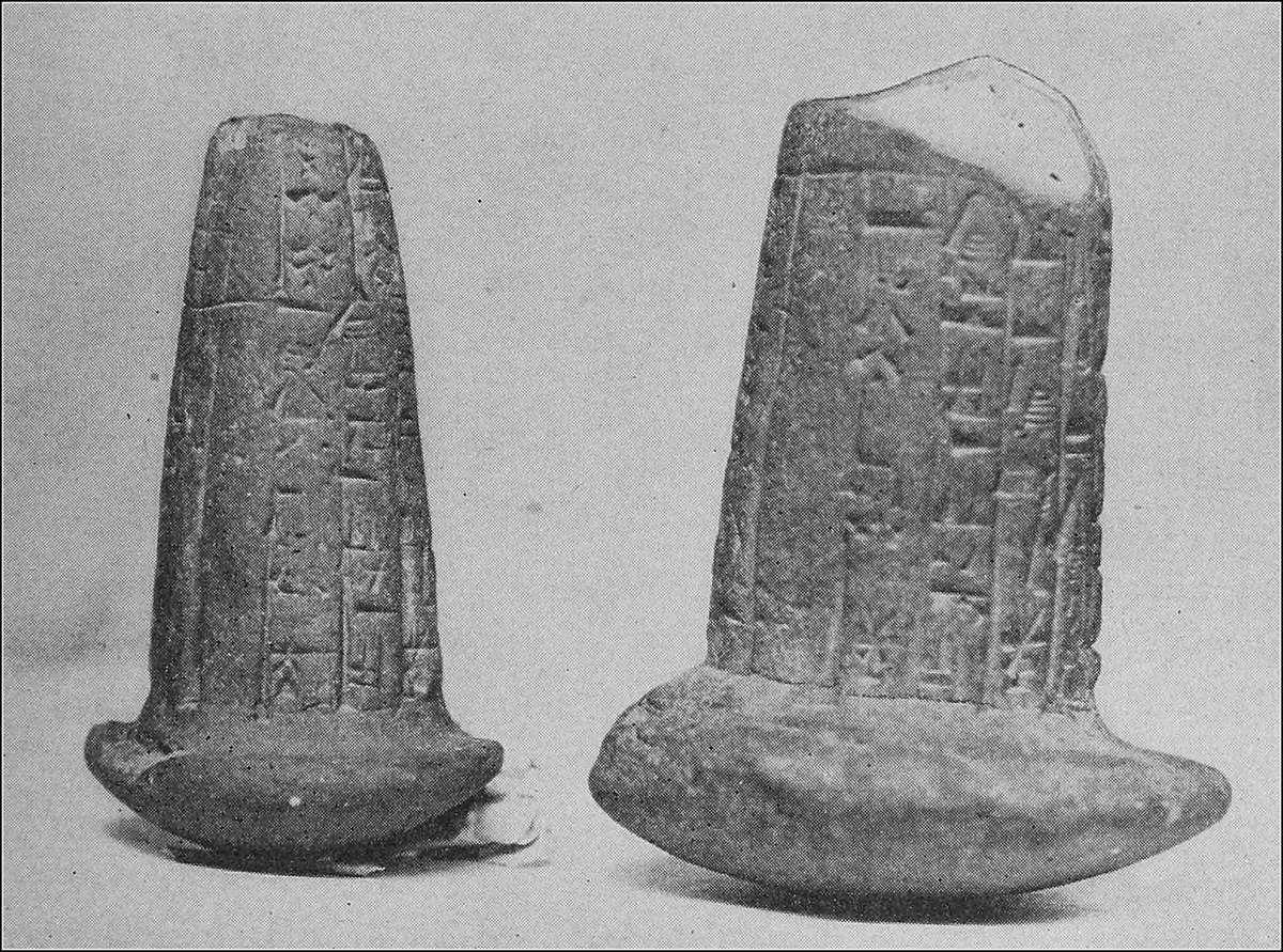
BUILDING NAILS (HELOS) USED IN PRE-HISTORIC TIMES
In the accompanying cut will be found a photograph of two building nails bearing the name and time of the reign of King Gudea in Ur, Chaldea, about 2500 B.C. Plaster casts of the above were presented to The First Institute of Podiatry by Fridtjov Anderson, Colonel in the Norwegian Artillery. These nails were called helos and because of their semblance to the foot excrescences, commonly called corns, the latter were therefore named helomata.
Synonyms. Corn, clavus, horn.
Definition. An heloma is a circumscribed, conical, deep-seated overgrowth of the epidermis, the apex of which presses down upon the derma. Corns usually occur about the toes, but may appear upon any part of the body subject to friction or pressure. They range in size from a pinhead to a ten cent piece.
According to their appearance, texture or composition, helomata are classified as follows:
Heloma durum, or hard corn. Heloma molle, or soft corn. Heloma vasculare, or vascular corn. Heloma miliare, or seed corn.
HELOMA DURUM
Heloma durum is a hard circumscribed overgrowth of the epidermis and may occur as above stated, but is usually found on the outer side of the fifth toe, the dorsum of the second, third and fourth toes, and on the plantar surface of the interphalangeal joint of the great toe.
Symptoms. The symptoms of an heloma durum may be classified as subjective and objective. The chief subjective symptom is pain in varying degrees. The formation of the growth produces a pressure upon the nerves which, in turn, gives rise to pain varying from a dull and mild sensation to a sharp and intense excitation. These pains are increased when the part affected is further irritated by ill-fitting shoes, by friction or by other pressure.
It is a well known fact that the pain in a corn is increased when the weather is about to change. Helomata, which at other times cause no inconvenience, will prove a source of annoyance at this time. This is due to certain physiologic changes that take place in the body as follows: the atmosphere preceding a storm becomes more and more charged with moisture. As this increases, the function of the skin, namely the elimination of liquid waste, is gradually diminished. This function is taken up and performed by the kidneys, and as the air becomes fully charged with moisture, the entire work of elimination is carried on by these latter organs. This physiologic change requires a readjustment of the blood supply and the nerves which control it, so as to bring about a proper equilibrium. In this latter procedure the little nerve fibres are unfavorably influenced, and pain results. The gradually increased uncomfortableness that is experienced is due to gradual increase in the humidity and when precipitation takes place and the air is freed from this atmospheric pressure, relief is afforded.
The patient will complain of pain while shoes and stockings are worn, but will feel relieved when they are removed, except in cases where inflammatory processes have commenced. The pains at these times are of the throbbing, pulsating variety, such as accompany all inflammations.
A clinical examination of an heloma durum shows a horny mass of epidermic cells crowded together with no regular formation within the growth. There is a sharp line of demarcation between it and the surrounding tissues and it is also distinguished by its darker, yellowish color in contradistinction to the healthy pink of the normal skin. Within the growth, and usually at its centre, may be seen a darker, more compact mass, which penetrates deeper into the tissues. This is the radix or nucleus, incorrectly termed the root, or the eye, of the corn. The skin immediately surrounding the heloma usually presents a red line, due to the somewhat lessened irritation that originally produced the corn. The color is due to congestion in the derma.
Etiology. Helomata dura are commonly caused by direct intermittent friction or pressure of ill-fitting shoes. The shoes may be too tight, thereby causing pressure, or they may be too loose, thereby causing friction. Helomata that appear on the plantar surface of the foot may be caused either by some roughness in the finish of the shoe or by soles which are too thin, or by downward displacement of the heads of the metatarsal bones beneath, caused by wearing shoes that are not anatomically correct.
Imperfections in the stockings, such as knots in the worsted or poor workmanship in darning, are also factors in producing helomata. Any concentrated or undue local pressure is capable of causing this overgrowth, provided, however, that the pressure is not primarily of sufficient intensity to set up such an amount of congestion and consequent inflammation as to lead at once to an ulceration of the part.
The same principle of intermittent friction and pressure, with counter-pressure, as a cause for helomata holds good on any part of the body surface.
Pathology. The pathologic changes accompanying the development of an heloma are mostly morphologic. The intermittent friction and pressure produce first an irritation or excitation of the cutaneous nerves; this causes an increased blood supply to the part and congestion takes place in the derma immediately beneath.
Were this pressure or friction to cease at this time, nature would restore the tissues to their normal condition in a very short time; but, as this pressure or friction is continued from day to day, the habitual congestion produces a chronic enlargement or hypertrophy of the papillæ of the derma.
The epidermic cells originate from the material supplied by the blood plasma, which is conveyed through the walls of the capillaries to the surface of the basement membrane by endosomis, where it forms into granules which contain nuclei of unusual size. These granules, the first organic shape of the future cells, gradually develop into nucleated cells.
Bearing in mind the development of the cells in the normal skin, with an increased blood supply, there will naturally be a more rapid proliferation or development of cells taking place over the enlarged papillæ than over the surrounding normal ones. This excessive development of cells causes an abnormal upward crowding of the preceding cells, with the result that the horny layer gradually becomes thickened. With this thickened layer acting as a counter-pressure from above, cornification of the tender cells takes place more rapidly with each succeeding new layer that is added to the under portion of that already formed. The growth at this time takes place at the interior and lateral portions of the heloma, principally the former.
This process in due time causes the epidermis to become transformed into a dense homogeneous mass of cells which is called a callosity. The size of the callosity is determined by the area of the papillæ affected.
The etiologic factors which cause the enlargement of the papillæ and the overgrowth of the epidermic cells continue, and that part of the growth which was first formed and is most usually central, becomes more dense than the surrounding callosity. This is due to the greater irritation to which it is subjected, and eventually this extends downward and penetrates the derma. The pressure of this new development, known as a “radix,” against the papillæ, causes these organisms to undergo progressive atrophy with the result that they are at times completely absorbed. Thus, we have the common heloma durum. The heloma now receives its nourishment from the lateral portions, which still continue to maintain their increased vascularity.
It often happens that an heloma is subjected to unusual pressure during its formation, which causes serous effusion or even subcutaneous hemorrhage in minute quantities. This effusion is absorbed by the soft cells which are in the process of transition into horny tissue. These cells take on a laminated appearance which present different tints, from a light yellow to a dark red.
The nerve filaments of the skin are also affected during the development of an heloma, and when this process is marked, the condition known as neuro-fibrous heloma is the result. These little neuromata very often become quite enlarged. The favorite location for this condition is the inner plantar edge of the great toe, and sometimes the outer plantar edge of the metatarsophalangeal joint of the fifth toe.
These helomata consist of two or three little irregularly shaped structures extending downward into the derma and in juxtaposition to each other, the septum dividing these structures being made up of one or more rows of enlarged papillæ which have become highly vitalized through the enlargement of the nerve fibres contained in them. The condition is very painful and great care must be exercised when operating upon them. The most logical reason for their appearance upon the plantar edges seems to be the fact that the long papillæ of the plantar surface leave off abruptly and are joined by the shorter ones of the dorsum. The line of junction is undoubtedly subjected to a greater degree of irritation than where the size of the papillæ is uniform.
Diagnosis. A typical heloma durum is a mass of epidermic cells, round in shape and varying in size. The color is usually yellow, but in cases where serous or bloody infiltration has taken place, the color may increase to red or even to dark purple.
Heloma durum may be mistaken for heloma vasculare or verruca, on account of the dark blood stains which are often in evidence, but when the top layers of the heloma are removed, the dark spots which were mistaken for blood vessels are not found in the growth proper, but are deep in the structure resting against the derma. When these spots, which are clots, are cut with the knife, no bleeding occurs; whereas, if the condition were one of heloma vasculare or verruca, hemorrhage would be produced.
Lesions of several skin diseases, such as eczema and psoriasis, very often produce scales or crusts on the feet which might be mistaken for helomata, for when the fingers are passed over them they feel as if they were an overgrowth of the skin. These scales or crusts, however, are usually loosened at the edges and the color is quite different, being red or white. Other indications of the presence of a skin affection will usually be found on other parts of the foot.
The radix, or nucleus, of the heloma is a characteristic which differentiates it from the ordinary callosity. The radix is a dark solid mass of epidermic cells which has an almost transparent appearance. When cutting an heloma, the radix may be felt by the tension produced as the knife passes through it.
Helomata dura found on the plantar surface of the foot over the heads of the metatarsal bones are not readily distinguished, due to the fact that they are covered by a layer of callous, which, when removed, exposes the nuclei to view.
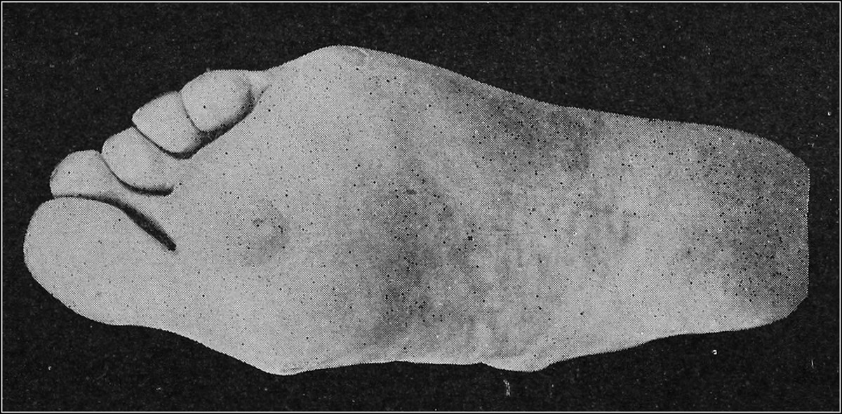
HELOMA DURUM ON PLANTAR SURFACE
Prognosis. The prognosis of heloma durum is uncertain. There are many cases on record which have disappeared after the first treatment and, on the other hand, some cases regularly recur notwithstanding many years of regular attention. This is due to two conditions: first, the cause of the heloma, namely, the continued wearing of the shoe, and, second, the papillæ beneath the growth remain enlarged and continue their function of excessive cell proliferation. The latter reason is more important than the first, for, even in cases where proper footgear is provided and no friction or pressure is permitted, the heloma may continue to grow. The only way to account for the total disappearance of some helomata, after treatment, is, that when the growth is removed, the size of the papillæ diminishes and normal function is restored. The situation of heloma durum is very important in considering the ultimate cure of the growth. Those on the outer aspect of the little toe rarely get well, due to the prominent position of that digit, while those on the plantar surface over the heads of the metatarsals are often completely cured after the proper treatment has been applied. When the heads of the metatarsal bones have been properly adjusted, the helomata produced by their downward displacement gradually disappear.
Treatment. The treatment of heloma durum is divided into three classes, viz.: preventive, palliative and operative.
Preventive treatment consists in securing freedom from friction of or pressure on the parts affected. Footgear of proper size and shape is essential. The shoes should be neither too tight nor too loose; they should fit snugly in the heel, and the toe box and front of the shoe should be broad enough to allow of freedom for the toes. The quality and kind of leather used for the shoe should also be considered. This should be soft and well seasoned, vici kid and calf skin being preferred. Patent leather and colt skin are undesirable, in that they are hard and almost air tight. Shoes made to measure on a last constructed from a plaster of Paris cast of the foot, are the best. In making a shoe, consideration should be given to other deformities, such as flat foot, metatarsalgia, etc., and means for correcting these ailments should be taken.
The stocking should be large enough to prevent pressure on either lateral side of the foot. There are stockings on the market which are cut for each foot and are known as right and left stockings. These are very desirable, as they prevent distortion of the toes, which is often produced by the regular stocking cut to a point at the third toe. The material is of little consequence, except that wool or cotton absorbs moisture excreted by the glands better than silk.
Palliative treatment includes the application of palliative agents, among which silver nitrate and salicylic acid are most serviceable. These remedies cause desiccation and shrinkage of the horny growth, which is thus made to shell out from the bed in which it lies. This method of treating an heloma durum is long and tedious, as many applications of the drugs are required to obtain a result. Great care must be exercised to prevent the applications from touching the surrounding normal skin. The virtue of so-called corn cures, which are very plentiful and are given artistic names, all depend upon some drug or chemical, chief among which is salicylic acid. The danger to the layman is readily seen, for when these agents are applied carelessly or in large quantities, the action is too severe, and ulceration and infection is the result.
When the practitioner finds it necessary to resort to palliative measures in the treatment of an heloma durum the following procedure is advised: the tissues surrounding the horny growth are protected by painting with several layers of collodion or glycerine jelly (Unna). A piece of salicylic acid plaster is cut to the size of the heloma and placed over it and a proper protection applied. Or, a shield can be fitted around the part and in the aperture is placed a 25% salicylic ointment, and the entire dressing is covered with adhesive plaster. This dressing is allowed to remain in contact with the part for two or three days, when the dressing is removed and the white, macerated tissues are scraped or cut away. The treatment is again applied and repeated as often as necessary. (See chapter, Shields and Shielding.)
Operative treatment may be divided into two classes, the radical and the non-radical. The radical method is painful, but with the use of local anesthetics, good results are obtained without discomfort to the patient. Alypin, 1 : 2%, quinine and urea hydrochloride, 2%, or novocaine, 1% (the latter being preferred by the writer) may be used to produce local anesthesia[5]. The parts are thoroughly cleansed and made aseptic by washing with the tincture of green soap, followed by alcohol, 60%, and finally painting the entire surface, including the surrounding parts with tincture of iodine. The hypodermic needle and syringe and all the instruments to be used in the operation are boiled in water for at least ten minutes. A spot is selected for the injection of the anesthetic, and ethyl chloride is sprayed on it, to make the primary injection of the needle painless. The needle is forced into the derma and pressure brought to bear on the piston of the syringe, as it moves forward into the tissues. By following the wheal thus produced, the entire area surrounding the heloma can be anesthetized.
When the anesthesia is complete, two semi-elliptic incisions, meeting at their extremities, are made through the skin, care being taken that they penetrate the subcutaneous tissue. These incisions should completely envelop the growth. The tissues between the incisions are seized with an artery forceps, and the entire wedge, including the derma and subcutaneous tissue, is dissected out. This produces free oozing, and it may be necessary to twist a small vessel. Hemorrhage is never severe. The edges of the wound are brought together (apposition should be perfect) by one or two fine sutures and primary union takes place in a few days, or, if the wound is not large, the part may be dressed and allowed to heal by granulation. Subsequent dressings should include shields for the prevention of friction or pressure.
Dr. Robert T. Morris, of New York City, recommends skin grafting with this operation, in order to prevent the formation of scar tissue, a very desirable consideration. After the tissue has been dissected out, some skin is taken from the fleshy part of the leg and is attached over the wound. This eliminates drawing the edges of the wound together, which procedure often causes harmful after-effects.
The non-radical operative treatment of heloma durum is the most popular and practical method employed by chiropodists today. The growth is removed down to the true skin, care being taken that this layer is not punctured and bleeding thus produced.
There are two general methods of procedure for the surgical treatment of callositas and helomata in vogue today. These are known as the paring or shaving method and the dissection or excision method. This nomenclature is derived from the operations themselves.
As the term implies, the paring method consists of removing the callouses with the chisel, knife or scalpel by shaving away the growth with a series of knife strokes or cuts, and the subsequent removal of the cone body or radix with the point of the same or a similar instrument.
Technic of the Shaving Method. The part is thoroughly cleansed with a standard antiseptic, such as phenol, 5%, lysol, 1%, or cresol, 1%, and this is followed by alcohol, 60%, which is allowed to remain in contact with the heloma for a few minutes. The instrument employed should be sterilized by boiling in water for at least fifteen minutes or by placing it in phenol, 95%, until the liquid clings to the blade, followed by alcohol, 95%.
After thorough asepsis has been obtained, the tissues adjoining the area to be removed are held firmly between the thumb and index finger of the left hand and the knife is grasped firmly with the fingers of the right hand.
By holding the tissues of the part firmly so that they may not move, the pain to the patient and the danger of invading the vascular tissues is minimized. A knife, no matter how sharp, is bound to pull the tissues overlying a tender and inflamed part unless these tissues are in some way prevented from moving.
After the overlying callouses have been thoroughly pared away, and the part is found to be soft and flexible, the nuclei are removed. In removing these hard bodies the operator who shaves or pares, practically becomes a dissector. With a sharp pointed knife the heloma is loosened from the surrounding soft parts until entirely freed, when it is lifted out. Should there be two or more helomata under one callous, the same procedure is adopted for each individual growth until all are removed and the whole area is found flexible.
The stroke of the knife in the shaving method is usually toward the operator, his fingers and thumbs being so arranged as to limit the distance which the blade may travel and so prevent cutting the patient or himself.
In shaving an heloma on the dorsum of the intermediate toes, the index finger of the left hand is placed anterior to the hardened area and the thumb, posterior. The knife is then held as a penholder and the strokes are made toward the operator, the toe being moved or rotated to bring all the surfaces of the growth under the blade of the knife.
On the plantar surface the operator, at times, is forced to work away from himself, but in most cases the blade is directed toward himself.
For operating on helomata between the toes, the knife is held as a penholder and the blade is directed toward the toe or web. Many of the practitioners who “shave” use a distinct dissection method for the removal of helomata in this location. They employ what is known as a “spoon”—a shallow flattened curette—and starting from the outer edge of the calloused area, work under the hardened layers until the entire growth is loosened, whereupon it is deftly removed.
The stroke of the chisel in the shaving method is usually away from the operator. This is in contradistinction to the stroke of the knife or scalpel. When operating on the dorsum of the toes, however, most operators using the chisel direct the blade toward themselves, unless, as is common, the operator moves around in front of the patient, when the cutting is done away from the operator.
Technic of the Dissection Method. One of the oldest, yet one of the most popular methods of operation is that known as dissection. As will be understood by the term, this method is a procedure whereby the growth is excised by the helotomon, as a whole, from the underlying tissues. This is in contradistinction to the paring or shaving method.
Two of the best known practitioners who use this method of procedure are E. C. Rice, M.D., of Washington, D. C., and Charles F. Stevens, of Elmira, N. Y. Dr. E. C. Rice has this to say on the subject:
“The limited knowledge of the chiropodists of earlier years did not apply to their handling of instruments, which was professionally scientific to the highest degree. The dissecting method was the method of operating in the early days.
“In the late fifties a practitioner by the name of Josiah Briggs taught many young men, among them Elliott W. Johnson, and the writer’s father, W. E. Rice. A Scotchman also instructed Nehemiah Kenison. They in turn taught others the dissection method. There will always be two professional methods of operating on helomata, the shaving (exfoliating) and the dissecting (excision) methods, and practitioners should understand both.
“Those who have a light touch, if determined to learn, can become skillful in the art of dissecting, and, when acquired, their life’s work will become a daily fascination. The procedure calls for skill that compares with that employed by the eye, nose and throat specialists and is appreciated by the most eminent surgeons.
“The word ‘dissect’ implies a separation, and this method permits the operator to separate the heloma from the normal tissue in one piece so that classification is made simple. In removing the growth in its entirety, it is possible to observe the various forms the nuclei take, and the classification the writer would make is as follows: granular, grain-like in appearance, sometimes called ‘surface corn’; crescent, forming a semi-circle about the joint; wedge, having a wedge-shaped nucleus, commonly found on the plantar surface; cone, from its shape; thumb tack, also named from its shape; multiple nuclei, resembling any of the above named and having numerous central points; soft, the gristly tissue between the toes; elevated, protruding, horn shaped; circular, because of the form of the nucleus.
“The instruments used in this method of operating are the chisel (helotomon—Dr. F. Oefele, editor,) and the iris mouse-toothed forceps.
“The original chiropodist’s instrument in this country was a cross between the surgeon’s scalpel and a chisel. The blade was shaped much like the human foot and was on a handle such as is used on the standard razor. From this first instrument of chiropody was evolved the only distinctive instrument of our profession, the chisel, which has been successfully used for more than half a century.
“It is distinctly a chiropodist’s instrument, as much as the plane is the tool of the carpenter. With this chisel the dissecting method is made possible, for its cutting edge, as its name suggests, is on the end of the instrument, and permits of the most delicate work. The chisel should be five and one-half inches long and should have a rough hexagon handle. The round or oval handles do not permit perfect finger control. The blade or cutting edge may be oblique, straight or oval, as the operator desires.
“The technic of the dissecting method of operating is as follows:
“The chisel is held in the right hand and the forceps in the left. Hold each as you would a correctly held penholder; to support and to steady the hands, let them rest gently on the fourth and fifth fingers; when operating between the toes, the supporting fingers rest upon and press aside the toes so as to give plenty of room for operating.
“The tissues at the periphery of the heloma are separated; with the forceps grasp the free edge and raise sufficiently to see the line of demarcation and use enough traction on the forceps to overcome the pressure of the chisel, not enough, however, to produce the sensation of pulling.
“When properly performed, the gentle lifting of the tissue prevents the nerve being pressed upon or pinched between the blade of the instrument and the underlying bone. The line of demarcation is made by the union of the light and dark shades of tissue, the normal being the light and the darker shade belonging to the heloma.
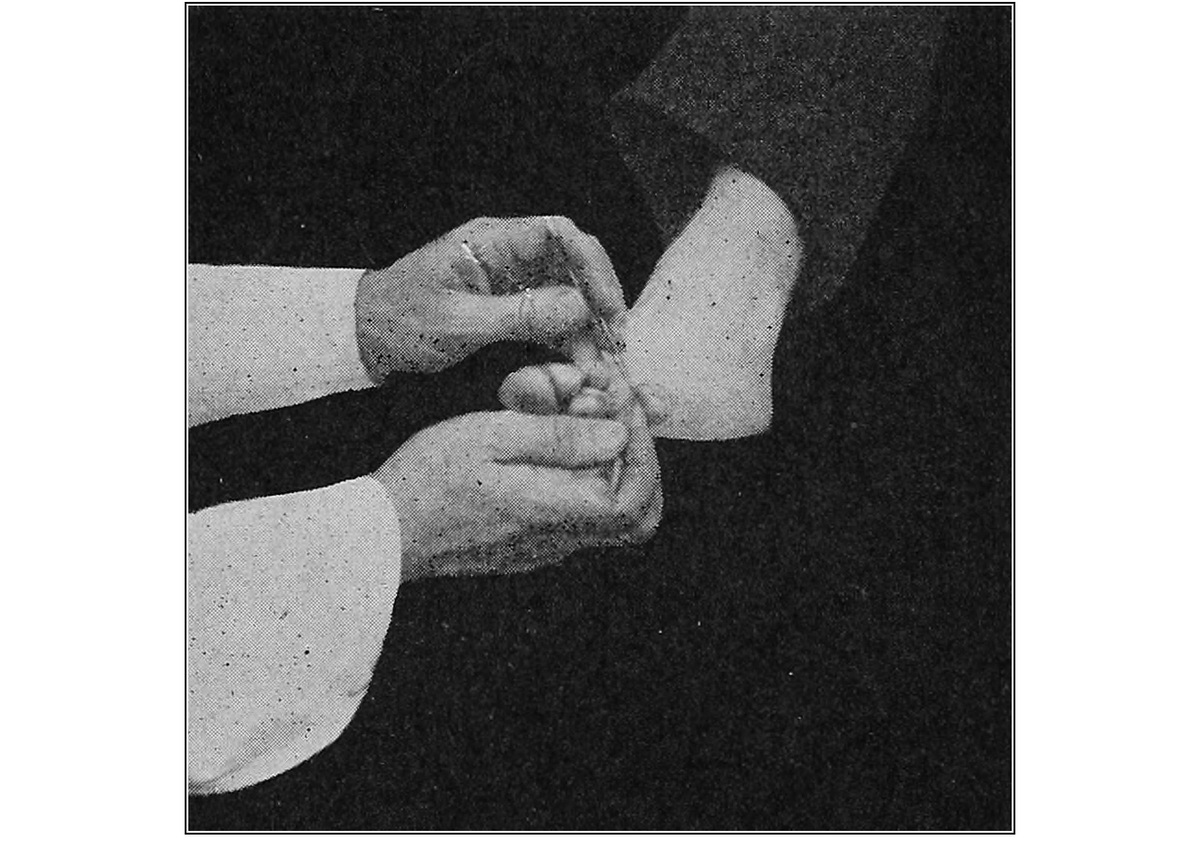
ILLUSTRATING METHOD OF SPREADING TOES APART BY USING FOURTH FINGER OF EACH HAND, MAKING OPERATION EASY
“Those who use the oval chisel find they can do more work and have fewer hemorrhages, and only occasionally do they find it necessary to use the nucleus dissector, which is required to remove an heloma of the thumb tack variety, as its shaft may extend to a depth of a fourth or a third of an inch. In dissecting this type of heloma, when the head of the same has been separated back to its shaft, the traction on the forceps is increased, the tissue being gently lifted and turned back. This tends to present the shaft toward the blade, and as cutting proceeds the shaft seems to be lifted up and out of the soft tissue into which it is embedded.
“To beginners, the writer would suggest the oval-shaped chisel and would advise practising first on the plantar surface.”
C. F. Stevens contributes the following:
“Speaking generally and taking the average heloma as an example, my method of procedure would be somewhat as follows:
“These growths are hardly ever deep, and are removed by the following process more easily than to chisel or pare. After the usual antiseptic precautions as to the operator’s hands, instruments and the patient’s foot have been taken, the operator grasps a sharp, pointed, slim instrument.
“Holding this knife with the right hand and with a small forceps in the left, he grasps the free part of the growth with the forceps, carefully raising this part to determine if possible how much is free and how much is attached to the deeper tissues.
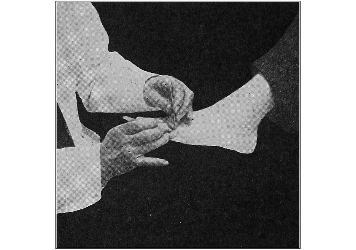
PROPER USE OF FINGERS ELIMINATES NECESSITY OF TOE SPREADERS
“With a sharp blade he makes a series of slight strokes, cutting but little at a time, on a line between the growth and the skin (the growth being darker than the skin), thus separating the excess deposit of horny cuticle from the skin, following every curve, deep part or point, until all is separated in the one mass. In this manner he is enabled to remove all in one piece. Dressings vary, according to the prominence of the part and the shoe worn.
“Since the writer was taught this method of procedure he has found it to be much easier to separate such a horny growth from its bed, than to try to pare it off in bits or shave it as the patient himself tries to do. Helomata being hard, very naturally resist the cutting of a knife, and the blade, therefore, when trying to pierce the hard mass, pulls on the sensitive tissues beneath, thus causing pain. Following the line between the normal and the abnormal tissues in operating, much softer integument is encountered; therefore, the cutting is easier and can be done with practically no pain.
“The cautious, careful operator will seldom invade the healthy tissues beneath sufficiently to cause capillary hemorrhage. A paring or shaving process could, of course, first be employed to remove the indurated callous, then proceed to carefully separate the deep parts as described in the case of heloma. Simply raising gently with the thumb forceps and cutting a very little at each stroke with a sharp pointed blade, following each wave or indentation indicated, as the work progresses, until each piece or mass is separated and removed, will be found a preferable procedure. However, we have found it as simple and easy to dissect the mass as a whole as to operate by paring and then removing the deeper parts.
“In case of a deep-seated hard corn where the toe is red, inflamed and very sensitive, the first described method (as in heloma) is usually best. Often upon reaching the lowermost layers, one finds a quantity of pus. When this escapes, as it does, the pressure on the inflamed tissues is lessened, and the patient will allow the operator to proceed faster.
“With a deep-seated heloma on the sole of the foot, the same method is followed, no matter how deep or serrated. The operator (after one or two small cuts) gently raises the edge with the forceps, while with the same style of blade he cuts down and around the growth, until the whole piece with its radix is lifted out.
“In connection with this the writer hears some one say, ‘even though you do remove the growth scientifically and without pain, severe pain will follow in an attempt to walk.’
“Of course, comfort depends in many instances on the dressing. The writer is not a great believer in heavy shielding and the method of dressing he employs is as follows: take a pledget of cotton which, when rolled, is about as large as the heloma just removed; place thereon a small amount of sedative in ointment form; place this in the cavity left by the removal of the growth, then cover all with a goodly sized piece of adhesive plaster. Instruct the patient to wear this for twenty-four hours, when he may remove the plaster. By this dressing the tissues that had been held up by the large heloma, are still held up by the rolled pledget of cotton, at the same time the cotton gradually flattens down with the patient’s weight. Thus the tissues are allowed to resume their normal position slowly and easily.
“When the adhesive plaster is removed, the cotton dressing comes with it. The tissues adjust themselves in from twelve to twenty hours and thus an equilibrium is painlessly established.
“Several years ago the writer chanced on to this method of dressing and since then he has used it and found it to be very efficacious in a large majority of cases; he has termed it the ‘filling dressing.’
“In operations on heloma molle the same surgical procedure is employed. It matters not whether the growth be on the side of the toe, or deep down between the toes on the web. The sharp, fine-pointed, narrow blade enables one to operate in a closely contracted space, and when used with short little cuts the blade reaches down, around and under the growth, thus loosening it completely and leaving its usually deep seat, clear and free from any parts which might remain, if chiseling or gouging were employed.”
(The authors of this work all operate with the knife or scalpel and have found that form of instrument very satisfactory. The beginner is advised to study and learn both methods so that he may be able to use both at any time. The fact that there are two methods does not mean that one is better than the other. There are many successful practitioners of both classes.)
Some persons have a great amount of dorsi-flexion of the toes, due to hammer toe or hallux flexus, and they usually develop an heloma on the distal end of the toe, under the nail. This is treated by cutting away the nail over the growth, and when the heloma is exposed to view it is treated in the same way as other helomata dura.
The subsequent dressings for helomata depend upon the state of the tissues beneath. Care should be exercised in operating so as not to cut too close to the normal skin, otherwise the parts become extremely sensitive.
The epidermis is a storehouse for bacteria, and when an heloma is removed, there is always a possibility that some of these bacteria may enter the body through some slight and invisible abrasion which does not necessarily bleed. It is, therefore, necessary to take precautions against this danger, and this is best done by painting all surfaces operated upon with a 4% solution of tincture of iodine (this may be made by diluting the official tincture with an equal amount of grain alcohol). This should be followed by painting these same surfaces with icthyolated collodion or nafalan collodion.
If the toe is inflamed it is treated with an agent that has the power to reduce inflammation. In severe inflammations, a wet dressing of Burow’s solution may be used to good advantage. The principal ingredient of this solution is aluminum acetate, which is astringent in its action, and a wet dressing applied for twenty-four hours will usually reduce the condition. In milder cases of inflammation, ointments of ichthyol, 10 or 15%, may be applied. This means of medication is very desirable whenever the application of a shield is indicated, because the aperture of the shield is a suitable place for ointment dressings.
When an heloma is found to be infected, the growth should be removed and the pus present evacuated. This should be followed by the application of hydrogen peroxide and the parts should then be irrigated with bichloride of mercury solution (¹⁄₄₀₀₀). The wound may now be treated with a wet dressing of Burow’s solution or, in severe cases in which there is an indication of the presence of cellulitis, bichloride of mercury solution (¹⁄₅₀₀₀) for twenty-four to forty-eight hours, should be similarly applied.
Subsequent dressings to stimulate granulation and promote healing may be applied, balsam of Peru or silver nitrate ointment or colloidal iodine being very efficacious.
The latest medication for infected areas, either great or small, and one of the many discoveries in surgical treatment since the beginning of the present war, is the Dakin solution. The worth of the application of this solution is based upon the helpful influence of free chlorine in small quantities, to tissues that have been mutilated either by injury or infection. Chlorazene tablets, purchasable in all drug stores, contain the elements desired for this treatment. Liquid chlorine ampules (J & J) also make an accurate Dakin solution.
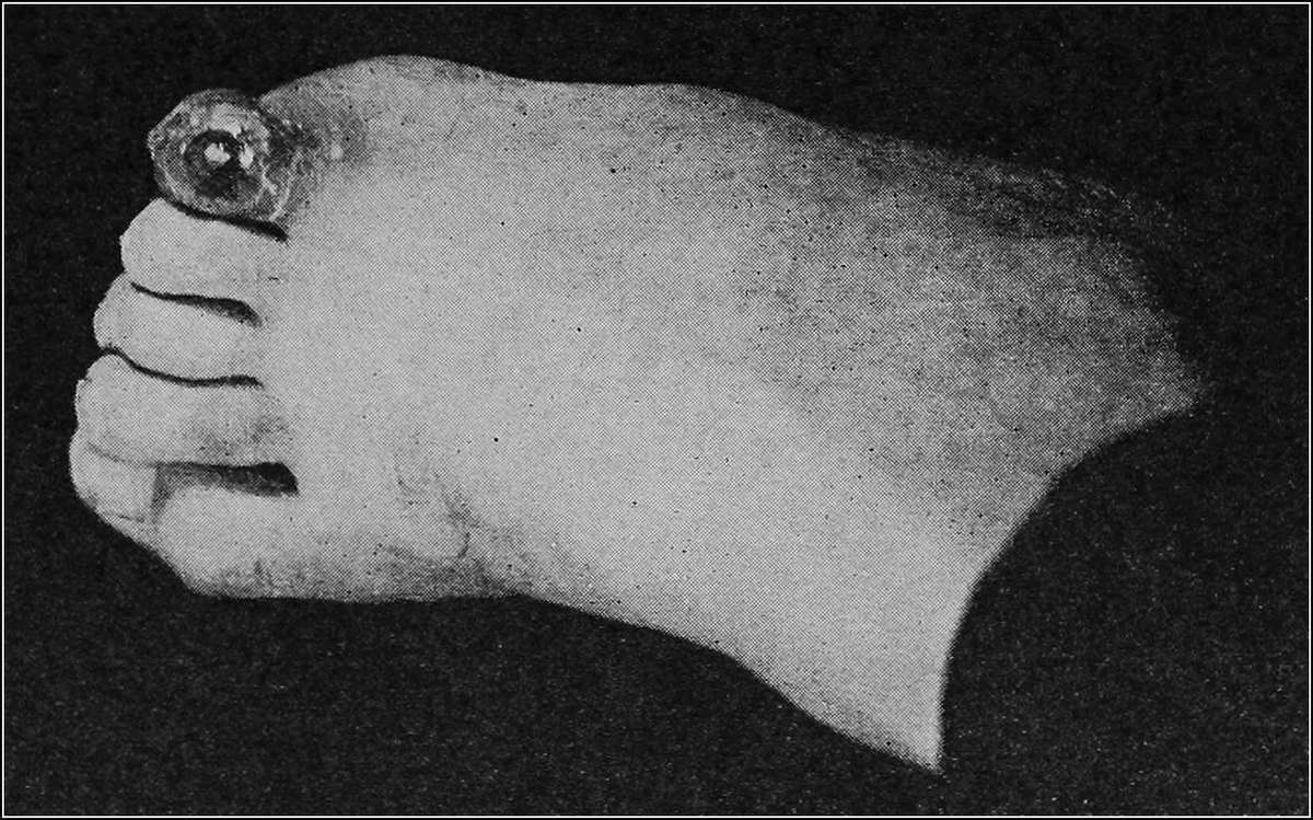
INFECTED HELOMA
If, upon examination, an infection shows that the deeper tissues, such as the periosteum or the bone, are involved, the patient should be sent to the surgeon, whose function it is to treat such cases, who will make incisions into the soft tissues so as to establish free drainage. The wound thus produced is packed with sterile gauze, and often with the aid of wet dressings, and nothing more, the wound is allowed to drain and heal.
The protection of the parts after an heloma has been removed, so as to insure comfort to the patient, is an all important part of the treatment of this ailment and a special chapter has been devoted to this feature of chiropody practice. (See Chapter Shields and Shielding.) There are certain types of helomata dura that are never relieved of pain, even after operation, unless a well-fitted shield has been applied.
HELOMA MOLLE
Definition. Heloma molle is a soft, white, macerated growth found between the toes, principally in the web of the fourth interosseous space and on the lateral sides of the interphalangeal joints of the toes.
Symptoms. The pain accompanying heloma molle varies with the degree of pressure brought to bear upon the toes. Where the heloma is situated in the web of the fourth and fifth toes, there is a sensation as if there were some foreign body, such as a pebble, between the toes, and as the growth develops the pain becomes gradually worse. The pain of an heloma molle, in other parts, is similar to the pain of heloma durum, and usually ceases when the foot is not encased in a shoe.
Upon examination, an heloma molle presents a white soft mass, having the consistency of rubber. There is no sharp line of demarcation between the lesion and the healthy skin. This is due to the blanching of all the tissues that come in contact with the excretions. In some instances there is a yellow ridge surrounding the neoplasm. The growth is superficial, due to its anatomic position. There is very little soft tissue between the epidermis and the lateral sides of the extremities of the phalanges, and therefore there is no possibility of the growth becoming deep-seated, as in heloma durum. The radix, or nucleus, when present, is of a dirty white color.
Helomata mollia found in the web of the fourth and fifth toes, have well defined nuclei which penetrate into the interosseous space between the metatarsal bones. These are easily distinguished since, as the surrounding callous is removed, they appear as a dirty white spot in an area of healthy pink skin.
Etiology. Helomata mollia are caused by shoes, the same as other types of helomata, but in this case the footgear acts as a secondary cause. Normally the phalanges are placed so that the base of one bone is opposite the head of another. When lateral pressure is brought to bear upon the toes, these bones press upon each other and thus produce an overgrowth of skin cells.
The sweat glands continue to functionate, but the parts being pressed together, do not allow the perspiration to evaporate; hence, there is an accumulation of moisture which acts upon the skin, producing a soft, white, macerated mass, with a rubber-like texture.
In the case of helomata mollia found in the web of the fourth and fifth toes, there is an outward rotation of the head of the fourth metatarsal bone, due to the lateral pressure on this region, causing the bone to drop and rotate outward; this in turn presses upon the base of the fifth proximal phalanx.
Pathology. The pathology of heloma molle is identical with that of heloma durum, except that the nucleus is rarely deep-seated. The epidermis composing heloma molle has no distinct cell formation, because of the macerated condition of the mass, but occasionally the nucleus of such a lesion, found in the web of the fourth and fifth toes, shows some of the original cell formation. Inflammation, terminating in suppuration, is very often encountered in this condition. Because of uncleanliness of the parts, bacteria thrive in this locality and the acidity of the moisture very often produces a fissure or abrasion in the tissues which may lead to infection and subsequent suppuration.
Diagnosis. The typical heloma molle is a mass of epidermic cells rarely larger than half the size of a dime. The color is white, with a dark grey centre, denoting the radix.
Very often an ordinary exfoliation of the epidermis between the toes may take on the appearance of an heloma molle, but careful examination will show that there is no overgrowth of epidermis. This exfoliation is easily loosened with a pair of forceps.
Fissured toe webs, accompanied by exudation and exfoliation of skin, may be mistaken for heloma molle, and treatment inaugurated for the latter condition will produce bad results, particularly if chemicals are used.
There need be no doubt about making a positive diagnosis if the color and texture of the growth be borne in mind. The finger passed over the affected surface will give the sensation of increased tissue.
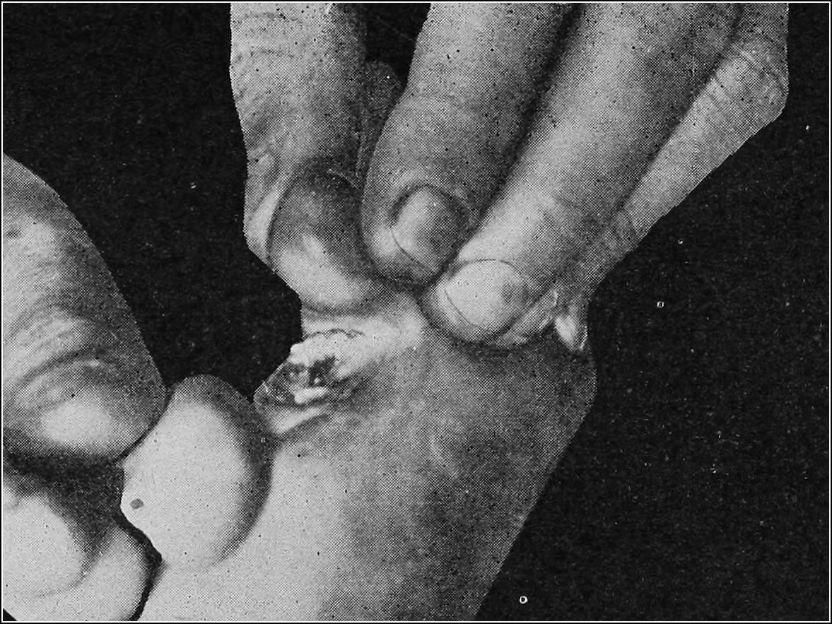
HELOMA MOLLE
Prognosis. The possibilities of the ultimate disappearance of helomata mollia is good. If the proper shoes are worn and the proper treatment be installed, the growths will gradually become smaller and will finally disappear.
The helomata that appear between the toes on the interphalangeal joints are most easily cured, by simply keeping the adjacent sides of the toes separated. Those that appear on the outer lateral side of the great toe do not respond to treatment as readily as the other types, for there is more soft tissue over this joint and usually the great toe is in a fixed position and does not easily straighten.
Helomata mollia that appear in the web of the fourth and fifth toes can also be permanently cured, but it is necessary to raise the head of the offending metatarsal bone, as well as to separate the toes.
Bearing in mind the etiology of heloma molle, and installing treatment which will correct or remove these causes, time and conscientious treatment will ordinarily insure a favorable outcome.
Treatment. Treatment of helomata mollia is divided into two classes: the non-radical surgical and the therapeutic. The latter method is the most popular, as it is very often impossible to use the knife. The texture of the skin, and the anatomic position of the growth often make it impossible to use an instrument with a cutting edge with a view to obtaining good results.
The non-radical surgical method consists of removing the corn in much the same way that an heloma durum is removed. The long cutting edges of knives and chisels are not well adapted for work between the toes, and for this purpose the “golf stick” and the “soft corn spoon” have been devised and are used extensively. The “golf stick” is an instrument which, as its name indicates, resembles the stick used by the golfer. Its cutting edge is almost at right angles to the handle and is about three-eighths of an inch long. This makes a very desirable instrument for removing helomata mollia on the lateral sides of the interphalangeal joints. The end of the instrument is rounded so as to allow for the removal of nuclei, if present. The cutting edge of the “soft corn spoon” extends almost around the entire instrument, and admits of a circular movement such as is employed in dissecting helomata dura. This instrument is used for removing soft corns that appear in the web of the toes, and is very efficient, inasmuch as by its use the operator is enabled to remove the growth without cutting into the tissues, as is often done with a knife or a chisel having a long straight edge.
The therapeutic method of treating heloma molle depends upon the caustic action of several drugs, among which may be mentioned salicylic acid, trichloracetic acid and silver nitrate. The two latter are used only occasionally, as they are powerful caustics, and unless applied with great caution they may produce harmful results.
Salicylic acid finds great favor among practitioners of podiatry, and the usual technic is as follows: after asepsis has been practised and the growth cannot be removed by the use of the knife, an ointment of salicylic acid, 15%, is applied over the growth, care being taken that the medication does not come in contact with the surrounding normal tissues. This is covered with a protective cocoon dressing, or the ointment may be applied into the aperture of the shield, if one is used. The dressing is allowed to remain in contact with the part for from four to seven days, depending upon the thickness of the skin. When the dressing is removed, the entire mass will be found, as a rule, to be loosened from the tissues beneath. If all of the growth is not thus loosed, the treatment is repeated and the patient is instructed to return in the prescribed length of time.
Shielding plays an important part in the treatment of heloma molle. For the type that forms on the lateral sides of the interphalangeal joints, a shield of the oval type with the aperture over the affected part, is most efficient, while for those that appear in the web of the toes, an oval shield with a semi-circular opening on the proximal end, which sets between the toes and protects the growth, is most desirable. In connection with a shield to protect the growth and separate the toes, it is necessary to raise the head of the affected metatarsal bone, which is the cause of this type of heloma molle. For this purpose, Alfred Ahrens, of New York City, has devised a dressing which he terms the “duck shield,” because of its resemblance to that animal. This shield has a dual function. It separates the toes and then passes down to the plantar surface of the foot over the metatarsal bone, and acts as a pad to raise the bone. The continuous application of this device to helomata mollia of this variety will produce good results.
HELOMA VASCULARE
Definition. Heloma vasculare, or vascular corn, is an overgrowth of the epidermis in which enlarged and elongated blood vessels are found.
Symptoms. The growths usually appear on the plantar surface of the foot, but occasionally they may develop in old callouses and helomata situated on the dorsal surface of the fifth toe. Pain is more severe than in other forms of helomata, the patient complaining of a burning sensation when not in a standing position. This form of growth is similar in appearance to heloma durum, having in addition small dark red spots scattered throughout it, which bleed upon being cut. These spots are not blood clots, such as are found in helomata dura as the result of injury, but are distinct blood vessels. The composition of the tissues is very dense, particularly when the growth is situated over the head of a bone, as is ordinarily the case when it appears on the plantar surface. The color of the entire mass is somewhat darker than in heloma durum, being grayish, or sometimes brownish, in appearance.
Etiology. As previously stated, helomata of all types are due to intermittent friction and pressure. The blood vessels that are found in this particular form are forced into the epidermis owing to lateral pressure of the shoes, or to the pinching of tight stockings. Why the blood vessels should be forced up into the epidermis is most peculiar, but helomata vasculare appear where the normal papillæ are longest, and this increased length of the vessels tends to force them up into the dead skin. Athletes, particularly runners and jumpers, are most commonly afflicted.
Pathology. Heloma vasculare consists of an overgrowth of epidermic cells in which are found the elongated vessels. There is an increase in epithelial tissue, but there is no increase in the quantity of the connective tissue and blood vessels, as in verruca. The blood vessels leave the papillary layer of the derma and enter directly into the epidermis, without any elevation of the surrounding connective tissue. On some occasions a nerve ending is found embedded in the callous mass. This adds considerably to the pain, but is not the true neuro-fibrous corn described under heloma durum, which has no accompanying blood vessels. The area surrounding an heloma vasculare is usually inflamed, but the inflammation rarely terminates in suppuration.
Diagnosis. The true heloma vasculare may be easily distinguished from verruca when the two conditions appear on the dorsal surface or any surface not subjected to extreme pressure, in that the latter is an overgrowth of all the layers of the skin, including the derma, and has a characteristic cauliflower appearance. However, when verrucæ appear on the plantar surface, they lose their cauliflower appearance and become flattened; they then resemble heloma vasculare, except that they are somewhat darker.
The blood vessels in heloma vasculare are not so numerous as in verruca, but this diagnostic point may not always manifest itself to the naked eye. A differential diagnosis between these two conditions is of no great importance, as the treatment is practically identical.
Heloma vasculare may be readily distinguished from heloma durum by the small red spots found therein which bleed when cut. Very often an heloma durum has a dark red spot at the base of the mass, due to the rupture of a small vessel and consequent clotting of the blood. This dark red spot does not bleed when the knife is passed through it, denoting the absence of blood vessels.
Prognosis. Heloma vasculare will always respond when the treatment is thorough. There may be a recurrence of heloma durum over the spot where the original growth was located, but the vascular condition, when once eradicated, should not return.
Treatment. The treatment of heloma vasculare may be divided into three classes, viz.: surgical, medicinal and mechanical.
The technic of the surgical method is as follows: the part is cleansed with tincture of green soap, followed by the application of tincture of iodine, 4%. The instruments having been sterilized, the part is anesthetized by the hypodermic method and a semi-elliptic incision is made a little to the outside of one-half the growth. The flap thus produced is seized with an artery forceps. The forceps are then raised and the rest of the growth is dissected out with a sharp knife or with a heavy pointed scissors. When the entire growth has thus been eradicated, a few layers of gauze should be placed over the part to produce pressure. A bandage should be applied over all to hold the dressing in place. This may be removed in three or four days, provided no inflammation is present, and the subsequent dressing should contain balsam of Peru or some other stimulant.
The medicinal or chemical treatment of heloma vasculare consists of the gradual destruction of the growth by means of chemicals, chief among which are nitric acid, potassium hydroxide and salicylic acid. If nitric acid is employed, the callous is removed so as to produce a slight oozing of blood, and a drop of the acid is allowed to fall in the centre of the mass. This is allowed to remain in contact with the part for two days, when the eschar produced is removed, and the acid is again applied. This treatment is continued as long as necessary to completely destroy the growth; when a slight exudation of pus is noticed, the application of the acid should cease. The subsequent ulcer thus produced is treated in the same manner as any other ulcer (see chapter Ulcers).
The salicylic acid method of treating heloma vasculare varies greatly depending upon the strength of the acid employed. If a weaker percentage is used, the treatment is practically the same as that with nitric acid. Several applications are necessary to completely remove the entire excrescence. The weaker solutions of this drug are the 10 to 15% ointments. The stronger ointments contain from 50 to 60% of the acid. The treatment with the 60% salicylic acid is preceded by cleansing the parts and removing the superfluous callous. The acid is then applied and the part protected. The dressing is allowed to remain in contact with the part for from ten days to two weeks, and when removed, the entire mass may be easily scooped out. When the stronger acid is used, it is often necessary to warn the patient that if there should be any throbbing pain experienced, he must return for treatment at once. This pain is due to the rapid action of the drug, and to a mechanical inflammation which ensues. Examination will usually reveal a newly formed ulcer, which must be cleansed and treated in the usual manner. The salicylic ointment method is finding great favor, particularly on account of the few treatments necessary. Those inclined to nervousness and imaginary fears, regarding chiropodical or any other operations, are also usually highly pleased with this non-surgical method of treatment because the use of the knife is avoided and cure is not long delayed.
In treating these cases medically, it is well to remember that the chemicals employed have a destructive action on the healthy tissue beneath the heloma as well as upon the heloma itself, and caution should be exercised in applying them. The case should be carefully watched and at no time should the operator allow the patient to remain away from the office for a greater length of time than above specified. It is also well to remember to warn the patient of the dull throbbing so characteristic of inflammation, which gradually increases as the pains become worse. These pains are due to a chemic inflammation produced by the action of the drug upon the normal tissue beneath the growth, and are always an indication to discontinue treatment, remove what is left of the destroyed tissue, and direct treatment to the healing of the parts.
The mechanical treatment of heloma vasculare consists of the removal of the growth by means of electricity. The fulguration spark and electrolysis are the two methods employed.
The fulguration spark is a concentrated violet ray, or high frequency current. The current is concentrated by passing it from the coil through a narrow glass electrode, at the far end of which is inserted a small piece of platinum or copper wire. As the current passes through the tube and the charged wire is brought in apposition to the excrescence, instead of the usual blue spark that is produced by the high frequency current in an ordinary vacuum electrode, there is a yellow spark produced which is quite painful to the body tissues. This spark has a caustic action, and after penetrating the superficial layers it enters into the deeper structures and there causes a destruction of the tissues.
Two, or at the most three, applications of this current, each of thirty seconds duration, will suffice for helomata vasculare which are situated on the dorsum of the foot. On the plantar surface, however, the tissues are more dense and many more treatments are required. It is on account of this density of the tissues that fulguration or any other form of electricity for the treatment of plantar growths is inadvisable.
Electrolysis consists of inserting a needle or other sharply pointed instrument to which the negative pole of a galvanic cell has been attached, beneath and around the growth. The positive pole is attached to a spot near where the condition is found, usually the calf of the leg. As the current is passing through the foot, the water in the tissues undergoes electrolysis, and after a time, as the hydrogen goes to the negative pole, bubbles of this gas are noticed around the free surface of the needles. This is an evidence that the decomposition has gone on sufficiently and the needle may be withdrawn. If the growth is a large one, the needle should be re-inserted at right angles to the original insertion, and the process repeated. If this is done properly, after two or three days, the entire mass will separate from the surrounding tissues. The greatest care must be observed in practising asepsis, as the electrolysis method is not an antiseptic one. The needle must be thoroughly boiled, and the part cleansed in the same manner as if a surgical operation were to be performed. This method, as well as the previous one, is not practical for helomata vasculare that appear on the plantar surface of the foot.
The carbon dioxide pencil may also be used in the treatment of this condition. This method, however, is not advised, as the parts become frozen from the contact and the pain of reaction is severe.
As previously stated, the treatment of heloma vasculare is almost identical with that of verruca and the reader is advised to consult the chapter on verruca for further knowledge along this line of treatment.
HELOMA MILIARE
Definition. Heloma miliare, or heloma disseminatum, or seed corn, is a small excrescence usually found in large numbers on the plantar surface of the foot, around the heel, or over the dorsal and inner lateral surface of the great toe joint. The growth is about the size of a millet seed.
Symptoms. This form of heloma does not produce the extreme pains caused by the other types of this growth, and only when they develop in great numbers do they become annoying. The patient then complains of an uncomfortable feeling, as if there were a foreign body in the shoe or stocking. Upon examination, several small helomata are seen, which appear to be all nuclei.
Etiology. Wrinkles produced by wearing loose stockings are a factor in producing helomata miliare; nails which protrude from the plantar surface of the shoe are also a fruitful cause of this condition. The wrinkling of the stockings produces an uneven surface over the length of the wrinkle and the weave of the material, usually wool, causes these helomata to develop. The nails found in shoes are usually caused by imperfect repairing. They do not extend out more than just the smallest fraction; in fact they protrude just enough to allow the patient to go along for several days or weeks without noticing that something is wrong.
Pathology. Hypertrophy of the epidermis takes place at the nucleus only, but the area immediately surrounding the heloma miliare feels hard and congested to the touch. Hypertrophy of the papillæ occurs, but only a small number are involved. There is no disturbance in the skin between the individual growths, each of the neoplasms having a distinct etiologic factor in its production.
Diagnosis. The heloma miliare is characteristic and cannot be mistaken for any other condition. As stated, the growth is rarely larger than a millet seed and appears to be all nucleus. There is an area of normal skin between these helomata, when they occur in numbers.
Prognosis. Careful operating and intelligent after-care will produce a cure in from four to five treatments. There are cases on record that have entirely disappeared after one treatment, but these are rare. The footgear, both shoes and stockings, should be examined and if found faulty should be corrected. This aids in a rapid cure and will, as well, prove a preventive.
Treatment. The removal of these helomata may be accomplished with the knife, but the ordinary scalpel is useless. It is necessary to have a very finely pointed small knife, and the procedure is the same as that followed in the treatment of the nucleus of heloma durum, except that more care must be practised, because of the smallness of the growth. The helomata miliare occur in groups containing as many as twenty or even thirty distinct minute growths, and it is necessary to take as much care with each one of them as with the first one removed. This is trying both to the patient and to the operator, but as it is essential to the successful cure to have the growths removed individually and carefully, patience is necessary. The after dressings may consist of ichthyol ointment, 15%, applied on a piece of lint, or balsam of Peru painted on after the helomata have been removed, and covered by a lint or cocoon dressing. Some practitioners apply tincture of iodine to the part without further dressing.
The therapeutic method of treatment consists of applying salicylic acid plaster, cut so as to fit over the affected area, and allowing this to remain in contact with the part for several days. This softens the tissues, so that the small growths may be easily removed, but care must be taken, as the acid will destroy the healthy tissue between the helomata unless each growth is isolated in treatment. The disintegrating process must not be allowed to continue to the extent that it does in the treatment of heloma molle or in the other conditions in which salicylic acid is employed. It is then often necessary to use the knife to remove the remaining tissue.
Recurrence is the rule in helomata miliare, but after persistent treatment the condition usually disappears. It must be borne in mind, however, that the footgear of the patient must be carefully examined and necessary corrections made. This, in itself, without the thorough treatment prescribed above, will often result in a cure of the most annoying cases of heloma miliare.