Chapter XX · ULCERS
Reading edition
Definition. An ulcer is a lesion of a cutaneous or mucous surface, caused by a molecular disintegration of the superficial parts, usually attended by more or less suppuration. A wound, or superficial loss of tissue due to traumatism, is not primarily an ulcer, but may become such if the healing process is arrested or the wound becomes infected with pyogenic microorganisms.
The following ulcers frequently come under the observation of the podiatrist:
Simple Ulcer, a local non-constitutional lesion attended with no marked pain or inflammation.
Indolent or Callous Ulcer, a chronic lesion, with hard, elevated edges and few or no granulations and showing no tendency to heal.
Varicose Ulcer, localized destruction of the skin over a varicose vein, usually of the leg, due to mechanical pressure, to nutritive disturbances, or to bacterial action.
Perforating Ulcer of the Foot, malum perforans pedis, a round, deep, trophic lesion of the sole of the foot, following disease or injury (in any part of its course from the centre to the periphery) of the nerve supplying the parts.
Syphilitic Ulcer, due to syphilis in late secondary or in tertiary stages.
Before describing the characteristics of the various forms of lesions just mentioned, together with their differential diagnosis, treatment, etc., it is deemed advantageous to briefly discuss the general etiology and pathology of all ulcers.
Etiology. One or several factors may be concerned in the etiology of ulcers, which are grouped under: (a) predisposing causes; (b) exciting causes. The first group includes local as well as general causes.
Predisposing Causes. Age can hardly be considered as a very important factor, except that old age is accompanied by retrogressive tissue changes, hardening of the arteries, impaired circulation, etc., and one would therefore expect the statistics to show a greater proportion of ulcers during the later years of life.
As regards sex, ulcer is more common among men than women in the ratio of about three to one. Alcoholism, syphilis, and traumatism may in some measure explain why ulcers are more common in men than women. Occupation seems to have little to do with the etiology beyond the fact that it may predispose to various forms of infection, and it is due to this element that we have the most important factors in the causation of ulceration. Varicose ulcer is always associated with varicose veins in the lower extremity, and these may be described as veins whose valves are incompetent. The most obvious cause of the breaking down of the valves is hard work, that is the lifting or carrying of heavy loads, as in the case of laborers, freight handlers, and longshoremen. The great strain occasioned by work of this kind lays a heavy load upon the veins of the legs. Whether the valves become useless through stretching of the vein walls, or are directly broken, is immaterial. The occupations which involve standing for long periods without moving the legs are, in a lesser degree, a source of valvular incompetence, and this is not from excessive back pressure but from stasis due to lack of muscular movement. Among women, the venous engorgement of the legs, so often seen in pregnancy, may, after the birth of several children, result in varicosity.
Many of the constitutional diseases such as gout, anemia, diabetes, syphilis, and tuberculosis, which lower the vitality of the tissues, and other conditions, such as valvular disease of the heart, general obesity, and arterial hardening which prevent proper circulation, predispose to the formation of ulcers when there is in addition some exciting cause.
Embolism, which cuts off the nutrition of the part, may also act as a local cause. Certain vasomotor disturbances, such as occur in frost bite and in Raynaud’s disease, may produce small areas of localized gangrene which subsequently become the seat of an ulcer.
Interference with the return of venous blood from a part predisposes to ulceration. Where phlebitis and periphlebitis occur, especially in the smaller venous radicles, small abscesses often form, the adjacent skin becomes involved, and an ulcer results; or the rupture of the diseased wall of one of the small veins may become infected, and ulcer develops.
Perforating ulcer of the foot is a frequent complication of tabes dorsalis. Myelitis, and other pathologic conditions of the nervous system may also, either through trophic changes or by reason of impaired sensation, bear a distinct relationship to ulcerative processes.
Exciting Causes. Traumatism is one of the most frequent causes of ulcer formation. Its degree may, of course, vary greatly, and whether it will produce an ulcer depends upon one or more of the predisposing causes already mentioned. Infection by any of the staphylococcus or streptococcus group of organisms as well as by the tubercle bacillus will produce ulcers; malignant ulceration also occurs.
Pathology. The pathology will vary according to the conditions causing the ulcer, although in the non-specific forms of ulcer the phenomena of congestion, exudative and necrotic inflammation, together with reparative inflammation or granulation, will only be in evidence. In the development of an ulcer the degenerative process predominates; in the healing stage, the reparative. When the ulcer develops from without, as when infection enters the skin through an abrasion, congestion first occurs. This is rapidly followed by the emigration of leucocytes, by a diapedesis of red blood cells which rapidly disintegrate, and by an exudation of serum and fibrin. At the same time there is a proliferation of the epithelial cells and also a proliferation of the connective tissue cells of the corium. The tissue next becomes softened by the exudate between the cells. Then, as a result of the pressure of the exuded serum, of the crowding by the leucocytes, and of the cutting off of the blood supply, and also in some measure through the effects of the toxins furnished by the bacteria, there occurs necrosis of the cells, which are thrown off from the surface with the products of exudation, until there is formed an ulcer with its base consisting of spheroidal and a few epithelioid cells developed from the connective tissue cells by proliferation.
When an ulcer in its complete stage of development is examined, the surface is found to be covered with a layer formed by the overproduction of new round cells, together with the exudate of fibrin, serum, and the cellular elements of the blood. When the discharge from the ulcer is profuse, this may be constantly washed away. When the ulcer is sluggish, it may be in a condition of coagulation necrosis. In this latter condition a croupous material covers the base of the ulcer, and below this is a more or less distinct layer, largely composed of cellular elements, with very little cellular substance, the cells being spheroidal and epithelioid in character and mingled with polynuclear leucocytes. As we go deeper, the amount of intercellular substance increases, and a number of transparent fibres and fusiform cells are found. In this layer of granulation tissue are also the newly formed blood vessels, the most superficial branches being vertical to the surface, and developing by a process of budding from the endothelial cells of the capillaries deeper down. This layer is paler in color than the layer made up of the cellular elements, but may contain pigment from the disintegration of the red blood cells. It gradually merges into a layer of cicatricial connective tissue which lies beneath the ulcer.
A section of a chronic ulcer would show an enlargement and prolongation of the papillæ, with a marked proliferation of the epithelial cells covering them. This is most pronounced in the condition known as callous ulcer, where the edges may, by proliferation, be considerably raised about the level of the surrounding skin, and often overhang the base of an ulcer. Under proper treatment the reparative process proceeds faster than the degeneration of the cells and the ulcer begins to heal by granulation. Small sprouts or buds of protoplasm protrude from the capillaries below or in the base of the ulcer, developing from the cells in their walls. These are hollowed out by the blood pressure and form new blood vessels which anastomose with others. Nuclei form in the protoplasm and thus endothelial cells develop. At the same time small spheroidal cells, developing from the connective tissue cells, become grouped around the blood vessels. These are closely crowded together at first, being separated by only a small amount of fluid intercellular substance. Some of the round cells then become larger and fusiform or branched. The larger cells are known as epithelioid cells. Some of the fusiform and branched cells, called fibroblasts, develop the new delicate fibrillar intercellular substance, while others form the connective tissue cells. Gradually the fibrous intercellular substance increases in amount, while the cells become fewer and flattened, and cicatricial tissue is formed. The contraction of this cicatricial tissue constitutes an important element in the healing of an ulcer.
During the process of granulation, more of the round cells are produced than are necessary. These die and are thrown off in the discharge. Healthy granulations should be small, even, and of a reddish pink color. Where the growth of the blood vessels proceeds more rapidly than the development of the cells and the formation of connective tissue, there is produced a soft, pale, flabby condition known as exuberant granulations, or proud flesh. On the other hand, both the cells and the blood vessels may develop very slowly, forming indolent or sluggish granulations. In order that the ulcer may heal it must eventually become covered with epithelium, and this can develop only from the epithelium at the edges of the ulcer. Under favorable conditions, when the granulations reach the level of the surrounding skin, the epithelium begins to spread in a thin bluish white line from the edges out over the surface, until the latter is entirely covered, when the ulcer is healed.
Simple Ulcers. By far the greatest number of ulcers coming under the observation and within the province of the podiatrist are of the simple variety. Heavy calloused areas which are neglected are apt to become so irritant as to cause the softer tissues underneath to break down and ulcerate, and a similar condition very often occurs in connection with helomata, particularly heloma molle.
Constitutional diseases, either trophic or specific, may be predisposing causes of these conditions but the exciting cause is surely traumatism.
Simple ulcerations are most generally found upon the plantar surfaces of the feet, under the heads of the first or fifth metatarsal bones. As has been previously mentioned, however, the interdigital surfaces are also prone to these conditions. In this latter location the amount of perspiration excreted in the locality undoubtedly has much to do with the lowering of the vitality of the skin covering the part, and renders it susceptible to disintegrative processes.
Treatment. All the overlying callous must be immediately removed so that the parts may be properly cleansed and so that drainage may be maintained. This may be done with a sharp sterile scalpel, but sufficient care should be exercised so that no hemorrhage is caused. After the hardened tissue has been cut away, all necrosed tissue adhering to the floor and edges of the ulcer should be removed. A spray of alcohol, 60%, may then be employed to obtain thorough asepsis and after the parts are thoroughly dried, a dressing is applied in keeping with the conditions present.
Wet Dressing. If infection is present, or if the parts be considerably inflamed, due simply to the traumatic irritation, a wet dressing of mercury bichloride, ¹⁄₄₀₀₀, liq. aluminum acetate, or alcohol and boric acid, equal parts, should be employed for a sufficient time to reduce all infective or other inflammatory symptoms. Bichloride of mercury should not be used for a prolonged period of time in these cases, for its corrosive action will prevent new granulations and thus retard healing. The aluminum acetate and alcohol, boric acid combination may be used without fear of toxic irritation. If simple inflammation is present in the parts, Goulard’s extract may be employed to reduce the acute symptoms, but care must be exercised and the parts watched so that no lead dermatitis shall develop from the drug.
It is unwise under any condition to prolong the use of wet dressings beyond a time when they are thought to be necessary. The constant moisture is not conducive to prolific or to healthy granulation and for this reason these applications are best discontinued as soon as possible.
Boroglycerine, a combination of boric acid and glycerine, applied to a simple ulceration, particularly one of the indolent type, is found to stimulate granulation and thus aid materially in the healing process. It is applied on sterile gauze and allowed to remain unchanged for from twenty-four to forty-eight hours.
Dry Dressings. Dry dressings, either of plain aseptic gauze or of dusting powders, are found effective in the treatment of simple ulcerations. The choice of the dusting agent is, of course, dependent upon the conditions present, but it should combine astringent and antiseptic properties.
Thymol Iodide, while not astringent, is a general favorite for most simple ulcerations. Contrary to the action of most powders, this combination of iodine and thymol induces a discharge rather than prevents it. This is due to the action of its constituent thymol and is desirable in dry ulcerations where more or less coagulation is present. This powder, known best by its trade name, aristol, has an energetic, antiseptic action due to the liberation of iodine and is used practically to the exclusion of all other iodine powders. It is principally used as an iodoform substitute, having none of the disagreeable odor of this drug.
Bismuth Subgallate, a combination of gallic acid and bismuth, is an efficient powder for use in these conditions. Its action is markedly astringent and it can be depended upon for antiseptic action as well.
Bismuth Subnitrate is also an astringent and antiseptic powder which may be substituted for the other bismuth salt in these conditions. The molecules of this powder are very fine and there is a tendency for it to cake so that when used, the dressing should be changed at regular and short intervals; the parts should be thoroughly cleansed of the dried powder from previous application before the new dressing is applied.
Zinc Powders, such as the oxide and the stearate, are also applicable in cases of simple ulcer. Zinc oxide may be combined with various other powders and numerous such combinations are now in the market. Zinc stearate is used alone and can be depended upon for a mild astringent action, although not comparable with either bismuth subgallate or subnitrate.
Ointment Dressings. The use of ointments is contra-indicated in the presence of a discharging surface and for this reason drugs in fatty or oily bases are not generally used in all stages of ulcer regeneration. Several ointments may be used, however, either for antiseptic or stimulative action after the acute discharge, if present, has subsided or if no great amount or exudation is present.
Ung. Hydrargyri Ammoniati, white precipitate of mercury, will be found useful where antiseptic action is desired.
Ung. Acidi Borici, an antiseptic ointment, is also used in this connection.
Ung. Acidi Tannici, twenty parts of tannic acid, twenty parts glycerine, sixty parts cerate, is an astringent ointment efficient in these cases.
Ung. Eucalypti is used as an antiseptic and stimulant application for indolent ulcers.
Ung. Zinci Oxidi is a soothing and mildly astringent ointment which can be used advantageously.
Ung. Balsam of Peru, a 3% to 10% ointment of Peruvian balsam in vaseline or lanolin, is both antiseptic and stimulant.
Scarlet Red, an ointment prepared from medicinal scarlet red (Biebrich), may be used in strengths from 1% to 8% as a stimulant and healing application.
In the use of all ointments it is advisable to place only a thin film of the mass over the parts. Avoid the tendency to use a large quantity of any ointment.
A shield may, at times, be used in connection with the application of the dry or of the ointment dressing. These appliances, however, particularly if made from a thick material, tend to arrest the circulation to the localized area, and, as free blood flow is to be desired at all times, the shield should be omitted in cases in which an ointment dressing is being used, unless it is sure that circulation is not being thereby impeded.
Squares of sterile gauze held in place by adhesive strips or by a soft cocoon dressing, are practical means of retaining a powder or an ointment to the part. In choosing the latter form of dressing, never use a great amount of collodion in binding down the cotton fibre. If applied too freely, it is absorbed by the cotton and is apt to come in contact with the ulcerated surface itself. The dressing, if applied over a discharging area, should be absorbent, and this possibility is nullified when it is hardened by collodion.
INDOLENT OR CALLOUS ULCER.
This form of ulcer occurs principally on the leg, but occasionally is found on the foot and ankle. Callous ulcers vary in size from a five cent piece to the entire circumference of the part attacked.
The surface is usually smooth and glistening and of a dirty yellow color, with perhaps a few badly formed granulations. The edges are hard and sharply cut and elevated considerably above the surface, while the surrounding skin may be inflamed over the margin and is either covered with sodden cuticle or is congested. The skin surrounding the part is often deeply pigmented from chronic congestion, the pigmentation starting in separate papillæ as maculae, which gradually coalesce. The discharge is purulent or serous and may be so abundant and irritating as to cause eczema of the skin. The base is adherent to the underlying tissues and this constitutes one of the main difficulties in healing, as contraction is thus prevented. If the ulcer is situated above a bone, such as the tibia, chronic periostitis may result. Such ulcers are sometimes very painful from pressure on cutaneous nerves, or from a localized cellulitis associated perhaps with inflammation of veins and lymphatics. Thrombosis not infrequently occurs in both sets of vessels, leading to chronic edema of the feet.
Etiology. General Causes: (a) Various devitalizing fevers and diseases such as typhoid, scorbutus, diphtheria, chronic nephritis, etc. (b) Mineral poisoning, such as is produced by phosphorus. (c) Anemia and debilitating conditions brought on by starvation, improper food, poor hygiene, overwork, lack of sleep, etc.
Local Causes: (a) Old scar tissue, the contraction of which has cut off the circulation. (b) Continuous pressure, from splints, lying in bed, etc. (c) Local destruction of the tissues such as is produced by extremes of heat and cold. (d) Local irritation or injury of tissues from violence. (e) Various diseases of the skin, for example, pemphigus.
Symptoms. These ulcers are most commonly found on the inner side of the lower third of the leg. They show great variety in size, shape and appearance, of base, edges, and surrounding area, and in accordance with these differences, many different names are applied to them. They may be round, very irregular, or funnel-shaped, as in perforating ulcer of the foot. When the granulations are large, irregular, and bleed easily, they are spoken of as exuberant or fungating; when pale, soft and flabby, as weak or edematous; when small and growing slowly, as indolent. Sometimes the base is covered with a grayish or yellowish-white necrotic layer formed of fibrin and necrotic cellular elements. When this is removed, no granulations appear, but instead it presents a smooth, shining base resembling mucous membrane. This form is known as the croupous ulcer. The edges also vary greatly. They may be irregular or sharply cut, moderately thickened, or very much so, due to chronic congestion and edema, with enlargement of the papillæ and proliferation of the epithelial cells. When this is a prominent feature, the name callous ulcer is applied. The edges may be adherent to the deeper structures, thus preventing contraction and healing; they may be rounded, elevated, undermined, or overhanging.
The discharge from an ulcer is usually slight in amount, serous in character, and contains very few pus cells. The surrounding area may be swollen, red, congested, pigmented, edematous, eczematous, or the ulcer may be surrounded by smaller sores, by vesicles, or by masses of varicose veins. As a rule, there is an absence of severe pain accompanying leg ulcers, unless there is an exposure or involvement of some nerve filaments; but frequently, after the patient has been on his feet for a long time, there is a dull, aching pain in the part, due to chronic congestion which causes tension in and about the ulcer.
Differential Diagnosis. The diagnosis of a chronic indolent or callous ulcer can be easily made by the character of the granulations and by the location of the ulcer itself. The history points usually to an injury or infection and the situation of the sore is at the site of the previous injury or infection. The base is shallow, inflamed and often of a grayish-yellow color, with no thickening or elevation of its edges. The surrounding area is usually round and inflamed. A varicose ulcer is differentiated by the history of varicose veins or phlebitis, by its occurrence at the lower third of the leg and by the undermined thickened and irregular-shaped edges. A syphilitic ulcer is diagnosed by the history of lues; by its usual occurrence at the upper third of the leg; by a dirty sloughing and deep base; by punched out, thin, dense, firm and undermined red edges; and by scars of a dusky red color. A tuberculous ulcer, by the history of previous glandular bone or lung disease; soft, pale, edematous granulations; thin undermined edges; involvement of glands and other signs of tubercular sinuses, bone disease, etc. A perforating ulcer, by the history of the case; the appearance of the ulcer upon the sole of the foot or in the vicinity of the heel; the presence of a sinus leading to necrosed bone; the pale, flabby granulations; all these signs should make the diagnosis easy.
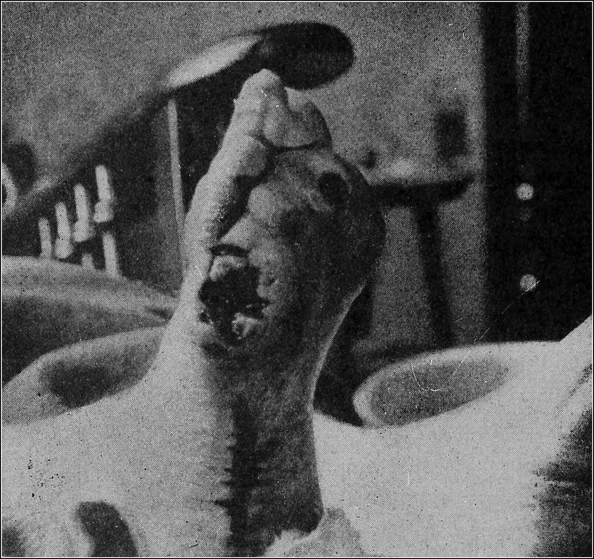
CHRONIC ULCER OF THE FOOT
(BEFORE OPERATION)
Treatment. This naturally depends upon the stage at which the ulcer is seen and the conditions present. If there is considerable inflammation, accompanied by marked cellulitis and pain, wet dressings are indicated. Two distinct therapeutic actions may be derived from the wet compress, depending upon whether or not an impervious covering is employed. These actions are antiphlogistic and hyperemic, and these in turn may be either antiseptic or astringent. The wet dressing, without a covering, is cleansing and heat reducing, because of evaporation. There should be frequent replenishment of the solution where there is considerable discharge, or where it is desirable to reduce inflammation. A wet dressing with an impervious covering is contra-indicated in the presence of pus, the warmth and moisture of such a dressing, being congenial to the growth and to the multiplication of bacteria. For the relief of pain and for the reduction of inflammation, wet dressings are the most effective form of treatment because (1) they are aseptic; (2) they permit free drainage; (3) no new granulations are disturbed in changing the dressing.
A great many different solutions are used and among these are: (1) sterile water; (2) ordinary saline solution (a teaspoonful of salt to a pint of water); (3) saturated solution of boric acid (prepared by dissolving a teaspoonful of boric acid in a pint of water); (4) Thiersch’s solution (prepared by dissolving 15 grains of salicylic acid and 90 grains of boric acid in a pint of water); (5) Burow’s solution (prepared by dissolving 675 grains of alum and 270 grains of lead acetate in a pint of water); (6) solution of bichloride of mercury (varying in strength from 1 to 3,000 to 1 to 10,000); (7) lead and opium wash (U. S. P.); (8) Dakin’s solution (hypochlorite of soda).
After the reduction of the inflammation, the next step is the cleansing and sterilization of the ulcer. Before healthy granulations can form, the removal of sloughs and the cleansing of the base must be accomplished as thoroughly as possible. Many means toward this end may be effective. A one-half to two per cent. creolin or lysol emulsion is very useful for those dirty ulcers from which a profuse, foul discharge escapes. A one per cent. solution of formalin is of great value for smaller ulcers, especially those due to tuberculous disease. The destruction and removal of sloughs may be hastened by cauterization with the solid stick of nitrate of silver. The use of certain ferments, such as brewer’s yeast, papoid, or protonuclein, may help to clean up a chronic ulcer. The most frequent means employed for the cleansing and sterilization of the ulcer, previous to the application of some stimulating dressing, is washing the part with tincture of green soap and water. Peroxide of hydrogen can next be used, then sulphuric ether, and finally ninety-five per cent. alcohol. Where there is an accompanying eczematous condition, the scales can best be removed with benzine.
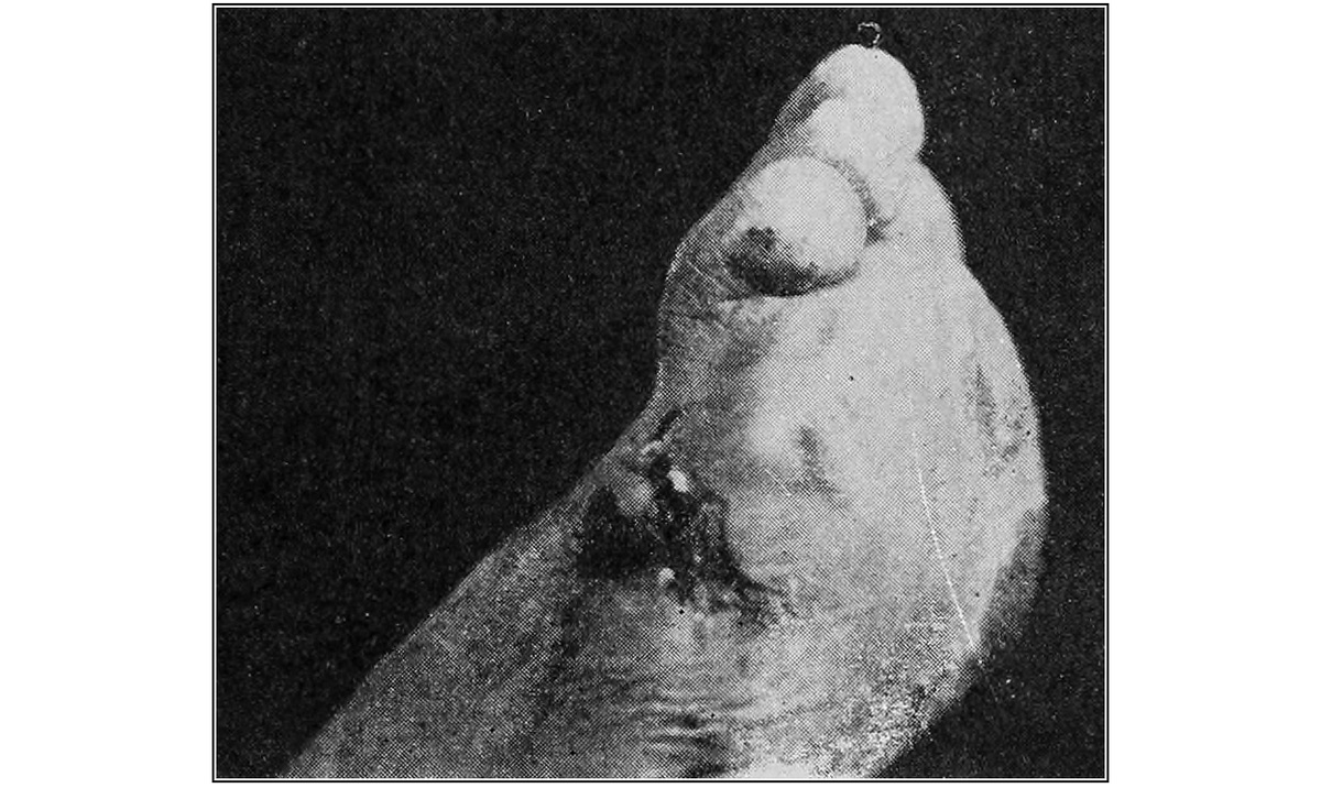
CHRONIC ULCER OF THE FOOT
(AFTER OPERATION)
Having reduced the inflammation and succeeded in cleansing the ulcer, the next thing to consider is the means by which granulations may be stimulated. This may be accomplished by applications in the form of powders, solutions, ointments and grafts.
Dusting powders are employed either as antiseptics or as astringents or for both purposes. Their use in this instance is limited, and they are employed only where the secretion is scanty. Among the various powders used are: aristol, dermatol, boric acid, orthoform, calomel, protonuclein, alum, zinc oxide, etc. Thymol iodide, or aristol, is a superior antiseptic powder and enjoys the advantage over iodoform of being inodorous. Iodoform should be used only in tuberculous conditions; calomel only in syphilitic cases. Dermatol, or bismuth subgallate, combines the astringent and mildly antiseptic qualities of bismuth and gallic acid. Boric acid is mildly antiseptic. Zinc oxide and alum are both astringent. Scarlet red, five per cent., with boric acid, ninety-five per cent., is indicated when the granulations are sluggish.
Among the various solutions used are silver nitrate in various strengths, zinc and copper sulphate, ichthyol, balsam of Peru, and calamine. Silver nitrate, zinc and copper sulphates are employed for their astringent action. Balsam of Peru, fifty per cent., with castor oil, fifty per cent., is used for its stimulating action.
Ointments are used in the treatment of ulcers either to stimulate the granulations or to soften thick epidermis. Ointments should never be employed where there is a profuse discharge. Many different kinds of ointments are used, prominent among them being: balsam of Peru, in a ten per cent. strength for the stimulation of the granulations; boric acid and ichthyol, in the same strength; Lassar’s paste (which consists of salicylic acid, one dram; starch and zinc oxide, each one ounce, and vaseline to make four ounces). This latter ointment is especially indicated when there is an eczema present. An ointment which has given good results is scarlet red, 1% to 5%. Scarlet red (Biebrich) was originally prepared as a dye for wool and silk, and is so named because of the fact that it was first manufactured in the town of Biebrich. Its application to granulating surfaces induces healing, not by the formation of scar tissue, but in every case by producing a high grade of normal skin which very soon becomes freely movable on the underlying tissue. The return of sensation in the healed area takes place from the periphery inward, instead of upward from the underlying tissue. Usually the dressing should be left undisturbed for from twenty-four to forty-eight hours, then reapplied, as indications warrant. In removing the dressing, if it be adherent to the granulations, peroxide of hydrogen should be used to loosen it. The skin about the granulating surface is best cleansed by benzine, as this removes all traces of scarlet red better than any other solution. The following formulas are recommended:
Scarlet red (medicinal Biebrich) fifteen grains; ungt. acidi borici, q. s. ad three ounces (one per cent.).
Scarlet red (medicinal Biebrich), forty-five grains; ungt. zinci oxidi, q. s. ad three ounces (three per cent.).
Scarlet red (medicinal Biebrich), seventy-five grains; balsam of Peru, seventy-five minims; petrolati, q. s. ad three ounces (five per cent.).
The first is indicated where scarlet red is desired over a large area and for a long time; the second, where an astringent action is required because the granulations are profuse; the third, where the granulations are sluggish and require stimulation.
VARICOSE ULCER.
Etiology. To chronic ulcers of the leg, associated with varicose veins, especially of the smaller venous radicles, the name varicose ulcer has been given. The usual development of this variety of ulcer is as follows: persons who suffer from varices of the leg usually complain, for some time before the external manifestation of the disease, of a dull, aching pain in the limb, with a sense of weight, fullness and fatigue. In a more advanced state of the disease the ankles swell after a day’s hard work, and the feet are constantly cold; an embarrassed state of circulation is denoted by these symptoms and the deep-seated veins begin to swell. After a time, which varies with the idiosyncrasy and occupation of the patient, small, soft, blue tumors are seen at different points of the leg, most of them disappearing on pressure, but returning when it is removed, or when the patient stands up. Each little tumor is caused by a vein, dilated at the point at which it is joined by an intramuscular branch. Around many of these tumors a number of minor vessels of a dark purple color are clustered; these are the small superficial veins which enter the dilating vein and in which the passage of the blood is retarded. An increasing area of veins gradually becomes involved and a number of irregular, knotty, consolidated tumors are developed, grouping themselves around the point at which the dilatation first began. The external and internal saphenous veins are those primarily affected, but long tracts of tortuous veins may extend up the leg and thigh. Dangerous and even fatal hemorrhage may ensue from the bursting of a varix through the skin. The vessels may become filled with clots and permanently obstructed, and ulceration with thrombosis or phlebitis may be the sequel. The capillaries become engorged with blood, and hence the assimilation changes are retarded and sometimes altogether checked. Gradually the entire circulation of the part is arrested. The vitality of the superficial structures becomes permanently impaired; consequently they are unable to resist the effects of slight injuries and repair fails to take place after a portion has been destroyed, and an open sore or ulcer is established.
Symptoms. The varicose ulcer is usually single, oval, round or irregular in outline, and is most often seen on the lower third of the leg near the internal or external malleolus. The edges are thick, everted, and swollen. The swelling is largely due to edema and is found to pit on pressure. The floor is generally covered with rather large granulations which bleed freely when touched. In a varicose ulcer the destruction of tissue often begins at the margin of a congested area and advances toward the centre. The size varies from the small ulcers, less than one-half inch in diameter, formed by the breaking down of an area of periphlebitis around a small vein, to those several inches in diameter. Several ulcers may be present on one limb. The granulations, as a rule, are weak and flabby. The discharge is thin, serous, mixed with débris, and may be blood-stained. The skin surrounding a varicose ulcer is often of a brownish blue color, due to a deposit of pigment. The recognition of varicose ulcers is usually easy; but the mere presence of enlarged veins, it should be noted, is not pathognomonic, because they may exist along with ulcers of other origin—the luetic, trophic, etc. The most frequent complication is phlebitis; cellulitis is also seen. This latter may sometimes be so severe as to necessitate operation. Complications such as necrosis of bone, involvement and ankylosis of the ankle joint, together with atrophy and contracture of muscles and adhesions of tendons (perhaps giving rise to various deformities of the feet, such as flat foot or even club-foot) are extreme and unusual complications.
Where the varicose ulcers have persisted for a long time and refuse to heal, it is always advisable to apply the Wassermann test in order to exclude the possibility of syphilis. In doubtful cases it is also advisable to test by the Noguchi luetin skin reaction.
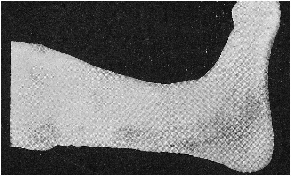
VARICOSE ULCER
Treatment. In these cases of varicose ulcers it is impossible to effect a cure until the chronic congestion of the limb is relieved and the blood supply of the part approaches normal. Often all that is necessary is a gauze, muslin, rubber or flannel bandage.
A bandage, when applied with moderate, even pressure, has for its purpose the relief of congestion. In a great many cases rubber has an irritating effect upon the skin, and that kind of a bandage should therefore be cautiously used. When the granulations are almost on a level with the skin, and also where there is considerable thickening of the edges of the ulcer, the best means of keeping up an even pressure and causing absorption of the thickened margins, as well as of hastening epithelial growth, is to apply zinc oxide adhesive plaster in strips, one-half to one inch in width. These strips should overlap to the extent of about one-third of their width, should extend about three-fourths of the way around the limb, and should be evenly and smoothly applied. They should be started about one inch below the ulcer and should run from two to three inches above it.
In order to effect a permanent cure, varicose veins must be operated upon, and a number of operations have been devised, as follows: the ligation of the internal saphenous, as advised by Trendelenburg; the multiple percutaneous ligations of Schede; the total extirpation of the internal saphenous, as recommended by Mayo; the dissection after the method of Madelung; and the spiral of Rindfleisch.
Perforating Ulcer of the Foot. This type of ulcer usually occurs where pressure and irritation are greatest and is therefore commonly found on the plantar surface of the foot under the heads of the first and fifth metatarsal bones, and on the under surface of the great toe. Occasionally, however, they develop on the dorsal surfaces or ends of the toes, in cases such as hammer toe.
Etiology. There are various theories relative to the causation of lesions of this nature. One claims injury to be the sole cause; another attributes it to arteriosclerosis and capillary thrombosis; still another charges it to chronic peripheral neuritis and alteration in the nerve terminals. One writer states that traumatism is an important factor in their development, conceding, however, that various systemic conditions must necessarily enter into the etiology, among them, locomotor ataxia and injuries to the spinal cord, diabetes and injuries to the peripheral nerves. This latter, known as the “mixed theory,” is the one most generally accredited and is in all probability most correct.
This type of ulcer is found more frequently in males than in females and it occurs almost exclusively in adult life (between 40 and 60 years). Occupation is a predisposing factor, and work demanding long periods of standing or walking unquestionably has much to do with the development of a perforating ulcer, all other conditions being equal.
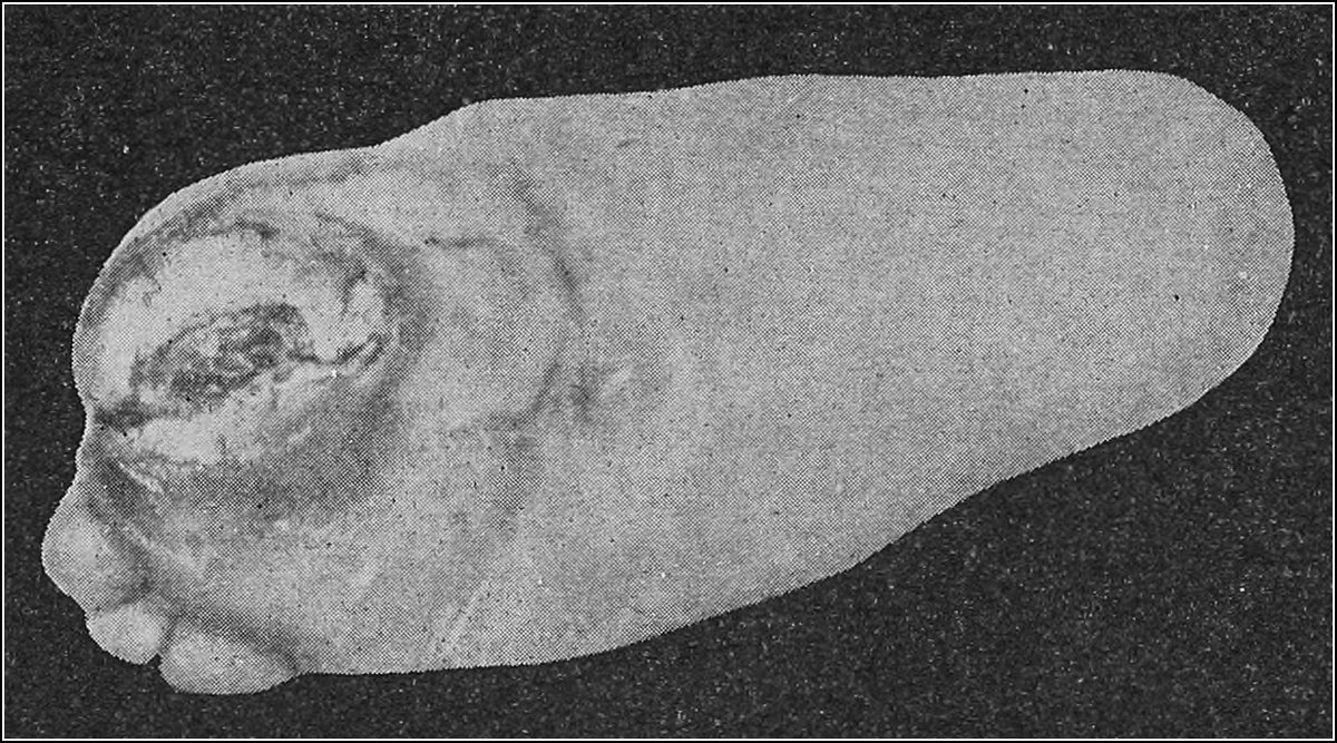
POST-OPERATIVE DIABETIC ULCER
Characteristics. The ulcer is usually found to be irregularly circular in shape, with a tendency to progressive development, involving the deeper soft tissues, finally attacking the periosteum and the bone itself, causing necrosis. The superficial edges of the ulceration are heavily calloused and the lesion shows little or no tendency to heal. One of the most marked characteristics is the entire loss of sensation. Many cases have been observed where the patient feels no pain, even when the lesion is deeply probed.
Symptoms. At times, particularly in diabetic patients, a purulent blister is the initial lesion, but in most instances these lesions develop under a heavy callous, the centre of which breaks down into an indolent superficial ulceration, discharging a thin, discolored, odorous pus, but never in great quantities.
The fact that changes in the peripheral nerve supply usually take place in the development of perforating ulcer probably accounts for the absence of pain, as above mentioned, and also explains the progressive degeneration which takes place, allowing the ulcerative process to progress into the deeper tissues.
Treatment. The systemic disturbances which may be present are important factors to be considered in the treatment of perforating ulcer, but local applications may be made and local conditions must be considered. If the ulcer be upon the plantar surfaces of the foot, walking and standing, which would bring continued pressure, must be avoided. Shoes must be well fitted and must not irritate the parts, and cleanliness must be obtained and maintained. All callous must be removed from the edges of the ulcer and proper drainage is of great importance. All necrosed tissue must necessarily be removed and any burrowing sinuses should be thoroughly opened. Artificial hyperemia, massage and electricity are found to be of benefit in improving the general circulation in the foot and leg.
In the local treatment of the ulceration itself, prolonged application of strong germicidal solutions is to be avoided at all times. Cleansing with warm normal salt solution is recommended as a non-toxic and stimulant application.
Dressings may be of plain aseptic or iodoform gauze packed lightly into the ulcer. These lesions are discouraging to treat, inasmuch as even after complete healing, relapses usually occur which leave the parts as bad or worse than the original lesion.
Stimulant applications may be employed locally, with some success in connection with internal medications for the systemic disturbance present. Balsam of Peru or scarlet red (1% to 3%) are advocated in this connection.
The prognosis in cases of perforating ulcer is bad, inasmuch as the progress of the lesion sooner or later involves sufficient tissue in the degenerative process to necessitate surgical interference—perhaps amputation of the foot. As has been previously mentioned, even when fully healed, relapse almost always occurs.
The Syphilitic Ulcer. The syphilitic ulcers do not properly come within the province of the podiatrist for treatment, but he should be able to recognize them. They may develop from pustules or begin as original lesions in the tertiary stages of the disease. Developing in this latter instance from gummata, they are immediately deep ulcers.
The worst superficial ulcers of syphilis may develop early in the course of the general disease.
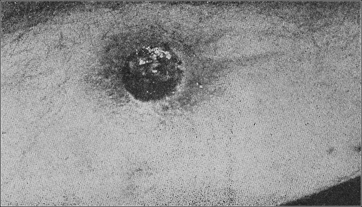
SYPHILITIC ULCER OF THE LEG
Symptoms. These ulcers vary in size from a quarter to a silver dollar and occur on the upper third of the leg, occasionally on the upper part of the middle third. During the early stages of the lesion it is surrounded by an inflamed area of skin at the ulcer and presents an even, “punched out” edge. Being a new growth, developed in the corium, the edges are usually more firm and dense than in other forms of ulcer. The floor of the lesion is of a dusky red or coppery color, and has a characteristic slough of a greenish color. The discharge is frequently bloody and is filled with broken-down tissue.
If on account of the presence of enlarged veins, it is difficult to distinguish a syphilitic from a varicose or other type of ulcer, a positive Wassermann test will confirm the diagnosis.
Being merely a local manifestation of a general infection, the systemic disturbance must be treated by a licensed physician. It is generally found that a lesion of this type, once healed, remains so.
Treatment. Treatment for syphilitic ulcers comprehends the use of mercurials as local applications. Mercury bichloride ¹⁄₁₀₀₀₀ may be employed with beneficent results in most cases where a profuse discharge is present. Where there is little or no discharge, calomel powder dusted into the ulcer will give good results.
As in most cases where a syphilitic lesion has developed locally on the leg, the patient is or has been under a physician’s care, practically none of these cases come to the podiatrist for his treatment alone. Many times, however, he is called in by the physician to do local dressings under his direction, and it has even happened that the podiatrist has been the first to recognize the significance of the local lesion.