Chapter VIII
Reading edition
ORTHOPTERA—FORFICULIDAE, EARWIGS—HEMIMERIDAE
Order II.—Orthoptera.
Insects with the mouth parts conspicuous, formed for biting, the four palpi very distinct, the lower lip longitudinally divided in the middle. The tegmina (mesothoracic wings), of parchment-like consistence, in repose closed on the back of the Insect so as to protect it. The metathoracic wings, of more delicate consistence, ample, furnished with radiating or divergent nervures starting from the point of articulation, and with short cross nervules forming a sort of network; in repose collapsing like a fan, and more or less completely covered by the tegmina (except in certain Phasmidae, where, though the wings are ample, the tegmina are minute, so that the wings are uncovered). In a few forms (winged Forficulidae and some Blattidae) the metathoracic wings are, in addition to the longitudinal folding, contracted by means of one or two transverse folds. The mode of growth of each individual is a gradual increase of size, without any abrupt change of form, except that the wings are only fully developed in the final condition. There is no special pupal instar. Species in which the wings are absent or rudimentary are numerous.
The Orthoptera are Insects of comparatively large size. The Order, indeed, includes the largest of existing Insects, while none are so minute as many of the members of the other Orders are; three millimetres is the least length known for an Orthopterous Insect, and there are very few so small, though this is ten times the length of the smallest beetle. The Order includes earwigs, cockroaches, soothsayers or praying-insects, stick- and leaf-insects, grasshoppers, locusts, green grasshoppers, and crickets.
The changes of form that accompany the growth of the individual are much less abrupt and conspicuous than they are in most other Insects. The metamorphosis is therefore called Paurometabolous. It has been supposed by some naturalists that Orthoptera go through a larger portion of their development in the egg than other Insects do. This does not clearly appear to be the case, though it seems that there are distinctions of a general character in the embryology; the period of development in the egg is prolonged, and the yolk is said by Wheeler[126] to be more than usually abundant in comparison with the size of the young embryo. The embryonic development may in tropical countries be accomplished in three weeks (see Mantidae), but in countries where winter supervenes, the period may in some species be extended over seven or eight months.
The external features of the post-embryonic development—a term that is more convenient in connexion with Orthoptera than metamorphosis—are as follows: the wings are never present when the Insect is first hatched, but appear subsequently, and increase in size at the moults; the form and proportions of the segments of the body—especially of the thorax—undergo much change; an alteration of colour occurs at some of the moults, and the integument becomes harder in the adult condition. Neither the development of the internal organs, nor the physiological processes by which the changes of external form are effected, appear to have been studied to any great extent.
Many of the Orthoptera do not possess wings fit for flight, and some species even in the adult state have no trace whatever of such organs. Flight, indeed, appears to be of minor importance in the Order; in many cases where the wings exist they are purely musical organs, and are not of any use for flight. The apterous and the flightless conditions are not confined to one division of the Order, but are found in all the families and in many of their subdivisions. As the front pair of wings in Orthoptera do not really carry out the function of flight, and as they differ in several particulars from the hinder pair, or true wings, it is usual to call them tegmina. The musical powers of the Orthoptera are confined to the saltatorial group of families.
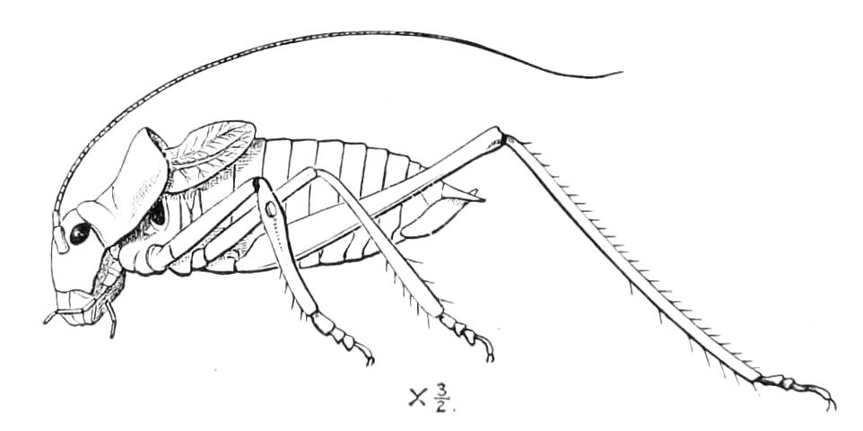
Fig. 101.—Poecilimon affinis ♂. Bulgaria. Alar organs serving only as musical organs. The ear on front tibia and aural orifice of prothorax are well shown.
The Cursoria are dumb or nearly so; it is a remarkable fact that also in this latter division the alar organs, though frequently present, have but little value for flight, and are in some cases devoted to what we may call purposes of ornament or concealment. This is specially the case in the Phasmidae and Mantidae, where the effectiveness of colour and pattern of these parts becomes truly astonishing. The tegmina frequently exhibit an extraordinary resemblance to vegetable structures, and this appearance is not superficial, for it may be seen that the nervures of the wings in their disposition and appearance resemble almost exactly the ribs of leaves. One of the most remarkable of the features of Orthoptera is that a great difference frequently exists between the colours of the tegmina and of the wings, i.e. the front and hind wings; the latter are concealed in the condition of repose, but when activity is entered on and they are displayed, the individual becomes in appearance a totally different creature. In some cases, contrary to what usually occurs in Insects, it is the female that is most remarkable; the male in Mantidae and Phasmidae being frequently a creature of quite inferior appearance and power in comparison with his consort. The musical powers of the saltatorial Orthoptera are, however, specially characteristic of the male sex. There is evidence that these powers are of great importance to the creatures, though in what way is far from clear. Some parts of the structures of the body are in many of these musical species clearly dominated by the musical organs, and are apparently specially directed to securing their efficiency. We find in some Locustidae that the tegmina are nothing but sound-producing instruments, while the pronotum is prolonged to form a hood that protects them without encumbering their action. In the males of the Pneumorides, where the phonetic organ is situated on the abdomen, this part of the body is inflated and tense, no doubt with the result of increasing the volume and quality of the sound. In the genus Methone (Fig. 185) we find a grasshopper whose great hind legs have no saltatorial function, and but little power of locomotion, but act as parts of a sound-producing instrument, and as agents for protecting some parts of the body in repose. Further particulars of these cases must be looked for in our accounts of the different groups.
The eggs of many Orthoptera are deposited in capsules or cases; these capsules may contain only one egg, or a great many.
The Order includes many species of Insects, though in Britain it is poorly represented: we have only about forty species, and this small number includes some that are naturalised. Only a few of the forty extend their range to Scotland. A revision of the species found in Britain has recently been made by Mr. Eland Shaw.[127] In continental Europe, especially in the south, the species become more numerous; about 500 are known as inhabitants of geographical Europe. In countries where the face of nature has been less transformed by the operations of man, and especially in the tropical parts of the world, Orthoptera are much more abundant.
The lowest number at which the species now existing on the surface of the earth can be estimated is 10,000. This, however, is probably far under the mark, for the smaller and more obscure species of Orthoptera have never been thoroughly collected in any tropical continental region, while new forms of even the largest size are still frequently discovered in the tropics.
We shall treat the Order as composed of eight families:—
Series, Cursoria: hind legs but little different from the others. 1. Forficulidae—Tegmina short, wings complexly folded; body armed at the extremity with strong forceps.

2. Hemimeridae—Apterous: head exserted, constricted behind.
3. Blattidae—Coxae of the legs large, exserted, protecting the lower part of the body.
4. Mantidae—Front legs very large, raptorial, armed with spines.
5. Phasmidae—Mesothorax large as compared with the prothorax.
Series, Saltatoria: hind legs elongate, formed for leaping, their femora usually thickened. 6. Acridiidae—Antennae short, not setaceous, of not more than 30 joints, tarsi three-jointed.

7. Locustidae—Antennae very long, setaceous, composed of a large number of joints, tarsi four-jointed.
8. Gryllidae—Antennae very long, setaceous, tarsi two- or three-jointed.
The first five of these subdivisions are amongst the most distinct of any that exist in the Insecta, there being no connecting links between them. The three groups forming the Saltatoria are much more intimately allied, and should, taken together, probably have only the same taxonomic value as any one of the other five groups.
Owing partly to the inherent difficulties of the subject, and partly to the fragmentary manner in which it has been treated by systematists, it has been impossible till recently to form any clear idea of the classification of Orthoptera. During the last twenty years Henri de Saussure and Brunner von Wattenwyl have greatly elucidated this subject. The latter of these two distinguished naturalists has recently published[128] a revision of the system of Orthoptera, which will be of great assistance to those who may wish to study these Insects. We therefore reproduce from it the characters of the tribes, placing the portion relating to each family at the end of our sketch thereof.
Fam. I. Forficulidae—Earwigs.
(DERMAPTERA OR DERMATOPTERA OF BRAUER AND OTHERS)
Insects of elongate form, with an imbricate arrangement of the segments of the body; bearing at the posterior extremity a pair of callipers or more distorted instruments. The hind wings (when present) folded in a complex manner, and covered, except at their tips, by a pair of short wing-covers (tegmina), of a leather-like consistence. Wingless forms are very numerous. The young is very similar to the adult.
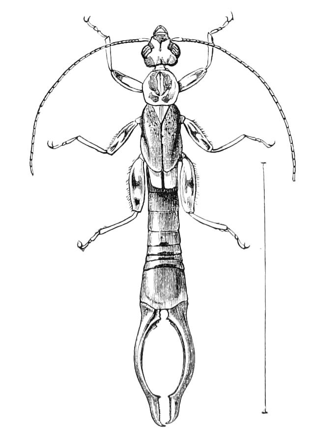
Fig. 102.—Pygidicrana hugeli. Java.
Although earwigs are said to be rare in most parts of the world, yet in Europe no Insect is better known than Forficula auricularia, the common earwig, it being very abundant even in gardens and cultivated places. In certain seasons it not unfrequently enters our houses, in which case it too often falls a victim to prejudices that have very little to justify them. This Insect is a good type of the winged earwigs. In the parts of the mouth it exhibits the structures usual in the Orthoptera; there is a large labrum, a pair of maxillae, each provided with two lobes and a palpus consisting of two very short basal joints and three longer joints beyond these; the mandibles are strong, with curvate pointed extremities; in the lower lip there is a ligula exposed in front of a very large mentum; it consists of two pieces, not joined together along the middle, but each bearing on its lateral edge a palpus with two elongate joints and a short basal one; this lip is completed by the lingua, which reposes on the upper face of the part, and completely overlaps and protects the chink left by the want of union along the middle line of the external parts of the lip. The antennae are elongate, filiform, and are borne very near the front of the exserted head. There are rather large facetted eyes, but no ocelli. The three segments of the thorax are distinct, the prothorax being quite free and capable of movement independent of the parts behind it: the meso- and meta-nota are covered by the tegmina and wings; these latter project slightly from underneath the former in the shape of small slips, that are often of rather lighter colour; the wing-covers are short, not extending beyond the insertion of the hind legs, and repose flat on the back, meeting together in a straight line along the middle. These peculiar flat, abbreviated wing-covers, with small slips (which are portions of the folded wings) projecting a little from underneath them, are distinctive marks of the winged Forficulidae.
The legs are inserted far from one another, the coxae being small; each sternum of the three thoracic segments projects backwards, forming a peculiar long, free fold, underlapping the front part of the following segment. The hind body or abdomen is elongate, and is formed of ten segments; the number readily visible being two less in the female than it is in the male. The segments are fitted together by a complex imbrication, which admits of great mobility and distension, while offering a remarkable power of resistance to external pressure: each segment is inserted far forward in the interior of that preceding it, and each also consists of separate upper and lower plates that much overlap where they meet at the sides (see Fig. 103). The body is always terminated by a pair of horny, pincer-like processes, which are differently shaped according to the sex of the individual.
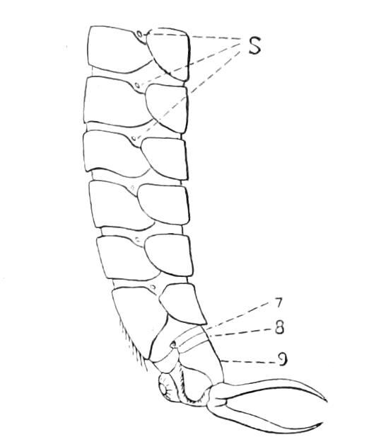
Fig. 103.—Lateral view of Forficula auricularia L. Female abdomen distended showing spiracles, S, and the small 8th and 9th dorsal plates (7 and 8 in Fig.).
The structure of the abdomen in the earwig has given rise to considerable discussion. In Fig. 103 we reproduce Westwood's diagram of it as seen fully distended in a female specimen; in this state the minute spiracles can be detected, though in the normal condition of the body they cannot be seen, being placed on the delicate membranes that connect the chitinous plates. Westwood's interpretation of the structure was not, however, quite correct, as the part which he considered to be the first dorsal plate is really the second; so that the segments numbered 7, 8, 9 in our figure are really 8, 9, 10. The common earwig is interesting as exhibiting, in an imperfect state, the union of the first dorsal plate of the abdomen with the thorax; a condition which is carried to so great an extent in the Hymenoptera as to quite obscure the nature of the parts, and which has consequently given rise to much perplexity and discussion. We represent this structure as seen in the common earwig in Fig. 104, where a represents the pronotum, b the mesonotum, c the metanotum, d the first, f the second abdominal segment; e being a delicate membrane of considerable size that intervenes between the two, and which is more exposed than are the corresponding membranes connecting the subsequent rings; a condition similar to that which is found in Cimbex, Cephus, and some other Hymenoptera.
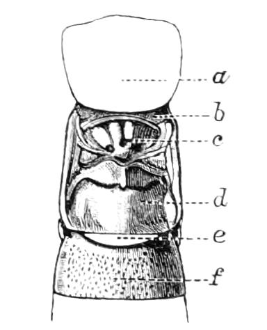
Fig. 104.—Dorsal portions of the middle segments of body of Forficula auricularia (tegmina and wings removed).
On the under surface of the abdomen of the earwig the full number of 10 plates cannot be superficially distinguished; but it is found by dissection that in the female the short eighth and ninth dorsal rings are joined on the ventral aspect by a delicate membrane, while the tenth ventral is of a less delicate nature, and forms a triangular plate at the base of each half of the forceps. Between the branches of the forceps there is a perpendicular plate, the pygidium of Orthopterists, possibly the unpaired terminal portion of the body seen in some embryos, and called the telson. The pygidium is a separate sclerite, though it looks as if it were only a portion of the large tenth dorsal plate bent downwards, and in some descriptive works is erroneously described as being such.
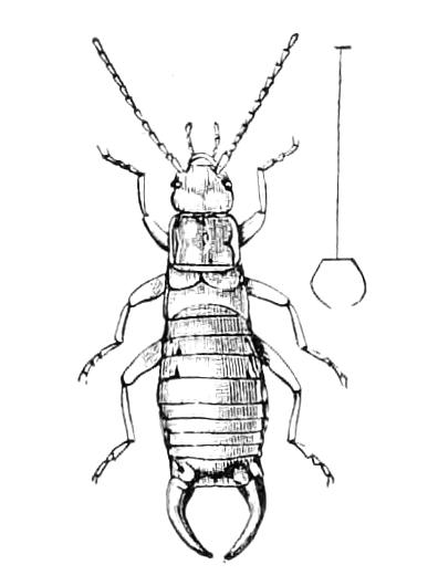
Fig. 105.—Chelidura dilatata, male. Pyrenees.
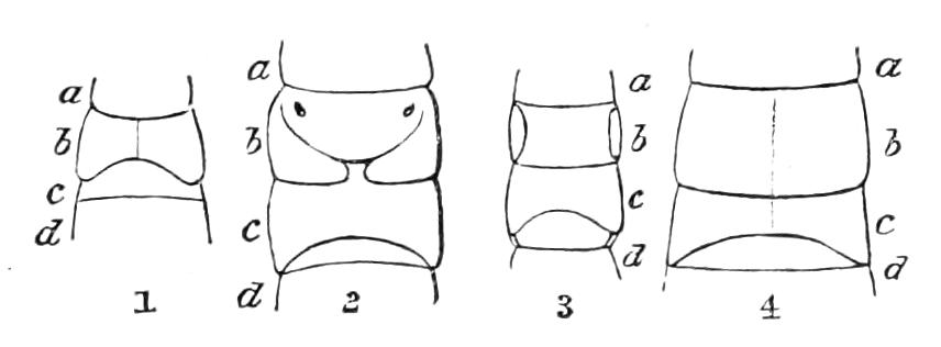
Fig. 106.—Tegmina and wings (visible in part or invisible) of apterous earwigs. 1, Chelidura sp.; 2, Chelidura dilatata; 3, Anisolabis moesta; 4. A. maritima. a, First thoracic segment; b, second; c, third; d, basal portion of abdomen.
A very large number of species of Forficulidae have the organs of flight undeveloped. Fig. 105 represents Chelidura dilatata, an apterous form that is very common in the Eastern Pyrenees. The condition of the meso- and meta-nota—the parts from which the tegmina and wings are developed, and to which they are attached when present—is very remarkable in these forms, and exhibits much variety. In Fig. 106 we represent the conditions of these parts in a few apterous forms. The tegmina or the segment from which they are developed (b), are seen in the shape of a plate which may extend all across the middle and be undivided (No. 4); in which case the appearance indicates entire absence of the tegmina; these are, on the contrary, evidently present in the form of slips grafted one to each side of the second thoracic segment in Anisolabis (No. 3); or they may look like short broad slips extending all across the body, and marking off a piece frequently called a scutellum, but which is really the mesonotum (some species of Chelidura, as No. 2); or, again, they may be nearly free tegmina, somewhat similar to those of the winged forms; this is the case with some species of Chelidura, as represented by No. 1. This last figure is taken from a species from the Sierra Nevada, apparently undescribed, allied to C. bolivari.
In the cases we are considering no analogous structures exist on the metanotum (the part of the body that in the winged forms bears the wings, and which is marked c in our diagrams, Fig. 106), so that the tegmina are to all appearance less rudimentary (or vestigial) than the wings. The metanotum forms a sort of flap, called by Fischer[129] "involucrum alarum"; he considered the part immediately behind this to be the metanotum; this piece is, however, no doubt really part of the abdomen (d in our Figure). This is apparently the view taken by Brunner.[130] The structure of these parts is important as bearing on the subject of the nature and origin of Insects' wings, a question to which no satisfactory answer has yet been given. The appearances we have remarked on are to some extent similar to the conditions existing in the immature state of the organs of flight in the common earwig (see Fig. 112, p. 212), but whether the varieties presented by the wingless forms have parallels in the immature conditions of the various winged forms is quite uncertain, the life-histories of earwigs being almost unknown.
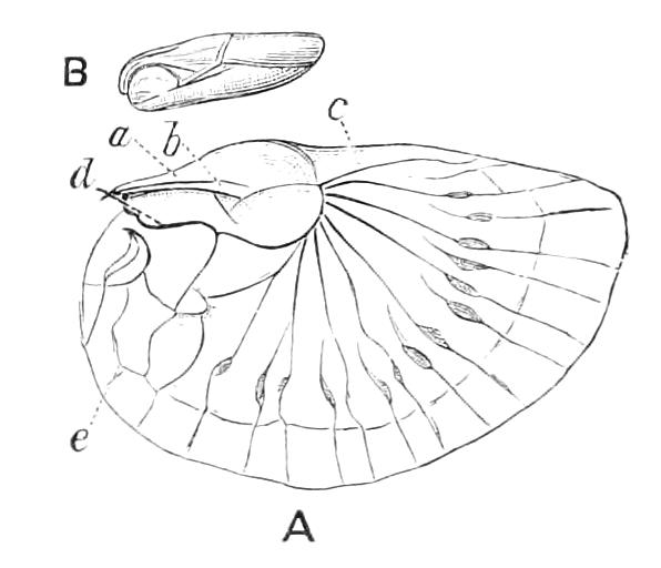
Fig. 107.—Wing of Forficula auricularia. A, Wing expanded, explanation in text; B, wing folded and packed.
The developed wings of earwigs are worthy of attention, both as regards their actual structure and the manner in which they are folded up in repose. When expanded they have a shape curiously suggestive of the human ear. The chief parts of the wing, as shown in Fig. 107, A, are a, b, two portions of the horny piece that forms the scale which covers the more delicate parts of the wing when it is folded, and which, according to Brunner, represents the radial and ulnar fields of the wings of Acridiidae and Locustidae (see Fig. 167); c is the small apical field limited below by the vena dividens; d is the vena plicata which runs along the under side of the scale as far as the apical field, where it gives off the axillary nerves; e is a vena spuria, or adventitious vein such as exists in many other Orthoptera with delicate wings. On the front part of the scale, a, and on a different plane so that it is not shown in our figure, there is a very delicate small band which is supposed to represent the marginal field of the wing of other Orthoptera. There are, however, grave difficulties in the way of accepting this view of the earwig's wing, amongst which we may mention the position of the vena dividens and its relation to the so-called radial and ulnar fields of the wing. The wings are remarkable for their delicacy; moreover, the way in which they fold up so as to be packed in the manner shown in B, Fig. 107, is very interesting, there being, in fact, no other Insects that fold up their wings in so complicated and compact a fashion as the earwigs do. The process is carried out somewhat as follows: the longer radii come a little nearer together, the delicate membrane between them falling into folds somewhat like those of a paper fan; a transverse fold, or turn-over, then occurs at the point where the radii, or axillary nerves, start from the vena plicata; then a second transverse fold, but in a reversed direction, occurs affecting the wing just close to the spots where the shorter radial nervures are dilated; then by a contraction close to the scale the whole series of complex folds and double are brought together and compressed.
It is quite a mystery why earwigs should fold their wings in this complex manner, and it is still more remarkable that the Insects very rarely use them. Indeed, though Forficula auricularia is scarcely surpassed in numbers by any British Insect, yet it is rarely seen on the wing; it is probable that the majority of the individuals of this species may never make use of their organs of flight or go through the complex process of unfolding and folding them. It should be remarked that no part of the delicate membranous expanse of the wing is exposed when the wings are packed in their position of repose; for the portion that projects from under the tegmina—and which, it will be remembered, is always present, for when wings exist in earwigs they are never entirely concealed by the tegmina—is, it is curious to note, of hard texture, and is frequently coloured and sculptured in harmony with the tegmen; in fact, one small part of the wing forms in colour and texture a most striking contrast to the rest of the organ, but agrees in these respects with the wing-covers. This condition is seen in Fig. 108, where B shows the sculpture of the tegmina t, and of the projecting tips of the wings w. There are numerous other instances in Orthoptera where one part of a wing or wing-case is exposed and the other part concealed, and the exposed portion is totally different in colour and texture from the concealed portion.
The wings of earwigs are attached to the body in a very unusual manner; each wing is continued inwards on the upper surface of the metanotum, as if it were a layer of the integument meeting its fellow on the mesial line; the point of contact forming two angles just behind the metanotum.
Some writers have considered that the tegmina of earwigs are not the homologues of those of other Orthoptera, but are really tegulae (cf. Fig. 56, p. 103). We are not aware that any direct evidence has been produced in support of this view.
The pair of forceps with which the body is armed at its extremity forms another character almost peculiar to the earwigs, but which exists in the genus Japyx of the Thysanura. These forceps vary much in the different genera of the family; they sometimes attain a large size and assume very extraordinary and distorted shapes. They are occasionally used by the Insects as a means of completing the process of packing up the wings, but in many species it is not probable that they can be used for this purpose, because their great size and peculiarly distorted forms render them unsuitable for assisting in a delicate process of arrangement; they are, too, always present in the wingless forms of the family. Their importance to the creature is at present quite obscure; we can only compare them with the horns of Lamellicorn Coleoptera, which have hitherto proved inexplicable so far as utility is concerned. No doubt the callipers of the earwigs give them an imposing appearance, and may be of some little advantage on this account; they are not known to be used as offensive instruments for fighting, but they are occasionally brought into play for purposes of defence, the creatures using them for the infliction of nips, which, however, are by no means of a formidable character.
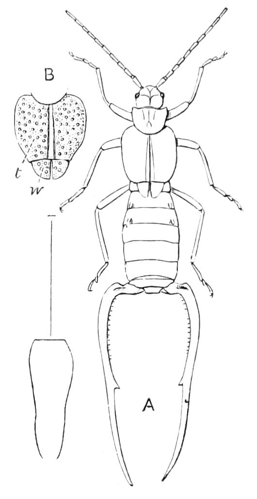
Fig. 108.—Anechura scabriuscula. Himalaya. A, Outline of the Insect; B, tegmina, t, and tips of wings, w, showing their similar sculpture.
These forceps are, in the case of the common earwig—and they have not been studied from this point of view in any other species—remarkable, because of the great variation in their development in the male, a character which again reminds us of the horns of Lamellicorn beetles: in the female they are comparatively invariable, as is also the case in the few species of Lamellicornia, which possess horned females. A and B in Figure 109 represent the forceps of different males of the common earwig, C showing those of the other sex. The subject of the variation of the male callipers of the earwig has been considered by Messrs. Bateson and Brindley,[131] who examined 1000 specimens captured on the same day on one of the Farne islands off the coast of Northumberland; 583 of these were mature males, and the pincers were found to vary in length from about 2½ mm. to 9 mm. (A and B in Fig. 109 represent two of the more extreme forms of this set of individuals.) Specimens of medium size were not, as it might perhaps have been expected they would be, the most common; there were, in fact, only about 12 individuals having the forceps of the medium length—4¾ to 5¼ mm., while there were no less than 90 individuals having forceps of a length of about 7 mm., and 120 with a length of from 2¾ to 3¼. Males with a medium large length of the organ and with a medium small length thereof were the most abundant, so that a sort of dimorphism was found to exist. Similar relations were detected in the length of the horns of the male of a Lamellicorn beetle examined by these gentlemen. In the case of the set of earwigs we have mentioned, very little variation existed in the length of the forceps in the female sex.
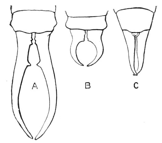
Fig. 109.—Forceps of the common earwig: A, of large male; B, of small male; C, of female.
In many earwigs—including F. auricularia—there may be seen on each side of the dorsal aspect of the true fourth, or of the fourth and neighbouring segments of the hind body a small elevation, called by systematists a plica or fold, and on examination the fold will be found to possess a small orifice on its posterior aspect. These folds are shown in Figs. 105 and 108; they have been made use of for purposes of classification, though no functional importance was attached to them. Meinert, however, discovered[132] that there are foetid glands in this situation, and Vosseler has recently shown[133] that the folds are connected with scent-glands, from which proceed, in all probability, the peculiar odour that is sometimes given off by the earwig. The forms destitute of the folds, e.g. Labidura, are considered to have no scent glands. There is a very peculiar series of smooth marks in the earwigs on the dorsal aspect of the abdominal segments, and these are present in the glandless forms as well as in the others.
The internal anatomy has been to some extent investigated by Dufour and Meinert. Dufour dissected F. auricularia and Labidura riparia, and found[134] that salivary glands exist in the latter Insect (called by him Forficula gigantea), though he was unable to discover them in the common earwig. According to Meinert,[135] there are, however, salivary glands affixed to the stipes of the maxillae in F. auricularia, while (in addition?) L. riparia possesses very elongate glands seated in the middle or posterior part of the breast. The alimentary canal is destitute of convolutions, but oesophagus, crop, and gizzard all exist, and the intestine behind the stomach consists of three divisions. The Malpighian tubes are numerous, 30 or 40, and elongate. The respiratory system is not highly developed. Earwigs—the European species at least—have, as already mentioned, very small powers of flight; the tracheal system is correspondingly small, and is destitute of the vesicular dilatations that are so remarkable in the migratory Locusts.
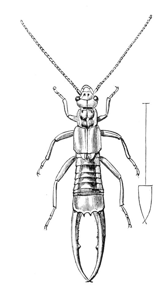
Fig. 110.—Labidura riparia, male. Europe.
The three thoracic spiracles[136] are readily observed in living individuals. There are seven pairs of abdominal spiracles, which, however, are very minute, and can only be found by distending the body as shown in Fig. 103. The ventral chain consists of nine ganglia (the sub-oesophageal centre is not alluded to by Dufour); the three thoracic are equidistant and rather small; the hindmost of the six abdominal ganglia is considerably larger than any one of the other five.
The ovaries of Labidura riparia and Forficula auricularia are extremely different. In L. riparia there are on each side five tubes, each terminating separately in an obliquely directed lateral part of the oviduct. In F. auricularia there is but one tube on each side, but it is covered by three longitudinal series of very short sub-sessile, grape-like bodies, each of the two tubes being much dilated behind the point where these bodies cease.
The testes in earwigs are peculiar and simple; they consist, on each side, of a pair of curvate tubular bodies, connected at their bases and prolonged outwards in the form of an elongate, slender vas deferens. The structures in the males of several species have been described at some length by Meinert,[137] who finds that in some species a double ejaculatory duct exists.
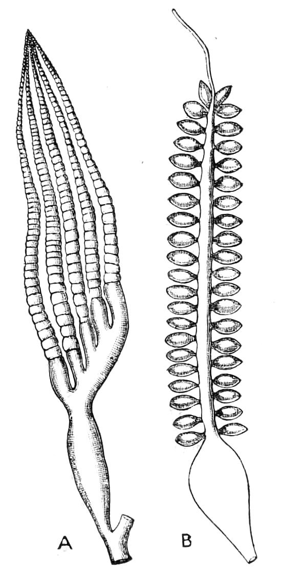
Fig. 111.—Ovaries of Labidura riparia, A; and Forficula auricularia, B. (After Dufour.)
The young is similar to the adult in form; in the winged forms it is always easy to distinguish the adult by the full development of the wings, but in the wingless forms it can only be decided with certainty that a specimen is not adult by the softer and weaker condition of the integuments. Scarcely anything appears to be known as to the life-history, except a few observations that have been made on the common earwig; Camerano found[138] that this Insect has certainly three ecdyses, and possibly an earlier one which he failed to notice, and his observation confirms the vague previous statement of Fischer. The eggs, in the neighbourhood of Turin, are deposited and hatched in the early spring; in one case they were laid on the 10th March, and the Insects issuing from them had completed their growth and were transformed into perfect Insects on the 22nd May. In the immature state the alar structures of the future imago may be detected. The tegmina-bearing sclerites, t, Fig. 112, look then somewhat like those of some of the apterous forms (Fig. 106) and, as shown in A and B, Fig. 112, do not differ greatly in the earlier and later stages. The wings, however, change much more than the tegmina do; at first (Fig. 112, A) there is but little difference between the two, though in the interior of the wing-flap some traces of a radiate arrangement can be seen, as shown at W in A, Fig. 112; in a subsequent condition the wing-pads are increased in size and are more divided, the appearance indicating that the wings themselves are present and packed about a centre, as shown in W of B, Fig. 112.
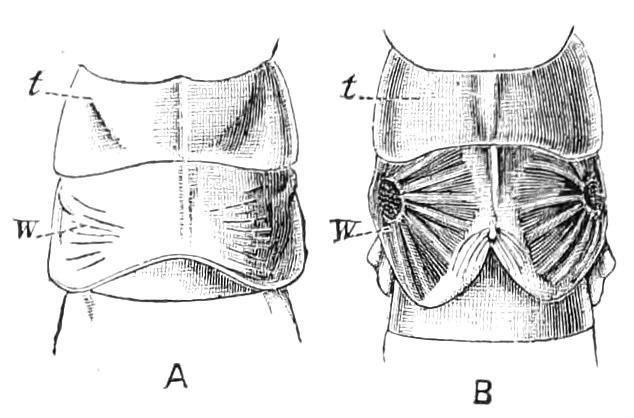
Fig. 112.—Notal plates from which the tegmina and wings of Forficula auricularia are developed in young, A, and more advanced, B, nymph.
In the young of the common earwig the number of joints[139] in the antennae increases with age. Camerano, l.c., says that before emergence from the egg there are apparently only 8 joints in the antennae, and Fischer states that the larvae of F. auricularia have at first only 8 antennal joints; later on 12 joints are commonly found, and, according to Bateson,[140] this number occasionally persists even in the adult individual. Meinert says[141] that the newly hatched Forficula has either 6 or 8 joints, and he adds that in the later portion of the preparatory stage the number is 12. Considerable discrepancy prevails in books as to the normal number of joints in the antennae of the adult F. auricularia, the statements varying from 13 to 15. The latter number may be set aside as erroneous, although it is, curiously enough, the one given in the standard works of Fischer, Brunner, and Finot. Meinert gives without hesitation 14 as the number; Bateson, l.c., found that 14 joints occurred in 70 or 80 per cent of adult individuals, that 13 was not uncommon, that 12 or 11 occasionally occurred, and that the number may differ in the two antennae of the same individual. These variations, which seem at first sight very remarkable, may with probability be considered as due to the fact that in the young state the number of joints increases with age, and that the organs are so fragile that one or more of the joints is very frequently then lost, the loss being more or less completely repaired during the subsequent development. Thus a disturbing agency exists, so that the normal number of 14 joints is often departed from, though it appears to be really natural for this species. Bateson has also pointed out that when the normal number of articulations is not present, the relative proportions of joints 3 and 4 are much disturbed. It is, however, probable that the increase in number of the joints takes place by division of the third or third and fourth joints following previous growth thereof, as in Termitidae; so that the variations, as was suggested by Bateson, may be due to mutilation of the antennae, and consequent incompletion of the normal form of the parts from which the renovation takes place; growth preceding segmentation—in some cases the growth may be like that of the adult, while the segmentation remains more incomplete. In the young the forceps of the two sexes differ but slightly; the form of the abdominal rings is, on the contrary, according to Fischer, already different in the two sexes in the early stage.
The common earwig has a very bad reputation with gardeners, who consider it to be an injurious Insect, but it is probable that the little creature is sometimes made the scapegoat for damage done by other animals; it appears to be fond of sweets, for it often makes its way to the interior of fruits, and it no doubt nibbles the petals, or other delicate parts of flowers and vegetables. Camerano, however, states, l.c., that the specimens he kept in confinement preferred dead Insects rather than the fruits he offered them. Rühl considers the earwig to be fond of a carnivorous diet, eating larvae, small snails, etc., and only attacking flowers when these fail.[142] It has a great propensity for concealing itself in places where there is only a small crevice for entry, and it is possible that its presence in fruits is due to this, rather than to any special fondness for the sweets. This habit of concealing itself in chinks and crannies in obscure places makes it an easy matter to trap the Insect by placing pieces of hollow stalks in the situations it affects; inverted flower-pots with a little hay, straw, or paper at the top are also effectual traps. We have remarked that it is very rarely seen on the wing, and though it has been supposed to fly more freely at night there is very little evidence of the fact. Another British species, Labia minor, a smaller Insect, is, however, very commonly seen flying.
Earwigs have the reputation of being fond of their young, and Camerano describes the female of the common earwig as carefully collecting its eggs when scattered, lifting them with its mandibles and placing them in a heap over which it afterwards brooded. De Geer[143] more than a century ago observed a fondness of the mother for the young. After the eggs were hatched, Camerano's individual, however, evinced no interest in the young. A larger species, Labidura riparia (Fig. 110) is said to move its eggs from place to place, so as to keep them in situations favourable for their development.
The name "earwig" is said to be due to an idea that these creatures are fond of penetrating into the ears of persons when asleep. Hence these Insects were formerly much dreaded, owing to a fear that they might penetrate even to the brain. There does not appear to be on record any occurrence that could justify such a dread, or the belief that they enter the ears. If they do not do so, it is certainly a curious fact that a superstition of the kind we have mentioned occurs in almost every country where the common earwig is abundant; for it has, in most parts of Europe, a popular name indicating the prevalence of some such idea. It is known as Ohren-wurm in German, as perce-oreille in French, and so on. The expanded wing of the earwig is in shape so very like the human ear, that one is tempted to suppose this resemblance may in former ages have given rise to the notion that the earwig has some connexion with the human ear; but this explanation is rendered very improbable by the fact that the earwig is scarcely ever seen with its wings expanded, and that it is a most difficult matter to unfold them artificially, so that it is very unlikely that the shape of the wings should have been observed by untutored peoples.
The group Forficulidae seems to be most rich in species in warm and tropical regions; several unwinged species are met with in the mountainous districts of Europe; indeed, in some spots their individuals are extremely numerous under stones. In Britain we have a list of six species, but only two of these are to be met with; the others have probably been introduced by the agency of man, and it is doubtful whether more than one of these immigrants is actually naturalised here. One of these doubtfully native species is the fine Labidura riparia (Fig. 110), which was formerly found near Bournemouth. Altogether about 400 species of earwigs are known at the present time, and as they are usually much neglected by Insect collectors, it is certain that this number will be very largely increased, so that it would be a moderate estimate to put the number of existing species at about 2000 or 3000. None of them attain a very large size, Psalis americana being one of the largest and most robust of the family; a few display brilliant colours, and some exhibit a colour ornamentation of the surface; there are two or three species known that display a general resemblance to Insects of other Orders. The remarkable earwig represented in Fig. 102 (and which appears to be a nondescript form—either species or variety—closely allied to P. marmoricauda) was found by Baron von Hügel on the mountains of Java; the femora in this Insect have a broad face which is turned upwards instead of outwards, the legs taking a peculiar position; and it is curious that this exposed surface is ornamented with a pattern. The feature that most attracts attention on inspecting a collection of earwigs is, however, the forceps, and this is the most marked collective character of the group. These curious organs exhibit a very great variety; in some cases they are as long as the whole of the rest of the body, in others they are provided with tynes; sometimes they are quite asymmetrical, as in Anisolabis tasmanica (Fig. 113); in Opisthocosmia cervipyga, and many others they are curiously distorted in a variety of ways. The classification of the earwigs is still in a rudimentary state; the number of joints in the antennae, the form of the feet, and (in the terrestrial forms) the shape of the rudimentary wing-cases and wings being the characters that have been made most use of by systematists; no arrangement into sub-families or groups of greater importance than genera is adopted.
The only particulars we have as to the embryological development of the earwig are due to Heymons.[144] The forceps spring from the eleventh abdominal segment, and represent the cerci of other Orthoptera. An egg-tooth is found to be present on the head for piercing the egg-shell. The embryo reverses its curved position during the development, as other Orthoptera have been observed to do, but in a somewhat different manner, analogous to that of the Myriapods.
Several fossil Forficulidae are known; specimens belonging to a peculiar genus have been described from the Lower Lias of Aargau and from the Jurassic strata in Eastern Siberia, but the examples apparently are not in a very satisfactory state of preservation. In the Tertiary formations earwigs have been found more frequently. Scudder has described eleven species of one peculiar genus from the Lower Miocene beds at Florissant in Colorado; some of these specimens have been found with the wings expanded, and no doubt that they were fully developed Forficulidae can exist. The fossil species of earwigs as yet known do not display so remarkable a development of the forceps as existing forms do.
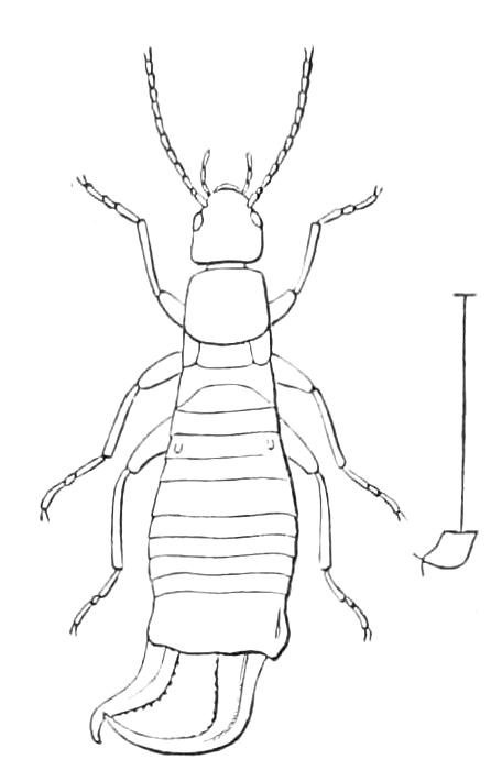
Fig. 113.—Anisolabis tasmanica ♂.
Brauer and others treat the Forficulidae as a separate Order of Insects—Dermaptera—but the only structural characters that can be pointed out as special to the group are the peculiar form of the tegmina and hind wings—which latter, as we have said on p. 206, are considered by some to be formed on essentially the same plan as those of other Orthoptera—the imbrication of the segments, and the forceps terminating the body. The development, so far as it is known, is that of the normal Orthoptera. Thus the Forficulidae are a very distinct division of Orthoptera, the characters that separate them being comparatively slight, though there are no intermediate forms. Some of those who treat the Dermaptera as a sub-Order equivalent to the rest of the divisions of the Order, call the latter combination Euorthoptera.
Fam. II. Hemimeridae.
Apterous, blind Insects with exserted head, having a constricted neck, mouth placed quite inferiorly; the thoracic sterna large, imbricate. Hind body elongate, the segments imbricate, the dorsal plates being large and overlapping the ventral; the number of visible segments being different according to sex: a pair of long unsegmented cerci at the extremity. Coxae small, widely separated. Development intra-uterine.
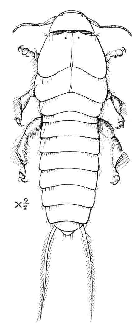
Fig. 114.—Hemimerus hanseni, female. Africa. (After Hansen.)
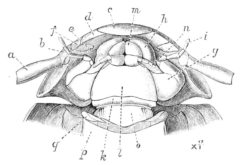
Fig. 115.—Under side of head and front of prothorax of Hemimerus. a, base of antenna; b, articulation of antenna; c, labrum; d, mandible; e, condyle of mandible; f, articular membrane of mandible; g, stipes of maxilla; h, exterior lobe; i, palpus of maxilla; k, submentum; l, mentum; m, terminal lobe of labium; n, labial palp; o, plate between submentum and sternum; p, prosternum; q, cervical sclerites. (After Hansen.)
In describing the labium of Mandibulata, p. 97, we alluded to the genus Hemimerus as reputed to possess a most peculiar mouth. When our remarks were made little was known about this Insect; but a very valuable paper[145] by Dr. H. J. Hansen on it has since appeared, correcting some errors and supplying us with information on numerous points. M. de Saussure described the Insect as possessing two lower lips, each bearing articulated palpi, and he therefore proposed to treat Hemimerus as the representative of a distinct Order of Insects, to be called Diploglossata. It now appears that the talented Swiss entomologist was in this case deceived by a bad preparation, and that the mouth shows but little departure from the ordinary mandibulate type. There is a large inflexed labrum; the mandibles are concealed by the maxillae, but are large, compressed, and on their inner edge toothed. The maxillae are well developed, are surmounted by two lobes and bear five-jointed palpi. The ligula appears to be broad and short, and formed of two parts longitudinally divided; the short palpi consist of three segments. The mentum is very large. The lingua is present in the form of a free pubescent lobe with a smaller lobe on each side. The structure of the pleura is not fully understood; that of the abdomen seems to be very like the earwigs, with a similar difference in the sexes. The cerci are something like those of Gryllidae, being long, flexible, and unsegmented. The legs have rather small coxae, and three-jointed tarsi, two of which are densely studded with fine hairs beneath, as in Coleoptera. It is difficult to detect the stigmata, but Dr. Hansen believes there are ten pairs.
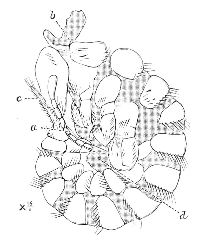
Fig. 116.—Foetus of Hemimerus. (After Hansen.) a, Antenna; b, organ from the neck; c, cerci; d, membrane (? cast skins).
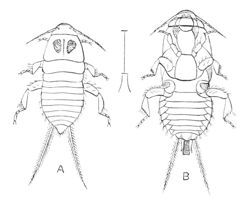
Fig. 117.—Hemimerus talpoides. Africa. (After de Saussure.) A, Upper; B, under surface.
The species described by Dr. Hansen as H. talpoides is probably distinct from that of Walker, though both come from equatorial West Africa. Dr. Hansen's species, which may be called H. hanseni, has been found living on the body of a large rat, Cricetomys gambianus; the Insect occurred on a few specimens only of the mammal, but when found was present in considerable numbers; it runs with rapidity among the hairs and apparently also springs. The nature of its food is by no means clear. Not the least remarkable fact in connexion with this peculiar Insect is its gestation. The young are borne inside the mother, apparently about six at a time, the larger one being of course the nearest to the orifice. Dr. Hansen thinks the young specimens are connected with the walls of the maternal passages by means of a process from the neck of each. But the details of this and other points are insufficiently ascertained; it is, indeed, difficult to understand how, with a process of the kind of which a fragment is shown in Fig. 116, b, the Insect could fix itself after a detachment for change of position. The young is said to be very like the adult, but with a simpler structure of the antennae and abdomen. On the whole, it appears probable that Hemimerus is, as stated by Dr. Hansen, a special family of Orthoptera allied to Forficulidae; further information both as to structure and development are, however, required, as the material at the disposition of the Swedish entomologist was very small.