Chapter XXI
Reading edition
NEUROPTERA CONTINUED—TRICHOPTERA, THE PHRYGANEIDAE OR CADDIS-FLIES
Fam. XI. Phryganeidae—Caddis-flies.
(TRICHOPTERA OF MANY AUTHORS)
Wings more or less clothed with hair, nervures dividing at very acute angles, very few transverse nervules; hind pair larger than the front, with an anal area which is frequently large and in repose plicately folded. Antennae thread-like, porrect, of many indistinct joints. Mandibles absent or obsolete. Coxae elongate and free but contiguous. Metamorphosis great; larvae caterpillar-like, usually inhabiting cases of their own construction. Pupa resembling the perfect Insect in general form, becoming active previous to the last ecdysis. Wingless forms of the imago excessively rare.
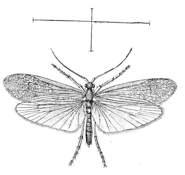
Fig. 319.—Halesus guttatipennis. Britain. (After M‘Lachlan.)
The caddis-flies are Insects of moth-like appearance, found in the neighbourhood of water; their larvae live in this element, where they may sometimes be found in abundance. Phryganeidae are not very attractive Insects, and there are few of large size; Hence they have been much neglected by entomologists, and very little is known about the exotic forms of the family. The habitations constructed by the larvae are, many of them, of a curious nature, and usually attract more attention than do the creatures they serve to protect.
The Phryganeidae form the division or series Trichoptera; the two terms are therefore synonymous; those entomologists who consider these Insects to form a distinct Order use the latter appellation for it.
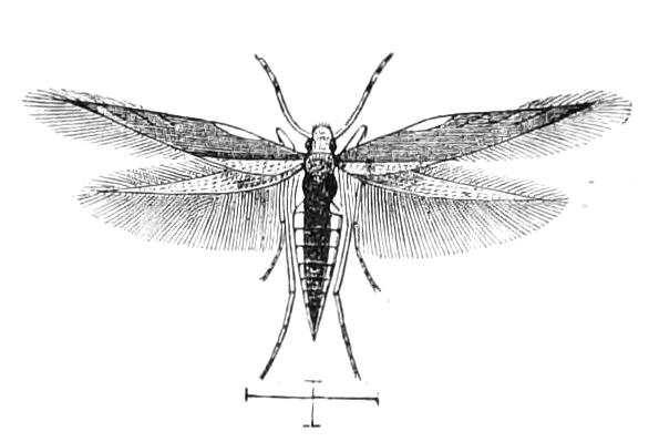
Fig. 320.—Hydroptila angustella ♀. Britain. (After M‘Lachlan.)
The perfect Insect, though the wings are usually ample, has but feeble powers of flight, and rarely ventures far from the water it was reared in; it has a moth-like appearance, and the wings in repose meet, at an angle, in a roof-like manner over the back (Fig. 326, E). The head is small, with the front inflexed; it has two large compound eyes, and usually three ocelli; the antennae are slender, thread-like, and occasionally attain a great length. The parts of the mouth are very peculiar, the labrum and the palpi—especially the maxillary palps—being well developed, while the lobes of the maxillae and labium are amalgamated and therefore indistinct. The labrum is more or less elongate, and is more mobile than is usual in mandibulate Insects; it is held closely applied to the maxillae. These latter are small, have usually only a single small free lobe; they are united to one another and to the labium by membrane in such a manner as to form a channel along the middle of the mouth, the labrum forming the roof of this channel. The palpi are in some cases (Sericostomatides) of a remarkable nature; their joints vary in number from three to five, and differ sometimes in the sexes of the same species. The lower lip appears as a plate supporting the labial palpi, which are three-jointed and do not exhibit any peculiarities of structure comparable with those we have mentioned as so frequently existing in the maxillary palps. Difference of opinion exists as to the mandibles, some entomologists declaring them to be entirely absent, while others state that a small tubercular process that may be seen in some species on each side of the labrum is their representative. The prothorax is very small, the notum is the largest piece but is quite short, the side-pieces are very small, and the sternum appears to consist only of membrane. The mesothorax is much the largest segment of the body; its sternum is large, but is nearly perpendicular in direction, and is much concealed by the elongate, free front coxae, which repose against it. The metathorax is intermediate in size between the pro- and meso-thorax; its side-pieces are rather large, but the sternum is membranous, with a heart-shaped piece of more chitinous consistence in the middle, entirely covered by the middle coxae. The side-pieces both of the meso- and meta-thorax are large, and are closely connected; the middle and posterior coxae are very large, elongate, and prominent, and the middle pair slope backwards, so that their tips are in contact with the tips of the hind pair. The abdomen is cylindric and rather slender; it looks as if formed of eight segments in addition to the terminal segment; this latter in the male usually bears remarkably modified appendages. The first ventral plate is sometimes, if not always, entirely membranous; indeed the texture of the segments is in general very delicate, so that they shrivel up to an extent that renders their comprehension from dried specimens very difficult. The legs are always elongate, the coxae attaining in some forms a remarkable length, and the tibiae and tarsi are armed with many spines; the tarsi are five-jointed, slender, frequently very elongate, terminated by two large claws and an apparatus, placed between them, consisting of a pair of hair-like processes with a membranous lobe.
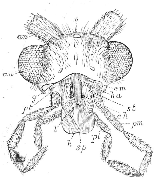
Fig. 321.—Front view of head of Anabolia furcata after removal of labrum. o, Ocellus; an, base of antenna; au, eye; cm, cardo; st, stipes; l, external lobe; pt, support of palpus; pm, palpus of maxilla; g, condyle of articulation of the absent mandible; ha, channel of haustellum; h, haustellum; sp, apex of channel of haustellum (not explained by Lucas); ch, chitinous point of external lobe of second maxilla; pl, labial palp. (After Lucas.)
The structure of the mouth-parts of the Phryganeidae has given rise to much difference of interpretation; it has recently been investigated by R. Lucas[401] in connexion with Anabolia furcata (Fig. 321). He agrees with other observers that mandibles are present in the pupa, but states that no rudiment of them exists in the imago. He calls the peculiar structure formed by the combination of the maxillae and labium a haustellum. He looks on the Trichoptera as possessing a mouth intermediate between the biting and sucking types of Insect-mouths. He considers that the Phryganeidae take food of a solid, as well as of a liquid, nature by means of the haustellum, but the solid matter must be in the form of small particles, and then is probably sucked up by the help of saliva added to it. Lucas says also that in the larvae certain parts of the salivary glands serve the function of spinning organs, and it is from these that the salivary glands of the imago are formed; those salivary glands of the larva that are not spinning glands disappearing entirely.
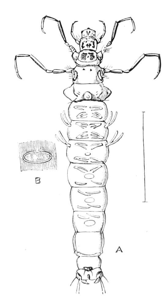
Fig. 322.—Anabolia nervosa. A, Larva extracted from its case; B, one of the dorsal spaces of the abdominal segments more strongly magnified.
The eggs are deposited in a singular manner; they are extruded in a mass surrounded by jelly; there may be as many as one hundred eggs in such a mass. This is sometimes carried about by the female after its extrusion from the interior of the body, but is finally confided to a suitable place in stream, spring, or pool. It is said that the female occasionally descends into the water to affix the egg-mass to some object therein, but this requires confirmation, and it is more probable that the egg-mass is merely dropped in a suitable situation. As soon as the larvae are hatched they begin to provide themselves with cases; they select small pieces of such material as may be at hand in the water, and connect them together by means of silk spun from the mouth. Particulars as to these tubes we will defer till we have considered the larvae themselves. These have the general appearance of caterpillars of moths; in order to move about they must put their head and the three pairs of legs at the front of the body out of their tube or case, and they then look very like case-bearing caterpillars. The part of the body that usually remains under cover is different in texture and colour, and frequently bears outstanding processes, or filaments, containing tracheae for the purpose of extracting air from the water. Some peculiar spaces of a different texture may be seen on certain larvae (Fig. 322, B); these may possibly be also connected with respiration. On each side of the extremity of the body there is a rather large hook by which the creature attaches its dwelling to its body, and there are also frequently present three large bosses on the anterior abdominal segment, which are supposed to assist towards the same end. The hold it thus obtains is so firm that it cannot be dragged out by pulling from the front; fishermen have, however, discovered a way of extracting it by a strategic operation: the cases are, as a rule, partially open behind, and by putting a blunt object in and annoying the larva it is induced to relax the hold of its hooks and advance forwards in the case, or even to leave it altogether. The firm hold of the larva is maintained in spite of the fact that the body does not fill the case. It is necessary that water should pass freely into and out of the case, and that there should be some space for the respiratory filaments to move in. The mouth of the case is open, and the posterior extremity is arranged by the larva in such manner as to allow a passage for the water; various ingenious devices are adopted by different species of larvae with the object of protecting the hind end of the body, and at the same time of permitting water to pass through the case.
The mode of changing the skin, or the frequency with which this occurs in the larval state of the caddis flies has not been recorded. The duration of life in this stage is usually considerable, extending over several months: indeed in our climate many species pass the winter in this stage, completing the metamorphosis in the following spring or summer; and as one generation each year appears to be the rule, it may be assumed that the larval condition in such cases lasts from seven to ten months. During this stage the Insects are chiefly vegetable feeders, some being said to feed on minute algae; animal diet is not, however, entirely avoided, and it is said by Pictet that not only do some of the Phryganeidae eat other Insects, but that they also sometimes devour their companions.
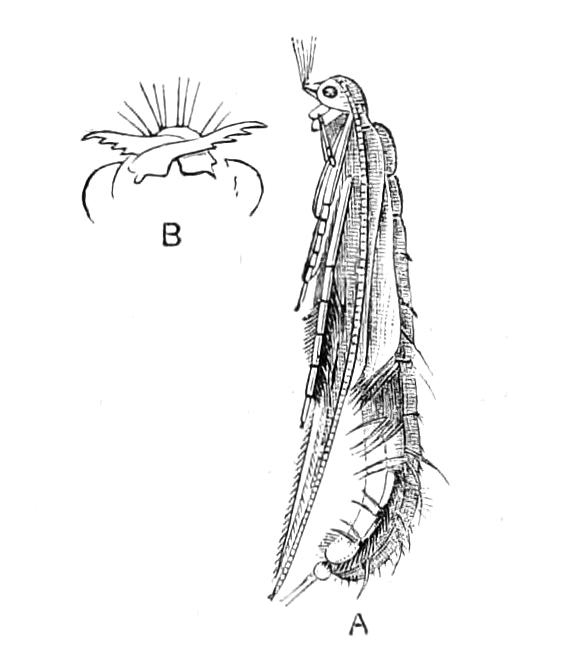
Fig. 323.—A, Pupa of Phryganea pilosa. (After Pictet.) B, Mandibles of pupa of Molanna angustata.
At the end of the larval period of existence the creature closes its case by a light web spun at each end, taking care not to prevent the ingress and egress of the water; it sometimes adds a stone or piece of stick, and having thus protected itself, changes to a nymph. During the first part of this metamorphosis the creature is completely helpless, for there is so great a difference between the external structures of the larva and nymph as to make the latter a new being, so far as these organs are concerned. The changes take place in the interior of the larval skin, and as they are completed this latter is shed piecemeal. The resulting pupa or nymph greatly resembles the perfect Insect, differing consequently very much from the larva. Pictet, who paid special attention to the nymph condition of these Insects, concludes, however, that many of the organs of the nymph are actually formed within the corresponding parts of the larva, and has given a figure that, if trustworthy, shows that the legs of the nymph, notwithstanding the great difference between them as they exist in the larva and in the perfect Insect, are actually formed within the legs of the larva; each nymphal leg being rolled up in the skin of the corresponding larval leg, in a spiral, compressed manner, and the only articulations that can be detected in the leg being those of the tarsus. The head of the nymph is armed in front with two curious projections that are, in fact, enormously developed mandibles (Fig. 323, B); they serve as cutting implements to enable the nymph to effect its escape from its prison; they are cast off with the nymph-skin, the perfect Insect being thus destitute of these organs. The abdomen of the nymph differs from that of the perfect Insect in possessing external respiratory filaments; the nymphs of some species have also the middle legs provided with swimming-hairs, that do not exist in the imago.
As a rule the larvae bring the respiratory filaments into contact with the water by moving the abdomen, but Fritz Müller found[402] that those of a Macronema move the gills themselves—after the manner of Ephemeridae—with much rapidity. Many kinds of larvae of Phryganeids possess at the posterior extremity of the body exsertile pouches in the form of finger-like, or even branched, processes into which tracheae do not enter. Müller observed that in the Macronema alluded to these pouches were generally not exserted; when, however, the larva ceased to move the tracheal gills, then these pouches were protruded. He is inclined to consider them blood-gills. Similar structures are found in Eristalis and some other Dipterous larvae that have to breathe under difficulties.
The imagines of certain species possess filaments—or something of the sort—on the abdomen. Palmén, who has examined these organs in Hydropsyche, thinks that they are the remains of gills that existed in the larva and pupa, and that they are functionless in the imaginal instar. M‘Lachlan thinks that in Diplectrona, where the filaments are elongate, they may be functionally active even in the imago.[403]
The skin of the nymph is at first very soft, but it soon hardens, and when about fifteen or twenty days have elapsed the nymph opens its case by means of the mandibular processes, and swims through the water with its back downwards till it reaches some solid object by which it can ascend to the air; the nymph skin then swells and splits, and the thorax of the imago protrudes; this is soon followed by the disengagement of the head and other parts, and the imago having thus escaped, the nymph skin remains a complete model of the external structure of the nymph, and contains a considerable number of tracheae. This sketch of the metamorphosis of a caddis-fly does not apply in all its details to all the forms of caddis-flies, there being exceptions, as we shall mention hereafter.
Dewitz has described[404] the first appearance and development of the wings in larvae of Phryganeidae. Each one appears at first in the form of a small thickening of the hypodermis, accompanied outwardly by a minute depression of the chitin (Fig. 324, A). He compares the structure in the earliest stage to the entothoracic projections into the interior of the body. The rudiment grows as the larva increases in size, the chitinous portion being duly shed at the ecdyses. When the rudiment is larger and more complex, a mesoderm layer appears in it (Fig. 324, B); this is derived from a nerve-sheath near the rudiment. During the resting state of the larva—after its case has been closed, but before the pupal form has appeared—the wing assumes the form and position shown in C, Fig. 324. Dewitz's description of the process leaves much to be desired, and it is doubtful whether in C the position of the wing on the exterior of the body is due to the stripping off of the chitinous integument, or to a process of eversion, or to both.
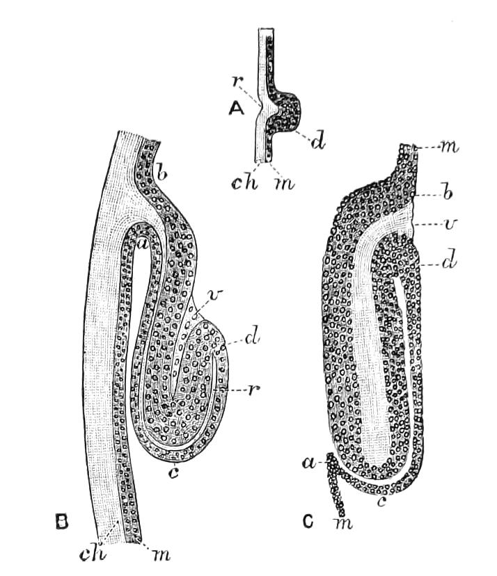
Fig. 324.—Development of wings of Phryganeidae. (After Dewitz.) A, Portion of body-wall of young larva of Trichostegia; ch, chitin, forming at r a projection into the hypodermis m; r and d forming thus the first rudiment of the wing. B, The parts in a largely grown larva; a, c, d, b, the much grown hypodermis separated into two parts by r, the penetrating extension of the chitin; v, mesoderm. C, Wing-pad of another Phryganeid freed from its case at its change to the pupa; b, d, outer layer of the hypodermis, m, of the body-wall; v, inner layer without nuclei.
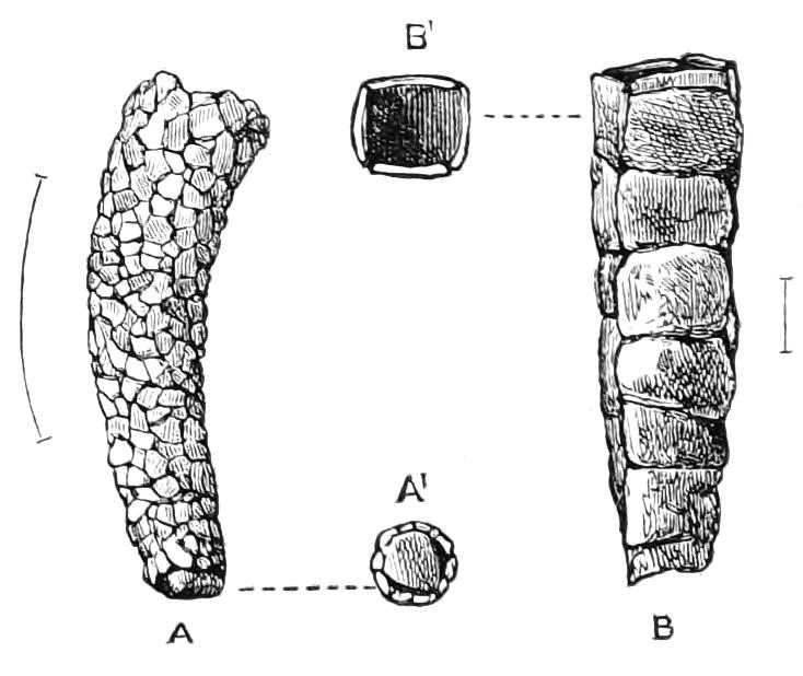
Fig. 325.—Cases of British Trichoptera. A, Of Odontocerum albicorne; A1, its termination; B, quadrangular case of Crunoecia irrorata; B1, mouth of case.
There are about 500 species of this family of Insects known as inhabiting the European region, and about 150 of this number occur in Britain. These are arranged by M‘Lachlan[405]—whose zealous and persevering work at this neglected family of Insects is beyond praise—in eight sub-families, on a system in which the structure of the maxillary palpi plays a principal part; they are called Phryganeides, Limnophilides, Sericostomatides, Leptocerides, Oestropsides, Hydropsychides, Rhyacophilides, Hydroptilides. The first three of these form the division "Inaequipalpia."
Phryganeides.—This group includes the largest forms of the family, and appears to be almost confined to the temperate regions of the northern hemisphere, though a few species are already known from the corresponding districts of the southern hemisphere. This feature in their geographical distribution is, however, by no means peculiar to them, for a similar discontinuity of distribution exists in numerous other groups of Insects, and even in other divisions of the Phryganeidae.
The Phryganeides almost without exception inhabit still waters, and it is more specially to them that the brief sketch of metamorphosis given in the preceding pages will be found to apply. The larva always has the respiratory filaments simple and thread-like, though elongate, and lives in a case that it carries about; this case is open at both ends, and the larva is said to occasionally cut off the end having the least diameter and increase the other end, thus accommodating the habitation to its own growth.
Limnophilides.—These Insects have only three, instead of four, joints in the maxillary palpi, but in most other respects agree with the Phryganeides. There is, however, greater variety in the habits of the larvae, though all live in free cases. In the genus Enoicyla (Fig. 326) we meet with the anomaly of a Trichopterous Insect that lives amongst moss and dead leaves, far away, it may be, from water. The cases of the Limnophilides are constructed of a great variety of materials, and are often decorated with shells containing living inmates.
In the genus Apatania the phenomenon of parthenogenesis is thought to occur, there being at least two species in which no male specimen has ever been discovered, though M‘Lachlan has made special efforts to discover the sex of A. muliebris. It should, however, be stated that these species have not been extensively investigated; A. arctica has been detected in the Arctic regions, and A. muliebris has occurred in several localities in Europe, in Britain chiefly near Arundel in a lake of intensely cold water.
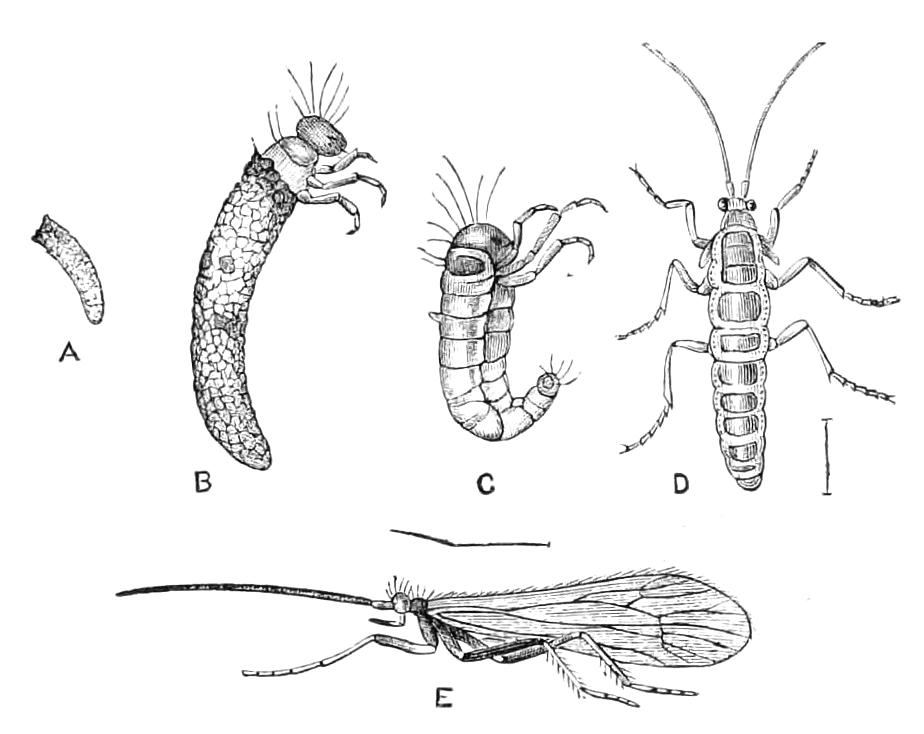
Fig. 326.—Metamorphoses of Enoicyla pusilla. (After Ritsema.) A, Case of full-grown larva; B, larva and case magnified; C, larva extracted; D, wingless adult female; E, male.
Sericostomatides, like the Limnophilides, is a group rich in species; the larvae are chiefly found in streams. They form portable cases out of sand and stones (Fig. 325, B, case of Crunoecia irrorata) in preference to vegetable matter. It is here that the genus Helicopsyche, which for long was an enigma to naturalists, is now placed. This genus consists of Insects whose larvae form spiral cases, similar to small snail shells, of sand or minute stones. These objects occur in various parts of the world. Fritz Müller[406] has informed us that the larva inhabiting one of them, when it withdraws entirely within its abode to repose, takes the precaution of anchoring its snail-like habitation, fixing it to a rock or stone by spinning some temporary silken threads. The respiratory filaments in this group are filiform.
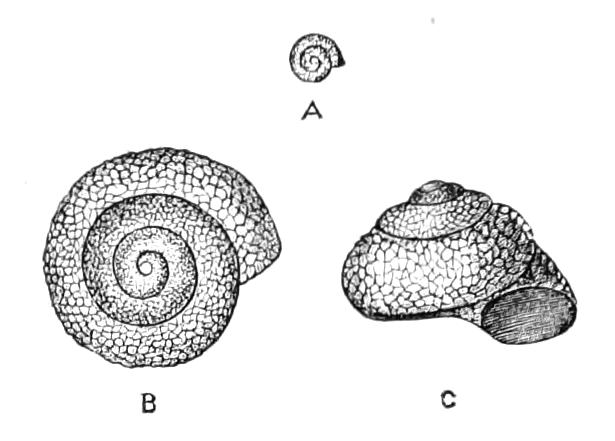
Fig. 327.—Cases of Helicopsyche shuttleworthi. (After von Siebold.) A, Natural size; B, C, magnified.
Leptocerides.—The first group of the division Aequipalpia; so that there are five-jointed maxillary palpi in both sexes; these organs are frequently developed in a remarkable manner. The antennae are usually extremely long and slender. The case of the larva is portable (Fig. 325, A, case of Odontocerum); the respiratory filaments are not very conspicuous; they form short tufts placed on various parts of the abdomen. Müller[406] has called attention to a species whose larva lives in Brazil between the leaves of Bromeliae on trees.
The Oestropsides is a small group, and has recently been reduced by M‘Lachlan to the rank of an inferior division.
Hydropsychides.—An extensive group, in which the larvae are believed to be chiefly of carnivorous habits. They vary, according to species, as to the nature of the respiratory filaments, and live in fixed abodes; these are less tubular than is the rule with the portable cases, and are formed from pieces of sand and stone spun together and fixed to larger stones under water. Sometimes several larvae live together in loosely compacted structures of this kind, and only form true cases when about to undergo their metamorphosis. Müller describes[406] a Brazilian species of Rhyacophylax as forming a case in which the mouth-end has a large funnel-shaped verandah, covered by a beautiful silken net. This larva lives in the rapids of various rivulets, and the entrance to the verandah is invariably directed towards the upper part of the rivulet, so as to intercept any edible material brought down by the water. Several of these larvae, moreover, build their cases so that they form a transverse row on the upper side of a stone; as many as thirty cases may be placed in one of these rows, and sometimes several rows are placed parallel with one another. This same larva has the habit of coming out of its case when necessary, and suspending itself in the water—as some caterpillars do in the air—by means of a silken thread. Other members of the Hydropsychides form tubes or covered ways of silk, earth and mud attached to stones, and in which they can move freely about. Some of the Hydropsychidae have been ascertained with certainty to be carnivorous in the larval state. A species of the genus Hydropsyche has been found by Howard[407] to help itself in the task of procuring food by spreading a net in the water in connexion with the mouth of its case. This net is woven in wide meshes with extremely strong silk, and supported at the sides and top by bits of twigs and small portions of the stems of water-plants. Small larvae brought down by the current are arrested by this net for the advantage of the larva that lurks in the tube. The breathing organs of the larvae of Hydropsychides are apparently of a varied character, and would well repay a careful study. Mr. Morton informs the writer that some of our British species of Philopotamus and Tinodes have no gills either in the larval or pupal state, and probably respire by means of modified tracts in the integument. In some of the allied genera, e.g. Polycentropus, the larvae are destitute of gills, but the pupae possess them.
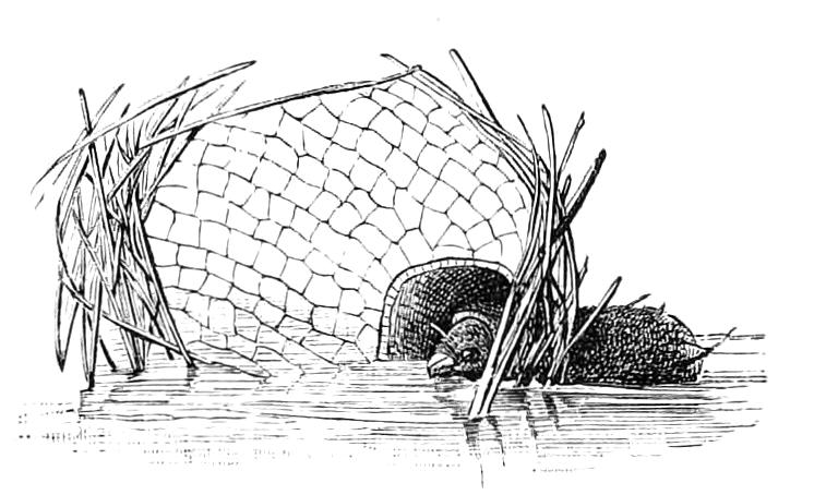
Fig. 328.—Case, with head of larva and snare of North American Hydropsychid. (After Riley and Howard.)
The Rhyacophilides is another group in which the larval habitations are fixed. Some of these larvae have no respiratory filaments, breathing only by means of the stigmata, but others have tufts of filaments. These Insects have a peculiarity in their metamorphosis inasmuch as the larva, instead of lying free, constructs a cocoon in its case or other habitation in which to change to a nymph. In the larvae that do not make use of a portable case the abdominal hooks are not essential, and are replaced by other organs differing much in structure, being sometimes apparently of a sensitive nature, in other forms possibly respiratory. Müller tells us of a carnivorous larva of this group in which the anterior legs are armed with powerful forceps for predatory purposes.
The Hydroptilides comprise the most minute of the Phryganeidae, and their species will probably prove to be very numerous in well-watered tropical regions, though few have yet been described from there. The perfect Insects (Fig. 320) bear an extreme resemblance to small moths of the group Tineidae. The larvae (Fig. 329) are destitute of respiratory filaments, and construct portable cases of a variety of forms, some resembling seeds. Müller has given particulars of a curious nature as to the cases of some Brazilian Hydroptilides; one species moors its dwelling to a stone by means of a long silken cable, by this artifice combining safety with the power of ranging over a considerable extent of water. In Diaulus there is only a narrow slit at each end of the case, but one side of it is provided with two chimneys to permit the flow of water for respiratory purposes.
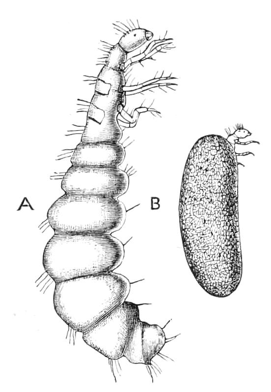
Fig. 329.—Hydroptila maclachlani. B, Case with larva magnified; A, larva more magnified. (After Klapálek.)
The larva of Oxyethira (Fig. 330) is a curious form, possessing comparatively long legs, and a head and thorax slender in comparison with the distended hind body. The cases are fastened, for the purposes of pupation, to a leaf of a water-lily.
Some very curious anomalies as regards the development of the wings exist in the Phryganeidae; Anomalopteryx, for instance, has the wings quite short and useless for flight in the male, while in the other sex they are ample; in Enoicyla—the curious Insect figured on p. 481, in which the larvae are of terrestrial habits—we find the females with only rudiments of wings, while in Thamastes the posterior wings are absent in both sexes. These anomalies are at present quite inexplicable; and we may here mention that we are in complete ignorance as to the functional importance of many of the peculiarities of the Phryganeidae. We do not know why the mouth is reduced from the normal state, the maxillary palpi being, on the other hand, extraordinarily developed; we do not know the importance of the numerous spines and of the spurs on the legs, nor of the hairs on the wings, although these are amongst the most characteristic of the special features of this group of Insects.
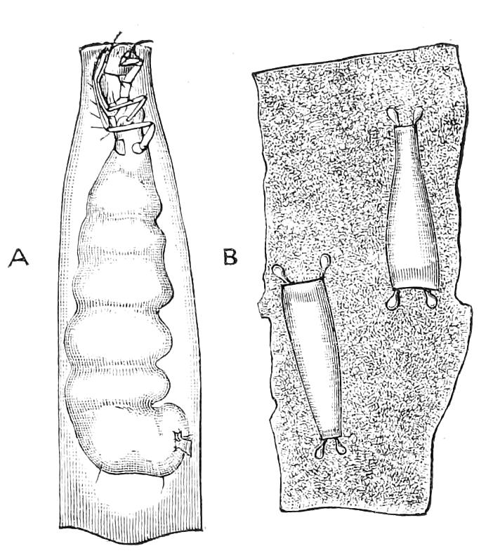
Fig. 330.—Oxyethira costalis. A, Larva in case; B, cases fastened to leaf for pupation. (After Klapálek.)
Fossils.—Abundant remains of Phryganeidae belonging to the Tertiary epoch have been discovered. They are common in amber, and it is a remarkable fact that a larval case has been found in amber. This seems almost inexplicable, except on the assumption that such larvae were of arboreal habits, a condition that, at the present time, must be excessively rare, though the terrestrial habits of Enoicyla warrant us in believing it may occur. In the Tertiary Lake Basin at Colorado the remains of Phryganeidae in the imago state are extremely abundant, so that it is curious that but few such remains have been found in Europe. In Auvergne the so-called indusial limestone, which is two or three yards thick over a wide area, is considered to be composed chiefly of the cases of larvae of this family.
In the Mesozoic epoch some wings found in the lower Purbeck strata are considered to be those of Phryganeidae; similar wings have been found in the Lias, but this is the only evidence of the existence of the family at that period except a tube, supposed to be a larval case, detected in the Cretaceous of Bohemia. Earlier than this nothing has been discovered that can be connected with the family, so that at present the palaeontological evidence appears unfavourable to the view held by some that the Phryganeidae may be considered forms allied to the early conditions of the Lepidoptera. It should be noted that the head in Phryganeidae is the most important part from a systematic point of view, and that fossils have been chiefly identified from the wings; this is a much more doubtful character, as the wings of the Phryganeidae have a simple system of neuration, and in shape have nothing very characteristic.
Extinct Order Palaeodictyoptera.
This seems to be the fittest place to notice the existence of some fossil remains from the Palaeozoic rocks that cannot be fitly, or certainly, assigned to any of our existing Orders, and to which the above name has consequently been given. These remains consist chiefly of wings in a more or less imperfect state of preservation, and it is therefore quite doubtful whether the course of assigning them to a separate Order supposed to be extinct be correct. This is all the more doubtful when we recollect that an Insect fossil, Eugereon bockingi, having the head with mouth-parts of a Hemipterous or Dipterous nature, has been found, the wings attached to it being such as, had they been found separate, would have been considered to be Neuropterous, or at any rate allied thereto. About forty-two forms of Palaeodictyoptera are assigned by Scudder to a section called Neuropteroidea, and may therefore be considered to have a special resemblance to our Neuroptera. These Neuropteroidea comprise numerous genera and no less than six families. Scudder's view is at the best tentative, and is not very favourably received by some entomologists. Brauer has, indeed, objected altogether to the formation of this Order Palaeodictyoptera, and Brongniart has published a list of the Palaeozoic Insects in which a system of arrangement different to that of Scudder is adopted. In his most recent work[408] Brongniart assigns some of these Neuropteroidea to the families Platypterides and Protodonates, which we have previously discussed. The whole subject of these Palaeozoic Insect remains is still in its infancy, and it would not be proper to accept any view as final that has yet been stated, nor would it be fair to dismiss the subject as unimportant because of the great divergence of opinion amongst the authorities who have investigated it.