Chapter XI
Reading edition
ORTHOPTERA CONTINUED—PHASMIDAE—WALKING-LEAVES—STICK-INSECTS
Fam. V. Phasmidae—Stick and Leaf Insects.
Head exserted; prothorax small, not elongate; mesothorax very elongate; the six legs differing but little from one another, the front pair not raptorial, the hind pair not saltatorial. The cerci of the abdomen not jointed, consisting of only one piece; the tarsi five-jointed. Tegmina usually small, or entirely absent, even when the wings are present and ample. The sexes frequently very dissimilar. Absence of alar organs frequent.
These Insects are amongst the most curious of natural objects. They are frequently of large size, some attaining 9 inches in length (Fig. 162, Palophus centaurus, one-half natural length). Their variety of form could scarcely be surpassed; their resemblance to products of the vegetable kingdom is frequently very great: some of the more linear species (Fig. 148, Lonchodes nematodes) look like sticks or stems of grass; some have a moss-like appearance, while others resemble pieces of lichen-covered bark. The members of the tribe Phylliides are leaf-like. A certain number of other Phasmids are covered with strong spines, like thorns (Fig. 149). The plant-like appearance is greatest in the female sex. When there is a difference between the two sexes as to the organs of flight, these are more fully developed in the male.

Fig. 148.—Lonchodes nematodes. Malay Archipelago. (After Westwood.)
The antennae are usually many-jointed, but the number of joints varies from 8 to more than 100; the head is exserted; the eyes are more or less prominent; ocelli are present in some cases. The prothorax is always small, and it is a remarkable fact that it undergoes but little elongation even in those species that are most linear and elongate in form (see Fig. 148, Lonchodes nematodes), and that have the meso- and metathoraces extremely long; it is very simple in structure, consisting apparently merely of a dorsal and of a sternal plate, nearly the whole of the side being occupied by the large space in which the coxae are inserted; the edges of the pronotum are not free. The mesothorax is frequently six times as long as the prothorax, though in the leaf-like and a few other forms it does not possess this great extension; still it is always of large size relatively to the other two thoracic segments. This is peculiar inasmuch as in other groups where the mesothorax is relatively large there are powerful mesothoracic wings; whereas the Phasmidae are remarkable for the obsolescence of the mesothoracic alar appendages. The middle legs and the tegmina or elytra, when present, are attached only to the posterior part of the mesothorax; the notum and the sternum are separated by two narrow slips on each side, the epimeron and episternum. The metathorax is formed like the mesothorax, except that the posterior part of the dorsal surface is considered to consist of the first ventral segment consolidated with the posterior part of the metanotum, the two being distinct enough in the winged forms. The hind body or abdomen is elongated except in the Phylliides; it consists of ten dorsal plates; the first frequently looks like a portion of the metanotum, and is treated as really such by Westwood, who describes the abdomen as consisting of nine segments. The flat apical appendages are attached behind the tenth dorsal plate. The ventral plates are similar to the dorsal in arrangement, except that in the female the eighth plate forms a sort of spoon-like or gutter-like process to assist in carrying or depositing the eggs, and that the two following segments are concealed by it, and are sometimes of more delicate texture. The legs vary greatly in the details of their shape: the coxae are short, oval, or round, never large; the trochanter is small; the front femora often have the basal part narrower than the apical, and they are frequently so formed that they can be stretched out in front of the head, concealing its sides and outline and entirely encasing the antennae. There is an arolium or cushion between the claws of the five-jointed tarsi. The front legs are frequently longer than the others. Only a very slight study has been made of the alar organs of Phasmidae; but according to Redtenbacher and Brauer, they differ greatly from those of Blattidae and Mantidae, inasmuch as the costal vein is placed not on the actual margin of the wing but in the field thereof, and in this respect they more resemble the Orthoptera saltatoria.
Very little information exists as to the internal anatomy of the Phasmidae. Many years ago a memoir of a fragmentary and discursive nature was published on the subject by J. Müller,[189] but his conclusions require confirmation; the nervous system, according to his account, which refers to Arumatia ferula, has the anterior ganglia small, the supra-oesophageal ganglion being apparently not larger than those forming the ventral chain.
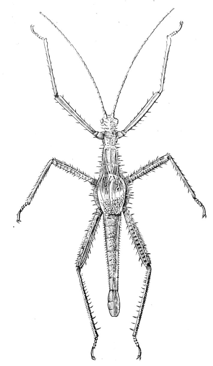
Fig. 149.—Heteropteryx grayi, male. Borneo. One-half natural size.
Joly's more recent memoir on the anatomy of Phyllium crurifolium[190] is also meagre; he states that the nervous system resembles that of the locusts (Acridiidae), though there are at least ten pairs of ganglia—one supra-, one infra-oesophageal, three thoracic, and five abdominal. He found no salivary glands; the Malpighian tubules are slender, elongate, and very numerous. The tracheal system has no air-vesicles. He found no distinction of crop and proventriculus, but the true stomach appears to consist of two different parts, the anterior being remarkably uneven externally, though destitute of coeca, while on the posterior part there are peculiar vermiform processes. There are eighteen or twenty tubes in each ovary.
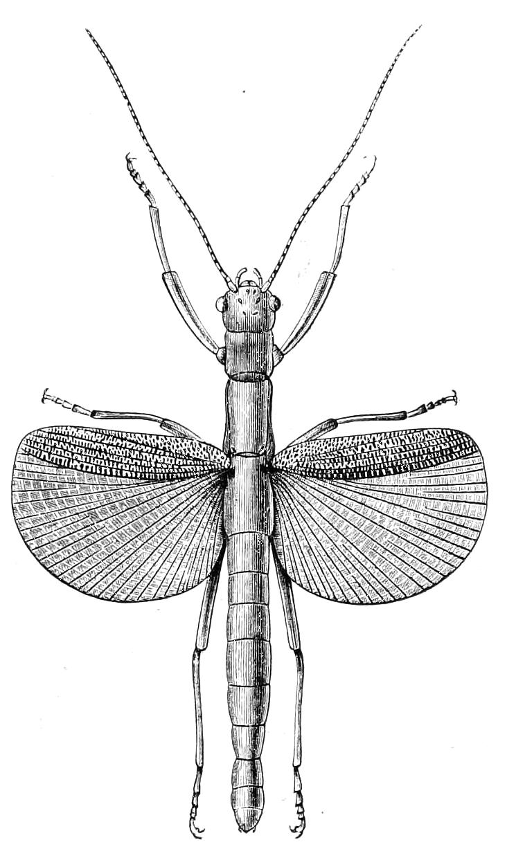
Fig. 150.—Aschipasma catadromus, female. Sumatra. Natural size. (After Westwood.)
When the young Insect is in the egg, ready for emergence, the meso- and meta-thorax are not remarkably elongate, so that the femora are not very far apart, but by the time the creature has fairly emerged from the prison of its embryonic life the thoracic segments have attained their usual proportions; much expansion of the body takes place as the Insect leaves the egg, so that it appears a marvel how it could have been contained therein; this expansion affects the parts of the body unequally.
The records as to the post-embryonic development of Phasmidae are very scanty, but indicate great differences in the length of time occupied by it. Bacillus patellifer is said to moult several times, Diapheromera femorata only twice. This latter species becomes full grown in six weeks, while, according to Murray,[191] Phyllium scythe required fifteen or sixteen months for growth, and did not moult until ten months after hatching; the number of ecdyses in the case of the Phyllium was three. At each change of skin an immediate increase in size, similar to that we have noticed as occurring on leaving the egg, takes place; each limb on being freed becoming about a fourth longer and larger than the corresponding part of the envelope from which it has just been withdrawn. After the second moult of Phyllium the tegmina and wings made their appearance, but remained of very small size until after the third moult, when they suddenly shot out to their full size; they came out of little cases about a quarter of an inch long, and in the course of a few minutes attained their full size of about two and a half inches of length. In the apterous species the difference between the young and adults in external characters is very slight.
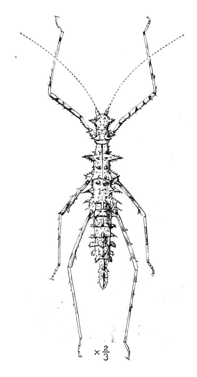
Fig. 151.—Ceroys saevissima. Brazil. (After Westwood.)
Phasmidae are very sensitive to cold; both in North America and Australia their lives are terminated by the occurrence of frost. They are all vegetable feeders, the cannibalism that has been attributed to them by several writers being probably imaginary. They are, however, excessively voracious, so that a pair will destroy a great quantity of foliage; they are consequently in some parts of the world classed amongst injurious Insects. In Fiji and the Friendly Islands, Lopaphus cocophagus eats the cocoa-nut foliage and causes a scarcity of food, so that it becomes a matter of necessity to destroy these Insects. One writer has gone so far as to attribute the occurrence of cannibal habits amongst the inhabitants of some of these islands to the want of food caused by the ravages of this Insect. Some, if not all, of the Phasmidae have the habit of ejecting a stinking fluid, that is said to be very acrid, and occasionally, when it strikes the eye, to cause blindness; this liquid comes from glands placed in the thorax. Some Phasmidae are much relished as food by birds; Diapheromera femorata is sucked by several bugs as well as eaten by birds, and another species is recorded to have harboured Ichneumon-flies in its body without suffering any apparent inconvenience from their presence or from their emergence. Notwithstanding the great amount of food they consume and their want of activity, they produce comparatively few eggs. From twelve to twenty or thirty is frequently mentioned as about the number, but in the case of Diapheromera femorata Riley speaks of upwards of one hundred. These eggs are not deposited in any careful way, but are discharged at random, simply dropping from the female; the noise caused by the dropping of the eggs of Diapheromera femorata from the trees on which the Insects are feeding to the ground is said to resemble the pattering of raindrops. The eggs of this species often remain till the second year before they hatch. The eggs in the Phasmidae generally are of a most remarkable nature, and nearly every one who mentions them speaks of their extreme resemblance to seeds. Göldi[192] has suggested that this is for the purpose of deceiving Ichneumons; it is, however, on record that the eggs are actually destroyed by Ichneumons. It is worthy of notice that the eggs are shed like seeds, being dropped loosely and, as we have said, remaining on the ground or elsewhere, sometimes for nearly two years, without other protection than that they derive from their coverings. Each egg is really a capsule containing an egg, reminding us thus of the capsule of the Blattidae, which contains, however, always a number of eggs. Not only do the eggs have a history like that of seeds, and resemble them in appearance, but their capsule in minute structure, as we shall subsequently show, greatly resembles vegetable tissue. The egg-capsule in Phasmidae is provided with a lid, which is pushed off when the Insect emerges (Fig. 157). This capsule induced Murray to suppose that the egg contained within is really a pupa, and he argued therefrom that in the Orthoptera the larval stages are passed in the egg, and that the Insect after its emergence should be looked on as an active pupa that takes food.
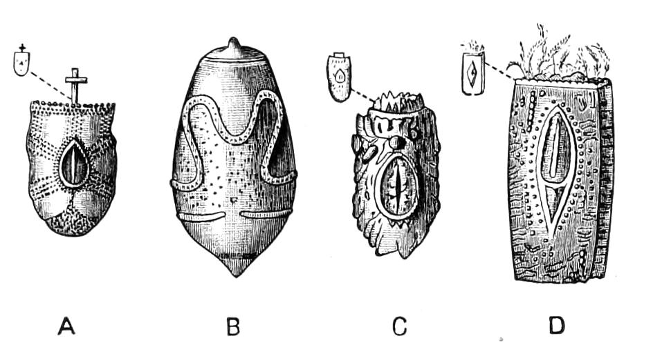
Fig. 152.—Eggs of Phasmidae: A, Lonchodes duivenbodi; B, Platycrania edulis; C, Haplopus grayi; D, Phyllium siccifolium. (After Kaup.)
The individuals of this group of Insects possess the power of reproducing a lost limb; and Scudder, who has made some experiments as to this,[193] states that if a leg be cut off beyond the trochantero-femoral articulation, the parts remaining outside of this joint are dropped before the next moult, and are afterwards renewed either as a straight short stump in which the articulations are already observable, or as a miniature leg, the femur of which is straight and the tibia and tarsus curved into a nearly complete circle; in the former case, the leg assumes at the next moult the appearance that it has in the second case; this latter form is always changed at the succeeding moult into a leg resembling the normal limb in every respect excepting size, and the absence of the fourth tarsal joint (Fig. 153). If the leg be removed nearer to the body than the trochantero-femoral articulation the limb is not replaced.
The sexes are frequently extremely different; the female is usually very much larger than the male. This latter sex often possesses wings when they are quite wanting in the other sex; the resemblance to portions of plants is often very much greater in the female than it is in the male.
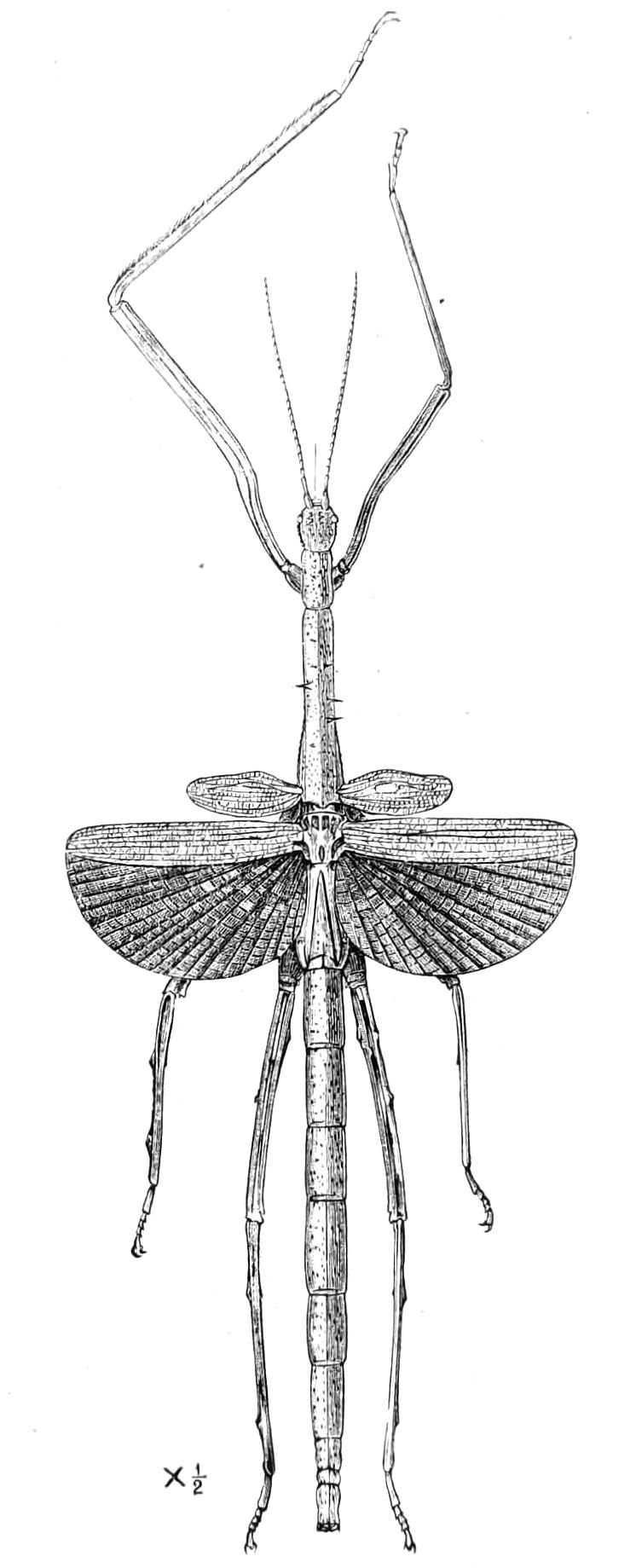
Fig. 153.—Cyphocrania aestuans; individual in which the right front leg has been renewed. Senegal. (After Westwood.)
We have pointed out that the tegmina or upper wings are usually of small size or absent (Fig. 150, Aschipasma catadromus), even in the species where the lower wings are very largely developed; in such cases the latter organs are folded in a complicated, fan-like manner, and repose on the back, looking as if they were really the tegmina (Fig. 159, Calvisia atrosignata); this appearance, moreover, is in some species enhanced much by the fact that the part of the wing which is outermost in the folded state is quite differently coloured from the rest of the organ. The colour of the body in many Phasmidae is said to be very variable, and if the tints be owing to chlorophyll or other plant juices, finding their way amongst the Insect-tissues, this is readily understood; in Diapheromera the young Insect is brownish on hatching, becomes green after feeding, and turns brown again when the leaves do so. The ocelli, too, are said to be very variable, and M‘Coy goes so far as to state[194] that they may be either present or absent in different individuals though of the same species and sex,—a statement so remarkable as to require minute examination, though it is to some extent confirmed by the remarks of other entomologists.
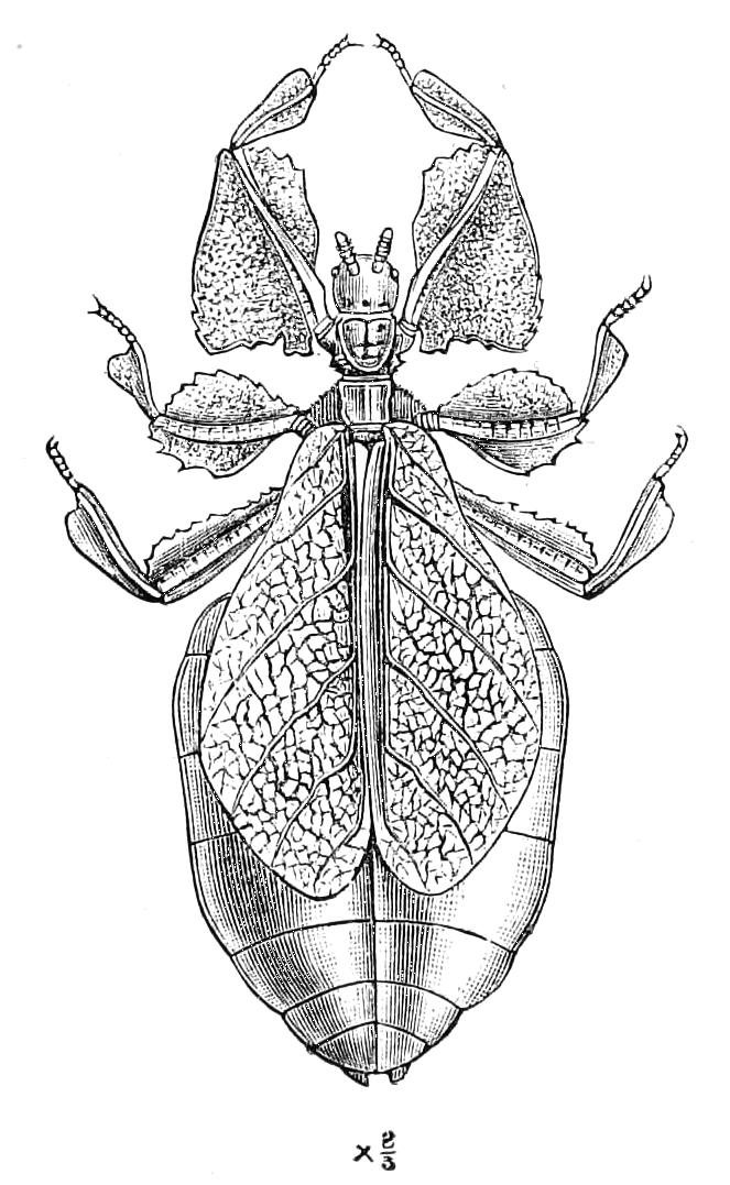
Fig. 154.—Phyllium scythe, female. Sylhet. (After Westwood.)
The resemblance presented by different kinds of Orthoptera to leaves is so remarkable that it has attracted attention even in countries where Natural History is almost totally neglected; in many such places the inhabitants are firmly convinced that the Insects are truly transformed leaves, by which they understand a bud developing into a leaf and subsequently becoming a walking-leaf or Insect. To them the change is a kind of metamorphosis of habit; it grew as a leaf and then took to walking.[195] It is usually the tegmina that display this great resemblance to vegetable structures, and there is perhaps no case in which the phenomenon is more marked than it is in the genus Phyllium, the members of which occur only in the tropical regions of the Old World, where they extend from Mauritius and the Seychelles to the Fiji Islands—possibly even more to the East—and have, it would appear, a peculiar penchant for insular life.
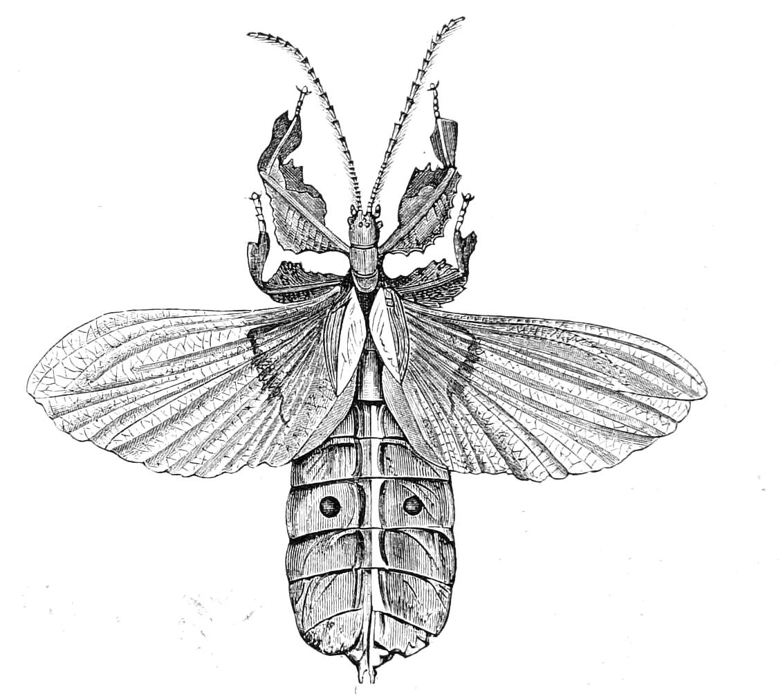
Fig. 155.—Phyllium scythe, male. Sylhet. (After Murray.)
The genus Phyllium constitutes by itself the tribe Phylliides. Although the characters and affinities of this group have been only very inadequately investigated, it will probably prove to be a very distinct and isolated one. The species are not well known, but are probably numerous, and the individuals are believed not to be rare, though the collections of entomologists are very badly supplied with them. The resemblance of the tegmina or front wings to leaves is certainly of the most remarkable nature. During the early life the Insect does not possess the tegmina, but it is said then to adapt itself to the appearance of the leaves it lives on, by the positions it assumes and the movements[196] it makes. When freshly hatched it is of a reddish-yellow colour. The colour varies at different periods of the life, but "always more or less resembles a leaf." After the young Insect has commenced eating the leaves it speedily becomes bright green; and when the metamorphosis is completed the female Insect is possessed of the leaf-like tegmina shown in Figs. 154, 156. Before its death the specimen described by Murray passed "through the different hues of a decaying leaf." Brongniart has had opportunities of observing one of these leaf-Insects, and has, with the aid of M. Becquerel, submitted their colouring matter to spectral analysis,[197] with the result of finding that the spectrum exhibits slight distinctions from that of solutions of chlorophyll, but does not differ from that of living leaves. Mr. J. J. Lister when in the Seychelles brought away living specimens of Phyllium; and these becoming short of food, nibbled pieces out of one another just as they might have done out of leaves. The Phasmidae are purely vegetable feeders, and these specimens did not seriously injure one another, but confined their depredations to the leaf-like appendages and expansions.
The males of this genus are totally different from the females; the foliaceous tegmina being replaced by appendages that are not leaf-like, while the posterior wings, which are large and conspicuous parts of the body, have no leaf-like appearance (Fig. 155).
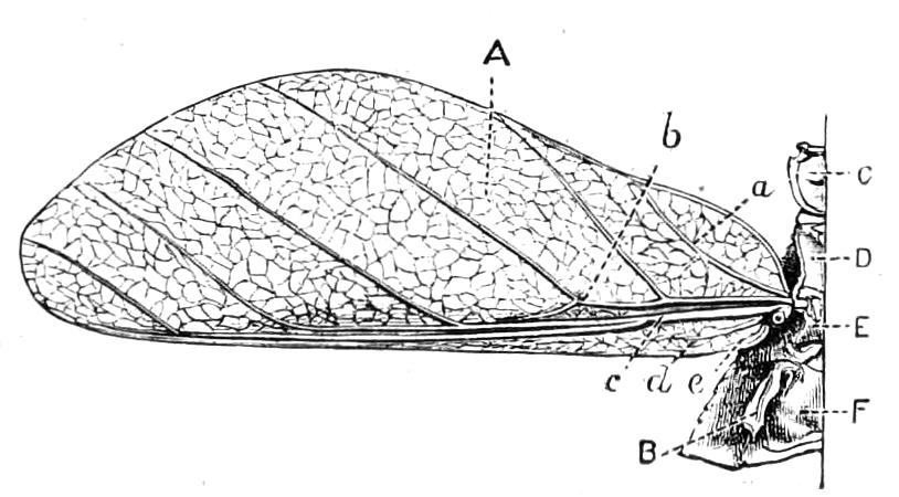
Fig. 156.—Alar organs and one side of thorax of Phyllium crurifolium: A, tegmen; B, rudiment of wing; C, pronotum; D, anterior division of mesonotum; E, posterior division; F, metanotum; a, b, c, d, e, chief wing-nervures; a, mediastinal; b, radial; c, ulnar; d, dividens?; e, plicata?.
In the female Phyllium the hind wings are not present, being represented by a minute process (Fig. 156, B). The tegmen of the female Phyllium is, from various points of view, a remarkable and exceptional structure. It is the rule that when there is in Insects a difference between the alar organs of the two sexes it is the male that has them largest; this is the case in Phyllium so far as the hind wings are concerned, but in the fore-wings the rule is departed from, the leaf-like tegmina of the female being very much larger than the rudimentary wing-covers of the male. In Phasmidae it is the rule that the tegmina are atrophied, even when the hind wings are largely developed. This is the case in the male of Phyllium, but in the female this normal condition is reversed. Although the alar organs of Phasmidae have received hitherto but a small amount of attention, it is probable that the female tegmen of Phyllium is as peculiar morphologically as it is in other respects. In Fig. 156 we give an accurate representation of the chief nervures in the tegmen of a female P. crurifolium. It is interesting to compare this with the diagrams we give of the tegmina of a Blattid (Fig. 121) and of an Acridiid (Fig. 167); the tegmen of the Phyllium is very different, the radial vein and all the parts behind it being placed quite close to the posterior edge of the structure. A similar view is taken by both Redtenbacher and Brauer. The latter says,[198] "In Phyllium (the walking-leaf) almost the whole of the front wing is formed by the praecostal and subcostal fields; all the other fields with their nervures, including even the costa, are compressed towards the hind margin into a slender stripe. In the hind wing the costa is, however, marginal." Unfortunately no examination appears to have been made of the male tegmen, so that we do not know whether that of the female differs from it morphologically as strongly as it does anatomically. It is, however, clear that the tegmina of the female Phyllium not only violate a rule that is almost universal in the Insecta, but also depart widely from the same parts of its mate, and are totally different—and, for a Phasmid, in an almost if not quite unique fashion—from the other pair of alar organs of its own body.
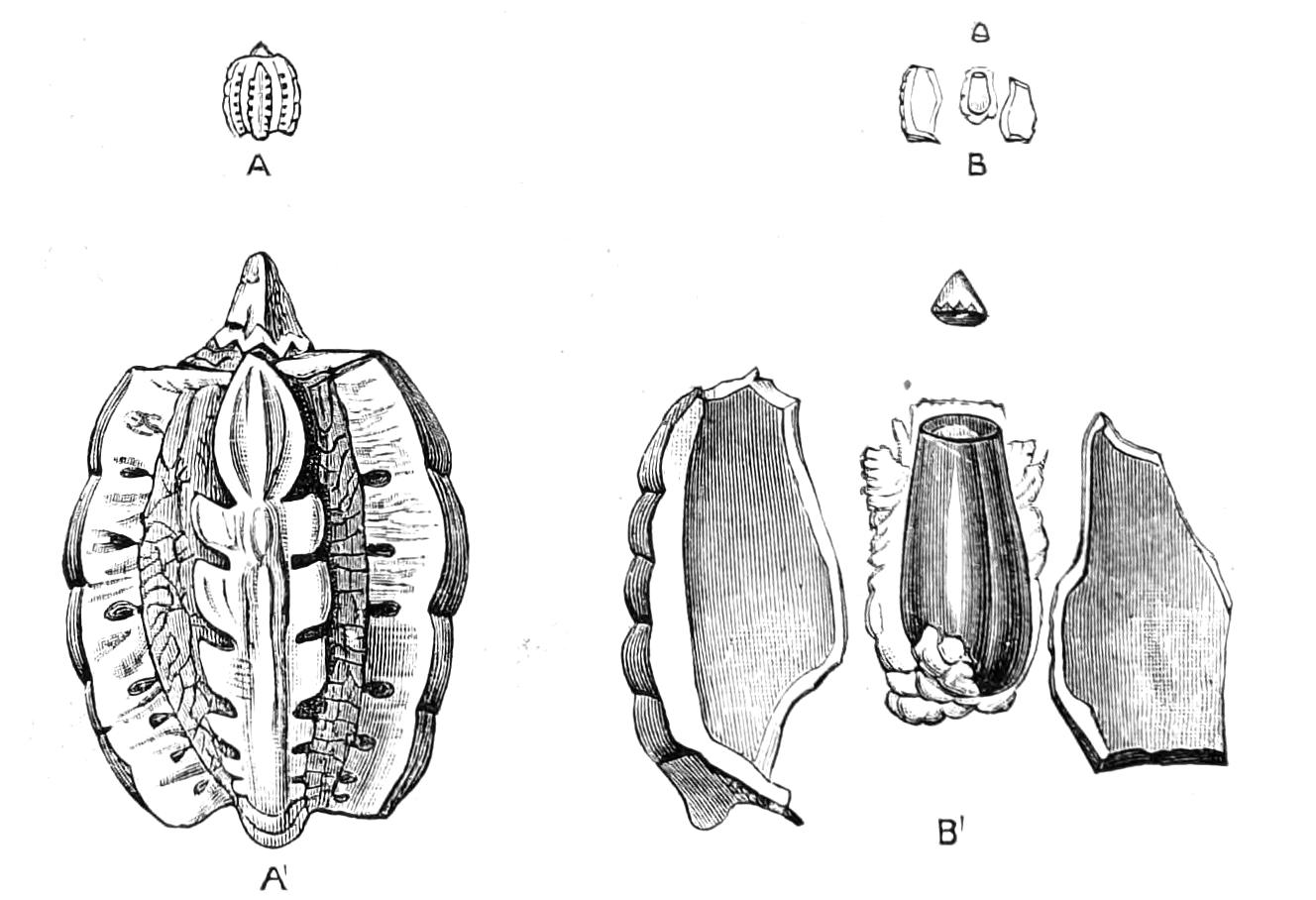
Fig. 157.—Egg of Phyllium scythe. (After Murray.) A, The whole egg, natural size; A', magnified; B, the capsule broken, showing the true egg inside, natural size; B', magnified.
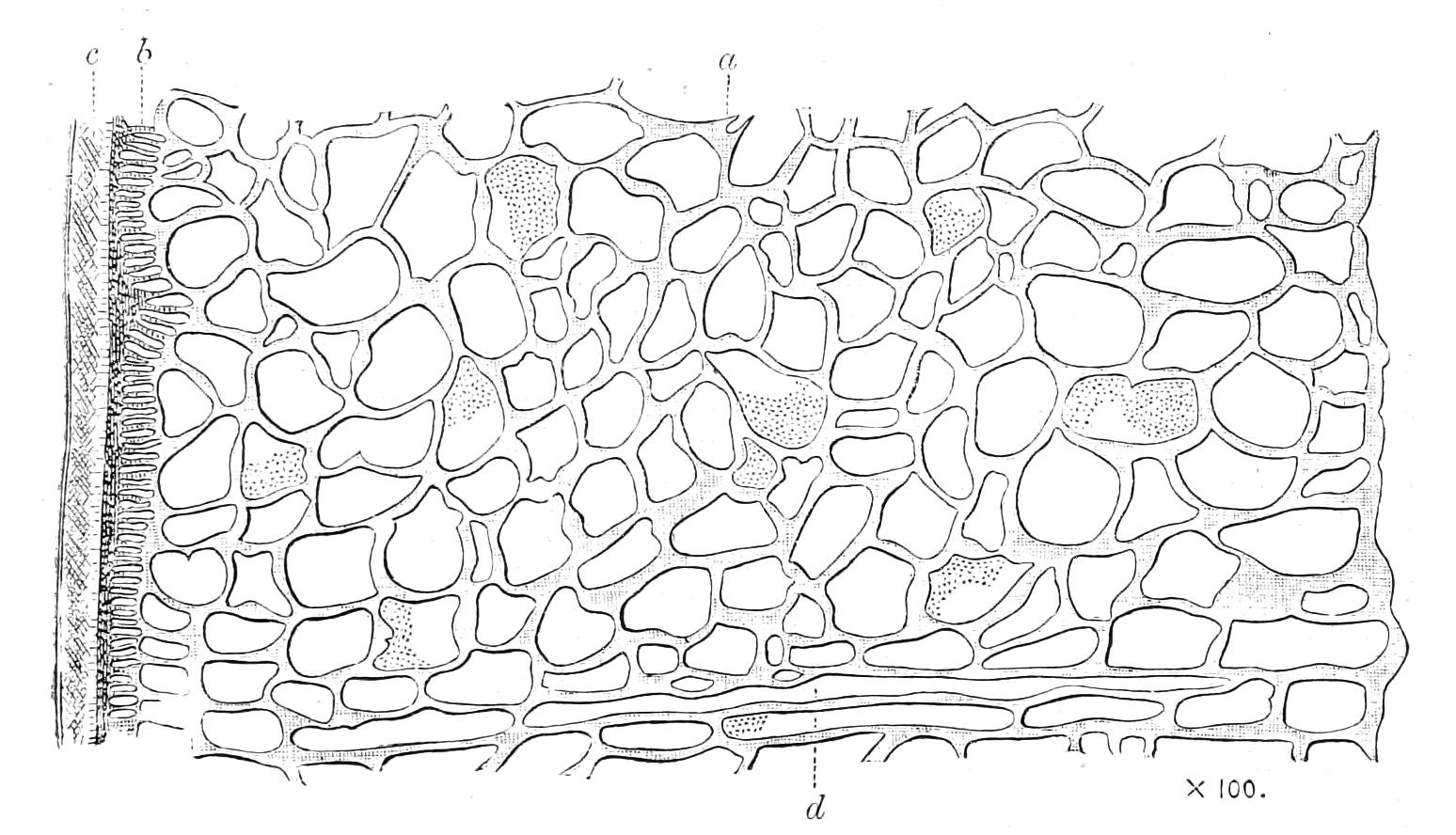
Fig. 158.—Portion of a longitudinal section of the egg capsule of Phyllium crurifolium: a, external; b, middle; c, inner zones; d, elongate alveoli. × 100. (After Henneguy.)
We have already alluded to the resemblance to seeds displayed by the eggs of Phasmidae. The eggs of Phyllium have been studied by several entomologists, and their resemblance to seeds excites general astonishment. Murray describes the egg-capsule of Phyllium scythe, and says: "It looks uncommonly like some seeds; if the edges of the seed of Mirabilis jalapa were rubbed off, the seed might be mistaken for the egg. The ribs are all placed at equal distances, except two, which are wider apart, and the space between them flatter, so that on the egg falling it rolls over till it comes to this flatter side, and there lies.... At the top there is a little conical lid, fitting very tightly to the mouth.... On removing the lid we see a beautiful porcelain chamber of a pale French-white colour, bearing a close resemblance to the texture of a hen's egg, but it is not calcareous, and has more the appearance of enamel." The eggs of P. crurifolium have been examined by Joly and Henneguy; their account confirms that of Murray. Henneguy adds that a prominent lozenge on the egg represents the surface by which the achene of an umbelliferous plant is united to the column, and that the micropyles are placed on this lozenge. The minute structure of the capsule has also been examined by several entomologists; and Henneguy,[199] who has described and figured some of the details of the capsule of P. crurifolium, says, "Almost every botanist, on examining for the first time a section of this capsule, would declare that he is looking at a vegetable preparation."
We may remark that, although there is difference of opinion on the point, the evidence extant goes to show that the egg-capsules are formed in the egg-tubes, only one egg being produced at a time in a tube,[200] the others in it remaining quite rudimentary.
About 600 species of the family are known; there are only four or five kinds found in Europe, and they are all confined to the south, only one of them extending as far north as Central France. The males of these European Bacilli are extremely rare in comparison with the females, which are common Insects. Phasmidae are of almost universal distribution in the warm parts of the world, and even the species whose individuals are of large size seem to be able to continue their existence in comparatively small islands. Australia is perhaps the region where they are most largely developed at present. Macleay says of Podacanthus wilkinsoni that it is rare in any part of Australia to find in the summer season a gum-tree without a few of these Insects grazing on it; and occasionally this Insect has been so abundant there that the trees for miles around have been denuded of their foliage by it, and the dead and dying Insects have been found lying beneath the trees almost in heaps. There are several Phasmidae in New Zealand, all wingless forms, and different from those found in Australia. In Brazil a species of the genus Prisopus has the peculiar habit of seeking shelter under the stones submerged in the mountain streams; to enable it to do this it is remarkably constructed, the under side of the body being hollowed, and various parts set with a dense fringe of hairs; the Insect is supposed to expel the air from the body in order to adhere to the upper surface of a stone, where it sits with its fore legs extended in front of its head, which is directed against the current. Attention has been called to a still more remarkable form said to be allied to the Prisopi, by Wood-Mason,[201] who calls the Insect Cotylosoma dipneusticum. This Insect is apparently known only by a single example of the female sex; it is 3 or 4 inches in length, has rudimentary organs of flight, and along the lower margins of the metathorax there are said to be on each side five conspicuous fringed plates of the nature of tracheal gills; these coexist with open stigmata for aerial respiration, as in the imago of Pteronarcys. The writer has examined this curious Insect, and thinks it very doubtful whether the plates are branchiae at all. The locality for this Insect is the island of Taviuni, not Borneo, as stated by Wood-Mason. These and one or two Acridiidae are the only Insects of the Order Orthoptera at present believed to possess aquatic habits.
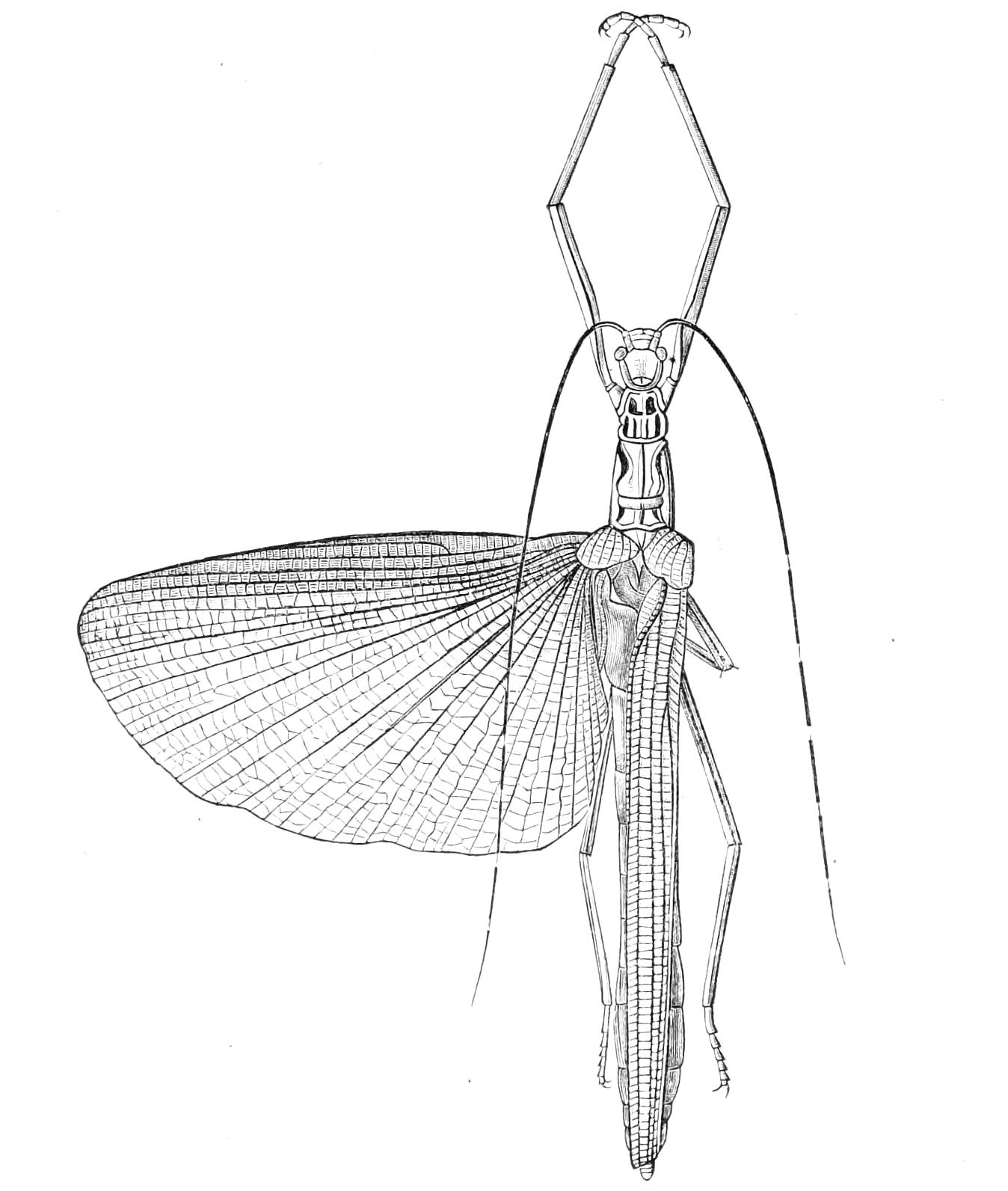
Fig. 159.—Calvisia atrosignata, female. Tenasserim. (After Brunner.)
Although the number of species of Phasmidae is small in comparison with what we find in many of the large families of Insecta, yet there is probably no other family that equals it in multiplicity of form and diversity of external appearance.
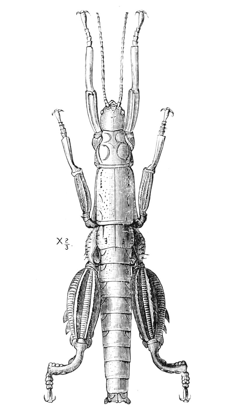
Fig. 160.—Eurycantha (Karabidion) australis, male. Lord Howe's Island. (After Westwood.)
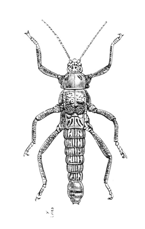
Fig. 161.—Anisomorpha pardalina. Chili. (After Westwood.)
Karabidion (Fig. 160), a genus found in some of the islands of the southern hemisphere, has the hind legs enormously thickened in the male. Some Phasmids, e.g. Orxines zeuxis, have the hind wings marked and coloured after the manner of butterflies or moths. Lamponius laciniatus has an elaborately irregular outline, looking like a mass of moss, and some species of Bacteria are so very slender that the linear body is scarcely equal in size to one of the legs it bears. Among the most interesting forms are the Insects for which the genera Agathemera and Anisomorpha (Fig. 161) have been established; they are remarkably broad and short, have the mesothorax but little elongated, with the tegmina attached to it in the form of two short, thick, leathery lobes; while the wings are seen as marks on the metanotum looking like a mere sculpture of the surface; these Insects have quite the appearance of larval forms, and it is worthy of note that the elongation of the mesothorax, which is one of the most marked features of the Phasmidae, is in these forms only very slight.
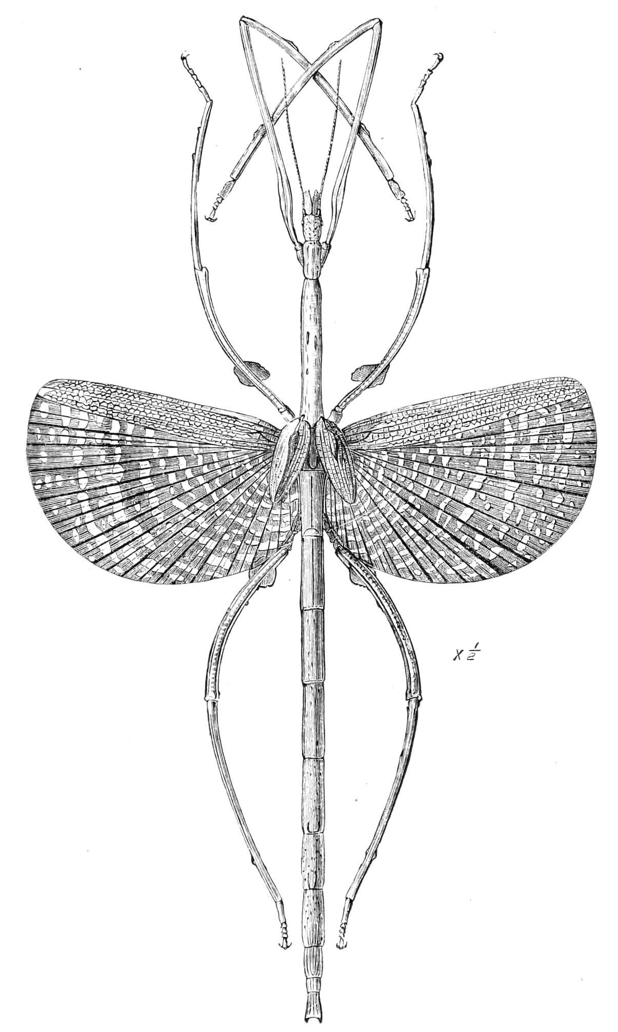
Fig. 162.—Palophus centaurus. Old Calabar. Half natural size. (After Westwood.[202])
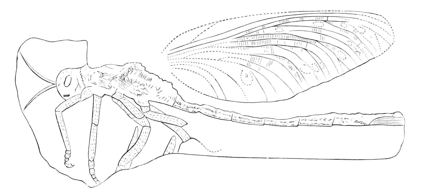
Fig. 163.—Titanophasma fayoli. Carboniferous formation at Commentry. × ⅕. (From Zittel.)
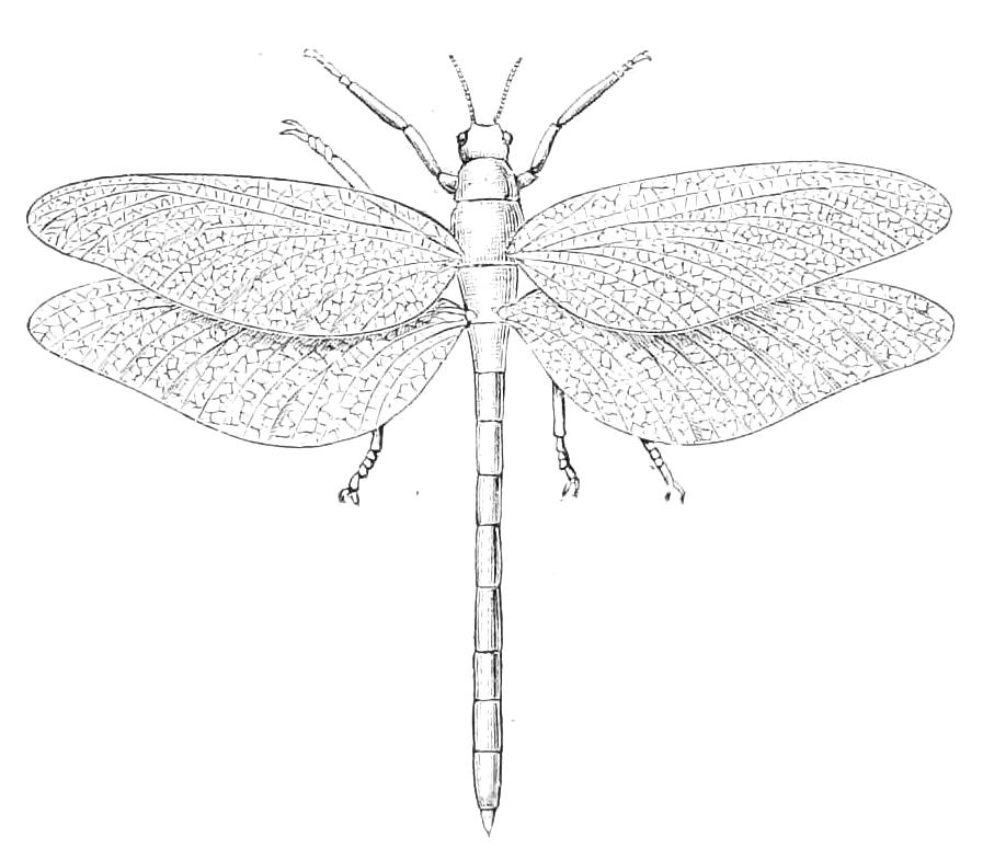
Fig. 164.—Titanophasma fayoli (restoration). × ⅒.
Some Insects said to belong to the genera Phasma and Bacteria have been found in amber. A single Insect-fossil found in the Tertiary strata in North America has recently been referred by Scudder to the family, and even to a genus still existing in the New World—Agathemera; the fragment is, however, so defective, and the characteristic points of the Phasmidae are so little evident in it, that not much reliance can be placed on the determination. No Phasmid has been unearthed from Mesozoic strata, so that, with the exception of the fragment just mentioned, nothing that evidently belongs to the Phasmidae has been discovered older than the remains preserved in amber. In the Carboniferous layers of the Palaeozoic epoch there are found remains of gigantic Insects that may possibly be connected with our living Phasmidae. These fossils have been treated by Brongniart and Scudder as forming a distinct family called Protophasmidae. The first of these authors says[203] that our Phasmidae were represented in the Carboniferous epoch by analogous types differing in the nature of the organs of flight: these ancient Insects were of larger size than their descendants, being 25 to 50 centimetres long, and as much as 70 in spread of wing. To this group are referred, on somewhat too inferential grounds, the fossil wings found in the Carboniferous layers, and called by Goldenberg Dictyoneura.
We reproduce from Zittel's handbook a figure (Fig. 162) of one of these gigantic Insects, and add an attempt at a restoration of the same after the fashion of Scudder (Fig. 163). From these figures it will be seen that the relation to our existing Phasmidae must at best have been very remote.[204] It will be noted that the larger of the two figures is on a ⅕ scale.
The classification of Phasmidae was left in a very involved state by Stål, but has recently been brought into a more satisfactory condition by Brunner von Wattenwyl. We give a translation of his table of the tribal characters:—
1. Tibiae beneath carinate to the apex, without an apical area.
2. Antennae much longer than the front femora, many jointed, the joints being above 30 in number and only distinct at the base and towards the apex.[205]
3. Median [true first abdominal] segment much shorter than the metanotum.[206] The species all apterous.
4. The anal segment of the males roof-like, more or less bilobate. The female has a supra-anal lamina. The species inhabit the Old World. Tribe 1. Lonchodides (Fig. 148, Lonchodes nematodes.)
4′. The anal segment of the males arched, straight behind. No supra-anal lamina in the female. The species are American. Tribe 2. Bacunculides.
3′. Median segment as long as, or longer than the metanotum. Species with the male or both sexes winged.
4. Females apterous or rarely possessed of short wings.[207] Males winged. Femora dentate beneath, or lobed, or at least armed with one tooth. Species occur both in America and in the Old World. Tribe 3. Bacteriides. (Fig. 162, Palophus centaurus.) 4′. Each sex winged. Femora smooth beneath. The species belong to the Old World. Tribe 4. Necroscides. (Fig. 159, Calvisia atrosignata.)
2′. Antennae (at any rate in the females) shorter than the front femora, the joints distinct, not more than 28 in number. The species belong to the Old World.
3. Median segment shorter than the metanotum. Apterous species. Cerci plump. Tribe 5. Clitumnides. (Fig. 160, Eurycantha australis.)
3′. Median segment longer than the metanotum. Species usually winged. Cerci (except in some genera of the group Platycraninae) flattened, elongate. Tribe 6. Acrophyllides. (Fig. 153, Cyphocrania aestuans.)
1′. Tibiae furnished beneath with a triangular apical area.
2. Antennae many jointed, longer than the front femora.
3. Median segment shorter than the metanotum. Apterous species.[208]
4. Either head, thorax, or legs spiny or lobed. Tribe 7. Cladomorphides. (Fig. 149, Heteropteryx grayi.)
4′. Head, thorax and legs unarmed. Tribe 8. Anisomorphides. (Fig. 161, Anisomorpha pardalina.)
3′. Median segment longer than the metanotum.
4. Claws unarmed. Tegmina lobe-like, either perfectly developed or entirely absent. The winged species are all American, the apterous are both African and Australian. Tribe 9. Phasmides.
4′. Claws toothed on the inner side. Tegmina spine-like. Wings well developed. The species are Asiatic. Tribe 10. Aschipasmides. (Fig. 150, Aschipasma catadromus.)
2′. Antennae shorter than the anterior femora,[209] formed of not more than 20 joints. Old World species.
3. Body slender. Apterous. Tribe 11. Bacillides.
3′. Body very broad, lamina-like. Either wings or tegmina present. Tribe 12. Phylliides. (Fig. 155, Phyllium scythe, male; Fig. 154, idem., female.)