Chapter XXIII
Reading edition
HYMENOPTERA PETIOLATA–PARASITIC HYMENOPTERA–CYNIPIDAE OR GALL-FLIES–PROCTOTRYPIDAE–CHALCIDIDAE–ICHNEUMONIDAE–BRACONIDAE–STEPHANIDAE–MEGALYRIDAE–EVANIIDAE–PELECINIDAE–TRIGONALIDAE.
We now pass to the consideration of the Hymenoptera of the sub-Order Petiolata, or Apocrita, as they are styled by Brauer. We should make use of the term Petioliventres, for it contrasts naturally by its termination with Sessiliventres, were it not that the word is so uncouth that we think it better to adopt the shorter and more euphonious expression, Petiolata.
The members of this sub-Order, without exception, have the hind body connected with the thorax by means of a deep constriction, so that the base of the abdomen (Fig. 336, B, b) is very narrow; the articulation between the two parts is effected by means of a complex joint allowing great play, and facilitating the operations of boring and stinging, processes that are of extreme importance in the economy of the great majority of the species. The petiole is sometimes extremely short, but it may be so long that it appears like a stalk, at whose extremity is borne the remaining part of the abdomen (Fig. 369). When the petiole is very short the abdomen reposes close to the back of the thorax (Fig. 331, C), and in this case the abdomen is usually described as sessile; while, when it is evidently stalked, it is said to be petiolate. These terms are, however, unsuitable, as the words sessile and petiolate should be reserved for the conditions characteristic of the two sub-Orders. We shall therefore use the terms pseudo-sessile and pedicellate for the two conditions of the Petiolata.
The Hymenoptera Petiolata comprises an enormous majority of the Order. Although it includes many of the most interesting and important of Insects, its classification is but little advanced, for a great many of the forms are still rare or unknown. Three series may be adopted for the purposes of nomenclature.
1. Parasitica.—Trochanters of two pieces, female with an ovipositor.
2. Tubulifera.—Trochanters undivided; abdomen consisting of only three, four, or five visible segments.
3. Aculeata.—Trochanters undivided; abdomen consisting of six or seven visible segments; female furnished with a retractile sting.
In the absence of any clear distinction between sting and ovipositor, these groups are merely conventional. The character furnished by the trochanters is unfortunately subject to some exceptions, there being a few parasitic forms in which the trochanters are not divided, and a few aculeates in which the reverse is more or less distinctly the case; moreover, the division, when it exists, is in some cases obscure, and the two pieces are of unequal size. Ratzeburg calls the upper division, which is frequently much larger than the other, the trochanter, and the lower division the apophysis. There is much reason for believing that the apophysis is really merely a secondary division of the femur. The Tubulifera are a comparatively small group, and will probably be merged in one of the other two, when the anatomy and morphology of the abdomen have been more thoroughly elucidated.
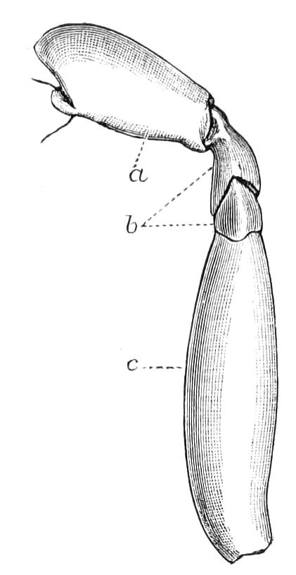
Fig. 345.—Divided (ditrochous) trochanter of an Ichneumon: a, coxa; b, the two divisions of the trochanter; c, femur. (For monotrochous trochanter see Fig. 335, A, c.)
Hymenoptera Parasitica or Terebrantia.
This is one of the most extensive divisions of the class Insecta. There can be little doubt that it contains 200,000 species, and possibly the number may be very much greater than this. It is, however, one of the most neglected of the great groups of Insects, though it is perhaps of greater economic importance to mankind than any other.
Insects derive their sustenance primarily from the vegetable kingdom. So great and rapid are the powers of assimilation of the Insect, so prodigious its capacity for multiplication, that the Mammal would not be able to compete with it were it not that the great horde of six-legged creatures has divided itself into two armies, one of which destroys the other. The parasitic Hymenoptera are chiefly occupied in destroying the tribes of vegetarian Insects; the parasites do this by the simple and efficient device of dwelling in the bodies of their hosts and appropriating the nutriment the latter take in. The parasites do not, as a rule, eat the structures of their host,—many of them, indeed, have no organs that would enable them to do this,—but they absorb the vegetable juices that, in a more or less altered state, form the lymph or so-called blood of the host. The host could perhaps starve out his enemies by a judicious system of abstention from food; instead, however, of doing this, he adopts the suicidal policy of persistent eating, and as the result of his exertions, furnishes sufficient food to his parasites, and then dies himself, indirectly starved. Ratzeburg considers that the traditional view that the larvae of parasitic Hymenoptera live by eating the fat-body of their host is erroneous. They imbibe, he considers, the liquid that fills the body of the parasitised Insect.[428]
The wide prevalence of Insect parasitism is appreciated only by entomologists. The destructive winter moth—Cheimatobia brumata—is known to be subject to the attacks of sixty-three species of Hymenopterous parasites. So abundant are these latter that late in the autumn it is not infrequently the case that the majority of caterpillars contain these destroyers. Although Lepidoptera are very favourite objects with parasitic Hymenoptera, yet other Insects are also pertinaciously attacked; there is quite a host of Insect creatures that obtain their sustenance by living inside the tiny Aphididae, or "green-flies," that so much annoy the gardener. A still larger number of parasites attack eggs of Insects, one or more individuals finding sufficient sustenance for growth and development inside another Insect's egg. As Insects have attacked Insects, so have parasites attacked parasites, and the phenomena called hyperparasitism have been developed. These cases of secondary parasitism, in which another species attacks a primary parasite, are extremely numerous. It is also pretty certain that tertiary parasitism occurs, and Riley is of opinion that even quaternary destruction is not outside the range of probability.
The physiological problems connected with Insect parasitism are of great interest to the entomologist; the modes of nutrition and respiration of these encaged creatures could not fail to be most instructive were we fully acquainted with them. It is obvious that when an Insect-egg is laid inside another Insect's egg, and the parasite has to undergo the whole of its growth therein, it is in the strangest condition as regards nutrition. It is unnecessary for the intruded egg to have yolk of its own; moreover, the embryonic mode of nutrition may be continued during what would, with other Insects, be the larval period. And it seems to be the case that both these conditions are actually met with in the lives of egg-parasites. The embryology and post-embryonic development of parasitic Hymenoptera have already been ascertained to be of the most extraordinary nature. Great variety, however, will no doubt be found to exist, as will be readily understood if we tabulate the conditions of the early life of various parasitic Hymenoptera.
1. The egg may be laid outside a larva, and the embryonic and larval developments may both be passed on the exterior.
2. The egg may be laid and the embryonic development passed through, outside the host, but the parasite on hatching may enter the host, so that the post-embryonic development is passed in the lymph of the host.
3. The egg may be laid inside the host, both embryonic and post-embryonic developments being gone through in the fluids of the host.
4. The egg may be laid inside another egg, the embryonic and post-embryonic developments being passed therein.
We shall find that all these conditions exist in the Insects we are about to consider.
We shall treat the series as composed of ten families; but we must remind the student that this great subject is still in a very unadvanced state; the combined efforts of generations of naturalists will be required to perfect it. Of the ten families five are comparatively insignificant in number of species. Many of the Cynipidae are not parasitic in habits, but live in galls. After what we have said as to the mode of nutrition of parasites it will be understood that the physiological conditions of life may not be so different in a gall-dweller and a parasite as would at first be supposed; and it is perhaps not a matter for much surprise that good characters cannot be found to separate the gallicolous from the parasitic forms.
Fam. I. Cynipidae—Gall-flies.
Wings with very few cells, with no dark patch (stigma) on the anterior margin; pronotum fixed to the mesonotum, and at each side extending back to the point of insertion of the front wing. Antennae not elbowed but straight, composed of a moderate number (12-15) of joints. Early stages passed either in galls or as parasites in the bodies of other Insects.
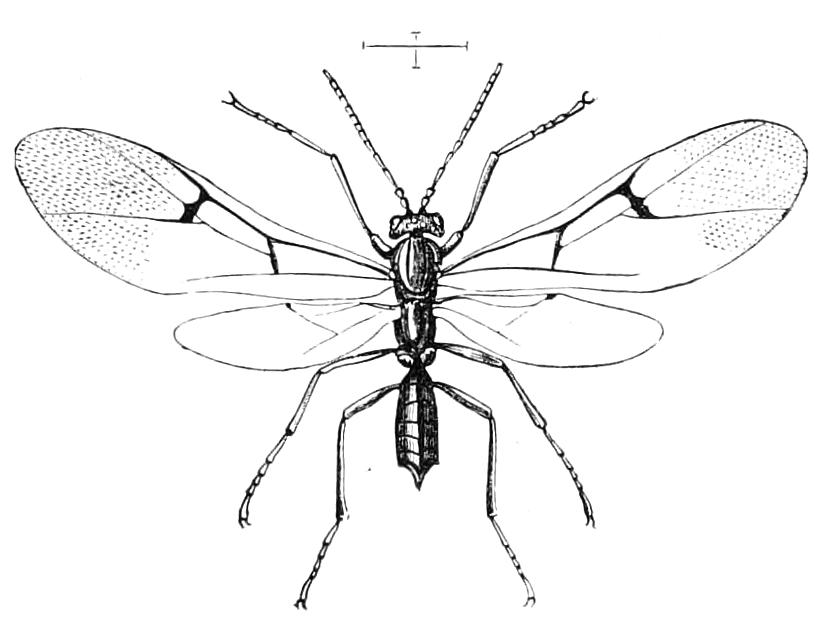
Fig. 346.—Neuroterus lenticularis. Britain.
The Cynipidae are always small, frequently minute, Insects; usually black or pitchy in colour. The simple structure of the antennae and the number of their joints are of importance as an aid in identifying a Cynipid. The mesonotum is usually remarkably convex, and has, behind, a prominent scutellum, which more or less overhangs the small metanotum and the median segment; these are perpendicular in their direction; the sculpture of these posterior parts of the alitrunk is usually deep and remarkable. The abdomen has usually only a short petiole, so as to be pseudo-sessile; but there are some genera in which this part is rather long. The abdomen is generally so very much changed in outer form that its structure is not easily understood. The visible portion is frequently in larger part made up of the greatly enlarged dorsal plate of the second or third segment, or of both. These large plates are really chiefly composed of free flaps, and on lifting them up the large ventral plates are disclosed, although these appeared previously to be nearly or quite absent. In the female there is a very slender ovipositor, of which only a small part protrudes, although the organ is really elongate; it is drawn into the abdomen by means of a peculiar series of structures, the modified terminal segments to which it is attached being folded over into the interior of the body in such a way that the posterior part becomes situated anteriorly. In conformity with this arrangement, the ovipositor is bent double on itself, the anterior and the middle portions of the borer being carried into the body, leaving only a small part projecting beyond the extremity. The Cynipid ovipositor is an instrument of much delicacy, and is capable of a great deal of movement; it is usually serrate just at the tip, and although it looks so very different from the cutting apparatus of the sawflies (Fig. 344), it seems that it is really composed of pieces similar in their origin to those of the Tenthredinidae.
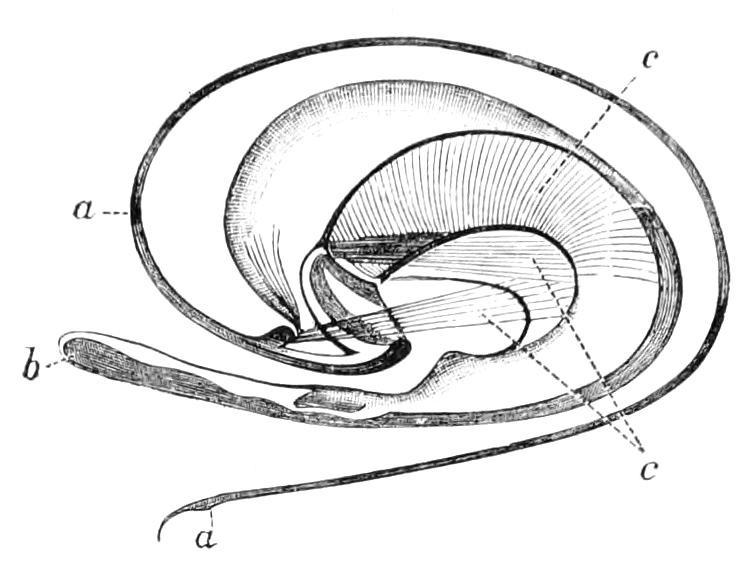
Fig. 347.—Ovipositor of Neuroterus laeviusculus. (After Adler.) a, a, The ovipositor partially coiled; b, extremity of posterior plate; c, c, muscles.
The wings frequently bear fine hairs; the paucity of nervures and the absence of the "stigma" are of importance in the definition of the family. The most important of the cells is one called the radial cell, situate just beyond the middle of the front part of the wing.
We cannot enter into a consideration of the classification of the family, as authorities are not agreed on the subject.[429] As regards their habits Cynipidae are, however, of three different kinds: (1) the true gall-flies, or Psenides, which lay an egg or eggs in the tissues of a growing plant, in the interior of which the larva lives after it is hatched; this mode of life may or may not, according to the species, be accompanied by formation of a peculiar growth called a gall: (2) Inquilines,[430] or guest-flies; these lay their eggs in the galls formed by the gall-makers subsequent to the growth of the galls, of which they obtain the benefit: (3) Parasites; these live, like most Ichneumon-flies, in the interior of the bodies of other living Insects; they prey on a considerable variety of Insects, but chiefly, it is believed, on Aphididae, or on Dipterous larvae. These parasitic flies belong to the sub-family Figitides.
A great deal of discussion has occurred relative to the nature and origin of galls, and many points still remain obscure. Considerable light has been thrown on the subject by the direct observations of modern naturalists. Previous to Malpighi, who wrote on the subject two hundred years ago, it was supposed that galls were entirely vegetable productions, and that the maggots found in them were due to spontaneous generation, it having been an article of belief in the Middle Ages that maggots in general arose from the various organic substances in which they were found, by means of the hypothetical process called, as we have said, spontaneous generation. Malpighi was aware of the unsatisfactory nature of such a belief, and having found by observation that galls arose from the punctures of Insects, he came to the further conclusion that the growth of the gall was due to the injection by the Insect into the plant of a fluid he termed Ichor, which had, he considered, the effect of producing a swelling in the plant, something in the same way as the sting of a bee or wasp produces a swelling in an animal. Réaumur also made observations on the gall-Insects, and came to the conclusion that the latter part of Malpighi's views was erroneous, and that the swelling was not due to any fluid, but simply to irritation caused by the prick; this irritation being kept up by the egg that was deposited and by the subsequent development of the larva. Observations since the time of Réaumur have shown that the matter is not quite so simple as he supposed, for though in the case of some galls the development of the gall commences immediately after the introduction of the egg, yet in other cases, as in the Cynipidae, it does not occur till some time thereafter, being delayed even until after the hatching of the egg and the commencement of the development of the larva. Galls are originated by a great variety of Insects, as well as by mites, on many plants; and it must not be concluded that a gall has been formed by Hymenoptera even when these Insects are reared from one. Extremely curious galls are formed by scale-Insects of the sub-family Brachyscelides on Eucalyptus trees in Australia; they are much inhabited by parasitic Hymenoptera, and Froggatt has obtained 100 specimens of a small black Chalcid from a single dead Brachyscelid.[431] The exact manner in which many of these galls originate is not yet sufficiently ascertained; but the subject of the galls resulting from the actions of Cynipidae has received special attention, and we are now able to form a conception of their nature. They are produced by the meristematic or dividing tissue of plants, and frequently in the cambium zone, which is caused to develop to an unusual extent, and in a more or less abnormal manner, by the presence of the Insect. The exact way in which a Cynipid affects the plant is perhaps not conclusively settled, and may be found to differ in the cases of different Cynipidae, but the view advocated by Adler and others, and recently stated by Riley,[432] seems satisfactory; it is to the effect that the activity of the larva probably affects the meristem, by means of a secretion exuded by the larva. The mere presence of the egg does not suffice to give rise to the gall, for the egg may be deposited months before the gall begins to form. It is for the same reason improbable that a fluid injected by the parent fly determines the gall's growth. It is true that the parent fly does exude a liquid during the act of oviposition, but this is believed to be merely of a lubricant nature, and not to influence the development. It is said that the gall begins to form in some cases before the larva is actually hatched, but the eggs of some Hymenoptera exhibit remarkable phenomena of growth, so that the egg, even during development of the embryo in it, may in these cases, exert an influence on the meristem. It is to reactions between the physiological processes of the meristem and the growing Insect that the gall and its form are due.
The investigations of several recent naturalists lend support to the view that only the meristematic cells of the plant can give rise to a gall. Riley says that the rate of growth of the gall is dependent on the activity of the meristem, galls on catkins developing the most quickly; those forming on young leaves also grow with rapidity, while galls formed on bark or roots may take months to attain their full size.
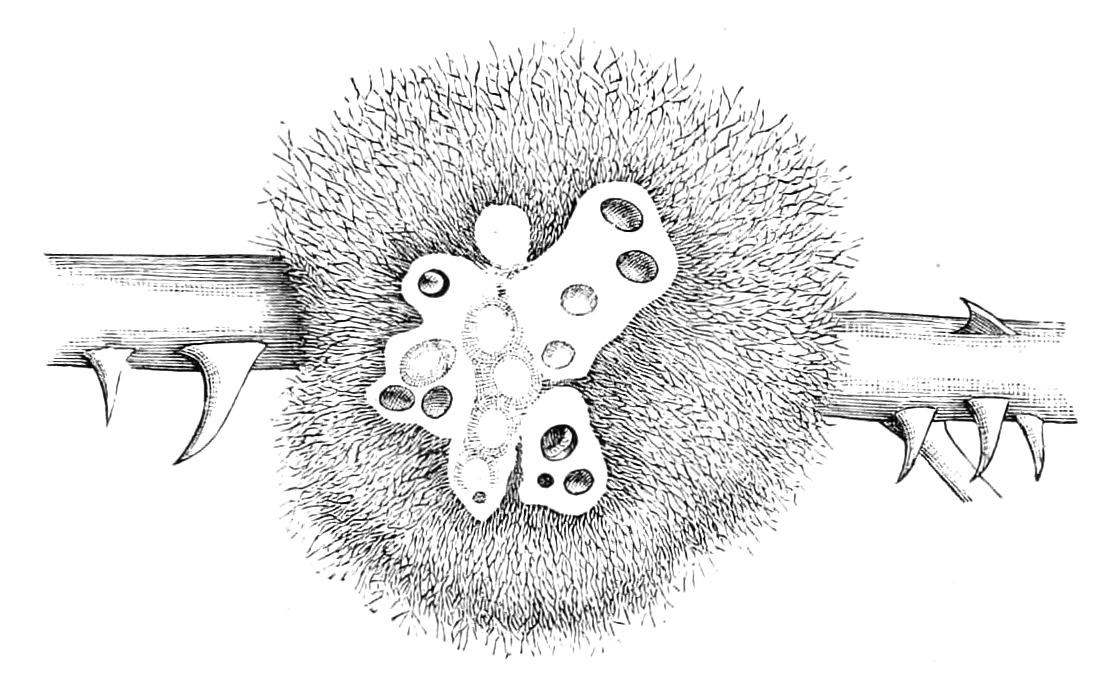
Fig. 348.—Bedeguar on rose, cut across to show the cells of the larvae; in some of the cells larvae are seen.
It is a curious fact that Cynipid galls are formed chiefly on oaks, this kind of tree supplying a surprising number and variety of galls. The plants that furnish Cynipid galls in Europe are not numerous. A list of them is given by Cameron.[433] Several species, of the genus Rhodites, attack rose-bushes. One of the best known of our British galls is the bedeguar, found in various parts of the country on both wild and cultivated rose-bushes (Fig. 348), and caused by Rhodites rosae (Fig. 349). This gall has the appearance of arising from a twig or stem, but it is really a leaf gall. Pazlavsky[434] has described the mode of formation of the bedeguar. The female Rhodites in the spring selects a rose-bud—not a flower-bud—that should produce a twig and leaves, and pricks this bud in a systematic manner in three places. The three spots of the bud pricked by the Insect are the three undeveloped leaves that correspond to a complete cycle in the phyllotaxis of the plant. The three rudiments do not develop into leaves, but by a changed mode of growth give rise to the bedeguar. Usually this gall, as shown in our figure, is of large size, and contains numerous cells; but abortive specimens are not infrequently met with; sometimes a small one is seated on a rose-leaf, and it is thought that these are due to a failure on the part of the Insect to complete the pricking operation. Cynipidae will not go through their gall-making operations except under natural conditions. Giraud[435] attempted to obtain oviposition, on gathered twigs of oak, from flies in confinement; but, although he experimented with thousands of specimens, they on no occasion laid their eggs in the fresh shoots placed at their disposal, but discharged their eggs in little heaps, without attention to the twigs. The same observer has also called attention to the fact that after being deposited in a bud the eggs of certain species of Cynips will remain dormant without producing, so far as can be seen, any effect on the tree for a period of fully ten months, but when the bud begins to develop and the egg hatches then the gall grows.
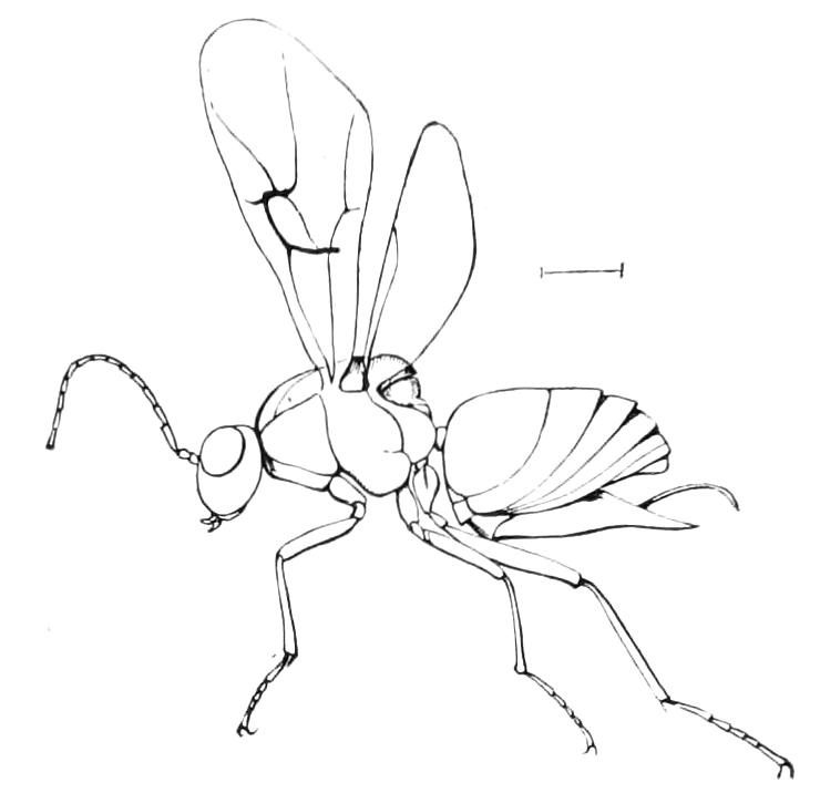
Fig. 349.—Rhodites rosae, female. Cambridge.
The exact mode in which the egg is brought to the requisite spot in the plant is still uncertain. The path traversed by the ovipositor in the plant is sometimes of considerable length, and far from straight; in some cases before it actually pierces the tissues, the organ is thrust between scales or through fissures, so that the terebra, or boring part of the ovipositor, when it reaches the minute seam of cambium, is variously curved and flexed. Now as the canal in its interior is of extreme tenuity, and frequently of great length, it must be a very difficult matter for the egg to reach the tissue where it should develop. The eggs of Cynipidae are very remarkable bodies; they are very ductile, and consist of a head, and of a stalk that in some cases is five or six times as long as the head, and is itself somewhat enlarged at the opposite end. Some other Hymenoptera have also stalked eggs of a similar kind (Fig. 357, A, egg of Leucospis). It has been thought that this remarkable shape permits of the contents of the egg being transferred for a time to the narrower parts, and thus allows the broader portion of the egg to be temporarily compressed, and the whole structure to be passed through a very narrow canal or orifice. It is, however, very doubtful whether the egg really passes along the canal of the borer. Hartig thought that it did so, and Riley supports this view to a limited extent. Adler, however, is of a different opinion, and considers that the egg travels in larger part outside the terebra. It should be remembered that the ovipositor is really composed of several appendages that are developed from the outside of the body; thus the external orifice of the body is morphologically at the base of the borer, the several parts of which are in longitudinal apposition. Hence there is nothing that would render the view of the egg leaving the ovipositor at the base improbable, and Adler supposes that it actually does so, the thin end being retained between the divisions of the terebra. Riley is of opinion that the act of oviposition in these Insects follows no uniform system. He has observed that in the case of Callirhytis clavula, ovipositing in the buds of Quercus alba, the eggs are inserted by the egg-stalk into the substance of the leaf, and that the egg-fluids are at first gathered in the posterior end, which is not inserted. "The fluids are then gradually absorbed from this exposed portion into the inserted portion of the egg, and by the time the young leaves have formed the exposed [parts of the] shells are empty, the thread-like stalk has disappeared, and the egg-contents are all contained within the leaf tissue." He has also observed that in Biorhiza nigra the pedicel, or stalk, only is inserted in the embryonic leaf-tissue, and that the enlarged portion or egg-body is at first external. The same naturalist also records that in the case of a small inquiline species, Ceroptres politus, the pedicel of the egg is very short, and in this case the egg is thrust down into the puncture made by the borer, so that the egg is entirely covered.
Some Cynipidae bore a large number of the channels for their eggs before depositing any of the latter, and it would appear that it is the rule that the boring of the channel is an act separate from that of actual oviposition. Adler distinguishes three stages: (1) boring of the canal; (2) the passage of the egg from the base of the ovipositor, where the egg-stalk is pinched between the two spiculae and the egg is pushed along the ovipositor; (3) after the point of the ovipositor is withdrawn, the egg-body enters the pierced canal, and is pushed forward by the ovipositor until it reaches the bottom.[436]
About fifty years ago Hartig reared large numbers of certain species of gall-flies from their galls, obtaining from 28,000 galls of Cynips disticha about 10,000 flies, and from galls of C. folii 3000 or 4000 examples of this species; he found that all the individuals were females. His observations were subsequently abundantly confirmed by other naturalists, among whom we may mention Frederick Smith in our own country, who made in vain repeated attempts to obtain males of the species of the genus Cynips. On one occasion he collected in the South of England 4410 galls of C. kollari (at that time called C. lignicola), and from these he obtained 1562 flies, all of which were females. A second effort was attended with similar results. Hartig, writing in 1843, after many years' experience, stated that though he was acquainted with twenty-eight species of the genus Cynips, he had not seen a male of any one of them. During the course of these futile attempts it was, however, seen that a possible source of fallacy existed in the fact that the Insects were reared from collected galls; and these being similar to one another, it was possible that the males might inhabit some different gall. Adler endeavoured to put the questions thus raised to the test by means of rearing females from galls, and then getting these females to produce, parthenogenetically, galls on small oaks planted in pots, and thus completely under control. He was quite successful in carrying out his project, and in doing so he made a most extraordinary discovery, viz. that the galls produced by these parthenogenetic females on his potted oaks, were quite different from the galls from which the flies themselves were reared, and were, in fact, galls that gave rise to a fly that had been previously considered a distinct species; and of this form both sexes were produced. Adler's observations have been confirmed by other naturalists, and thus the occurrence of alternation of generations, one of the two generations being parthenogenetic, has been thoroughly established in Cynipidae. We may mention one case as illustrative. A gall-fly called Chilaspis lowii is produced from galls on oak-leaves at Vienna at the end of April, both sexes occurring. The female thereafter lays eggs on the ribs of the leaves of the same kind of oak, and thus produces a different gall from that which nourished herself. These galls fall off with the leaves in the autumn, and in July or August of the following year a gall-fly is produced from them. It is a different creature from the mother, and was previously known to entomologists under the name of Chilaspis nitida. Only females of it occur, and these parthenogenetic individuals lay their eggs in the young buds of the oak that are already present in the autumn, and in the following spring, when the buds open and the leaves develop, those that have had an egg laid in them produce a gall from which Chilaspis lowii emerges in April or May. In this case therefore the cycle of the two generations extends over two years, the generation that takes the greater part of the time for its production consisting only of females. Adler's observations showed that, though in some species this alternation of generations was accompanied by parthenogenesis in one part of the cycle, yet in other species this was not the case. He found, for instance, that some gall-flies of the genus Aphilothrix produced a series of generations the individuals of which were similar to one another, and were all females and parthenogenetic. In some species of the old genus Cynips no males are even yet known to occur. A very curious observation was made by the American, Walsh, viz. that of galls gathered by him quite similar to one another, some produced speedily a number of both sexes of Cynips spongifica, while much later on in the season the remainder of the galls gave rise to females only of an Insect called Cynips aciculata. It is believed that the galls gathered by Walsh[437] were really all one species; so that parts of the same generation emerge at different times and in two distinct forms, one of them parthenogenetic, the other consisting of two sexes. It has, however, been suggested that Cynips spongifica and C. aciculata may be two distinct species, producing quite similar galls.
Turning now to the questions connected with inquiline or guest-flies, we may commence with drawing attention to the great practical difficulties that surround the investigation of this subject. If we open a number of specimens of any kind of gall it is probable that several kinds of larvae will be found. In Fig. 350 we represent four kinds of larvae that were taken out of a few bedeguar galls gathered on one day in a lane near Cambridge. It is pretty certain that No. 1 in this figure represents the larva of Rhodites rosae, and that Nos. 2 and 3 are larvae of inquilines, possibly of Synergus, or of a parasite; while No. 4, which was engaged in feeding on No. 3 in the position shown, is possibly a Chalcid of the genus Monodontomerus, or may be Callimome bedeguaris. It is clear that, as we cannot ascertain what is inside a gall without opening it, and thereby killing the tenants, it is a most difficult matter to identify the larvae; the only safe method is that of observation of the act of oviposition; this may be supplemented by rearing the flies from galls, so as to ascertain what variety of flies are associated with each kind of gall. This last point has been well attended to; but the number of cases in which oviposition of inquiline gall-flies in the galls formed by the Psenides has been ascertained by direct observation is still very small; they are, however, sufficient to show that the inquilines deposit their eggs only after the galls are formed.
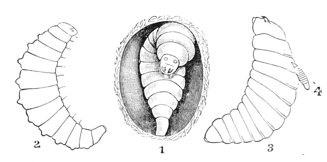
Fig. 350.—Larvae inhabiting bedeguar gall at Cambridge. 1, Rhodites rosae in cell; 2 and 3, larvae of inquilines; 4, larva of a parasitic Hymenopteron.
Bassett recorded the first case of the kind in connexion with a North American species, Cynips (Ceroptres) quercus-arbos Fitch. He says: "On the first of June galls on Quercus ilicifolia had reached their full size, but were still tender, quite like the young shoots of which they formed part. Examining them on that day, I discovered on them two gall-flies, which I succeeded in taking. They were females, and the ovipositor of each was inserted into the gall so deeply that they could not readily free themselves, and they were removed by force."
The great resemblance of the inquiline gall-fly to the fly that makes the gall both dwell in, has been several times noticed by Osten Sacken, who says "one of the most curious circumstances connected with the history of two North American blackberry galls is, that besides the Diastrophus, which apparently is the genuine originator of the gall, they produce another gall-fly, no doubt an inquiline, belonging to the genus Aulax, and showing the most striking resemblance in size, colouring, and sculpture to the Diastrophus, their companion. The one is the very counterpart of the other, hardly showing any differences, except the strictly generic characters! This seems to be one of those curious instances, so frequent in entomology, of the resemblance between parasites and their hosts! By rearing a considerable number of galls of D. nebulosus I obtained this species as well as its parasite almost in equal numbers. By cutting some of the galls open I ascertained that a single specimen of the gall frequently contained both species, thus setting aside a possible doubt whether these Insects are not produced by two different, although closely similar galls."[438]
The substance of which galls are composed, or rather, perhaps, a juice they afford, is apparently a most suitable pabulum for the support of Insect life, and is eagerly sought after by a variety of Insects; hence by collecting galls in large quantities many species of Insects may be reared from them; indeed by this means as many as thirty different kinds of Insects, and belonging to all, or nearly all, the Orders, have been obtained from a single species of gall. Some galls are sought by birds, which open them and extract their tenants, even in cases where it might be supposed that the nauseous flavour of the galls would forbid such proceedings.
Not more than 500 species of Psenides and Inquiline Cynipidae are known from all parts of the world; and of described Parasitic Cynipidae there are only about 150 species. The British forms have recently been treated by Cameron in the work we have already several times referred to.[439]
A few Cynipidae have been found in amber; and remains of members of the family, as well as some galls, are said by Scudder to have been found in the Tertiary strata at Florissant.
Fam. II. Proctotrypidae, or Oxyura.
Small Hymenoptera, with few, or even no, nervures in the wings: the pronotum closely adherent to the mesothorax, and at the sides reaching backwards to the points of insertion of the wings. The abdomen is pointed, and the pointed apex is frequently deflexed; the ovipositor is not coiled, but is retractile, and when extruded is of tubular form, and apparently a continuation of the tip of the body. The earlier stages are passed in the bodies, or in the eggs, of other Arthropods.
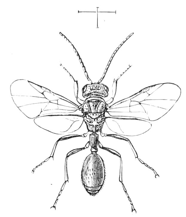
Fig. 351.—Helorus anomalipes. Britain.
The Proctotrypidae is one of the most difficult groups of Hymenoptera to define; some of its members exhibit a great resemblance to Aculeate Hymenoptera. This is the case with the Insect we figure (Fig. 351). It, however, is an undoubted Proctotrypid, but there are other forms that approach very closely in appearance to the Aculeata, or stinging Hymenoptera; so that until a better comprehension is reached as to the distinction between a sting and an ovipositor the separation between Proctotrypidae and Aculeata must be considered somewhat arbitrary.
There is extreme variety in the family; the wings differ considerably in shape and neuration; they are not infrequently altogether absent in one or both sexes. The chief distinction of the family from other parasitic Hymenoptera is the tubular form of the ovipositor; which part appears to be a continuation of the tip of the body. This latter is more definitely acuminate than usual, and has given rise to the term Oxyura, by which name the Proctotrypidae are distinguished in many books. From the Chalcididae they are distinguished also by the angles of the pronotum attaining the tegulae. In this character they agree with the Cynipidae, but the ovipositor and abdomen are very different in form in these two groups, and the Proctotrypidae very frequently have a pigmented spot or stigma on the front wings which is absent in Cynipidae. As if to add to the difficulties the systematist meets with in dealing with this family, some of its members have the trochanters undivided, as in the case of the stinging Hymenoptera. The larvae of all that are known lead a completely parasitic life in the bodies or eggs of other Insects or of Spiders. Sometimes half a dozen specimens may find the means of subsistence during the whole of their development in a single Insect's egg. Usually Proctotrypids pupate in the position in which they have fed up, enclosed each one in a more or less distinct cocoon. In Fig. 352 we represent a very remarkable case of Proctotrypid pupation; a larva of some beetle has nourished many specimens of a species of the genus Proctotrypes, and the pupae thereof project from the body of the host, a pair of the parasites issuing from each segmental division in a remarkably symmetrical manner.
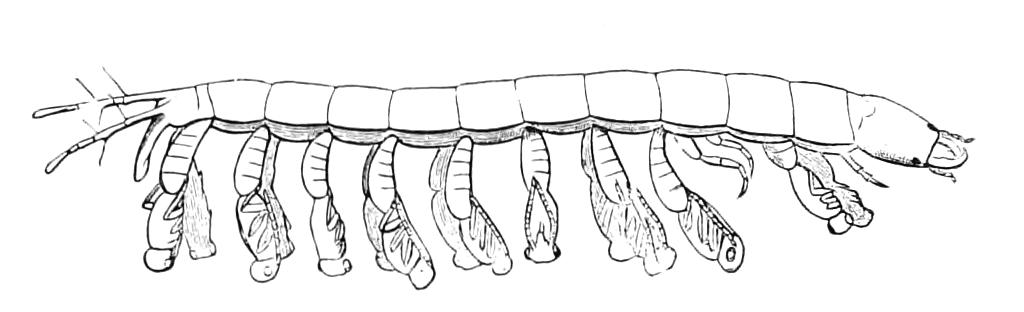
Fig. 352.—Pupation of Proctotrypes sp. in body of a beetle larva.
Comparatively little is known as to the habits of the members of this family, but such information as has been obtained leads to the conclusion that great variety will be found to exist in this respect. We have already mentioned that numerous species have been ascertained to feed inside the eggs of Insects or of Spiders; others have been reared from larvae or from galls of the minute Dipterous midges of the family Cecidomyiidae; others have been obtained from Cynipid galls, a few from ants' nests and from green-fly; some species are known to attack Coleoptera. The distinguished Irish entomologist, Haliday, has written an account of the proceedings of a species of Bethylus,[440] from which it has been supposed that this Insect carries off living caterpillars, and stores them in a suitable receptacle as food for its progeny, thus anticipating, as it were, the habits of the fossorial division of the Aculeata, in which group this instinct has, as we shall subsequently relate, attained an astonishing degree of perfection. Haliday's observation was unfortunately incomplete and has not been subsequently confirmed. The Bethylides are remarkable for their great approach in structure to the Aculeates, so much so that entomologists are not agreed as to whether certain Insects are Proctotrypids or Aculeates. Pristocera, with a very wide distribution, may be mentioned as illustrative of these doubtful forms; but other genera of the Bethylides are in many respects very similar to the Aculeates, and it is not matter for surprise that Haliday should have considered the Bethylides to be a tribe of the stinging Hymenoptera. The genus Scleroderma consists of small Insects much resembling ants, and, as well as some of its allies, is of great interest from the remarkable phenomena of polymorphism presented by certain species. The males in this genus are winged, the females completely apterous; yet at times winged females are produced—as exceptional individuals in a brood of wingless specimens—the females in these cases being not only winged, but possessed of ocelli like the females of other winged Hymenoptera. Particulars of a case of this kind have been given by Sir Sidney Saunders,[441] and Ashmead also mentions[442] the exceptional occurrence of these winged females. Westwood[443] was of opinion that there are three forms of the female sex. This subject is of importance in connexion with the production of the various castes in ants. Although the presence of wings in these Insects is always accompanied by the existence of ocelli (which, it will be remembered, are normally absent from the wingless individuals), yet the converse is not always the case, for a form of the female of Cephalonomia formiciformis, without any wings, yet having ocelli, as well as eyes, well developed, is figured by Westwood.[444]
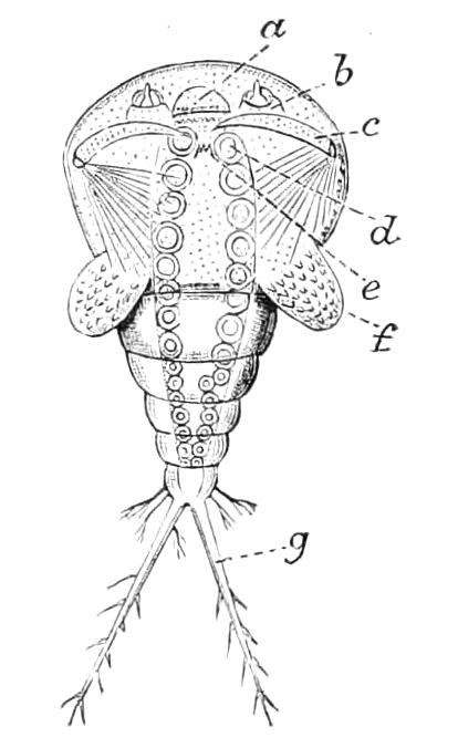
Fig. 353.—Cyclops form of larva of Platygaster sp. (After Ganin.) a, Mouth; b, antenna; c, claw-limb; d, lower lip (the pointing line is a little too short); e, doubtful "zapfenförmig" organ; f, wing-like lobe; g, branch of the tail.
The development of some of the Proctotrypids has been partially described by Ganin and others, and is of an extraordinary character. Ganin's observations[445] were most complete in the case of a species of Platygaster, which he found in the larva of a very minute Dipteron of the genus Cecidomyia. The Platygaster larva changes its form very much in the course of its life, resembling at first a minute Crustacean rather than an Insect-larva; it has a very large rounded anterior portion, while behind it terminates in two, or more, tail-like processes. By a very peculiar kind of metamorphosis this Cyclops-like larva changes into an almost unsegmented, oviform larva, destitute of appendages; by a second change this creature assumes a third condition, in which it is similar to the ordinary form of parasitic Hymenopterous larvae. Sometimes several of the Platygaster larvae are found in a single host, but only one of them reaches this third stage. Afterwards the third larval instar passes into the pupal stage, which lasts five or six days, and then the perfect Insect appears. It is worthy of remark that the internal organs undergo quite as remarkable a change as the outer form does. The metamorphoses of some other Proctotrypidae have been examined by Ganin, and appear to be of an equally interesting character.[446]
There is reason to suppose that these Platygaster parasites are of great economic importance as well as of scientific interest, for Platygaster herrickii is one of the enemies of the larva of the destructive Hessian fly, Cecidomyia destructor.
The Proctotrypidae are no doubt extremely numerous in species, but as yet they have been very little studied; a good work on the British species is much required. A valuable contribution has recently been made to the study of the family by Ashmead, in the book we have already referred to. This volume includes much information on the natural history of these Insects, and the outline figures give some idea of the great variety of external form.
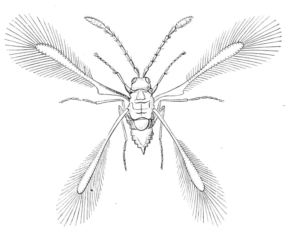
Fig. 354.—Alaptus excisus, Westwood. Britain. (Probable size about ½ millim.)
Many entomologists include the Mymarides in Proctotrypidae, but Ashmead considers that they should be treated as a separate family. Alaptus excisus Westw. (Fig. 354) has been frequently said to be the smallest known Insect, the measurement given for it by Westwood[447] being a length of ⅙ of a millimetre—about 1⁄150 of an inch. Mr. Enock has recently examined Westwood's type in the Museum at Oxford, and from his information we may conclude that this Insect is probably the same as Alaptus fusculus Hal., and that the measurement mentioned by Westwood is erroneous, the Insect being really about half a millimetre long. The Mymarides are, however, very minute, some of them not exceeding one-third of a millimetre in length. Whether any of them are smaller than the beetles of the family Trichopterygidae, some of which are only one-fourth of a millimetre long, may be doubted.
The Mymarides are recognisable by their very minute size, and by their peculiar wings. These are slender, destitute of nervures, fringed with long, delicate hairs, and stalked at the base. Probably Mymarides may all prove to be dwellers in eggs of other Insects. The group is remarkable from the fact that it contains some of the very few Hymenoptera with aquatic habits. Two species were discovered in their winged condition in the water of a pond near London by Sir John Lubbock[448]; one of them—Polynema natans Lubbock—probably, according to Mr. Enock, the same as Caraphractus cinctus Hal., uses its wings freely for swimming under water, while the other—Prestwichia aquatica—performs this operation by the aid of its legs. This latter Insect seems to be very anomalous, and its position quite doubtful. The embryogeny of Polynema is very peculiar, and takes place in the egg of a dragon-fly—Calepteryx virgo—under water. According to Ganin,[449] in the earliest stages the developments of the embryos of the Calepteryx and of the Polynema progress simultaneously, but that of the dragon-fly does not proceed beyond the formation of the ventral plate. The Polynema appears to leave its own egg at an extremely early stage of the embryonic development. It would appear, in fact, that there is no definite distinction between embryonic and larval stages. The information given by Ganin leads to the conclusion that a complete study of this remarkable mode of development is necessary before forming any general ideas as to the nature of Insect embryogeny and metamorphosis.
Fam. III. Chalcididae.
Pronotum with some freedom of movement, its angles not extending to the insertion of the front wings. Antennae elbowed, consisting of from seven to thirteen joints. Wings without a system of cells; with a single definite nervure proceeding from the base near the front margin, or costa; afterwards passing to the costa, and giving off a very short vein more or less thickened at its termination. The species are, with few exceptions, of parasitic habits.
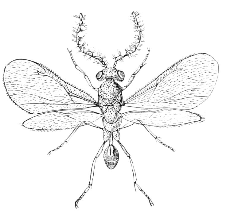
Fig. 355.—Eurytoma abrotani, male. Britain. Hyper-parasite through Microgaster of Liparis dispar, and according to Cameron, parasite of Rhodites rosae and other gall-flies in Britain, × 10. (After Ratzeburg.)
The Insects of this family—the Pteromalini of Ratzeburg—are frequently of brilliant colours and of remarkable form; the species are very numerous, some 4000 or more having already been described. Of this number nearly 3000 are European, and as there is good reason for supposing that Chalcididae are quite as numerous in the Tropics and in the New World as they are in Europe, the family will probably prove to be one of the largest in the class. About twenty sub-families have already been proposed for the classification of the group; they are based chiefly on the number of joints in the tarsi, and the details of the antennae and of the ovipositor. This latter exhibits great variety in external appearance, due chiefly to the modification in form of the basal, or of the following ventral abdominal plates, one or more of which may be prolonged and altered in form or direction, giving rise in this way to considerable diversity in the shape of the abdomen. Correlative with this is a great variety in the mode of parasitism of the larva. Many live in galls, feeding on the larvae of the makers of the galls or on those of the inquilines; others attack caterpillars, others pupae only; some flourish at the expense of bees or other Hymenoptera, or of Coccidae and Aphididae, and some deposit their eggs in the egg-cases of Blattidae. The details of the life-history are well known in only a few cases.
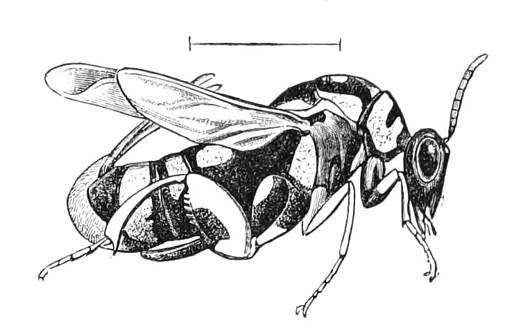
Fig. 356.—Leucospis gigas, female. Gibraltar.
The career of Leucospis gigas has been investigated by Fabre, and exhibits a very remarkable form of hypermetamorphosis.[450] This Insect is of comparatively large size and of vivid colours, wasp-like, black contrasting with yellow, as in the case of the wasps; and like these it has the wings folded or doubled. The female bears a long ovipositor, which by a peculiar modification is packed in a groove on the back of the Insect. This species lives in Southern Europe at the expense of Chalicodoma muraria, a mason-bee that forms cells of a hard cement for its nest, the cells being placed together in masses of considerable size; each cell contains, or rather should contain, a larva of the bee, and is closed by masonry, in the construction of which the bee displays much ability. It is the mission of the Leucospis to penetrate the masonry by means of its ovipositor, and to deposit an egg in the cell of the bee. The period chosen for this predatory attack is the end of July or the beginning of August, at which time the bee-larva is in the torpid and powerless condition that precedes its assumption of the pupal state. The Leucospis, walking about leisurely and circumspectly on the masonry of the nest, tests it repeatedly by touching with the tips of the antennae, for it is most important that a proper spot should be selected. The bee's cell is placed in a mass of solid masonry, a considerable part—but a part only—of whose area is occupied by the group of cells; every cell is closed by hard mortar, making an uneven surface, and the face of the masonry is rendered more even by a layer of hardened clay outside the rougher material; it is the task of the Leucospis to detect a suitable spot, in the apparently uniform external covering, and there to effect the penetration so as to introduce an egg into a cell. By what sensations the fly may be guided is unknown. After a spot has been selected and the ovipositor brought into play, the masonry is ultimately pierced by patient work; sometimes a quarter of an hour is sufficient for the purpose, but in other cases three hours of uninterrupted effort are required before the end is attained. Fabre expended much time in watching this operation, and after the Insect had completed it, he marked with a pencil the exact spot of the masonry that was penetrated, and the date on which it was done, and he states that he afterwards found that without any exception a proper spot had been selected, and a cell consequently penetrated. Admirable as the instinct of the parasite appears from this point of view, it is nevertheless accompanied by a remarkable deficiency in two other respects. The first is that though the spot selected by the Leucospis invariably gives entrance to a cell, yet in the majority of the cases the selected cell is not a suitable one; a large number of the cells of the Chalicodoma are not occupied by living larvae on the point of pupation—though in that case only can the egg of the Leucospis hatch and successfully develop—but by dead and shrivelled larvae, or by mouldy or dried-up food. And yet, in each case of penetration, Fabre believes that an egg is deposited, even though it may be impossible that it can undergo a successful development. Strange as this may appear, it is nevertheless rendered less improbable by the second deficiency in the instinct of the parasite. The Insect has no power of recognising a cell that has been previously pierced either by itself or by another of its species. One bee larva can only supply nourishment for a single larva of the parasite, and yet it is a common occurrence for a cell to be revisited, pierced again and another egg introduced; indeed Fabre, by means of the cells he had marked, was able to assure himself that it is no uncommon thing for this to be done four times; four eggs, in fact, are sometimes deposited in a cell that cannot by any possibility supply food for more than one larva. The egg of the Leucospis is a curious object (Fig. 357, A), very elongate oval, with one end drawn out and bent so as to form a hook; it is not placed at random in the cell of the bee, but is suspended on the delicate cocoon with which the Chalicodoma larva is surrounded at the period of pupation.
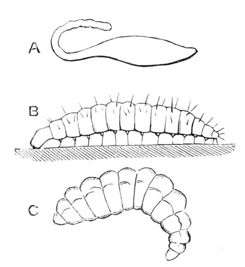
Fig. 357.—Leucospis gigas. A, Egg; B, primary, C, secondary larva. (After Fabre.)
Fabre allowed sufficient time to elapse for the hatching of the larvae from the eggs, and then opened some cells where Leucospis eggs had been deposited, in order to obtain the larvae; when doing this he was surprised that he never found more than one Leucospis larva in a cell. Even in cells where he had observed more than one act of oviposition, and which he had marked at the time, only one larva existed. This induced him to think that it was possible that no egg was deposited by the Leucospis at the second penetration. He accordingly examined cells soon after the eggs were laid, and thus discovered some that contained more than one egg,—indeed in one cell he observed no less than five eggs suspended from the cocoon of the Chalicodoma; he was also able to demonstrate that eggs were actually deposited in some cells that contained no means of support for the larva. How then could these two facts be reconciled—four or five eggs deposited in a cell, only one larva present afterwards? It is of course impossible to observe the operations of a larva shrouded in the obscurity of a cell formed of masonry, so he transferred some bee larvae with their destructive companions to glass tubes, in which he was able to note what took place. He found that the egg deposited by the Leucospis hatches and produces a very peculiar larva, having little resemblance to the Leucospis larva that he had found eating the Chalicodoma larva. The primary larva (Fig. 357, B) of the Leucospis is an arched worm, moderately deeply segmented, a millimetre or a little more in length, with a remarkably large and abruptly-defined head. The body bears erect setae, the most remarkable of which are a pair on the ventral aspect of each of the segments, each of these ventral setae being borne on a small conical prominence. These prominences and setae serve as ambulatory organs, and are supplemented in their function by a protuberance at the posterior extremity. The little creature has considerable powers of locomotion; it moves, after the fashion of many other larvae, by contracting and arching the body so as to bring the posterior part nearer to the anterior; then fixing the hinder part, the anterior is extended and fixed, the posterior being again brought nearer to the front. The Leucospis larva when hatched does not at once attack the bee larva which is to be its future food, but every few hours makes excursions over its surface, and even explores the walls of the cell; returning, however, always to the cocoon for repose. The object of these excursions is, Fabre believes, to ascertain if another Leucospis egg has been laid in the cell, and in that case to destroy it. For the food, as we have said, being only enough for one larva, and the mother Leucospis frequently laying more than one egg in a cell, it is necessary that all the eggs except one should be destroyed. Fabre did not actually observe the act of destruction, but he found repeatedly in his glass tubes that the supernumerary eggs were destroyed, being, in fact, wounded by the mandibles of the first-hatched larva. After several days of this wandering life the tiny destroyer undergoes a first moult, changing its skin and appearing as a very different creature (Fig. 357, C); it is now completely destitute of any means of locomotion, very deeply segmented, curved at one extremity, with a very small head, bearing extremely minute, scarcely perceptible, mandibles. The sole object of its existence in this state is to extract the contents of the Chalicodoma larva, and appropriate this material to the purposes of its own organisation. This it accomplishes not by wounding, tearing, or destroying the larva, for that apparently would not answer the purpose; the contents must be conveyed while still in their vital state to itself; and this it effects by applying its mouth to the extremely delicate skin of the victim, the contents of whose body then gradually pass to the destroyer, without any visible destruction of the continuity of the integument. Thus the Leucospis larva gradually grows, while the bee larva shrinks and shrivels, without, however, actually suffering death. The process of emptying the bee larva apparently does not occupy the Leucospis more than two or three weeks, being completed by about the middle of the month of August; afterwards the larva remains in the cell by the side of the shrivelled skin of its victim for ten or eleven months, at the end of which time it assumes the pupal condition, and very shortly thereafter appears as a perfect Insect.
Monodontomerus cupreus is another member of the Chalcididae that lives parasitically at the expense of bees of the genus Chalicodoma. Its habits have been sketched by Fabre,[451] and exhibit considerable difference from those of Leucospis. It is much less in size, and can accommodate itself to a greater variety of food; it will, in fact, eat not only the larva of Chalicodoma, but also that of another bee, of the genus Stelis, that is frequently found shut up in the cell of the Chalicodoma, at whose expense the Stelis also lives parasitically. The Monodontomerus bores a hole through the masonry of the bee and deposits its eggs in the cell after the fashion of the Leucospis; one bee larva is, however, sufficient food for several individuals of the young of this smaller parasite. There is no hypermetamorphosis, the early larval condition resembling the later. This Insect attacks not only Chalicodoma and Stelis, as already mentioned, but also other bees; and a single larva of some of the larger kinds will afford sufficient food for fifty young of the Monodontomerus. They feed on the bee larva, as the Leucospis does, without wounding it. This fly has the power of recognising what is suitable provender for its young by the use of the antennae, even when the conditions are so changed that it is clear the sense of sight has nothing to do with the recognition. Fabre relates that he had extracted a number of the bee larvae from their cells of masonry, and that as they were lying on his table enclosed in their cocoons, the Monodontomerus recognised the latter as containing the desired provender for its young by examining them with its antennae; after which, without hesitation, the Monodontomerus pierced the cocoon with its ovipositor and deposited the eggs in a suitable position. This observation, together with those made on Leucospis, seem to indicate that it is neither by sight nor smell that these Insects discover the desired object, but by some sense we do not understand, though its seat is clearly in the antennae of the Insect.
Newport discovered a Monodontomerus, which he described as M. nitidus,[452] in the cells of the bee Anthophora retusa, and demonstrated that the alimentary canal, as is usual in Petiolate Hymenoptera, is closed behind until the Insect is about to enter the pupal state, when it becomes perforated and faecal matters are for the first time passed from it. "These matters were composed of the refuse of digestion and of epithelial cells accumulated during the period of feeding, and retained in the digestive sac until the period of its perforation. In this way the food and abode of the Insects are maintained pure and uncontaminated, and the digestive apparatus is completed, and the refuse of nutrition ejected only when the whole of the food has been consumed."
In the cells of the same bee Newport discovered another curious parasitic Chalcid, Anthophorabia retusa.[453] The male has short wings, and the compound eye is replaced by an ocellus on each side of the head, the female having fully developed wings and eyes. A variation may occur in the metamorphosis of this Insect, inasmuch as when the growth is completed during the month of August, the Insect changes to a pupa, the imago appears ten or twelve days thereafter, and the perfect Insect then hibernates for seven or eight months; but should the completion of growth be deferred till after the end of August, hibernation takes place in the larval condition. A large and brilliant Chalcid Eucharis myrmeciae, has been described by Cameron as preying on the formidable Australian ants of the genus Myrmecia.
The development of Smicra clavipes has been partially described by Henneguy.[454] This Insect lives in the interior of the aquatic larva of Stratiomys strigosa, a Dipterous Insect. As many as fifty eggs of the parasite are found in one larva, but a large number of embryos die during development, so that he has never found more than two or three well-grown larvae in one Stratiomys larva. It has been ascertained that the eggs of many of these parasitic Insects are deficient in yolk, and the ovum of Smicra is said to obtain the nutritive materials necessary for the development of the embryo from the blood of its host by endosmosis. For a long time after the assumption of the larval condition, the larva appears to nourish itself only at the expense of the blood of its host. The segmentation of the ovum is total, and a single embryonic membrane appears at an early period, before the formation of the embryo, by a process very different from that giving origin to the amnion of the majority of Insects.
A very interesting sketch of the development of Encyrtus fuscicollis has been given by Bugnion.[455] This small parasite passes its earlier stages in the interior of the larva of Hyponomeuta cognatella or other Lepidoptera. The female Encyrtus deposits her eggs in the interior of a caterpillar, in the form of a series of 50 to 100 or more eggs enclosed in a sac; the origin of the sac is obscure, but the embryonic development and the early part of the larval life are passed in the sac, which contains a supply of nutritive matter. The larvae of the Encyrtus are at first entirely confined to this sac, but when they have consumed all the nutritive matter in it, they leave it and pass the remainder of their larval and pupal existence in the body-cavity of the caterpillar. They live at first on the lymph (blood) of the Insect, and apparently do it no harm; nevertheless the strength of the caterpillar is so much enfeebled that it fails to undergo the transformation to a pupa; the parasites then devour its interior, and use the empty skin as a nidus for their own pupation; they form cocoons which divide the area into compartments. Usually the individuals disclosed from one Hyponomeuta are all of one sex, which may be either male or female. Unfortunately the most interesting points of this development, viz. the history of the common sac for the larvae, the nature of the eggs, the earlier embryonic stages, and the nutriment in the sac, are still without elucidation. The account given by Bugnion raises a great desire for information on these points.
We have in a previous page described the remarkable mode of oviposition of Mantis. Captain Xambeu[456] has made a very curious observation to the effect that a minute Chalcid, Podagrion (Palmon) pachymerus, shelters itself under the wings of the Mantis so as to be in a position to oviposit in the eggs of the latter when it shall be forming its peculiar ootheca.
The genus Isosoma consists of Insects that differ in habits from their congeners, being phytophagous instead of parasitic. I. tritici and I. hordei live in the stalks of corn, and in North America, where they are known to the agriculturist as joint-worms, are frequently very injurious to crops. They are sometimes obtained in large numbers without any males appearing, and a wingless as well as a winged form of the female occurs. Owing to the fact that the allies of these Insects are parasitic, it has been frequently maintained that this may also prove to be the case with Isosoma, but the observations of Riley[457] and others leave no doubt that the Insects of this genus are really plant-feeders.
Riley has called attention[458] to some facts in connection with I. tritici and I. grande, that make it clear that these two supposed species are really alternate generations, and that both generations are probably in larger part, if not entirely, parthenogenetic. Some species of the genus Megastigmus are known to be of phytophagous habits.[459]
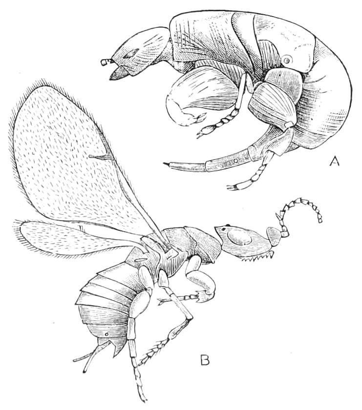
Fig. 358.—Blastophaga grossorum. A, Male, × 22; B, female, × 15. (After Mayer, Mitt. Stat. Neapel, iii. 1882.)
The most interesting of all the forms of Chalcididae are perhaps those called fig-Insects. A considerable number of species are now known, and amongst them we meet with the unusual phenomenon of species with wingless males, the females possessing the organs of flight normally developed. The wingless males exhibit the strangest forms, and bear no resemblance whatever to their more legitimately formed partners (Fig. 358, A, B). Many of the fig-Insects belong to a special group called Agaonides. Others belong to the group Torymides, which contains likewise many Chalcididae of an ordinary kind; possibly some of these may be parasitic on the Agaonides. Some of these Torymid fig-Insects have winged males, as is normal in the family, but in other cases winged and wingless forms of the male of one species may be present.
The most notorious of these fig-Insects is the one known as Blastophaga grossorum (Fig. 358), this being the chief agent in the custom known as caprification of the cultivated fig-tree. This process has been practised from time immemorial, and is at the present day still carried on in Italy and the Grecian archipelago. The Greek writers who describe it say that the wild fig-tree, though it does not ripen its own fruit, is absolutely essential for the perfection of the fruit of the cultivated fig. In accordance with this view, branches from the wild fig are still gathered at certain seasons and suspended amongst the branches of the cultivated fig-trees. The young fig is a very remarkable vegetable production, consisting of a hollow, fleshy receptacle, in which are placed the extremely numerous and minute flowers, the only admission to which is by a small orifice at the blunt end of the young fig; this orifice is lined with projecting scales, that more or less completely fill it up or close it; nevertheless inside this fruit the Blastophaga grossorum develops in large numbers. The males are, as we have seen, wingless creatures, and do not leave the fruit in which they were bred, but the females make their way out of the wild fig, and some of them, it is believed, enter the young fruit of the cultivated trees and lay their eggs, or attempt to do so, therein; and it has been supposed by various writers that these proceedings are essential to the satisfactory development of the edible fruit. It is a curious fact that the Blastophaga develops very freely in the wild fig—so much so, indeed, as to be a means of preventing it from coming to maturity; but yet the Insect cannot complete its development in the cultivated fruit. This is due to the fact that the fly must lay its egg in a particular part of the fig-ovule, so that when the egg hatches the larva may have a proper supply of food. In the cultivated fig the structure of the flower differs somewhat from that of the caprificus, as the wild fig is called, and so the egg, if deposited at all, does not reach a proper nidus for its development. Hence the Blastophaga can never live exclusively on the cultivated fig, and if it be really necessary for the development of the latter, must be brought thereto by means of the caprifig. Whether the Blastophaga be really of use, as has been for so long supposed, is, however, a matter for doubt. The reasons for this are (1) that those who think caprification beneficial do not agree as to the mode in which they suppose it to be so; (2) that there is but little reason for believing that when introduced amongst the cultivated figs the Blastophaga occupies itself to any great extent therewith; and (3) that in some parts of the world caprification is not performed, but the cultivated fig nevertheless ripens its fruit there. Hence many writers on the subject—Solms-Laubach,[460] Mayer,[461] and Saunders[462]—entertain considerable doubt as to whether caprification is at present anything more than an old custom destitute of practical utility. On the other hand, Riley states[463] "that the perfect Smyrna fig, the most esteemed of the edible species, can be produced only by the intervention of the Blastophaga psenes [grossorum]."
Although the questions connected with the effect the Blastophaga is supposed to produce on the fruit are of a botanical rather than a entomological nature, we may briefly say that two views have been held: (1) that, as in the fruit of the cultivated fig, only female flowers are produced, the Blastophaga is necessary for their fertilisation and the subsequent development of the fruit; (2) that the Insects stimulate the fig by biting parts thereof or by burrowing in it, and so give rise to the processes that have as their result the edible fruit. There seems to be little doubt that the Insect agency is necessary to the fertilisation of some species of figs. Cunningham, who has recently carried out an elaborate investigation as to the fertilisation of Ficus roxburghii,[464] concludes that in this fig, and probably also in other kinds, the perfect development is dependent on the access of the fig-Insects to the interior of the receptacular cavity. Should access fail to occur, both male and female flowers abort, without the formation of pollen grains by the former or seeds by the latter. The access of the Blastophaga is thus as necessary for the perfect evolution of the normal male and female flowers as it is for that of the modified ♀ or gall-flowers, with their contained ova and Insect-embryos. Whether the successful fertilisation of the flowers is really essential to the production of the edible fig is not a question for our discussion.
Fig-Insects are apparently more numerous in South America than they are in any other part of the world; and Fritz-Müller has discovered[465] a number of species there of a very extraordinary character, several of them possessing two forms of the male, one winged like the female, the other wingless and so different in character that they were considered to belong to a different genus. The wingless male of a species found in Madagascar, Kradibia cowani, has the peculiarity of possessing only four legs, the middle pair being represented merely by minute two-jointed rudiments. Some of these Insects live in galls on the figs. The fig-Insects were formerly considered to belong to the Proctotrypidae or to the Cynipidae (gall-makers), but there can be no doubt, notwithstanding they differ so much in their habits from the parasitic Chalcididae, that they probably belong to the same family. If treated as different from Chalcididae, they should be separated as a distinct family rather than united with the Cynipidae.[466]
It is impossible for us to do more than allude to the extraordinary shapes exhibited by some Chalcididae. The genus Thoracantha is specially remarkable in this respect. T. latreillei is said to resemble a beetle of the family Mordellidae, and has the wings concealed by false wing-cases—really projections from the thorax—so that from above the Insect bears no resemblance to the other Insects of the Order it really belongs to.
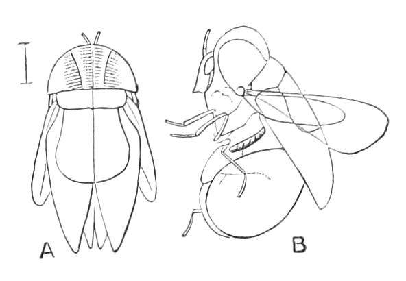
Fig. 359.—Thoracantha latreillei. Bahia. A, Upper, B, lateral aspect. (After Waterhouse.)
Howard has called attention to some peculiarities in the pupation of Chalcididae.[467] Like the Cynipidae, they do not make a silken cocoon, but some of them that change to pupae inside the victims on which they were nourished have the power of forming oval cells in which to undergo their transformation, and they thus cause a peculiar inflation of the skin of their deceased victim, which after death still continues to serve as a protection to the destroyers. The statement made by Haliday, and repeated subsequently in various works, to the effect that Coryna spins a cocoon under the Aphis in which it has lived, is an error, the cocoon being really formed by Praon, a Braconid that is a parasite of the Aphis, and on which the Chalcid Pachycrepis (Coryna) lives as a hyperparasite. The pupae of some species differ from those of other Hymenoptera, in that the integument is hard, and the limbs are soldered to the body as in Lepidoptera. These forms pupate external to the victim.
Fritz Müller has recorded that the pupa of an unnamed species of Chalcid that attacks a Brazilian ant (Azteca instabilis Forel) is suspended on the wall of the cell the ant lives in by its posterior extremity, just like the chrysalis of a butterfly.
Notwithstanding the small size of Chalcididae, their remains have been detected in the tertiary strata of both Europe and North America.
Fam. IV. Ichneumonidae (Ichneumon-flies).
Wings with a well-developed series of nervures and cells; the space on the front wing separating the second posterior cell from the cubital cells is divided into two cells by a transverse veinlet. The abdomen is attached to the lower or posterior part of the median segment. Larvae parasitic in habits.
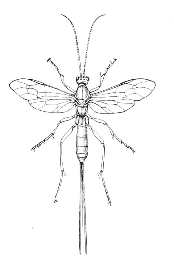
Fig. 360.—Lissonota setosa, ♀. Britain. Parasite of the goat-moth, etc. (After Ratzeburg.)
The Ichneumonidae form a family of enormous extent, containing nearly 6000 described species. The study of the family is but little advanced, owing to their parasitic habits and to this bewildering multiplicity in their specific forms. Most of the species, in the larval state, live inside the larvae of Lepidoptera, and they thus keep the myriads of caterpillars within bounds, the number of these destroyed by Ichneumons being prodigious. Some of the family are, however, external parasites, and some are known to attack Spiders and Insects of other Orders than Lepidoptera. Their antennae are not elbowed and are many-jointed, the joints being closely compacted, especially towards the extremity. This character readily distinguishes Ichneumonidae from the families we have previously considered. The ocelli are well developed even in the apterous forms, and are placed in a triangular position on the vertex. The pronotum is small in front; and extends backwards at the sides to the points of insertion of the front wings; it is fixed to the mesonotum. The wings (Fig. 367, A) have a more complex neuration than those of most of the other parasitic Hymenoptera, but are occasionally absent in one or both sexes of a species. The metathorax is very small, and the middle and hind legs are placed close together. The propodeum is very large, and is frequently covered with a highly-developed sculpture.
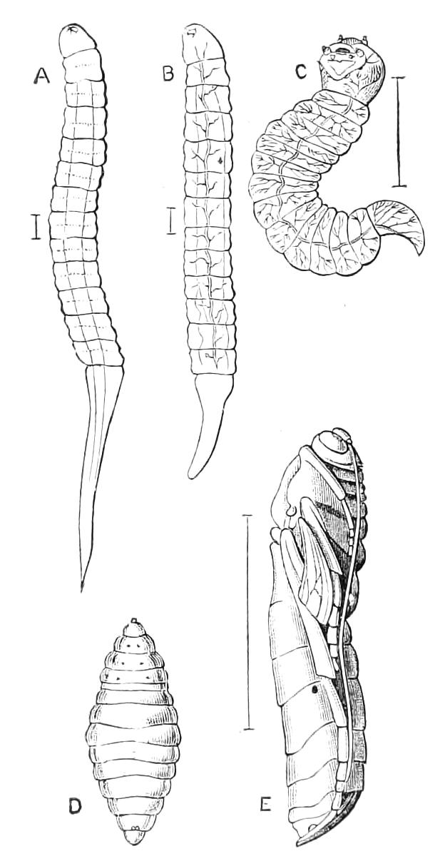
Fig. 361.—Anomalon circumflexum, larval development. (After Ratzeburg.) A, First instar; B, second instar; C, the larva in the third or encysted stage extracted from its cyst; D, the mature larva; E, pupa.
The hind body springs from the lower part of the propodeum; it is usually of slender form, and its segmentation is very conspicuous. The females bear an ovipositor, which differs greatly in length according to the species, and is known in the case of one species to attain a length six times that of the whole of the rest of the body.[468] The egg is deposited by some species on the skin, by others within the body of the victim; it varies much in form and colour, some eggs being stalked and of peculiar shape. The larvae issuing from the eggs are legless maggots with a delicate integument of pallid white or creamy colour. If the eggs are laid on the surface of the body, the resulting larvae (except in the cases of the external parasites) soon bore into the interior of their victim, and disappear therein. The changes that take place in the lifetime of the larvae have been studied in only a few cases; but if we can judge from Ratzeburg's history[469] of the changes that take place in Anomalon, they are of great interest. From observation of the differences existing amongst a great number of larvae of A. circumflexum he distinguished four stages. It is of course impossible to follow directly the growth of one individual, because it is concealed in the interior of the caterpillar in which it lives, and to open this involves the death of both caterpillar and Ichneumon-larva. The life history must therefore be constructed from a great number of separate observations; and it is not ascertained that the four instars described by Ratzeburg represent the number of moults of the larva that actually take place. He, however, entertained no doubt that all the forms he observed were stages in the development of one species. In the earliest stage, when only one millimetre in length and about as thick as a horse-hair, the larva is free in the interior of the caterpillar's body, and has a small head armed only with a pair of mandibles. There are, in addition to the head, thirteen segments, and the last of these is an elongate tail forming nearly one-half the length of the creature. No trace of tracheae can be discovered. In the second stage the larva is still free, an elongate tracheal tube exists, the tail has diminished to half the length, the head has become much larger, and rudimentary antennae of one joint are visible; possibly stigmata are present at this stage, though they cannot afterwards be detected. In the third stage (Fig. 361, C) the larva is encysted, the head is large, the parts of the mouth are all developed, the tracheal system is extensive, and the caudal termination of the body is quite short; notwithstanding the extensive development of the tracheal system, no stigmata can be found. In the fourth stage the larva is still encysted, the tail has disappeared, the head and mouth parts are reduced in size and development, and the creature has now the appearance of a normal larva. The changes to pupa and perfect Insect take place within the body of the victim, in some cases, if not usually, after it has undergone its metamorphosis into a chrysalis. Very little information is extant as to the duration of the various stages, but it appears to be the rule that only one generation appears annually, though in some cases there are pretty certainly two.
It is very difficult to observe the act of oviposition; the Ichneumon-flies usually decline to notice caterpillars with which they are placed in confinement. Ratzeburg thinks they will only attack caterpillars that are in a deficient state of health or vitality. Occasionally we may by a happy chance observe the act in Insects at large, and from the records of observers it may be deduced with tolerable certainty that the sense of sight takes no part in the operation. Ratzeburg relates that he saw a Pimpla alight on a leaf of Rhus and thrust its ovipositor through the leaf. On looking to the under-side of the leaf he found that a cocoon of Bombyx neustria was concealed there in such a position that it could not have been seen by the Ichneumon.
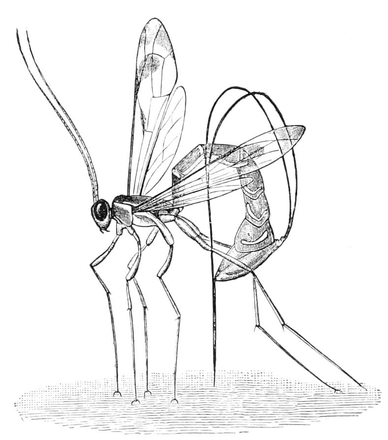
Fig. 362.—Thalessa lunator. Oviposition. (After Riley.)
Among the most remarkable of the Ichneumon-flies are the Insects of the genera Rhyssa and Thalessa. These fine Insects have an ovipositor three or four inches in length, and are parasitic on species of the family Siricidae, which, as we have previously described, live in solid wood. In order therefore to deposit the egg in a suitable place, the wood must be pierced by the Ichneumon. The ovipositor is not only of extreme length, but is also furnished with serrations on its apical part, so that it forms a very effective boring apparatus. It is brought into use by being bent on itself over the back of the Insect (Fig. 362), so as to bring the tip vertically down on to the wood, through which it is then forced by a series of efforts; the sheaths do not enter the wood. The egg is laid anywhere in the burrow of the Sirex; the young larva seeks its prey, and lives on it as an external parasite (Fig. 342, D). Erne, however, states[470] that the young larva of Rhyssa persuasoria enters its victim, and remains within the latter till its death occurs. This happens when the young Rhyssa is two or three lines in length, and it then makes its exit from the interior of the body and gradually eats it up. Should the larva it has attacked be of large size, it of itself affords sufficient food for the completion of the growth of the Rhyssa. Should the Rhyssa, however, have attacked a small larva, this does not furnish it with sufficient food, and it consequently dies without seeking another larva. Erne says, indeed, that it will not eat another if offered to it, so that in order to rear the Rhyssa in captivity, the victim it has first attacked must always be given to it. The same observer states that the Rhyssa larva is sometimes transported by the Sirex deep into the wood, so that when it has completed its metamorphoses the Ichneumon-fly may find itself buried in solid wood to a depth of about two inches. In that case it excavates the wood with its mandibles, and should it fail to gain the exterior after three days of work, it dies. In the case of Thalessa it is stated that it sometimes bores into wood where there are no larvae, but Riley thinks this erroneous; it is, on the other hand, certain that the Insect after penetrating the wood is frequently unable to withdraw the ovipositor, and consequently dies.
Packard has recorded,[471] without mentioning the species, the oviposition of an Ichneumon of which the egg is deposited externally. It was placed on the head of the caterpillar, and speedily hatched; the young larva at once bored through the prothoracic segment of the victim, the head of the latter then became swollen, and covered the opening into the prothorax, made by the parasite.
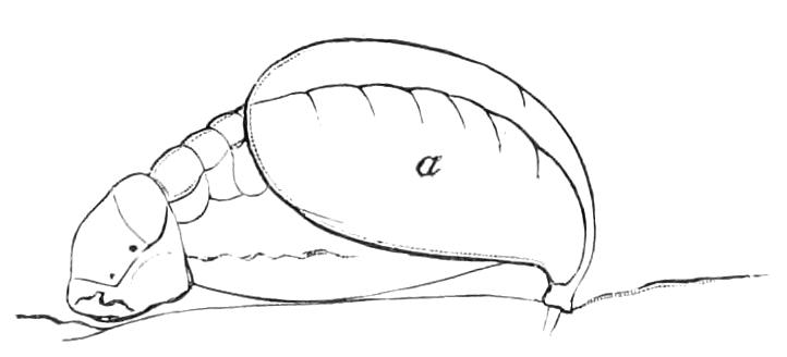
Fig. 363.—Young larva of Paniscus in position of feeding on the skin of Mamestra. (After Newport.) a, The egg-shell.
The history of an Ichneumon larva that feeds as an external parasite has been sketched by De Geer and Newport. The observations of the latter[472] refer to Paniscus virgatus; he found small, shining, black bodies attached to the skin of the larva of a moth, Mamestra pisi; these were the eggs of the Ichneumon. They are furnished with a short peduncle, which is implanted in the skin of the victim; the egg, according to De Geer, being retained more firmly by the peduncle subsequently swelling, so as to form two knobs. The hatching takes place by the egg-shell splitting longitudinally, while from the split protrudes the little head of the destroying larva. This becomes fixed to the caterpillar, from which the nutriment is to be drawn; the Paniscus larva does not, however, leave the egg-shell, but, on the contrary, becomes adherent to it, so that the parasite is in this manner fixed by the two ends to its victim. In fifteen days the parasite was full-grown, and had become half an inch in length. At first no tracheae were to be seen, but these were detected after the second day. Moulting took place three times, and in a peculiar manner, very different from that described by Ratzeburg as occurring in the internal parasites (which, he states, change their very delicate skin by detaching it in almost imperceptible fragments). In the external parasite the skin remains entire, and is shuffled down to the extremity of the body, but cannot be completely detached owing to the anchoring of the posterior part of the body to the caterpillar; the cast skins thus remain as envelopes to the posterior part of the body. Newport states that if the mouth of the parasite be detached, it usually cannot again seize hold of the victim, and consequently perishes. It is a curious fact that more eggs than one caterpillar can support are habitually placed on it, and some of the resulting larvae of necessity perish during the period of growth. Poulton, who has recently made some additional observations on the development of Paniscus,[473] says that if three larvae are close together, it is the middle one that perishes, and suggests that this is due to some simple physical condition. From Newport's account it may be gathered that the Mamestra retains sufficient vitality to form its cocoon, and that the Paniscus larvae likewise construct their own cocoons within that of the Mamestra. In the case of Paniscus cephalotes feeding on Dicranura vinula, Poulton relates that the latter died after the twelfth day of attack. The parasites, having relaxed their hold on the victim just previous to this event, then thrust their heads into the dead body, and devoured the larva, leaving only a dried and empty integument. These larvae span a loose sort of web in which to undergo their metamorphosis. In a natural state, however, they form cocoons inside the cocoon of the Dicranura. The period passed in the pupal condition was about four weeks. This parasite only attacks the Lepidopterous larva during the last stage of its existence as a larva, but the eggs may be laid on the victim in an earlier stage; and in such case De Geer has stated, and Poulton has confirmed the observation, that though the larva sheds its skin it does not get rid of the eggs.
The little Ichneumons of the genus Pezomachus are quite destitute of wings and somewhat resemble ants; they are common Insects in Britain. Only the female sex is known, and it is believed that the winged Ichneumons assigned to the genus Hemiteles—of which no females are known—are the males of Pezomachus. Repeated efforts have been made to place this beyond doubt, but they have usually failed, for when a brood of these parasites is reared, the individuals generally prove to be either all Hemiteles or all Pezomachus. It is to be hoped that this interesting case will be fully elucidated.
Although the Ichneumonidae are perhaps the most purely carnivorous of all the great families of Hymenoptera, there is nevertheless reason for supposing that some of them can be nourished with vegetable substances during a part at any rate of the larval existence, Giraud and Cameron[474] having recorded observations that lead to the conclusion that some species of the genus Pimpla may inhabit galls and live on the substance, or juices thereof.
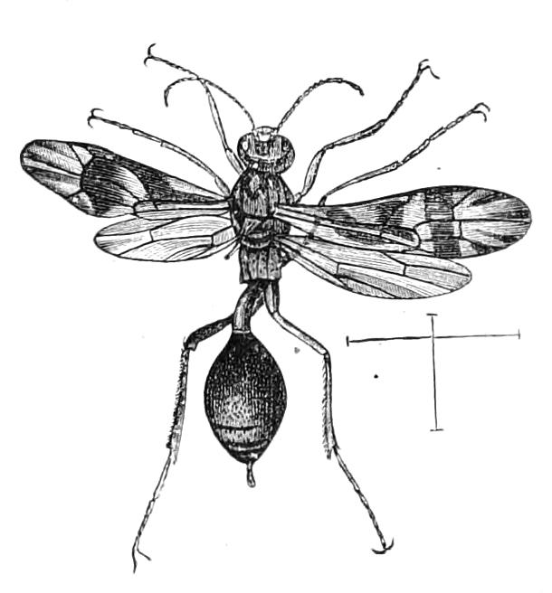
Fig. 364.—Agriotypus armatus, female. Britain. (After Curtis.)
Over 1200 species of Ichneumonidae are known to inhabit Britain, and there can be no doubt that this number will be increased as a result of further observation. Unfortunately no general work has yet been published on this department of our fauna, and the literature is very scattered.[475] The species of North America have not received so much investigation as those of Europe, and the Ichneumon fauna of the tropics remains almost uninvestigated. Six sub-families are recognised: Agriotypides, Ichneumonides, Cryptides, Tryphonides, Pimplides, Ophionides. Of these the first is the most remarkable, as it consists of an Insect having aquatic habits. It has for long been known that the unique species Agriotypus armatus, a rare Insect in our islands, is in the habit of going under water and remaining there for a considerable period, and it has now been satisfactorily ascertained that it does this for the purpose of laying its eggs in the larvae of Trichoptera.[476] The resultant larva lives inside the cases of species of Silo, Goëra, etc. It undergoes a sort of hypermetamorphosis, as its shape before assuming the pupal condition is very different to what it was previously. It changes to a pupa inside the case of the Trichopteron in a cocoon attached to the walls of the case. Previous to making this, however, the Agriotypus forms a curious, elongate, string-like process attached to the anterior extremity of its cocoon. The use of this is unknown. Full information as to the life-history of this aquatic Hymenopterous larva, especially as to its respiratory functions, would be of great interest. The affinities of this remarkable Insect are still doubtful. It may probably prove to be between Proctotrypidae and Ichneumonidae.
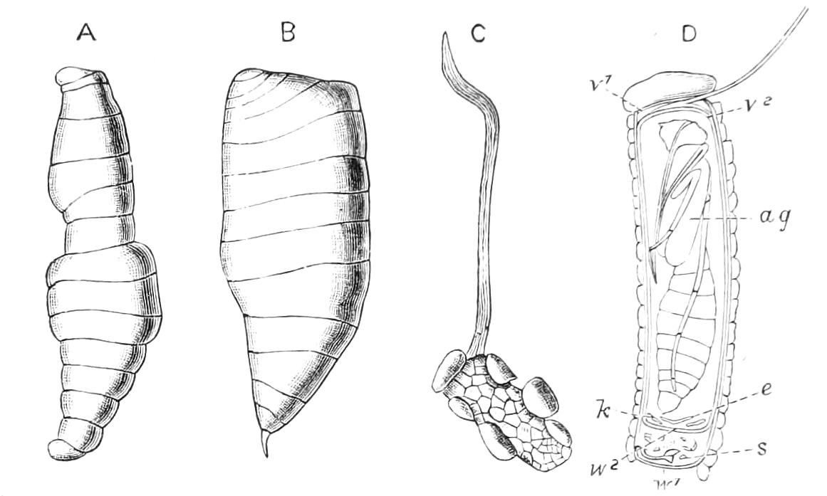
Fig. 365.—Metamorphosis of Agriotypus. (After Klapálek.) A, Larva; B, sub-nymph; C, case of the Silo with the string of attachment formed by Agriotypus; D, section of the case: v1, operculum of case; v2, cocoon; ag, pupa of Agriotypus; e, exuvia of Agriotypus; w2, wall of cocoon; s, remains of Silo; w1, closure of case.
Remains of Insects that may be referred with more or less certainty to Ichneumonidae have been found in some abundance in various tertiary strata both in Europe and North America, but nothing indicative of the existence of the family has yet been found in the older rocks.
Fam. V. Braconidae—Supplementary Ichneumon-flies.
Antennae with many (nearly always more than fifteen) joints, not geniculate. Wings with a moderate number of cells, which on the anal part of the front wing are more or less imperfect, the anal (i.e. the second posterior) cell being separated from the cubital cells by a large space in which there is no cross-nervure. Abdomen with but little mobility between the segments; the suture between the second and third usually absent, or obsolete. Larvae living parasitically in—possibly exceptionally outside—the bodies of larvae or pupae of Insects.
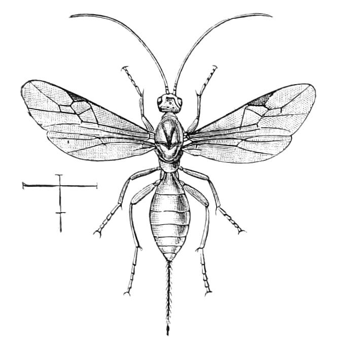
Fig. 366.—Bracon palpebrator, female. Europe. (After Ratzeburg.)
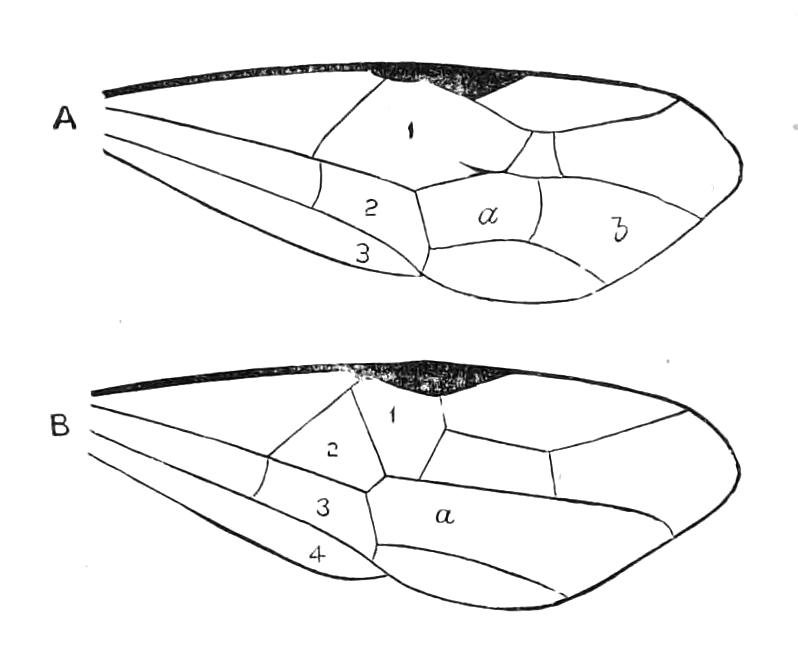
Fig. 367.—Diagram of wing of Ichneumonid (A) and of Braconid (B). 1, 2, 3, 4, series of cells extending across the wing; a, b, divided cell of the Ichneumonid wing, corresponding with a, the undivided cell of the Braconid wing.
The Braconidae are the Ichneumones, or Ichneumonides, adsciti of the older Hymenopterists. They are extremely similar to the Ichneumonidae, but the hind body has a much less degree of mobility of its segments, and there are some constant distinctions in the wings. Although there is a great deal of difference in the various forms of each of the two families, yet there are two points of distinction easily appreciated; the series of cells running across the wing (Fig. 367) being only three in the Ichneumonides (Fig. 367, A), but four in the Braconids (Fig. 367, B); besides this the space a of the Braconid wing is divided into two (a, b) in the Ichneumonid wing. A glance at these characters enables us at once to separate correctly the thousands of species of the two families.
The habits of the Braconidae are similar to those of Ichneumonidae, it being believed that all are parasites. Usually they attack larvae, but they are bred in great numbers from pupae, and even from imagos of other Insects. Elasmosoma is one of the few parasites known to attack ants. As many as 1200 specimens of Microgaster have been reared from a single Lepidopterous larva. Although such parasitism raises a feeling of repulsion, yet there is reason for supposing that there may be little or no cruelty or acute suffering connected with this mode of life. The victim attacked is not eaten, the parasites in the interior taking in the lymph of the caterpillar either by the mouth or by endosmosis, but not biting their host. The latter displays no sign of sickness, but eats voraciously, so that it serves merely as a sort of intermediary between the juices of the plant and the larvae inside itself. It is only when the metamorphosis is at hand that the host sickens, but this does not always happen: parasitised larvae frequently change to pupae, and they may occasionally even become perfect Insects. Cases are known in which imagos have appeared with some of the small parasites embedded in some of the outer parts of their bodies. These cases are, however, very rare; in the enormous majority of instances the host is destroyed either when it is in the larval stage or before the pupa has advanced to any great extent on its metamorphosis to an imago. Particulars as to various species will be found in the valuable work of Ratzeburg we have already referred to.[477] Reference may also be made to Goureau's account of Microgaster globatus,[478] this latter including some suggestions by Dr. Boisduval on some of the difficult physiological questions involved in the lives of these parasites.
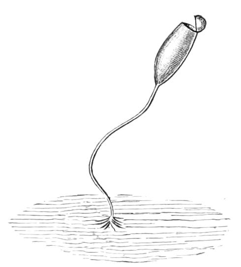
Fig. 368.—Stalked cocoon of Apanteles formosus. (After Marshall.)
The metamorphosis of Microgaster fulvipes has been studied by Ratzeburg, and an epitome of his observations is given by Marshall.[479] The larva goes through a series of changes somewhat similar to those we have already sketched in Anomalon circumflexum. Usually these Insects after emerging from the body of their host spin a mass of cocoons more or less loosely connected together. A most curious case has, however, been recorded by Marshall[479] of a stalked cocoon (Fig. 368) being formed as an exceptional act by Apanteles formosus. Mr. Marshall has recently received other specimens of this cocoon as well as the Insects reared therefrom in France, and inclines to the opinion that the stalked cocoon may be the usual form, and is sometimes departed from by the Insect for unknown reasons.
This family is of enormous extent; we have several hundred species of it in Britain,[480] and there are no doubt many thousands of undescribed exotic forms. To Apanteles glomeratus we are indebted for keeping our cabbages and kindred vegetables from destruction by the caterpillars of the white butterflies. The larvae of the various species of Pieris, as well as those of other Lepidoptera, are attacked by this little Insect, the masses of whose cocoons may frequently be found in numbers in and near cabbage gardens. The tropical species of Braconidae are greatly neglected, but many large and remarkable forms—some of brilliant colours—have been brought from there, so that we are justified in believing that Insects of this family will prove to be very numerous. There are but few apterous Braconidae. Both sexes of Chasmodon apterus are destitute of wings; the females of one species of Spathius, and also those of Pambolus and Chasmodon are apterous; in a small number of species of various genera the wings are so minute as to be incapable of serving as organs of flight. In the genus Alloea the wings of the male are shorter than those of the female.
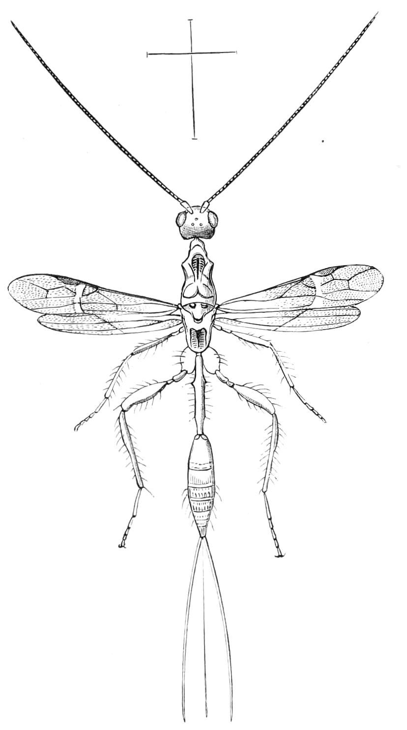
Fig. 369.—Stenophasmus ruficeps, female. Aru Islands. (After Westwood.)
Fam. VI. Stephanidae.
Antennae composed of many (thirty to seventy) joints; hind body attached to the lower and posterior part of the median dorsal plate. Wings with a distinct costal cellule; head globose, posterior femora frequently toothed.
This is a doubtful family, consisting of a few anomalous Insects. Schletterer assigns to it only two genera, Stephanus and Stenophasmus;[481] both have a wide distribution over the world, though we have no species in Britain. Nothing is known of their habits, and they are apparently all very scarce Insects. The definition is compiled from those of Cameron and Schletterer. There seems very little to distinguish these Insects from Braconidae.
Fam. VII. Megalyridae.
Hymenoptera with short broad hind body, which is not separated by a pedicel from the thorax. The female has a very long bristle-like ovipositor. Antennae with fourteen joints.
This family is constituted by the Australian genus Megalyra,[482] one of the most interesting of the numerous extraordinary Insect-forms found in that region; the species appear to be very rare and not numerous. Apparently nothing is known as to their habits. It is quite possible that these Insects will prove to be anomalous Braconidae.
Fam. VIII. Evaniidae.
Petiole of the abdomen attached to the upper part of the median dorsal plate; antennae not elbowed, of thirteen or fourteen joints. Wings with a moderate number of nervures. Larva of parasitic habits.
This family is composed of only three genera—Evania, Gasteruption, and Aulacus, each possessing a considerable number of species; they agree in the characters mentioned above, and may be readily recognised by the peculiar insertion of the hind body. This character occurs outside the limits of the Evaniidae only in one or two genera of Chalcididae and Braconidae; it is to this latter family that the Evaniidae must be considered most closely allied.
The species of the genus Evania are believed to live at the expense of cockroaches (Blattidae), and to deposit their eggs in the egg-capsules of those Insects. The species of Gasteruption live, in the larval state, on the larvae of other Hymenoptera, more especially of such as form nests in wood. Very little is known as to the habits of the species of Aulacus, but it is believed that they are parasitic on members of the Hymenopterous families, Siricidae and Oryssidae. Only the most meagre details as to the life history of any of the Evaniidae have been recorded. The species of Evania are met with most freely where cockroaches abound, and are said, hence, to be frequently observed on board ship. Two or three species of each of the two genera Evania and Gasteruption occur in Britain. The latter genus is more widely known under the name of Foenus.[483]
Fam. IX. Pelecinidae.
Sexes very different; the female without exserted ovipositor, but with extremely long abdomen. Articulation between the femur and trochanter oblique and elongate, but without division of the trochanter.
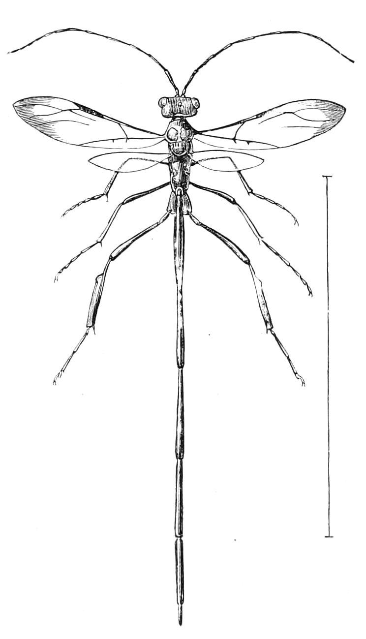
Fig. 370.—Pelecinus polyturator, ♀. Mexico.
This family at present comprises, according to Schletterer,[484] only the three genera Pelecinus, Ophionellus, and Monomachus. The systematic position of the Insects is very doubtful, and their habits are but little known. Pelecinus polyturator (Fig. 370) appears, however, in the female sex, to be a common Insect over a large part of the warmer regions of the New World; it is in all probability parasitic in its habits, the elongate ovipositor of the female Ichneumon being in this Insect replaced by an extraordinary linear extension of the abdomen itself. Doubleday has recorded that he saw twenty or thirty specimens of this species that had perished with their elongated hind bodies inserted into the stem of a tree, from which they could not extricate themselves. On the other hand, Patton thinks they are parasitic on locusts.[485]
The male in Pelecinus has the proportions of the parts of the body normal, there being no elongation of the abdomen; it thus differs very much in appearance from the female. There seems to be very little to distinguish Pelecinus from Proctotrypidae. The undivided trochanters have led to these Insects being placed, by some, among the Aculeate Hymenoptera. This character, as we have already shown, occurs also in Proctotrypidae.
Fam. X. Trigonalidae.
Abdomen ovate, not separated by a pedicel from the thorax. Antennae twenty-five-jointed. Trochanters imperfectly two-jointed. Both the anterior and posterior wings provided with a well-developed neuration. Abdomen composed of only five apparent segments. Larva (in some cases) parasitic on Aculeate Hymenoptera.