Chapter XIII
Reading edition
ORTHOPTERA CONTINUED—LOCUSTIDAE, GREEN GRASSHOPPERS, KATYDIDS
Fam. VII. Locustidae—Green Grasshoppers.
Orthoptera, with very long delicate antennae composed of many more than thirty joints; hind legs longer than the others, thicker at the base. Tarsi with four joints. Front tibiae usually provided with tympanal organs placed below the knee; stridulating apparatus of males, when present, situate on the basal part of the tegmina. Females usually with an elongate exserted ovipositor, formed by the apposition of six pieces. Wingless forms numerous.
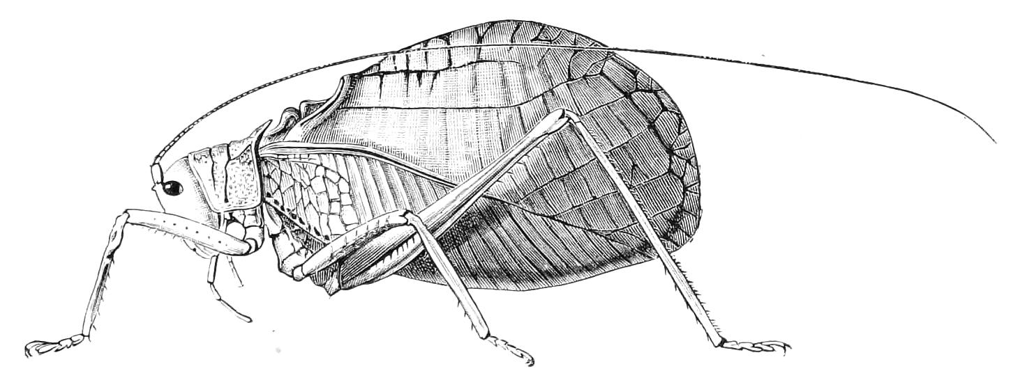
Fig. 187.—Cyrtophyllus crepitans, male. West Indies.
An unfortunate confusion has long existed as to the term Locustidae, and has resulted in the application of the name to a group of Insects that contains none of the locusts of ordinary language. Some entomologists therefore use the term Phasgonuridea for this family, but the great majority prefer the term Locustidae.
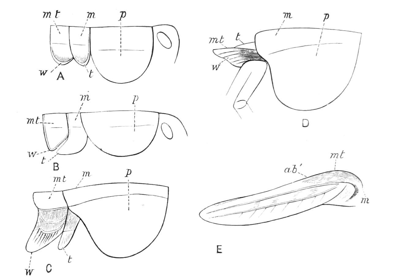
Fig. 188.—Development of wings in Platycleis grisea: A, B, C, D, E, consecutive stages; p, prothorax; m, mesothorax; mt, metathorax; t, tegmen; w, wing; ab′, position of first abdominal segment. In C, D, and E, m points to the part by which the m, shown in A and B, is concealed; in D and E only the positions of mt are indicated. (After Graber.)
The Locustidae are, as a rule, more fragile Insects than the Acridiidae, from which they can be readily distinguished by the characters we have mentioned in our definition. According to Dufour, there are no air vesicles connected with the tracheal system in this family; possibly to this it may be due that none of the family undertake the long flights and migratory wanderings that have made some of the Acridiidae so notorious. Very little is known as to the life histories of the members of this extensive family of Orthoptera. Graber, however, has given some particulars as to the development of Platycleis grisea, and of one or two other species. He recognises five instars, but his first is probably really the second, as he did not observe the Insect in its youngest condition. Although his figures are very poor, we reproduce them, as they give some idea of the mode of growth of the wings, and of the correlative changes in the thoracic segments. It will be seen that in the first three of these instars the alar organs appear merely as prolongations of the sides of the posterior two thoracic rings, and that in D a great change has occurred in the position of these segments, so that the alar organs are free processes, the two posterior thoracic rings being insignificant in size in comparison with the now greatly developed prothorax. In E the tegmen is shown fully developed, the positions of some of the rings covered by it being indicated by the letters m, mt, ab′. These changes are very similar to those we have described in Acridiidae, the chief difference being the greater development of the dependent wing-pads previous to the fourth instar.
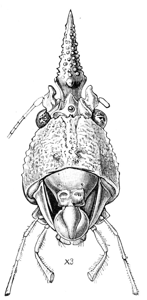
Fig. 189.—Front of head of Copiophora cornuta, female. Demerara.
The ocelli in Locustidae are much more imperfect than they are in Acridiidae, and are frequently rudimentary or nearly totally absent, or there may be but one instead of three. They are, however, present in a fairly well-developed state in some species, and this is the case with the one whose face we portray in Fig. 189, where the anterior of the three ocelli is quite conspicuous, the other two being placed one on each side of the curious frontal cone near its base. The peculiar head ornament shown in this figure exists in both sexes, and something similar occurs in a large number of Conocephalides. We have not the slightest idea of its import. Individuals of one or more species of this curious South American genus are occasionally met with alive in gardens near London. They are, no doubt, imported as eggs, for they are sometimes met with in the juvenile state, but in what way they are introduced is not known.
The ovipositor frequently attains a great length in these Insects, so as to exceed that of the body. It is used in different ways, some of the family depositing their eggs in the earth, perhaps in vegetable matter under the surface; but other species place the ova in twigs or stems of plants, arranging them in a very neat and compact manner in two series, as depicted by Riley[245] in the case of Microcentrum retinerve (Fig. 190). These eggs are laid in the autumn, and in the following spring become more swollen before hatching. The Insect undergoes a moult during the process of emerging from the egg. By the time the emergence is completed the Microcentrum has expanded so much in size that it is a matter of astonishment how it can ever have been packed in the egg; the young commence jumping and eating leaves in a few minutes. Including the ecdysis made on leaving the egg, they cast their skins five times. The post-embryonic development occupies a period of about ten weeks. The larvae eat their cast skins. When the final moult occurs the tegmina and wings are at first quite soft and colourless, but within an hour they assume their green colour. These Insects, as remarked by Riley, make interesting pets. The people of the Amazon valley are in the habit of keeping a species in cages, and our British Locusta viridissima does very well in confinement. One of the most curious habits of these Locustidae is a constant licking of the front paws. Riley says that M. retinerve bestows as much attention on its long graceful antennæ as many a maiden does upon her abundant tresses, the antennæ being drawn between the jaws and smoothed by the palpi. This American naturalist also tells us that he reared three successive broods in confinement, and that the Insects gradually deteriorated, so that the eggs of the third generation failed to hatch.
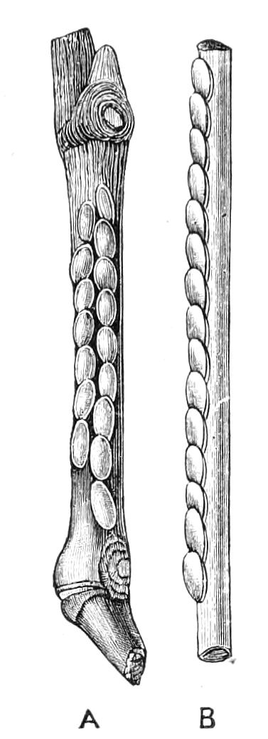
Fig. 190.—Eggs of Katydid (Microcentrum retinerve): A, the two series at deposition; B, side view of a single series. (After Riley.)
The ovipositor, which is one of the most characteristic features of the Locustidae, is not present in the newly-hatched Locustid (Fig. 191, A), the organ being then represented only by two papillae placed on the penultimate segment. The structure and development of the ovipositor in Locusta viridissima have been described by Dewitz.[246] Fig. 191, A, shows the young Insect taken from the egg just as it is about to emerge. The abdomen consists of ten segments, the terminal one bearing at its extremity two processes, the cerci, a′. These persist throughout the life of the Insect, and take no part in the formation of the ovipositor. The tenth segment subsequently divides into two (a, a'′, Fig. 191, C), giving rise to the appearance of eleven abdominal segments, and of the ovipositor springing from the antepenultimate.
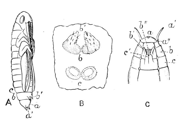
Fig. 191.—Development of ovipositor of Locusta viridissima: a, terminal segment; a′, cerci; a″, secondary division of terminal segment; b, penultimate (ninth) segment; b′, primary papillae of this segment; b″, secondary divisions thereof; c, eighth segment; c′, its papillae. (After Dewitz.) A, embryo ready for emergence; B, portion of integument of the ventral plates of eighth and ninth segments; C, the appendages in a condition somewhat more advanced than they are in A.
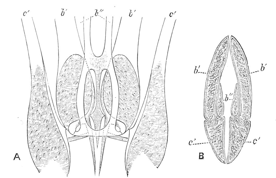
Fig. 192.—Structure of ovipositor of Locusta viridissima: A, arrangement of parts at base, c′ being separated and turned outwards; B, transverse section. The parts of the appendage bear the same lettering as in Fig. 191. (After Dewitz.)
Near to one another, on the middle of the ventral aspect of the true ninth abdominal segment, are seen the two papillae (b′), which at first are the only visible indications of the future ovipositor. If, however, the integument be taken off and carefully examined, it will be found that there exist on the eighth abdominal plate two spots, where there is a slight thickening and prominence of the integument (Fig. 191, B, c). From these two spots the two lower rods of the ovipositor are produced; these two, together with the two growths from the ninth segment, form the four external rods of the ovipositor. Inside these there exist in the completed structure two other rods (Fig. 192, B, b'′). These are produced by a growth from the inner parts of the two papillae of the ninth segment. The relations of the six rods in their early condition are shown in Fig. 191, C, where the two primary papillae b′ of the ninth segment are seen with their secondary offshoots b″; c′ being the papillae of the eighth segment. The subsequent relations of the pieces are shown in Fig. 192; A exhibiting the base of the organ with the lower rods turned on one side to show the others, the shaded parts indicating muscular attachments; B is a transverse section of the organ. In these figures the different parts of the appendages bear the same lettering as they do in Fig. 191. It will be seen that in the completed structures the parts c′ have become very intimately connected with the parts b′ and b″, which belong to another segment.
The Locustidae resemble the Acridiidae in the possession of specialised ears and sound-producing organs; neither of these is, however, situate in the same part of the body as in Acridiidae. The ears of Locustidae are placed on the front legs, below the knee; a tympanum (Fig. 193, A), or a crack giving entrance to a cavity in which the tympanum is placed (Fig. 193, B), being seen on each side of each of the anterior pair of limbs. In this family, as in the Acridiidae, three kinds of ear are recognised according to the condition of the tympanum, which is either exposed (Fig. 193, A) or closed by an overgrowth of the integument (Fig. 193, B), or in a condition to a certain extent different from either of these. The existence of ears placed on the legs is a curious fact, but it is beyond doubt in the Locustidae, and there is good reason for believing that analogous organs exist in this situation in other Insects that have special means of sound-production, such as the ants and the Termites.
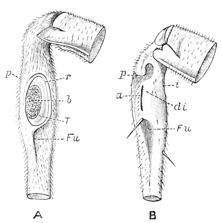
Fig. 193.—Ears of Locustidae: A, portion of front leg of Odontura serricauda, adult; p, prominence of integument; r, rim of ear; T, tympanum; b, thickened area thereof; Fu, remains of groove in which the structure was developed. B, portion of front leg of Thamnotrizon apterus; i, inner margin; a, slit-like external aperture of ear; di, overlapping cover of the ear. (After Graber.)
The structure of these organs in the Locustidae has been investigated by Graber,[247] and their acoustic functions placed beyond doubt, though to what special kind of sounds they may be sensitive is not ascertained, this point being surrounded by even greater difficulties than those we have discussed in the case of the Acridiidae. In the Locustidae there is a special structure of a remarkable nature in connexion with the ears. In Acridiidae a stigma is placed close to the ear, and supplies the internal structures of the organ with air. There are no stigmata on the legs of Insects, consequently admission of air to the acoustic apparatus in Locustidae is effected by means of a gaping orifice at the back of the prothorax, just over the base of the front leg (Fig. 101); this communicates with its fellow of the other side, and from them there extend processes along the femora into the tibiae, where they undergo dilatation, so as to form vesicular cavities, one of which is in proximity to each drum of the ear. These leg-tracheae are not connected with the ordinary tracheal system; the prothoracic stigma exists in close proximity to the acoustic orifice we have described, but is much smaller than it. It is not yet clear why the acoustic apparatus should require a supply of air apart from that which could be afforded by the ordinary tracheal system. This special arrangement—to which there is hardly a parallel in Insect anatomy—has still to be accounted for; we do not know whether the necessity for it may be connected with the respiratory system or the acoustic organ.
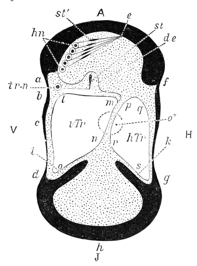
Fig. 194.—Diagram of arrangement of parts of the ear as seen in transverse section of the tibia of a Locustid. A, J, V, H, outer, inner, anterior, posterior aspects of leg; a, d, thin part of integument forming anterior tympanum; b, c, thicker portion of same; f, g, posterior tympanum; a, f, and d, h, g, thick portions of integument; i, k, internal protuberances of same; l, m, n, o, walls of the anterior tracheal vesicle, vTr; p, q, s, r, walls of the posterior tracheal vesicle, hTr; o″, projection of tympanal orifice of prothorax; tr-n, tracheal nerve-end organ, crista acustica; st, rod; de, curtain-membrane; hn, e, supra-tympanal, nerve-end organ; hn, ganglion cells; st′, rods; e, point of integumental fixation of nerve endings. (After Graber.)
The chief features of the acoustic apparatus of the legs of Locustidae will be gathered from the accompanying diagrammatic transverse section through the tibia. In this figure the deep black parts indicate the outer wall of the tibia and its prolongations, the white spaces indicate the parts filled with air, while the dotted portions are occupied by blood or some of the body organs;[248] the circular space o″ is not part of the actual structure, but represents the area of the external acoustic orifice of the prothorax; it is not, however, so large as it should be.
Although the tibial ears of Locustidae are very perfect organs, there is great difficulty in deciding on the exact nature of their functions. They would appear to be admirably adapted to determine the precise locality from which a sound proceeds, especially in those cases—and they are the highest forms—in which the tympanum is placed in a cavity the external orifice of which is a slit (Fig. 193, B); for the legs can be moved in the freest manner in every direction, so as to bring the drum into the most direct line of the vibrations. But as to what kinds of vibrations may be perceived, and the manner in which they may be transmitted to the nerves, there is but little evidence. On reference to the diagram it will be noticed that the tympanum, the tympanal vesicles, and the nervous apparatus are not in close connexion, so that even the mode by which the impulses are transmitted is obscure.
The musical organs of the Locustidae are different from those of the Acridiidae, and are invariably situate on the basal part of the tegmina. They are found, in the great majority of cases, only in the male; in the tribes Ephippigerides and Callimenides they exist in each sex. One of the wings bears a file on its inner surface, while the other—on the right side of the body—is provided with a sharp edge placed on a prominent part of its inner margin. By slightly tilting the tegmina and vibrating them rapidly, the edge passes under the file, and a musical sound is produced. These structures are limited to the small anal area of the wing, and when the tegmina are very greatly reduced in size, it is this part that still remains. There is much variety in the details of the structure. The nervures of this part of the tegmina are different in the male from what they are in the female, and, moreover, the two wing-covers of the male differ from one another. It is apparently the vibrations of the right tegmen that produce the sound, and this part usually bears a space of a glassy nature, which probably improves the character of the sound produced. Our chief British songster of this group, Locusta viridissima, is only provided with phonetic organs (Fig. 195) of a somewhat imperfect character, but in the genus Mecopoda there is great perfection of the structures. The anal areas of the two tegmina are in this case very different; that of the left one, which bears the file, being similar in texture to the rest of the wing-cover, while the corresponding part of the other tegmen is rigid and transparent, and greatly distorted, so as to create a cavity which, no doubt, improves the sound; the scraper too is very perfectly formed. The difference between this form of musical organ and that of L. viridissima is curious, inasmuch as in the better instrument the important modifications are confined to one tegmen, while in the other form both tegmina are largely changed. The difference appears to be that in Locusta the left tegmen, as well as the right one, acts as a sounding-board, while in Mecopoda it does not do so, but when the wings are closed quite covers and conceals the musical instrument.
The Locustidae, notwithstanding the fact that their alar organs are generally more ample than those of the Acridiidae, seem to be, as a rule, of more sedentary habits, and more nocturnal in their activity. The musical powers of the different species are very varied. Locusta viridissima produces a shrill and monotonous but not disagreeable, sound, and is capable of sustaining it for a quarter of an hour without any intermission, except a break for the sake of starting again immediately with greater force, like a performer on a flute. It occasionally chirps in the day, but the act is then very brief. Bates informs us that one of these singing grasshoppers, called Tananá by the natives of the Amazon valley, is much admired for its singing, and is kept in little cages. The Amazonian naturalist thought the music of this species superior to that of any other Orthopterous Insect he had heard. The name of this grasshopper is Thliboscelus camellifolius. It is very similar in appearance to Cyrtophyllus crepitans, the Insect we have represented in Fig. 187.
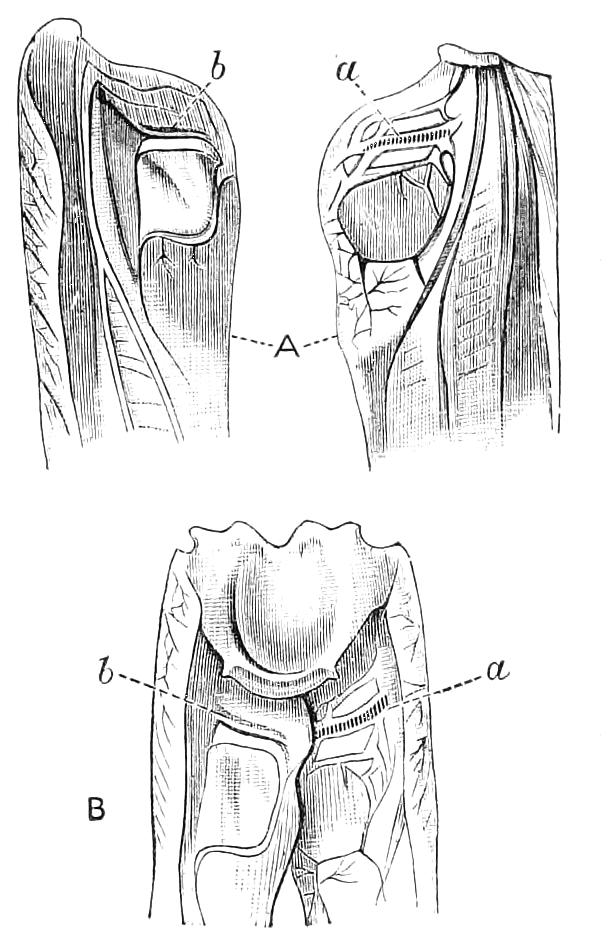
Fig. 195.—Inner face of base of tegmina of Locusta viridissima: A, the two wing-covers separated; B, in natural position with mesonotum connecting them, showing file and edge scraping it; a, the stridulating file; b, the rudimentary file on other tegmen.
The most notorious of the musical Locustids are the Katydids of North America. There are several species of them—they belong, indeed, to more than one genus,—but it seems that sounds somewhat resembling the words Katy-did are perceptible in most of their performances. These sounds are frequently repeated with slight variations—Katy-did, O-she-did, Katy-did-she-did. Riley describes the music of the Katydid we represent in Fig. 196 as follows:[249] "The first notes from this Katydid are heard about the middle of July, and the species is in full song by the first of August. The wing-covers are partially opened by a sudden jerk, and the notes produced by the gradual closing of the same. The song consists of a series of from twenty-five to thirty raspings, as of a stiff quill drawn across a coarse file. There are about five of these raspings or trills per second, all alike, and with equal intervals, except the last two or three, which, with the closing of the wing-covers, run into each other. The whole strongly recalls the slow turning of a child's wooden rattle, ending by a sudden jerk of the same; and this prolonged rattling, which is peculiar to the male, is invariably and instantly answered by a single sharp 'chirp' or 'tschick' from one or more females, who produce the sound by a sudden upward jerk of the wings."
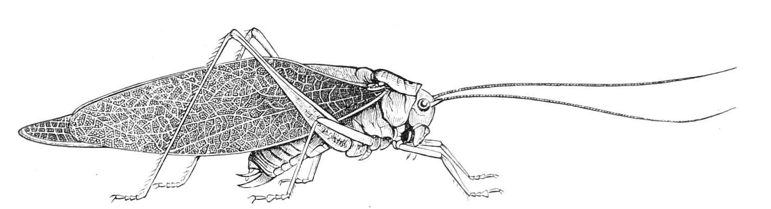
Fig. 196.—Katydid, Microcentrum retinerve. N. America. (After Riley.)
Pertinacity is one of the most curious features of the performance of musical Locustids. One would say they desire to distinguish themselves as much as possible. Harris says that Cyrtophyllus concavus mounts on the uppermost twigs of trees and there performs its Katy-did-she-did in rivalry with others. He says even the female in this species gives forth a feeble noise. Scudder says that some of the Katydids sing both by day and night, but their day song differs from that of the night. "On a summer's day it is curious to observe these little creatures suddenly changing from the day to the night song at the mere passing of a cloud, and returning to the old note when the sky is clear. By imitating the two songs in the daytime the grasshoppers can be made to respond to either at will; at night they have but one note."
Although but little is known as to the habits of Locustidae, it is ascertained that they are less exclusively herbivorous in their food habits than the Acridiidae are; many seem to prefer a mixed diet. Locusta viridissima will eat various leaves and fruits, besides small quantities of flesh. It has been recorded that a specimen in confinement mastered a humble-bee, extracted with its mandibles the honey-bag, and ate this dainty, leaving the other parts of the bee untouched. Many of the Locustidae are believed to be entirely carnivorous. Brunner considers a minority to be exclusively phytophagous. The species very rarely increase to large numbers; this, however, occurs sometimes with Orphania denticauda and Barbitistes yersini in Europe, and Anabrus purpurascens in North America. We have already mentioned that the eggs of some species are deposited in parts of plants, and of others in the earth. The British Meconema varium deposits its eggs in the galls of Cynips in the autumn; these eggs do not hatch till the following spring. Xiphidium ensiferum has somewhat similar habits in North America, the gall selected for the reception of the eggs being the scales formed by a species of Cecidomyia on the leaves of willows. It has been ascertained that the development of the embryo in the last-named species is commenced in the autumn, but is suspended during the winter, being only completed in the following spring, eight or nine months afterwards. We owe to Wheeler[250] a memoir on the embryology of this Insect.
Some of the species have the peculiar habit of dwelling in caves. This is especially the case with the members of the tribe Stenopelmatides (Fig. 197), which frequently possess enormously long antennae and legs, and are destitute of alar organs and ears. The species with this habit, though found in the most widely separated parts of the world, have a great general resemblance, so that one would almost suppose the specimens found in the caves of Austria, in the Mammoth cave of Kentucky, and in the rock-cavities of New Zealand to be one species, although they are now referred by entomologists to different genera.
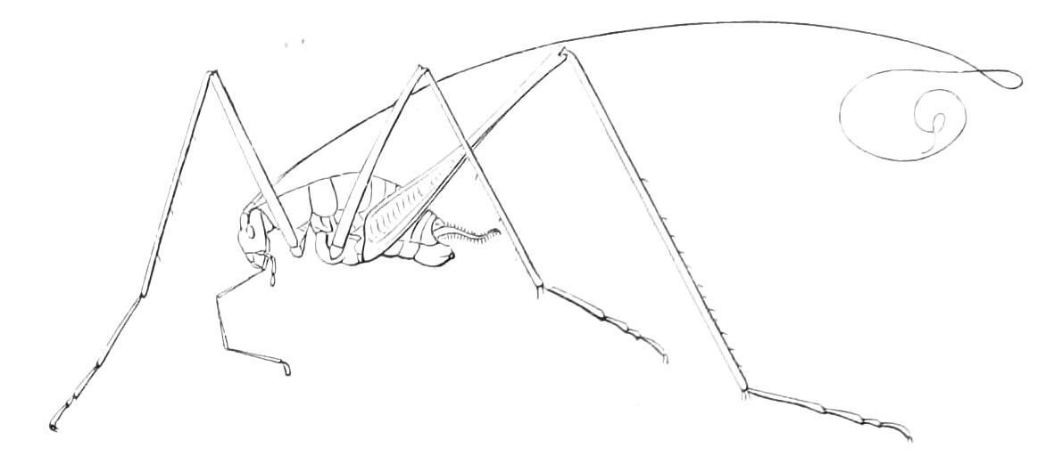
Fig. 197.—Dolichopoda palpata, male. Dalmatia. (After Brunner.)
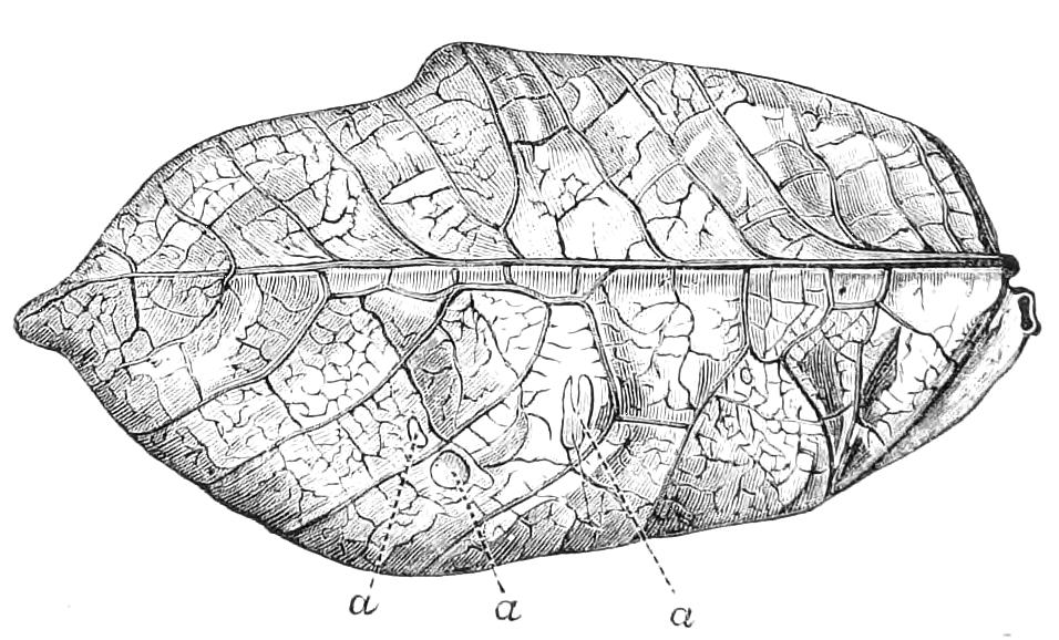
Fig. 198.—Leaf-like tegmen of Pterochroza ocellata: a, a, a, marks like those made by Insects on leaves.
The Locustidae display in the greatest possible perfection that resemblance of the tegmina to leaves which we mentioned when speaking of the general characters of the Orthoptera. The wing-covers are very leaf-like in colour and appearance in many Locustidae, but it is in the tribe Pseudophyllides and in the South American genus Pterochroza (Fig. 198) that the phenomenon is most remarkable. The tegmina in the species of this genus look exactly like leaves in certain stages of ripeness or decay. In the tegmina of some of the species not only are the colours of faded leaves exactly reproduced, but spots are present like those on leaves due to cryptogamic growths. Perhaps the most remarkable feature of these resemblances is the one pointed out by Brunner von Wattenwyl,[251] viz. that the tracks and spots formed on leaves by the mining of Insects in their tissues are also represented in the leaf-like wing-covers of the Pterochroza; transparent spots (a, a, Fig. 198) being present, just as they are in many leaves that have been attacked by Insects. Brunner was so much impressed by these facts that he came to the conclusion that they cannot be accounted for on the grounds of mere utility, and proposed the term Hypertely to express the idea that in these cases the bounds of the useful are transcended. We will mention here another peculiar case of resemblance described by Brunner as occurring in a Locustid. Two specimens of a little Phaneropterid were brought from the Soudan by the Antinori expedition, and have been described by Brunner under the name of Myrmecophana fallax. The Insect is said to bear an extraordinary resemblance to an ant. The most peculiar feature in the resemblance is shown in Fig. 199, A, B. The most characteristic point in the external form of an ant is the stalked abdomen, this structure being at the same time quite foreign to the Orthoptera. In the other parts of the body and in the colour generally, the Myrmecophana resembles an ant, but the abdomen of the Orthopteron is not stalked; it has, however, the appearance of being so, in consequence of certain parts being of a white colour, as shown in our figure. If abstraction be made of the white parts, the form of the stalked abdomen of the ant is nicely reproduced. The specimens brought from the Soudan were wingless and destitute of ovipositor, and may be immature, but Brunner suggests that they may prove to be really mature, the ovipositor, tegmina, and wings being permanently absent. The existence of a long ovipositor would certainly detract greatly from the ant-like appearance of the Orthopteron.
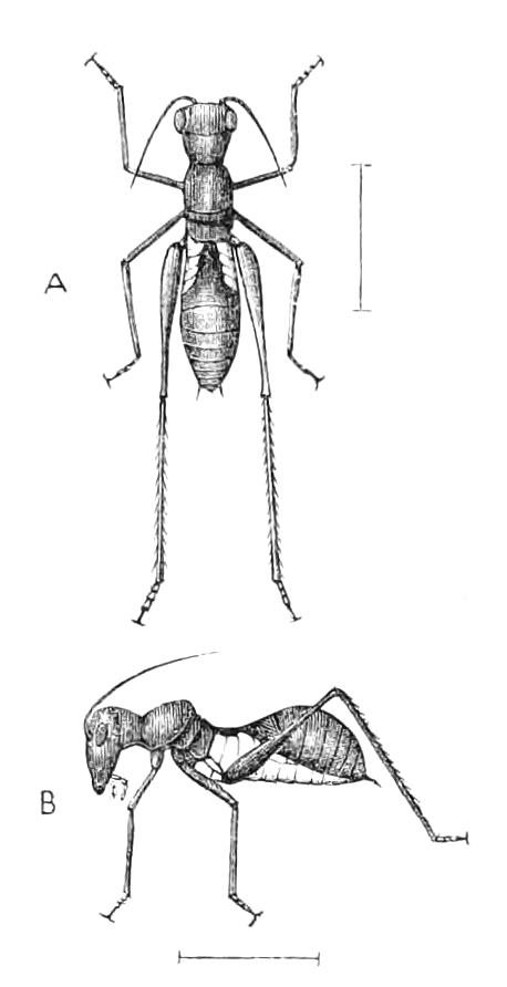
Fig. 199.—Myrmecophana fallax.
It is certain that the plant-like appearance of some of the Locustidae renders them inconspicuous to the human eye in the situations they frequent. It is a matter of common observation that though the noise of their chirpings may be heard to such an extent as to make it certain that many individuals must be in the immediate neighbourhood, yet at the same time it may be most difficult to detect even a single individual. M. Boutan noticed this phenomenon in the case of Ephippigera rugosicollis, and tells us that the human eye can, with a little practice, acquire the art of detecting these concealed creatures. This consists apparently in making use, not of a general inspection, but of a scrutiny of the outlines of the leaves and twigs of a tree. By this means, when the eye is accustomed to the task, the Insects can be detected with comparative ease; much in the same way, M. Boutan says, as a figure, placed in an engraving in such a way as to elude the eye, is appreciated with ease after the eye has once perceived it.
Some of the Locustidae are provided with means of defence of a positive nature. The Algerian Eugaster guyoni ejects two jets of a caustic orange-coloured fluid from two pores situate on the sides of the mesosternum, and covered by the anterior coxae. This species is carnivorous as well as herbivorous, and produces a sound more like humming than stridulation.[252]
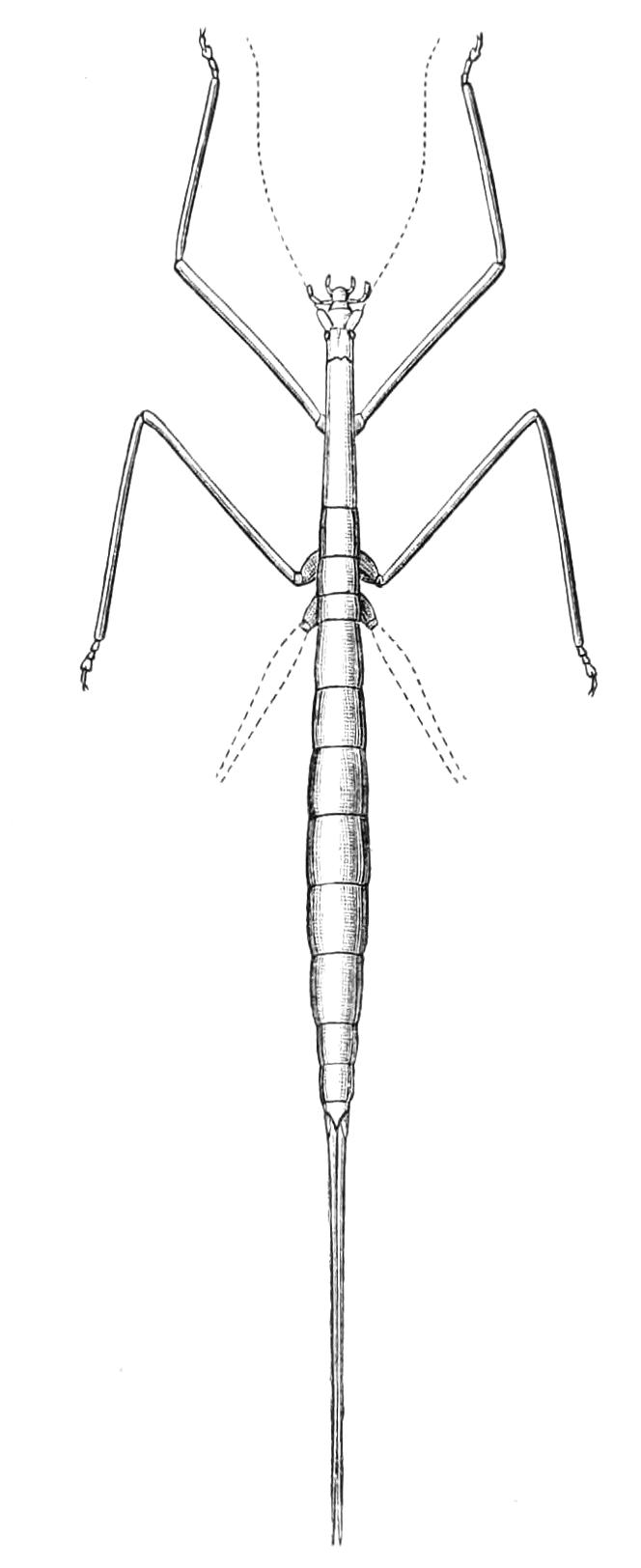
Fig. 200.—Phasmodes ranatriformis,female. Australia. (After Westwood.)
We have previously pointed out that some of the Acridiidae resemble the stick-Insects rather than the members of their own group; and similar cases occur amongst the Locustidae. Such a resemblance has, however, only been found in a few species of the tribe Prochilides. We figure one of these, Phasmodes ranatriformis, a native of South-West Australia. The very elongate linear form and the total absence of alar organs give this Insect a considerable resemblance to the stick-Insects or apterous Phasmidae. Prochilus australis is allied to this curious Locustid, but the alar organs are present in both sexes, and the Insect bears a great resemblance to the winged Phasmidae. This is due not only to the general form and colour, but also to the fact that the tegmina are very narrow, which causes them to look like the coloured slip on the anterior parts of the wings of some of the Phasmidae (cf. p. 266). Another case of a Locustid with elongate, slender form is found in the extraordinary Peringueyella jocosa of South Africa, a member of the tribe Sagides. It has minute organs of flight, and reproduces, to a considerable extent, the form and appearance of Proscopides or of some Tryxalides.[253]
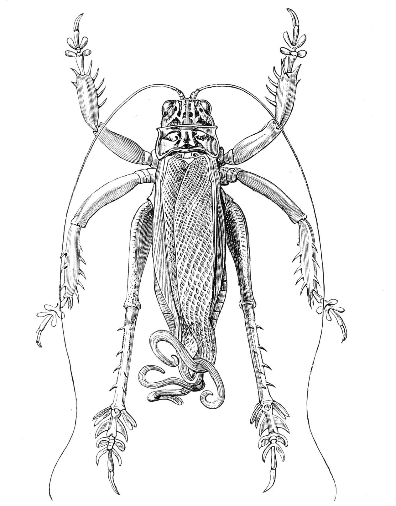
Fig. 201.—Schizodactylus monstrosus, male. Natural size. East India.
We follow Brunner in placing among the Locustidae the large Insect we represent in Fig. 201. It is remarkable on account of its tegmina and wings; these have their extremities much prolonged and curled; moreover, the flat interior area and the abruptly deflexed exterior area make them look more like the wings of Gryllidae. This species has no ocelli, and is said to be destitute of ears. The inflated condition of the anterior and middle tibiae suggest that it possesses auditory structures, though there appears to be no external opening for them. This Insect is found in India, where it is said to be common on the banks of sandy rivers, living there in burrows of the depth of three feet. Very little is known, however, as to this curious Insect. It has recently been reported[254] as being injurious to tobacco and other crops on high ground in Durbungha by cutting off their roots. The local name for the Insect is bherwa. We should think it somewhat doubtful whether this refers really to S. monstrosus.
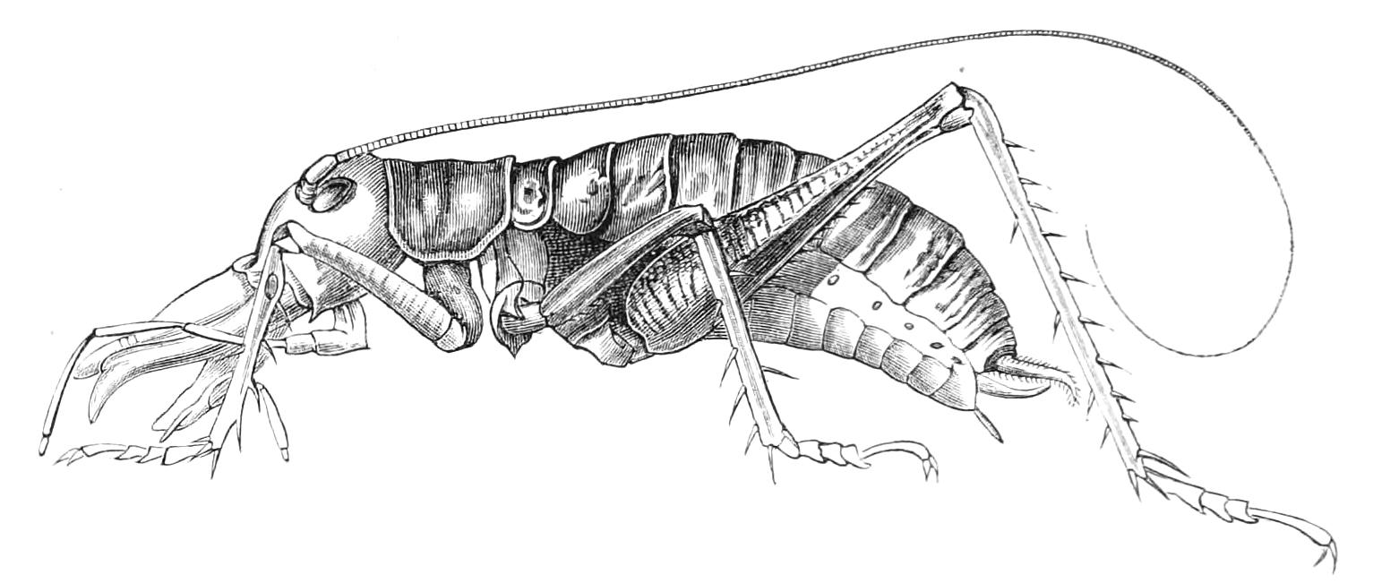
Fig. 202.—Anostostoma australasiae, male. Australia.
In number of species the Locustidae are perhaps scarcely inferior to the Acridiidae, and in variety of form they surpass this latter family. Many of the most gigantic forms are apterous, and these very often have a repellant aspect. The genus Anostostoma is remarkable for its large head. Allied to it is Deinacrida heteracantha, the "Weta-punga" of the New Zealand natives, an Insect formerly abundant in the forests north of Auckland, but of late years become extremely rare. The head and body of this Insect may measure more than 2½ inches in length, and when the antennae and legs are stretched out the total length may be 14 or 15 inches. Although bulky and absolutely wingless, yet, as Buller informs us,[255] it climbs with agility, and is sometimes found on the topmost branches of lofty trees. When disturbed it produces a clicking, accompanied by a slow movement of its hind legs. A second species, D. thoracica, lives in decayed wood, and a third, D. megacephala, is remarkable from the very large size of the head and mandibles in the male sex. The fact that a clicking noise is produced by the Weta-punga is of some interest, for the genus Deinacrida is among the Locustidae that possess ears, but are said to be destitute of sound-producing organs.
Amongst the most remarkable of the Locustidae are the two species of which Brongniart has recently formed the genus Eumegalodon and the tribe Eumegalodonidae, which is not included in Brunner's table of the tribes of Locustidae. The ovipositor is large and sabre-shaped; the male is unknown. The genus Megalodon is placed by Brunner in the tribe Conocephalides; it also consists of extremely remarkable Insects.
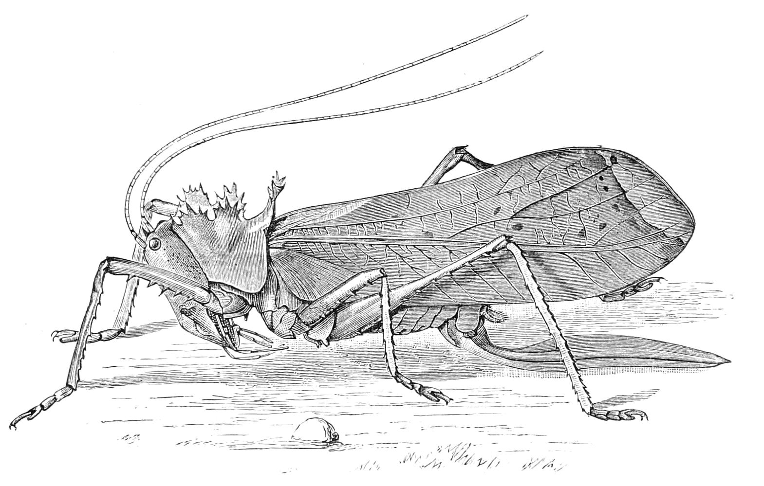
Fig. 203.—Eumegalodon blanchardi, female. Borneo. × ⅘. (After Brongniart.)
The Locustidae appear to be of slow growth, and the autumns of Britain are usually not warm enough for them. Hence we have but nine British species, and of this number only three or four are known to occur north of the Thames. The only one that attracts attention is Locusta viridissima, which in some districts of the south of England occurs in considerable numbers, and attests its presence by its peculiar music. It is called the green grasshopper.
The geological record is rather obscure in the matter of Locustidae. Scudder considers that a fair number of Tertiary forms are known, and says that they represent several of the existing tribes and genera. One or two have been found in Mesozoic rocks.
Table of the Tribes of Locustidae
1. Tarsi more or less depressed.
2. Front tibiae furnished with auditory cavities.
3. Antennae less distant from the summit of the occiput than from the labrum; inserted between the eyes.[256]
4. First two joints of the tarsi laterally smooth. (Posterior tibiae furnished on each side with an apical spine.) Tribe 1. Phaneropterides. (Fig. 196, Microcentrum; Fig. 199, Myrmecophana. Fig. 101, Poecilimon affinis.)
4′. First two joints of the tarsi laterally, longitudinally sulcate.
5. Foramina of the anterior tibiae normally open. (Fig. 193, A.)
6. Posterior tibiae furnished on each side with apical spines.
7. Prosternum unarmed. Tribe 2. Meconemides.
7′. Prosternum bispinose or bituberculate. Tribe 3. Mecopodides.
6′. Posterior tibiae with no apical spines. (Head prognathous.) Tribe 4. Prochilides. (Fig. 200, Phasmodes.)
5′. Foramina of the anterior tibiae forming a chink, or protected by a scale. (Fig. 193, B.)
6. Anterior tibiae with no apical spines.
7. Margins of the scrobes[257] of the antennae prominent. Tribe 5. Pseudophyllides. (Fig. 187, Cyrtophyllus crepitans; Fig. 198, Pterochroza ocellata.)
7′. Margins of the scrobes of the antennae not prominent.
8. Posterior tibiae furnished above on each side with apical spines, or with a single spine on the side.
9. Posterior tibiae either furnished with apical spines on each side, or only on the inner side. Tribe 6. Conocephalides. (Fig. 189, Copiophora cornuta.)
9′. Posterior tibiae furnished above with an apical spine placed only on the outer side. Tribe 7. Tympanophorides.
8′. Posterior tibiae without apical spines. Tribe 8. Sagides.
6′. Anterior tibiae furnished with an apical spine on the inner side.[258]
7. The first joint of the posterior tarsi destitute of a free sole-lobe. Tribe 9. Locustides.
7′. The first joint of the posterior tarsi furnished with a free sole-lobe. Tribe 10. Decticides.
3′. Antennae more distant from the summit of the occiput than from the labrum, inserted either beneath the eyes or on their inferior border. Tegmina and wings greatly abbreviate, scale-like; when tegmina are present they are furnished in each sex with a tympanum.
4. Third joint of the posterior tarsi shorter than the second. Both anterior and posterior tibiae furnished on each side with a spine. Tribe 11. Callimenides.
4′. Third joint of posterior tarsi longer than the second joint. Anterior tibiae with no apical spine on the inner side, and posterior tibiae with no apical spine on the outer side.
5. Antennae inserted at the edge of the eyes. Pronotum unarmed. Tegmina present in each sex. Anterior tibiae furnished on the outer side with an apical spine. Posterior tibiae furnished beneath with four apical spines. Tribe 12. Ephippigerides.
5′. Antennae inserted distinctly below the eyes. Pronotum spinous. Elytra in the females wanting. Anterior tibiae without apical spine on either side. Posterior tibiae beneath with two apical spines or with none. Tribe 13. Hetrodides.
2′. Anterior tibiae without auditory cavities. Tegmina with no tympanum. Tribe 14. Gryllacrides. (Fig. 201, Schizodactylus monstrosus.)
1′. Tarsi distinctly compressed (most of the species apterous.) Tribe 15. Stenopelmatides. (Fig. 202, Anostostoma australasiae; Fig. 197, Dolichopoda palpata.)