Chapter VIII
Reading edition
ROTIFERA, GASTROTRICHA, AND KINORHYNCHA
ROTIFERA—HISTORY—EXTERNAL FEATURES—MOVEMENT—ANATOMY—REPRODUCTION—EMBRYOLOGY—CLASSIFICATION—DISTRIBUTION—AFFINITIES—GASTROTRICHA—KINORHYNCHA
The Rotifera are microscopic animals, the largest not exceeding one-eighth of an inch in length. According to Hudson and Gosse,[233] they are first recorded in an observation of the Rev. John Harris, in 1696, of "an Animal like a large Maggot which could contract itself into a Spherical Figure, and then stretch itself out again; the end of its Tail appeared with a Forceps like that of an Ear-wig."[234] This was certainly a Bdelloid Rotifer.
In 1703 Leeuwenhoek[235] gave a fuller description of a tubicolous form, probably Limnias, and noted the peculiar appearance of the ciliary wreath as "two wheels thickset with teeth as the wheel of a watch." He also noted a little later[236] the way in which Melicerta (see p. 206) builds its tube, and was the first to observe the revivification of certain species after drying.[237] Joblot, a French professor of mathematics, in 1718 figured and described a large number of new genera and species with more or less fantastic details. Baker's figures[238] are a considerable advance on Joblot's, and his descriptions of habits are still fresh and accurate. Eichhorn found a number of new and interesting forms; and O. F. Müller, influenced by the new discipline of Linnaeus, not only figured many species, but gave good short diagnoses of their characters. Ehrenberg in 1838 brought out his magnificent Infusionsthierchen, which contains descriptions and figures of what are now divided into Protophyta, Protozoa, Rotifera, and Gastrotricha. Dujardin's monograph on the "Infusoires," in the Suites à Buffon,[239] was in several respects an advance on Ehrenberg, whose power of observation was so great as to render his mistakes the more inexplicable. But Ehrenberg ever adhered to his errors as firmly as to his facts.
The occurrence of Rotifers among microscopic plants induced the botanists Cohn and Williamson[240] to work at their structure; the group has been studied by men engrossed in other professional cares, such as Gosse, Bedwell, Moxon, Rousselet, and Maupas. Huxley,[241] Leydig,[242] and Cohn[243] studied Rotifers in the '50's and early '60's with a precision the more remarkable when we remember the imperfect methods then available. This period was closed by the valuable monograph published in Arlidge's (4th) edition of Pritchard's Infusoria,[244] under the supervision of W. C. Williamson. Leidy began the study of the American Rotifers. Eckstein[245] gave a careful and interesting account of the species about Giessen in a richly illustrated paper. In recent times the modern methods of histological and embryological research have been applied by Vallentin,[246] Plate,[247] Tessin,[248] and Zelinka,[249] the three Studien ueber Rotatorien of the last author being indispensable to every student, and containing a full bibliography.
Hudson and Gosse's Monograph (1886-89) contains a history of the class to which, as to the whole book, we are deeply indebted; and a full systematic account of all published species.[250] C. Rousselet has introduced a method[251] of preparation of Rotifers in microscopic slides which enables workers to preserve the types they figure and describe for future identification and comparison. Gunson Thorpe has collected and studied Rotifera in China and Australia. It would be unfair not to record here the invaluable services of the late Thomas Bolton, and his son of the same name, both of Birmingham, and of J. Hood of Dundee, who have found and widely distributed living specimens of new, rare, and interesting species.
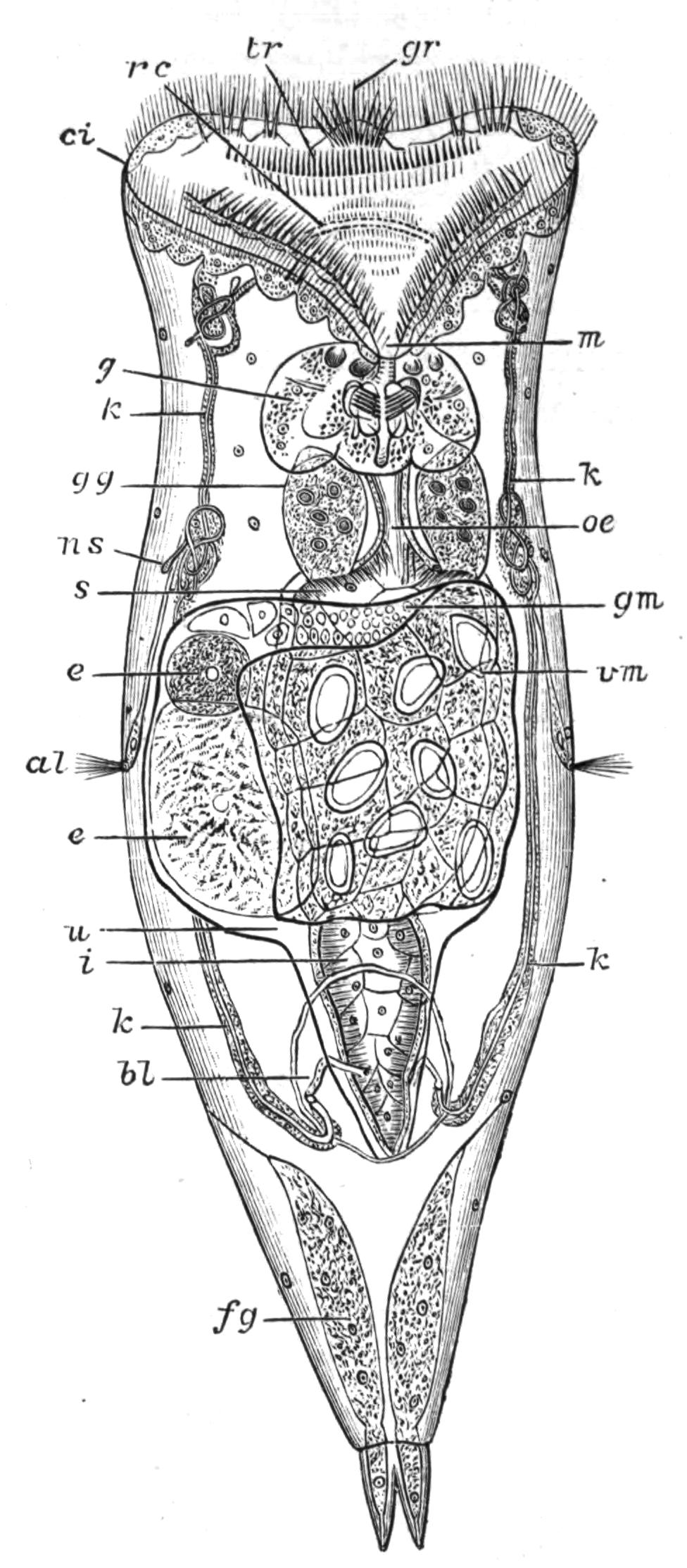
Fig. 106.—Hydatina senta, ventral view. (After Plate.) al, Lateral antenna; bl, bladder; ci, cingulum; e, e, eggs in uterus; fg, foot gland; g, gizzard; gg, gastric gland; gm, germarium or ovary; gr, ciliated lobes of "groove"; i, intestine; k, k, kidneys; m, mouth; ns, nephrostome; oe, oesophagus; rc, renal commissure, transverse tube uniting kidneys above mouth; s, stomach overlaid by reproductive organs; tr, trochus; u, uterus; vm, vitellarium or yolk-gland.
Definition of the Class.—We may define Rotifera as a class of minute bilaterally symmetrical animals, with a chitinous integument, a soft terminal "disc" fringed by a complex ciliary "wreath," an anterior or subventral mouth, and a dorsal cloacal aperture, beyond which the body is usually prolonged into the "foot" or process bearing cement glands, and serving for attachment, temporary or permanent. The body-cavity has no epithelial lining, and is traversed by nerves and muscles. The alimentary canal possesses a chitinous gizzard or mastax of peculiar arrangement, and it usually opens into a cloaca. The nervous centre consists of a ganglion on the dorsal side of the pharynx, to which a second one on the ventral side is sometimes connected to form a complete ring; eyes and bristle-bearing feelers are usually present as sense-organs. A paired system of renal tubes serves for excretion, opening through a median contractile bladder into the ventral side of the cloaca. The sexes are distinct; but the males (Fig. 107), which mostly lack digestive organs, occur rarely, and the females are usually viviparous, or carry about the eggs till they are hatched; while, owing to the rarity of the males, parthenogenesis is habitual. Fission and budding are alike unknown. The fertilised eggs are of the kind termed "winter" or "resting" eggs, and resist conditions adverse to life.
The Rotifera are of cosmopolitan distribution; most of the species inhabit fresh water, whilst some are brackish, and a few are marine; 84 genera and about 700 species have been described.
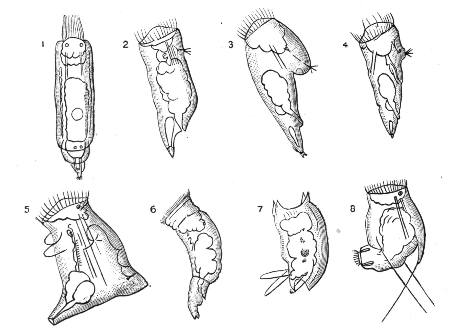
Fig. 107.—Male Rotifers. (After Hudson.[252]) 1, Floscularia campanulata; 2, Lacinularia socialis; 3, Notops brachionus; 4, Synchaeta tremula; 5, Asplanchna ebbesbornii; 6, Brachionus urceolaris; 7, Salpina mucronata; 8, Pedalion mirum.
External Features.[253]—The body is divided into three regions: (1) the head, ending in the disc, which bears the ciliary wreath; (2) the trunk, containing the viscera; (3) the foot, which only contains muscles, nerves, and cement-glands. The general form of the BODY varies greatly: it is spherical in Trochosphaera, ovoid in Asplanchnidae, conical in Scirtopoda, Triarthridae, and Synchaeta; moderately elongated in the majority of the Ploima, among which some forms are very flat, like Pterodina, Metopidia, and Brachionus; shortly elongated and cylindrical in Hydatina (Fig. 106), Notommatidae, and many others. In Taphrocampa it is cylindrical and segmented, while the segments are telescopic in the Bdelloida, both ends being retractile into the middle segment. In most attached, tube-dwelling forms the body is ovate, tapering behind into the elongated stalk-like foot.
The FOOT at the hinder end of the body is usually more or less jointed; in Pterodina and Brachionus it is long, transversely wrinkled, and retractile. Usually it terminates in a couple of acute, mobile toes, perforated at the tips by the ducts of the pedal glands (Fig. 106, fg), whose viscid secretion serves to anchor the animal. In Rotifer there are three of these toes, which are retractile, and in addition there are in this genus, as in most of the Bdelloida, toe-like pointed spurs in pairs on the more proximal joints of the foot. In Callidina the spurs are often perforated, and the toes are replaced by numerous openings on the last joint of the foot (Fig. 109, A); while in Discopus the end of the foot expands into a large disc, with numerous pores for the exudation of the pedal cement, and there are no spurs. In Pedalion mirum the foot is represented by two tubular processes ciliated at the apex and at the outer side near the base (Fig. 117, f). These are inconstant in size and form, that of one side being sometimes reduced or absent, while both are absent in the closely allied species P. fennicum.
In Melicertidae and Flosculariidae the long foot ends in an expanded disc, which is cupped and ciliated in the larva (Fig. 112, B) and in the larva-like male (Fig. 107); but in two species it is prolonged into a long flexible thread which is not contractile. The foot is also elongated in the Bdelloid genus Actinurus and the Ploimal genus Scaridium. It forms a mere ventral disc in Apsilus (and Atrochus?), and is absent in Asplanchnidae (except Asplanchnopus), Triarthridae, and Anuraeidae, and in the genera Trochosphaera (Melicertaceae) and Pompholyx (Pterodinidae).
The fringed spines of Triarthridae are jointed appendages moved by powerful muscles; in Triarthra one is median and ventral, the others being attached to the shoulders. In Polyarthra, there are twelve flattened and serrated spines, a bunch of three being attached to the dorsal and ventral faces of either shoulder. An easy transition leads to the hollow appendages of Scirtopoda, which end in a fringe of bristly hairs, themselves feathered with finer hairs (Fig. 117). These processes are in Pedalion six in number, two median (respectively dorsal and ventral), two antero-lateral, and two postero-lateral. As they contain proper muscles, and the postero-lateral pair contain part of the nephridia and bear the lateral antennae, they are true outgrowths of the body, and are not homologous with the spines of Triarthridae.
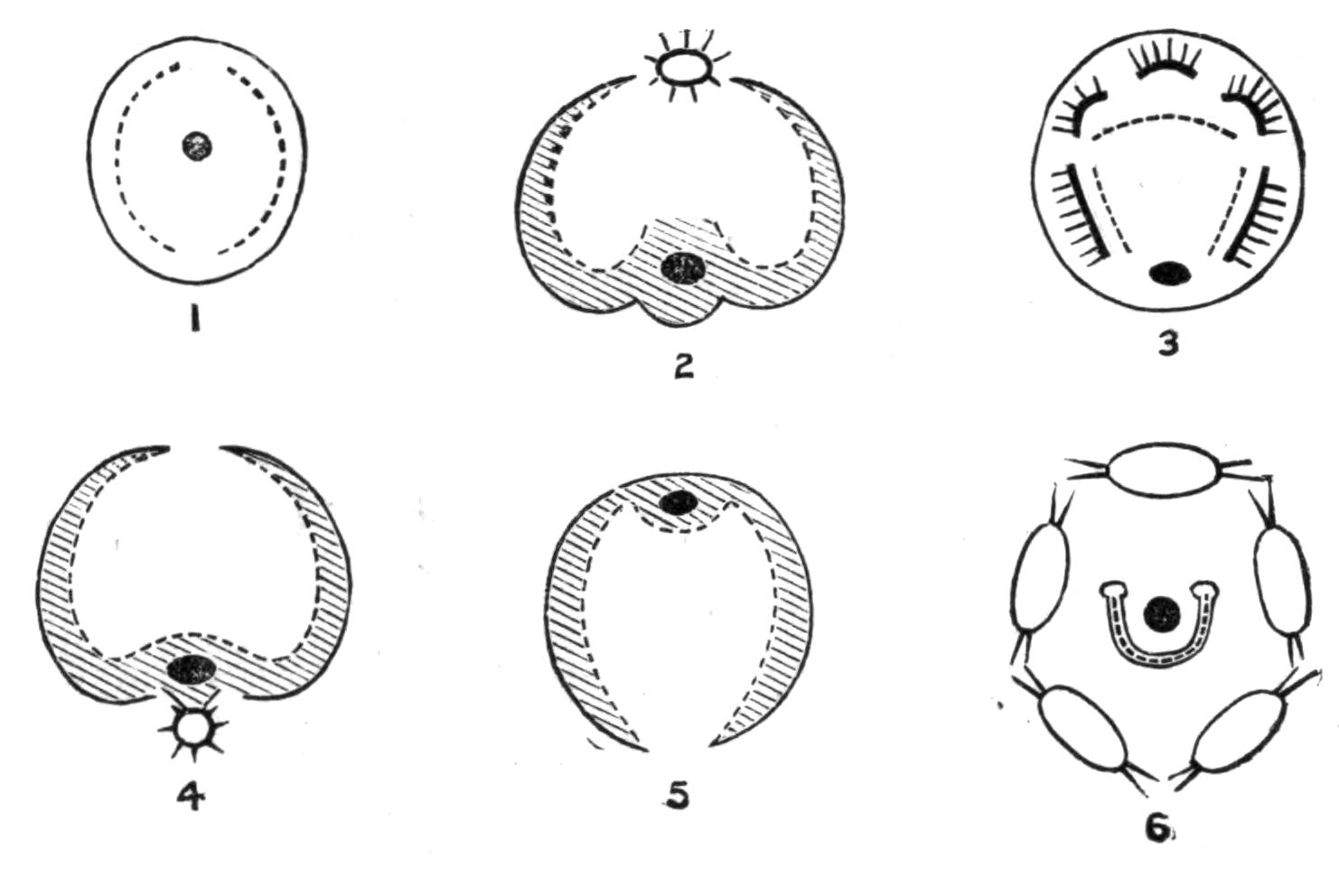
Fig. 108.—Diagrammatic views of disc of Rotifers. Cingulum represented by a black line, groove shaded; trochus dotted; the black spot represents the mouth. 1, Simple disc of Microcodon; 2, Bdelloid disc of Rotifer or Callidina, the star represents the ciliated proboscis; 3, disc of Hydatina, groove represented by lobes bearing ciliated styles; 4, disc of Melicerta, the star represents the ciliated ventral cup with openings into it from the groove; 5, disc of Conochilus; 6, disc of Stephanoceros, cingulum (?) of setose lobes, trochus horseshoe-shaped, mouth central.
The front of the body constitutes the HEAD, which is scarcely distinct, though usually separated by a slight neck-like constriction. The DISC, which terminates the head, varies greatly in shape and in the arrangement of its parts. Imagine a circular funnel, finely ciliated within, and with the mouth at the bottom, the prominent rim bearing two zones of cilia, the inner or anterior being the coarser, and termed the "trochus" or hoop; the outer finer, and termed the "cingulum" or girdle, while a very finely ciliated groove lies between the two zones. Either or both of these zones may be interrupted on the dorsal or ventral median line, or both; and the funnel-shaped mouth may be shifted—usually ventrally, so that it forms only a dilatation of the ciliated groove. Again, the wreath as a whole may be festooned or lobed; or the lobing may be confined to the area between the cingulum and trochus, as in most Ploima (Figs. 106 and 108, 3). Very frequently on these lobes adjacent cilia are fused together during life, producing "vibratile styles," whose true nature is only revealed after death. In Microcodonidae the structure of the disc (Fig. 108, 1) nearly conforms to the primitive type; but the ciliated groove is absent, and the "trochus" is in two separate half-elliptical bands. In the Flosculariaceae (Fig. 108, 6) the mouth is also central, the disc is funnel-shaped, and the trochus is a horseshoe-shaped ridge, with its ends dorsal and raised into prominent knobs. The margin of the funnel is in Flosculariidae (Fig. 115) usually lobed, and furnished either with exceptionally strong cilia, or else with very long bristles which are usually passive. However, by the retraction of the lobes that bear them they are clasped together like casting-nets to enclose prey brought into the funnel by the action of the trochal cilia. An external ring of cilia in Floscularia mutabilis and F. pelagica serves for swimming. In Apsilidae the margin of the disc bears neither cilia nor bristles, but is either simple and ring-like, or is produced into tentacles (Fig. 112, C). The oral funnel is probably represented in Flosculariaceae by the continuation of the small central mouth into a ciliated tube (Fig. 115, C, tf), open below, and hanging freely down into the crop.
In all other cases the mouth is displaced, and lies in the groove and on its ventral side (except in Conochilus, where it is dorsal, Fig. 108, 5). In the Bdelloida the disc is prolonged into two great lobes like kettle-drums, round the posterior, external, and ventral edges of which run the trochus, cingulum, and ciliated groove (Fig. 108, 2). All three are interrupted behind in the median line; ventrally the groove widens into the oral funnel, the cingulum is continued into a sort of spout-like lower lip (Fig. 109, C, D, l), and the trochus is absent. The body is prolonged dorsally above the lobes into a two-jointed proboscis, ending in a ciliated cup overhung by two dorsal flaps: this we regard as a detached portion of the wreath.
This "Bdelloid" type of wreath occurs also in Scirtopoda (Fig. 117), and in the Ploimal genera Triarthra, Pterodina, and Pompholyx. A simpler wreath of essentially the same type occurs in Asplanchnaceae and Melicertaceae; the disc is not prolonged into drum-shaped lobes, but is thin at the rim, where it bears the triple ciliated zone, interrupted on the dorsal median line and depressed ventrally into the oral funnel. In the Melicertidae, moreover, the disc is widened into a great plate-like extension, often beautifully lobed; and in many of the species a ciliated cup lies ventral to the lips, and is connected with the groove by a short ciliated channel on either side (Figs. 108, 4, and 116). Even the simpler wreath of Asplanchnidae is complicated by stronger lobes on either side bearing vibratile styles.
The most complex discs are found in Ploima, especially in Brachionus, Hydatina, and Synchaeta, since the groove is replaced by a zone of lappets, as above mentioned. In Proales the whole face of the disc is strongly ciliated. The wreath is reduced in the parasitic genera Drilophagus, Albertia, Balatro, and the Seisonaceae; in Adineta and Taphrocampa it is only represented by a general but scanty ciliation of the disc.
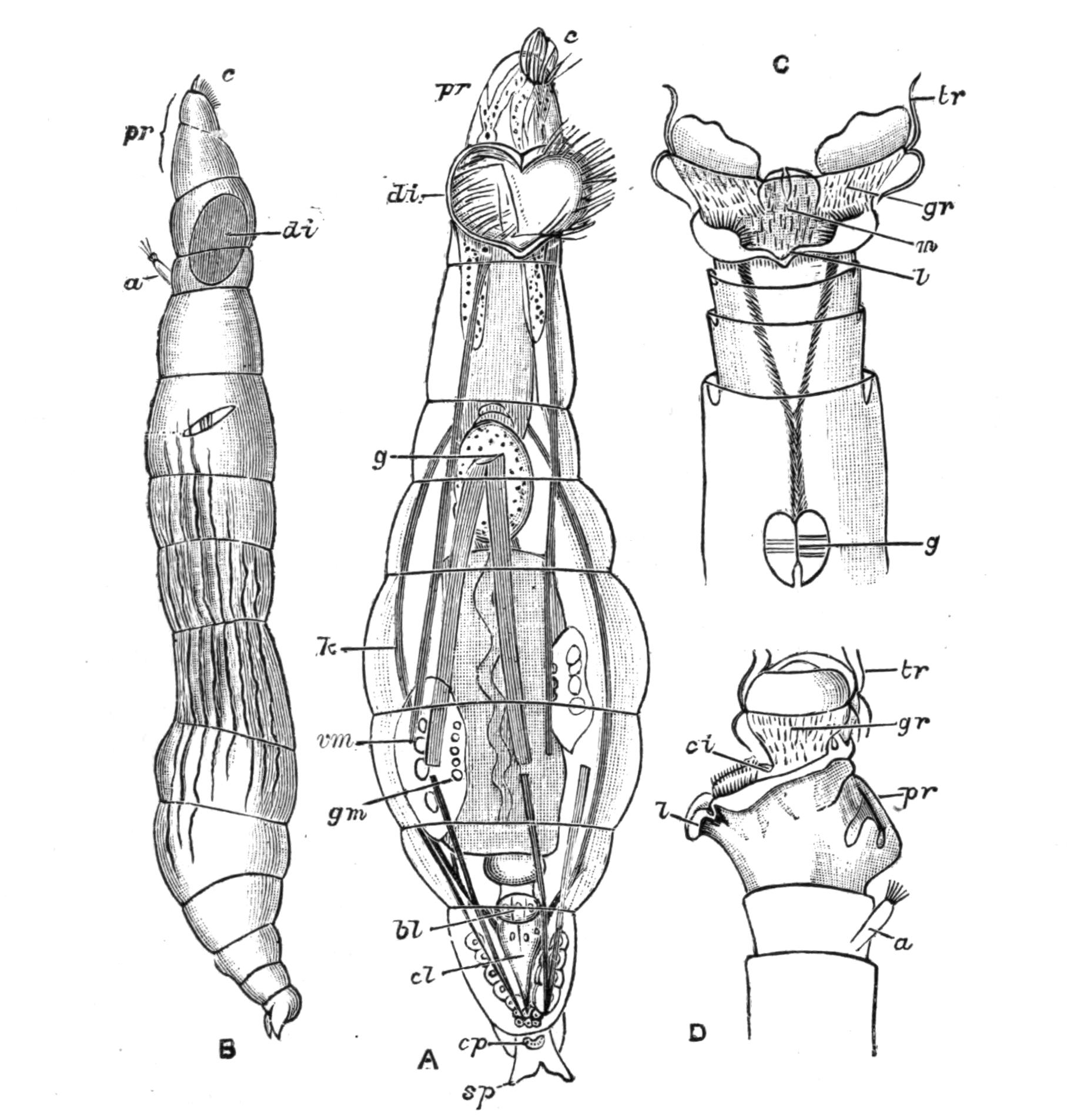
Fig. 109.—Callidina symbiotica. (After Zelinka.) A, Ventral view, with the disc half expanded, proboscis extended; B, lateral view, proboscis extended; C, ventral view of anterior segments with expanded disc; D, lateral view of same (proboscis retracted). a, Antenna; bl, bladder (enlargement of rectum); c, ciliated cup of the proboscis; ci, cingulum; cl, cloaca; cp, group of pores, the openings of cement glands; di, disc; g, gizzard; gm, germarium (that of the opposite side seen at a higher level); gr, ciliated groove; k, kidney; l, lip; m, mouth; pr, proboscis; sp, spurs of foot; tr, large cilia of trochus, showing vertical movements; vm, yolk-gland. The body muscles are represented by shaded bands.
The head is very frequently retractile, as a whole, by strong muscles. In Bdelloida the disc proper is retracted when the animal crawls, while the proboscis is exserted (Fig. 109). Ciliated patches occurring outside the region of the disc point to a primitive condition when the whole surface of the body was ciliated, as does the partial ciliation of the foot in certain groups. Synchaeta and many Notommatidae possess a pair of lateral, hollow, ciliated pits on the body, which can be everted to serve as additional swimming organs; these are termed "auricles."
The cuticle varies much in texture. It may be smooth and flexible, dotted or shagreened, or in the Loricata firm and of definite shape, constituting a lorica, which may be more or less distinctly divided up into areas or separated into distinct pieces. In this case it resists decomposition, and several species are only known by this "skeleton." In Ploesoma it is much thickened and looks like a honeycomb. A regular alternation of harder and softer zones effects the annulation of the body in certain genera.
The hypoderm or protoplasmic layer of the skin has no cellular boundaries, though it contains large and distinct nuclei; it is usually somewhat granular. It forms the wall of the body-cavity, which contains a transparent liquid without corpuscles.
The principal external glands are the pedal or cement-glands, which secrete a viscid substance that sets in water and serves to anchor the animal. They are formed from an ingrowth of the hypoderm, are usually paired, and open by fine ducts on or near the apex of the toes, when these processes of the foot are present (Fig. 106, fg). These glands are mostly absent when there is no foot, as in most Asplanchnidae and in Anuraeidae, but in Asplanchna herrickii a small gland on the ventral side of the cloacal aperture appears to represent the last rudiment of the foot.
In addition to these, the ciliated ventral cup below the disc of many Melicertidae secretes a viscid substance (Fig. 116, p); and possibly the whole surface of the body is secretory in those species of this group, and of the Flosculariidae, whose tube (Fig. 115, A) is uniform and not made of pellets. In several other species belonging to Bdelloida and Ploima-Illoricata a viscid secretion of the surface of the body renders it "sordid" with adherent particles of dirt.
When the secretion takes the form of a tube, the body can be wholly withdrawn into it by the contraction of the foot. In Floscularia, Stephanoceros, and Conochilus the tube is hyaline and thin-walled; in Oecistes and Cephalosiphon it is more or less floccose; and in Limnias it is thin, firm, and annulated. In Melicerta and some species of Oecistes the tube thus secreted by the body is only formed in a very young state. In M. janus and M. pilula it is increased by the successive deposition of ovoid faecal pellets on to the rim. In M. ringens (Fig. 116) and M. conifera pellets are formed of the excess of the food particles brought to the disc by the ciliary current; they are carried through the gutters on either side of the projecting ventral lip or "chin" into the ciliated glandular cup on that side of the head. Here, as they revolve, they are cemented together into a pellet which is spheroidal in the former species, cylindro-conoidal with a basal hollow like a rifle-bullet in the latter. After a pellet is completed the animal stoops down and deposits it on the edge of the tube. This may easily be verified by furnishing a young Melicerta with water containing solid particles of carmine. M. tubicolaria forms a thick tube which is laminated, the laminae being directed upwards and outwards, and having diatom shells, etc., between the layers. In this case we have observed that the faeces are pellucid, and sometimes are so ejected as to lie in a sheet against the funnel-shaped mouth of the tube, and we are inclined to believe that the tube itself is formed altogether in this way. A similar process probably occurs in Oecistes crystallinus and Oe. umbella.
The muscles are simple elongated fibres, usually having near the middle a mass of granular protoplasm containing a nucleus; they may be smooth or striated. The principal muscles of the body are conspicuously striated in many active free-swimming forms (Pedalion, Synchaeta, Pterodina, Triarthra).
The muscles of the body-wall are transverse and longitudinal. They are best seen in Bdelloida. The principal muscles of the body-cavity are longitudinal; the most conspicuous and constant are the retractors of the disc and of the foot, protraction of these organs being usually accomplished by the contraction of the transverse muscles. Special muscles effect the vigorous springing of the Triarthridae and Scirtopoda; in the former group the muscles only raise the spines, and their elastic recoil is the actual mechanism of progression; but in the latter (Fig. 117) special flexor muscles of the limbs are the effective agents of the leaping movements.
Movements.—The Rotifera vary very greatly in their movements. The cilia of the disc, and especially of the trochus, are the principal organs of prehension of food, and also of swimming when the animal is not fixed by its foot. In some cases, as in Bdelloida, the cilia lash downwards successively in the longitudinal plane of the body (Fig. 109, C, D); this motion during fixation produces a hollow vortex ring, like the rings of a skilled cigarette-smoker, but when the animal is free it determines a simple forward progression through the water. In other cases the animal rotates on its long axis, or may even turn somersaults (Synchaeta). The appearance of the spokes of a wheel is a pure illusion due to the greater visibility of the cilia in their slow recovery than in their instantaneous down-lash. The finer cilia of the groove and cingulum play a very minor part in the act of swimming, and in the production of the great vortices at the edge of the disc when the animal is fixed; they serve to direct the particles brought by the vortices to the edge of the disc onwards towards the mouth. It is easy to see that the stream must be in opposite directions on opposite sides of the groove; its prolongation across the dorsal median line would be useless, which explains the existence of the dorsal median gap. At the ventral side we usually find a prominent ciliated lip, whose cilia work outwards, and carry off the excess of food particles as by an overflow spout. In many cases among the Notommatidae, Coluridae, etc., the disc serves as much for creeping over organic débris as for swimming.
We have already noticed the springing bristles and limbs of the Triarthridae and Scirtopoda respectively; the great foot of Scaridium is also used for leaping. The Bdelloida have the power of retracting their disc and progressing in loops like a leech or looper (Geometrid) caterpillar.
Baker, in a letter addressed to Martin Folkes, Esq., President of the Royal Society, dated London, 16th January 1744-5,[254] gives the following lively account of the aspect and movements of Philodina roseola belonging to this group, with figures, some of which we reproduce from the original copper-plate engraving:—"I call it a Water Animal, because its Appearance as a living Creature is only in that Element. I give it also for Distinction Sake the Name of Wheeler, Wheel Insect or Animal; from its being furnished with a Pair of Instruments, which in Figure and Motion appear much to resemble Wheels. It can, however, continue many Months out of Water, and dry as Dust; in which Condition its Shape is globular, its Bigness exceeds not a Grain of Sand, and no Signs of Life appear. Notwithstanding, being put into Water, in the Space of Half an Hour a languid Motion begins, the Globule turns itself about, lengthens by slow Degrees, becomes in the Form of a lively Maggot, and most commonly in a few Minutes afterwards puts out its Wheels, and swims vigorously through the Water in Search of Food; or else, fixing by its Tail, works them in such a Manner as to bring its Food to it. But sometimes it will remain a long While in the Maggot Form and not shew its Wheels at all....
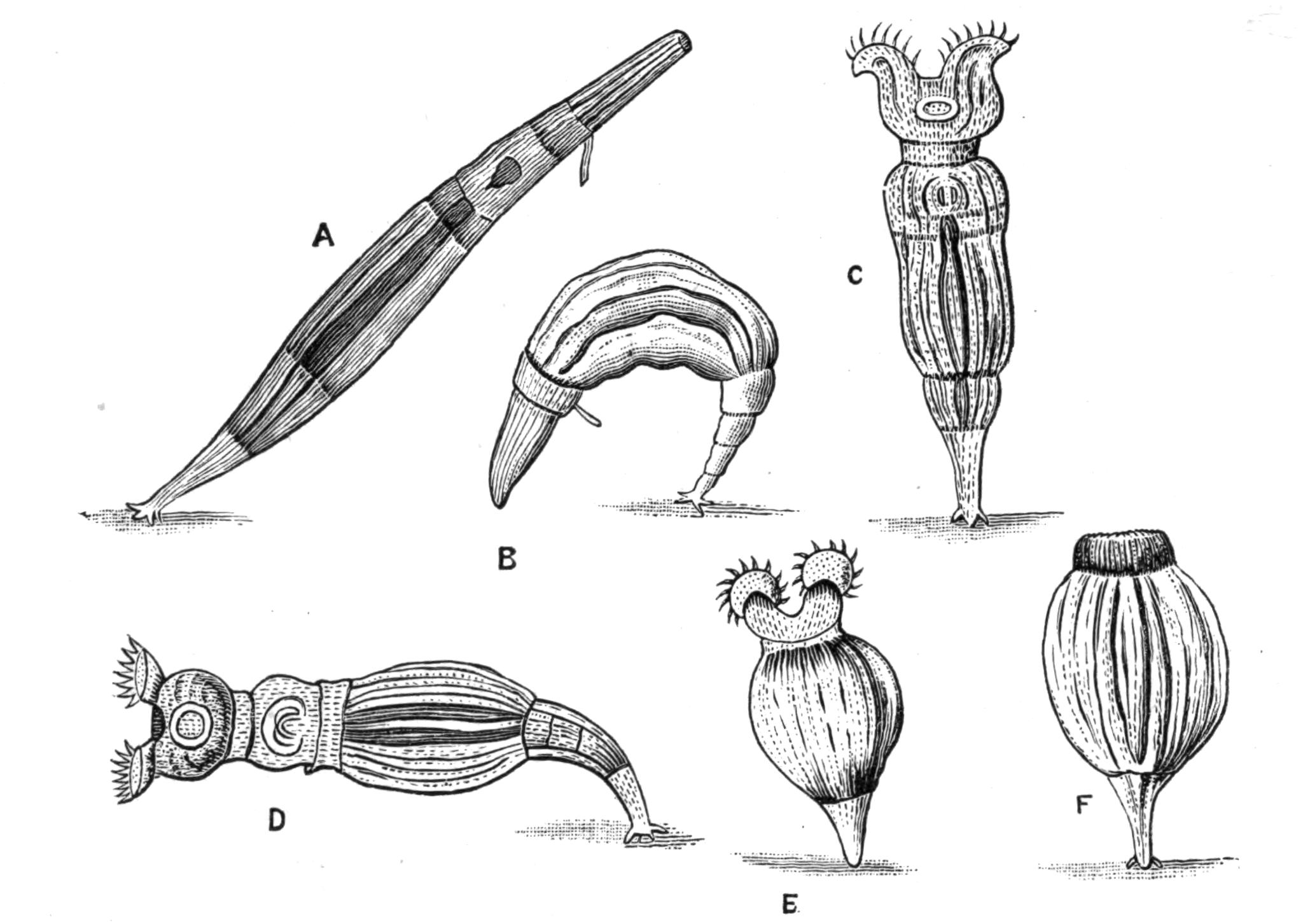
Fig. 110.—Philodina roseola. (After Baker.) A, B, Crawling, with extended proboscis, and showing antenna; C, D, E, attached, with "wheels" extended for catching food; F, attached, with anterior end retracted.
"If the Water standing in Gutters of Lead, or the slimy Sediment it leaves behind, has any Thing of a red Colour, one may be almost certain of finding them therein,[255] and, if in Summer, when all the Water is dried away, and nothing but Dust remains, that Dust appears red, or of a dark brown, one shall seldom fail, on putting it into Water, to discover Multitudes of minute reddish Globules, which are indeed the Animals, and will soon change their Appearance, in the Manner just now mentioned....
"A Couple of circular Bodies, armed with small Teeth like those of the Balance-Wheel of a Watch, appear projecting forwards beyond the Head, and extending sideways somewhat wider than the Diameter thereof. They have very much the Similitude of Wheels, and seem to turn round with a considerable Degree of Velocity, by which Means a pretty rapid Current of Water is brought from a great Distance to the very Mouth of the Creature, who is thereby supplied with many little Animalcules and various Particles of Matter that the Waters are furnished with.
"As these Wheels (for so from their Appearance I shall beg Leave to call them) are every where excessively transparent, except about their circular Rim or Edge on which the Cogs or Teeth appear, it is very difficult to determine by what Contrivance they are turned about, or what their real Figure is, though they seem exactly to resemble Wheels moving round upon an Axis....
"As the Animal is capable of thrusting these Parts out, or drawing them in, somewhat in the Way that Snails do their Horns, the Figure of them is different in their several Degrees of Extension and Contraction, or according to their Position to the Eye of the Observer, whereby they not only appear in all the various Forms before represented, but seem at certain Times as if the circular Rim of the Wheel or Funnel were of some Thickness, and had two Rows of Cogs or Teeth, one above and the other below that Rim."
Digestive Organs.—The pharynx is usually a narrow ciliated tube, which varies in length from genus to genus, but in no other important point, save in Flosculariidae, where it assumes the form of a crop, into which the mouth hangs freely down as a narrow ciliated tube. At its lower end is an enlargement, the mastax or gizzard.[256] This is a strong muscular sac containing the trophi or hard chitinous chewing organs, with an antero-ventral inlet from the pharynx, and a postero-dorsal outlet through which the food passes into the stomach either directly or through a slender gullet (Fig. 106, oe). In the ventral wall of the gizzard of most Ploima is a median piece, the fulcrum, from which run forwards and upwards two pieces, the rami, which are hinged on the fulcrum. The Y-shaped structure formed of these three pieces is called the incus (anvil). At either side of the gizzard and at a higher level is a paired piece, the malleus, so called from its resemblance to a hammer, of which the manubrium (handle) looks backwards, and is embedded in the side walls of the mastax, while the toothed claw or uncus looks forwards and inwards, and is hinged at its inner side with the tip of the ramus. As the unci and rami are usually strongly toothed, this gizzard forms a very efficient apparatus for chewing. In some cases, when the pharynx is short and dilatable, the points of the unci and rami may be protruded for biting, for clinging to the host (in the parasitic genera Albertia and Drilophagus), or for the prehension of food (Rattulidae, etc.).
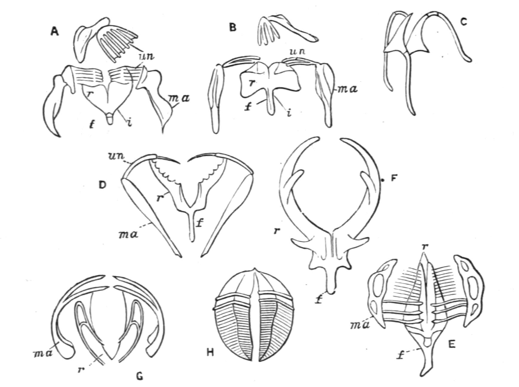
Fig. 111.—Diagram of trophi. (After Hudson.) A, Malleate; B, submalleate; C, virgate; D, forcipate; E, malleoramate (Melicerta); F, incudate (Asplanchna); G, uncinate (Stephanoceros); H, ramate (Rotifer). f, Fulcrum; i, incus; ma, manubrium (malleus in G); r, ramus; un, uncus.
The type we have just described is termed the "malleate" type (Fig. 111, A). If all the trophi are slender and scarcely toothed, we have the "virgate" type (C), which is frequently asymmetrical. In the "submalleate" type (B) the mallei only are slender; in the "forcipate" type (D) both the unci and rami are slender and sharply pointed.[257] In the "malleoramate" type (E) the manubrium is a curious looped structure, while the uncus is formed of a number of parallel slender elongated teeth; this characterises the family Melicertidae, and the genera Triarthra, Pterodina, and Pedalion. In the "uncinate" type (G) the mallei are simply incurved hooks with a few teeth at the free end, the rami are simple or absent, and there is no fulcrum; this type occurs in Flosculariaceae only. In Asplanchnidae the rami are large and hooked, constituting the "incudate" mastax (F); but here reduced mallei are often present, and in Asplanchnopus they are almost as well developed as in Melicertidae, affording a transition to the malleoramate type. In this group too the mastax has a very peculiar form; it is divided into two chambers, dorsal and ventral. The dorsal chamber forms a great purse-like sac or crop, with a framework of four longitudinal bars: into this the gullet and pharynx open. The ventral pouch is much smaller, and in its base the large rami are inserted, so that they can be protruded into the crop. This ventral sac with the rami may even be everted through the crop and the mouth, to swallow the small Rotifers and Entomostraca which form the food of this group, or to eject the undigested remains of the food. Two lateral sacs open at the junction of the ventral pouch and the crop, but whether they play a part in the deglutition of food or in the disgorging of faeces is uncertain. The fact that the whole of this apparatus is lined by a non-ciliated chitinous cuticle justifies our view that it is simply an enlargement and specialisation of the mastax.
The trophi in Bdelloids also are only represented by the rami, which have the form of segments of a sphere, excavated on the curved sides for the attachment of muscles, and transversely ridged on the two flat sides; the gizzard is here called "ramate" (H).
It will be seen that the characters of the gizzard are very useful for classification, only breaking down indeed in the Ploima; for though the majority of these present one or other of the four varieties of the malleate type, Triarthra and Pterodina (but not the other genera of their respective families) have the gizzard malleoramate.
The oesophagus is, when present, a contractile ciliated tube in which the food makes no sojourn on its way to the stomach.
The stomach may be nearly spherical, ovoid, or elongated and cylindrical. Its walls are formed of large cells, often granular and sometimes brownish, whence a hepatic function has been assigned to them. Its apertures are both surrounded by constricting muscular fibres. The intestine may be simple or divided by a similar constriction into intestine proper and rectum. The whole of the alimentary tract, with the exception of the mastax, is richly ciliated within. The rectum opens into the slender non-ciliated cloaca. The intestine is sharply bent upwards and towards the back in the tubicolous forms, but is nearly straight elsewhere; in Trochosphaera and Apsilus it is bent ventrally. In Asplanchnaceae and in Paraseison there is no rectum, the stomach being a blind sac.
The so-called salivary glands, usually two in number, open into the pharynx or mastax; and the paired gastric glands (Fig. 106, gg) open into the oesophagus or stomach. While the prehension of food is usually accomplished by the ciliary current of the disc and pharynx, we have seen that a more active swallowing action takes place in Flosculariaceae and Asplanchnidae, which devour whole Algae, Infusoria, and even other Rotifers, the long spines of Triarthra not availing as a protection. Many Ploima put out the tips of their trophi to nibble at débris, or, in the case of Diglena and Distemma, to attack Desmids, or the Infusorian Stentor. But this use of the trophi is most efficient in Ploesoma. Bilfinger[258] writes: "It has the courage to attack larger Rotifers; thus I was able to observe under the microscope how it fell upon a Rattulus but little smaller than itself and destroyed it. First it plunged the sharp prongs of its mastax deep into the tender frontal area of its unhappy victim; then followed a pumping action of the gizzard, and stroke by stroke the whole contents of the victim's body passed into the brigand's stomach." From this it is an easy transition to the ectoparasitism of Drilophagus, Balatro, and some species of Albertia, which cling to their host by the exserted trophi.
Renal Organs.—The kidneys consist of a pair of convoluted tubes, formed of a succession of perforated, so-called "drainpipe" cells (Fig. 106, k); they open directly or indirectly into the cloaca. Their walls are thin in the straight parts, but thick and glandular in the coils which occur at intervals. These tubes bear little tag-like appendages, hanging freely into the body-cavity, often widening towards the free end, and flattened or circular in section (Fig. 106, ns). They show during life a peculiar flickering motion in their interior, like the equivalent "flame-cells" of many Platyhelminthes (see p. 25), and are in function the representatives of the multicellular renal funnels of Annelids. On one side, especially on the edge of the flattened tags, the appearance is as of a tapering whip-like lash, attached by its base to the free end of the tag and waving in its cavity; but the side view of the flattened tags shows an appearance of successive transverse or oblique waves. In many if not all cases the free end of the tag is closed by a vacuolated plug of protoplasm, which sometimes at least bears two flagella waving freely in the body-cavity. The probable explanation of the two distinct wave appearances within the tag is that the protoplasmic plug bears on its inner face a row or tuft of long cilia hanging down into the cavity of the tag. The tags probably keep up a current of liquid through the kidneys, while the contents of the body-cavity are constantly replenished by osmosis.
The two renal tubes may end blindly below the disc, or else join by a short transverse dorsal communication in front of the brain, as in Stephanoceros, Atrochus (Fig. 112, C), and Apsilus among Flosculariaceae, Lacinularia among Melicertidae, and Hydatina among the Illoricate Ploima (Fig. 106, rc). In some species of Asplanchna, if not all, a recurrent branch occurs opening at either end into the main tube of its own side.
The kidneys unite to discharge into the cloaca near its orifice, and on its distal (primitively ventral) side in many Melicertidae. In Bdelloida the common duct formed by their fusion opens into the ventral side of a dilated bladder-like section of the cloaca (Fig. 109, A, bl), which contracts rhythmically to discharge the liquid; while in the majority of the class they open singly or by a common duct into a separate contractile vesicle or bladder, which also discharges at regular intervals into the cloaca on its ventral or distal side (Figs. 106, bl and 112).
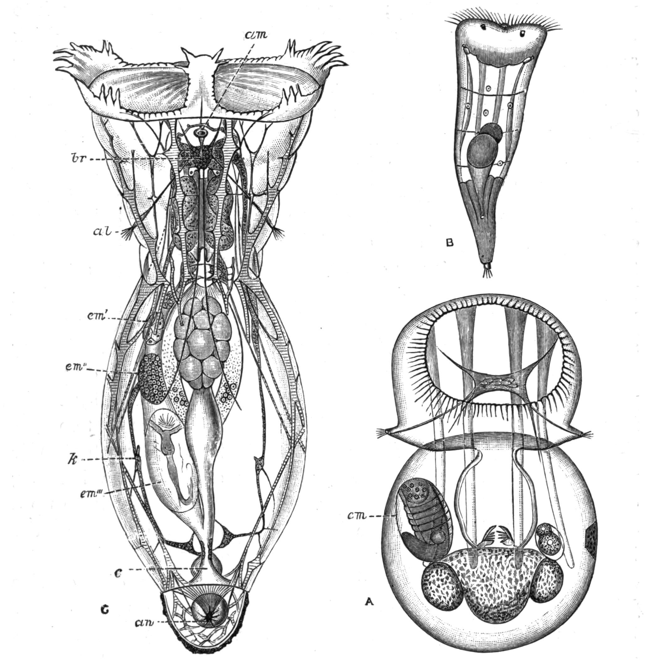
Fig. 112.—Apsilidae: A, Apsilus lentiformis, ♀, dorsal view (after Metschnikoff); the square brain is seen with nerves to the lateral antennae; B, larva of A. lentiformis (?), showing the paired eyes and ciliated cupped foot; C, adult of Atrochus appendiculatus, ♂ (after Wierzejski). al, Lateral antennae; am, median antenna (just in front is seen the renal commissure); an, anus; br, brain, below which the paired eyes are seen; c, cloaca; em, embryo; em', em', em''', three successive stages of embryos in the uterus of C; k, kidney. The coarser muscles are striated.
This bladder may reach when expanded one-third the diameter of the whole animal, and contract as often as three times per minute; so that in a period of nine minutes a bulk of water equal to that of the animal must have diffused through the body-wall, to be removed by the kidneys. It is obvious that while the function of the kidneys is primitively excretory, the passage of the water through the body must bring in the oxygen dissolved in the external medium, and carry off the carbonic acid formed in the tissues, and so fulfil the act of respiration. This mechanism is physiologically comparable with that of the contractile vacuole of fresh-water Protozoa. In a few genera (Conochilus, Lacinularia, Pterodina) the kidneys open separately after a slight dilatation into the cloaca.
Nervous System.—The nervous centre of the Rotifera is the brain (Fig. 112, C, br), a ganglion lying dorsal to the pharynx; and when this is short it may be immediately below the surface of the disc (Microcodon). In Bdelloida a second ganglion is present below the pharynx, and is connected with the former by lateral cords which contain ganglion cells. From the brain, nerves are given off to the disc, to the muscles, and to the integument of the body, as well as to the sense organs. The largest nerves are two given off from the sides of the brain, each of which divides into a lateral and a ventral trunk, which run nearly the whole length of the animal.
The brain of several Notommatidae has a curious appendage, white by reflected light and very opaque; it is a sac full of chalky mineral matter, which dissolves readily in dilute acids.
Sense Organs.—The most widely diffused sense organs are the antennae or feelers, which may serve for touch or smell, or possibly both. Each antenna is a conical or tubular outgrowth of the skin; from its apex projects a fine pencil of sense hairs borne on a protoplasmic cushion, which receives a nerve. Often the antenna is elongated, and may then contain a muscle by which it is retractile (lateral antennae of Melicerta); sometimes it is reduced to a slight prominence bearing the setae (dorsal antenna of this genus). There are usually three antennae—a median dorsal (Figs. 109, B, a, and 112, C, am) and two lateral (Figs. 106, 112, C, and 115, A, al), often approximated towards the ventral surface, and sometimes all but fused on the middle line, or completely united (Conochilus dossuarius, Copeus caudatus).[259]
Most Rotifers possess an organ of sight. This in its simplest form is a refractive globule seated in a red pigmented cup through which the nerve passes; in other cases it lies directly on the brain. Very frequently the eye is paired (Figs. 112, B, and 115, A); and these paired eyes may lie on the brain, and then are so close together that the pigment-cups have the shape of an x, or else they are seated in the dorsal region of the head behind the disc. In some cases they lie just under the ciliary wreath, or even within the region of the disc, and pass towards its ventral side in Pedalion (Fig. 117, A, e). In Rotifer they lie just under the dorsal side of the proboscis just below its apex. The median and two lateral eyes often exist together, as in Eosphora; and sometimes additional paired eyes exist. In Furcularia longiseta, var. grandis a pair of pigment spots (eyes?) occurs at the hinder end of the body just in front of the foot.
The active Ploima show a spontaneity of movement and marked power of avoiding obstacles, etc. This is still more marked in the very active Pedalion, which, as Rousselet notes, clearly avoids capture by the dropping tube, aided by its sense of sight, as he suggests, or by the tactile or olfactory powers of the antennae. They must rank as psychically high in the scale of creatures of simple organisation.
Reproductive Organs and Reproduction.—The most conspicuous organ in the female is the large yolk-gland or vitellarium (Figs. 106 and 109, A, vm), which was regarded as the ovary by all the older observers. It consists usually of eight cells, with conspicuous nuclei, lying on the ventral side of the stomach, and frequently displaced to one side; but in most Asplanchnidae it forms a broad transverse band of numerous cells. In Pterodina it is horseshoe-shaped, while in Seisonaceae and Bdelloida it is paired, either gland containing four or eight cells. The true ovary or germarium (Fig. 106, gm) lies more or less hidden between the yolk-gland and the stomach; it is composed of numerous minute rounded cells, of which the hindmost for the time being enlarges by nutrition from the yolk-gland, and finally receives a membranous shell. This true ovary is somewhat lateral in most Rotifers, but is median in Asplanchnidae, and paired in Pterodina, Bdelloida, and Seisonaceae. A membranous covering is common to the ovary and yolk-gland (paired when these are paired); it is continued into a thin-walled tube or oviduct, which opens into the cloaca on its ventral side beyond the bladder or common renal duct. In the viviparous species the mature ovum (Fig. 112, em) usually lies in the oviduct, dilating it into a sort of "uterus" until the birth of the young. The ordinary eggs or "summer eggs" are formed without any fertilisation, and develop immediately; they are often hatched within the tube of the tubicolous species.
Under certain conditions the unfertilised females produce exclusively smaller eggs, which develop into males. Maupas[260] has demonstrated that a rise in temperature to a minimum of 26° C. (79° F.) is the efficient factor. But as Bergendal points out,[261] the critical temperature probably varies with the antecedent conditions of the race, since males occur in Greenland at a very much lower temperature; and it would seem probable that a temperature approaching that at which the pools habitually dry up is what is necessary for the production of males, as a provision for those fertilised eggs, which, having a hard shell often adorned with prickly prominences, and usually remaining for some time before development, are capable of withstanding drought; such eggs are termed "winter eggs," but a better term would be "resting eggs" (German, "Dauereier").[262]
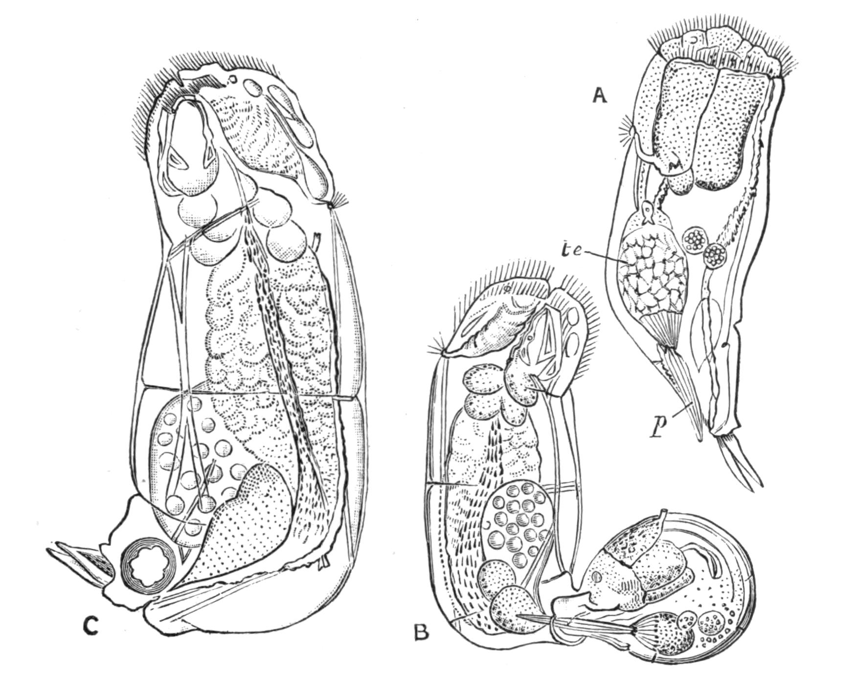
Fig. 113.—Diglena catellina. (After Weber.) A, Male; B, the pair in copula; C, female, p, Penis; te, testis.
The male organs consist of a testis (Fig. 113, A, te) with accessory glands, a large seminal vesicle, and a protrusible or projecting penis (p). In Notommata and Diglena true intromission at the cloaca (B) has been seen by many observers; but it appears equally certain that in many cases the male bores into the body-wall of the female at any point, and deposits the spermatozoa in the body-cavity, so that they must pass through the wall of the oviduct to effect fertilisation. Maupas finds that the process of fertilisation is ineffective except upon such newly-hatched females as would otherwise be the parents of small male eggs; that fertilisation is inoperative even for these at a later age when their eggs have begun to mature; and that it is wholly useless for those that lay ordinary summer eggs. The parent of male or winter eggs would thus be comparable to the queen bee, which if not fertilised produces drones. These sexual relations find a close parallel in the Ostracod and Phyllopod Crustacea, as well as in many plant-lice (Homoptera).
Development.—This has only been fully studied in the summer egg; in Brachionus by Salensky,[263] in Melicerta by Joliet[264]; in Eosphora digitata and several other species by Tessin[265]; in Callidina and Melicerta by Zelinka,[266] the last two observers having utilised modern methods of research.[267] We shall base our account on Zelinka's observations. As in the case of most "parthenogenetic" eggs, the ovarian egg begins by a very uneven division to form two cells: the minute "first polar body" which undergoes no further development; and the definitive egg, which by its repeated divisions gives rise to the tissues and organs.
Segmentation is very unequal, and recalls that of Molluscs in several respects. The first division gives rise to a smaller and a larger cell. Both of these divide again, the latter unequally, so that now there are three smaller cells and one large one; and after repeated divisions of the small cells and unequal divisions of the larger one, a stage is reached where there are a number of small cells and one large one, which sinks in and is overgrown by the small ones. Just prior to this the large cell undergoes equal divisions; its cells are the "hypoblast" cells (Fig. 114, hyp), and give rise to the gullet, stomach, and intestine, with their appendages, and the generative organs; while the smaller cells constitute the "epiblast" (ep), which gives rise to the body-wall and muscles, to the cement glands, nervous system, pharynx and mastax, and probably to the kidneys.
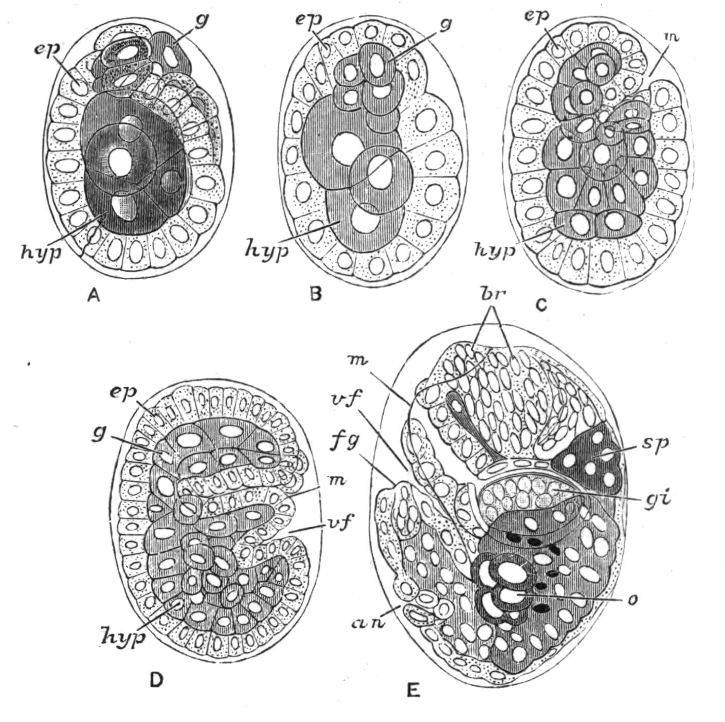
Fig. 114.—Development of Callidina. (After Zelinka.) A, Early stage showing involution of granular cells (g), to form the mastax or gizzard. B, Involution complete. C, Second involution of epiblast cells to form pharynx. D, The embryo bent on itself at ventral fold (vf). E, Showing ingrowth of epiblast to form brain (br): an, involution of epiblast to form cloaca; br, brain; ep, epiblast; fg, involution to form cement glands of foot; g, granular cells; gi, gizzard; hyp, hypoblast; m, mouth; o, ovary; sp, salivary glands; vf, limiting body from foot.
Owing to the elongation of the body within the narrow space of the egg the hinder part is bent up on the ventral surface (D, E); and this part, narrower than the rest, forms the foot, the centre of which is at first occupied by a column of hypoblast. The cloaca is now formed by a dorsal ingrowth of epiblast (the "proctodaeum") at the junction of the foot and the body (an). The hypoblast in the body anterior to the cloacal ingrowth forms the digestive apparatus; the part immediately behind forms the reproductive organs (o); and the hindmost part apparently disappears. An ingrowth of epiblast at the extreme tip of the foot gives rise to the cement glands (fg). The muscles arise from the epiblast cells. The disc arises from the modification of epiblast cells lateral to and behind the mouth, enclosing a so-called "polar area"; it is completed by the transformation of cells on the ventral side of the mouth. The brain (br) is formed by the multiplication of epiblast cells; and in Bdelloida a ventral ingrowth below the mouth forms the sub-oesophageal ganglion. The ciliated cup in Melicerta is formed as a ventral hollow, only later on united with the ciliated furrow of the wreath by the lateral grooves.[268] In Melicerta the two eyes are formed in the polar area. The young as hatched differs from the adult in the greater simplicity of its ciliary wreath; and in the tubicolous forms the cupped end of the foot-gland is ciliated, and two eyes are present on the polar area, which later sink in, and often disappear more or less completely. It is stated that the young hatched from winter eggs do not pass through this larval state.
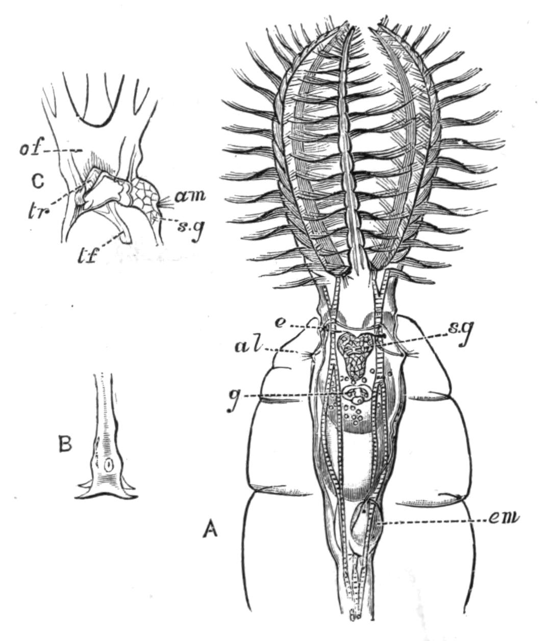
Fig. 115.—Stephanoceros eichhornii. (After Cubitt.) A, Dorsal view of the upper part in its tube: al, lateral antenna; e, eye; em, developing embryo in uterus; g, gizzard; s.g, median salivary gland. B, Extremity of foot. C, Lateral view of base of disc: am, median antenna; of, oral funnel; s.g, median salivary gland; tf, in the crop, indicates the ciliated tube prolonging the funnel; tr, horseshoe-shaped trochus.
Classification.[269]
Order I. Flosculariaceae.—Females mostly tubicolous, attached by a long contractile foot. Disc produced into a wide funnel-shaped contractile cup, produced into lobes with long setae (Floscularia) or coarse cilia (Stephanoceros), or entire (Apsilidae); an outer row of fine cilia rarely present; trochus a horseshoe, open behind. Oral funnel a slender tube hanging freely into a large pharyngeal crop; trophi uncinate projecting freely into the crop. Kidneys often united by an anterior cross-piece. Body-wall often containing a definite system of canals, filled with refractive granules, and serving by their contraction to dilate the disc. Males (Fig. 107, 1) and larvae vermiform with a ciliated pedal cup, and a simple wreath, with two eyes on the disc.
Fam. 1. Flosculariidae: Floscularia Oken, Stephanoceros E. (Fig. 115).
Fam. 2. Apsilidae: Apsilus Metschnikoff (Fig. 112, A), Acyclus Leidy, Atrochus Wierzejski (Fig. 112, C).
The family Flosculariidae contains some most exquisite forms; Stephanoceros, the "Crown Animalcule," being probably the most lovely of the Class, and many of the Floscules coming not far behind. The Apsilidae are mostly mud-dwellers.
Order II. Melicertaceae.—Females (except in Trochosphaera) attached or tubicolous; tube variable. Disc with a dorsal gap (except Conochilus) often two-lobed or corolla-like; a ventral lip often separating off a ventral ciliated cup continuous by a pair of gutters with the ciliated groove; trochus of stronger cilia than the cingulum. Trophi malleoramate in a distinct mastax. Intestine much curved dorsally, cloaca long eversible (except Trochosphaera). Males and larvae as in Order I.
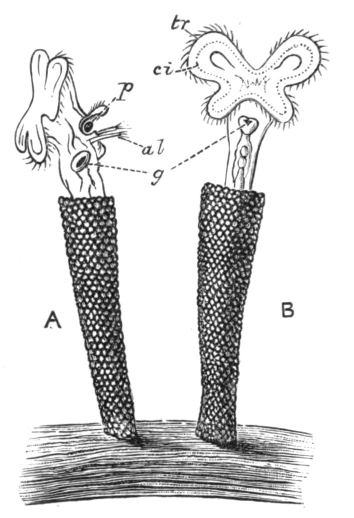
Fig. 116.—Melicerta ringens. (After Joliet). A, Side view; B, dorsal view. al, Lateral antennae; ci, cingulum seen by transparency; g, gizzard; p, pellet in ciliated cup, about to be deposited on edge of tube; tr, trochus.
Fam. 3. Melicertidae: Melicerta E. (Fig. 116), Limnias Schrank, Cephalosiphon E., Oecistes E., Lacinularia E., Megalotrocha E., Conochilus E., Octotrocha Thorpe.
Fam. 4. Trochosphaeridae: Trochosphaera Semper (Fig. 118, D).
The Melicertidae embrace a large number of tubicolous forms, many of which are social. This habit is especially noticeable in Lacinularia socialis, which forms a gelatinous incrustation easily seen by the naked eye; and in Conochilus volvox, which forms free-swimming globular aggregates, the young attaching themselves when hatched to the centre of the ball, and the ball splitting up into two as soon as undue pressure is exerted at the periphery by overcrowding. In this genus the eyes are very conspicuous in the adult, as they are in the similar free-swimming aggregates of Lacinularia racemovata.
Trochosphaera (Fig. 118, D) is remarkable for its peculiar spherical shape, the absence of a foot, the limitation of the viscera to the lower hemisphere, and the dorsal position of the ovary. But a reference to the figure will show that the outgrowth of a foot in the quadrant between the mouth and anus and the flattening of the upper hemisphere would bring its organs on the whole into close correspondence with those of the rest of the Order. It is recorded from South China, the Philippines, and North-East Australia, and has only been seen by Semper, the founder of the genus, and by Thorpe, who saw the male of the first species, and described a second.[270]
Order III. Bdelloida.—Females creeping like a leech, as well as swimming (males unknown), susceptible of desiccation and revival ("anabiotic"). Body telescopic at both ends. Disc (except in Adineta) chiefly composed of two dorsal lobes like kettle-drums, wholly retractile; a dorsal proboscis or trunk-like prolongation of the body ends in a ciliated, sensory, and adhesive cup used in crawling, and overhung by a pair of membranous flaps. Trophi ramate; brain with a ventral ganglion, forming a complete ring. Eyes, two on the proboscis or brain, or absent. Bladder a mere dilatation of the rectum. Foot often possessing blind spurs, as well as two or three retractile perforated toes, or forming a terminal disc perforated by numerous pores of the cement glands, rarely ciliated.[271]
Fam. 5. Philodinidae: Philodina E. (Fig. 110), Rotifer Schrank, Actinurus E., Callidina E. (Fig. 109), Adineta H.
This group is remarkable for the great resisting powers of its members to drought and to heat and cold when dried, a fact which may explain the absence of males, though Janson records the occurrence of winter eggs in four species of Callidina and in Adineta vaga. The body is often strongly pigmented; red in Philodina roseola, Callidina scarlatina, and C. russeola, yellow in P. citrina, Rotifer citrinus, and Discopus synaptae. Most of the species are dust- or moss-dwellers; some, such as Rotifer vulgaris, are equally common in organic débris in infusions, pools, and ditches. Discopus adheres to the skin of the Holothurian Synapta.
Order IV. Asplanchnaceae.—Females ovoid, footless except in Asplanchnopus. Disc often bearing a pair of antennae; circular, often prolonged at the margin into two rounded lobes, interrupted dorsally, depressed at the ventral side into a deep ventral funnel. Trophi incudate (virgate in Ascomorpha), mastax enlarged dorsally into a wide crop; stomach large, blind. Kidneys large, with a "recurrent duct" and numerous tags; bladder large. Brain large, with a median eye, and frequently paired smaller eyes at the base of the marginal processes of the disc; anterior antennae paired, relatively far back on dorsal surface. Males (Fig. 107, 5) relatively large, frequently found.
Fam. 6. Asplanchnidae: Asplanchna G., Asplanchnopus De Guerne, (?) Ascomorpha, Perty, (?) Dinops Western.
Order V. Scirtopoda.—Females of conical shape, with the body prolonged into hollow limb-like expansions (see p. 201) moved by strong muscles, and ending in branched setose fins like the limbs of Crustacea. Disc as in Bdelloids, but not retractile. Foot represented by two subventral toes, ciliated, inconstant or absent. Trophi malleoramate. Eyes two, latero-ventral, on the disc. Male (Fig. 107, 8) conical, with simple setae.
Fam. 7. Pedalionidae: Pedalion H. (Fig. 117),[272] Hexarthra Schmarda.[273]
Order VI. Ploima.—Free-swimming forms, more rarely parasites, often adherent by their trophi to a host. Disc variable, often bearing within the cingulum a number of lobes fringed with coarse compound cilia. Foot rarely absent, marked off by a sharp constriction. Mastax variable, rarely malleoramate, never incudate or uncinate. Intestine not blind. Males small.[274]
Sub-Order A. Illoricata.—Ploima with a soft flexible integument; disc variable; ciliated auricles sometimes present (Synchaetidae, Notommatidae); foot rarely absent; trophi usually malleate.
Fam. 8. Microcodonidae: Microcodon E., Microcodides Bergendal.
Fam. 9. Rhinopidae: Rhinops H.
Fam. 10. Hydatinidae: Hydatina E. (Fig. 106), Notops H., Hudsonella Zach., Cyrtonia Rouss.
Fam. 11. Synchaetidae: Synchaeta E.
Fam. 12. Notommatidae: Notommata E., Pleurotrocha E., Copeus G., Proales G., Furcularia G., Eosphora G., Triophthalmus E., Diglena E. (Fig. 113), Distemma E., Triphylus E., Taphrocampa G., Albertia Duj., Balatro Clap.
Fam. 13. Drilophagidae: Drilophagus Vejdovsky.
Fam. 14. Triarthridae: Triarthra E., Polyarthra E., Pteroessa G., Pedetes G.
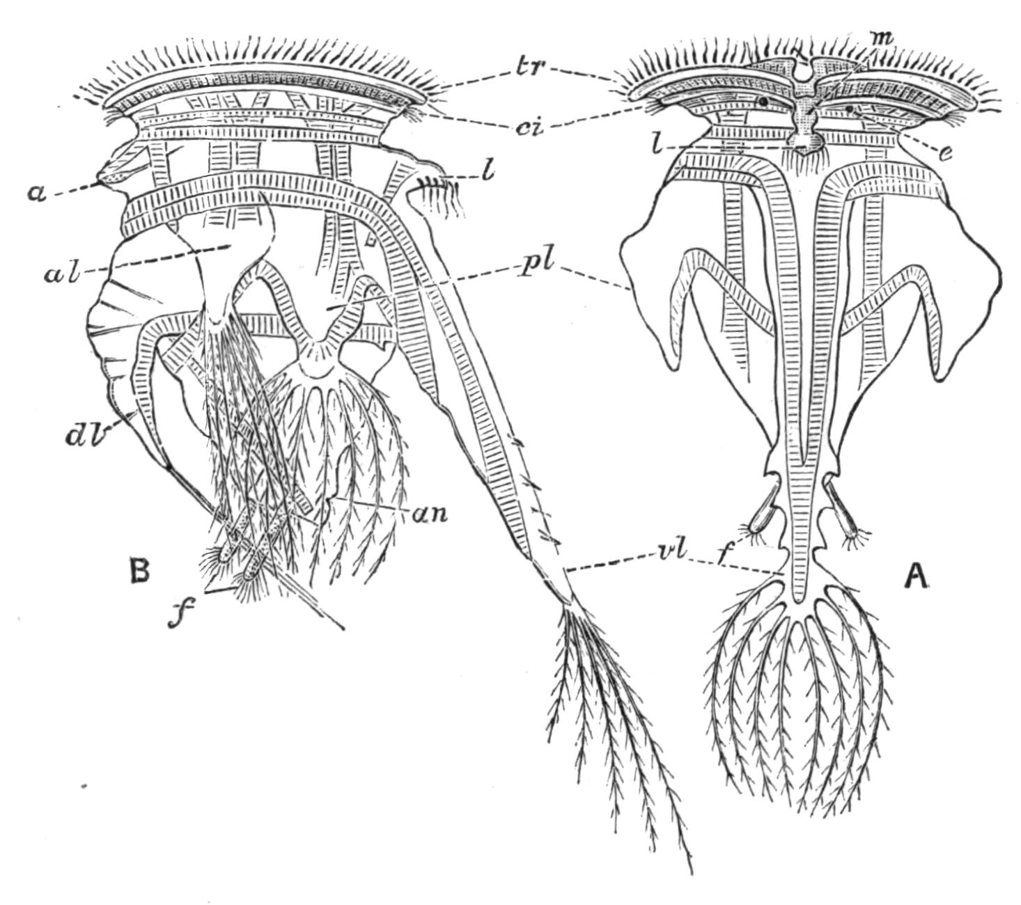
Fig. 117.—Pedalion mirum, female. (After Hudson.) A, Ventral view; B, side view. a, Median antenna; al, antero-lateral limb; an, anus; ci, cingulum; dl, dorso-median limb; e, eye; f, ciliated pedal processes; l, lip; m, mouth; pl, postero-lateral limb; tr, trochus; vl, ventro-median limb.
To this group belongs the eyeless Hydatina, a classical object of study, common in greenish pools, whose male was the first male Rotifer to be figured by Ehrenberg (1838), though he did not recognise its nature, and gave it the name of Enteroploea hydatina. Rhinops has the back of the corona curiously prolonged forwards into a sort of proboscis bearing two eyes. Some species of Notommata and Proales are distinctly annulated; in Taphrocampa the segmentation is so marked as to give the appearance of mesenteric septa extending inwards from the body-wall to the intestine. Microcodon has a wreath which is very peculiar in its extreme simplicity, with the mouth nearly central, and the eye lying just dorsal to the mouth. The Triarthridae, which resemble the Scirtopoda in having strong leaping spines fringed by fine bristles, should perhaps be placed in the next sub-Order.
Sub-Order B. Loricata.—Ploima with a firm elastic cuticle of definite form, persistent after death, continuous, or divided by thinner strips into plates or shields, which again may be areolated. The cuticle may also be shagreened or embossed in various ways.
Fam. 15. Rattulidae: Rattulus E., Mastigocerca E., Coelopus G., Diurella (?) Eyfurth.
Fam. 16. Dinocharididae: Dinocharis E., Scaridium E., Stephanops E.
Fam. 17. Salpinidae: Salpina E., Diaschiza G., Ploesoma Herrick, Diplax G., Diplois G.
Fam. 18. Euchlanididae: Euchlanis E., Dapidia G., Apodoides Joseph.
Fam. 19. Cathypnidae: Cathypna G., Distyla Eckstein, Monostyla E.
Fam. 20. Coluridae: Colurus E., Metopidia E., Monura E., Mytilia G., Cochleare G., Dispinthera G.
Fam. 21. Pterodinidae: Pterodina E., Pompholyx G.
Fam. 22. Brachionidae: Brachionus E., Noteus E., Schizocerca Daday.
Fam. 23. Anuraeidae: Anuraea E., Notholca G., Eretmia G.
The group includes a number of very minute forms, besides others conspicuous both for size and beauty. A soft dorsal flap above the head occurs in Stephanops; also in Coluridae, a large family of minute species, where the flap is movable, and looks in profile like a hook overhanging the forehead. The genus Pterodina, like Pedalion and Triarthra, combines a Bdelloid disc with malleoramate trophi, while its exsertile wrinkled foot ends in a ciliated cup like that of a larval tubicolous species.
Brachionus, a large, often flat, transparent form, with a long wrinkled foot, is a very common genus, known to the earlier observers, and repeatedly figured by them. Pompholyx has a sack-like lorica, no foot, and carries its immense egg suspended by an elastic thread from the cloaca. The Anuraeidae lack the foot, and often have great spines or bristles projecting from the lorica, which no doubt facilitate floating. They are abundant in the "plankton" or floating fauna of large lakes far from the shore. Many marine species belong to this family.
Order VII. Seisonaceae.—Marine Rotifers parasitic on the Crustacean Nebalia; males resembling the females. Body elongated, with a slender retractile neck, a much reduced disc, an elongated foot with a terminal perforated disc as in Callidina. Trophi virgate exsertile. Genito-urinary cloaca opening at the base of the neck in the male, at the hinder end of the body in the female. Intestine complete (Seison) or blind (Paraseison).[275]
Fam. 24. Seisonidae: Seison Grube; Paraseison Plate; Saccobdella Van Beneden and Hesse.
Habits.—The habitat of Rotifers is well known to the student of pond life. Every dip from a greenish pool will give us a supply, if there be not an excessive contamination by manure; and such pools give us some of the largest and most beautiful forms, such as Hydatina and Brachionus, swimming about among the fibrous Algae and feeding on the organic débris among them. Almost any organic infusions freely exposed to the open air will yield Ploima shortly after the active putrefaction is completed. The finer water-weeds yield most of the beautiful tubicolous forms. A whole group of species and genera are quasi-pelagic in fresh and salt water, constituting a large proportion of the "plankton" or floating life near the surface; and some of these are found in deep water or in the depths of the lakes. Among them are the Asplanchnidae, Triarthridae, and Anuraeidae. A number of Loricates, such as Notholca and Eretmia, are armed with long spines, which doubtless render floating easier.
Among tubicolous forms Conochilus volvox and Lacinularia racemovata have this pelagic habit, forming floating globular or ovoid colonies, and two species of Floscularia also float freely in their tubes.
The following forms occur in salt or brackish water,[276] those marked with an asterisk (*) also occurring in fresh water:—
Floscularia campanulata.* Melicerta tubicolaria.* Rotifer citrinus.* Discopus synaptae. Synchaeta baltica, S. monopus, S. apus, S. tremula,* S. longipes, S. tavina. Asplanchna girodi.* Asplanchnopus syringoides. Hexarthra polyptera. Notommata naias, N. reinhardti. Proales decipiens. Furcularia forficula,* F. gracilis, F. reinhardti, F. marina, F. neapolitana. Diglena catellina,* D. suilla, D. putrida. Pleurotrocha leptura. Distemma raptor, D. marinum, D. platyceps.* Bothriocerca longicauda. Polyarthra platyptera.* Triarthra longiseta.* Rattulus calyptus. Diurella marina, D. brevidactylus, D. brevis. Diaschiza fretalis. Euchlanis luna. Monostyla quadridentata, M. lunaris. Colurus amblytelus, C. uncinatus,* C. dactylotus, C. coelopinus, C. pedatus, C. rotundatus, C. truncatus, C. caudatus.* Mytilia tavina. Pterodina clypeata. Brachionus bakeri,* B. mülleri. Anuraea valga,* A. biremis,* A. aculeata,* A. tecta,* A. cochlearis.* Notholca striata,* N. scapha,* N. thalassia, N. spinifera, N. inermis, N. jugosa, N. rhomboidea. Seison grubei, S. annulatus. Paraseison asplanchnus, P. nudus, P. proboscideus, P. ciliatus. Discobdella nebaliae.
Thus about seventy species are recorded as marine. Synchaeta baltica is truly pelagic, and contributes to the phosphorescence of the ocean.
Other forms again are parasitic. Proales werneckii is found in Vaucheria, a coarse, dark green, thread-like Alga found in fresh water; and the closely allied P. parasita is not uncommon in the beautiful floating green spheres of Volvox.[277] Albertia, Drilophagus, and Balatro are parasitic on or in fresh-water Oligochaetes; the curious Seisonaceae are parasitic on Nebalia, a small Crustacean easily obtained in masses of whelk's eggs; the aberrant Bdelloid Discopus attaches itself to the surface of the Holothurian Synapta. Similarly among this last Order Callidina parasitica attaches itself to the limbs of the fresh-water Crustacea Gammarus and Asellus. These are rather commensals than true parasites. The species of Brachionus often attach themselves temporarily to the common water-flea Daphnia.
Besides a few Ploima, the vast majority of the Bdelloids live in or among mosses and their roots. Many Callidina inhabit cup-like hollows in the leaves of the scale mosses (Jungermanniaceae), especially of the genus Frullania. Almost all the members of this Order are susceptible of desiccation and revival; certain species, such as Rotifer vulgaris, Philodina roseola, Adineta vaga, etc., can be readily obtained by moistening gutter dust. The mechanism of the process is as follows: when desiccation is gradual the animals close up their telescopic bodies and excrete gelatinous plugs at either end, which effectually seal them against further drying; if, however, they be dried on a slide without any débris, the process is too rapid for them to protect themselves, and they therefore die. This was dimly seen by others, and clearly demonstrated by H. Davis,[278] who records the following experiment:—The Rev. E. J. Holloway, having found Philodina roseola in gutters, placed strips of paper there in the rainy season, and succeeded in obtaining clean gatherings, taking dry groups of a hundred together, having a varnish-like covering all over; and being glued to one another, mostly in one plane, and to the paper, forming a pavement. In the dry condition they resist extremes of temperature; thus Zelinka found Callidina revive after an exposure of -20° C. (-4° F. or 36° of frost), and immersion in hot water at 70° C. (158° F.). They will also resist deprivation of air in a vacuum of an ordinary air-pump, but not the all but perfect exhaustion of the Sprengel pump.
A very curious fact in relation to this Class is that often when a new form is once described from a single locality, fresh and widely distant stations for it rapidly become known.[279] Thus Pedalion mirum, first found at Clifton in 1872 by Hudson, was a few years after captured in a small pool above tide-marks on a rocky islet in Torres Straits. Since then it has been recorded from many different European stations, and a second closely allied species has been found in Finland. So a species of Ehrenberg's[280] was not seen again till within the last decade or so; but since then it has been independently found and described by six observers, who have given it as many distinct generic names. In the case of Pedalion it may well be that, as Hudson suggests, the species is of southern origin and has followed the flag, the winter egg being conveyed in dust by ships or travellers.
The above account of the habits gives the key to the collection of the various forms. The weed-loving species are collected with the weeds, and will keep with these in vessels if screened from direct sunlight and protected against dust. The free-swimming forms may be collected by sweeping with a net of fine gauze, with a bottle fixed in the bottom.
Except for their power of resisting desiccation, Rotifera are not very long-lived, and the males are especially short-lived; the most exact observations are those of Maupas on Hydatina. He found that the greatest age of the unfertilised female was thirteen days, during which it could produce some fifty eggs; the fertilised female lives for seven or eight days, producing about sixteen eggs; while the male dies in two or three days.
The preservation of Rotifers has been recently reduced to a fine art by Rousselet, who uses a solution consisting of cocaine hydrochlorate, 1 gramme; water, 50 cc.; and methylated spirit, 12 cc. This will keep without deterioration. When in use it must be diluted in the proportion of two volumes to three of water. This solution is added cautiously to the capsule in which the Rotifers lie, and they are watched till their ciliary motions slacken; when this happens a drop or two of osmic acid solution (½ to 1 per cent) is added; the Rotifers are then sucked up by a capillary pipette, and transferred to fresh water; and then into a solution of "Formaline" diluted to contain 2½ per cent of formic aldehyde. In this solution they are transferred to shallow cells, ground out of the centre of an ordinary glass slide, covered with thin glass, and sealed.[281] Other methods of preparing Rotifers for minute study will be found in the papers of Plate, Tessin, and Zelinka.
The zoological affinities of the Rotifers have long been a subject of keen interest. As early as 1851 Huxley[282] suggested that they represent a primitive form, preserved, with modifications, in the larva of Molluscs, Annelids and other worms, and Echinoderms. Similar views were later maintained by Lankester,[283] who termed the larva of Polychaets, etc., a "trochosphere," for which "trochophore" has been substituted in order to avoid confusion with the Rotifer Trochosphaera; Balfour,[284] Hatschek,[285] Kleinenberg,[286] and others have developed these views. Serious difficulties, however, arise in the detailed comparison of Rotifers with this type; and the special students of this Class have found it practically impossible to agree in the identification of the various parts, a difficulty especially felt in the case of the Rotiferan genus Trochosphaera, though this is just the one which presents the closest superficial resemblance to the Trochophore larva. I have been induced to take a view of the structure of Rotifers that brings it into close relationship with the lower Platyhelminthes, and with the more primitive larva of the Nemertines termed Pilidium (Fig. 60, p. 113). This is hemispherical, ciliated all over, with the mouth in a ventral funnel lined by fine cilia; while the edge is fringed with two rows of strong cilia, separated by a finely ciliated groove, like those of the ciliary wreath of a Rotifer.
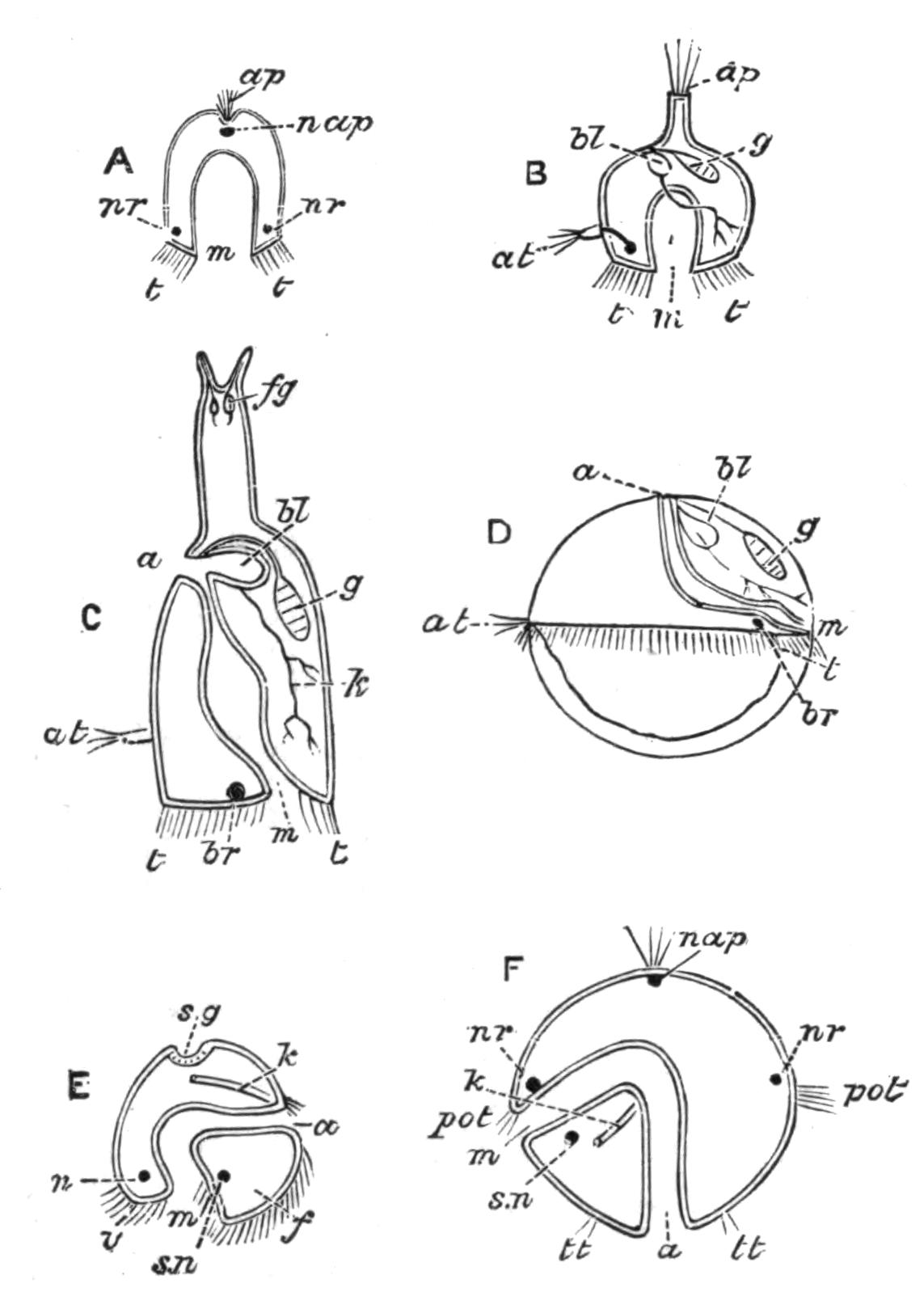
Fig. 118.—Diagram explaining the possible relations of Rotifers. A, Pilidium; B, hypothetical Rotifer modified from Asplanchnopus; C, a Ploimal Rotifer; D, Trochosphaera aequatorialis (modified from Semper, the extension of the ovary into the posterior ventral quadrant being omitted); E, Mollusc larva (Veliger); F, Trochophore larva of Annelid. a, Anus; ap, apical organ; at, median antenna (near which, in B, is a black spot, the brain); bl, bladder (receiving the ramified kidney in B, C, and D); br, brain; f, foot; fg, cement-glands, replacing apical organ; g, ovary; k, kidney; m, mouth; n, supra-oesophageal ganglion; nap, nerve of apical organ; nr, nerve-ring in section; pot, praeoral portion of trochus; s.g, shell-gland; s.n, sub-oesophageal ganglion; t, trochus or ciliary wreath; tt, posterior ciliated ring; v, velum, or expanded praeoral part of trochus.
The sides are produced on either side into lappets, which we do not take into account. A cup-shaped depression at the apical pole is lined by sense-cells, bearing long cilia which are probably sensory. A ring of nerve-cells passes within the ciliated rim of the hemisphere, and the stomach is a blind sac. If we compare this organism with a Rotifer, we find that the wreath corresponds in both, the funnel of the disc in such forms as Flosculariidae and Microcodon leading to the mouth of Pilidium, while the gut is blind in Asplanchnidae and in some of the highly developed Seisonidae. The circular nerve-ring of Pilidium is in many Rotifers only represented by its anterior part, the brain; though in Bdelloids a sub-oesophageal ganglion completes the ring. This leaves a difficulty with regard to the apical sense organ; but it is easy to understand that an organ of sensation should become an organ of fixation. In this case the foot with its glands would correspond to the sense organ of the Trochophore larva; and it retains its primitive ciliated character in the larvae and males of many Rotifera, and the adult female of Pterodina and Callidina tetraodon. Embryology tells us that the anus of Rotifers cannot be homologous with that of Annelids, etc., for it is formed outside the area of the blastopore: it is an independent formation, probably due to the coalescence of the originally blind intestine at its extremity with the earlier genito-urinary cloaca. On this view we must change the orientation of the Rotifer, and place it, like a Cuttlefish, mouth downwards: for "anterior and posterior" we must substitute oral (or basal) and apical; for "dorsal" and "ventral" we must use anterior and posterior; while "right" and "left" are unchanged. And this correctly expresses the actual space-relations in those Ploima like Rattulus that swim with their disc in contact with the organic débris on which they feed, with the foot turned outwards and backwards. As these views are now published for the first time, I have thought it wiser to keep to the accepted relations in the general description, a course which has the advantage of avoiding difficulties in the study of the literature of the Class.
The supposed resemblance of Pedalion to the Crustacea is probably the result of convergence, not of consanguinity. The Polyzoa are a group of freely-budding organisms whose structure otherwise recalls in many respects that of the attached Rotifers; but a close investigation reveals so many differences in structure, orientation, and development, that we cannot regard the two groups as at all closely allied.
Thus the Rotifers may be regarded as a group apart, but probably representing an early offshoot from a free-swimming Platyhelminth, probably a Rhabdocoele; the modifications being the loss of the general ciliation of the surface, the arching of the back into an elongated vault, the conversion of the inner half of the pharynx into a gizzard, the change of position of the genital and urinary apertures to the antero-dorsal surface, and the opening of the intestine into the genito-urinary cloaca.
Gastrotricha.
This small and very homogeneous group consists of minute fresh-water organisms, closely resembling many Ciliate Infusoria in their movements, habit and habitat. They were first described in detail by Ehrenberg, and placed by him and Dujardin in the neighbourhood of Rotifers. In recent years A. C. Stokes[287] in America and C. Zelinka[288] in Germany have contributed, the former a careful description of a number of new species and their habits, the latter a complete monograph of everything that is known of the Order.
The Gastrotricha dwell among filamentous Algae and organic débris, and are of frequent occurrence with Protozoa and Rotifera of similar habit. The largest known measures only 400 µ (1⁄60 in.) in length, and the smallest run as low as 74 µ (1⁄300 in.).
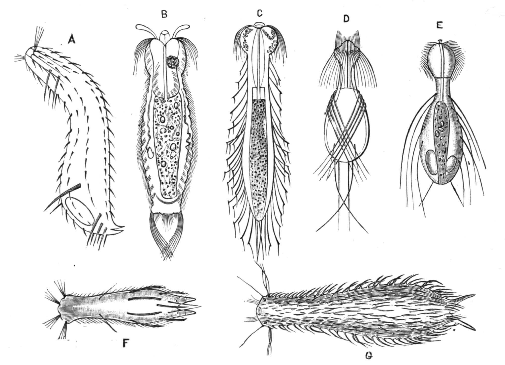
Fig. 119.—Gastrotricha. (From Zelinka.) A, Chaetonotus bogdanovii, side view (after Schimkewitsch); B, Gossea antenniger (after Gosse); C, Dasydetes goniathrix (after Gosse); D, Dasydetes saltitans (after Stokes); E, D. longisetosum (after Metschnikoff); F, Chaetonotus spinulosus (after Stokes); G, Chaetonotus schultzei (after Gosse and Bütschli). (Magnified.) B-F, × about 390; G, × about 125.
We shall follow Zelinka in his description of the common species Chaetonotus larus as a type. The body is nearly circular in section, flattened a little on the ventral side. The apertures are the terminal mouth; the anus, nearly terminal and slightly dorsal; the two kidney openings, ventral, nearly half-way down the trunk; besides the pore of a cement-gland on either terminal process. The short ventral and post-anal portion of the trunk with its processes therefore corresponds to the foot of a Rotifer. The integument of the body is a thin nucleated hypoderm, not distinctly divided into cells, covered by a chitinised cuticle; it bears cilia, sensory hairs, and peculiar scale-like processes, sometimes produced into long bristles.
The cilia are chiefly arranged in two ventral bands, each extending nearly the whole length of the body, and composed of a series of transverse rows of single cilia; along these bands the hypoderm is thickened and more richly nucleated. The sides of the head also bear numerous long cilia.
The scales are hollow processes of the cuticle overlapping from before backwards. A ventral row lies between the ciliary bands; two series of alternating dorsal rows lie on the back and sides of the animal, and in the hirsute species it is these that are produced backward into bristles. A single large scale, the "frontal shield," protects the head above and behind, but does not extend down to the ventral surface. On either side of the head is a pair of flattened oval areas, the "lateral fields." From between these on either side springs a tuft of motile sensory hairs. Two pairs of similar tufts arise dorsally on the front margin of the frontal shield, and a fourth pair spring from the ventral surface a little behind the mouth. These hairs are distinguished from ordinary cilia by their length, and their insertion on large nucleated cells receiving nerves; two pairs of similar hairs lie farther back on the dorsal surface, one in the front of the neck, one near the base of the pedal processes.
The muscles lie some in the body-wall, and some traverse the body-cavity; only six pairs occur, simple, unstriated, and longitudinal. There are neither transverse nor circular muscles.
The alimentary canal is very simple and nearly straight from mouth to anus; it may be divided into pharynx, gullet, stomach, and rectum. The mouth is circular, and looks forwards and a little downwards. From the mouth opens the pharynx, a short chitinous tube, capable of eversion by being pushed forwards by the gullet; it bears half-way down a circlet of curved hooks, which open out when it is everted; within these are tooth-like thickenings.
The oesophagus or gullet is thick and muscular, extending through the whole of the neck of the animal; its cavity, as well as the opening from the pharynx, is triradiate like a leech-bite, but can be dilated by the action of the muscular walls, inserted into a firm external cuticle; the internal wall is also cuticulised, not ciliated as in Rotifers. The hinder end of the gullet is produced into a short, wide, membranous funnel projecting freely into the midgut or stomach. The latter is elongated and oval, composed of four rows of hexagonal cells, with large nuclei. This is separated by a distinct constriction or sphincter from the short pear-shaped rectum, which opens by a minute anus on the back just in front of the pedal processes.
The food is chiefly organic débris; but Gastrotricha have been seen to attack large Infusoria by nibbling, and to swallow the protoplasm as it exudes from the wound in their prey.
The nervous system is chiefly composed of the large brain, a ganglion lying like a saddle above and on the sides of the gullet, and in direct continuity with the nerve-cells of the cephalic sense-hairs. A pair of dorsal nerve-trunks extend along the whole length of the gullet. The sense-hairs described with the general integument may be organs of external taste ("smell") or of touch. Eyes have been described in several species; and though Zelinka has failed to verify this, I have myself seen a pair of minute red eyes in the back of the head of an animal (probably a Chaetonotus), whose hasty escape into a mass of débris prevented my determining its species.
The kidneys are paired tubes lying at the sides of the front of the stomach, and sending a simple loop into the neck. Each tube is much convoluted, and ends at the one extremity in a long "flame-cell," like that of a Rotifer much drawn out, and at the other by a minute pore on the outer side of the ventral row of scales.
Reproductive Organs.—Only the female is with certainty known to occur; and the eggs, though recalling in their thick ornamented shell the fertilised winter eggs of Rotifers, are probably unfertilised and parthenogenetic like the summer eggs. The ovaries are two minute patches of cells lying at the junction of the stomach and rectum. The eggs, as they mature and enlarge, press against the side and back of the stomach, where they attain a length of one-third to one-half that of the mother. The extrusion of the egg has not been observed; but it is laid in the angles of weeds, the moulted shells of Entomostraca, etc., where its development may be studied. The sculpture of the shell serves to anchor it if laid among weeds. When hatched the head, trunk, and pedal processes are of the full adult size, all subsequent growth being limited to the neck.
The function of testis has been ascribed by Ludwig to a minute granular organ between the ovaries above the rectum; if this view be correct the Gastrotricha are hermaphrodite.
The movements of the Gastrotricha are very elegant, recalling those of the long-necked Ciliate Infusoria, like Amphileptus, Lacrymaria, etc., with the characteristic exception that they always swim forwards; the grace of their movements being due to the bending of the head and neck on the body. Those which are provided with long motile bristles like Dasydetes, alternate their gliding with leaps, like the springing Rotifers.
The Gastrotricha are divided into two sub-Orders—Euichthydina, with two pedal appendages, containing the genera Ichthydium Ehr., Lepidoderma Zel., Chaetonotus Ehr., and Chaetura Metsch.; and the Apodina, with no pedal appendages, comprising Dasydetes G. and Gossea Zel.
Their geographical distribution, like that of most microscopic fresh-water organisms, is cosmopolitan. Few observers have enumerated the members of this group; of their extra-temperate occurrence we have only the single observations of Ehrenberg, Schmarda, and Voeltzkow for Nubia, Ceylon, and Madagascar respectively.
Of the thirty-two species described, twelve are recorded by A. C. Stokes from Maine and New Jersey only, besides five others that occur also in Europe. In Europe nineteen species are recorded, one of which, Ichthydium podura, has also been found in Nubia and Ceylon. One species, Chaetonotus tabulatus Schmarda, has been recorded by its author from Colombia (in South America). As of the nineteen European species only seven have been recorded as British, we may expect to find that careful study will well repay the student in these islands.
The affinities of this group are probably with the Turbellarians and the Nematodes; they differ from the former in the highly developed alimentary canal, and from the latter in the possession of the ciliated ventral bands and wreath. The general chitinisation of the skin, the primitive body-cavity, the character of the alimentary canal, the ventral opening of the renal canals far in front of the anus are characters shared by the Nematodes, many of which possess bristles like this group. But their affinity must be rather to some hypothetical ancestral group than to any living Nematodes, which are destitute of cilia. To the Rotifers the affinity, dwelt on by Zelinka, is less close.
Kinorhyncha.
This Class and Order comprises but one genus, Echinoderes (Fig. 120), founded in 1851 by Dujardin.[289] Reinhard's monograph[290] is the generally accepted authority on this subject, and contains a full bibliography, with diagnoses of the individual species, eighteen in number.
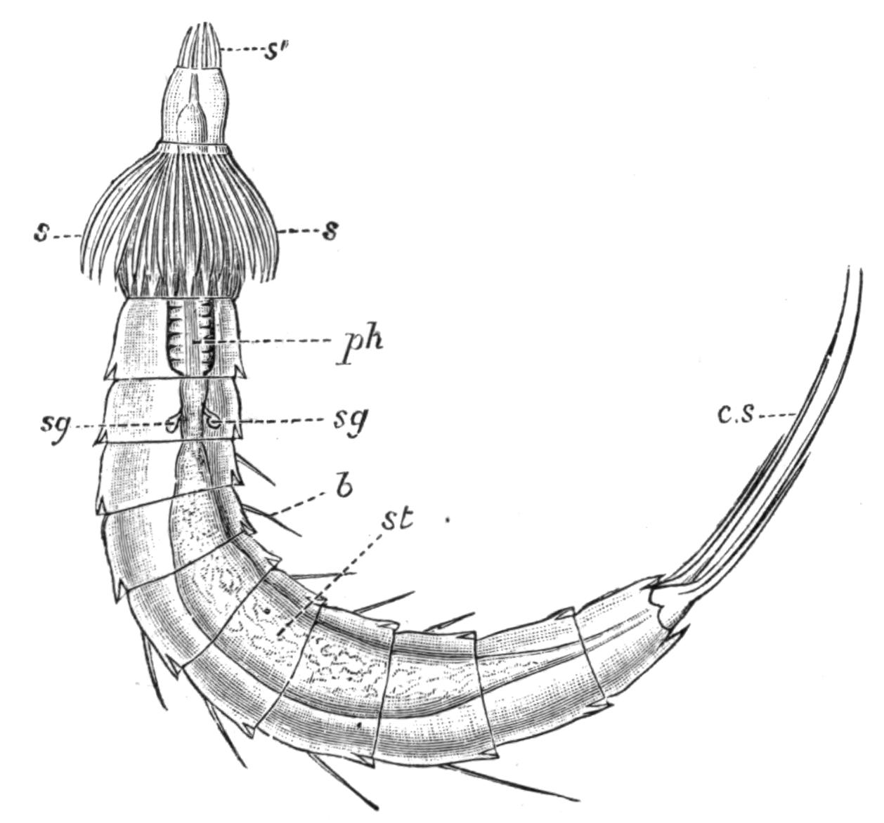
Fig. 120.—Echinoderes dujardinii (?), drawn from a preserved specimen taken at Worthing. × about 210. b, Bristle; c.s, caudal spine; ph, pharynx; s and s', the spines on the two segments of the proboscis; s.g, salivary glands; st, stomach.
The animals of this group are found in shallow seas with muddy bottom, below low-water mark, and feed on organic débris. They have been taken in the Black Sea, Mediterranean, British Channel, and North Sea, and off the Canary Islands (Lanzarote, Porto Pi, Palma di Mallorca). Their size varies from 0.86 mm. × 0.22 mm. in Echinoderes spinosus, to 0.14 mm. × 0.03 mm. in E. kowalevskii.[291]
The body is protected by a strong chitinous cuticle distinctly annulated, forming eleven rings, besides a retractile proboscis obscurely divided into two segments at the apex of which the mouth opens. The anus opens on the extreme end of the last segment, which is frequently retracted; the genital pores open right and left of the anus; and the renal pores lie on either side of the back of the ninth segment. The first ring may be undivided, or else distinctly divided into four plates, one dorsal, two latero-ventral, and one ventral. In the remaining segments each ring has only three plates, one dorsal and two ventral, the two latter being sometimes more or less fused in the last or ventral segment. These plates all overlap from before backwards.
As the name Echinoderes implies (Thorn-skin), the cuticle is produced into points, bristles, or spines. The last segment frequently bears a large pair of these, which have been compared, on the flimsiest grounds, with the furcal processes of Crustacea and the perforated toes of Rotifers and Gastrotricha.
The proboscis when extruded has the form of a truncated cone, obscurely divided into two segments, a ring of strong spines marking the boundary between them, and a second double ring of spines surrounding the apex. The eversion is of the type termed by Lankester pleurembolic or acrecbolic, the sides being first withdrawn, the apex first extruded.
As in so many Invertebrata, the epidermis is not separated by boundaries into distinct cells. This layer sends out processes each of which lies in a hollow in the thick cuticle, and perforates it to end in a fine bristle. Minute orange pigment-granules occur at irregular intervals in this hypoderm.
The muscles of Echinoderes are simple striated bands. Numerous bands lie within and attached to the body-wall, extending its whole length; paired dorsi-ventral muscles separate the intestine from the reproductive organ on either side, and a complex system effect the movements of the proboscis.
Alimentary Canal.—The pore at the tip of the proboscis leads into a short thin-walled tube, which is rarely evaginated; into the base of this tube projects the short bluntly conical apex of the large ovoid muscular pharynx (or gullet?); this is lined by an epithelial layer of nucleated protoplasm, which secretes a strong cuticle. The stomach is a wide tube, somewhat dilated in each segment between the paired dorsi-ventral muscles, and tapering behind to end in the terminal anus. Four minute glands open at the junction of the pharynx and stomach.
Kidneys.—These are a pair of blind pear-shaped sacs, ciliated within (the only case of ciliation in Echinoderes), lying in the eighth segment, and opening by the taper ends right and left on the back of the ninth segment.
Nervous System.—All that has been clearly defined of this is a small brain or ganglion lying dorsally at the junction of the pharynx and stomach. From two to eight eye-spots have been described by earlier writers, but Reinhard was unable to find them in the (distinct) species which he principally worked at, though he noted their existence in the solitary specimen of the original species, E. dujardini, which he obtained.
Reproductive Organs.—The sexes are distinct. The reproductive glands form a pair of tubular sacs, opening ventrally on either side of the anus, and extending forwards beside the gut as far forwards as the fifth to the second segment in the male, but only to the fourth at furthest in the female. The ova are large nucleated cells embedded in the protoplasmic lining of the ovarian sac, and acquiring a distinct shell as they approach its opening. Three-quarters of the testis sac is occupied with granular protoplasm containing a quantity of small nuclei; the lower part alone contains mature spermatozoa. Adjoining each external opening in the male are a pair of short hollowed spines, which may perhaps serve as organs of copulation; but nothing is really known of this process or of the development of the egg. It is almost certain, from the absence of developing eggs within Echinoderes, that the genus is not viviparous.
From the foregoing description it is obvious that Echinoderes approaches the Nematoda very closely: the two main points of difference are its ciliated kidneys and its bilaterally paired sexual organs. Possibly the study of such forms as Desmoscolex (Fig. 81, p. 159) may reveal closer affinities.
[Zelinka (Verh. D. Zool. Ges., 1894 and 1898), has given a preliminary account of a new research on this group. The principal addition is the discovery of a ventral nerve-cord, with a ganglionic dilatation in each segment, lying in the ectoderm of the body-wall, as indeed do the brain and nerve-collar. He divides the genus into two Orders according as the orifice of the retracted fore-part of the body is slit-like or circular. The former (Homalorhagae) retract the first two segments with the proboscis; they are mud-dwellers, sluggish, eyeless: the latter group (Cyclorhagae) only retract the first segment with the proboscis; they crawl among algae, and mostly have paired pigmented eye-spots, each with a lens, imbedded in the brain.—M. H., Jan. 1901.]