Chapter VI · (continued 2)
Reading edition
In addition to the larva of Sialis lutaria, the first larval stage has also been found in the larva of Ephemera, Tanypus, Corethra, and Chironomus; the second in Carabus hortensis Fabr., Procerus (Carabus) coriaceus Linn., Calathus fuscipes Goeze, Molops elatus Fabr., several species of Pterostichus, and a number of other beetles. It is probable that its normal hosts are S. lutaria and Pt. niger, but it is clear that it often comes to rest in other insects. The view that the Gordiidae have no special hosts, but may either pass the whole of their life-history within one and the same animal, or, on the other hand, may inhabit animals belonging to very different groups, is held by Villot, who has paid great attention to the subject. He finds the first larval form encysted in the walls of the alimentary canal in fishes, such as Leuciscus phoxinus, the minnow, Cobitis barbatula, the loach, and Petromyzon planeri, the lamprey; in the larvae of Diptera, Ephemera, and beetles, in Planorbis (a water snail), in Enchytraeus (an Oligochaet); the second larval form in all kinds of insects, spiders, Crustacea, fish, frogs, birds (Otis), and in man, and these various habitats lead him to the conclusion that "Les Gordiens n'ont pas d'hôtes spéciaux." On the other hand, as von Linstow points out, it is contrary to our knowledge of parasites that a single species should develop equally well in the body of warm and cold-blooded Vertebrates and of Insects, and the explanation of the presence of the larvae in these various forms may either be that they belong to different species of Gordius or, more probably, that they are accidentally present, having passed into their hosts with drinking water.
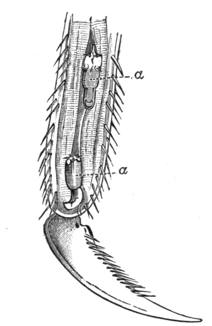
Fig. 91.—Tarsal joint of an Ephemerid larva into which two Gordius larvae (a, a) have penetrated. Magnified. (From G. Meissner.)
The number of species of Gordius is large; over 100 are enumerated in the Compendium der Helminthologie,[214] the great majority of which inhabit insects.
The life-history of Nectonema is practically unknown; the adults have been found swimming near the surface of the sea at two places only: Newport, R.I., and Wood's Holl, Mass., on the south coast of New England. It has been fished close to the shore, from the end of June to the beginning of October, when the tide is going out at evening and there is no moon. This seems to indicate that it avoids the light. When first caught the worms move actively about, coiling themselves into figures of eight and then uncoiling; at the same time there is a rhythmical movement caused by waves of muscular contraction passing down each side of the body alternately; by this kind of motion they make rapid and definite progress through the water.
It seems probable that the adult Nectonema is preceded by one or more larval stages, and what appears to be a young form has been obtained from the thoracic cavity of a prawn, Palaemonetes,[215] which has thus some claim to be regarded as the host of this species, but nothing is known about its early life-history.
Sub-Order III. Acanthocephala.
The Acanthocephala, which form the third class of the Nemathelminthes, consists of but few genera; there are, however, numerous species of very different size, varying from 10 to 65 cm. long in the female Gigantorhynchus (Echinorhynchus) gigas, to quite minute forms a few millimetres in length. The adult stage occurs in the alimentary canal of Vertebrates, as a rule in those which live in, or frequent water; the larvae are found in the bodies of certain Invertebrates, very frequently small Crustacea.
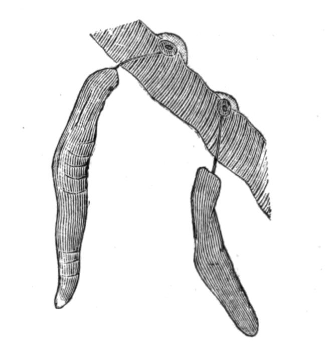
Fig. 92.—Two specimens of Echinorhynchus proteus Westrumb., with their anterior ends embedded in the wall of the intestine of a Pike. Magnified with a lens. (From Hamann.)
Anatomy.—The body of the mature forms can usually be divided into three sections—the proboscis, the neck, and the trunk, but the middle region is not always discernible. The proboscis is armed with rings of hooks (Fig. 93) arranged in longitudinal rows; they are usually of two kinds, but in E. proteus of three. They have a certain specific value, but not much stress can be laid on the number of rings, e.g. in E. angustatus the number varies from eight to twenty-four. The recurved hooks serve to fasten the parasite very firmly to the tissues of the host. The proboscis is hollow and retractile; it can be withdrawn into the body by means of muscles attached internally to its tip. It does not, however, pass straight into the body-cavity, but is retracted into a special cavity—the proboscis sheath—with a double muscular wall. The proboscis sheath may perhaps be looked upon as a septum, such as is found in some of the Nematomorpha, dividing the body-cavity into two parts. It is inserted into the body-wall at the junction of the neck and trunk or of the proboscis and trunk. In addition to the muscles which withdraw the proboscis into its sheath, there are two retractors running from the outside of the sheath to the body-wall; these serve to retract the whole sheath and its contents into the body-cavity of the trunk.
The structure of the skin is essentially like that of Nematodes, but the details are much more complicated. The whole body is covered by a thin cuticle secreted by the epidermis, which, as in the other groups, breaks down and forms a syncytium called the sub-cuticle. The minute fibrils which penetrate this layer are much more definitely arranged than in Nematodes; the largest of them run from without inwards, others run concentrically round the body. Large oval or spherical nuclei are scattered in the sub-cuticle, which is further honeycombed by a number of lacunae or spaces which are described below.
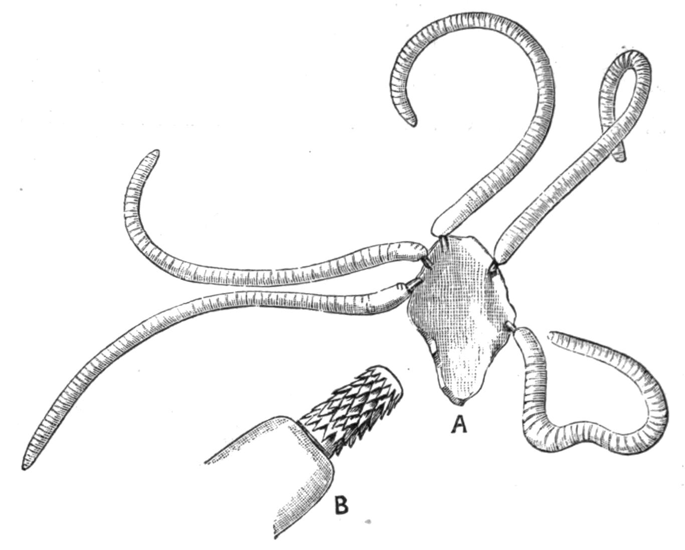
Fig. 93.—A, Five specimens of Echinorhynchus acus Rud. attached to a piece of intestinal wall, × 4; B, the proboscis of one still more highly magnified.
Within the sub-cuticular layer is found a sheath of circularly-arranged muscle-fibres, and within this again a sheath of longitudinal muscles which do not extend into the proboscis; this inner layer lines the body-cavity, there being no epithelium within it. In their minute structure the muscle-cells resemble those of Nematodes.
The canals in the sub-cuticle form a very curious system of anastomosing spaces, in which a clear fluid containing fat globules circulates. The extent to which the system is developed varies in different species, but in all there is a pair of longitudinal canals which are situated laterally, and which give off the subsidiary channels in their course. The above description applies to the lacunar spaces in the skin of the trunk; those of the proboscis are quite distinct, and there is no communication between the two sets of spaces; in fact, the sub-cuticle in which the lacunae are formed is not continuous across the line of junction of the proboscis and the neck, or, when the latter is absent, of the proboscis and the trunk, but it is interrupted by the ingrowth of a thin ring of cuticle which reaches down to the muscular layers (Fig. 94).
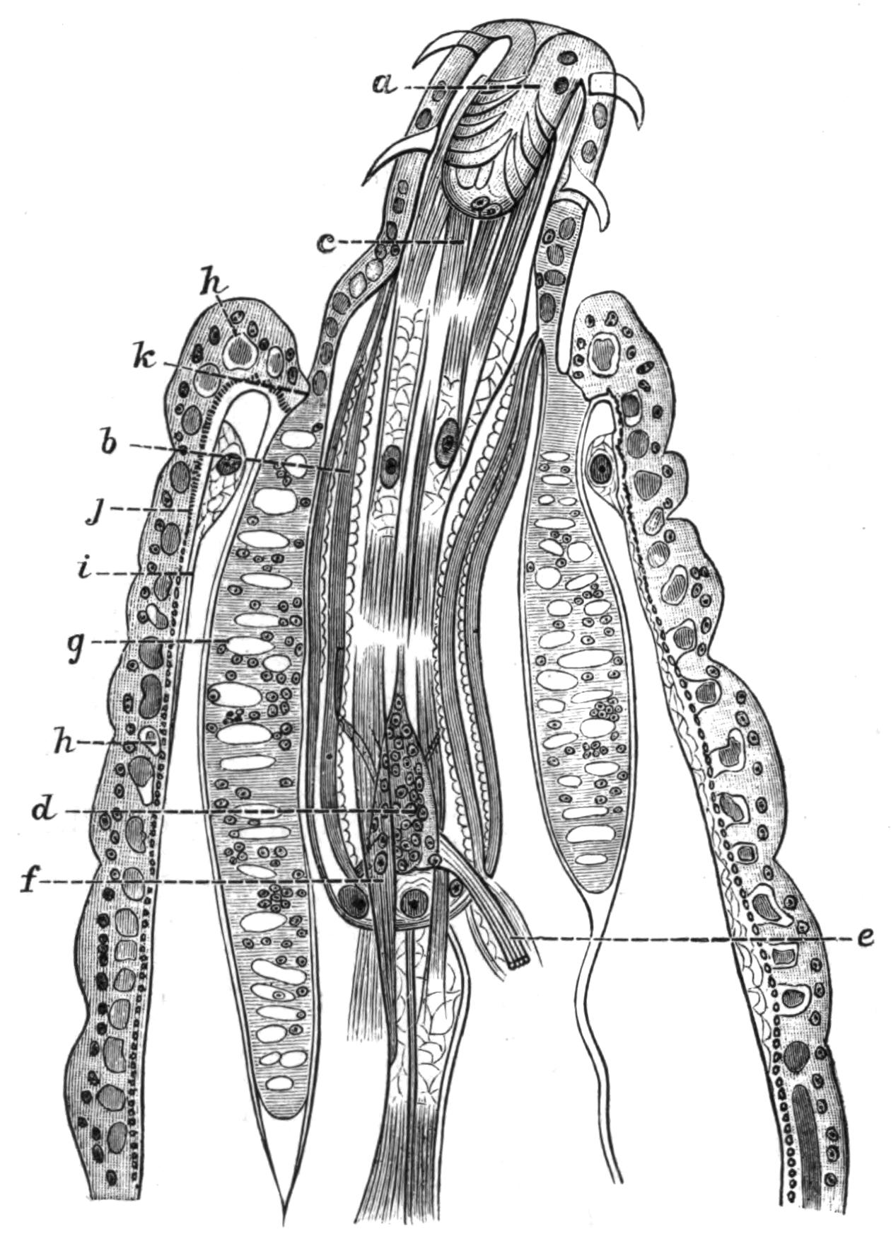
Fig. 94.—A longitudinal section through the anterior end of Echinorhynchus haeruca Rud. (From Hamann.) a, The proboscis not fully expanded; b, proboscis-sheath; c, retractor muscles of the proboscis; d, cerebral ganglion; e, retinaculum enclosing a nerve; f, one of the retractors of the sheath; g, a lemniscus; h, one of the spaces in the sub-cuticular tissue; i, longitudinal muscular layer; j, circular muscular layer; k, line of division between the sub-cuticular tissue of the trunk and that of the proboscis with the lemnisci.
All the spaces in the skin of the proboscis open ultimately into a circular canal situated round its base; on each side the canal opens into a sac-like structure which extends through the body-cavity towards the posterior end of the animal. These two lateral diverticula are termed the lemnisci. They have always attracted considerable attention from the workers at the group, and numerous functions have from time to time been attributed to them. They are more or less hollow, and their walls consist of sub-cuticular tissue surrounded with a scanty muscular coat; they contain the same fluid as the lacunae of the skin of the proboscis, with which they are placed in communication by means of the circular canal; and it seems most probable that, as Hamann[216] suggests, they act as reservoirs into which the lacunar fluid retires when the proboscis is retracted, and which, by means of the contractions of their muscular coat, force the fluid into the lacunae when the proboscis is everted, and thus aid in its protrusion.
The parasitic habits of Echinorhynchus have had a deeper influence on the structure of the body than is the case with the Nematoda. All traces of an alimentary canal have disappeared, and the animals live entirely by the imbibition through the skin of the already elaborated fluids of their hosts. The power of absorbing fluids is shown by the fact that they swell up and become tense when placed in fresh water.
Until recently no definite excretory organs had been recognised, and the function of excreting the nitrogenous matter was by some assigned to the lemnisci. In 1893 Kaiser[217] described in G. gigas two organs which he called nephridia, placed dorsally to the ducts of the male and female reproductive organs. Each nephridium, which somewhat resembles a cauliflower, consists of a stalk or duct, opening at one end into the reproductive ducts, and at the other branching and breaking up into a number of secondary and tertiary twigs. The end of each twig is closed by a membrane pierced with a number of most minute pores, by means of which it communicates with the body-cavity; on the inner side the membrane bears a number of long cilia, which keep up an active flickering. The presence of these cilia is interesting, as elsewhere they are unknown throughout the Nemathelminthes.
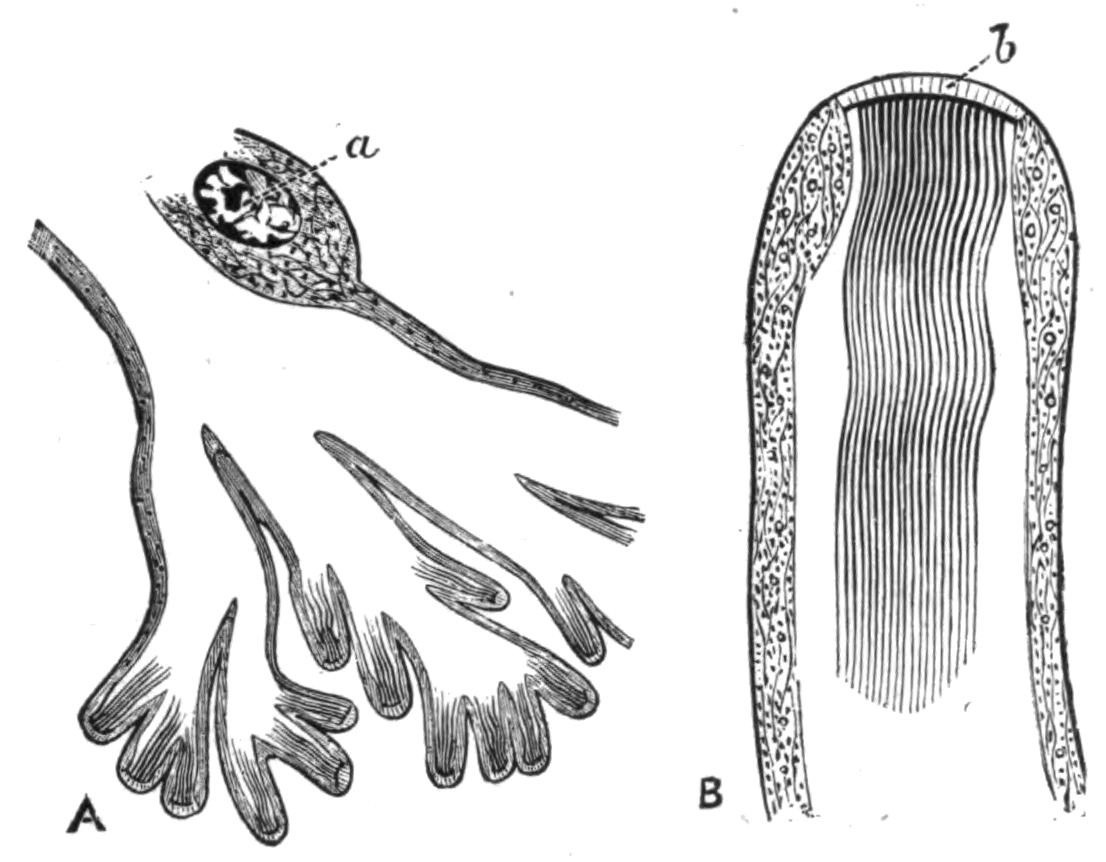
Fig. 95.—A, A longitudinal section through the terminal twigs of the nephridium of Gigantorhynchus gigas. (From J. E. Kaiser.) Highly magnified. a, Nucleus. B, A terminal twig more highly magnified; b, the porous membrane.
The nervous system consists of a central ganglion situated in the proboscis sheath; it is oval and flattened in shape. The ganglion gives off nerves to the proboscis, and two main trunks which pierce the proboscis-sheath and run backward surrounded by a cluster of muscle-fibres, the whole being termed the retinaculum; in the male they are in connexion with a special genital ganglion which lies near the ductus ejaculatorius.
With the exception of certain sensory papillae in the neighbourhood of the male genital orifice, and of three similar papillae mentioned by Kaiser on the proboscis, the Acanthocephala are devoid of sense organs.
The Acanthocephala are dioecious; their generative organs are developed in connexion with the ligament, a cord-like structure which arises between the inner and outer layer of the hinder end of the proboscis sheath and traverses the body-cavity, ending posteriorly in connexion with the genital ducts. The testes lie in this ligament; they are paired oval bodies which open each into a vas deferens. The vasa deferentia each bear three lateral diverticula, the vesiculae seminales; and three pairs of cement glands pour their secretion into a duct which opens into the vasa deferentia; the latter unite and open by a penis which is withdrawn into a genital bursa, but is capable of being extruded.
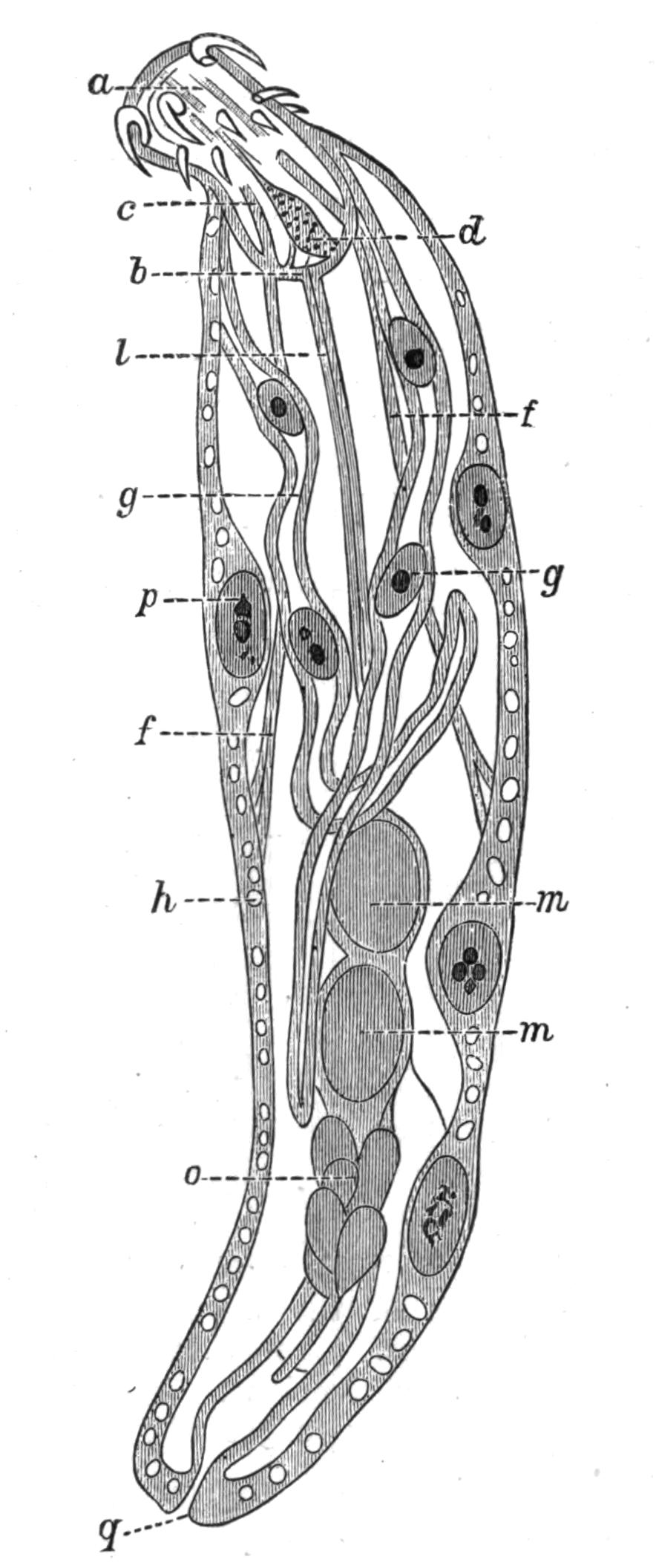
Fig. 96.—An optical section through a male Neorhynchus clavaeceps Zed. (From Hamann.) a, Proboscis; b, proboscis sheath; c, retractor of the proboscis; d, cerebral ganglion; f, f, retractors of the proboscis sheath; g, g, lemnisci, each with two giant nuclei; h, space in sub-cuticular layer of the skin; l, ligament; m, m, testes; o, glands on vas deferens; p, giant nucleus in skin; q, opening of vas deferens.
The two ovaries are formed in the ligament of the female in a corresponding position to that occupied by the testes in the male, but at an early stage they break down into packets of cells, of which those of the peripheral layer develop into ova at the cost of the central cells, which serve them as a food supply. As these masses grow and increase in number they rupture the walls of the ligament, and escape into the body-cavity, in which they float. The ova are fertilised whilst floating in the fluid of the body-cavity. The eggs segment and the embryo is formed whilst still in the body of the mother.
The embryos escape by means of a complicated apparatus the details of which vary in the different species, but which, like many of the organs in these animals, consists of very few cells with very large nuclei. This apparatus consists of three parts: the bell, the uterus, and the oviduct. The bell is a large funnel-shaped structure, which opens into the body-cavity, and is connected with the end of the ligament; near its lower end, where it is continuous with the uterus, is a second smaller opening situated dorsally. By the contraction and expansion of its lips the oval embryos are swallowed and pass on through the uterus to the oviduct, which opens at the posterior end of the body. If the bell takes in any of the less mature eggs which are spherical in shape, they are passed back into the body-cavity through the above-mentioned dorsal opening, and the same orifice permits the passage of the spermatozoa even when the bell is full of embryos.
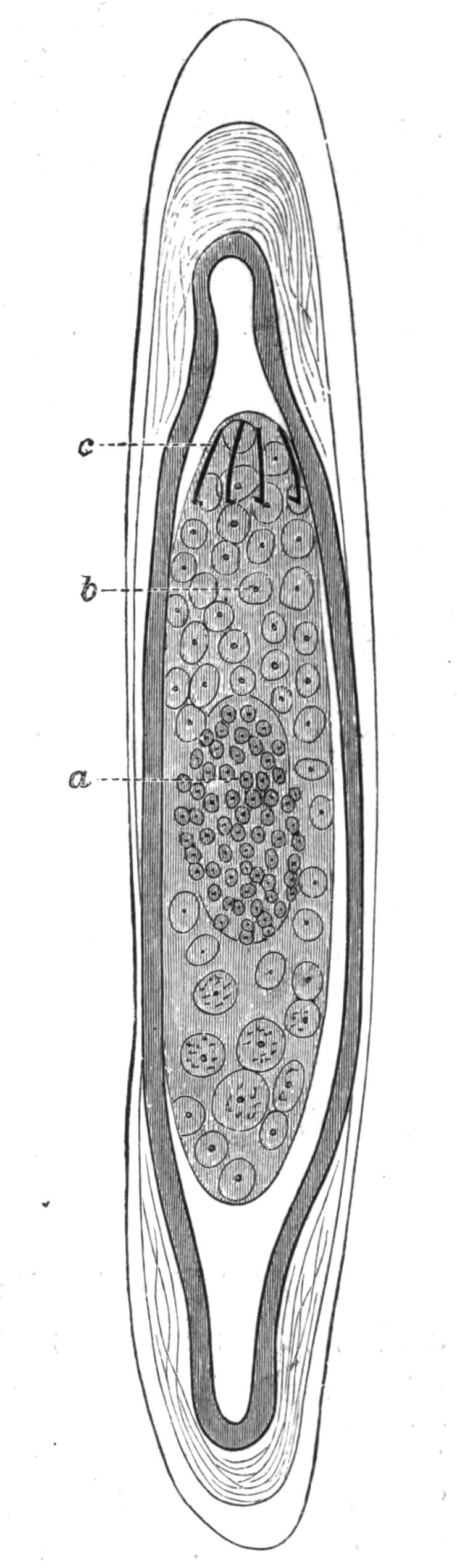
Fig. 97.—An egg of Echinorhynchus acus Rud. surrounded by three egg-shells. Highly magnified. The egg has segmented, and the cells are differentiated into a, the entoblast, and b, the ectoblast; c, spines. (From Hamann.)
Embryology.—After fertilisation the egg surrounds itself with several egg-shells, three of which are usually distinguished; the embryo is already far advanced in its development by the time it leaves the body of the mother and passes out into the alimentary canal of the Vertebrate host. It leaves the body of this second host with the faeces, and is eaten by the first or larval host, usually a small Crustacean or water-insect, but in some cases a fish, within whose alimentary canal it casts its membranes and becomes actively mobile. By means of a ring of hooks developed round the anterior end it bores its way through the wall of the alimentary canal, and after some time—three weeks in E. proteus—comes to rest in the body-cavity of its host. By this time most of the organs of the adult, with the exception of the reproductive glands, are already well established; the latter only attain maturity when the first host is eaten by the second, and the larvae find themselves in the intestine of a Vertebrate.
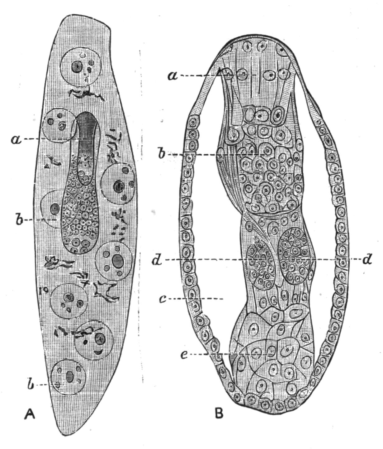
Fig. 98.—A, A larval Echinorhynchus proteus Westrumb. further developed than in Fig. 97. Highly magnified. The entoblast has developed inside it the proboscis a; b, b, the giant nuclei of the ectoblast. B, The entoblast at a more advanced stage, the ectoblast is not shown. The outermost layer of cells will form the muscles of the body-wall; the body-cavity has appeared; a, proboscis; b, cerebral ganglion; c, body-cavity; d, d, the testes beginning to appear in the ligament; e, cells which will form the generative ducts.
Some of the details of the development are very remarkable, and a short account of them may be given. The segmentation of the egg is unequal; it results in the formation of a central biscuit-shaped mass of small cells and a peripheral mass of larger cells; the former is called by Hamann[218] the entoblast, the latter the ectoblast. From the entoblast arise all the organs of the body but the sub-cuticle and the associated lemnisci, which are formed from the ectoblast. The latter has a remarkable history; the cells begin to break down and lose their outlines, whilst their nuclei fuse together and form a small number of giant nuclei, which lie scattered throughout the syncytium thus formed. The syncytium surrounds the entoblast on all sides; by this time the anteriorly-placed hooks have appeared; in E. proteus there are ten of these, but the number is not the same in all species. The syncytium is in a fluid state, with a few gigantic nuclei floating in it; these now lose their spherical shape, and throwing out processes become amoeboid; in this way they bud off small portions of their substance, and from these the oval nuclei of the sub-cuticle and the lemnisci arise. The rest of the syncytium hardens into the fibrillar matrix of the sub-cuticle, leaving, however, scattered spaces which form the sub-cuticular sinuses of the adult. An interesting feature of N. clavaeceps and Arhynchus hemignathi is that the skin of the adult retains the larval features, and it and the lemnisci consist of a syncytium with a very few giant nuclei scattered through it. Hamann counted only eight in the skin and two in each lemniscus in the example figured on p. 178.
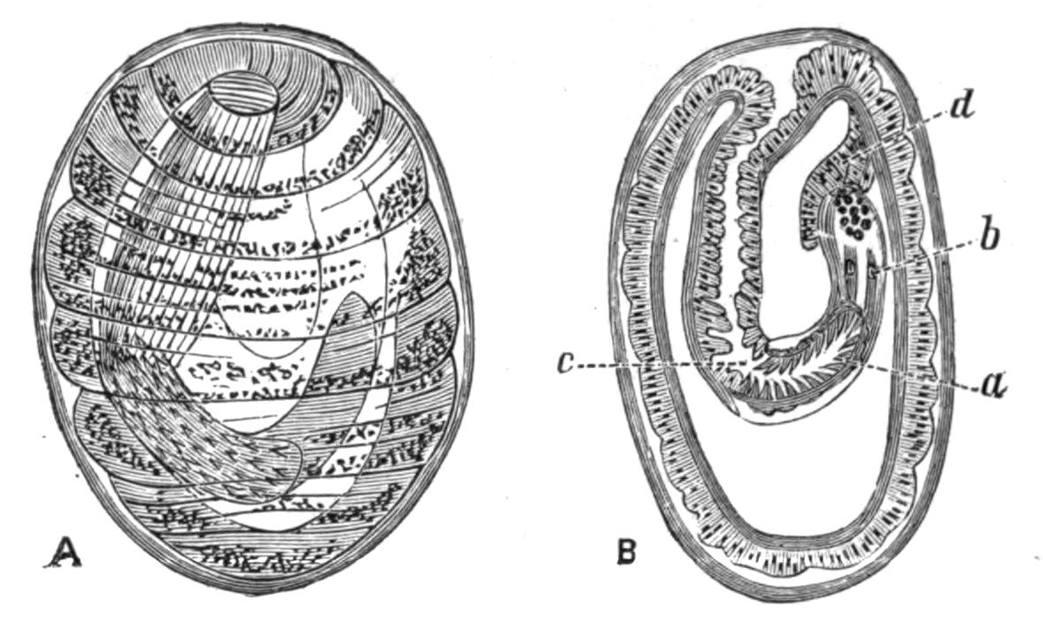
Fig. 99.—A, The larva of Echinorhynchus proteus from the body-cavity of Phoxinus laevis, with the proboscis retracted and the whole still enclosed in a capsule. B, A section through the same; a, the invaginated proboscis; b, proboscis sheath; c, beginning of the neck; d, lemniscus. Highly magnified. (Both from Hamann.)
The whole of the rest of the body is formed by the entoblast. Within the latter a circular split arises which separates a single layer of outermost cells from an axial strand of many cells (Fig. 98, B). The split is the future body-cavity; the axial strand forms the proboscis, its sheath, the cerebral ganglion, muscles, etc., and the ligament with the contained generative organs; the outermost layer of cells forms the muscular lining to the skin. It is interesting to note that these cells destined to become muscle-fibres are at first arranged as a single layer of cubical epithelial cells lining the body-cavity; most of them become circular muscle-fibres, but a few are pushed inwards so as to lie next the body-cavity, and these become the longitudinal fibres.
Classification.—Until recently the Acanthocephala were supposed to include but one genus, Echinorhynchus, with several hundred species, but Hamann[219] has pointed out that these species present differences which enabled him to divide the group into three families, each with a corresponding genus. To these I have ventured to add a fourth family, to include a remarkable species, Arhynchus hemignathi, described below. The characters of the first three families in the account given below are taken from Hamann's paper.
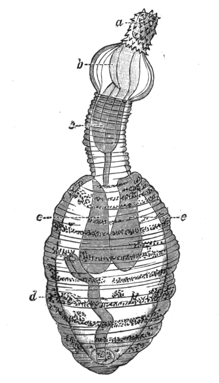
Fig. 100.—Fully formed larva of Echinorhynchus proteus from the body-cavity of Phoxinus laevis. (From Hamann.) Highly magnified. a, Proboscis; b, bulla; c, neck; d, trunk; e, e, lemnisci.
Family I. Echinorhynchidae.—The body is elongated and smooth. The proboscis-sheath has a double wall, and the proboscis is invaginated into it. The central nerve-ganglion lies in the middle line, as a rule on the posterior blind end of the proboscis-sheath. The papillae which bear the hooks are only covered with a chitinous cap at their apex, and the hooks have a process below. This family is by far the largest; a few species only can be mentioned. Echinorhynchus proteus lives in its mature form in fishes; the young forms, up to a centimetre in length, are found living freely in the intestine of numerous fresh-water fishes. Those found in Gobio fluviatilis, the gudgeon; Leuciscus virgo; Lota vulgaris, the burbot or eel-pout; young trout; Thymallus vulgaris, the grayling, seldom surpass this size, but those found in Acerina cernua, the pope fish; in Abramis bipunctatus; in Esox lucius,the pike, and in older trout, attain or surpass double the length. As the parasites grow older they bury their proboscis and neck in the wall of the intestine, the inner surface of which is studded with the orange-coloured bodies of the parasites. The proboscis is so deeply sunk in the wall of the alimentary canal as to form a papilla on its outer surface (Fig. 92). The larvae of E. proteus are found in the body-cavity of Gammarus pulex, one of the Amphipod Crustacea, and also in the same position in numerous fresh-water fishes; they must have passed into this first host by the mouth and alimentary canal. If the liver of an infested minnow, Leuciscus phoxinus, be examined, it will be found to contain on its surface numerous spherical or egg-shaped capsules of an orange colour, 2 to 2.5 mm. in length; these contain the larval forms of the parasite. They develop into the adult form when the first host is eaten by a carnivorous fish, but a complication may take place when the larval form is found in Gammarus, as the latter, the first host, may be eaten by a fish (intermediate host) in which the larva does not become mature, and only develops sexual organs when eaten by a carnivorous fish (second host). The larval form is also found in Nemachilus barbatulus, Gobio fluviatilis, and the sticklebacks Gasterosteus aculeatus and G. pungitius.
E. clavula Duj. is found in Salmo fario, Abramis brama, Cyprinus carpio, Gobius niger, Lepadogaster gouanii, etc.; E. linstowi Ham. in Leuciscus idus, Abramis ballerus, Abramis bipunctatus, and Acipenser huso; E. lutzii Ham. was found by Dr. Lutz in Brazil in the intestine of Bufo agua; E. angustatus Rud. occurs in such numbers in the perch, Perca fluviatilis, as to almost occlude the lumen of the intestine, and one out of every three or four fish in certain districts is infested by it. It is also found in the pike, Esox lucius, and the barbel, Barbus vulgaris. The first or larval host of this species is the Isopod Asellus aquaticus. E. moniliformis Brews. is stated to attain maturity in the human intestine. Except for the fact that G. gigas has once been observed in the same place, this is the only human parasite amongst the Acanthocephala. Its normal second hosts are Mus decumanus and Myoxus quercinus, and its first or larval host, the larvae of the beetle Blaps mucronata. E. porrigens Rud. is found in considerable numbers in the small intestine of a fin-whale (Balaenoptera sibbaldii), and E. strumosus Rud., in the small intestine of a seal (Phoca vitulina), and in the body-cavity of the angler fish (Lophius piscatorius). E. acus is common in the whiting, Gadus merlangus.
Family II. Gigantorhynchidae.—Large forms with ringed, flattened, and Taenia-like bodies. The hook-papillae are covered all over with transparent chitinous sheaths with two root-like processes. The proboscis-sheath is muscular and without a lumen. The central nervous system is excentrically placed below the middle of the so-called sheath. The lemnisci are long twisted tubes with a central canal.
Hamann places three species in this family: Gigantorhynchus echinodiscus, G. spira, and G. taenioides; but as he points out that E. gigas resembles these in its more important structural features, it seems advisable to include it here under the name G. gigas. The members of the first family often present a transversely ringed appearance after death, but the Gigantorhynchidae are ringed when alive, and the circular canals in the skin show a certain regularity, being arranged one between each two rings. There is no lumen in the proboscis-sheath, which is not attached to the boundary between the proboscis and the trunk, but to the inner surface of the proboscis, and the whole can be retracted within the anterior portion of the body, which is invaginable. There are always eight cement-glands, and other differences exist in the musculature, hooks, and position of the nervous system.
G. gigas occurs in the adult state in the small intestine of swine; in Europe its first or larval host is believed to be the grubs of Melolontha vulgaris and Cetonia aurata, but these beetles are absent from America, though the parasite infests American hogs. Stiles[220] has recently made some experiments which tend to show that in the United States the source of infection is some species of the beetle Lachnosterna, and he has succeeded in infecting the grub of L. arcuata by feeding it on the eggs of the parasite; from one larva he took 300 parasites six weeks after feeding it. L. arcuata is, like M. vulgaris, phytophagous, but the grubs of both the beetles are fond of frequenting manure heaps and patches of dung, and thus are much exposed to the dangers of infection.
G. echinodiscus inhabits the intestine of ant-eaters, having been found in Myrmecophaga jubata and Cycloturus didactylus. G. spira lives in the king vulture Sarcorhampus papa, and G. taenioides in Dicholophus cristatus, a species of Cariama.
Family III. Neorhynchidae.—Sexual maturity is reached in the larval stage. The proboscis-sheath has a single wall. A few giant nuclei only are found in the sub-cuticle and in the lemnisci. The circular muscle layer is very simply developed. The longitudinal muscle-cells are only present in certain places.
This family includes two species, Neorhynchus clavaeceps and N. agilis, which afford interesting examples of paedogenesis. The sub-cuticle and the lemnisci are dominated by a few giant nuclei, which remain in the embryonic state and do not break up into numerous nuclei as in other forms. The musculature is but little developed and the longitudinal sheath hardly exists. The proboscis-sheath consists of a simple muscular layer, and the short proboscis has few hooks and presents an embryonic appearance.
The sexually-mature form lives in the carp, Cyprinus carpio; the larval form is found, according to Villot,[221] encysted in the fat bodies of the larva of Sialis lutaria, one of the Neuroptera, and in the alimentary canal of the leech Nephelis octocula, and successful experiments have been made in infecting some species of the water snail Limnaea. N. agilis occurs in Mugil auratus and M. cephalus.
Family IV. Arhynchidae.—Short forms with the body divided into three well-marked regions—head, collar, and trunk. The head is pitted, the collar smooth, and the trunk wrinkled, not annulated, in spirit specimens. There is no eversible introvert, and no introvert sheath and no hooks. The sub-cuticle and the lemnisci have a few giant nuclei, and the lemnisci are long and coiled.[222]
This family resembles the Gigantorhynchidae in the length and curvature of its lemnisci, and the Neorhynchidae in the persistence of the embryonic condition of the nuclei in the sub-cuticle and the lemnisci; but in the shape of the body, its division into three well-marked regions, the absence of eversible proboscis, proboscis sheath, and hooks it stands alone, though it is nearer to the Neorhynchidae than to either of the other families.
The single species Arhynchus hemignathi was found attached to the skin around the anus of a Sandwich Island bird, Hemignathus proceros. The bird is a member of a family Drepanididae, which is entirely confined to the Sandwich Island group. Professor Newton tells me that it is probable that the "food of Hemignathus consists entirely of insects which it finds in or under the bark of trees," hence it is probable that the second host of this parasite, if such exists, must be looked for amongst the Insecta.