Chapter XV
Reading edition
GEPHYREA
INTRODUCTION—ANATOMY—DEVELOPMENT—SIPUNCULOIDEA—PRIAPULOIDEA—ECHIUROIDEA—EPITHETOSOMATOIDEA—AFFINITIES OF THE GROUP.
The animals included in the above-named group were formerly associated with the Echinodermata. Delle Chiaje[468] states that Bohadsch of Prague in 1757 was the first to give an accurate description of Sipunculus under the name of Syrinx, but Linnaeus, who noted that in captivity the animal always kept its anus directed upwards, re-named it Sipunculus. Lamarck[469] placed the Gephyrea near the Holothurians; and Cuvier[470] also assigned them a position amongst the Echinoderms. He mentions Bonellia, Thalassema, Echiurus, Sternaspis, and three species of Sipunculus, one of which, S. edulis, "sert de nourriture aux Chinois qui habitent Java, et qui vont la chercher dans le sable au moyen de petits bambous préparés."
The name Gephyrea[471] was first used by Quatrefages, who regarded these animals as bridging the gulf between the Worms and the Echinoderms. He included in this group the genus Sternaspis (vide p. 335), now more usually classed with the Chaetopoda.
The Gephyrea are exclusively marine. They are subcylindrical animals, which can either retract the anterior end of their body—the introvert—carrying the mouth into the interior; or are provided with a long flexible but non-retractile proboscis. The latter is easily cast off. They usually bear spines or hooks of a hard chitinous character, secreted by the epidermis or outermost layer of cells. The mouth is at the base of the proboscis or at the end of the protractile part, the anus is at the other end of the body or on the dorsal surface. The nervous system consists of a ring round the mouth and of a ventral nerve-cord. A vascular system is present as a rule. Nephridia are found which act as excretory organs, and in most cases also as ducts for the generative cells. The Gephyrea are bisexual, and the male is sometimes degenerate.
The group may be divided into four Orders:—(i.) Sipunculoidea; (ii.) Priapuloidea; (iii.) Echiuroidea; (iv.) Epithetosomatoidea; of these the first is by far the largest, both in number of genera and of species.
The Anatomy of Sipunculus nudus.
External Characters.—The body of S. nudus when fully extended may attain a length of a foot, or even a little more; in this condition it is seen to consist of two portions, the anterior of which is, however, retracted into the other when the animal is disturbed. The retractile portion is sometimes termed the proboscis, but as its nature is entirely different from that of the proboscis of the Echiuroidea, it is better to refer to it as the introvert. Special retractor muscles are attached on the one hand to the body-wall about half-way down the body, and on the other hand are fused into a muscular sheath which surrounds the gullet, just behind the mouth. When these muscles contract, they withdraw the introvert into the rest of the body or trunk in much the same way as the finger of a glove may be drawn into the hand, by a thread fastened to the inside of its apex. The introvert is protruded by the contraction of the circular muscles of the body-wall. These exert a pressure on the fluid which fills the body-cavity, and by this means the sides of the introvert are forced forward until finally the head is exposed.
The introvert occupies about one-sixth or one-fifth of the total body length. It is somewhat narrower than the trunk, and is covered by a number of small flattened papillae, some of which lie with their free ends directed backward, overlapping one another like tiles on a roof. In some other genera, as Phymosoma, the introvert bears rows of horny hooks, which are apt to fall off as the animal grows old.
The trunk has from thirty to thirty-two longitudinal furrows, the elevations between which correspond with a similar number of muscles lying in the skin. This longitudinal marking is crossed at right angles by a circular marking of similar origin, the elevations of which correspond with the circular muscles in the skin. These two sets of markings thus divide the skin of the trunk into a number of small square areas, very regularly arranged (Fig. 212).
The outline of the trunk is more or less uniform, but it is capable of considerable change according to the state of contraction of its muscles. The circular muscles, for instance, may be contracted at one level, thus causing a constriction at this spot. The colour of S. nudus is a somewhat glistening greyish-white.
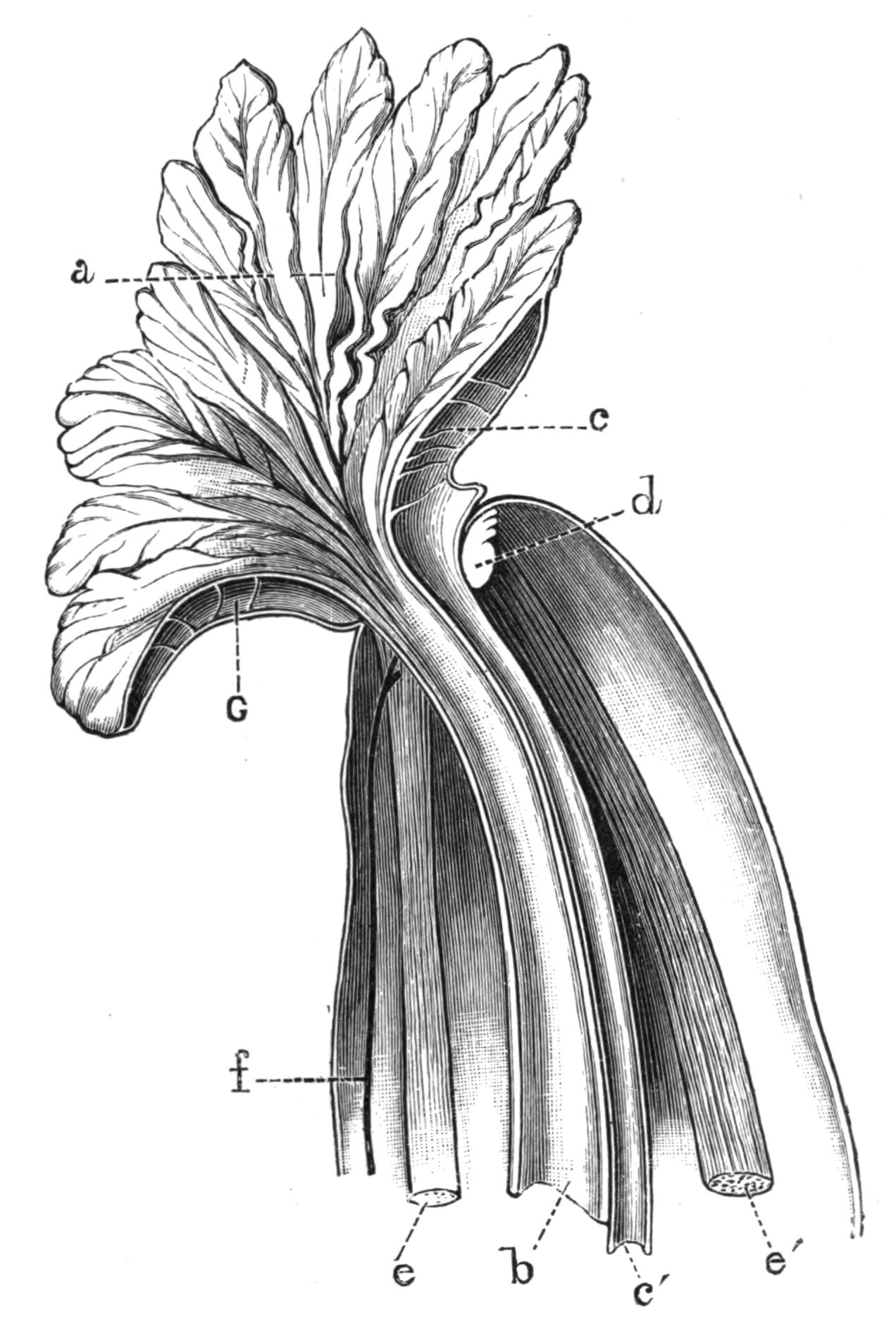
Fig. 211.—Right half of the anterior end of Sipunculus nudus L., seen from the inner side and magnified. a, Funnel-shaped grooved tentacular crown leading to the mouth; b, oesophagus; c, strands breaking up the cavity of the tentacular crown into vascular spaces; c', heart; d, brain; e, ventral, and e', dorsal retractor muscles; f, ventral nerve-cord; G, vascular spaces in tentacular crown.
The anterior end of the fully-expanded Sipunculus may be termed the head; here the skin is produced into a frayed fringe which stands up in the shape of a funnel round the mouth. This fringe is grooved on its internal surface with numerous little gutters, all of them lined with cilia, which by their constant motion keep up a current which sweeps food into the mouth. The fringe may be in the form of a simple ring round the mouth, or the ring may be folded in at the dorsal side so as to take the form of a double horse-shoe (Figs. 211 and 212).
Body-wall.—The glistening appearance of Sipunculus is due to the cuticle, a chitinoid layer which is secreted by the external layer of cells, the epidermis. Beneath this lies a layer of connective tissue, which is not always present in other Gephyrea; within this lies a layer of circular muscles arranged in bundles, then comes a very thin sheath of oblique muscular fibres, then a thicker layer of longitudinal muscles, and finally a layer of peritoneal epithelial cells, which in Sipunculus are for the most part ciliated.
Scattered over the surface of the body, and opening by narrow tubes which pierce the cuticle, are a number of glandular bodies which may be either bi- or multi-cellular. The glandular cells are apparently enlarged and modified epidermal cells; they are arranged in a cup-shaped manner, with their apices directed towards the orifice. They are crowded with granules, which are presumably poured out over the cuticle, but the exact function of the secretion is entirely unknown. They have a well-developed nerve supply.
Digestive System.—The mouth lies in the centre of the fringe, and is not provided with any kind of jaw or biting armature; it leads directly into the thin-walled alimentary canal, the first part of which is ciliated. The alimentary canal is not marked out into definite regions, but passes as a thin-walled semi-transparent tube to the posterior end of the body, and then turns forward again and opens to the exterior by an anus situated about an inch below the junction of the introvert with the trunk, on the median dorsal line. The descending and ascending limbs of the alimentary canal are coiled together in a spiral, which may be more or less close in different individuals. The whole is supported by numerous fine muscular strands, which pass from the walls of the intestine to the skin, and by a spindle-muscle, which runs from the extreme posterior end of the trunk up the axis of the spiral and terminates in the skin close to the anus.
No glands open into the alimentary canal at any point of its course, but near the anus a simple diverticulum, or pocket, of unknown function arises. The size of this outgrowth differs enormously in different individuals. The alimentary canal near the anus also bears two tuft-like organs, which, however, do not open into the intestine, but probably have some function in connexion with the fluid in the body-cavity.
Along the whole course of the alimentary canal there runs a ciliated groove, into which the food does not pass, but the cilia of which probably keep in motion a current of water whose function may be respiratory.
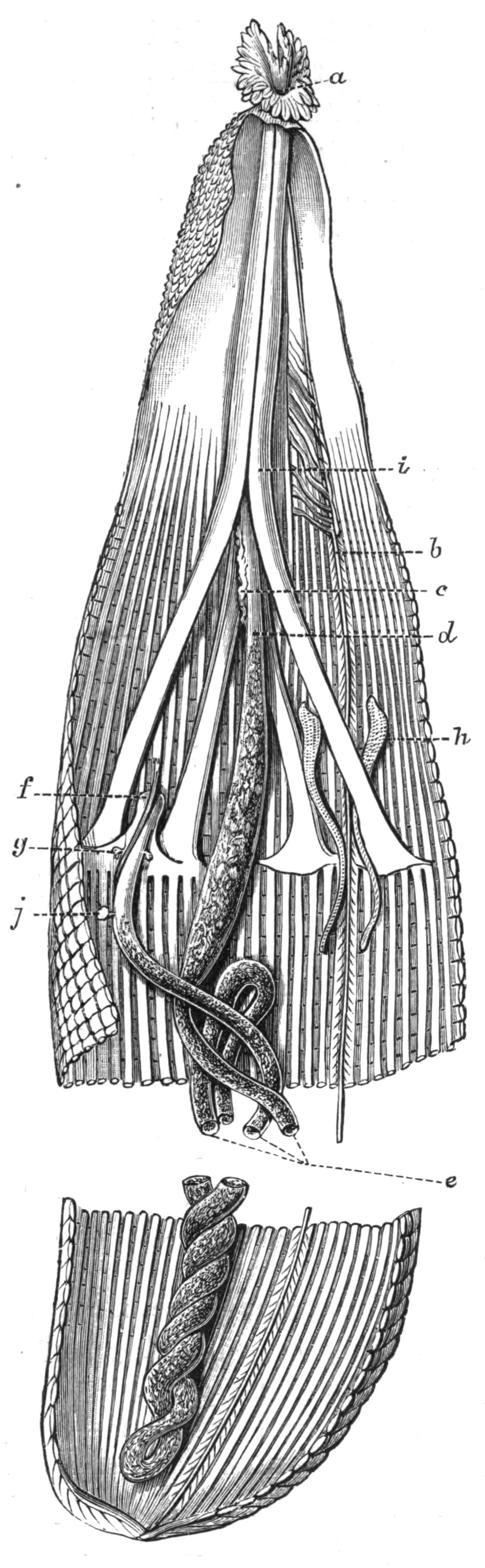
Fig. 212.—Sipunculus nudus L., with introvert and head fully extended, laid open by an incision along the right side to show the internal organs. × 2. a, Mouth; b, ventral nerve-cord; c, heart; d, oesophagus; e, intestine; f, position of anus; g, tuft-like organs; h, right nephridium; i, retractor muscles; j, diverticulum on rectum. The spindle-muscle is seen overlying the rectum.
Vascular System.—On the dorsal surface of the anterior end of the alimentary canal lies a contractile vessel, usually termed the heart. It is a tube about an inch long, ending blindly behind, but opening in front into a ring-shaped space surrounding the mouth and partially enveloping the brain. From this ring-like vessel numerous branches are given off which pass into the fringe round the mouth, and probably the chief function of the heart is by its contraction to force fluid into this fringe, and so to extend it. The heart contains a corpusculated fluid. A similar but shorter tube is found on the ventral surface of the anterior end of the alimentary canal in the species in question; it also opens into the ring which surrounds the mouth.
Respiratory System.—There are no special respiratory organs, and it has long been a matter of dispute where the respiration of Gephyrea is carried on. The oxygenation of the blood probably takes place to some extent through the walls of the oral fringe, but the blood which receives its oxygen at this spot is limited in its distribution, and could only supply the brain and head. It seems probable that the remaining organs are supplied with oxygen by the fluid of the body-cavity, which bathes them on all sides. This might obtain its oxygen from the blood in the heart, or more probably, through the thin walls of the intestine, from the stream of water which is maintained by the ciliated groove described above. Quite recently a form—S. mundanus, var. branchiata—has been described[472] with thin-walled papillae covering parts of the skin. These papillae are full of corpuscles, and are regarded by their discoverer as branchiae.
Body-Cavity.—The pinkish fluid of the body-cavity contains numerous corpuscles, the products of the reproductive organs (either ova or spermatozoa), and some curious unicellular bodies known as "urns." The latter are shaped like a bowl with a ciliated rim, and are formed from the budding of certain cells on the walls of the dorsal blood-vessel.[473] Their function is unknown, but they resemble certain multicellular bodies found in the body-cavity of Phascolosoma. The generative cells found in the body-cavity are further considered below. The true corpuscles are either biconcave round corpuscles coloured with a chemical substance, the haemerythrin of Krukenberg, which apparently plays the same rôle as haemoglobin in other animals; or amoeboid corpuscles, which, though rare in Sipunculus, are very numerous in Phascolosoma.
Nervous System.—The nervous system of Sipunculus consists of a brain or cerebral ganglion, a circumoesophageal ring surrounding the gullet, and a ventral nerve-cord. The brain is a small bi-lobed nervous mass situated on the dorsal surface of the oesophagus, in the angle between the right and left dorsal retractor muscles close to their point of insertion. Numerous nerves arise from it, and pass to the fringe surrounding the mouth and to neighbouring parts. At the sides, the brain is continued into two stout nerve-cords which encircle the oesophagus, and meeting, fuse together in the median ventral line to form the ventral nerve-cord (Fig. 211). The latter is of the same diameter throughout, and shows no signs of segmentation; it is oval in section, and consists of small ganglion cells heaped up on the ventral surface, i.e. next the skin, and of numerous fibres situated dorsally. The cord gives off many nerves, which usually arise in pairs. These pass into the skin, and forming rings, run round the body, and give off finer nerves as they go.
The nerve-cord is supported by numerous strands of muscle which pass to it from the skin. These are especially long in the region where the introvert joins the trunk, and thus allow free play to the nerve-cord when the former is being protruded or retracted.
Sipunculus is not well provided with sense-organs, but in an animal which lives buried in sand we should not expect to find these very highly developed. On the introvert there are certain patches of epithelium bearing long stout cilia, which have been regarded as tactile in function, and there is a tubular infolding reaching the brain, which almost certainly has some sensory function. Ward[474] has termed this "the cerebral organ." It consists of a duct lined with ciliated cells, which opens to the exterior in the middle dorsal line outside the tentacular fringe. The duct leads down to the brain, and expands at its lower end into a saucer-shaped space, covering that portion of the brain where its substance is continuous with the external epithelium. In Phymosoma this cavity is produced into two finger-shaped processes, which are sunk into the brain and are lined by cells crowded with a dense black pigment.[475] They are probably rudimentary eyes, perhaps distinguishing only between darkness and light. The pits appear to be absent in Sipunculus nudus, but Andrews states they are found, although without pigment, in S. gouldii.[476]
Excretory System.—The excretory organs or "brown tubes" are typical nephridia, that is to say, they consist of tubes with glandular walls which open on the one side to the exterior, and on the other by means of a ciliated funnel-shaped opening into the body-cavity. In Gephyrea one wall of the tube is produced into a long diverticulum or sac which hangs down into the body-cavity, and is usually supported by muscle-fibres running to the body-wall. The lower end of the sac is broken up into a number of crypts or pits, lined by large glandular cells crowded with brown pigment. The pigment-granules are secreted into the cavity of the sac, and leave the body through the external opening; they probably consist of the nitrogenous excreta of the animal. The upper end of the sac, into which both the external and internal orifices open, is usually enlarged, and its walls are very muscular. As in so many other animals, the nephridia serve as ducts through which the reproductive cells leave the body of the parent.
Reproductive System.—The Gephyrea are bisexual. In Sipunculus the testes and ovaries are found in the same position in the two sexes, and are indistinguishable without microscopic investigation. They each consist of small ridges situated at the lower end of the ventral retractor muscles, just where the latter take their origin from the longitudinal muscles of the skin. At this level the cells which line the body-cavity on the inside of the skin are heaped up, and become modified in the one case into ova or eggs, and in the other into the mother-cells of the spermatozoa. This method of forming the reproductive organs from modified cells lining the body-cavity is very common in the higher animals; but it is seen in its simplest and least modified form in the Sipunculidae.
The eggs break away from the ovary in a very undeveloped condition, but whilst floating about in the body-cavity they increase in size and secrete a thick membrane around them. They have a well-marked nucleus, and are oval in outline.
The mother-cells of the spermatozoa also break away in an immature condition, and complete their development in the nutritive fluid of the body-cavity. They divide into a number of spermatozoa, usually eight or sixteen, which remain in contact. They each develop a tail, which projects outwards, and aids the cluster in swimming along. These clusters of spermatozoa are about the same size as the ova of the female, and, like them, make their way into the "brown tubes." The exact way in which this is accomplished is not very clear, but the cilia on the funnel-shaped internal opening of the tube seem to have some power of selecting the generative cells when they come within their reach, and of passing them on, whilst they reject the much smaller corpuscles of the perivisceral fluid, which are never found in the nephridia.[477] Once inside the internal opening, the clusters break up and the spermatozoa escape singly into the sea. Here they meet with and fertilise the eggs which have escaped from the body of the female.
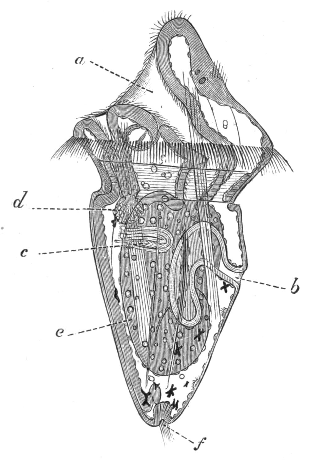
Fig. 213.—Larva of Sipunculus nudus L. × 150. (After Hatschek.) a, Mouth; b, anus; c, excretory organ; d, glandular appendage of oesophagus; e, wall of stomach over which the retractor muscle runs; f, invaginated sense-organ at aboral pole.
Development.—Hatschek,[478] who investigated the development of Sipunculus nudus at Pantano, an inlet of the sea near Messina, states that the spawning takes place during the night, and ceases about July 10. The rate of development depends upon the temperature, but the larvae usually free themselves from the egg-membrane during the third day. When hatched the embryos lengthen out a good deal, and take the form represented in Fig. 213. The larva swims actively by means of a ring of stout cilia, which encircle the body just behind the mouth. Other shorter cilia are found on the head, continuing into the lining of the mouth, and a little bunch of them is situated at the extreme posterior end. The alimentary canal is already formed, and is twisted, so that the anus lies dorsally, but not so far forward as it does in the adult. A glandular structure opens into the mouth, and another body of unknown function is connected with the oesophagus; both these disappear during larval life. A pair of excretory tubules, the forerunners of the brown tubes, are found, and the chief muscle tracts are already established. The nervous system is still in close connexion with the skin, from the outer part of which it is derived; the cerebral thickening bears two eye-spots.
The fluid of the body-cavity contains corpuscles, which are kept in active circulation by the constant contractions of the body-wall, and by numerous tufts of cilia which are borne on the inner surface of the skin. The dorsal blood-vessel is one of the latest organs to arise.
The larva swims actively about for a month, during which time it increases greatly in size; it then undergoes a somewhat sudden metamorphosis. The ciliated ring and the structures related to the oesophagus begin to disappear, the distinction between the head and the rest of the body is obliterated, and the head becomes relatively small. The mouth changes its position, and becomes terminal instead of being somewhat ventral, and the tentacular membrane begins to appear. At the same time the larva relinquishes its free-swimming life, and sinks to the bottom; it begins creeping amongst the sand by protruding and retracting the anterior part of its body, and takes on all the characters and habits of the adult.
I. Order Sipunculoidea.
Besides the genus Sipunculus, the Order Sipunculoidea includes ten other genera. A key to these, taken for the most part from Selenka's admirable monograph, is given on page 424.
Phascolosoma contains, in comparison with Sipunculus, only small species, and it is easily distinguished by the fact that the longitudinal muscles are fused into a continuous sheath. As a rule the skin is smooth. A few species bear hooks, which are generally scattered irregularly and not arranged in transverse rows, as in Phymosoma (Fig. 214) and most of the other genera.
The fold which in S. nudus surrounds the mouth may be in the same species bent in so as to take the form of a double horse-shoe, the opening of which is always dorsal, just above the brain; in this case the mouth is crescentiform. In other genera the fold is broken up into discrete tentacles, and these are variously arranged; in Dendrostoma they are grouped together in four or six bundles round the mouth, but the more usual arrangement is the horse-shoe-like row of tentacles which overhang the crescentiform mouth, as in Phymosoma and some species of Aspidosiphon.
The ventral side of each tentacle is grooved and ciliated, and the grooves are continued into the ciliated mouth. Their dorsal surface is pigmented, and in the hollow of the horse-shoe lies a deeply pigmented epithelium covering the brain.
A blood-vessel courses up each tentacle, and usually two channels return the blood to the vascular ring which surrounds the mouth. In those forms which possess tentacles on the dorsal side of the mouth only, the ventral part of the vascular ring lies in the lower lip, which is tumid and swollen. The brain supplies a nerve to each tentacle.
When the introvert is retracted the tentacular ring is withdrawn and to some extent collapsed; in this condition it would be almost touching the rough external surface of the introvert. In some species of Phymosoma the delicate appendages of the head are guarded from the hooks on the introvert by a thin membrane or collar,[479] which completely ensheaths the retracted head.
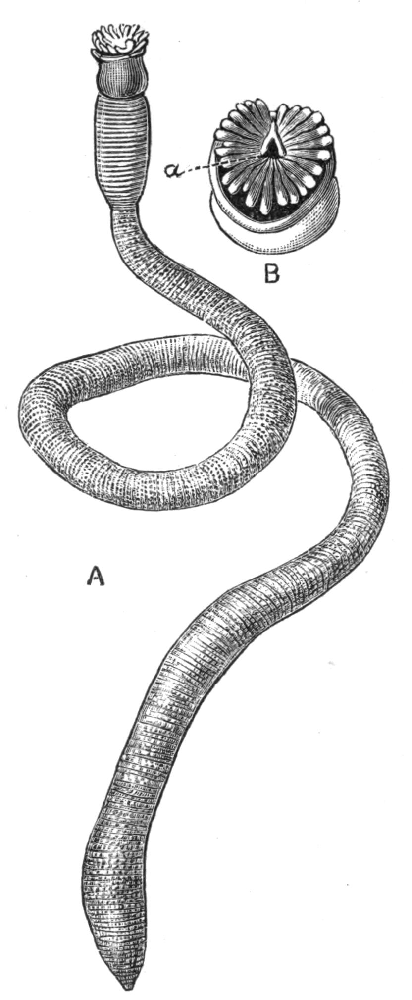
Fig. 214.—A, Phymosoma granulatum F. S. Leuck. × 2. B, Head of the same. × 4. a, Pigmented pit leading to brain. The crescentiform mouth on the lower side of the figure is overhung by the tentacles.
When the introvert is fully extended the dorsal blood-vessel contracts and sends its blood forward into the vascular ring, and thence into the tentacles or tentacular fold, which are thus erected. In several species of Sipunculus, as S. nudus, S. norvegicus, S. robustus, S. tesselatus, there is a ventral blind tube as well as a dorsal, into which the blood is withdrawn when the head is retracted. In many other species in various genera, such as Phymosoma weldonii and Ph. asser, Dendrostoma signifer, S. vastus, the lumen of the dorsal vessel is increased by numerous hollow blind processes which it bears, hanging freely into the body-cavity. Three very small genera of Sipunculids—Onchnesoma, Petalostoma, and Tylosoma—are devoid of all trace of vascular system and of tentacles; the mouth opens in the centre of the anterior end of the introvert. In Onchnesoma the dorsal part of the lip is somewhat produced, so that the head has somewhat the shape of a Doge's cap, and in Petalostoma there are two leaf-like processes of the body-wall which guard the mouth.
The extent to which the intestine is coiled varies very much even in the same species; the axis of the coil is often supported by a spindle-muscle, but this is sometimes absent. The caecum, which opens into the rectum of S. nudus, is again a very variable structure, and when it is present varies remarkably in size.
The food of Sipunculids seems to consist almost entirely of sand, and their only nourishment must be such small microscopic organisms or particles of animal and vegetable débris as are to be found mixed with the sand. The alimentary canal is, as a rule, quite full of sand, and yet in spite of the tenuity of its walls they never seem to be ruptured. If the contents of the digestive tube be washed out with a pipette, it will be found that it requires considerable force to dislodge many of the sand-particles lying next the wall. These are more or less embedded in crypts or pockets of the wall, and as the sand passes along the intestine they probably serve as more or less fixed hard points, against which the sharp edges of the sand particles are worn off. Amongst the sand are usually to be found pieces of shell, sometimes with a diameter equal to that of the alimentary canal; these are usually rounded, but their angles may have been removed by attrition before they entered the mouth of the Sipunculid.
In S. tesselatus the sand is to some extent held together by a mucous deposit; in those cases where there is no sand in the intestine, there is always a coagulum of mucus, and the walls are contracted and thick; when full of sand the walls are tensely stretched and very thin. This thinness of the wall of the alimentary canal seems ill-adapted to a diet of sand, nevertheless it is also met with in other great sand-eating groups of animals, such as the Echinids and the Holothurians.
The enormous amount of sand and mud which passes through the bodies of the Sipunculids shows that they must take a considerable part in modifying the mineral substances which form the bottom of the sea. Just as earthworms, as shown by Darwin, play a considerable rôle in the formation of soil, so must these animals, in conjunction with Echinids and Holothurians, effect considerable modifications in the sand and mud which pass through their bodies. Mr. J. Y. Buchanan[480] is "led to believe that the principal agent in the comminution of the mineral matter found at the bottom of both deep and shallow seas and oceans, is the ground fauna of the sea, which depends for its subsistence on the organic matter which it can extract from the mud." The minerals at the bottom of the sea are exposed to a reducing process in passing through the bodies of the animals which eat them, and subsequently to an oxidising process due to the oxygen dissolved in the sea-water acting on the minerals extruded from the animals' bodies.
The rate at which the sand passes through the body of Sipunculus is unfortunately unknown, but that at any one moment a considerable quantity is contained in the intestine is shown by the fact that the average weight of five specimens of S. nudus from Naples, taken at random, was 19.08 grms., whilst the average weight of sand washed out of their alimentary canal was 10.03 grms. The sand contained in five other specimens of the same species measured respectively 6 c.c., 7 c.c., 6.5 c.c., 7.5 c.c., and 7.5 c.c., giving an average of 6.9 c.c. for each individual.
Onchnesoma and Tylosoma have only one retractor muscle; Aspidosiphon and Phascolion have, as a rule, two; Phymosoma and Sipunculus have four, and perhaps this is the more usual number.
Phascolion, Tylosoma, and Onchnesoma have but one "brown tube"; in Phascolion this is the right, in Onchnesoma it is sometimes the right and sometimes the left that persists. Most other genera retain two, but there are many exceptions; for instance, Phascolosoma squamatum has but one, and so has Aspidosiphon tortus, and in both cases it is that of the left side. No Sipunculid has more than two. It has been pointed out by Selenka that those species which have but one brown tube are, as a rule, inhabitants of tubes or shells, and do not move actively about in the sand.
The eggs of all members of the family, with the exception of the genus Phymosoma, are spherical, but those of the last-named genus are elliptical. They are always surrounded by a thick membrane, the "zona radiata," pierced by numerous pores.
Aspidosiphon (Fig. 215) is easily recognised by the presence of two symmetrically-arranged cuticular shields, one at each end of the trunk. These are formed by the fusion of minute cuticular plates, such as exist in the skin of most Sipunculids. The posterior shield is radially symmetrical, but the anterior is somewhat like the shell of a Pecten, and symmetrical only about one plane. The introvert is protruded from the acute angle of the anterior shield, and when extended lies almost at right angles to the trunk, instead of being, as is usually the case, in the same straight line with it. In many specimens, and these seem as a rule to be the older ones, a deposit of calcium carbonate takes place over these shields, covering over and concealing their external markings.
Cloeosiphon (Echinosiphon) has a calcareous ring, consisting of four or five rows of lozenge-shaped calcareous bodies forming a close mosaic, arranged round the base of the introvert, which when extended is in the same straight line as the trunk. Each piece bears a brown spot, which is said to be the pore of a gland (Fig. 217). Golfingia Lankester, has a cylindrical horny thickening at the anterior end of the trunk and another at the posterior.
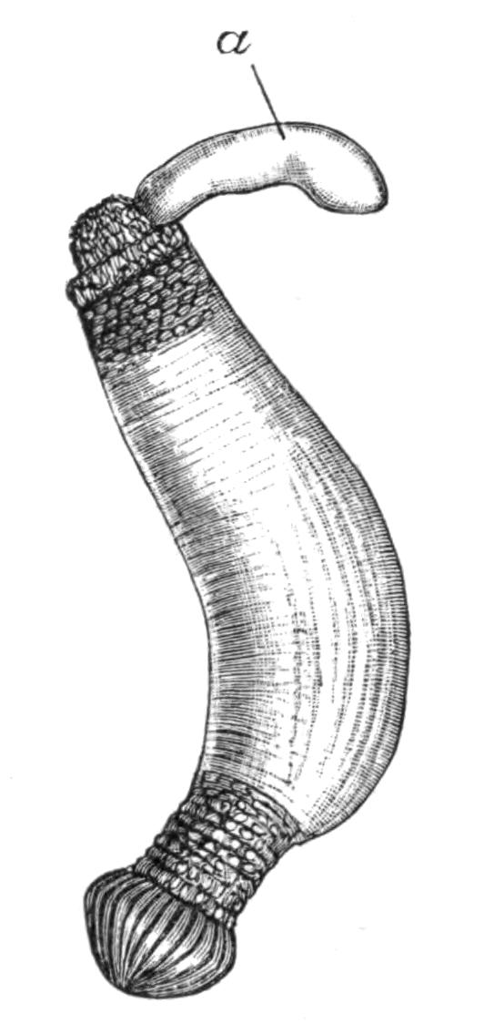
Fig. 215.—Aspidosiphon truncatus Kef. × 2. a, Introvert partially extended, but not sufficiently to show the head.
Key to the Genera of Sipunculoidea.[481]
I. The longitudinal muscles in the body-wall divided into 17-41 distinct bundles. Four retractor muscles.
A. Body covered with papillae. Numerous filiform tentacles which seldom (or never?) surround the mouth, but stand above and dorsal to it in a horse-shoe, with the opening dorsal. No rectal caecum. Hooks usually present. Four retractors (in Ph. Rupellii only two?). Heart almost always without caeca. Eye-spots always present. Eggs oval, flat, reddish. Almost entirely small tropical species
1. Phymosoma
B. Body devoid of papillae. Tentacular membrane surrounds the mouth in a circlet. Rectum with one or more caeca (except S. edulis?). Hooks absent except in S. australis. Eggs spherical. The tentacular membrane contains a vascular network. A ventral contractile vessel usually present in addition to the heart. Mostly large forms. Found in all seas
2. Sipunculus
II. The longitudinal muscles in the body-wall form a continuous sheath, and are not split up into bundles.
A. Two brown tubes. Numerous tentacles form a wreath round the mouth. Alimentary canal forms a complete spiral, free behind except in Ph. Hanseni. Spindle-muscle usually present. One or more ligaments present, but only on the anterior convolutions of the intestine. Adhesive papillae always absent. Hooks very frequently absent. Eggs spherical. Found in all seas.
3. Phascolosoma
B. Two free brown tubes. Only four or six plumed tentacles. A complete intestinal spiral, not attached behind. Spindle-muscle always present. One or more ligaments present, but only on the anterior convolutions of the intestine. Hooks are present, but sometimes fall off early in life. Heart usually bears caeca. Found only in the tropics.
4. Dendrostoma
C. Only one brown tube, that of the right side, present; it is attached to the body-wall throughout its entire length. Numerous tentacles form a circle round the mouth. The alimentary canal forms no spiral, or an incomplete one. No spindle-muscle, but the intestine is attached to the body-wall throughout its length by numerous ligaments. Adhesive papillae often present. Not more than two retractors. Spherical eggs. Inhabits Mollusc shells or tubes. Found in all seas
5. Phascolion
III. At both ends of the trunk a distinct horny shield, or tube-like cornification, or a calcareous ring at the anterior end of the trunk. Hooks sometimes present. Longitudinal muscles continuous or split up into bundles.
A. A shield at both ends of the trunk. Introvert excentric, arising from the ventral side of the anterior shield. Tentacles small and few in number, arranged in a horse-shoe above the mouth. A spindle-muscle, which arises from the posterior end of the body, traverses the intestinal coil. Two retractors only, these are the ventral; they are frequently fused together from their point of origin.
6. Aspidosiphon
B. A calcareous ring surrounds the anterior end of the trunk, from the middle of which the introvert is extruded. Longitudinal muscles continuous. Hooks bifid. Tropical.
7. Cloeosiphon
C. A corneous ring, from which the introvert issues, surrounds the anterior end of the trunk, and the posterior end of the trunk is produced into a corneous spike. Six pinnate tentacles encircle the mouth. Four retractors. Hooks present on the introvert. Longitudinal muscles continuous. Intestine not coiled throughout in a spiral nor fastened posteriorly. Spindle muscle present.
8. Golfingia
IV. No tentacles, but two leaf-like extensions of the body-wall guard the mouth. Four retractors. Few intestinal loops, quite free. No vascular system.
9. Petalostoma
V. No tentacles, no vascular system. One retractor, and one segmental organ.
A. Introvert long. Body small, pear-shaped.
10. Onchnesoma
B. No introvert (?). Body cylindrical, thickly covered with papillae, which are larger and more crowded at both ends of the trunk.
11. Tylosoma
Species of Sipunculoidea.—The genus Phymosoma (Fig. 214) contains more species than any other genus of Sipunculoidea, and they are all of fair size. Twenty-seven species are known, of which seventeen occur in the Malay Archipelago, thirteen being found there alone. Phymosoma affects shallow water, the deepest specimens being taken at a depth of about 50 fathoms; this may be due to the fact that they flourish only in comparatively warm water. With very few exceptions, they are found only in tropical seas, very often living in tubular excavations made in soft coral rock.
The genus Sipunculus contains sixteen species. They are the largest and the most conspicuous members of the group. They have a very wide distribution, some species, as S. nudus (Fig. 212) and S. australis, being almost cosmopolitan. They are most common in temperate and tropical seas, but S. norvegicus and S. priapuloides are found far north, but always at considerable depths, 100 to 200 fathoms.
The following account of the habits of Sipunculus gouldii is taken from Mr. Andrews'[482] paper on that species:—
"This Sipunculus is very abundant in certain small areas of compact, fine sand darkened by organic matter and not laid bare at ordinary low tide. In such places, only a few square metres in extent, they pierce the sand in all directions to a depth of more than half a metre, making burrows with persistent lumen running from the surface downward and then laterally, but with no regularity in direction.
"Kept in aquaria, the dependence of the animal upon the nature of the sand and its method of locomotion may be readily observed. A vigorous individual buries itself in a few moments in the following manner: Running out the introvert to nearly its full extent, and applying it to the surface of the sand till some spot of less resistance is found, the animal still further expands the introvert so that it penetrates the sand, provided this is not too dense and firm, for then the body is merely shoved backward. When the introvert is inserted, the contraction of the longitudinal muscles of the body-wall brings the whole body forward somewhat, in case the introvert is fixed in the sand. In case soft ooze was present, this fixation did not take place, and the introvert was merely pulled out again, but when the sand was of the right consistency the introvert was fixed by becoming much swollen at the tip, and then constricted just posterior to this swollen area. This bulb-like area exerts lateral pressure on the sand, as could be seen by movements of the grains. The swelling of the anterior end of the introvert is brought about by the body-wall contracting elsewhere, and forcing in liquid to distend that end. Owing to the curved form assumed by the body in the normal contracted state when first removed from its burrow, the entrance of the introvert may often be nearly vertical, and hence the entire body is soon raised nearly upright in the water above the sand. If the body has thus been warped forward sufficiently to become somewhat fixed in the sand, the introvert is rolled in and again thrust forward from this new point of resistance, and so on till the animal is entirely buried. This locomotion increases in speed as the creature becomes more completely surrounded by sand, and is the only means of moving from place to place.
"On a smooth surface, or on one not presenting the right degree of resistance, the Sipunculus does not change its position, but remains till death finally occurs, rolling its introvert in and out and contracting its body-wall to no purpose.
"The essential factors in the mechanism bringing about this hydrostatic locomotion are an elongated contractile sac filled with liquid, and some means of definitely co-ordinating the contractions of the sac.
"In natural environment the animals are found with sometimes one, sometimes the other end nearer the surface of the sand: in the aquaria the same was observed, but when the water became stagnant and impure the anterior end with expanded branchiae was often protruded somewhat above the surface of the sand."
The genus Phascolosoma contains at least twenty-five species, for the most part small. Ph. margaritaceum, however, measures[483] 10 cm. in length, and Ph. flagriferum, 13 cm. The latter is produced at the hinder end of its trunk into a long whip-like process, which recalls the horny spike of Golfingia. Most species live free, but a few inhabit the shells of dead Gasteropods or of Dentalium, or the abandoned tubes of worms. They occur in practically all seas.
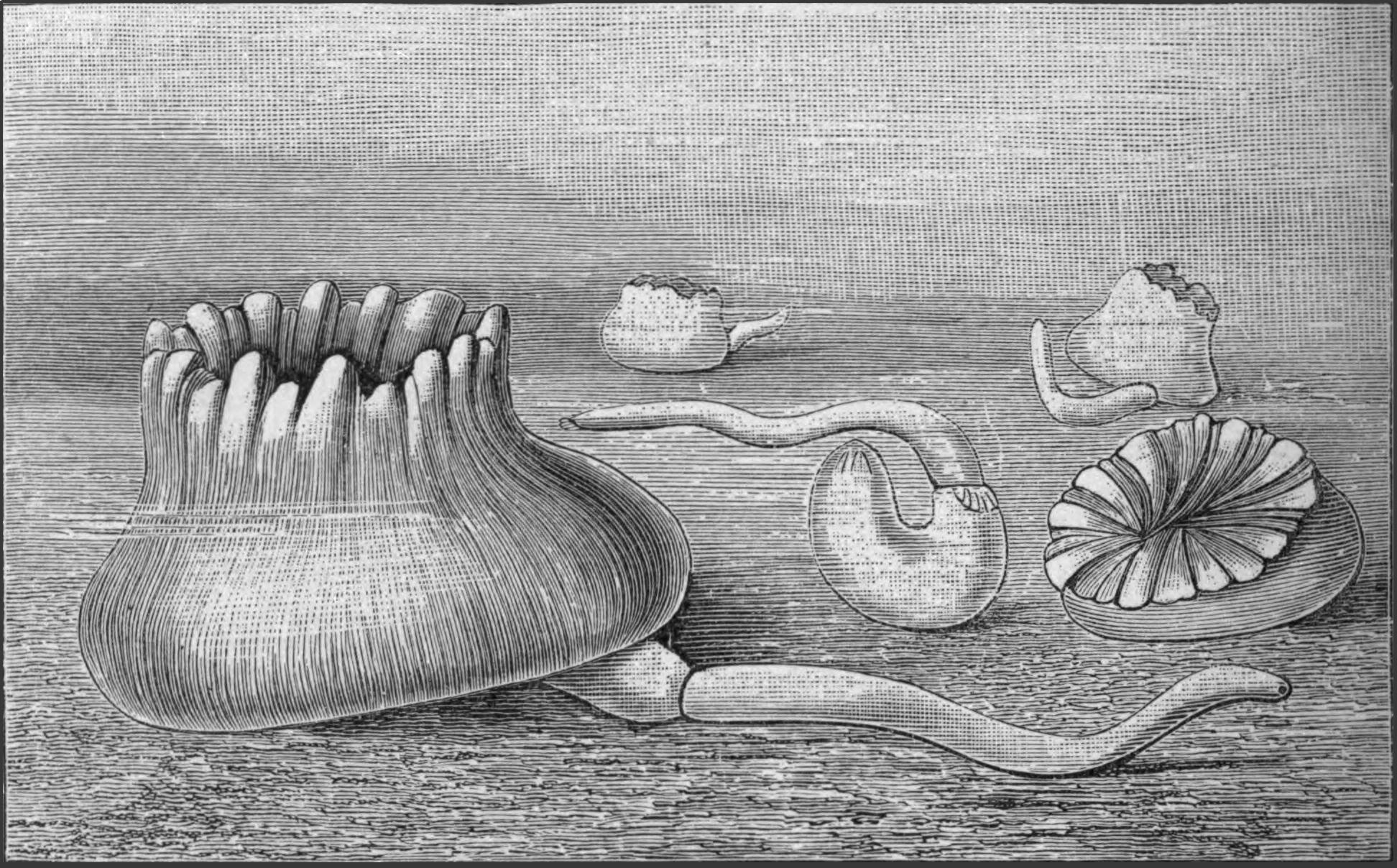
Fig. 216.—Specimens of the Coral Heteropsammia cochlea, with Aspidosiphon heteropsammiarum or A. michelini living in a state of commensalism with them. (From Bouvier.)
Dendrostoma contains but five species, which are all found within the tropics in the Pacific or in the West Atlantic. They are shallow-water forms, and some are found between tide-marks.
Phascolion is a smaller genus, containing but ten species, which may have been derived independently from different species of Phascolosoma, and in this case the genus should be broken up. The members of this genus live in Mollusc shells, such as Dentalium, Turritella, Buccinum, Chenopus (Aporrhais), Nassa, Strombus, and generally acquire the coiled shape of their host. They are usually attached to the shell by means of certain adhesive papillae found on their posterior end. Ph. strombi fills its shell with mud, which must be kept together by some secretion of the animal. The body lies in a tube in this mud, and the introvert projects from the small round opening at the end of the tube, and explores the ground in every direction. They are found in all seas, but more especially in the colder waters.
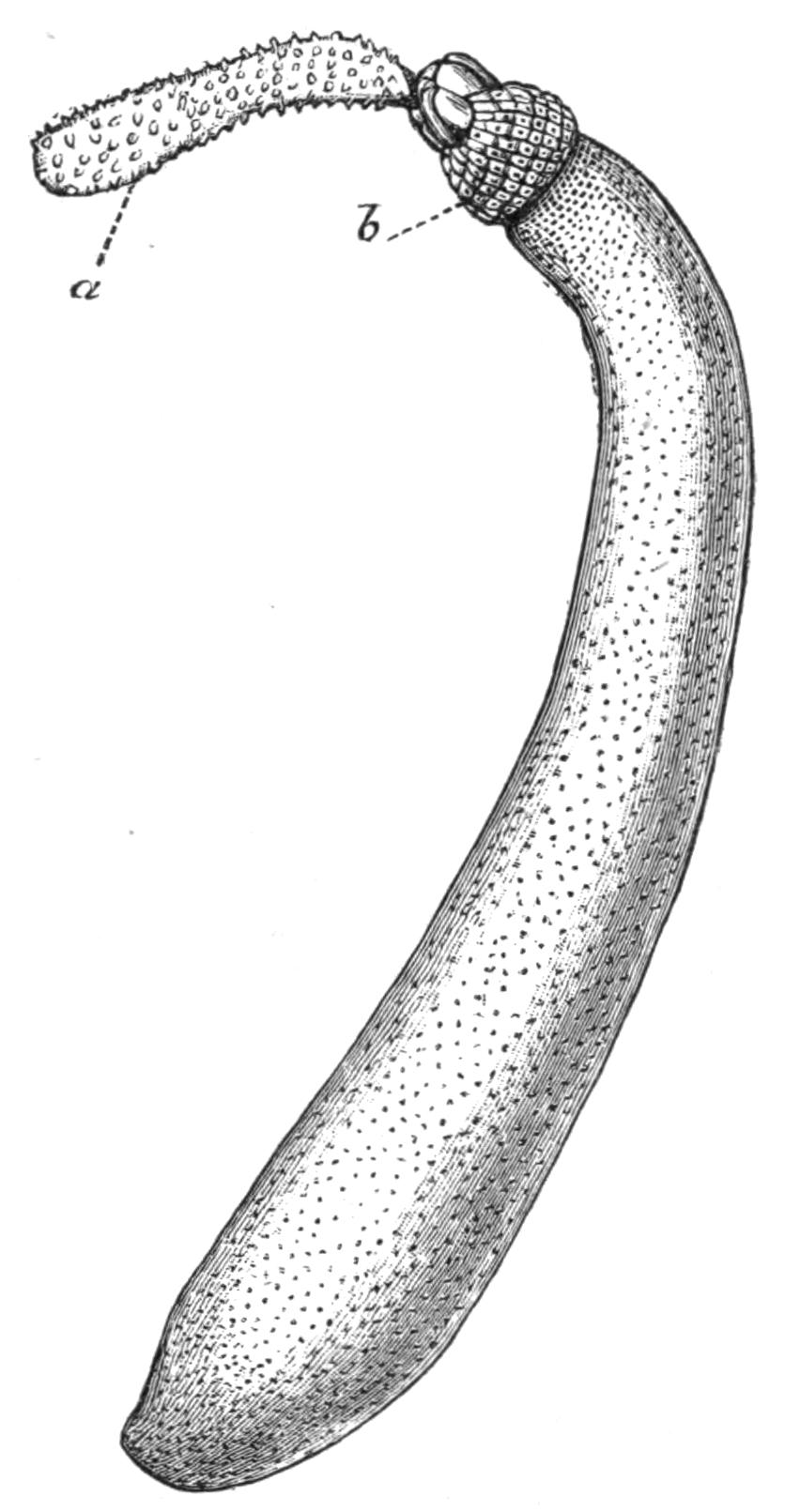
Fig. 217.—Cloeosiphon aspergillum Quatr. × ½. a, Introvert covered with spines and partially extended, but not sufficiently to show the head; b, calcareous plates surrounding the point of origin of the introvert.
The genus Aspidosiphon includes nineteen species, which are, with few exceptions, exclusively confined to the Indian Ocean and neighbouring seas, including the Red Sea. The exceptions are A. armatus from the Norwegian coast, and A. mülleri from the Mediterranean and Adriatic. A. truncatus is also stated to occur at Panama, the Bahamas, and at Mauritius. The remaining species almost all occur in the Malay Archipelago and neighbouring islands, and as was the case with Phymosoma, this part of the world seems to be the headquarters of the genus. A. mülleri lives in the interstices of rocks and stones, and occasionally in disused Mollusc shells.
Two species of Aspidosiphon have been described by Bouvier[484] living in a state of commensalism with two species of Madreporarian corals, Stephanoceris rousseaui and Heteropsammia cochlea, which live on and surrounding the shells of certain Molluscs at Aden (Fig. 216). Apparently the Gephyrean takes up its abode within its house at a tender age, and according to Bouvier, it provides for its increasing bulk by secreting a coiled calcareous tube, the outer surface of which affords space for the growth of the coral.
The genus Cloeosiphon, the Echinosiphon of Sluiter, includes three species: C. aspergillum (Fig. 217), C. molle, and C. javanicum. The first named occurs at Mauritius, the Malay Archipelago, and neighbouring islands; the others are confined to the last-named area, which thus again forms the headquarters of a genus.
Golfingia, described by Lankester from a single specimen, was dredged in St. Andrews Bay, at the depth of 10 fathoms.
Petalostoma comprises but one species, P. minutum, which is found in the English Channel.
Onchnesoma comprises two species, O. steenstrupii and O. sarsii, both found off the coast of Norway at considerable depths between 200 and 300 fathoms.
Tylosoma comprises one species, T. lütkenii, also from the Norwegian coast. It is dredged from stony ground in 50 to 80 fathoms.
II. Order Priapuloidea.
Anatomy.—This Order consists of the two genera Priapulus and Halicryptus. Both are cylindrical animals with the mouth at one end and the anus at the other. The introvert is short, and is covered with rows of chitinous spines, which are continued to some extent over the body.
The skin is folded in a series of rings, and the body is usually somewhat swollen posteriorly. P. caudatus bears a curious caudal appendage, beset with a number of hollow lobes somewhat grape-like in appearance. This is situated ventral to the anus; its lumen is continuous with that of the body-cavity, but it can be separated from it by the action of a sphincter muscle. Two such appendages exist in P. bicaudatus.
There cannot be said to be any head in the Priapuloidea; they have no tentacles or tentacular fringe, no proboscis, and no distinct brain; simply a round aperture, the mouth, which is surrounded by a groove in the skin, at the bottom of which the circumoesophageal nerve-cord lies. The mouth leads into a very muscular pharynx lined with stout chitinous teeth; this passes into an intestine, which is as a rule straight, but in P. glandifer it has a single loop.
The Priapuloidea possess no vascular system and no brown tubes. Their skin has in the main the same structure as that of the Sipunculids, with spines, glandular bodies, and papillae with sensory hairs which resemble similar structures on Phymosoma varians. Retractor muscles arise from the longitudinal muscles of the skin, and are inserted into the pharynx; they are short and not constant in number.
The nervous system has retained throughout its primitive connexion with the epidermis. In almost all animals the nervous system is formed from the epiblast or outermost cellular layer of the embryo; it usually, however, breaks away from this and sinks into the body. Thus in Sipunculus it lies within the body-cavity, and has retained its primitive connexion with the outer layers of the skin only in the region of the brain; but in the Priapulids the nervous system, which consists of a ring round the mouth and of a ventral cord, lies embedded in the skin, and the nerve cells are directly continuous with the cells of the epidermis. The nerve-ring lies at the base of a groove in the skin, which forms a kind of gutter round the mouth; the ventral nerve-cord is visible exteriorly as a light line which marks the ventral surface of the animal. In no place is the ring or cord differentiated in any way, and there cannot be said to be any brain or special sense-organs. Numerous nerves are given off from the ring to the pharynx and intestine, and from the cord to the body-wall.
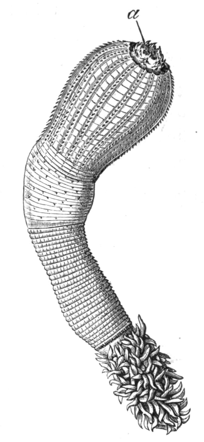
Fig. 218.—Priapulus caudatus Lam. Nat. size. a, Mouth surrounded by spines.
The sexes are distinct, but they differ from the other Gephyrea in the nature of their reproductive organs. In mature specimens the ovaries or testes are easily recognisable, lying to the right and left of the alimentary canal. The reproductive glands are continuous with ducts, which act as oviducts and vasa deferentia respectively. Both glands and ducts are attached to the body-wall by a mesentery.
The excretory function is performed in the Priapuloidea by the ducts of the generative organs. These are primarily connected with a number of branching canals of small size which project into the body-cavity. According to Schauinsland,[485] one or more pear-shaped cells are found at the end of each branch, and each is continued into a long cilium which hangs down into the lumen of the canal, and by its movement produces a flickering motion. Beyond the free end of the large cilium the canal is lined with ciliated cells. The remarkable resemblance this form of excretory organ presents to that of the Platyhelminthes (vide p. 25) and of certain Chaetopods is worthy of attention. In the young Priapuloidea the duct with its branching canals is not masked by the generative organs, but as the animals become mature, diverticula from the duct arise, and the cells covering these become modified into ova in the female, and into spermatozoa in the male. The presence of these follicles masks the excretory part of the gland. The ova and spermatozoa escape through the ciliated ducts which open to the exterior one on each side of the anus, and, contrary to what is the case with other Gephyrea, leave the body without having ever been in the body-cavity.
Nothing is known of the embryology of either member of this family, but both genera appear to be sexually mature from the end of May until October.
Classification.—The two genera which make up the Order Priapuloidea are characterised as follows:—
Priapulus.—The body is continued into one or two caudal appendages, beset with hollow papillae; these are ventral to the anus. The introvert forms ¼ to ⅓ of the total body-length; it is covered with spines in conspicuous longitudinal rows, the rest of the body being ringed. The retractor muscles are numerous, and are attached to the body-wall, some anteriorly and some posteriorly.
The genus includes the following five species:—
P. caudatus Lam. (Fig. 218). Hab. Coasts of Greenland, Norway, Great Britain, the North Sea, and the Baltic.
P. bicaudatus Dan. Hab. North Sea and Arctic Ocean.
P. glandifer Ehlers. Hab. Coast of Greenland, North Sea.
P. brevicaudatus Ehlers. Hab. North Sea and Baltic, from ten fathoms.
P. tuberculato-spinosus Baird. Hab. Falkland Islands.
Halicryptus.—No caudal appendages. Introvert ⅒ to 1⁄12 of the total body length, with numerous spines arranged in close circles. Retractors numerous and all attached to the body-wall anteriorly.
H. spinulosus v. Sieb. (Fig. 219). Hab. North Sea, Arctic Ocean, and Baltic, in from two to fifty fathoms.
It will be noticed that with the exception of P. tuberculato-spinosus, described by Baird from a single specimen, the whole family is confined to northern seas.
Habits.—The newly-captured specimens of both P. caudatus and H. spinulosus are of a flesh colour, with a somewhat metallic sheen. According to Apel, the latter lived in an aquarium for more than five months, whilst the former died during the first month. When first introduced into the aquarium they immediately began to busy themselves in the mud or sand at its bottom, and very seldom showed themselves above it. They forced their way into the sand by alternately contracting and extending their introvert, and the Priapulus arranged itself so that a portion, often a very small one, of its caudal appendage was exposed to the water; this fact supports the view that the appendage is respiratory in function. When the animal buries itself deeply, the appendage does not relinquish its position at the surface of the sand, but stretches itself until it in some cases surpasses the length of the body. On the other hand, Halicryptus (Fig. 219), according to the same observer, lies with the anterior end, the mouth, projecting from the surface of the sand, or else it curves itself, so that both ends project into the water.
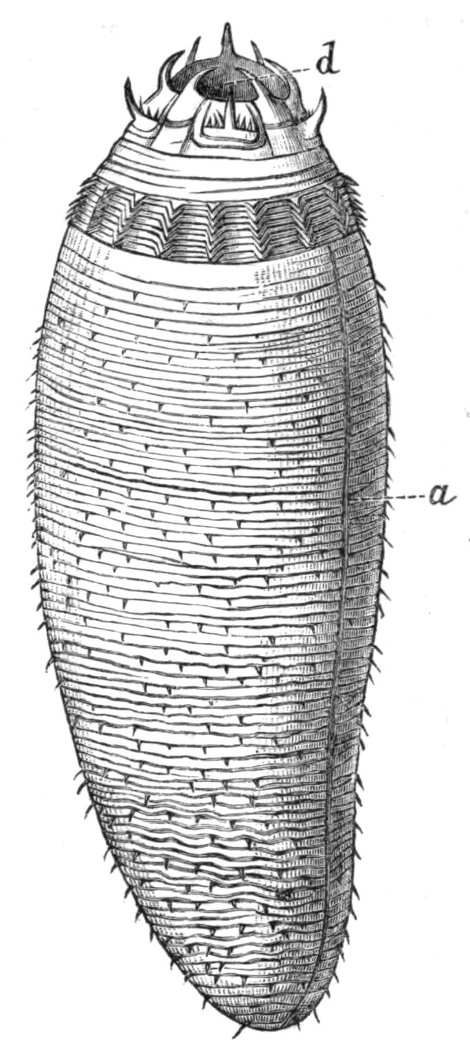
Fig. 219.—Halicryptus spinulosus v. Sieb. × 6. a, Dark line indicating the position of the ventral nerve-cord; d, mouth surrounded by spines.
Leckenby, who described specimens of P. caudatus which were found by fishermen searching for worms for bait in the outer harbour at Scarborough at half tide, states that they live in sandy clay in U-shaped tubes, at a depth of about 9 inches, the tubes opening at each end on to the surface of the sand. The fishermen of this district call them "sea mushrooms."
Halicryptus casts its cuticle in May and September; it becomes loose first at the hinder end, and the split between it and the skin grows forward until the animal lies free in a cuticular mantle. After some days this is split, and the animal frees itself from it; the cast-off cuticle includes for a short distance the lining of the mouth, the anus, and the two generative pores.
III. Order Echiuroidea.
Anatomy.—The most striking peculiarity of the Echiuroidea, as opposed to the other two families of the Gephyrea, is the presence of a solid dorsal outgrowth of a portion of the head, forming the proboscis. The nature of this proboscis is something quite different from that of the introvert of the Sipunculoidea; it would appear to correspond to an extension, in the members of the last-named Order, of that part of the head which is dorsal to the mouth and is covered by a peculiar pigment-epithelium, often in continuity with the brain. In its outgrowth this portion of the body has carried with it the nerve-ring and the vascular ring, which both surround the mouth. The proboscis is found in all the genera with the exception of the aberrant genus Saccosoma.
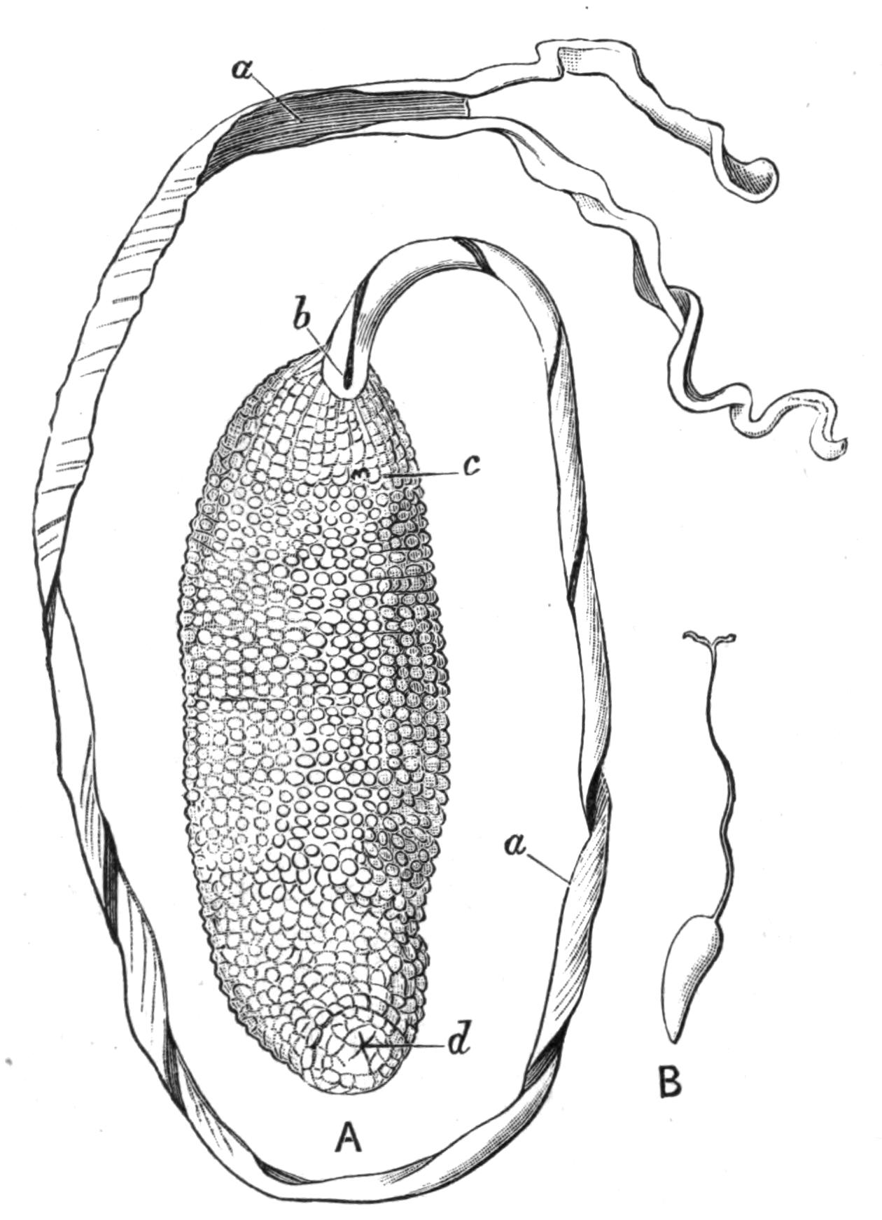
Fig. 220.—A, Bonellia viridis Rol., ♀; B, B. fuliginosa. Both nat. size. a, Grooved proboscis; b, mouth; c, ventral hooks; d, anus.
The body of the female Bonellia viridis, one of the best known species of Echiurids, is shaped like a small sausage, and is usually about 2 inches long. The proboscis arises from the anterior end, and is extremely extensible. At the distal end the proboscis splits into two short arms, which are often recurved; along the whole ventral surface runs a groove lined with cilia, which by the approximation of its edges can be converted into a tube. At the bottom of the proboscis the groove opens into the mouth. Echiurus; Thalassema, and the female Hamingia have short proboscides, which do not bifurcate but otherwise resemble those of the female Bonellia.
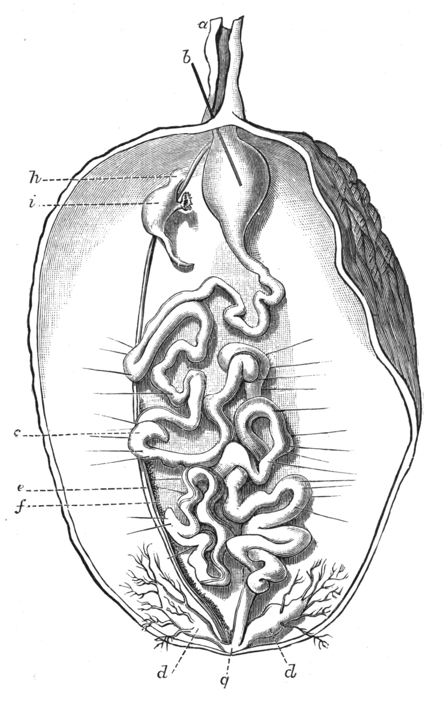
Fig. 221.—View of a female Bonellia viridis Rol., opened along the left side, × 2. a, Proboscis cut short; b, a bristle passed through the mouth into the pharynx; c, convoluted intestine; d, anal tufts or vesicles; e, ventral nerve-cord; f, ovary borne on ventral vessel running parallel with e; g, position of anus; h, points to position of external opening of nephridium; i, nephridium. This line is on a level with the internal funnel-shaped opening.
The green colour of B. viridis is due to a special pigment, "Bonellein," which at one time was thought to be identical with chlorophyll. A similar green colour is found in Hamingia arctica, Thalassema baronii, and the larvae of many forms.
A short distance behind the mouth, on the ventral surface, the female Bonellia and both sexes of Thalassema and Echiurus bear two incurved stout chitinous hooks; these gave the name Gephyrea Armata to the above-mentioned genera. In addition to these, Echiurus has a row of chitinous bristles surrounding the posterior end of the body; the row is single in E. unicinctus, double in E. pallasii. These bristles are formed, like the hooks on the introvert of the Sipunculoidea, by epidermal cells; those of B. minor and of the posterior rings in Echiurus are said to arise each from a single cell, just as the bristles do in Chaetopods.
The skin consists of very much the same layers as does that of Sipunculus; the cuticle is thin, the epidermis is modified into numerous glandular cells, papillae, and pits, from which the bristles arise. A third layer of oblique or circular fibres is usually found inside the longitudinal muscle-layer. The proboscis is solid, and contains much connective-tissue and numerous muscle-fibres running in all directions; the ventral groove is ciliated.
The alimentary canal in the Echiuroidea consists of a long thin-walled tube with numerous convolutions; it is not coiled as in Sipunculids, but the loops are irregularly arranged, and are supported by numerous fine muscular strands which run from the skin. There is a ciliated groove running along one side of the intestine, as in the Sipunculids. The anus is terminal. The most striking peculiarity of the alimentary canal of the Echiurids is the existence of a collateral intestine or "siphon." This is a narrow tube which arises from the main canal not very far from the mouth, and re-enters it again lower down. A similar structure occurs in some Echinids, and in the Capitelliformia (pp. 272, 305). Its function is not certainly known.
Another characteristic feature of the Echiurids is the presence of "anal vesicles," branching structures which unite into a common stem opening into the intestine close to the anus. The free end of each of the branches terminates in a ciliated funnel-shaped opening. The function of these structures may be excretory, or they may control the amount of fluid in the body-cavity.
A closed vascular system exists in Echiurids, consisting of a contractile dorsal vessel running along the dorsal surface of the anterior end of the alimentary canal, and continued along the axis of the proboscis. At the tip of the proboscis it bifurcates, and each branch descends along the edge until it reaches the base where, having encircled the oesophagus, the two unite, and are continued as the ventral vessel which runs along the dorsal surface of the nerve-cord, and eventually ends blindly. There is also a vessel which passes from the ventral vessel and encircles the intestine, opening into the posterior end of the dorsal vessel. In Echiurus the same vessel encircles a stout muscle which runs from the base of one of the ventral bristles to the other. In Thalassema Lankester states that the fluid within the vessels is colourless, and does not contain corpuscles similar to those in the body-cavity fluid.
The "brown tubes" or nephridia vary in number in the Echiurids. In the female Bonellia there is but one; in B. viridis the right, in B. minor the left usually persists. In shape, colour, contractility, and minute structure they closely resemble those of Sipunculus. Hamingia is said to have a pair of brown tubes; Echiurus has two pairs, except E. chilensis, which has three; their internal openings are produced into long coiled slits in some genera. Thalassema gigas has one pair; Th. neptuni, Th. baronii, Th. formosulum, and Th. exilii, two; whilst Th. vegrande, Th. moebii, Th. erythrogrammon, Th. caudex, and Th. sorbillans have three pairs.
The nervous system consists of a ventral cord lying in the body-cavity, as in the Sipunculoidea, but attached to the skin, and of a circumoesophageal ring. With the growth of the proboscis this ring is drawn out, and the two branches run along the sides of the proboscis and unite at the tip. There is no specialisation of brain, nor are any special sense organs present, but the ventral cord gives off paired nerves at regular intervals, which, uniting dorsally, form rings in the skin in some and probably in all species.
The perivisceral fluid is of a dark brown colour in Thalassema, containing numerous spherical corpuscles deeply impregnated, according to Lankester, with haemoglobin, and also containing granules of a brown pigment. Haemoglobin is also found in certain of the muscles and in part of the epithelial lining of the body-cavity. Lankester also describes the presence of haemoglobin in the corpuscles of the perivisceral fluid in Hamingia.
The genital glands are, like those of the Sipunculoidea, formed by a special development of the cells lining the body-cavity. These cells are massed together along the wall of the ventral blood-vessel. In Echiurus and in Thalassema the cells break off and float in the body-cavity, developing into ova and spermatozoa. In Bonellia each cell does not become an egg, but a mass of cells breaks off, one of which increases in size at the expense of the others and forms the ovum. The mature sexual cells leave the body through the nephridia.
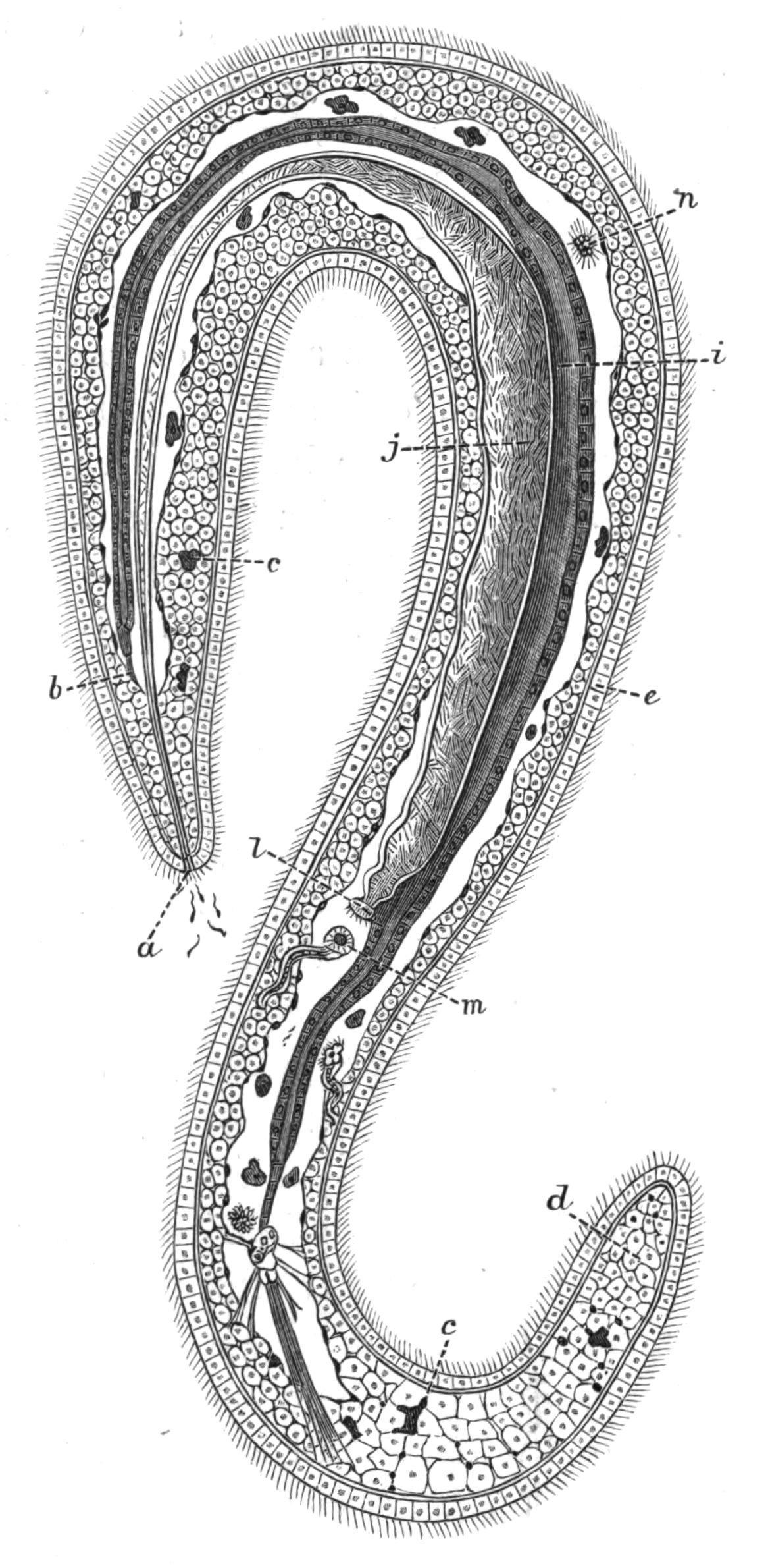
Fig. 222.—An adult male Bonellia viridis Rol. The original was 1.5 mm. long. The nervous system is not shown. (After Selenka.). a, Generative pore with spermatozoa coming out; b, anterior blind end of intestine attached to the parenchymatous tissue by muscular strands; c, green wandering cells containing chlorophyll; d, parenchymatous connective-tissue; e, epidermis; i, intestine; j, vas deferens; l, internal opening of vas deferens; m, the left anal vesicle; n, spermatozoa in the body-cavity.
Bonellia and Hamingia present very interesting cases of sexual dimorphism. In both genera the female is an animal of considerable size with the normal structure of the Echiuroidea, but the male (Fig. 222) is a microscopic Planarian-like animal, which lives in the mouth and in the nephridia of the female. Both in Bonellia[486] and in Hamingia the male is provided with a pair of hook-like ventral bristles; these are wanting in the female Hamingia. The surface of the male is ciliated, and the skin contains circular and longitudinal muscle-fibres. The body-cavity is developed, but does not reach to either end of the body. The alimentary canal is closed, neither mouth nor anus existing; it is supported by regularly arranged dorso-ventral muscle strands. A nerve-ring and a ventral cord exist. There are also two rudimentary organs corresponding with the anal vesicles of the female, and a single nephridium which acts as a duct for the spermatozoa; the latter arise from modified cells lining the body-cavity.
In both sexes the larvae develop to a certain stage without showing any trace of sexual differentiation, but after this stage, the development of the male is to a certain extent arrested; in some respects, indeed, it undergoes retrogressive changes. At this time it is found clinging to the proboscis of the female, thence it makes its way to the mouth, where it undergoes its final change; and then creeping out, finds its way into the nephridium of the female, and spends the rest of its life there in a special recess cut off by a fold from the excretory part of this organ. In Hamingia, however, Lankester, who first described the male, did not find any in the nephridia, but found five specimens, each 1⁄12 inch long, within the dilated pharynx of the female.
Development.—In Bonellia and Hamingia it seems probable that the ova are fertilised in the nephridium of the female; in the other genera they are fertilised in the water after leaving the body of the mother.
In Thalassema and Echiurus the growth of the embryo results in the formation of a typical Trochosphere larva, a type widely spread in the animal kingdom, being found in the Chaetopoda (Fig. 145, A), Polyzoa (p. 510), and Mollusca. The large prae-oral lobe persists in the Echiuroidea as the proboscis; the mouth is ventral in position, with usually a ring of cilia encircling the body in front of and behind it; the anus is posterior and terminal. A pair of larval excretory organs are present, and a special nervous aggregation of cells at the apex of the prae-oral lobe is usually indicated by the presence of a bunch of long cilia.
The trunk of the Trochosphere is unsegmented, and in certain groups of animals it remains so, but in Chaetopods, and in Echiurus and Thalassema, it elongates and becomes divided up into a series of somites or segments. Of these there are fifteen in Echiurus, and apparently eleven in Th. mellita; in this stage the Gephyrean larvae have again so close a resemblance to the segmenting Chaetopod larvae as to be easily mistaken for them. The segmentation is shown in the following way: (i.) the middle layer of cells or mesoblast is typically segmented, and forms septa, which separate each segment from its neighbours; (ii.) the ventral nerve-cord arises as segmentally-arranged thickenings of the epiblast, which fuse together, but retain their segmented appearance for some time; (iii.) the skin shows the segmentation of the body both by the arrangement of the pigment and by bands of cilia. The latter are replaced in the adult by rows of spines, and on the fourteenth and fifteenth segments in Echiurus pallasii by the two peri-anal circles of bristles. Each bristle, like those of Chaetopods, originates from a single cell.
The anal vesicles arise quite late in the development; when they have acquired their openings into the body-cavity, they seem to take in water. In Thalassema, as described by Conn, this is accompanied by remarkable changes, amounting almost to a metamorphosis. The body increases in bulk fourfold, the cilia of the prae-oral ring disappear, and the animal now moves only by means of its proboscis; the pigment is absorbed, and all traces of segmentation disappear. A similar intaking of water is described by Spengel in Bonellia. In this genus the larva, which is coloured bright green, and has two brown eye-spots, is not such a typical Trochosphere as is that of Echiurus and Thalassema.
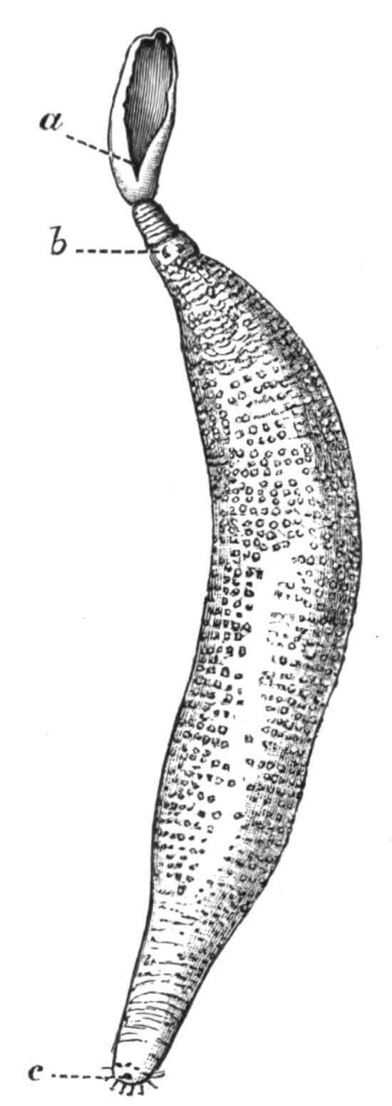
Fig. 223.—Echiurus pallasii Guér. × ½. a, Mouth at the end of the grooved proboscis; b, ventral hooks; c, anus.
Nothing is known of the development of Hamingia or of Saccosoma.
Species of Echiuroidea.—Echiurus. Proboscis not bifurcated at the end. Two ventral hooks and a single or double peri-anal ring of bristles. The body is to a varying extent marked by rings bearing spines. Two or three (E. chilensis) pairs of nephridia, their external orifice often lengthened and spirally coiled. Both sexes alike.
Greef[487] mentions six species of Echiurus, viz. E. pallasii, E. forcipatus, E. sitchaensis, E. chilensis, E. carabaicus, and E. chrysacanthophorus; to these must be added E. unicinctus. It seems probable that E. forcipatus of Reinhardt is identical with E. pallasii, although bigger, whilst E. sitchaensis, E. carabaicus, and E. chrysacanthophorus are inadequately described. The distribution of the remaining three species is as follows:—
E. pallasii Guérin (Fig. 223). North Sea, Atlantic, English Channel.
E. unicinctus Drasche. Japan.
E. chilensis Max Müller. Chili.
Thalassema.—Proboscis rather pointed at the end, not bifurcated. No peri-anal bristles, but two ventral hook-like bristles placed anteriorly. One to three or four pairs of nephridia. The sexes resemble each other.
Greef mentions eight species of Thalassema and Rietsch thirteen; three of these, however, Th. grohmanni, Th. lessonii, and Th. pelzelnii, were not seen by either author, and their description is taken from Diesing. There is some reason for thinking that the two first-named species are identical with Th. neptuni. Conn has established a new species for the specimens whose embryology he worked out at Beaufort, Virginia, and Selenka described a new species from the Challenger material.
With the exception of the three doubtful species mentioned above, the list of species of Thalassema is as follows:—
Th. neptuni Gaertner (Fig. 224). English Channel (Devonshire coast), Concarneau, Mediterranean (Gulf of Marseilles), Irish coast (Dungarvan).
Th. gigas Max Müller. Trieste.
Th. vegrande Lampert. Philippine Islands.
Th. baronii Greef. Canary Islands (Lanzarote).
Th. formosulum Lampert. Shanghai and Philippine Islands.
Th. exilii Fr. Müller. Brazil (Desterro).
Th. moebii Greef. Mauritius.
Th. erythrogrammon Max Müller. Red Sea and East Indies (Billiton).
Th. caudex Lampert. Red Sea and Indian Ocean.
Th. sorbillans Lampert. Philippine Islands.
Th. mellita Conn. West Atlantic (Beaufort).
Th. faex Selenka. North of the Faroe Islands.
Bonellia.—Proboscis very extensible and bifurcated at the end. The body and proboscis are coloured a bright green. Two ventral hook-like bristles, but no peri-anal ring. A single nephridium. The above applies to the female; the males are degenerate, and live in the nephridium or pharynx of the female.
Three (or four?) species of this genus are known.
B. viridis Rolando (Fig. 220). Mediterranean, Adriatic, North Sea (Bergen).
B. minor Marion. Mediterranean (Gulf of Marseilles, Naples).
B. suhmii Selenka. Off Nova Scotia. Male not known.
B. fuliginosa Rolando? (Fig. 220). Mediterranean (Naples).
Hamingia.—Proboscis not bifurcated, about as long as body. No ventral hook-like bristles. One or two nephridia, which open at the apex of one or two well-marked papillae. The above applies to the female; as in the genus Bonellia, the male is minute and parasitic. It has two well-marked hook-like bristles situated behind the genital pore.
This genus was first described by Koren and Danielssen as H. arctica. Two specimens were afterwards described by Horst as H. glacialis. Later Lankester described two other specimens; he was the first to find the male in the pharynx of the female. He is of the opinion that all three descriptions apply to the same species, and for this the original name H. arctica must be retained.
Hamingia arctica K. and D. Two hundred miles north of North Cape and in the Hardanger Fjord.
Saccosoma.—No proboscis. The body is flask-shaped. The mouth and anus are terminal. The ovary is anterior, and there is only one nephridium. No bristles.
Our knowledge of this remarkable Gephyrean is very incomplete, but such as it is, it is due to the careful investigations of Koren and Danielssen, who had only a single specimen at their disposition.
Saccosoma vitreum K. and D. North of the Faroe Islands.
Habits of the Echiuroidea.—As a rule the members of this group conceal their bodies in clefts and fissures of rocks and stones, keeping up communication with the outer world by means of their proboscis. Rietsch[488] describes a specimen of Bonellia minor, which he placed in an aquarium, exploring with its proboscis the nature of the bottom; when the animal had found a convenient crevice, it fixed its proboscis in it by means of the bifurcated end, and by its contraction drew the body up, and entered the hole, proboscis first. It then turned round, and during this operation doubtless the ventral hooks came into play; and then stretching out its proboscis, it began to explore the neighbourhood. The proboscis is evidently very sensitive, and in addition to being a locomotor organ, it is also used for the prehension of food. If cut off near the mouth, the animal does not long survive, but if a considerable portion is left the scar heals, and the lost part is probably regenerated. In captivity the animals frequently change their place of residence.
Eisig some years ago described the great extensibility of the proboscis of B. viridis when confined in the tanks of the Zoological Station at Naples. When contracted the proboscis was but a few inches long, but at times it was extended till it reached the length of 1½ metre, shining through the water as a transparent green thread. The body of the Bonellia was hidden under stones, but the proboscis could be seen seizing between its two ends the bodies of certain Ascidians which covered the inside of the tank, tearing them off the walls, and conveying them to the mouth along its grooved ventral surface.
The food of the Echiuroidea consists of organic matter, in the main of animal nature, but the group differs from the Sipunculoidea in not eating sand.
Rietsch describes Thalassema neptuni as being more active in its movements and less sedentary than B. minor. The proboscis is still the chief organ of locomotion, but the trunk plays a greater part in the movements of the animal than it does in the last-named species. Th. neptuni is found in cavities of stones or in the chambers worn out by the Mollusc Gastrochaena; when withdrawn from its house the body is found to be covered by a thick layer of tenacious viscid mucus.
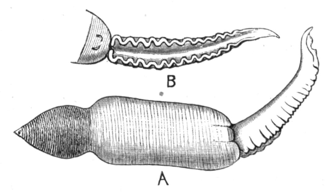
Fig. 224.—Thalassema neptuni Gaert. × 2. A, The animal lying on its ventral surface. B, Ventral view of the anterior end, showing the grooved proboscis ending behind in the mouth, and the ventral hooks.
Th. mellita was so named by Conn because it is found sheltering in the Echinid Mellita. "It enters the shell at the oral opening while yet very small, but once within its house it grows to its adult size, and is obliged therefore to remain during the rest of its life a prisoner." Each shell thus inhabited acquires a reddish brown horse-shoe-shaped marking, which affords a conspicuous signal that the shell contains a Thalassema.
Thalassema is seldom found living in sand, and Bonellia never, but Echiurus is almost always found in U-shaped tubes or passages in the sand, which it digs out for itself by the rapid contractions of its body-wall aided by its bristles. It, like the other two genera named above, does not long remain in the same hole, but frequently changes its home. As a rule the Echiurus sits near the mouth of its tube, which is often a foot or even two in depth, and sends out its proboscis in every direction; at the least sign of disturbance it withdraws into the deeper recesses. The walls of the tube are kept from falling in by a layer of mucus, which makes a smooth lining to the passage. The peri-anal bristles, which can be withdrawn or protruded at will, enable the animal to fix itself at any level in the tube.
The Echiuroidea are sometimes used by fishermen as bait. In Echiurus pallasii Greef found three parasites, all of them new species. One, a Gregarine, he named Conorhynchus gibbosus; the others were Platyhelminthes, and were named by him Distomum echiuri and Nemertoscolex parasiticus respectively.
IV. Order Epithetosomatoidea.
This Order includes the single Family Epithetosomatidae, which was established by Koren and Danielssen to contain the remarkable Gephyrean they described in 1881 under the name Epithetosoma norvegicum (Fig. 225).
Unfortunately only two specimens were at their disposition, and these were badly preserved, so that many details of their structure could not be made out. The animals are of an olive-green colour, and consist of a trunk about 12 mm. long, and of a proboscis 30 mm. in length; the latter differs essentially from the proboscis of the Echiuroidea inasmuch as it is hollow, and seems to be a whip-like tubular extension of the skin. Its lumen opens into the body-cavity. Ventral to the base of the proboscis is the mouth; the intestine is straight, and terminates in the anus, which is posterior. The nervous system lies between the circular and the longitudinal muscles of the body-wall, and contains a tube, the nature of which is obscure. No vascular system is known. The ovary is attached to a mesentery ventral to the anterior part of the alimentary canal, and there is a single nephridium. No anal vesicles exist.
The most remarkable feature of the genus is a series of pore-like openings, which are stated to lead from the outside into the body-cavity (Fig. 225, a). These are arranged four on each side, at the bottom of two slit-like depressions in the skin, which lie one on each side of the base of the proboscis, slightly dorsal to it.
These remarkable structures are without parallel amongst the Gephyrea, and, together with the peculiar character of the proboscis, justify the Norwegian naturalists in adding a new family to the group.
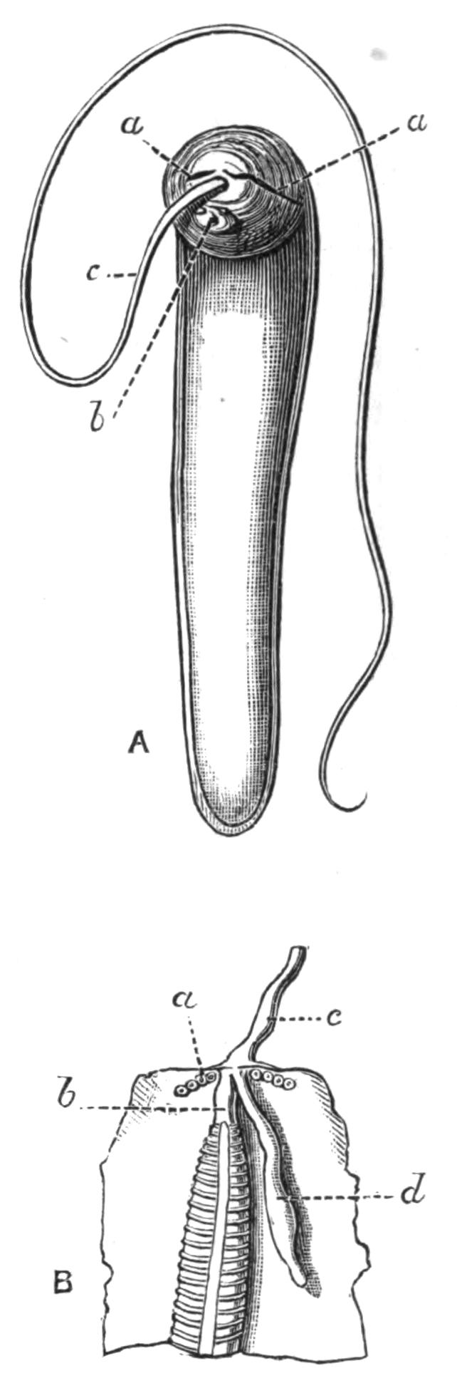
Fig. 225.—A, Epithetosoma norvegicum K. and D., magnified. a, a, Right and left slits leading to the pores; b, mouth; c, proboscis: B, the same animal opened dorsally; a, pores; b, oesophagus; c, proboscis; d, brown tube. (After Danielssen and Koren.)
Affinities of the Gephyrea.
Before considering to what other groups of animals the Gephyrea may be allied, it is advisable to discuss the relationship of the four Orders which compose the group.
Quatrefages, in the year 1865, divided the Gephyrea into I. Gephyrea Armata, with which he included the Echiuroidea and Sternaspis,[489] and II. Gephyrea Inermia or Sipunculoidea. The Gephyrea Inermia, sometimes called the Achaeta, have been extended to include the Order Priapuloidea, and opposed to the smaller sub-group the Gephyrea Armata or Chaetifera. In my opinion, however, these names now are no longer in accordance with our knowledge of the structure of the animals they attempt to describe, and they should be given up. Both names had reference to the presence or absence of the two hook-like bristles described on the ventral surface of some of the Echiuroidea, but of the five genera of this family, two, Saccosoma and Hamingia (the latter in the female or normal form), are without these bristles, and can therefore be described neither as Armata nor as Chaetifera. On the other hand, hook-like chitinous bristles of somewhat the same nature, though smaller in size and varying in position, are very common on the introvert of Sipunculoidea and on the body of the Priapuloidea.
Again, the association of the two last-named Orders in one sub-group is, to my mind, an error. The Priapuloidea have little in common with the Sipunculoidea; almost the only real point of resemblance is the power of protruding the anterior part of the alimentary canal, and withdrawing it by the aid of retractor muscles. But in the Priapuloidea this power exists to a very small extent, and it is a power shared by very many animals besides the Gephyrea. The terminal anus of the former is a feature shared by the Echiuroidea and by Epithetosoma, but these have little else in common with the Priapuloidea. On the other hand, the entire absence of any head appendages, such as the proboscis of the Echiuroidea and the tentacles or tentacular membrane of the Sipunculoidea, the absence of a vascular system, of nephridia or anal vesicles, taken together with the straight intestine which occurs elsewhere only in Epithetosoma, the persistent connexion of the nervous system with the epidermis, the unique character of their excretory system and of the reproductive organs, are all features in which the Priapuloidea differ from the more normal members of the other three Orders. These constitute a list of peculiarities which are at least as important, and probably even more important, than those which characterise the Sipunculoidea and the Echiuroidea. Thus the Priapuloidea should, I think, be regarded as a distinct Order, which occupies a very isolated position in the group.
Until we know something about the development of Halicryptus and of Priapulus, it will be difficult to say whether the Order is more nearly allied to one or the other of the two great Orders of Gephyrea, whether it is very primitive or very specialised. The connexion of the entire nervous system with the epidermis and the absence of a vascular system are both rather primitive features, and so is the Platyhelminthine character of the excretory organs. With regard to the vascular system, however, it should be pointed out that it arises very late in the larva of those Gephyrea whose development is known, and that it does not seem to correspond with the vascular system of other animals; it has no fine vessels or capillaries connected with it, and apparently does not act so much as the channel of the circulatory medium, but more as a mechanism for the expansion of the head appendages, the tentacles in the Sipunculoidea and the proboscis in the Echiuroidea; moreover, it is absent in some genera of the former, such as Onchnesoma, Tylosoma, and Petalostoma, where there are no tentacles.
The conclusion of the whole matter seems to be that the Priapuloidea are an isolated Order retaining many primitive features, and having no closer affinities to the Sipunculoidea than to the Echiuroidea.
Hatschek came to the conclusion, from his work on the development of Echiurus, that the Echiuroidea are true "Annelids," and from the presence and mode of formation of the bristles, that they are related to the Chaetopods. In this view he is confirmed by Conn, who worked out the development of Thalassema. This relationship is further confirmed by the discovery of Sluiter's that Sternaspis, the genus of Chaetopods which in other respects most nearly resembles the Gephyrea, has in one of its species (S. spinosa) a well-marked bifid proboscis, which, like that of the Echiuroidea, is thrown off at the least disturbance. Thus it seems fairly well established that the Echiuroidea are closely connected with the Chaetopoda, for although the only traces of segmentation they retain in the adult are the serially-repeated nephridia of Thalassema and Echiurus pallasii, and the two rows of peri-anal bristles in the latter, and possibly the circular nerves given off from the ventral cord, yet the larva is fully segmented, and in other respects is almost typically Chaetopodan.
The relationship of the Sipunculoidea to the Echiuroidea is a more doubtful point. Hatschek is inclined to separate them, and in this he is again supported by Conn. Embryology unfortunately does not help us much. The early stages and larvae of Sipunculus nudus and of Phascolosoma elongatum have been investigated by Hatschek and by Selenka respectively. In neither genus is there any trace of segmentation or of Annelid features, with the possible exception of the bristles on the larval Phascolosoma. On the other hand, it must be remembered that the development of Sipunculus is remarkably abbreviated, and that such stages may have dropped out, the larvae hardly differing more from the Trochosphere of Echiurus and Thalassema than does that of Bonellia, an undoubted Echiurid. Still the facts that there is never a head-kidney present, that there is no trace of segmentation, and that at no stage is the anus terminal, must have a certain weight.
If we leave out of account the larval history, which, although pointing to a difference in the nature of the two families, is by no means decisive, and consider the adult structures, we find very considerable evidences of affinity. Taking firstly the main points of difference, we find these to be (i.) the nature of the cephalic appendages, either a proboscis or some modification of tentacles; (ii.) the position of the anus; (iii.) the presence of anal vesicles; (iv.) the number of the nephridia, never more than one pair in Sipunculids; and (v.) the difference in origin of the chaetae. Of these most undoubtedly the first is the most important. The Echiuroidea have retained the prae-oral lobe of the larva in the form of a solid outgrowth of the body, which outgrowth has carried with it the nerve-ring and vascular ring which surround the mouth. This has been lost in the Sipunculoidea, but is, I think, represented by a modified patch of epidermis which lies dorsal to the mouth and just above the brain. A solid extension of the skin in this region, which involved the nervous and vascular systems, would bring about the same relation of parts as is found in the Echiuroidea. The tentacular membrane or tentacles of the Sipunculoidea have such a variety of form and arrangement, whilst all subserving the same end, that I am inclined to believe that they have originated within the limits of the family.
The position of the anus in the Sipunculoidea is one common to very many animals which live embedded in sand or in tubular holes; it is probably not primitive, as in the larva of Sipunculus it is near the posterior end, and becomes more dorsal as the larva elongates.
The anal vesicles of the Echiuroidea probably have no representative in the Sipunculoidea. In appearance and position they are very like the little tufts which are found on the rectum of Sipunculus, but since these open neither into the body-cavity nor into the alimentary canal, it is hardly fair to compare them.
The resemblances between the Orders seem to me, on the whole, to outweigh the differences. The general structure of the skin, the coiled alimentary canal, with its ciliated groove, supported by strands of muscles, the vascular system which gives off no capillaries, the structure of the brown tubes, the existence of chitinous hooks or bristles, the nervous system with its single unsegmented ventral cord, the formation of the generative organs, all point to a sufficiently close resemblance to justify us in classing the two Orders together. In addition to these there are considerable histological resemblances which cannot be discussed here, but which have a certain weight.
To sum up, it seems probable that the Echiuroidea are derived from the Chaetopoda, and that their nearest ally in this group is Sternaspis; and that the Sipunculoidea are allied to the Echiuroidea, but have further departed from the Annelid stock, and have lost even those traces of affinity with the parent group which have been preserved in the development of Echiurus and Thalassema.
So little is known of Epithetosoma that it is difficult to discuss its affinities. The presence of the hollow proboscis and the pores leading into the body-cavity undoubtedly justify its being placed in a separate Order, but beyond the presence of a terminal anus, in which it resembles the Echiuroidea, there is nothing in its structure which connects it more nearly with one than with the other of the three larger Orders of Gephyrea.
List of Gephyrea found in the British Area as defined by Canon Norman.
Phascolosoma vulgare Blainv. English Channel and North Sea.
Phasc"losoma elongatum Kef. English Channel.
Phasc"losoma papillosum Thom. English coast.
Phasc"losoma eremita Sars North Sea.
Phasc"losoma procerum Moeb. Bass Rock.
Phascolion strombi Mont. English coast (Plymouth).
Sipunculus nudus L. North Sea, English Channel (Paignton, Teignmouth).
Golfingia macintoshii Lank. East coast of Scotland (St. Andrews Bay).
Petalostoma minutum Kef. English Channel (Plymouth).
Priapulus caudatus Lam. Scarborough, Outer Hebrides.
Echiurus pallasii Guérin Coast of Scotland, English Channel.
Thalassema neptuni Gaertner English Channel (Coast of Devonshire).